sensory system unit- l. special senses the eye the eye 1” in diameter 1” in diameter protected...
TRANSCRIPT

Sensory SystemSensory System
Unit- LUnit- L

Special SensesSpecial Senses The EyeThe Eye
1” in diameter1” in diameter Protected by orbital Protected by orbital
socket of skullsocket of skull, , eyebrows, eyelashes eyebrows, eyelashes and eyelids.and eyelids.
Bathed in fluid from Bathed in fluid from Lacrimal GlandsLacrimal Glands
( ( tears empty into tears empty into nasal cavitynasal cavity))
Conjunctiva – thin Conjunctiva – thin membrane that lines membrane that lines the eyelids the eyelids and covers and covers part of the eye.part of the eye.

The EyeThe Eye
Wall of Eye is made up of 3 layers or Wall of Eye is made up of 3 layers or coatscoats Sclera, Choroid Coat, and the RetinaSclera, Choroid Coat, and the Retina
SCLERASCLERA- Outer layer- Outer layer White of the eyeWhite of the eye Tough, fibrous capsule helps maintain shape Tough, fibrous capsule helps maintain shape
of eye and protects the structure within.of eye and protects the structure within. EXTRINSIC MUSCLES- muscle responsible for EXTRINSIC MUSCLES- muscle responsible for
moving the eye that are attached to the moving the eye that are attached to the sclera.sclera.

The Other EyeThe Other Eye
CorneaCornea – Front of – Front of sclerotic coat (part sclerotic coat (part of sclera)of sclera) Clear part (no blood Clear part (no blood
vessels) – called the vessels) – called the “window of the eye”“window of the eye”
Transparent so light Transparent so light rays can pass rays can pass through through
Gets O2 and nutrients Gets O2 and nutrients through lymphthrough lymph

I can see you….I can see you….
Choroid CoatChoroid Coat Middle layerMiddle layer Contains blood vesselsContains blood vessels Circular opening in front is the PUPILCircular opening in front is the PUPIL Colored, muscular layer Colored, muscular layer surrounding pupil surrounding pupil
is is IRISIRIS INTRINSIC MUSCLES - change size of iris to INTRINSIC MUSCLES - change size of iris to
control amount of light entering through control amount of light entering through the pupil – exposed to light = constrictionthe pupil – exposed to light = constriction

Can you see me??Can you see me??
LensLens Crystalline structure located behind iris and pupil – Crystalline structure located behind iris and pupil –
focuses light rays on retina – function is focuses light rays on retina – function is accommodation = adjusting for near / far visionaccommodation = adjusting for near / far vision
Elastic, disc-shaped, biconvexElastic, disc-shaped, biconvex
Situated between the anterior and posterior Situated between the anterior and posterior chamberschambers

ANTERIOR CHAMBER filled with AQUEOUS HUMOR, a watery fluid.
POSTERIOR CHAMBER filled with transparent, jellylike substance - VITREOUS HUMOR
Both substances maintain eye’s spherical shape and refract light rays
Hey…where did you go??Hey…where did you go??

Oh no!! It’s dark in here!!Oh no!! It’s dark in here!! RetinaRetina
Innermost layer – 3Innermost layer – 3rdrd coat of the eye – located coat of the eye – located between the posterior chamber and choroid coatbetween the posterior chamber and choroid coat
Light rays focus an image on the retinaLight rays focus an image on the retina The image then travels to the cerebral cortex via The image then travels to the cerebral cortex via
the OPTIC NERVE.the OPTIC NERVE.
If light rays do not focus correctly on the retina, the If light rays do not focus correctly on the retina, the condition may be corrected with properly fitted condition may be corrected with properly fitted contact lenses, or eyeglasses, which bend the light contact lenses, or eyeglasses, which bend the light rays as required.rays as required.

Finally…the eye is almost Finally…the eye is almost over!over!
Retina contains specialized cells, visual receptors - Retina contains specialized cells, visual receptors - rods and conesrods and cones
RODSRODS- sensitive to - sensitive to dim light…problems driving at dim light…problems driving at night….damage to rodsnight….damage to rods
CONESCONES – sensitive to bright light – responsible for – sensitive to bright light – responsible for color visioncolor vision
OPTIC DISC- OPTIC DISC- on the on the retinaretina, known as the blind spot- , known as the blind spot- nerve fibers gather here to form the optic nervenerve fibers gather here to form the optic nerve, , no rods or cones.no rods or cones.


The EyeThe Eye
Pathway of VisionPathway of Vision
LIGHTLIGHT
RODS & CONESRODS & CONES( Pick up stimulus)( Pick up stimulus) OPTIC NERVEOPTIC NERVE BRAINBRAIN
CORNEACORNEA PUPILPUPILLENS (Light LENS (Light
rays are refracted)rays are refracted)
RETINARETINA

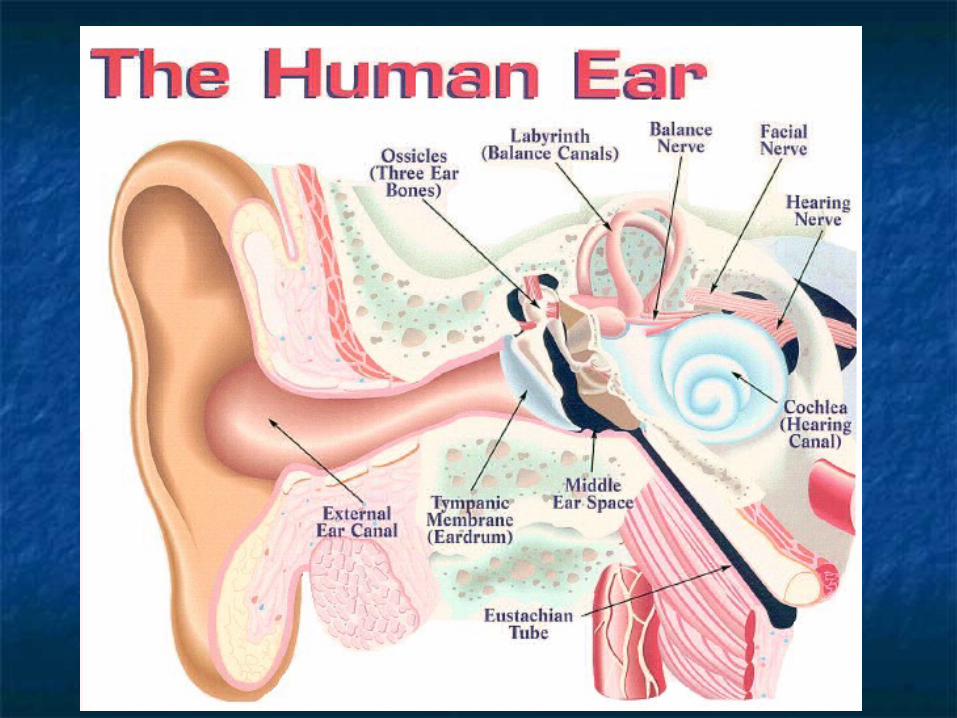
The EarThe EarOuter EarOuter Ear PINNAPINNA (AURICLE)- (AURICLE)-
outer ear, collects outer ear, collects sound wavessound waves
EXTERNAL AUDITORY EXTERNAL AUDITORY CANAL- ear canalCANAL- ear canal
CERUMENCERUMEN- earwax, - earwax, protects the earprotects the ear
TYMPANIC TYMPANIC MEMBRANE- ear MEMBRANE- ear drum, separates drum, separates outer and middle ear.outer and middle ear.
Can you hear me??Can you hear me??
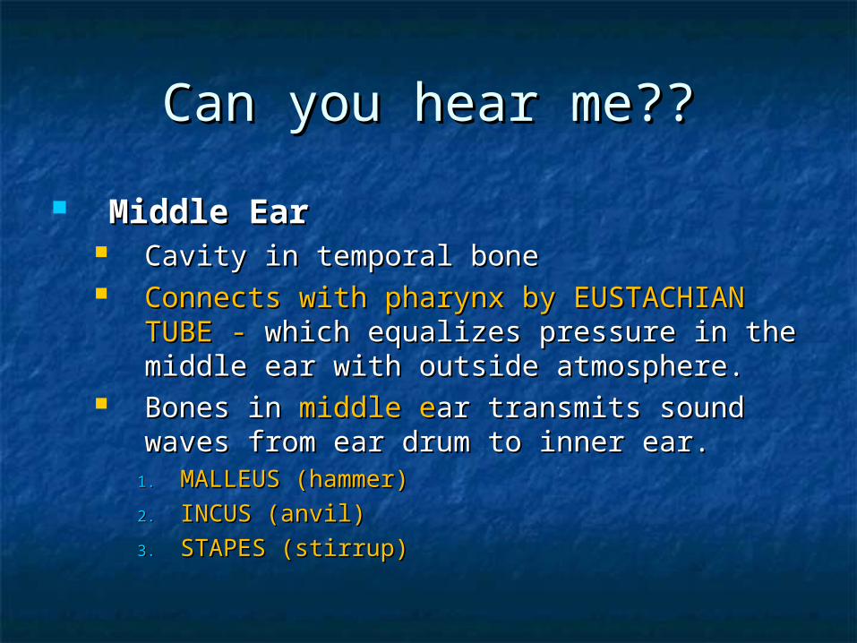
Middle EarMiddle Ear Cavity in temporal boneCavity in temporal bone Connects with pharynx by EUSTACHIAN TUBE Connects with pharynx by EUSTACHIAN TUBE
- - which equalizes pressure in the middle ear which equalizes pressure in the middle ear with outside atmosphere.with outside atmosphere.
Bones in Bones in middle emiddle ear transmits sound waves ar transmits sound waves from ear drum to inner ear.from ear drum to inner ear.
1.1. MALLEUS (hammer)MALLEUS (hammer)
2.2. INCUS (anvil)INCUS (anvil)
3.3. STAPES (stirrup)STAPES (stirrup)

I can hear you I can hear you Inner EarInner Ear
ContainsContains spiral shaped organ of hearing - the spiral shaped organ of hearing - the COCHLEACOCHLEA
The cochlea contains a membranous tube, the The cochlea contains a membranous tube, the cochlear duct- which is filled with fluid that vibrates cochlear duct- which is filled with fluid that vibrates when sound waves are transmitted by the stapes.when sound waves are transmitted by the stapes.
ORGAN OF CORTI-ORGAN OF CORTI- in the cochlea in the cochlea - delicate hairlike - delicate hairlike cells that pick up vibrations of fluid and transmit cells that pick up vibrations of fluid and transmit them as a sensory impulse along the auditory nerve them as a sensory impulse along the auditory nerve to the brain.to the brain.
SEMICIRCULAR CANALS-SEMICIRCULAR CANALS- three structures in the inner three structures in the inner ear, contain liquid that is set in motion by head and ear, contain liquid that is set in motion by head and body movements- impulses sent to cerebellum to body movements- impulses sent to cerebellum to help maintain body balance (equilibrium).help maintain body balance (equilibrium).

Where does the sound go?Where does the sound go? Pathway of HearingPathway of Hearing
Sound waves Sound waves PinnaPinna External External Auditory CanalAuditory Canal Tympanic Tympanic MembraneMembrane Ossicles ( malleus, Ossicles ( malleus, incus, & stapes)incus, & stapes) Cochlea CochleaAuditory nerveAuditory nerve BrainBrain

The Nose & TongueThe Nose & Tongue
NoseNose Smell accounts for 90% of tasteSmell accounts for 90% of taste Tissue in the nose, olfactory Tissue in the nose, olfactory
epithelium, contains specialized nerve epithelium, contains specialized nerve cell receptors.cell receptors.
Those receptors stimulate the Those receptors stimulate the OLFACTORY NERVE OLFACTORY NERVE to the brain.to the brain.
TongueTongue Mass of muscle tissueMass of muscle tissue Bumps, projections, on the surface are Bumps, projections, on the surface are
papilla, they contain the TASTE BUDS.papilla, they contain the TASTE BUDS. Receptors in the taste buds send Receptors in the taste buds send
stimuli through 3 cranial nerves to the stimuli through 3 cranial nerves to the cerebral cortex.cerebral cortex.

Common Sensory Disorders Common Sensory Disorders
Disorders of the EyeDisorders of the Eye CONJUCTIVITISCONJUCTIVITIS
Pink eyePink eye Inflammation of Inflammation of
conjunctival membranes conjunctival membranes in front of the eyein front of the eye
Redness, pain, swelling, Redness, pain, swelling, and dischargeand discharge
Highly contagious Highly contagious Rx- antibiotic eye dropsRx- antibiotic eye drops

GlaucomaGlaucoma Excessive intraocular pressure Excessive intraocular pressure causing destruction causing destruction
of the retina and atrophy of the optic nerveof the retina and atrophy of the optic nerve Caused by over production of aqueous humor, lack Caused by over production of aqueous humor, lack
of drainage, or aging.of drainage, or aging. Symps- develop gradually – mild aching, loss of Symps- develop gradually – mild aching, loss of
peripheral vision, halo around the lightperipheral vision, halo around the light TONOMETER- measures intraocular pressureTONOMETER- measures intraocular pressure Rx – drugs or laser surgery to decrease intraocular Rx – drugs or laser surgery to decrease intraocular
pressurepressure
Eye disordersEye disorders

Common Sensory DisordersCommon Sensory Disorders
CATARACTSCATARACTS Lens of eye gradually becomes cloudyLens of eye gradually becomes cloudy Frequently occurs in people over 70Frequently occurs in people over 70 Causes a painful, gradual blurring and loss of visionCauses a painful, gradual blurring and loss of vision Pupil turns from black to milky whitePupil turns from black to milky white Rx- surgical removal of the lensRx- surgical removal of the lens
Macular DegenerationMacular Degeneration Eye disorder that occurs with agingEye disorder that occurs with aging The macula ( part of the retina responsible for sharp color The macula ( part of the retina responsible for sharp color
vision) degeneratesvision) degenerates Vision is reduced but usually doesn’t cause total blindness.Vision is reduced but usually doesn’t cause total blindness.

Still problems with the eyeStill problems with the eye Detached RetinaDetached Retina
May occur with aging- accident can cause it at younger May occur with aging- accident can cause it at younger ageage
Retina detaches from choroid Retina detaches from choroid Symps- loss of peripheral vision and then central visionSymps- loss of peripheral vision and then central vision Rx- laser or freezing technique Rx- laser or freezing technique
STY ( HORDEOLUM)STY ( HORDEOLUM) Abscess at the base of an eyelash (in sebaceous gland)Abscess at the base of an eyelash (in sebaceous gland) Symps- red, painful and swollenSymps- red, painful and swollen Rx- warm, wet compressesRx- warm, wet compresses

Eye InjuriesEye Injuries
Tears are effective in cleaning the eyeTears are effective in cleaning the eye If glass or fragments get in eye, cover both If glass or fragments get in eye, cover both
eyes and see medical treatment. (DO NOT eyes and see medical treatment. (DO NOT remove the object)remove the object)
Night blindness = NYCTALOPIA- due to Night blindness = NYCTALOPIA- due to inactive rodsinactive rods
Color blindness- cones are affected – Color blindness- cones are affected – genetic disorder that carried by the female genetic disorder that carried by the female and transmitted to males.and transmitted to males.

Do you need glasses?Do you need glasses?
PRESBYOPIAPRESBYOPIA Lens loses elasticityLens loses elasticity, cant focus on close or distant objects, cant focus on close or distant objects Usually occurs after age 40Usually occurs after age 40 Rx- bifocals Rx- bifocals
HYPEROPIAHYPEROPIA FarsightedFarsighted Focal point beyond the retina because eyeball too shortFocal point beyond the retina because eyeball too short Convex lenses helpConvex lenses help
MYOPIAMYOPIA Nearsighted – Nearsighted – can’t see far awaycan’t see far away Eyeball too longEyeball too long Concave lenses helpConcave lenses help
Eye doctor =
Eye doctor =
Opthamologist
Opthamologist

Still more eye problemsStill more eye problems AMBLYOPIAAMBLYOPIA
Reduction or dimness of visionReduction or dimness of vision
ASTIGMATISMASTIGMATISM Irregular curvature of the cornea or lens, causing blurred vision and Irregular curvature of the cornea or lens, causing blurred vision and
eye straineye strain Rx- corrective lensesRx- corrective lenses
DIPLOPIA- double visionDIPLOPIA- double vision
STABISMUS ( cross-eyes)STABISMUS ( cross-eyes) Eye muscles do not coordinate their actionsEye muscles do not coordinate their actions Usually in childrenUsually in children Rx – eye exercises or surgeryRx – eye exercises or surgery
Opthalmoscope = used
Opthalmoscope = used
to examine the eye
to examine the eye

Disorders of the EarDisorders of the Ear
Loud noiseLoud noiseHearing is fragile! Loud noise over a Hearing is fragile! Loud noise over a period of time can cause period of time can cause hearing losshearing loss..
Symptoms- Symptoms- TINNITUSTINNITUS (ringing in ears) and (ringing in ears) and difficulty understanding what people are difficulty understanding what people are sayingsaying
Conductive hearing lossConductive hearing loss….may be from ….may be from excessive wax in ear canalexcessive wax in ear canal

Disorders of the EarDisorders of the Ear
OTITIS MEDIAOTITIS MEDIA
Infection of the middle earInfection of the middle ear Often a complication of a common cold in Often a complication of a common cold in
childrenchildren Rx- antibioticsRx- antibiotics If chronic or if fluid builds up- MYRINGOTOMY If chronic or if fluid builds up- MYRINGOTOMY
(opening in the tympanic membrane) with (opening in the tympanic membrane) with tubes inserted will relieve the pressure.tubes inserted will relieve the pressure.
Sxs – Sxs – fever, pain, fluid drainagefever, pain, fluid drainage

Other Sensory ProblemsOther Sensory Problems
Phantom PainPhantom Pain After an amputation your brain still sends signals to After an amputation your brain still sends signals to
amputated limb. Neural pathways are still intact.amputated limb. Neural pathways are still intact.
RhinorrheaRhinorrhea Allergies cause constant, clear drainage Allergies cause constant, clear drainage
from nose and down pharynx from from nose and down pharynx from maxillary sinuses. May cause chronic maxillary sinuses. May cause chronic pharyngitis.pharyngitis.

Other Sensory ProblemsOther Sensory Problems
ThrushThrush (oral yeast infection characterized by white patches on (oral yeast infection characterized by white patches on tongue and cheeks)tongue and cheeks)
Antibiotics wipe out normal flora which allow Antibiotics wipe out normal flora which allow fungi to flourish…vaginal and oral yeast fungi to flourish…vaginal and oral yeast infections are common with antibiotic useinfections are common with antibiotic use