separation of the poly(glycerophosphate) lipoteichoic acids of enterococcus faecalis kiel 27738,...
TRANSCRIPT
Eur. J. Biochem. 196,475-482 (1992)
001429569100166F
0 FEBS 1991
Separation of the poly(g1ycerophosphate) lipoteichoic acids of Enterococcus faecalis Kiel27738, Enterococcus hirae ATCC 9790 and Leuconostoc mesenteroides DSM 20343 into molecular species by affinity chromatography on concanavalin A Klaus LEOPOLD and Werner FISCHER Institut fur Biochemie der Medizinischen Fakultat, Universitat Erlangen-Nurnberg, Federal Republic of Germany
(Received September 3, 1990) - EJB 90 1048
This study shows for the first time microheterogeneity of 1,3-1inked poly(g1ycerophosphate) lipoteichoic acids. The lipoteichoic acids investigated were those of Enterococcus faecalis Kiel27738 (I), Enterococcus hirae (Streptococcusfaecium) ATCC 9790 (11), and Leuconostoc mesenteroides DMS 20343 (111). Lipoteichoic acids I1 and 111 are partially substituted by mono-, di-, tri-, and tetra-a-D-glUCOpyranOSyl residues with (1 + 2) inter- glycosidic linkages. Lipoteichoic acid I is substituted with a-kojibiosyl residues only. Lipoteichoic acids I and I11 additionally carry D-alanine ester.
Lipoteichoic acids were separated on columns of concanavalin-A - Sepharose according to their increasing number of glycosyl substituents per chain. It was evident that all molecular species are usually glycosylated and that alanine ester and glycosyl residues occur on the same chains. The chain lengths of lipoteichoic acid I and I1 vary between 9 -40 glycerophosphate residues, whereas those of lipoteichoic acid I11 appear to be uniform (33 f 2 residues). Molecular species differ in the extent of glycosylation but their content of alanyl residues is fairly constant. All lipoteichoic acids contain a small fraction ( 5 - 15%) different in composition from the bulk and most likely reflecting an early stage of biosynthesis.
Two procedures for chain length determination of poly(g1ycerophosphate) lipoteichoic acids are described.
Lipoteichoic acids are components of the cytoplasmic membrane of numerous Gram-positive bacteria [l]. Most widespread is the classical type which contains a single unbranched 1,3-1inked poly(g1ycerophosphate) chain consis- tently attached by a phosphodiester bond to C-6 of the non- reducing hexosyl terminus of a di- or trihexosyldiacylglycerol [ l , 21. Some of the glycerol residues of the chain are substituted with glycosyl residues or D-alanine ester or with both. These chain substituents have the capacity to greatly modify the biological activities of lipoteichoic acids. D-Alanine ester and glycosyl substituents block the carrier activity of lipoteichoic acid in the reaction of wall teichoic acid polymerases [ 3 , 41. D- Alanine ester modifies the inhibitory effect of autolysins [5] and both substituents drastically reduce the ability to activate the mammalian complement system [6].
Although poly(g1ycerophosphate) lipoteichoic acids were discovered 20 years ago [7, 81, a number of structural details has remained unexplored. Lipoteichoic acids of various bac- teria contain alanine ester and glycosyl substituents but whether the two substituents occur on separate chains or together on the same chain has not previously been clarified. Since substitution is commonly incomplete, another question is whether these lipoteichoic acids are composed of fully sub-
Correspondence to W. Fischer, Institut fur Biochemie der Medizinischen Fakultit, Universitat Erlangen-Nurnberg, Fahr- strasse 17, W-8520 Erlangen, Federal Republic of Germany
Abbreviations. Abbreviations for oligosaccharides follow IU B Recommendations, see Eur. J . Biochem. 126,433 -437 (1982).
stituted and unsubstituted species or all chains are partially substituted. This question has so far been answered only for the D-alanyl lipoteichoic acid of Staphylococcus aureus [9]. With one exception [2], it is also not known whether molecular species of a given lipoteichoic acid have a uniform chain length or display microheterogeneity like the lipopolysaccharides of Gram-negative bacteria [lo - 131.
Answers to all three questions are presented in this report in which we describe affinity chromatography of lipoteichoic acids on concanavalin A, a lectin of the jack bean [14]. To be recognized by this lectin, lipoteichoic acids were selected that carry a-D-glUCOpyranOSyl and (1 + 2)-linked a-D-glUCOpy- ranosyl oligosaccharide substituents on their chains.
MATERIALS AND METHODS
Materials and reference compounds
Concanavalin-A - Sepharose was purchased from Phar- macia LKB GmbH (Freiburg, FRG). Enzymes and cosubstrates were obtained from Boehringer Mannheim GmbH (Mannheim, FRG). Methyl a-D-mannopyranoside and Triton X-100 were from Sigma Chemie GmbH (Deisen- hofen, FRG).
Glc(a1- 2)Glc(al- 3)acy12Gro and Glc(a1- 2),acyl + 6Glc(al- 3)acyl,Gro were obtained from previous work [15]. Man(al-2)Man(al-3)acy12Gro [16] was isolated from the crude lipid extract [I71 of Micrococcus luteus ATCC 4698 by column chromatography on DEAE-cellulose [18] and
476
deacylated as described [ 191. Glycosylglycerol standards were prepared from the H F hydrolysate of the lipoteichoic acid of Enterococcus hirue (Streptococcus faecium) ATCC 9790 (see below). After being dried. the hydrolysis products were dis- solved in water, phosphate was removed by passage through a small column of anion-exchange resin (acetate form) and the effluent was taken to dryness. Glycosylglycerols were peracetylated (acetic anhydride/pyridine, 2: 1, by vol.; 60 ' C , 14 h), and separated by preparative TLC on silica gel plates developed twice with chloroform/methanol/ethyl acetate (100: 1 : 6, by vol.). The compounds, visualized by spraying the plates with water, were extracted from silica gel with chloroform/methanol (1 : 1, by vol.). Silica gel was removed by centrifugation and subsequent phase partition [20] and the compounds were deacetylated in chloroform/methanol/0.4 M NaOH (1:2:1, by vol.) at 37°C for 1 h. After addition of chloroform and water, the resulting aqueous layer was deionized by passage through a small column of cation-ex- change resin (Hf form).
Preparation of lipoteichoic acids
Lipoteichoic acids were prepared from Enterococcus fue- calis Kiel27738 (DSM 20371), Enterococcus hirae (Strepto- coccus juecium) ATCC 9790 (obtained from Dr G. Shockman, Temple University, Health Science Center, Philadelphia, PA, USA), Leuconostoc mesenteroides DSM 20343, Staphylococ- cus aureus H gol-I 4R71 (obtained from Dr J. Douglas, De- partment of Microbiology, University of Glasgow, Glasgow, UK), and Streptococcus pyogenes I1 D 698 (obtained from Laboratory of Culture Collection, Institute of Medical Sci- ence, Tokyo, Japan). The bacteria were grown as described [2]. From freshly harvested bacteria, lipoteichoic acids were extracted by a phenol/water procedure [21] and purified in one step by hydrophobic interaction chromatography on octyl- Sepharose as described [21, 221. Purified lipoteichoic acids were free of nucleic acid, polysaccharide, and protein contami- nants.
Analytical procedures
D-Alanine [23], fatty acids [22], D-glucose [24], glycerol [25], and phosphorus [26] were measured as described in the references given.
GLC was done on a Hewlett Packard gas chromatograph 5840 A equipped with a cold injection system (Gerstel GmbH, Muhlheim, FRG). A Durabond capillary column (DB5,30 m, internal diameter 0.25 mm, film thickness 0.25 pm) was used at 150-250°C with a temperature rise of 5"C/min for the separation of trifluoroacetylated glycosylglycerols, at 135 - 250 "C with a temperature rise of 6 "C/min for the separation of fatty acid methyl esters. For quantitative analysis peak areas of trifluoroacetylated glycosylglycerols were corrected by molar response factors which were determined with glycosylglycerol standards (see above). Related to GlczGro (1.00), they were 0.7,1.13, and 1.18 for GlcGro, G1c3Gro, and Glc,Gro, respectively.
TLC was done on silica gel plates (Merck 60). Glycolipids were separated with chloroform/methanol/water (65 : 25 : 4, by vol.) and chloroform/acetone/methanol/acetic acid/water (80:20: 10: 10:4, by vol.), glycosylglycerols with propan-1-ol/ ethyl acetate/water (7 : 2: 2, by vol.) and propan-I-ol/pyridine/ water (7: 4: 3, by vol.). Compounds were visualized with l-naphthol/H2SO4 reagent.
Affinity chromatography
All steps were done at 4°C. A column (1.6 x 30 cm) of concanavalin-A - Sepharose was used. The column was stored in 0.1 M sodium acetate/acetic acid pH 6, containing 1 M NaC1,0.02% NaN,, 1 mM each of MnCl2, MgClZ, and CaClZ (A). Before use, the column was washed in sequence with 3 bed vol. each of 0.1 M Tris/HCl pH 6.8 containing 0.25 M NaCl (buffer B) and 0.02 M Tris/HCl pH 6.8 containing 0.25 M NaCl and 0.1% (massjvol.) Triton X-100 (buffer C). At a flow rate of 3 ml . h - ', lipoteichoic acid (approximately 40 pmol phosphorus) was loaded onto the column in buffer C (5 - 15 ml) containing glycerol (1 - 2 pmol) as a marker. The column was eluted at a flow rate of 5 ml. h-' , first with buffer C, then with buffer C containing a linear gradient of methyl a-D-mannopyranoside (MeMan) as indicated under results. Finally MeMan was eluted with two bed vols. of 0.02 M Tris/HCl pH 6.8 containing 1 M NaC1. Column frac- tions (2 ml) were analysed for phosphorus and, where appro- priate, for glycerol (marker) and, after hydrolysis (2 M HCl, 100"C, 2.5 h), for MeMan. The lipoteichoic acid of selected tubes was hydrolyzed (2 M HC1, 100"C, 2.5 h) and analysed for phosphorus, D-alanine, and glucose. Before analysis, Tri- ton X-100 and fatty acids were extracted from the hydrolysate with petroleum ether/chloroform (4: I , by vol.), and the re- sidual organic solvent was blown off in a stream of nitrogen. For hydrolysis with H F (see below), lipoteichoic acid was freed of MeMan by chromatography on octyl-Sepharose using a scaled-down centrifugation procedure [2, 271, whereby MeMan was eluted with 15% propanol in 0.1 M sodium ace- tate pH 5, lipoteichoic acid with 50% propanol in water. From larger volumes of combined tubes, MeMan was removed by dialysis.
Analyses of lipoteichoic acids
For compositional analysis, lipoteichoic acids were hydro- lyzed in 2 M HCl at 100'C for 2.5 h. For the characterization of glycosylated glycerols and the determination of the chain length, lipoteichoic acids were hydrolyzed in 48% (by mass) H F (2"C, 36 h) as described [22]. Prior to this treatment, fatty acid and alanine ester were hydrolyzed in 0.2 M NaOH (37 "C, 1 h). The alkali hydrolysates were passed through small columns of cation-exchange resin (H' form) and fatty acids were extracted from the effluent with petroleum ether/chloro- form (4: 1, by vol.). The aqueous layer was neutralized with NH40H and taken to dryness. The H F hydrolysate, after drying over KOH in vacuo at 2"C, was analysed for free glycerol and phosphorus; glycosylglycerol (Glc,Gro) was cal- culated as the difference and its molar ratio to phosphorus (A) calculated:
free glycerol - - 1----
phosphorus phosphorus Glc,Gro
A =
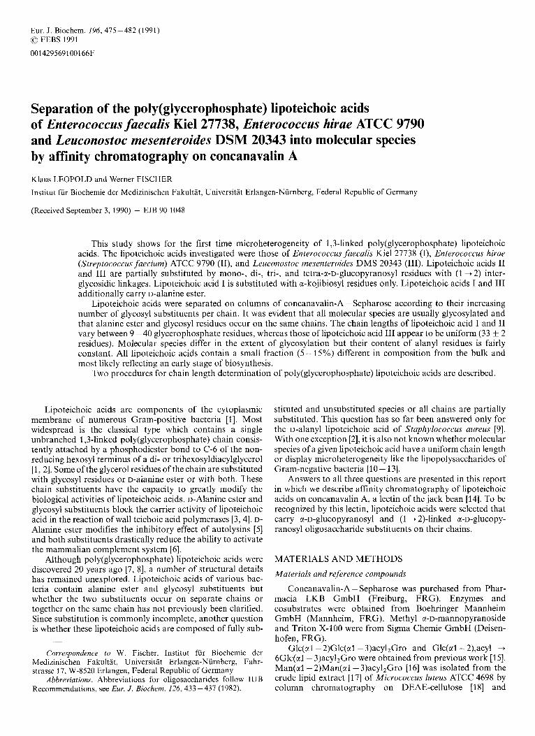
Another sample of the H F hydrolysate was treated with trifluoroacetic anhydride, containing 19'0 (by vol.) pyridine (room temperature, 1 h) and was then analysed by GLC as shown in Fig. 1. Using the corrected peak areas of chain glycosylglycerols (Glc,Gro; x = 1 -4) and the deacylated gly- colipid anchor (Glc,Gro'), the molar ratios of Glc,Gro/chain (B) and chain glucose/phosphorus (C) were calculated:
Glc Gro CGlc Gro chain Glc2Gro'
B = -"_- = --x-_
477
- ,
chain glucose phosphorus
C =
C Glc,Gro . x Glc - - CGlc,Gro.x + 2 Glc2Gro’ phosphorus ’
for which glucose and phosphorus were measured after hy- drolysis with HC1 (see above).
Accordingly, the molar ratio of glycolipid glucose/phos- phorus (D) is given by:
glycolipid glucose phosphorus
D =
2 Glc2Gro’ Glc - - CGlc,Gro ‘x + 2 Glc2Gro’ phosphorus
The molar ratio Glc/Glc,Gro (E) was calculated as:
- CGlc,Gro . x -
Glc E =
Glc,Gro C Glc,Gro
Chain lengths (m and m’ number ofglycerophosphate residues per chain) were calculated by
B 2 m = -, and m‘ = -
A D ’
Under Results (Tables 3 and 4) mean values of m and m‘ are given; the difference between m and m‘ was, on average, less than 10% and 15% with the lipoteichoic acids of E. faecalis and E. hirae, respectively. When the molar ratio Glc,/phos- phorus was 5 0.2 (Results, Tables 1 and 2), chain lengths were determined by adding Man(a1- 2)Man(al- 3)Gro to the H F hydrolysate before trifluoroacetylation:
Man2Gro phosphorus Glc2Gro Man2Gro
m” =
where the first therm gives the peak areas (molar response factors 1 : l), the second the molar amounts present in the samples to be analysed. In these experiments molar ratios of Glc,Gro/phosphorus (A‘) were calculated by
Glc,Gro C phosphorus E
- - A’ = -
RESULTS
Composition of lipoteichoic acids
On purification by column chromatography on octyl- Sepharose the lipoteichoic acids of both E. faecalis and E. hirae separated into two peaks, the major one (86% and 71 YO) containing Glc(a1- 2)Glc(al- 3)acy12Gro as lipid moi- ety, the minor one Glc(ct1- 2),Ptd + 6Glc(al -3)acy12Gro [2, 211. The lipoteichoic acid of L. mesenteroides DSM 20343 also separated into two peaks (not shown). The lipid moieties of the major (79%) and the minor fraction released by hy- drolysis with HF cochromatographed on TLC with Glc(a1- 2)Glc(ctl- 3)acy12Gro and Glc(a1- 2),acyl+ 6Glc(al- 3)acy12Gro, respectively. The deacylation products were indis- tinguishable from authentic Glc(a1- 2)Glc(al- 3)Gro on TLC and, after trifluoroacetylation, on GLC.
The major fractions of these lipoteichoic acids containing Glc(a1- 2)Glc(al- 3)acy12Gro as lipid anchor were used in this study. Chain analyses are given in Tables 1-4. In accor- dance with earlier results [ S , 281, the lipoteichoic acid of E. faecalis was found to be substituted with D-alanine ester
3
W
N h
3 3
3 m
3 m
=I N N 3
w r l N r l i -! I N . m.
m 3
N s In 3
Fig. 1. Gas-liquid chromatography of tr@uoroacetylated glycosylgly- cerols released from the lipoteichoic acid of” E. hirae by hydrolysis with HF. Native (A) and deacylated lipoteichoic acid (B) were hydrolysed and the products of (A) were separated by phase partition [20]. (A) The chain glucosylglycerols, Glc(al-2),Glc(al-2)Gro (n = 0- 3); (B) in addition, the deacylated glycolipid moiety, Glc(a1- 2)Glc(al- 3)Gro. Man2Gro, used in some experiments as internal standard, appeared ahead of the chain Glc2Gro (not shown)
and a-kojibiosyl residues, the lipoteichoic acid of E. hirae lacked D-alanine ester and was substituted with a-D- glucopyranosyl, a-kojibiosyl, a-kojitriosyl and a-kojitetraosyl residues (Fig. 1). The lipoteichoic acid of L. mesenteroides DSM 20343 contained D-alanine ester and mono-, di-, tri-, and tetraglucosyl substituents which, released as glycosyl- glycerols by HF, were indistinguishable from the glucosylgly- cerols of E. hirae when tested on TLC and, as trifluoro- acetates, on GLC. The (1 + 2) interglycosidic linkage was confirmed by methylation analysis [29, 301, the a-anomeric bonds by resistance to oxidation with Cr03 [31, 321. This chain substitution contrasts with that of the lipoteichoic acid of L. mesenteroides DSM 20188 which contained D-alanine ester only [S].
Chromatographic behaviour of glucosyl lipoteichoic acids on concanavalin-A - Sepharose
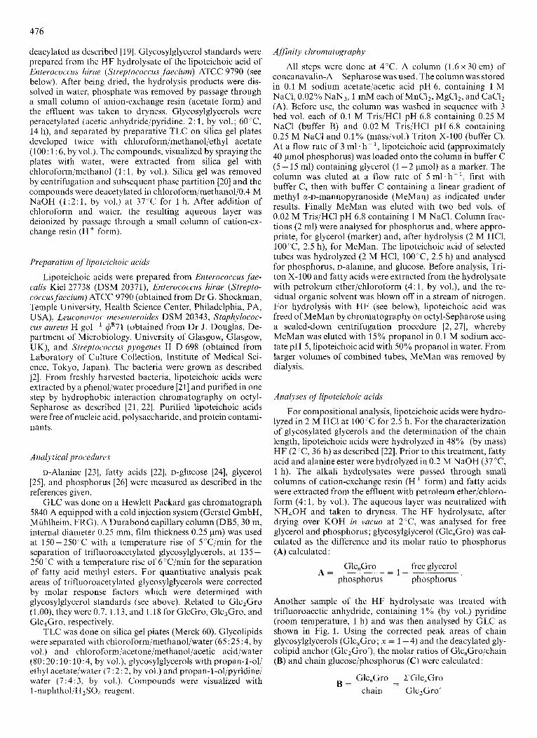
When Leuconostoc lipoteichoic acid was passed through a column of concanavalin-A - Sepharose, it was completely retained (Fig. 2A). On subsequent elution with a MeMan gradient, MeMan and lipoteichoic acid appeared in the efflu- ent synchronously, although MeMan was expected to elute 25 tubes earlier. The MeMan concentration in the effluent first increased rapidly and then slowed to the increase calculated for the gradient applied. Extrapolation of this line to the abscissa suggests that MeMan in the first SO ml ofthe gradient was actually adsorbed by concanavalin-A - Sepharose. A more detailed analysis of MeMan in the effluent revealed that the rapid increase followed a saturation curve which changed to the linear increase of the gradient.
The recovery of lipoteichoic acid from the column was better than 90%. When Triton X-100 was omitted from the starting and elution buffer, MeMan concentrations as high as SO mM failed to elute lipoteichoic acid (not shown) suggesting that, owing to the aggregation of a-glucosyl residues, lipoteichoic acid micelles bind more firmly to the lectin than do individual lipoteichoic acid molecules segregated in micelles of Triton X-1 00.
478
0 I 1.0
3 0 4 0 5 0 6 0 7 0 8 0 9 0 100
T U B E N U M B E R
20 30 40 50 60 70 80 9 0
T U B E N U M B E R
Fig. 2. Chrornatography on concunavalin-A - Sepharose of Leuconostoc lipoteichoic acidalone ( A ) and in separate mixtures with non-glycosylated mlunine-ester-containing and alanine-free Iipoteichoic acid of S. aureus ( B ) . ( 0 ) Native alanyl lipoteichoic acid of L. mesenteroides, preparation 1 (40 pmol phosphorus); ( 0 ) alanine ester-containing and (0) alanine-free lipoteichoic acid of S. aureus (10 and 2 pmol phosphorus, respectively); (-.-.--) glycerol (marker); ( x - - ~ x ) concentration of MeMan in the effluent; arrow: theoretical appearance of MeMan in the effluent if not absorbed by the lectin. The MeMan gradient (150 ml each of buffer C and buffer C containing 6 mM MeMan) was applied at tube 16. For other details see text
i
j: ! \ 1 : * 3 0 1 0 7 0 8 0 9 0 100 110 120 130
T U B E NUMBER
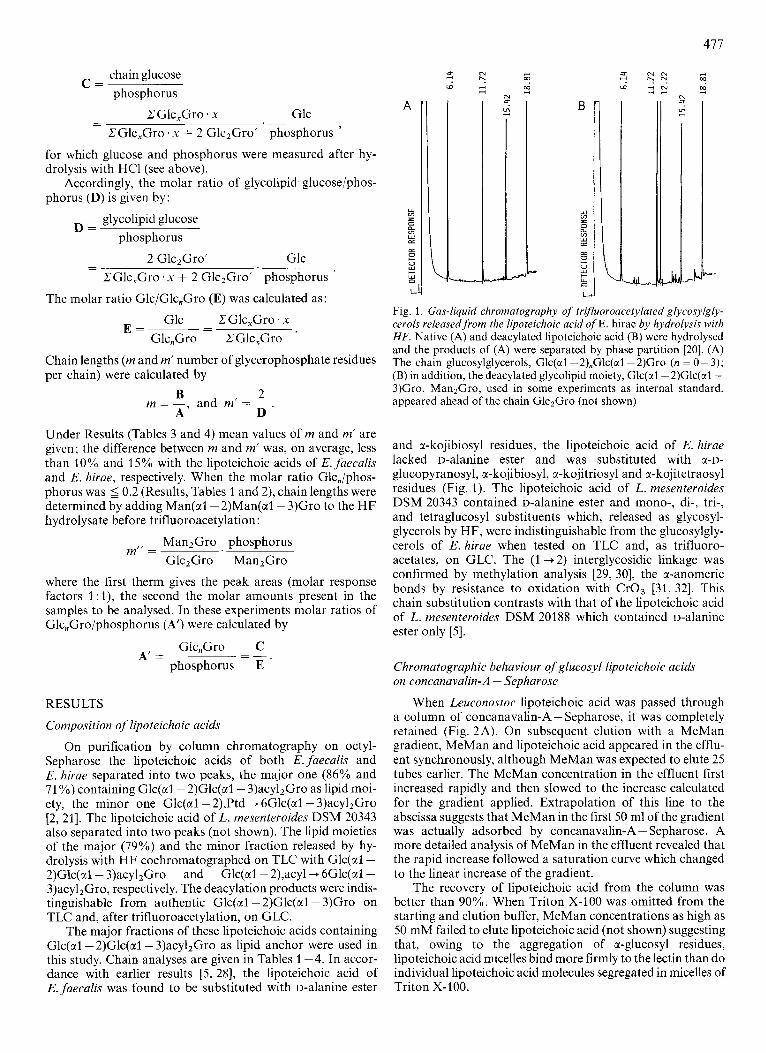
Fig. 3. Fractionution by column chromatography on concanavalin-A - Sepharose of native a1aninc.-ester-containing lipoteichoic acid of' L. mesenteroides, preparation I . The MeMan gradient (150 ml each of buffer C and buffer C containing 3 mM MeMan) was applied at tube 16. For arrow and symbols, see Fig. 2
Fig. 2B shows two experiments in which mixtures of Leu- conostoc lipoteichoic acid and nonglycosylated lipoteichoic acid of 5'. nureus H gol-' 4R71 [l , 331 were used. Nongly- cosylated lipoteichoic acid eluted completely ahead of glucosylated lipoteichoic acid, independently of the amount and whether or not it contained alanine ester. Also not re- tained on the column was the lipoteichoic acid of Streptococ- cus pyogencs which lacks a-u-glucopyranosyl substituents on the chain but contains Glc(xl-2)Glc(al- 3)acy12Gro as the lipid moiety [34]. This observation confirms that retention on the column is indicative of a-glucosyl substituents on the chain.
T U B E NUMBER
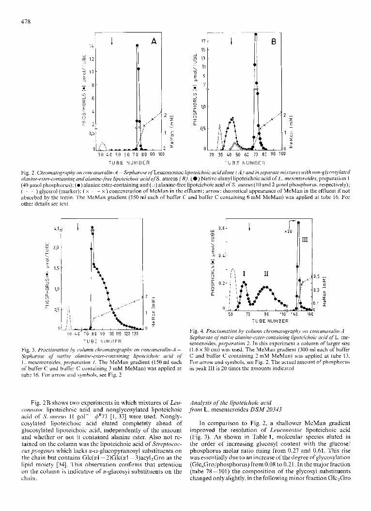
Fig. 4. Fractionation by column chromatogruplij) on concunavalin-A - Sepharose of' native alanine-ester-contain in^ lipoteichoic acid o j L. me- senteroides, preparation 2. In this experiment a column of larger size (1.6 x 50 cm) was used. The MeMan gradient (300 ml each of buffer C and buffer C containing 2 mM MeMan) was applied at tube 13. For arrow and symbols, see Fig. 2. The actual amount of phosphorus in peak 111 is 20 times the amounts indicated
Analysis of the lipoteichoic acid from L. mesenteroides DSM 20343
In comparison to Fig. 2, a shallower MeMan gradient improved the resolution of Leuconostoc lipoteichoic acid (Fig. 3). As shown in Table 1, molecular species eluted in the order of increasing glucosyl content with the glucose/ phosphorus molar ratio rising from 0.27 and 0.61. This rise was essentially due to an increase of the degree of glycosylation (Glc,Gro/phosphorus) from 0.08 to 0.21. In the major fraction (tube 78 - 101) the composition of the glycosyl substituents changed only slightly, in the following minor fraction Glc2Gro

479
Table 1. Analyses oflipoteichoic acid, preparation 1 .from Leuconostoc mesenteroides DSM 20343 before and after separation on concanuvalin- A - Sepharose (Fig. 3 ) Glucose values were corrected for the glucose of the glycolipid moiety
Component ratios Values in ~
parent lipo- tube number teichoic acid
78 83-85 92 - 94 98-101 104-108 112-114 126-135
GlcGro/Glc,Gro Glc2Gro/Glc,Gro Glc3Gro/Glc,Gro Glc,Gro/Glc,Gro
Alanine/phosphorus Glucose/phosphorus Glc,Gro/phosphorus
Phosphorus/glycolipid
mol/mol ~-
0.14 0.08 0.21 0.57
0.29 0.45 0.14
36
0.15 0.03 0.23 0.59
0.27 0.27 0.08
34
0.14 0.05 0.22 0.60
0.30 0.39 0.12
37
~
0.14 0.07 0.22 0.58
0.28 0.50 0.15
37
~
0.13 0.09 0.22 0.56
0.31 0.56 0.17
36
0.14 0.13 0.23 0.50
0.29 0.56 0.18
40
0.15 0.18 0.22 0.45
0.29 0.60 0.20
34
0.12 0.26 0.23 0.38
0.28 0.61 0.21
33
Table 2. Analyses of lipoteichoic acid, preparation 2,from Leuconostoc mesenteroides DSM 20343 before and after separation on concanavalin- A - Sephavose (Fig. 4 ) Glucose values were corrected for the glucose of the glycolipid moiety; tr, trace amount
Component ratios Values in
parent lipo- tube number teichoic acid 60-73 79-103 140-142 143 144 145 146 147/148 149-151 152-160
mol/mol
GlcGro/Glc,Gro 0.15 0.29 0.28 0.15 0.15 0.14 0.15 0.13 0.15 0.15 0.13 Glc2Gro/Glc,Gro 0.05 tr 0.05 0.04 0.04 0.04 0.05 0.05 0.05 0.06 0.07 Glc3Gro/Glc,Gro 0.15 tr 0.31 0.16 0.15 0.15 0.15 0.15 0.14 0.15 0.15 Clc,Gro/Glc,Gro 0.65 0.71 0.30 0.66 0.67 0.67 0.65 0.66 0.66 0.64 0.66
Alanine/phosphorus 0.52 0.87 0.89 0.52 0.47 0.45 0.40 0.41 0.43 0.45 0.42 Glucose/phosphorus 0.33 < 0.01 0.11 0.27 0.31 0.35 0.40 0.44 0.47 0.49 0.50 Glc,Gro/phosphorus 0.10 0.002 0.04 0.08 0.09 0.10 0.12 0.13 0.14 0.15 0.15
Phosphorus/glycolipid 31 25 26 34 33 34 33 34 35 33 36
increased, whereas Glc4Gro decreased. The chain length (phosphorus/glycolipid, 36 & 2) and the alanine/phosphorus molar ratio (0.29 2 0.01) were essentially unaltered through all fractions.
The results obtained with another lipoteichoic acid prep- aration from a separate batch of the same L. rnesenteroides strain are summarized in Fig. 4 and Table 2 . Compared with preparation 1, the second preparation had , on average, shorter chains (31 k 3 glycerophosphate residues), lower mo- lar ratios of glucosyl and Glc,Gro to phosphorus (0.33 and 0.20, respectively), and a higher alanine/phosphorus molar ratio (0.52). From concanavalin-A - Sepharose two peaks containing 4% and 11 YO of the total phosphorus, respectively, eluted at and immediately behind the location of nonglycosylated lipoteichoic acid (Fig. 4; I, 11). This material contained equimolar amounts of glycerol and phosphorus and had the same fatty acid pattern and glycolipid structure as the major fraction of peak 111 (data not shown). The poly(g1ycero- phosphate) chains, however, were shorter (25 glycerophos- phate residues) and had a remarkably higher alanine/phos- phorus molar ratio (0.88 k0.01). Moreover, peak I contained almost no glycosyl substituents and peak I1 no more than one
per chain. In the major fraction (tubes 142 - 157) the molar ratios of glucose and Glc,Gro to phosphorus increased from 0.27 to 0.50, and 0.08 to 0.15, respectively, and accordingly the number of glycosyl substituents per chain from 2.7 to 5.4. Through all fractions of peak 111 the glycosylation pattern and the chain length (34 & 1 glycerophosphate residues) remained essentially unchanged. The alanine/phosphorus molar ratio initially decreased slightly and then remained constant (0.44 & 0.04).
Three peaks of a similar composition in almost the same proportions as seen with the second preparation were also observed with a third one. The reason for the absence of peak I and I1 from preparation 1 is not clear. All preparations were from over-night cultures but, due to different lag phase du- ration, at harvesting the bacteria might have been at the end of logarithmic growth and in the stationary phase, respectively.
Analysis of the lipoteichoic acid from E. faecalis Kiel27738
From concanavalin-A - Sepharose no phosphorus ap- peared before MeMan in the effluent (not shown). As shown in Fig. 5 , approximately 6% of the lipoteichoic acid eluted

480
Table 3. Analyses Qf lipoteichoic ucid,from E. faecalies KielZ7738 before and after separation on concanuvalin-A - Sepharose (Fig. 5 ) Glucose values were corrected for the glucose of the glycolipid moiety
Component ratios Values in
parent lipo- tube number teichoic acid
92-95 111-115 131-133 151-153 171-173 190-195 205-210
mol/mol
Alanine/phosphorus 0.39 0.42 0.39 0.38 0.37 0.36 0.34 0.33
GlcZGro/phosphorus 0.47 0.23 0.44 0.50 0.57 0.59 0.59 0.58
Phosphorus/glycolipid 24 11 17 23 32 33 38 40
Glucose/phosphorus 0.90 0.38 0.82 1 .oo 1.11 1.21 1.24 1.21
Tdbk 4. Analyse~T of lipoteichoic acid f rom E. hirae ATCC 9790 before and after separation on concunavulin-A - Sepharose (Fig. 6 ) Glucose values were corrected for the glucose of the glycolipid moiety
Component ratios Values in ~~
parent lipo- tube number teichoic acid ~ _ _ _ _ _ ~ -
86-89 108-113 129-131 151-153 169-173 188-192 207-215 220-250
mol/mol -
GlcCro/Glc,Gro 0.59 0.84 0.70 0.64 0.62 0.58 0.59 0.62 0.55 Glc,Gro/Glc,Gro 0.23 0.09 0.18 0.19 0.22 0.24 0.26 0.27 0.29 Glc,Gro/Glc,Gro 0.03 0.01 0.02 0.03 0.01 0.02 0.03 - 0.03 Glc4Gro/Glc,Gro 0.15 0.06 0.1 1 0.14 0.15 0.15 0.13 0.11 0.14
Glc,Gro,'phosphorus 0.78 0.34 0.68 0.72 0.83 0.83 0.90 0.95 0.79
Phosphorus/glycolipid 16 9 13 15 20 22 25 29 36
Glucose,/p hosphorus 1.42 0.50 1.16 1.24 1.52 1.58 1.64 I .79 1.91
80 90 100 110 120 130 140 150 160 170 180 190 200 210 T U B E N U M B E R
Fig. 6. Fractionation by column chromatogruphy on concanavalin-A - Sepharose of native non-alanyluted lipoteichoic acid of E. hirae. MeMan gradient as in Fig. 5
T U B E N U M B E R
Fig. 5. Fractionation hj. column chromatography on concanavalin-A - Sephurose of nutire ulanine-ester-containing lipoteichoic acid of E. faecalis. The MeMan gradient (300 ml each of buffer C and buffer C containing 2.5 mM MeMan) was applied at tube 16
initially as a sharp peak and was followed by the major frac- tion which appeared as a broad peak of symmetrical shape. The analyses, summarized in Table 3, suggest the molecular species of the forepeak to form a separate entity characterized by particularly low molar ratios of glucose and Glc2Gro to phosphorus. Over all fractions, the molar ratios of glucose and Glc2Gro to phosphorus and the chain length (phosphorus/
glycolipid) increased 3.2-, 2.5- and 3.5-fold, respectively, re- sulting in an approximately 9-fold increase of the glycosylated glycerol/chain. The alanine/phosphorus molar ratio showed a slight decrease from 0.42 to 0.33.
Analysis of the lipoteichoic ac id , f rom E. hirae ATCC 9790 The elution profile from concanavalin-A - Sepharose of
this lipoteichoic acid (Fig. 6) was similar to that of E. faecalis (Fig. 5 ) although the mean degree of glycosylation was con- siderably higher and the glycerol residues were substituted with an oligosaccharide series (Table 4). The forepeak contained 5% of the total phosphorus and represented again

48 1
a distinct entity on the basis of its composition (Table 4). Over all fractions, the molar ratios of glucose and Glc,Gro to phosphorus and the chain length (phosphorus/glycolipid) increased 3.8-, 2.3- and 3.8-fold, respectively, resulting in a 9- fold increase of the glycosylated glycerol/chain. Among the chain substituents, the molar proportion of GlcGro decreased from 0.84 to 0.55, that of GlczGro increased from 0.09 to 0.29. The proportion of Glc,Gro remained constant, and that of Glc4Gro increased initially and then slightly decreased.
DlSCUSSION
The results reported here indicate that poly(g1ycerophos- phate) lipoteichoic acids containing a-D-glucopyranosyl resi- dues as chain substituents are separated by affinity chroma- tography on concanavalin-A - Sepharose according to the number of glycosyl substituents per chain. This involves the extent of glycosylation and the length of the chain, and re- quires lipoteichoic acid molecules separated from each other by incorporation into micelles of Triton X-100. Whether, in the case of di-, tri-, and tetrasaccharide substituents, only the nonreducing a-glucopyranosyl terminus or the intra-chain residues also are recognized by the lectin cannot yet be answered. Earlier investigations of malto- and isomalto-oligo- saccharide series revealed that the concanavalin A combining site is complementary to a single, nonreducing terminal WD- glucopyranosyl residue [35] whereas the binding power of a series of (1 + 2)-linked a-D-manno-ohgosaccharides increased with increasing number of a-D-mannosyl residues [36, 371. The kojibiosyl residue of the lipid anchor of lipoteichoic acids is apparently not recognized, presumably because its non- reducing terminus carries the poly(g1ycerophosphate) chain [2]. As can be seen from Fig. 4 and Table 2 nonglycosylated species separate from those that carry no more than one gly- cosy1 substituent, and approximately two or three substituents per chain bind lipoteichoic acid so firmly to the lectin that competition by MeMan is required for elution (Tables 1 - 4). On gradient elution with MeMan, separation is achieved of species that contain between 3 and 29 substituents per chain (Fig. 6, Table 4).
In this study it became evident that, except for a small amount in one sample of Leuconostoc lipoteichoic acid, all molecular species are glycosylated and that alanine ester and glycosyl substituents occur on the same rather than on sepa- rate chains. Moreover, microheterogeneity was detected con- cerning the extent of glycosylation and, in a species- or genus- specific manner, the chain length. Accordingly, variation of chain lengths between 11 and 40, and between 9 and 36 gly- cerophosphate residues was observed with the lipoteichoic acid of E. faecalis and E. hirae, respectively. The actual range may be still larger because the forepeak containing the short- chain species was analysed as a whole (Figs 5 and 6). Chain lengths between 30 and 65 glycerophosphate residues were recently found with the naturally nonglycosylated lipoteichoic acid of Lactobacillus casei when the glycolipid-deprived de- rivative was separated by gel permeation chromatography [2]. By contrast, the lipoteichoic acid of L. mesenteroides DSM 20343 seems to contain molecular species of practically invariable length. Independent evidence for this would be desirable because, in principle, highly glycosylated short-chain species might elute from concanavalin A together with slightly glycosylated long-chain species.
Investigations into the biosynthesis of the lipoteichoic acid of E. hirae (S . faecium) ATCC 9790 revealed that the chain is
elongated by addition of glycerophosphate residues at the terminus distal to the lipid moiety and elongation of pre- formed chains appears to be possible [38]. Incorporation of glycosyl substituents appears not to be dependent on chain growth, and preformed glycosyl substituents may be elongated [28]. The data obtained in the present study with the lipoteichoic acid of this strain further suggest that chain growth may stop at any point, giving rise to a lipoteichoic acid population with a Gaussian distribution of chain lengths (Fig. 6; Table 4). They also demonstrate the presence of a short-chain fraction whose nascent character is suggested by the preponderance of monoglucosyl substituents. During further growth of these chains, the monoglucosyl substituents may be elongated and new glycosyl substituents added, re- sulting in the observed increase of Glc,Gro/phosphorus molar ratios from 0.34 to 0.95 as the chain length increases from 9 to 29 glycerophosphate residues (Table 4).
The glycerophosphate polymerase of L. mesenteroides seems to be different from that of E. hirae and E. faecalis as it produces chains of a uniform length (Tables 1 and 2). The slightly shorter chains in peaks I and 11, as compared to the bulk, seen with two preparations (Fig. 4; Table 2), might then indicate small amounts of non-completed chains. The high alanine ester content in peaks 1 and I1 and the absence of glycosyl residues from peak I suggests that alanine ester might be added before glycosylation occurs and later partially lost, presumably by spontaneous hydrolysis [3, 39, 401.
It would be desirable to test these hypotheses on lipoteichoic acid biosynthesis by appropriate pulse/chase ex- periments and separation of radiolabelled molecular species by the procedure described in this work.
We gratefully acknowledge the reliable and expert help of Edeltraud Ebnet and Birgitta Reiter. This work was supported by the Deutsche Forschungsgemeinschuft (Fi 21 8/4-7).
REFERENCES 1.
2.
3.
4.
5.
6.
7.
8.
9. 10.
11.
12.
13.
14.
Fischer, W. (1988) in Advances in microbiulphysiologjl (Rose, A. H. & Tempest, D. W., eds) pp. 233-302, Academic Press, London.
Fischer, W., Mannsfeld, T. & Hagen, G. (1990) Biochem. Cell Biol. 68, 33 -43.
Fischer, W., Koch, H. U., Rosel, P. & Fiedler, F. (1980) J. Biol. Chem. 255,4551-4562.
Koch, H. U., Fischer, W. & Fiedler, F. (1982) J . Bid. Chem. 257,
Fischer, W., Rosel, P. & Koch, H. U. (1981) J . Bucteriol. 146,
Loos, M., Claas, F. & Fischer, W. (1986) Infect. Zmmun. 53, 595-
Wicken, A. J. & Knox, K. W. ( 3 970) J . Gen. Microbiol. 60,293 -
Toon, P., Brown, P. E. & Baddiley, J. (1912) Biochem. J . 127,
Fischer, W. & Rosel, P. (1980) FEBS Lett. 119, 224-226. Palva, E. T. & Mlkell, P. H. (1980) Eur. J . Biochem. 107, 137-
Munford, R. S., Hall, C. L. & Rick, P. D. (1980) J . Bucteriol.
Goldman, R. C. & Leive, L. (1980) Eur. J . Biochem. 107, 145-
Galanos, C., Jiao, B., Komuro, T., Freudenberg. M. A. &
Goldstein, 1. J. & Hayes, C. E. (1978) A&. Curho/~j.~/~. . C ’ h ~ m .
9473 - 9419.
461-415.
599.
301.
399 - 409.
143.
144,630-640.
153.
Liideritz, 0. (1988) J . Chromutogr. 440, 397-404.
Biochem. 35, 128 - 340.
482
15. Fischer, W., Lainc, R. A., Nakano, M., Schuster, D. & Egge, H.
16. Lennarz, W. J. & Talamo, B. (1966) J . B id . Chem. 241, 2707-
17. Bligh, E. G. & Dyer, W. J. (1959) Can. J . Biochem. Physiol37,
18. Fischer, W. (1984) in Glycogiycerolipids (Mangold, H. K., ed.)
19. Kates, M. (1990) in Handbook of lipid research (Kates, M., ed.)
20. Folch, J., Lecs. M. & Stanley, G. H. S. (1957) J . Bid. Chem. 226,
21. Fischer, W., Koch, H. U . & Haas, R. (1983) Eur. J . Biochem. 133,
22. Fischer, W. (1987) Eur. J . Biochem. 165, 639-646. 23. GraRl, M. & Supp, M. (1985) in Methods of enzymatic analysis
(Bergmeyer, H . U.. Bergmeyer, J. & GraD1, M., eds) 3rd edn, vol. VIII, pp. 336 - 340, VCH Verlagsgesellschaft, Weinheim.
24. Kunst, A., Draegcr, B. & Ziegenhorn, J. (1984) in Methods qf enzymatic analysis (Bergmeyer, H. U., Bergmeyer, J. & GraRI, M., eds) 3rd edn, vol. VI, pp. 163-172, VCH Verlags- gesellschaft. Weinheim.
25. Nagele, U., Wahlefeld, A. W. & Ziegenhorn, J. (1985) in Methods ofenzymrztic czna/~vsis (Bergmeyer, H. U., Bergmeyer, J. & GraBl, M., eds) 3rd edn, vol. VIII , pp. 2-12, VCH Verlagsgesell- schaft, Weinheim.
(1978) Chenz. Phys. LipidxSf, 103-112.
2719.
911 -917.
pp. 555 - 587, CRC Press, Boca Raton.
vol. 6, p. 18, Plenum Press, New York, London.
497 - 509.
523 - 530.
26. Schnitger, H., Papenberg, K., Ganse, E., Czok, R., Biicher, T. &
27. Koch, H. U., Haas, R. & Fischer, W. (1984) Eur. J . Biochem. f38,
28. Cabacungan, E. & Pieringer, R. A. (1985) FEMSMicrobiol. Lett.
29. Ciucanu, I. & Kerek, F. (1984) Carbohydr. Res. 131, 209-217. 30. Lindberg, B. (1972) Methods Enzyrnol. 48, 178-195. 31. Angyal, S. J. & James, K. (1970) Austr. J . Chem. 23,1209-1221. 32. Hoffman, J., Lindberg, B. & Svenson, S. (1972) Acta Chem.
33. Heckels, J. E., Archibald, A. R. & Baddiley, J . (1975) Biochem.
34. Fischer, W., Koch, H. U., Rosel, P., Fiedler, F. & Schmuck, L.
35. Smith, E. E. & Goldstein, I. J. (1967) Arch. Biochem. Biophys.
36. So, L. L. & Goldstein, I. J . (1968) J . B id . Chem. 243,2003 - 2007. 37. Goldstein, I. J . (1975) Adv. Exp. Med. Bid. 55, 35-53. 38. Cabacungan, E. & Pieringer, R. A. (1981) J . Bacferiol. 147, 75-
39. Archibald, A. R. & Baddiley, J. (1966) Adv. Carboh.ydr. Chem. 21,
40. Childs 111, W. C. & Neuhaus, F. C. (1980) J . Bucteriol. 143,293-
Adam, H. (1959) Biochem. 2. 332,167-185.
357 - 363.
26,49 - 52.
Scand. 26,661 -666.
J . 149,637-647.
(1980) J . B id . Chem. 255,4550-4556.
121,88-95.
79.
323 - 375.
301.