sequence analysis of the small (s) rna segment of viruses in the
TRANSCRIPT

SEQUENCE ANALYSIS OF THE SMALL (S) RNA SEGMENT OFVIRUSES IN THE GENUS ORTHOBUNYAVIRUS
Maizan Mohamed
A Thesis Submitted for the Degree of PhDat the
University of St. Andrews
2007
Full metadata for this item is available in the St AndrewsDigital Research Repository
at:https://research-repository.st-andrews.ac.uk/
Please use this identifier to cite or link to this item:http://hdl.handle.net/10023/434
This item is protected by original copyright
This item is licensed under aCreative Commons License

SEQUENCE ANALYSIS OF THE SMALL (S) RNA
SEGMENT OF VIRUSES IN THE GENUS
ORTHOBUNYAVIRUS
MAIZAN MOHAMED
University of St. Andrews, UK
A thesis submitted for the degree of Doctor of Philosophy at the
University of St. Andrews
July, 2007

2
Abstract
Viruses in the genus Orthobunyavirus (family Bunyaviridae) are classified serologically
into 18 serogroups. The viruses have a tripartite genome of negative sense RNA composed
of large (L), medium (M) and small (S) segments. The L segment encodes the polymerase
protein, the M segment encodes two glycoproteins, Gc and Gn, and a non-structural protein
(NSm), and the S segment encodes nucleocapsid (N) and NSs proteins, in overlapping
reading frames (ORF). The NSs proteins of Bunyamwera and California serogroup viruses
have been shown to play a role in inhibiting host cell protein synthesis and preventing
induction of interferon in infected cells.
To-date, viruses in only 4 serogroups: Bunyamwera, California, Group C and Simbu, have
been studied intensively. Therefore, this study was conducted with the aim to sequence the
S RNA segments of representative viruses in the other 14 orthobunyavirus serogroups, to
analyse virus-encoded proteins synthesised in infected cells, and to investigate their ability
to cause shutoff of host protein synthesis.
S RNA segment sequences were obtained from cloned RT-PCR products. They were
compared with the available sequences and each other. Complete S RNA sequences of
Anopheles A (ANAV) and Tacaiuma virus (TCMV) [Anopheles A serogroup], Anopheles
B (ANBV) and Boraceia virus (BORV) [Anopheles B serogroup], Eretmapodites (E147V)
and Nyando virus (NDV)[Nyando serogroup], Bwamba virus (BWAV) [Bwamba
serogroup], M’Poko virus (MPOV) [Turlock serogroup], Tete (TETEV) and Batama virus
(BMAV) [Tete serogroup], and Gamboa (GAMV) and San Juan 2441 virus (SJ244V)
[Gamboa serogroup], and partial sequences of Patois virus (PATV) [Patois serogroup],
Guama (GMAV) and Bertioga virus (BERV) [Guama serogroup], Capim virus (CAPV)
[Capim serogroup] and Palestina virus (PLSV) [Minatitlan serogroup] were obtained.
Complete S segment sequences revealed that viruses in the same serogroup have same
length of N and NSs proteins, except for the viruses in Gamboa serogroup which were
found to have two lengths of NSs protein. Viruses in 4 serogroups (Anopheles A,
Anopheles B, Tete and Capim) were found not to encode an NSs ORF, presenting the first
report of naturally isolated orthobunyaviruses without an NSs protein. Most of these
viruses were found to have longer N proteins compared to those with NSs protein, with the
largest N protein observed to date in TETEV and BMAV (258 amino acids). Other viruses

3
(EREV, NDV, GAMV, SJ2441V, BWAV and MPOV) were found to encode both N and
NSs proteins in their S segment with the largest and smallest NSs protein detected to date
in SJ2441V (137 amino acids) and MPOV (70 amino acids) respectively. The conserved
CA rich motif in 5’ non coding region (NCR) of Bunyamwera and California serogroups
viruses was absent in BWAV and MPOV, while ANBV and BORV were found to have
two copies of this motif. Repeated sequences, as observed previously in the 5’ NCR of
genomic-sense RNA of Lumbo virus (LUMV), were also detected in BWAV and TCMV S
RNA segments.
Sequence comparisons and phylogenetic analyses of the sequences determined in this
study were in agreement with previous serological classification of the viruses, except for
BERV and TCMV. BERV, in the Guama serogroup, was found to have a closer
relationship with CAPV compared to GMAV. However high sequence identities (>70%)
were observed between these 3 viruses, suggesting that they are derived from the same
ancestor. N protein and nucleotide sequence identities of TCMV with ANAV were only
53% and 59% respectively. However, Neighbour-Joining (NJ) plot based on complete N
amino acid sequence and Maximum Parsimony (MP) plot based on partial N sequence
supported previous serological classification which placed this virus in the same clade as
ANAV.
This study first reports on the proteins synthesised by Bakau, Bwamba, Koongol, Gamboa,
Minatitlan, Olifantsvlei and Tete serogroup viruses. Analysis of radio-labelled cell extracts
revealed similar protein migration patterns for all the studied viruses compared with other
viruses in the genus Orthobunyavirus. Shutoff of host cell protein synthesis, similar to that
seen in Bunyamwera virus (BUNV)-infected cells was only observed in ACAV, BAKV,
BWAV, CAPV, PAHV, PATV and WONV-infected cells. However, this shutoff was
found not related to the presence of NSs protein. In general, viruses in the same serogroup
were found to have almost same size of plaque and plaque-size did not correlate with the
presence of NSs protein and the virulence of the virus in the mice.
In vitro transcription and translation (TnT) using rabbit reticulocyte and wheat germ lysate
expression systems further confirmed the sequencing results that no NSs protein was
expressed from S cDNA clones of ANAV, TCMV, ANBV, BORV, BMAV and TETEV. S
RNA segments shutoff almost similar to BUNV-infected cells was observed in A549 cells

4
infected with TCMV, suggesting that TCMV might use a different mechanism to induce
shutoff. No significant shutoff was observed in Hep2, Hep2/V and C6/36 cells infected
with any of the viruses.
RT-PCR specific for IFN- ß mRNA in 293 infected cells and IFN reporter gene assays
revealed that TCMV was capable of counteracting IFN production similar to wt BUNV,
whereas the other NSs minus viruses (ANAV, ANBV, BORV, TETEV and BMAV) were
found to be capable of inducing IFN in infected cells. However, only low level of IFN- ß
mRNA and weak activation of the IFN- ß promoter was detected in ANAV and BMAV-
infected cells.

5
Declaration
(i) I, Maizan Mohamed, hereby certify that this thesis, which is approximately
27,000 words in length, has been written by me, that it is the record of work
carried out by me and that it has not been submitted in any previous application
for higher degree.
Date………………. Signature of candidate………………..
(ii) I was admitted as a research student in October 2003 as a candidate for the
degree of Doctor of Philosophy in Molecular Virology; the higher study for
which this is a record was carried out in the Faculty of Biomedical and Life
Sciences at the University of Glasgow between 2003 to 2005 and Faculty of
Science at University of St. Andrews between 2006 to 2007.
Date………………. Signature of candidate………………..
(iii) I hereby certify that the candidate has fulfilled the conditions of the Resolution
and Regulations appropriate for the degree of Doctor of Philosophy in the
University of St. Andrews and the candidate is qualified to submit this thesis in
application for that degree.
Date………………. Signature of supervisor……………….
Unrestricted copyright declaration
In submitting this thesis to the University of St. Andrews I understand that I am
giving permission for it to be made available for use in accordance with the
regulations of the University Library for the time being in force, subject to any
copyright vested in the work not being affected thereby. I also understand that
the title and abstract will be published, and that a copy of the work may be
made and supplied to any bona fide library or research worker.
Date……………… Signature of candidate………………..

6
Table of Contents
Page
List of Tables 13
List of Figures 14
Acknowledgements 16
List of Abbreviations 17
1. Introduction 23
1.1 The Bunyaviridae 23
1.1.1 Virion characteristics and properties 23
1.1.2 Classification of the viruses 32
1.2 The Orthobunyavirus genus 32
1.2.1 Transmission of the virus 34
1.2.2 Impact of Orthobunyavirus disease 34
1.2.3 Epidemiology 36
1.2.3.1 Anopheles A serogroup 36
1.2.3.2 Anopheles B serogroup 36
1.2.3.3 Bakau serogroup 36
1.2.3.4 Bunyamwera serogroup 37
1.2.3.5 Bwamba serogroup 37
1.2.3.6 Group C serogroup 37
1.2.3.7 California serogroup 38
1.2.3.8 Capim serogroup 38
1.2.3.9 Gamboa serogroup 38
1.2.3.10 Guama serogroup 38
1.2.3.11 Koongol serogroup 39

7
1.2.3.12 Minatitlan serogroup 39
1.2.3.13 Nyando serogroup 39
1.2.3.14 Olifantsvlei serogroup 39
1.2.3.15 Patois serogroup 39
1.2.3.16 Simbu serogroup 40
1.2.3.17 Tete serogroup 40
1.2.3.18 Turlock serogroup 40
1.2.4 Genome organisation and protein function of
orthobunyaviruses 40
1.2.4.1 L segment and protein 40
1.2.4.2 M segment and protein 41
1.2.4.3 S segment and proteins 42
1.2.4.3.1 Nucleocapsid (N) protein 42
1.2.4.3.2 The NSs protein 43
1.2.4.3.3 Non-coding region (NCR) 44
1.2.5 Virus replication 45
1.2.5.1 Attachment and entry 45
1.2.5.2 Transcription and replication of RNA 47
1.2.5.3 Morphogenesis and release of the virus
from the cell 50
1.3 Viral evolution 50
1.4 Effect of virus replication in host cells 53
1.4.1 Effects on host-cell metabolism 53
1.4.2 Effects on host antiviral response 53
1.4.3 Apoptosis of the infected cells 56
1.4.4 Persistent infection 57
1.4.5 Defective interfering RNA 57

8
1.5 Phylogenetic and sequence analyses of Orthobunyavirus
genus 57
1.5.1 Sequences and phylogenetic analysis of S segment 58
1.5.2 Sequences and phylogenetic analysis of M segment 62
1.5.3 Sequences and phylogenetic analysis of L segment 62
1.6 Aims of the project 63
2. Materials and Methods 64
2.1 Materials 64
2.1.1 Media 64
2.1.1.1 Cell culture media 64
2.1.1.2 Plaque assay medium 64
2.1.2 Cells 64
2.1.3 Enzymes 65
2.1.4 Reagents, chemicals and solutions 65
2.1.4.1 Reagents and chemicals 65
2.1.4.2 Oligonucleotides/ primers 66
2.1.4.3 Reverse Transcription (RT) and
Polymerase Chain Reaction (PCR) 66
2.1.4.4 Competent cell preparation,
transformation, and plasmid extraction 66
2.1.4.5 Transfection of cultured cells 69
2.1.4.6 RNA preparation 69
2.1.4.7 In vivo protein labelling, polyacrylamide
gel electrophoresis (PAGE) and in vitro
transcription and translation (TnT) 69
2.1.5 Plasmids 70
2.1.6 Bacterial strains 71
2.1.7 Viruses 71
9
2.2 Methods 71
2.2.1 Maintenance of mammalian and insect cell lines 71
2.2.2 Preparation of virus stock 74
2.2.3 Plaque Assay 74
2.2.4 Plaque purification of viruses 75
2.2.5 Preparation of SDS-PAGE 75
2.2.6 In vivo protein labelling 76
2.2.7 Preparation of virions for RNA extraction 76
2.2.8 Preparation of viral nucleocapsid 76
2.2.9 RNA Extraction 77
2.2.10 RNA ligation 77
2.2.11 Reverse Transcription-Polymerase Chain Reaction
(RT-PCR) 78
2.2.12 3’/5’ RACE RT-PCR 78
2.2.13 Modified 3’/5’ RACE 79
2.2.14 Rolling circle amplification-RACE (RCA-RACE) 80
2.2.15 Cloning 81
2.2.15.1 Ligation 81
2.2.15.2 Preparation of competent E. coli cells 81
2.2.15.3 Transformation 81
2.2.15.4 Plasmid preparation 82
2.2.15.5 Restriction endonuclease digestion of
plasmid DNA 82
2.2.16 Sequences analysis and alignments 82
2.2.17 In vitro Transcription and Translation (TnT) assay 83
2.2.17.1 Transcription of T7 transcription plasmid 83
2.2.17.2 Transcription and Translation reaction 84

10
2.2.18 Interferon assay 84
2.2.18.1 IFN reporter gene assays 84
2.2.18.1.1 Dual Renilla luciferase assay
system 84
2.2.18.1.2 IFN-specific RT-PCR 85
2.2.19 Shutoff of host cell protein synthesis in virus-
infected cells 86
2.2.19.1 In vivo protein labeling at different time points 86
2.2.19.2 Luciferase expressing plasmid based assay 86
3. Characterization of Viral Growth and Protein Profiles 87
3.1 Introduction 87
3.2 Results 88
3.2.1 Virus propagation 88
3.2.2 Plaque morphologies produced by viruses 88
3.2.3 In vivo protein labelling in Vero-E6 cells 99
3.2.4 Shutoff of host protein synthesis 99
3.3 Discussion 105
3.3.1 Virus propagation 105
3.3.2 Plaque characteristic of the viruses 106
3.3.3 Protein synthesis in virus infected cells 106
3.3.4 Shutoff of host protein synthesis 107
4. Sequence Analysis of the S RNA Segments of Orthobunyaviruses 108
4.1 Introduction 108
4.2 Results 108
4.2.1 RT – PCR, cloning and sequencing of the S segments 108
4.2.2 ‘Architecture’ of S segment 111
4.2.3 Identification of potential NSs ORF in the S segments 130

11
4.2.4 Sequences analysis of 3’/5’ NCR 136
4.2.5 N nucleotide and protein comparison 140
4.2.6 NSs protein comparison 145
4.2.7 Phylogenetic of S segments 145
4.2.8 Partial m and L sequences 151
4.3 Discussion 156
5. Interferon Response and Shutoff of Host Protein Synthesis
caused by NSs Minus Orthobunyaviruses 160
5.1 Introduction 160
5.2 Results 160
5.2.1 In vitro transcription and translation 160
5.2.2 Shutoff of host protein synthesis 162
5.2.2.1 Shutoff in A549 infected cells 162
5.2.2.2 Shutoff in Hep2 infected cells 162
5.2.2.3 Shutoff in Hep2/V cells infected with the
viruses 162
5.2.3 Shutoff of protein synthesis using a luciferase
reporter based assay 165
5.2.3.1 Shutoff in infected BHK-21 cells 165
5.2.3.2 Shutoff in C6/36 infected cells 168
5.2.4 Induction of IFN-α/β in infected cells 168
5.2.4.1 Reporter gene assay 168
5.2.4.2 IFN – specific RT – PCR 170
5.3 Discussion 170
5.3.1 In vitro transcription and translation 173
5.3.2 Shutoff of host protein synthesis 173
5.3.3 Production of interferon in infected cells 177

12
6. General Discussion and Conclusion 178
6.1 Virus growth, protein synthesis and ability of the viruses to
cause shutoff of host protein synthesis 178
6.2 Sequence analysis of the S RNA segments of
orthobunyaviruses 179
6.3 Ability of NSs minus viruses in inhibiting the production of
interferon and in causing the shutoff of host protein
synthesis 182
7. References 184
8. Appendix 198

13
List of Tables
Table 1.1 Pattern of Bunyaviridae protein sizes (kDa).
Table 2.1 Sequences of the oligonucleotides used.
Table 2.2 Viruses used in this study.
Table 3.1 Propagation of the virus in Vero-E6, BHK-21 and
LLCMK2 cells.
Table 3.2 Summary of the plaque produced by the viruses in infected
Vero cells.
Table 3.3 Summary of the estimated size of protein synthesized by the
viruses and shutoff caused by the viruses in infected Vero
cells.
Table 4.1 Primers used in the RT – PCR.
Table 4.2 Summary of the positive-sense RNA sequences obtained
from the study and also from the prototype virus for each
published serogroups; Bunyamwera (BUNV), California
(CEV), Group C (ITQV) and Simbu (OROV).
Table 4.3 Nucleotide and amino acid sequence identity between
studied viruses and representative virus of Bunyamwera,
California, Group C and Simbu serogroups.
Table 4.4 NSs amino acid sequences identities of the viruses.

14
List of Figures
Fig. 1.1 Schematic structure of an Orthobunyavirus virion.
Fig. 1.2 Coding strategies of Bunyaviridae genome segments.
Fig. 1.3 Terminal consensus sequences of the S, M and L genome
segments of each genus of the family Bunyaviridae.
Fig. 1.4 Complementary sequences and possible base-paired
structures between the 3’ and 5’ termini of BUNV genomic
RNA segments.
Fig. 1.5 Antigenic relationships among viruses and serogroups
within genus Orthobunyavirus.
Fig. 1.6 Transmission cycle of La Crosse virus of Orthobunyavirus
genus.
Fig. 1.7 Replication cycle of viruses in the family Bunyaviridae.
Fig. 1.8 Bunyavirus transcription and replication strategies.
Fig. 1.9 Patterns of genome subunit exchange among members of
the genus orthobunyavirus.
Fig. 1.10 Mechanism of type I IFN induction, signaling and action.
Fig. 1.11 Phylogenetic tree of aligned N ORF of members of the
Bunyamwera, California, Group C and Simbu serogroups.
Fig. 3.1 Plaques formed by the viruses in infected Vero cells.
Fig. 3.2 Viral protein synthesis and shutoff of host protein synthesis
of the viruses in Vero infected cells.
Fig. 4.1 Nucleotide sequences of positive-sense S segments of the
viruses.
Fig. 4.2 Alignment of initiation codon of N and NSs proteins of the
5’ region of positive-sense S segment to indicate their
spacing.
Fig. 4.3 Open reading frames of ANAV, ANBV, BORV, BMAV,
CAPV, TCMV, TETEV and control virus BUNV using the
DNAMAN programme (Lynnon biosoftware).
Fig. 4.4 Alignment of complete 5’ NCR of virus-sense S RNA
segments.

15
Fig. 4.5 Presence of repeated sequences in 5’ NCR of virus-sense
RNA.
Fig. 4.6 Alignment of complete N protein of the viruses.
Fig. 4.7 Alignment of NSs protein.
Fig. 4.8 Phylogeny of the studied viruses based on complete N ORF
amino acid sequence of ANAV, ANBV, BWAV, BORV,
E147V, GAMV, MBOV, NDV, SJ2441V and TETEV, and
partial N ORF of BERV, GMAV and PLSV.
Fig. 4.9 Nucleotide and amino acid sequences of positive-sense of
partial M (A) and L segments (B) of PATV and partial L of
CAPV (C).
Fig. 4.10 Position of L sequence obtained for CAPV and PATV (A),
and M sequence obtained for PATV (B).
Fig. 5.1 Proteins expressed from orthobunyavirus S segments in the
in vitro TnT system using wheat germ lysate and rabbit
reticulocyte lysate system.
Fig. 5.2 Shutoff of host protein synthesis in infected A549 cells at
18 h pi.
Fig. 5.3 Shutoff of host protein synthesis in infected Hep2 cells at 18
h pi.
Fig. 5.4 Shutoff of host protein synthesis in infected Hep2/V cells at
18 h pi.
Fig. 5.5 Shutoff of host cell protein synthesis in infected BHK-21
cells at 12 and 24 h pi from expression of pRL-SV40.
Fig. 5.6 Inhibition of host cell protein synthesis in infected C6/36
cells from the expression of phRL-CMV at 18 h pi.
Fig. 5.7 Activation of IFN-β promoter.
Fig. 5.8 Detection of IFN-β and cellular γ-actin mRNA.

16
Acknowledgements
I would like to express my sincere thanks to my supervisor, Professor Richard Elliott for
his advice, patience and enthusiasm throughout the years of work.
I would also like to acknowledge my assessors, Prof. Barklie Clements from Glasgow
University and Prof. Rick Randall from St. Andrews University, for helpful discussions
and comments of my work.
Thanks to everyone from RME group: Angela, Ping Li, Alain, Saleh, Anice, Carol,
Xiaohong, Russell, Gjohn and Vincent for their cheerfulness, help and advice. My thank
also goes to Dan Young, Monica and Bernie from Rick Randall’s Group, Alex from
Richard Iggo’s Group and Pablo from Martin Ryan’s Group for helping me in some of the
technical work.
Gratitude also goes to the administration and support staff of BMS, St. Andrews University
and Virology Section, Glasgow University.
Finally, special thanks to my husband, parent, children, brothers and sisters for their
enduring patience, support and encouragement.

17
List of Abbreviations
AP Anchor primer
APS Ammonium persulphate
ATF Activated transcription factor
BHK Baby hamster kidney
BSA Bovine serum albumin
cDNA Complementary deoxyribonucleic acid
CF Complement fixation
CIP Calf intestinal phosphatase
cpe Cytopathic effect
CTD C-terminal domain
DEPC Diethyl pyrocarbonate
dH2O Deionised H2O
DI Defective interfering
DMEM Dulbecco’s modified Eagle’s medium
DNA Deoxyribonucleic acid
dsRNA Double-strand RNA
E. coli Escherichia coli
FBS Foetal bovine serum
GMEM Glasgow modified Eagle’s medium
HI Hemagglutination inhibition
IAP Inhibitor of apoptosis
IFA Immunofluorescent antibody
IFN Interferon
IFNAR IFN alpha receptor
IRF IFN regulatory factor
ISRE IFN-stimulated regulatory element
JAK Janus kinase
LAR Luciferase assay reagent
LB Luria broth

18
MAbs Monoclonal antibodies
MED Mediator
MEM Minimum essential medium
ML Maximum-likelihood
MOI Multiplicity of infection
NA North America
NCR Non coding region
NJ Neighbor-joining
NS Non-structural
nt nucleotide
OAP Oligo (dt) anchor primer
ORF Open reading frame
PAGE Polyacrylamide gel electrophoresis
PBS Phosphate buffer saline
PCR Polymerase chain reaction
PEG Polyethylene glycol
PFU Plaque forming unit
pi Post-infection
PKR Protein kinase R
PLB Passive lysis buffer
RACE Rapid amplification of cDNA ends
RCA Rolling circle amplification
RNA Ribonucleic acid
RNP RNA polymerase
RT Reverse transcription
SA South America
SDS Sodium dodecylsulphate
ssRNA Single-strand RNA
STAT Signal transducer and activator of transcription
TBE Tris-Borate EDTA
TEMED NNN,N,-tetramethylethylenediamine
TMD Transmembrane domain
TnT Transcription and translation
TPB Tryptose phosphate broth

19
TSB Transformation and storage buffer
VN Viral neutralisation
VSP Virus specific primer
VSPF Virus specific primer forward
VSPR Virus specific primer reverse

20
Abbreviations of virus names
ACAV Acara virus
AINV Aino virus
AKAV Akabane virus
ANAV Anopheles A virus
ANBV Anopheles B virus
BAKV Bakau virus
APEUV Apeu virus
BATV Batai virus
BERV Bertioga virus
BMAV Batama Virus
BORV Boraceia virus
BOTV Botambi virus
BUNV Bunyamwera virus
BWAV Bwamba virus
CAPV Capim virus
CARV Carapayu virus
CEV California encephalitis virus
CMV Cytomegalovirus
CVV Cache Valley virus
DUGV Dugbe virus
EREV Eretmapodites virus
GAMV Gamboa virus
GERV Germiston virus
GMAV Guama virus
GROV Guaroa virus
ILEV Ilesha virus
INKV Inkoo virus
ITQV Itaqui virus
JATV Jatobal virus
JCV Jamestown Canyon virus
JSV Jerry Slough virus
KETV Ketapang virus

21
KEYV Keystone virus
KOOV Koongol virus
KRIV Kairi virus
LACV La Crosse virus
LOKV Lokern virus
LUMV Lumbo virus
MADV Madrid virus
MELV Melao virus
MMLV Murine leukaemia virus
MNTV Minatitlan virus
MPOV M’Poko virus
MTBV Marituba virus
MURV Murutucu virus
NDV Nyando virus
NEPV Nepuyo virus
NORV Northway virus
OLIV Olifantsvlei virus
ORIV Oriboca virus
OROV Oropouche virus
OSSAV Ossa virus
PAHV Pahayokee virus
PATV Patois virus
PEAV Peaton virus
PGAV Pangola virus
PLSV Palestina virus
RESV Restan virus
RVFV Rift Valley Fever virus
SHOV Shokwe virus
SHUV Shuni virus
SJ2441V San Juan 2441 virus
SSHV Snowshoe hare virus
SV40 Simian virus 40
TAHV Tahyna virus
TCMV Tacaiuma virus

22
TENV Tensaw virus
TFV Telok Forest virus
TRV Tanjong Rabok virus
TURV Turlock virus
TVTV Trivittatus virus
UMBV Umbre virus
UUKV Uukuniemi virus
WONV Wongol virus
WYOV Wyeomyia virus
XINV Xingu virus
ZELV Zelga virus

23
1 Introduction
1.1 The Bunyaviridae
1.1.1 Virion characteristics and properties
The family Bunyaviridae is a large group of more than 350 viruses which are mainly
arthropod-borne. The members of this family share characteristics such as particle
morphology, molecular composition, mode of transmission, genome structure and coding
strategies (Fenner, 1975). The viruses are spherical in shape with a diameter of 80-110 nm,
with two surface glycoproteins (Gc and Gn) embedded in a lipid envelope. The envelope is
usually derived from cellular Golgi membranes or rarely from surface membranes, and
surrounds the tripartite single-stranded negative sense RNA genome. The three segments
are designated large (L), medium (M) and small (S) (Bishop et al., 1980). The segments
are present in virions as ribonucleoproteins (RNPs) and each of them consists of a single
genomic RNA encapsidated by the viral nucleocapsid (N) and L proteins (Fig. 1.1)
(Obijeski et al., 1976).
The buoyant densities of virions in sucrose and CsCl are 1.16-1.18 and 1.20-1.21 g/cm3
respectively, and virus particles are labile to heat, lipid solvents, detergents, formaldehyde,
70% ethanol and ultraviolet radiation (Bishop et al., 1980). The composition of virus
particles was found to be 1-2% RNA, 58% protein, 20-30% lipid and 2-7% carbohydrates
by weight (Obijeski and Murphy, 1977).
Members of the Bunyaviridae family are divided into five genera: Orthobunyavirus,
Hantavirus, Nairovirus, Phlebobovirus and Tospovirus, based on their serological
relationships and supported by biochemical analyses (Elliott, 1990). They are commonly
referred as orthobunyaviruses, hantaviruses, nairoviruses, phleboviruses and tospoviruses,
respectively (Elliott, 1997).
The lengths of the S, M and L segments vary among genera, ranging from 6.3-13 kb for the
L segment, 3.5-5 kb for the M segment and 0.8-2.9 kb for the S segment. The sizes and
coding strategies of proteins encoded by viruses with in each genus are similar (Fig. 1.2A,
1.2B, 1.2C and Table 1.1) (Elliott, 1990). The 3' and 5' terminal nucleotide sequences of

24
Fig. 1.1. Schematic structure of an Orthobunyavirus virion. The surface
glycoproteins Gc ( ) and Gn ( ) are embedded in lipid bilayer as
heterodimers. The three nucleocapsids are helical, circular and
comprise one each of the unique ssRNAs (L, M and S) encapsidated
by N protein ( ) and associated with the L protein ( ) to form
ribonucleoproteins (RNP). The size of the virion is about 80-120 nm.
M
S
L
Gc
Gn
N
L
80 – 120nm

25
A. L segment
Fig. 1.2 Coding strategies of Bunyaviridae genome segments. Genomic RNAs are
represented by purple boxes, black boxes indicate 3’/5’ NCR, mRNAs are
shown as green boxes, red boxes indicate host-derived primer sequence
at 5’ end, and arrowheads indicate truncated 3’ end, and nt indicates
nucleotides. Gene products, with their size in kilodaltons (kDa) are
represented by light and dark blue boxes. Two examples of Phlebovirus
M segments are given which differ with respect to the presence or
absence of NSm (Adapted from Elliott, 1996).
3’ 5’
5’ 3’
259 kDa
3’ 5’
6530 nt
247 kDa
12225 nt
259 kDa
6875 nt
5’
5’ 3’
3’
6404 nt
8897 nt
238 kDa
332 kDa
Bunyavirus
BUNV
Hantavirus
HTNV
Nairovirus
DUGV
Tospovirus
TSWV
Phlebovirus
RVFV

26
B. M segment
3’
5’
NSm 14 kDa Gn 55 kDa Gc 62 kDa
3’
3’
3’
3’
5’
5’
5’
5’
3’ 5’
3616 nt
4888 nt
3884 nt
3231 nt
4821 nt
Gc 70 kDa Gn 55 kDa
Gn 35 kDa Gc 73 kDa
Gc 72 kDa Gn 67 kDa
Gn 46 kDa Gc 75 kDa NSm 37 kDa
Gn 32 kDa NSm 18 kDa Gc 110 kDa
4458 nt Bunyavirus
BUNV
Hantavirus
HTNV
Nairovirus
DUGV
Phlebovirus
RFV
Phlebovirus
UUKV
Tospovirus
TSWV

27
C. S segment
3’ 5’
N 23 kDa
N 29 kDa
NSs 52 kDa
2916nt
N 28 kDa NSs 32 kDa
N 50 kDa
N 48 kDa
NSs 11 kDa
1720 nt
1712 nt
1696 nt 5’
5’
5’
5’ 3’
3’
3’
3’
Bunyavirus
BUNV
Hantavirus
HTNV
Nairovirus
DUGV
Phlebovirus
RFV
961 nt
Tospovirus
TSWV

28
GENUS RNA
Protein Orthobunyavirus Hantavirus Nairovirus Phlebovirus Tospovirus
L segment
L
kDa
259-263
kDa
246-247
kDa
459
kDa
238-241
kDa
330-332
M segment
Gn
Gc
NSm
29-41
108-120
15-18
68-76
52-58
none
30-45
72-84
78-85, 92-115
50-70
55-75
None or 78
52-58
72-78
34
L segment
N
NSs
19-25
10-13
50-54
none
48-54
none
24-30
29-31
52
29
Table 1.1 Pattern of Bunyaviridae protein sizes (kDa) (Taken from Elliott, 2001).

29
the three RNA segments are conserved within a genus, but differ from those of other
genera (Figure 1.3). They are base-paired, forming non-covalently closed and circular
RNAs (Obijeski et al., 1976). In most of the orthobunyaviruses, the terminal 11 bases of S,
M and L segment are complementary except a non-canonical pairing at the position 9 and -
9 (Fig. 1.4) (Elliott, 1990). The viral mRNAs are truncated at the 3' end compared to the
genomic RNAs but they do not appear to be polyadenylated (Bishop et al., 1980).
In contrast to the other enveloped RNA viruses, bunyaviruses do not have an internal
matrix protein, suggesting that morphogenesis of the virus involves the RNPs interacting
directly with the cytoplasmic tails of the glycoproteins embedded in the Golgi membrane
(Smith and Pifat, 1982). Viral RNA transcription is primed by host RNA sequences
derived by a “cap-snatching” process which is similar to that of orthomyxoviruses
(Kolakofsky and Hacker, 1991). Virus replication occurs in the host cell cytoplasm and
virions mature by budding into intracytoplasmic vesicles from the internal membranes of
the Golgi apparatus (Bishop et al., 1980; Schmaljohn and Dalrymple 1983; Elliott, 1990).
Bunyavirus particles are composed of four structural proteins: two external glycoproteins
(Gc and Gn) encoded by the M segment, the N protein encoded by the S segment and the L
transcriptase protein encoded by the L segment. Schematic representations of the genome,
coding strategies and encoded products from a representative member of each genus are
shown in Figures 1.2A, 1.2B and 1.2C. Viruses in Orthobunyavirus, Phlebovirus and
Tospovirus genera also encode non-structural (NS) proteins such as NSm on the M
segment and NSs on the S segment (Elliott et al., 1991). The N and NSs proteins of
orthobunyaviruses are encoded in overlapping open reading frame (ORF) of the virion
complementary S RNA, whereas the NSs protein of phleboviruses and tospoviruses, and
NSm protein of tospoviruses are encoded in ambisense coding strategies (Figures 1.2B and
C). Two NSm proteins about 70 and 85 kDa are encoded by Dugbe virus (DUGV) in the
Nairovirus genus, which are precursors for the viral glycoproteins (Marriott et al., 1992).
For phleboviruses, the NSm protein is encoded by M segment of Rift Valley Fever virus
(RVFV) as part of the glycoprotein precursor, whereas Uukuniemi virus (UUKV) does not
encode an NSm. The ORF of each segment is flanked by 3’/5’ non-coding region (NCRs)
sequences (Obijeski et al., 1980). No NS protein is encoded by the S and M segments of
hantaviruses (Elliott et al., 1991).

30
Orthobunyavirus 3’ UCAUCACAU---
5’ AGUAGUGUG---
Hantavirus 3’ AUCAUCAUCUG---
5’ UAGUAGUAUGC---
Nairovirus 3’ AGAGUUUCU---
5’ UCUCAAAGA---
Phlebovirus 3’ UGUGUUUC---
5’ ACACAAAG---
Tospovirus 3’ UCUCGUUA---
5’ AGAGCAAU---
Fig. 1.3. Terminal consensus sequences of the S, M and L genome segments of each
genus of the family Bunyaviridae (Elliott, 1996).

31
U G C-G-A A-C- / \ / \ / \ / 3´ U-C-A-U-C-A-C-A-U-G-A-G-G-U-G-G-A-U-U-U-U-G-A-A U-A S | | | | | | | | | | | | | | | | | | | | | | | | | | 5´ A-G-U-A-G-U-G-U-G-C-U-C-C-A-C-C-U-A-A-A-A-C-U-U A-U \ / \ A-A-A A-C-
S
A-G U A- / \ / \ / 3´ U-C-A-U-C-A-C-A-U-G-A-U-G-G-C-U-A-U-G-U U-G-U-U-G-G-A-A M | | | | | | | | | | | | | | | | | | | | | | | | | | 5´ A-G-U-A-G-U-G-U-G-C-U-A-C-C-G-A-U-A-C-A A-C-A-G-C-C-U-U \ / \
A-A G- M
A A-U U-
/ \ / \/ 3´ U-C-A-U-C-A-C-A-U-G-A-G-G-A-U-G-U-A-U-U-C-U-U-U-U-A-A U L | | | | | | | | | | | | | | | | | | | | | | | | | | | | 5´ A-G-U-A-G-U-G-U-G-C-U-C-C-U-A-C-A-U-A-A-G-A-A-A-A-U-U A \ / \ G-U C- L
Fig. 1.4 Complementary sequences and possible base-paired structures between
the 3’ and 5’ termini of BUNV genomic RNA segments. The terminal 11
nucleotides (left of black line) are conserved in all genome segments; red
letters represent the next four nucleotides (only 3 in M segment) which
are conserved for each segment within the orthobunyaviruses, and blue
letters represent the non-canonical pairing at position 9 and -9 (Taken
from Elliott et al., 1991).

32
Except for hantaviruses which have no arthropod vector and are transmitted by rodent
excretions, viruses in each genus have their specific biting arthropods for their
transmission; orthobunyaviruses are transmitted by mainly mosquitoes or midges,
nairoviruses mainly by ticks, phleboviruses mainly by sandflies or ticks, and tospoviruses
are transmitted by thrips (Bishop and Shope, 1979; Pringle, 1996).
1.1.2 Classification of the viruses
Serological tests based on complement fixation (CF), hemagglutination inhibition (HI),
indirect immunofluorescent antibody (IFA) and viral neutralisation (VN) were used to
study the antigenic relationships among viruses in the Bunyaviridae family and their
classification into genus or serogroup (Hunt and Calisher, 1979; Karabatsos, 1985). Many
bunyaviruses are still uncharacterised and remain outside the existing generic structure
(Zeller et al., 1989).
VN and HI assays are used to detect the relationships among the viral glycoproteins, while
the CF assay is used to detect the relationships among the conserved N proteins (Beaty and
Bishop, 1988). Viruses are grouped into a genus based on the CF test, while VN and HI
tests are used to divide them into serogroups (Calisher, 1996). Because of the antigenic
relationships between Bunyamwera virus (BUNV) and certain other groups of viruses,
Casals and Whitman (1960) and Whitman and Shope (1962) suggested placing the viruses
into the Bunyamwera supergroup. This supergroup was later known as Bunyavirus genus
in the family of Bunyaviridae because of their similarity in molecular, morphogenetic,
structure and mode of replication (Fig. 1.5) (Bishop et al., 1980). The International
Committee for Taxonomy of Viruses later decided to rename this genus as
Orthobunyavirus (Elliott et al., 2000). Based on varying degrees of their serological
relationships, viruses in the Orthobunyavirus, Phlebovirus and Nairovirus are further
divided into serogroups, complexes, subtypes, variants, varieties and strains.
1.2 The Orthobunyavirus genus
There are more than 170 viruses in this genus which are divided into 18 serogroups;
Anopheles A, Anopheles B, Bakau, Bunyamwera, Bwamba, Capim, California, Gamboa,

33
Fig. 1.5. Antigenic relationships among viruses and serogroups within genus
Orthobunyavirus. Anopheles A, Bakau and Nyando serogroups are not
included in this figure (Taken from Calisher, 1988).
CALIFORNIA
PATOIS
GUAMA
KOONGOL
Bahig Buttonwillow
Patois BWAMBA
Bwamba
Pahayokee
GAMBOA
Gamboa
M’poko Koongol
MINATITLAN
Minatitlan
Shark river
Nepuyo
La Crosse Guaroa
X Serogroup X –Bridging virus between
serogroups
TURLOCK
ANOPHELES A
GROUP C
OLIFANTSVLEI
TETE
SIMBU
CAPIM
Anhembi
BUNTAWERA

34
Guama, Group C, Koongol, Minatitlan, Nyando, Olifanstlei, Patois, Simbu, Tete and
Turlock, with a few additional as yet ungrouped viruses (Calisher, 1996).
1.2.1 Transmission of orthobunyaviruses
Members of this genus are capable of replicating alternately in vertebrates and several
arthropod vectors. Their life cycle involves two types of vertebrate hosts, amplifier and
dead end hosts (Fig. 1.6). For example in La Crosse virus (LACV) life cycle, chipmunks
serve as an amplifier host where the infection is asymptomatic. During the viremic stage,
infection of further mosquitoes occurs following ingestion of a blood meal. In this cycle,
humans are considered a dead-end host as they are unlikely to transmit the virus back to
mosquito and they became infected through the biting of infected mosquitoes (Borucki et
al., 2002). Venereal and transovarial transmission in certain arthropod vectors has been
reported for some members of the genus (Bishop et al., 1980; Thompson and Beaty, 1977).
These two modes of infection are important for the maintenance of the virus in the
mosquito population, especially during the winter season (Beaty and Bishop, 1988).
1.2.2 Impact of orthobunyavirus disease
Some members in the Orthobunyavirus genus are capable of causing disease in humans
such as LACV, Cache Valley virus (CVV), Tahyna virus (TAHV) and Oropouche virus
(OROV) (Elliott, 1997). BUNV, the prototype member of the family Bunyaviridae and the
genus of Orthobunyavirus, causes febrile illness with headache, arthralgia and rash in
humans in Sub-Saharan Africa (Nichol, 2001). In 1998, Garissa virus, a reassortant of
BUNV and Batai virus (BATV) was isolated and found to be responsible for causing
hemorrhagic fever outbreaks in Kenya and Somalia (Bowen et al., 2001). LACV and
Jamestown Canyon virus (JCV) have been reported to be responsible for the majority of
paediatric viral encephalitis cases in the United States (US) (Kappus et al., 1983). CVV has
also been reported to be related to meningitis cases in humans in the US (Sexton et al.,
1997; Campbell et al., 2006). OROV has been shown to cause an acute febrile dengue-like
illness called Oropouche fever in Brazil, South America, Trinidad, Panama and Peru
(Pinheiro et al., 1981; Saeed et al., 2000).

35
Fig. 1.6 Transmission cycle of La Crosse virus of Orthobunyavirus genus. This
cycle involves two types of vertebrate hosts; small mammals as an
amplifier-host and humans as dead-end host. Mosquitoes are the vector
for this virus, which are capable of transovarial and venereal
transmission of this virus (Taken from Borucki et al., 2002).

36
These viruses not only pose threat to humans. Some members in the Simbu serogroup
could cause severe disease in animals. Akabane virus (AKAV) and Aino virus (AINV)
were found to be associated with abortions, stillbirths, and congenital defects in cattle,
sheep and goat (Inaba et al., 1975). These viruses are widely distributed in Southeast Asia,
Australia, and East Asia leading to a devastating economic impact on the livestock in these
countries. CVV which is found throughout the United States, Canada and Mexico, is
associated with fetal death, stillbirths and multiple congenital malformations in sheep
(Edwards et al., 1989)
1.2.3 Epidemiology
1.2.3.1 Anopheles A serogroup
With the exception of Tacaiuma virus (TCMV), which was also isolated from humans and
primates, most members of this serogroup were isolated from various mosquitoes
especially Anopheline and Culicine recovered in South and North America (Karabatsos,
1985). Data obtained from serology surveys indicate that some of the viruses in this
serogroup cause natural infections in a range of species in Brazil such as livestock animals,
birds and rodents. Serologic cross-reaction has been detected between Anopheles A and
California serogroup viruses (Bishop, 1996).
1.2.3.2 Anopheles B serogroup
These viruses were obtained from mosquitoes collected in South America. Little is known
about their vertebrate hosts (Karabatsos, 1985).
1.2.3.3 Bakau serogroup
Viruses in this serogroup were isolated from mosquitoes and ticks in Asia and Africa
(Karabatsos, 1985). BAKV, Tanjong Rabok (TRV) and Telok Forest (TFV) viruses were
isolated from monkeys. Serological surveys indicated that they also infect birds, bats,
flying squirrels and rodents. Although antibody surveys have indicated that the viruses
infect humans, no isolation has been made to date (Bishop, 1996).

37
1.2.3.4 Bunyamwera serogroup
Most of the viruses in this serogroup are transmitted by mosquitoes, except for Lokern
(LOKV) and Main Drain (MDV), which have also been isolated from Culicoides
(Karabatsos, 1985). Many of the viruses such as BUNV, Germiston (GERV), Guaroa
(GROV), BATV, Ilesha (ILEV), Shokwe (SHOV), Wyeomyia (WYOV) and Xingu
(XINV) have been isolated from human. Some of the viruses such as BUNV, GERV,
GROV, BATV, ILEV, Tensaw (TENV) and WYOV are associated with human infections
(Karabatsos, 1985). MDV was isolated from horses with encephalitis. WYOV has been
reported in South and North America while BATV has been detected in Asia (Karabatsos,
1985). BUNV was first isolated from Aedes mosquitoes in Uganda in 1943 and also from
viremic humans in Africa (Karabatsos, 1985). Antibodies to this virus have been detected
in humans, primates, rodents, birds and livestock animals. CVV has been isolated from a
variety of mosquitoes in North America and infection of sheep causes embryonic and fetal
death, stillbirth, and multiple congenital malformations (Edwards et al., 1989; McConnell
et al., 1987). Recently, this virus has also been associated with infection in humans (Sexton
et al., 1997; Campbell et al., 2006).
1.2.3.5 Bwamba serogroup
Viruses in this serogroup are geographically restricted to Africa and most of them were
isolated from humans with febrile illness (Karabatsos, 1985). Bwamba virus (BWAV) was
first isolated in 1937 from an infected man in Bwamba, Uganda. Antibodies to these
viruses have been detected in humans, livestock animals, and avian sera in Africa (Bishop
and Shope, 1979). These viruses are serologically related to the California serogroup
(Casals, 1963).
1.2.3.6 Group C serogroup
These viruses have been isolated from mosquitoes, rodents and marsupials in South and
North America (Karabatsos, 1985). Some of the viruses such as Carapayu (CARV),
Oriboca (ORIV), Itaqui (ITQV), Nepuyo (NEPV), Apeu (APEUV), Marituba (MTBV),
Murutucu (MURV), Restan (RESV), Ossa (OSSAV) and Madrid (MADV) viruses have

38
been associated with human disease such as self-limited and dengue-like illness including
fever, headache, myalgia, nausea, vomiting and weakness (Nunes, 2005).
1.2.3.7 California serogroup
The prototype virus of this serogroup, California encephalitis (CEV) was isolated in 1943
from mosquitoes in North America (Hammon and Reeves, 1952). Viruses in this serogroup
have a widespread distribution covering North and South America, Europe, Asia and
Africa (Calisher, 1983). Besides mosquito vectors, these viruses have also been obtained
from rodents and other animals (Karabatsos, 1985). Many of the viruses such as CEV,
JCV, LACV, Inkoo (INKV), Snowshoe hare (SSHV), TAHV and Trivittatus (TVTV)
cause infection in humans with encephalitic symptoms.
1.2.3.8 Capim serogroup
Culex mosquitoes serve as vectors for viruses in this serogroup. These viruses are
associated with rodent hosts and a number of livestock animals, but no infection has been
reported in humans. These viruses have been detected only in North and South America
(Karabatsos, 1985).
1.2.3.9 Gamboa serogroup
These viruses have been isolated from Culicine mosquitoes collected in Central and South
America (Karabatsos, 1985). Their natural vertebrate hosts are not known and none of the
viruses infect humans (Bishop, 1996).
1.2.3.10 Guama serogroup
These viruses are restricted to South and North America. Mosquitoes of many different
genera can act as vectors, and the viruses are associated with rodent and marsupial hosts
(Karabatsos, 1985). They have also been isolated from birds, bats and a variety of livestock
animals. Some of the viruses such as Catu (CATV) and Guama (GMAV) viruses have
been associated with disease in humans (Vasconcelos et al., 2001).

39
1.2.3.11 Koongol serogroup
To date only two viruses were identified in this serogroup, Koongol (KOOV) and Wongol
(WONV) viruses which were isolated from Culex annulirostris mosquitoes in Australia
(Doherty et al., 1963). Although antibodies to these viruses were detected in cattle and
other vertebrates such as marsupials and birds, no virus isolation has been made from these
animals (Bishop and Shope, 1979).
1.2.3.12 Minatitlan serogroup
Only two viruses, Minatitlan (MNTV) and Palestina (PLSV) have been identified in this
serogroup, which were isolated from Culex mosquitoes and hamsters in Mexico and
Equador (Karabatsos, 1985). To date, no information is available on their vertebrate host
and infection in humans.
1.2.3.13 Nyando serogroup
Viruses in this serogroup have been isolated from mosquitoes in Africa. Nyando virus
(NDV) has been recovered from humans with a febrile illness in central Africa and
antibodies to this virus have been detected in human sera in Kenya and Uganda
(Karabatsos, 1985).
1.2.3.14 Olifantsvlei serogroup
These viruses are geographically restricted to Africa and were isolated from Culex
mosquitoes. Little is known about their vertebrate hosts and these viruses have no
association with disease in humans (Karabatsos, 1985).
1.2.3.15 Patois serogroup
These viruses are geographically restricted to Central, North and South America. They are
vectored by mosquitoes and are associated with rodent hosts. Although serological surveys
indicated that Patois (PATV) and Zelga (ZELV) viruses infect humans, to date, no
isolation has been made from them (Karabatsos, 1985).

40
1.2.3.16 Simbu serogroup
These viruses are widely distributed in Asia, Australia, Africa, North and South America
(Karabatsos, 1985). They are vectored by culicoids and mosquitoes. Some of the viruses
have been recovered from birds and a number of vertebrate species including cattle and
pigs. Some of them are human pathogens such as OROV and Shuni virus (SHUV), while
AKAV and AINV are of significant veterinary importance (Karabatsos, 1985). OROV
infects hundreds of human in Brazil. Serological survey results indicated that this virus can
also infects monkeys, birds and rodents (Bishop, 1996).
1.2.3.17 Tete serogroup
Most of the viruses in this serogroup were isolated from birds and are vectored by Ixodid
ticks or Culicoides species (Karabatsos, 1985). They have been isolated in Europe, Asia
and Africa but none of them has been associated with human disease (Karabatsos, 1985).
1.2.3.18 Turlock serogroup
Viruses in this serogroup are vectored by Culicine mosquitoes from North and South
America, Africa, Asia and Europe. Some of the viruses such as Turlock (TURV) and
Umbre virus (UMBV) have been isolated from birds (Karabatsos, 1985).
1.2.4 Genome organisation and protein function of orthobunyaviruses
The genome of BUNV was the first virus in this family to be sequenced completely and
has a size of 12294 nucleotides, of which 95.3% encodes amino acids (Elliott, 1989). The
genome is richer in A + U than C + G residues (Elliott, 1990).
1.2.4.1 L segment and protein
Based on published sequences of complete L segment of Bunyamwera, California and
Simbu serogroup viruses, their size lengths are in the range of 6.8-6.9 kb. This segment
encodes the L protein (RNA dependent RNA polymerase) in a negative-sense coding
strategy (Figure 1.2A). In virus-infected cells, only small amounts of this protein are

41
detected (Elliott et al., 1991). To date, no other coding region beside polymerase ORF was
reported in this segment (Elliott, 1989). Examination of the mRNAs synthesized by
recombinant L protein revealed that they contain a 5’ cap and host derived-primer
sequence which is needed for transcription, suggesting that L protein has an endonuclease
activity to mediate the ‘cap-snatching’ process (Jin and Elliott, 1993). Overall, little
homology was observed between the L proteins of viruses in different genera. However,
amino acid sequence alignment of LACV L protein with sequence of other polymerases
from other members of Bunyaviridae, has identified the presence of conserved motifs
containing a polymerase module common to all RNA dependent polymerase (Roberts et
al.1995). It has been shown that the L protein of California serogroup viruses also plays a
role in mouse neurovirulence and neurovasiveness, but the mechanism behind it is still
unclear (Endres et al., 1991).
1.2.4.2 M segment and protein
Published nucleotide sequences of the M segments of orthobunyaviruses revealed that they
are 4458-4534 nucleotides in length (Eshita and Bishop, 1984; Lees et al., 1986; Grady et
al., 1987; Pardigon et al., 1988; Brockus et al., 1999; Campbell and Huang, 1999; Wang et
al., 2001; Yanase et al., 2003). Similar to the N protein, the M sequences of the viruses
from different serogroups exhibit limited sequence homology, while viruses in the same
serogroup showed closer similarity, about 66% for Gn, 50% for NSm and 40% for Gc
protein (Pringle, 1991).
The M segment of orthobunyaviruses encodes 3 proteins, two surface glycoproteins: Gc
(108 to 125 kDa) and Gn (29 to 41 kDa), and a non-structural protein, NSm (15-18 kDa) in
the form of a polyprotein precursor. The polyprotein has not been detected in infected
cells, suggesting that it is co-translationally cleaved to give the mature proteins (Lappin et
al., 1994). The gene order of the M segment of orthobunyaviruses is 5’-Gn-NSm-Gc-3’in
the genome-complementary sense (Figure 1.2B) (Fazakerley et al., 1988, Fuller and
Bishop, 1982; Nakitare and Elliott, 1993).
Alignment of the M segment sequences of Bunyamwera and California serogroup viruses
identified three potential glycosylation sites that are relatively rich in cysteine, and N and
C terminal hydrophobic domains which are conserved in Gc and Gn proteins of these

42
viruses (Lees et al., 1986; Pardigon et al., 1988; Brockus and Grimstad, 1999). However,
OROV was found to have only two glycosylation sites of which only that in Gc was
conserved with the other two serogroups (Wang et al., 2001). These conserved regions are
believed to involve in neutralising and protective epitopes (Wang et al., 1993). Najjar et
al. (1985) have suggested that these protective epitopes are clustered within a single
immunodominant antigenic site. The conserved regions in the Gn glycoprotein are found to
contain type-specific antigenic determinants for hemagglutinating and neutralizing
antibodies that are used to place the viruses into serogroups (Cheng et al., 2000). The Gn
glycoprotein also contains the Golgi targeting and/or retention signals which are required
for the Gc to interact with Gn to localize to Golgi compartment (Lappin et al, 1994; Bupp
et al, 1996; Shi et.al., 2004). It has been shown that the N terminal domain of NSm is also
required for the virus growth in cell cultures (Pollitt et al., 2006). Furthermore, NSm also
contains some hydrophobic and non-hydrophobic domains that may be required for virus
assembly and as an internal signal sequence for Gc glycoprotein (Shi et al., 2006).
The Gc is found to be responsible for fusion activity and also as a major determinant for
viral attachment to mammalian cells (Pobjecky et al., 1986; Pekosz et al., 1995).
Homologies with Sindbis virus E1 have suggested that LACV Gc is a class II viral fusion
protein (Plassmeyer et al., 2005). Beside the L protein, the Gc protein is also shown to play
a major role in determining the virus virulence (Gonzalez-Scarano et al., 1985; Elliott,
1990).
1.2.4.3 S segment and proteins
1.2.4.3.1 Nucleocapsid (N) protein
The N protein is the most abundant protein and first to be expressed in virus-infected cells.
Alignment of nucleotide and amino acid sequences of three serogroup viruses of
Orthobunyavirus genus (Bunyamwera, California and Simbu) revealed the presence of
certain conserved regions in their N proteins (Akashi et al., 1984; Dunn et al., 1994;
Bowen et al., 1995; Saeed et al., 2001). These regions have been suggested to be associated
with the complement fixation antibodies that cross-react among the viruses in the same
genus (reviewed by Calisher, 1996). Nucleotide and amino acid sequence comparisons of
these viruses reveal no sequence identity between viruses in different genera but with at

43
least 40% identity between the viruses in the same genus (Dunn et al., 1994; Bowen et al.,
1995; Saeed et al., 2001; Nunes et al., 2005). As mentioned in Section 1.1.1, N is used to
encapsidate the genomic and antigenomic RNA to form RNPs (Jin and Elliott, 1991). The
N protein of BUNV was shown to bind specifically to the 5′ terminus of the S genome
segment, which may contain the signal to initiate N encapsidation (Osborne and Elliott,
2000). Residues 17 to 20 at the amino terminus of N protein contain the motif FDPE
conserved in almost all orthobunyaviruses and have been suggested to be structurally
essential for N protein folding and/or stability (Leonard et al., 2005).
1.2.4.3.2 The NSs protein
For orthobunyaviruses, the NSs protein is translated from the same mRNA species as N
protein using an alternative start codon in an overlapping reading frames +1 frame (Akashi
and Bishop, 1983; Elliott, 1989). The size of NSs protein of orthobunyaviruses is between
10-13 kDa (Table 1.1). Most NSs proteins in Bunyamwera and California serogroup
viruses are initiated with a double methionine whereas this feature is not shown by Group
C and Simbu serogroup viruses. There is no sequence conservation between the NSs
proteins of the viruses in different genera but some sequence identities were observed
within the viruses in the same genus (Dunn et al., 1994).
A lot of studies have been carried out to determine the functions of the orthobunyavirus
NSs protein. For BUNV, it was found to contribute to the viral pathogenesis by acting as
an interferon antagonist that blocks the transcriptional activation of IFN-β (Weber et al.,
2002). It is also able to inhibit host cell protein synthesis (Bridgen et al., 2001) and capable
in delaying the early stage cell death by inhibiting IRF-3 mediated apoptosis (Kohl et al.,
2003). In contrast to other IFN antagonists, NSs inhibits dsRNA-dependent IFN induction
but has no effect on the dsRNA-activated PKR and RNase L systems (Streihtenfeld et al.,
2003). Furthermore, the NSs protein of LACV was also found to be able to counteract
RNA silencing directed against cellular and viral RNA (Soldan et al., 2005). The functions
of NSs protein will be discussed in detail in sections 1.4.1, 1.4.2 and 1.4.3.

44
1.2.4.3.3 Non-coding region (NCR).
The ORF of each RNA segment of bunyaviruses are flanked by 3’/5’ NCRs. Although the
terminal 11 nucleotides of these NCRs are conserved between the three segments within a
genus (Section 1.1.1), the internal regions are unique to each segment and largely non-
conserved between different viruses (Haaster and Bishop, 1980). Mini-replicon systems
based on reporter genes flanked by NCR sequences have been developed for BUNV
(Weber et al., 2001; Kohl et al., 2006) and LACV (Blakqori et al., 2003), which
demonstrated that the presence of NCRs alone are adequate to allow transcription,
replication, encapsidation and packaging of mini-genome segments by viral proteins. By
using this system, Kohl et al. (2004) have showed that the terminal 15 nt sequences of the
3’/5’ NCR are required for BUNV S promoter to be functional. These complementary
sequences are thought to be important in providing signals for recognition by the virus-
encoded polymerase and are involved in packaging of the viral genome (Elliott, 1990).
Furthermore, the cooperation between 3’ and 5’ NCR sequences through base-pairing
interaction is found to be required for BUNV RNA synthesis (Barr and Wertz, 2004). In
addition, Barr and Wertz (2005) have showed that the non-canonical (G-U) at position 9
and -9 (Figure 1.4) is crucial for the signalling of BUNV transcription.
Recombinant BUNV with deletions in the 3’/5’ NCR have shown that the 5' NCR is
essential for viral growth, while the internal 3' NCR is important for the regulation of viral
RNA synthesis (Lowen and Elliott, 2005). Furthermore, competition assays with a variety
of viral RNAs have identified a region within the 5’ terminus of the BUN S segment for
which N had a high preference for binding to the 5’ end of the S segment, suggesting that
this site may constitute the signal for the initiation of encapsidation by N (Osborne and
Elliott, 2000). Computer-assisted RNA folding models used by Kohl et al. (2006) revealed
that a conserved sequence within nt 20-33 of the 3’/5’ end of the genome segments was
necessary for efficient transcription. The 5’ NCRs of S segment genome also contain
transcription termination signals which are responsible for the truncation of BUN S-
segment mRNA (Elliott, 1990; Schmaljohn, 1996). The GU-rich region in the 5’NCR has
been shown to play an essential role in directing this termination (Lowen and Elliott,
2005). Barr et al. (2006) have identified two transcription termination signals located in
5’NCR of BUNV: one at the nt 91(3’ GUCGAC 5’) which plays a major role in

45
termination signalling and the other one is a functionally independent termination signal at
the nt 59 (3’ UGUCG 5’).
A recombinant virus called BUN MLM, in which the L segment open reading frame
(ORF) is flanked by the M segment NCRs, was employed to investigate the segment-
specific functions of the NCRs. In comparison to wt virus, BUN MLM virus was shown to
be attenuated in cultured mammalian cells, had slower disease progression in mice,
produced a smaller plaque size, expressed reduced levels of L mRNA and RNA
polymerase protein, synthesized less L genomic and anti-genomic RNA, and had an
increased particle-to-PFU ratio. The rescued of this BUN MLM mutant virus indicates that
BUN NCRs have different efficiency in packaging of the viral genome (Lowen et al.,
2005).
1.2.5 Virus replication
The process of bunyavirus replication involves attachment and penetration of the virus into
the cell, primary transcription and translation, replication of the viral RNA, secondary
transcription and translation, virus assembly and morphogenesis, and release of the virus
from cells (Fig. 1.7).
1.2.5.1 Attachment and entry
Attachment is mediated by an interaction of the viral glycoproteins with unknown host
receptor, while entry of the virus into the host cell and uncoating are thought to occur by
receptor-mediated endocytosis of virions and fusion of viral membranes with endosomal
membranes. The viral genomes and polymerase are released into the cytoplasm, where
primary transcription is initiated (Schmaljohn and Hooper, 2001). The only cellular
receptor identified is the β3 integrin family which was used for cell attachment of some
hantaviruses (Gavrilovskaya et al., 1999) but it is unlikely that the same receptor is used by
other genera (Pekosz et al., 1995). Treatment of LAC virions with proteases (bromelain or
pronase), which only degraded Gc, was found to abolish the infectivity of the virus to
vertebrate host cells, suggesting that Gc is responsible in binding to vertebrate host cells
(Kingsford and Hill, 1983), while Gn is shown to be responsible for the binding to
mosquito cell lines and midgut cells (Ludwig et al., 1991). Exposing the endosomes at low

46
Fig. 1.7 Replication cycle of viruses in the family Bunyaviridae. Viruses enter the
cell by receptor-mediated endocytosis (I). RNPs are released through
fusion process of the viral envelope with the endosomal membrane (II).
Three segments of genomic RNA are transcribed to mRNA by virion-
associated polymerase; S and L mRNAs are translated by free ribosomes
in the cytoplasm and M mRNAs by ER-bound ribosomes (III).
Processing and glycosylation of envelope protein Gc and Gn (IV). Newly
synthesized viral proteins mediate the replication of genomic RNAs to
antigenomic RNAs, the templates to synthesize new genomic RNAs (V).
Budding of the virus in the Golgi, assembly and release from the cells by
endocytosis (VI)(Courtesy from Dr. Alain Kohl).
Nucleus
+strand
RNA
I
S
L
M
ER
N
NSs
NSm
Gc
Gn
golgi
L
N
S
I
II
III
IV
V
VI
S M
L
S
M
L
-strand
RNA

47
pH is thought to promote conformational change in Gc, causes fusion of the viral and cell
membrane, suggesting a role of Gc in virus entry. Furthermore, treating the cells with
ammonium chloride to prevent the acidification of endosomes inhibited infection by CEV
(Hacker and Hardy, 1997). However, expression of Gc alone was found not to cause cell-
cell fusion, suggesting that an interaction of Gc and Gn is needed for the membrane to be
fused (Bupp et al., 1996).
1.2.5.2 Transcription and replication of RNA
Bunyavirus transcription involves the synthesis of mRNAs by virion-associated RNA
polymerase or transcriptase which is complementary to genomic templates using host cell-
derived capped primers. Primary translation involves the translation of L and S segment
mRNAs by free ribosomes and M segment mRNAs by membrane-bound ribosomes to
yield the viral structural and non-structural proteins. Primary glycosylation and co-
translational cleavage of a precursor to yield envelope proteins (Gc and Gn) and NSm for
some of the viruses occurs at the endoplasmic reticulum (ER) (Bishop, 1996). During this
process, the L protein also copies vRNA into antigenomic RNA (Schmaljohn and Hooper,
2001). A model for transcription and replication of the family Bunyaviridae is presented in
Figure 1.8.
Primary transcription of the vRNA to complementary mRNA is initiated by interaction of
the virion-dependent RNA polymerase with the three viral RNAs (Bouloy and Hannoun,
1976; Bishop, 1996; Schmaljohn and Hooper, 2001). Studies on co-expression of viral
proteins using minigenome RNAs have demonstrated that L and N proteins are necessary
for the transcription process, implying that only RNPs and not free RNA act as a template
(Dunn et al., 1995). In this transcription process, polymerase starts to synthesise mRNA at
the 3’ end of template with a capped primer and stop at nt 50-91 before the 5’ end of the
template, in response to mRNA termination signal (Barr et al., 2006).
The cap-snatching of the transcription process resulted in the presence of viral mRNAs that
contain non-templated, host-derived capped primers (about 10 to 20 heterologous
nucleotides) at the 5' terminus and are about 50-100 nucleotides shorter than full-length
transcript but not polyadenylated at the 3’ end ( Eshita et al., 1985; Hacker et al., 1990; Jin
and Elliott, 1993; ). This cap-snatching mechanism was proven by the study of Patterson et

48
Fig. 1.8. Bunyavirus transcription and replication strategies. (A) from negative
sense genome segments, and (B) from ambisense genome segments. Black
boxes represent 5’ cap structures. Solid blue boxes represent mRNAs,
brown boxes represent nucleocapsid protein of genomic and antigenomic
RNPs, pink circles represent L protein, red circles represent N protein
and green and brown chain represent viral proteins.
5’ 3’ - strand genome RNA
Viral proteins
3’ 5’
+strand antigenome RNA
5’ 3’ mRNA
replication
translation
transcription
A
5’ 3’ - strand genome RNA
3’ 5’
+strand antigenome RNA
Viral proteins
Viral proteins
5’
5’
3’
3’
mRNA
mRNA
2nd
transcription
translation
B

49
al. (1984) which showed that purified LACV virions possess an endonuclease activity
which specifically cleaves alfalfa mosaic virus RNA 4 containing a methylated cap group
which could be selected by anti-cap antibodies (Patterson et al., 1990). Sequencing the 5’
terminal region of the viral mRNAs revealed that these host-derived capped primers are
rich in C and G residues and possess a U or a C adjacent to the viral sequence (Bouloy et
al., 1990). Cleavage of the capped primers is mediated by endonuclease activity of the L
protein (Jin and Elliott, 1993). However, unlike the influenza virus, this process occurs in
the cell cytoplasm (Rossier et al., 1986) and is not affected by actinomycin D or α-
amanitin (Vezza et al., 1979).
Efficient transcription of bunyaviruses requires simultaneous translation (Bellocq et al.,
1987; Bouloy, 1991). It has been shown in vitro that full length mRNAs could be
synthesized only in the presence of rabbit reticulocyte lysate, which was active in
translation (Bouloy, 1991). Blocking of translation resulted in the synthesis of incomplete
viral transcripts, suggesting that the translation of the nascent bunyavirus mRNA is
required to prevent premature termination of the transcription process (Bellocq and
Kolakofsky 1987).
After primary transcription and translation, replication, which involves the synthesis of
antigenomes to act as template for synthesis of further genomic strands, occurs
(Kolakofsky and Hacker, 1991). However replication does not involve the ‘cap-snatching’
mechanism as no host primer sequences are observed at the 5’end of antigenomic RNA,
suggesting that initiation is at the exact 5’ end of the RNA and is primer independent
(Bishop et al, 1983). Therefore a switch from transcription to replication is required. For
bunyaviruses, the polymerase protein must first function as cap-dependent endonuclease to
generate a primer for transcription and then switch to a process of independently initiating
transcription to produce a full-length of complementary RNA (cRNA). Most likely some
viral or host factor is required to initiate RNA replication and to suppress the transcription-
termination signal responsible for the generation of truncated mRNA (Bishop, 1996).
However, the mechanism involved has yet to be defined. It is likely that viral proteins such
as L, N and NSs might be involved in this process since addition of translational inhibitors
such as cycloheximide was found to prevent genome replication and secondary
transcription (Vezza et al., 1979).

50
1.2.5.3 Morphogenesis and release of the virus from the cell
Maturation of Bunyavirus is usually occurs by budding at the smooth membrane vesicles
in the Golgi complex (Lyons and Heyduk, 1973; Murphy et al., 1973). During this process,
RNPs are also present at the same site, suggesting that budding is induced by a
transmembrane recognition between the viral glycoproteins and the N protein (Smith and
Pifat, 1982). The new virions are then released by exocytosis, which involves fusion of
cytoplasmic vesicles with the plasma membrane (Matsuoka et al., 1991). This process was
found to be inhibited by monensin, a monovalent ionophore (Cash, 1982).
The Golgi targeting and retention signal of BUNV glycoproteins was shown to reside in
the transmembrane domain (TMD) of the Gn protein (Shi et al., 2004). The role of NSm in
transport and Golgi retention are not known. However, Shi et al. (2006) have shown that
domain I and part of domain II of the N-terminal region of NSm are required for virus
assembly and hydrophobic domain V of C-terminal region may be function as an internal
signal sequence for the Gc glycoprotein.
1.3 Virus evolution
Two major mechanisms are involved in the evolution of RNA viruses: genetic drift and
genetic shift (Holland and Domingo, 1998). Genetic drift occurs through the accumulation
of point mutations, deletions, duplications and inversions of viral RNA because of the
RNA-dependent RNA polymerases lack the proofreading capabilities during the genome
replication process. Genetic shift occurs through the reassortment of viral RNA segments
resulted from a mixed infection in host cells (Beaty et al., 1985; Beaty and Calisher, 1991;
Pringle, 1996; Elliott, 1996). Reassortment has been demonstrated to occur only between
closely related viruses within the same serogroup but not with the viruses from different
serogroups (Pringle, 1996).
Naturally occurring reassortant viruses have been reported to occur among Bunyamwera
(Dunn et al., 1994; Bowen et al, 2001), Simbu (Akashi et al., 1997; Saeed et al., 2001),
Group C (Nunes et al., 2005), California (Chandler et al., 1991) and Patois serogroup
viruses (Ushijima et al., 1981). Studies of these reassortants have shown that they usually
share the same L and S segments but different M RNA segment (Pringle, 1996). This

51
reassortment could confer beneficial traits on the progeny reassortants (Elliott, 1990),
which sometimes could lead to the emergence of a variant with different pathogenicity and
tropism (Elliott, 1996). Amongst orthobunyaviruses, Garissa virus, which caused
haemorrhagic fever outbreaks in East Africa, was found to have the L and S segment
sequences almost identical to those of BUNV and the M segment sequence closely related
to BATV, an orthobunyavirus first detected in Malaysia and which has not been isolated
from humans (Briese et al., 2006).
The chances of dual infection and natural reassortment of orthobunyaviruses are enhanced
in the arthropod vector, especially in ticks because they feed on different hosts at different
life stages (Bishop, 1996). This may be one of the main reasons contributing to the
existence of so many virus serotypes, subtypes, variants and varieties of these viruses
(Bishop, 1996). In the case of transovarial transmission, the viruses may persist through
generations of infected arthropods without being transmitted to a vertebrate host and this
can promote the emergence of a new strain of viruses via both genetic drift and genetic
shift (Beaty and Bishop, 1988; Calisher, 1988).
Pringle (1996) has suggested that there are different gene pools within the different
serogroups in the genus Orthobunyavirus (Figure 1.9). In this figure, he summarised the
outcome of in vitro heterologous recombination experiments which involved the crossing
of ts mutants and determining the phenotypes and genotypes of non-ts reassortments. In
this study, all the six viruses in the California serogroup were able to exchange genome
segments. However in the Bunyamwera serogroup viruses, only five out of eight were able
to exchange segments. The three viruses: MDV, Kairi virus (KRIV) and GROV are
genetically isolated and less able to reassort, suggesting that the pattern of restriction
within the Bunyamwera serogroup is in agreement with their serological relationships
(Hunt and Calisher, 1979); the more divergent their antigenic relationship, the greater the
degree of restriction for the virus to reassort. It also illustrates that the tendency of the
viruses to undergo reassortment is not related to the known geographical range of these
viruses. For example in MAGV which is thought to be limited to South America, can
exchange genome segments with African, Eurasian and North American Bunyamwera
serogroup viruses, suggesting that there is no genetic barrier for these viruses to reassort
(Iroegbu and Pringle, 1981). In contrast, Northway (NORV), MDV and KRIV, which are
reported only in North America cannot reassort among themselves, suggesting that

52
GEOGRAPHIC RANGE
Africa Eurasia North South
America America
Batai Batai Northway Maguari
Bunyamwera
Bunyamwera Germiston
Serogroup Main Drain Guaroa
Kairi
California Lumbo La Crosse
Encephalitis Tahyna Tahyna Snowshoe Hare Serogroup
Trivittatus
Simbu Sathuperi Sathuperi
Serogroup
Fig. 1.9. Patterns of genome subunit exchange among members of the genus
Orthobunyavirus. The large rectangles enclose serologically related
viruses. Viruses which can exchange genome sub-units by genetic
reassortment and can be regarded as a common gene pool are contained
within the same heavily lined box. Viruses in different boxes cannot
exchange genome sub-units although they belong to the same serogroup.
The viruses are arranged vertically by geographic range; all except Batai,
Tahyna, and Sathuperi are restricted to a single continent (Taken from
Pringle, 1991)

53
restricted host-vector pairing has led to the genetic isolation and stability of the virus
(Calisher, 1988).
Based on CF tests, GROV was previously included in the Bunyamwera serogroup virus.
However, with NT tests, it exhibits some reactivity similar to California serogroup viruses
(Whitman and Shope, 1962), and therefore is considered as a bridging virus or reassortant
virus between Bunyamwera and California serogroups (Bowen et al., 1995; Pringle, 1996).
However, sequencing of both S (Dunn et al., 1994) and M segment (Briese et al., 2004) has
showed that it has closer relationship to Bunyamwera than California serogroup, therefore
confirming that GROV is a bridging virus between these two serogroups.
.
1.4 Effect of virus replication in host cells
1.4.1 Effects on host-cell metabolism
Shutoff of host protein synthesis has been noted in mammalian cells infected with
orthobunyaviruses (Lazdins and Holmes, 1979; McPhee and Westaway, 1981; Bridgen et
al., 2001; Blakqori and Weber, 2005) and some phleboviruses (Ikegami et al., 2006). It has
been suggested that the shutoff of host protein synthesis caused by LACV in mammalian
cells could be due to mRNA instability produced by the virus, most probably mediated by
the endonuclease activity of the viral transcriptase during the cap-snatching process (Raju
and Kolakofsky, 1988). A drastic reduction in shutoff has been observed in cells infected
with BUNdelNSs, a mutant BUNV lacking the NSs protein, suggesting that NSs plays an
important role in the shutoff of host protein synthesis (Bridgen et al., 2001). No shutoff
was observed in cells infected with hantaviruses and nairoviruses which do not encode the
NSs protein in their S segment (Elliott et al., 1984; Watret et al., 1985). Shutoff is
important in decreasing competition between the virus and cell for cellular factors such as
transcription and translation components (Lyles, 2000).
1.4.2 Effects on host antiviral response
Production of interferon (IFN) in infected cells is the first line of host defence against virus
infection. Infected cells secrete α/β IFN which stimulates the neighbouring cells to

54
synthesize antiviral factors, thereby limiting the spread of virus infection (Weber et al.,
2002). Induction and activation of specific host molecules by IFN blocks virus infection at
several levels, including transcription, translation and RNA degradation. Viruses have
evolved several ways for evading the IFN responses; sequestering double-stranded RNA or
inhibition of activation of the double-stranded RNA-dependent protein kinase R (PKR),
NF-kB, and other IFN regulatory factors (IRF), e.g., IRF-1 and IRF-3, and inhibition of
IFN signalling at different levels (signalling of IFN receptors, JAK/STAT activation, and
signalling of p48 and ISGF3 transcriptional factors) (Liu et al., 2005). The onset of IFN-
α/β response in virus-infected cells occurs on viral entry and release of viral components,
including ds-RNA intermediates (Figure. 1.10). These viral components activate the
transcription factors IRF-3, IRF7 and NF-kB, and activated transcription factor 2 (ATF-2).
Secreted IFN- α/β binds to the IFN alpha receptor (IFNAR) on the surface of both infected
and neighbouring cells, resulting in activation of Janus kinase (JAK) and signal transducer
and activator of transcription (STAT) pathway and transcription of numerous genes from
promoters containing IFN-stimulated regulatory elements (ISRE), resulting in the
induction of antiviral state (Munoz-Jordan et al., 2003).
For orthobunyaviruses, the NSs protein of BUNV and LACV has been found to suppress
induction of IFN-α/β, inhibit the action of small interfering RNAs and interfere with host-
cell apoptosis signalling pathway. Mutant viruses, BUNdelNSs and rLACVdelNSs, which
do not express NSs protein, were found to be strong inducers of IFN- α/β. IFN induction
by BUNdelNSs correlated with the activation of NF-kB and the IFN transcription factor
IRF-3 by the virally-produced double-stranded RNA (dsRNA). In IFN-nonresponsive cells
and mice, both BUNdelNSs and wt BUNV were found to replicate almost to the same
level. However in IFN-competent systems, wild-type BUNV grew more efficiently than
BUNdelNSs, confirming that the NSs protein is an IFN antagonist that blocks the
transcriptional activation of IFN-alpha/beta (Weber et al., 2002) and has a role in
contributing to viral pathogenesis (Bridgen et al., 2001).
The NSs protein of BUNV inhibits IFN-β gene expression in the mammalian host by
dysregulating the phosphorylation at serine 2 in the heptapeptide repeat (YSPTSPS) of the
C-terminal domain (CTD) of RNA polymerase (RNAP) II, which would prevent the
elongation step of transcription without disturbing initiation of transcription. Interestingly,
no interference with CTD phosphorylation of RNAP II was observed in insect cells

55
Fig. 1.10. Mechanism of type I IFN induction, signaling and action. (A) ds RNA
(by-product of virus replication) activates the transcription factors NF-
kB and IRF-3. These actions are required for full activation of the IFN-
ß promoter. IRF-3 is phosphorylated by the kinases IKK and TBK-1
which are activated by the RNA-sensing complex of RIG-I, MDA5 and
IPS-1/MAVS. A second signaling pathway involves endosomal TLR-3
and TRIF. (B) Newly synthesized IFN- ß binds to the type I IFN
receptor (IFNAR) and activates the expression of numerous ISGs via
the JAK/STAT pathway. IRF-7 amplifies the IFN response by inducing
the expression of several IFN- ß subtypes. SOCS and PIAS are negative
regulators of the JAKSTAT pathway. Mx, ISG20, OAS and PKR are
proteins with antiviral activity (Taken from Haller et al., 2006).
A B

56
(Thomas et al, 2004). The ability to inhibit both host transcription and the interferon
response is linked to the interaction of BUNV NSs protein with the MED8 component of
Mediator, a protein complex necessary for host mRNA production. The interacting domain
on NSs was mapped to the C-terminal region, which contains 3 amino acids (LPS motif)
conserved among orthobunyavirus NSs proteins. A recombinant virus in which the
interacting domain in NSs was deleted had strongly reduced ability to inhibit host protein
expression and was unable to inhibit the interferon response (Leonard et al., 2006).
1.4.3 Apoptosis of the infected cells
.
Apoptosis is the process by which abnormally behaving cells are induced to die to cause
minimum disruption to neighboring cells. This mechanism is necessary in clearance or
removal of infected, cancerous or damaged cells. If there is no clearance, the cell will die
necrotically, releasing harmful intracellular components. Successful viral replication
requires not only the efficient production and spread of progeny, but also evasion of host
defense mechanisms that limit replication by killing infected cells. In addition to inducing
immune and inflammatory responses, infection by most viruses triggers apoptosis or
programmed cell death of the infected cell (Aigner, 2002).
Some viruses seem to use apoptosis as a mechanism of cell killing and virus spread. The
NSs protein of California serogroup viruses has been shown to have some sequence
similarity to Reaper, a proapoptotic protein from Drosophila. Although NSs protein is
lacking in the Reaper N-terminal motif critical for Inhibitor of Apoptosis (IAP) inhibition,
they do retain other functions of Reaper at their conserved C-terminal regions. Similar to
Reaper, NSs protein of California serogroup viruses also induces mitochondrial
cytochrome c release and caspase activation in cell-free extracts and promotes neuronal
apoptosis and mortality in a mouse model. Independent of caspase activation, NSs proteins
of these viruses also share with Reaper the ability to directly inhibit cellular protein
translation (Colon-Ramos et al., 2003)
In mammalian cells, the NSs protein of BUNV has been found to delay cell death in the
early stages of BUNV infection by inhibiting IRF-3-mediated apoptosis. BUNdelNSs,
which does not express NSs was found to induce apoptosis more rapidly than wt BUNV.
Screening for apoptosis pathways revealed that both BUNV and BUNdelNSs could

57
activate the proapoptotic transcription factor IRF-3, but only BUNV was able to suppress
signaling downstream of IRF-3 (Kohl et al., 2003). However in contrast with BUNV NSs
protein, the NSs protein of LACV was found to exhibit a strong proapoptotic response
(Blakqori and Weber, 2005).
1.4.4 Persistent infection
Infection of mammalian cells by orthobunyaviruses is generally cytolytic while infection
of mosquito cells e.g. Aedes albopictus C6/36 cells is asymptomatic and persistent (Rossier
et al., 1988; Scallan and Elliott, 1992). Persistent infection resulted in generation of viruses
with variable genetic and phenotypic characteristics (Elliott and Wilkie, 1986). Reduction
of viral RNA and protein synthesized, and the presence of defective RNAs in infected cells
were suggested to contribute to maintenance of persistence (Newton et al., 1981).
1.4.5 Defective interfering RNA
Passages at high multiplicity of infection (e.g. moi 10) of BUNV in insect and mammalian
cells have been reported to produce defective interfering (DI) particles (Kascsak and
Lyons, 1978; Newton et al., 1981). In general, the presence of DI particle has been shown
to interfere with the virus growth and contributed to the establishment of persistent
infection (Giachetti and Holland, 1989). DI RNAs which have deletions in L were
described before in BUNV (Patel and Elliott, 1992). However these deletions were found
still in-frame, allowing the translation of truncated L polypeptides to occur, suggesting that
the signals necessary for replication, transcription and translation are still retained and this
interference only occurred at certain steps in the replication process. The presence of
defective L RNA segments and resultant DI particles were also reported in GERV
(Cunningham and Szilagyi, 1987).
1.5 Phylogenetic and sequence analyses of Orthobunyavirus genus
Because of the high error rate of RNA polymerase during RNA replication, RNA viruses
have been shown continually to evolve rapidly in vivo and in vitro (Holland et al., 1982).
For orthobunyaviruses, analyses of oligonucleotide fingerprints of a number of LACV

58
isolates from various ecological niches have showed that every isolate has a different L, M
and S genome sequence, suggesting that genetic drift plays a major role in orthobunyavirus
evolution (El Said et al., 1979; Klimas et al., 1981).
Although serological techniques have mostly been used to characterise the viruses in
Bunyaviridae family, there are yet many isolates which cannot be characterised using these
methods and remain as unclassified viruses. Based on numbers and genetic diversity of
these viruses, it is very difficult to get a panel of monoclonal antibodies (MAbs) that could
recognise all epitopes of the viruses (Kingsford, 1991). Therefore other techniques are
needed especially for new virus isolates.
Gene sequencing is found to be a rapid and accurate method for characterisation of viruses.
Furthermore, there is also a need to understand at the molecular level the basis of genetic
diversity and its direct result on antigenic relationship, host susceptibility, pathogenesis,
transmission by vector, etc (Kingsford, 1991). In addition, sequencing data will also
provide us information on the gene functions of the viral proteins in replication and protein
recognition by the immune system (Elliott, 1990).
1.5.1 Sequences and phylogenetic analysis of S segment
Data on the presently available S segment sequences of Bunyamwera, California, Group C
and Simbu serogroup viruses clearly show that the patterns of sequence relationships are
mostly in agreement to the previous serological classification (Dunn et al., 1994; Bowen et
al., 1995; Saeed et al., 2001; Nunes et al., 2005).
Comparison of the sequences of the S segments of 34 viruses in four serogroups of the
Orthobunyavirus genus (Bunyamwera, California, Group C and Simbu) revealed that the
lengths of the segments are between 850 to 1077 nucleotides, with variable lengths of 3’/5’
NCR (Dunn et al., 1994; Bowen et al., 1995; Saeed et al., 2001; Nunes et al., 2005). The
GU-rich motif in the 3’NCR of the positive-sense RNA is conserved in Bunyamwera,
Group C and California serogroup viruses but this motif was not present in Simbu
serogroup viruses (Dunn et al., 1994; Bowen et al., 1995; Nunes et al., 2005). The 3’ NCR
of positive-sense RNA of Bunyamwera, California, Group C and Simbu serogroup viruses
are always longer than 5’NCR. The longest 3’NCR was observed in Lumbo virus

59
(LUMV), which was found to have a duplicated sequence including two GU-rich motifs
which are separated by 79 nt (Dunn et al., 1994). Nucleotide alignment of the terminal
3’/5’ NCR of positive-sense S RNA sequences of California serogroup viruses revealed
that the first 33 nt of 5’ terminus and the first 22 nt of the 3’ terminus were highly
conserved between viruses within this serogroup (Bowen et al., 1995).
The encoded N proteins are 233 amino acid residues for Bunyamwera and Simbu
serogroups, except for OROV and Jatobal virus (JATV), which have 231 amino acids, 234
amino acids for Group C and 235 amino acids for California serogroup. The NSs proteins
of the orthobunyaviruses are found to be more variable, 91 to 96 amino acids for Simbu
serogroup, 83 to 109 amino acids for Bunyamwera serogroup, 92 or 97 amino acids for
California serogroup and 91 or 98 for Group C serogroup, with a lower sequence identity
and fewer conserved residues between viruses in different serogroups (Dunn et al., 1994;
Bowen et al., 1995; Saeed et al., 2001; Nunes et al., 2005).
In general, viruses within the same serogroup exhibit close similarity between each other
(Pringle, 1991). N amino acid sequence identity of the viruses in the same serogroup is
found to be more than 62% identity, and between serogroups is more than 40% (Dunn et
al., 1994; Bowen et al., 1995; Saeed et al., 2001; Nunes et al., 2005), with Group C viruses
closer to Simbu serogroup and Bunyamwera serogroup viruses closer to California
serogroup. Viruses in Group C and Simbu serogroups were found to display larger
divergence in the N sequence (more than 40% divergence) compared to Bunyamwera and
California serogroup viruses (Saeed et al., 2001; Nunes et al., 2005). The possible reasons
for these divergences might be that these viruses existed earlier than Bunyamwera and
California serogroup viruses, and they have broader geographical distribution and vector
association compared to Bunyamwera and California serogroup viruses (Saeed et al.,
2001).
Alignments of the N and NSs protein sequences of the viruses in Bunyamwera, California
and Simbu serogroups revealed that conserved amino acid motifs of the N protein are
located at amino acid residues 39 to 43, 90 to 116 and 125 to 165. These regions might be
responsible in inducing the CF antibodies that cross react between the viruses in the genus
Orthobunyavirus and might be functional domains that are responsible in the interaction of
N protein with L protein or viral RNA (Dunn et al., 1994; Bowen et al., 1995). Alignments

60
of the NSs protein of Bunyamwera and California serogroup viruses identified the
variation in length to be at the carboxy terminus, and the presence of a conserved amino
acid motif LPS near this terminus. However this LPS motif was not observed in GROV,
Group C and Simbu serogroup viruses. The NSs protein was found to be more divergent
than the N protein (Dunn et al., 1994).
Phylogenetic analysis of Bunyamwera and California serogroup viruses by Bowen et al.
(1995) led the authors to conclude that the trend in orthobunyaviruses evolution is toward a
smaller NSs protein by truncation at the carboxy terminus. However, GROV is found not
to fit this pattern, with its NSs protein being shortened at both amino and carboxy termini,
resulting a very short NSs (83 residues) and low identity with other members of the
Bunyamwera serogroup. Shortening of NSs has been suggested as an evolutionary
advantage to the S segment of the virus by freeing additional ORF codons from the
constraint of encoding both N and NSs proteins (Bowen et al., 1995).
Based on phylogenetic analyses of N ORF of viruses in Bunyamwera, California, Group C
and Simbu serogroups (Fig. 1.11), each serogroup was divided into several clades. For
example, viruses in Bunyamwera serogroup are divided into four clades; BUN, GER, GRO
and KRI (Dunn et al., 1994), California serogroup into three clades; MEL, CE and TVT
(Bowen et al., 1995), Group C serogroup into three clades; I, II, III with MADV
representing a distinct lineage (Nunes et al., 2005) and Simbu serogroup into five clades; I,
II, III, IV and V (Saeed et al., 2001).
Viruses in Bunyamwera and California serogroups are found to have 16 nt between the
spacing of N and NSs AUG initiation codon, while Group C and Simbu serogroups are
found to have 19 nt (Dunn et al., 1994; Bowen et al., 1995; Saeed et al., 2001; Nunes et al.,
2005). However, GROV is found not to follow these spacing with 31 nt (Dunn et al.,
1994). With the exception of GROV, GERV, AINV and Group C viruses, most of the
viruses in California (Bowen et al., 1995), and Bunyamwera (Dunn et al., 1994)
serogroups and certain OROV strains in Simbu serogroup (Saeed et al., 2001) start their
NSs ORF with two successive ATG codons.

61
Caraparu_BeAn3994Itaqui_BeAn12797
Vinces
93
Murutucu_BeAn974Restan_TRVL51144
70
99Marituba_BeAn
Nepuyo_TRVL18462
82
100Gumbo_LimboFE-371H
50
Madrid_BT4075
100
MaguariCache_ValleyBataiBunyamwera
84
97Main_Drain
79
Guaroa
100
Kairi
55
100
100
Jameston_CanyonMEL
Keystone
39
TahynaLum
California_encephalitis
98
La_CrosseSSH
89
82
91
83
TVT
96
65
100
AinoSHU
PEA
33
AkabaneTINYABA
100
SABO
86
DOUSAT
97
100
100
39
MERING
FPOropouche
BUT26
100
40
100
97
0.05
III
II
I
GROUP C
BUNYAMWERA
SEROGROUP
CALIFORNIA
SEROGROUP
SIMBU
SEROGROUP
III
V
II
IV
I
KRI
GRO
MD
BUN
TVT
CE
MEL
Fig. 1.11. Phylogenetic tree of aligned N ORF of members of the Bunyamwera,
California, Group C and Simbu serogroups. The tree was constructed
by the Neighbor-Joining (NJ) method. Numbers adjacent to each
branch represent bootstrap support calculated for 100 replicates. The
viruses in each serogroup were divided into several complexes:
Bunyamwera serogroup into BUN, KRI, GRO and MD complexes,
California serogroup into CE, MEL and TVT complexes; Group C
into 3 groups (I, II and III), with the exception of MADV which was
outside the group and Simbu serogroup into 5 groups (I, II, III, V and
IV) ( Generated using references from Dunn et al., 1994; Bowen et al.,
1995; Nunes et al., 2005; Saeed et al., 2001)

62
1.5.2 Sequences and phylogenetic analysis of M segment
Comparison of the M segment sequences of BUNV (Lees et al., 1986), LACV (Grady et
al., 1987) and GERV (Pardigon et al., 1988) with SSHV (Eshita and Bishop, 1984)
revealed an overall identity of 60% for Gc, 50% for NSm and 40% for Gn with the
conserved sequence of –Lys-Ser-Leu-Arg-Ala/Val-Ala-Arg- at the carboxy terminal of Gc
(Elliott 1990). Phylogenetic analyses of M sequences of viruses in the California serogroup
(Campbell and Huang, 1999) and some viruses in Simbu serogroup (Wang et al., 2001)
were found to be consistent with the previously reported phylogeny based on S RNA
sequences (Bowen et al., 1995 and Saeed et al., 2001). However, the phylogenetic tree of
AKAV, AINV and PEAV in the Simbu serogroup based on the M ORF sequences revealed
that PEAV first forms a cluster with AKAV, and is distant from AINV, while the tree
based on N ORF sequences showed closer relationships of Peaton virus (PEAV) with
AKAV and AINV. This suggested that RNA segment reassortment had occurred among
the ancestors of these viruses (Yanase et al., 2003). Based on complete S and partial M
segment sequences of Group C viruses, reassortments were also observed in these viruses
(Nunes et al., 2005).
1.5.3 Sequences and phylogenetic analysis of L segment
To date, only limited sequence data are available for the L segments of orthobunyaviruses.
Comparisons of these sequences data indicate that generally little similarity was observed
between viruses in the different genera. However, phylogenetic analysis of these viruses
was found to be correlated with their current serological classification (Elliott, 1996).
Comparison of the L protein sequences of BUNV with LACV and SSHV reveal that 46
amino acid residues of the amino-terminal show strong identity (63%) (Elliott,1989).
Phylogenetic analyses of the complete L sequences of nine members of four genera
revealed that orthobunyaviruses are more related to TSWV in Tospovirus genus than the
other genera (Roberts et al, 1995). Sequence comparison of L amino acid sequences of
OROV, BUNV and LACV showed that BUNV and LACV are more closely related to each
other than to OROV.
Sequence data of the viruses from other serogroups in Orthobunyavirus genus are needed.
Therefore, in this study, I made an attempt to sequence viruses in the other 14 serogroups

63
to give fuller understanding of their phylogenetic relationships and to provide a greater
insight of their evolution.
1.6 Aims of the project
The main objective of this project was to sequence the S segments of the viruses in the
unstudied 14 serogroups of the Orthobunyavirus genus and to compare these sequences
between themselves and with the published sequences of other orthobunyaviruses. The S
segment was chosen in this study because it displays relatively large sequence divergence
(Dunn et al., 1994), and also because of its size, being the smallest (less than 1000
nucleotides) among the three segments. I hope in this study to get a clearer picture of the
phylogenetic relationships and evolution of the viruses in all eighteen serogroups of viruses
in the Orthobunyavirus genus.
The second objective was to better characterize the viruses in terms of their ability to grow
in different cell lines, analysis of viral-encoded proteins synthesised in infected cells and
investigation of the function of the NSs protein in inhibiting the production of interferon
and causing shutoff of host protein synthesis.

64
2 Materials and Methods
2.1 Materials
2.1.1 Media
2.1.1.1 Cell culture media
Foetal bovine serum (FBS), Dulbecco modified Eagle’s medium (DMEM), Glasgow’s
modified Eagle’s medium (GMEM), Liebowitz (L15) medium, and L-glutamine were
purchased from Invitrogen. 1x trypsin (0.025%) and versene (0.02% EDTA) were
purchased from Sigma.
2.1.1.2 Plaque assay medium
Minimum essential medium 2x (MEM) (Gibco BRL) supplemented with 2% FBS and 2
mM L-glutamine,
2.1.2 Cells
A549 is a cell line derived from human alveolar basal epithelium. This cell was obtained
from MRC Virology Unit, University of Glasgow.
293 is a cell line derived from human embryo kidney (Graham et al., 1977), which was
obtained from Prof. Rick Randall.
BHK-21 clone 13 (Macpherson and Stoker, 1962), a cell line derived from baby hamster
kidney.
C6/36 cells (Igarashi, 1978) were derived from the mosquito Aedes albopictus

65
Hep2 and Hep2/V cells were obtained from Prof Rick Randall. Hep2 is a human negroid
cervical carcinoma cell line and Hep2/SV5-V is a Hep2 cell expressing the V protein of
Simian virus 5 (SV5) (Andrejeva et al., 2002).
LLCMK2 (ATCC CCL 7) is a monkey kidney cell line.
Vero-E6 (ATCC no. CRL-1586) is an African green monkey kidney cell line.
BHK-21 cells were maintained in GMEM supplemented with 10% FBS, 2 mM L-
glutamine and 10% tryptose phosphate broth (TPB) (Gibco BRL). A549, Vero-E6, 293,
Hep2, Hep2/V and LLCMK2 cells were maintained in DMEM containing 10% FBS and 2
mM L-glutamine. C6/36 cells were maintained in L-15 medium supplemented with 10%
FBS, 2 mM L-glutamine and 10% TPB.
Except for C6/36 cells which were incubated at 28oC in a tightly sealed container, the other
cells were grown at 37oC in the presence of 5% CO2 in a humidified incubator.
2.1.3 Enzymes
Restriction endonuclease enzymes, T4 RNA and DNA ligases, and calf intestinal
phosphatase (CIP) were purchased from Promega. All the reactions using these enzymes
were carried out according to the manufacturer’s instructions.
2.1.4 Reagents, chemicals and solutions
2.1.4.1 Reagents and chemicals.
All chemicals and reagents as listed below were purchased from BDH Chemical Ltd,
Fisher Scientific or Sigma Chemicals except as stated.
DNA loading buffer: 0.25% (w/v) bromophenol blue; 20% (w/v) Ficoll 400; 0.1 M
Na2EDTA, pH8.0; 1% SDS; 0.25% (w/v) xylene cyanol;
Ampicillin, agarose HSA, and DNA size markers were purchased from Research Biolabs.

66
Ethidium bromide and nuclease-free water were purchased from Promega.
10x Tris-borate EDTA (TBE): 1.0 M Tris; 0.9 M boric acid; 0.01 M EDTA was obtained
from Invitrogen.
Multi-purpose agarose was obtained from Roche.
Penicillin/Streptomycin: 10 000 U/ml penicillin and 10 000 µg/ml streptomycin sulphate in
0.85% saline solution was purchased from Gibco BRL.
Phosphate buffered saline (PBS): 170 mM NaCl; 3.4 mM KCl; 10 mM Na2HPO4; 1.8 mM
KH2PO4, pH 7.4; 0.68 mM CaCl2; 0.49 mM MgCl2.
lysis buffer (0.15 M NaCl; 0.05 M Tris HCl, pH7.5; 0.6% NP40).
2.1.4.2 Oligonucleotides/ primers
Oligonucleotides were purchased from Sigma-Genosys. Sequences of the oligonucleotides
used are listed in Table 2.1.
2.1.4.3 Reverse Transcription (RT) and Polymerase Chain Reaction (PCR)
Access RT-PCR one step system, Taq DNA polymerase, 10 mM deoxynucleotide
triphosphate (dNTPs), Avian myeloblastosis virus (AMV) reverse transcriptase, and
recombinant ribonuclease inhibitor (RNasin) were purchased from Promega.
5’/3’ RACE (rapid amplification of cDNA ends) (2nd
generation) kit was obtained from
Roche.
2.1.4.4 Competent cell preparation, transformation, and plasmid extraction
Ampicillin (Penbritin): 50 mg/ml
L- broth (LB): 10 g NaCl; 10 g bactopeptone; 5 g yeast extract per litre.

67
Oligonucleotide Sequences (5’-3’) Reference
BUN S+ AGTAGTGTACTCCAC Dunn et al.,
1994
BUN S- AGTAGTGTGCTCCAC Dunn et al.,
1994
S13+ GTCACAGTAGTGTACYCC This study
S13- CTGACAGTAGTGTGCYCC This study
S25F GTCACAGTAGTGTACTCCACWDNAA This study
S400F TCCCAGGAACAGAAATGTT This study
S400R AACATTTCTGTTCCTGGGA This study
MPO400PR AGAAACATTTCTGCACCWG This study
S350F TCATCAACCCAATTGCTGA This study
ORO2S AGTAGTGTGCTCCACAT Saeed et al.,
2001
BWAPR CTGTATATGCCAATTGCTAGTGG This study
BERPF CTGCTGCAGCCAGAGCCTTCCT This study
BUNCAL2 CCCCTACCACCCACCC Bowen et al.,
2001
CAPPF ATCGTGAACCCGATTGCTG This study
CAPPR AGTGAACGTAGCAGTGCCAA This study
PGTR AAAAACATCTCTGTGCCAGG This study
GMAPF GTCTGCTGCAGCCAGAAACTTCT This study
GMAPR GCTCKRTARATTCCRATTGC This study
NIFFF TTAAGGATTTTCTTCCTCAATGC This study
NNHR ACTTGTTGTGGTTATT This study
PATNNHR CAACCATAACCCAAGACTGG This study
PATPF CTCTGTAGATCCCGATGGCAAGTGG This study
PLSPF TCATAAACCCGATTGCAGCA This study
SJPF CTGCAAGAATGATTGAGTTCTTC This study
Oligo (dT)
anchor primer
(OAP)
GACCACGCGTATCGATGTCGACT
TTTTTTTTTTTTTTTV
RACE kit
(Roche)
PCR anchor GACCACGCGTATCGATGTCGAC RACE kit

68
primer (AP) (Roche)
DT88 GAAGAGAAGGTGGAAATGGCGTTTTGG[3d_A] Li et al.,
2005
DT89 CCAAAACGCCATTTCCACCTTCTCTTC Li et al.,
2005
Exo-oligo
random hexamer
NNnnN Polidoros et
al., 2006
InvR TGGACATAAGCCGTAGCATG Polidoros et
al., 2006
5’ Xba/BUN S+ TCTAGAAGTAGTGTACTCCAC
This study
5’Kpn/BUN S-. GGTACCAGTAGTGTGCTCCAC
This study
IFN-βF GACGCCGCATTGACCATCTA Spiegel et
al., 2005
IFN-βR CCTTAGGATTTCCACTCTGACT Spiegel et
al., 2005
human γ-actin F GCCGGTCGCAATGGAAGAAGA Spiegel et
al., 2005
human γ-actin R CATGGCCGGGGTGTTGAAGGTC Spiegel et
al., 2005
Table 2.1. Sequences of the oligonucleotides used. [3d_A] indicates endocypin- blocked
adapter primer, D stands for A/G/T, K for G/T, N for A/C/G/T, R for A/G, V for A/C/G, W
for A/T and Y for C/T.

69
LB agar: L-broth plus 1.5% (w/v) agarose.
transformation and storage buffer (TSB): L-broth + 10% PEG (3350 Mwt); 5% DMSO; 10
mM MgCl2; 10 mM MgSO4.
TSB glucose: 0.36% (w/v) glucose in TSB.
QIAquick gel extraction kit, Mini and Midi plasmid preparation kits were obtained from
Qiagen.
5-bromo-4-chloro-3-indolyl-b-D-galactopyranoside (X-gal) and isopropyl-1-thio-ß-D-
galactoside (IPTG) were obtained from Invitrogen.
2.1.4.5 Transfection of cultured cells.
Lipofectamine 2000 was purchased from Invitrogen
Luciferase reporter gene assay kit was obtained from Promega.
2.1.4.6 RNA preparation
TRIzol reagent (a mono-phasic solution of phenol and guanidine isothiocyanate) was
obtained from Invitrogen.
diethyl pyrocarbonate (DEPC)-treated dH2O: DEPC was added to double distilled water to
0.01%, mixed and incubated overnight at room temperature before autoclaving.
2.1.4.7 In vivo protein labelling, polyacrylamide gel electrophoresis (PAGE) and in
vitro transcription and translation (TnT).
Acrylamide/bis-acrylamide stock solution: 30% (w/v) acrylamide; 0.8% bis-acrylamide,
final ratio 37.5:1.

70
ammonium persulphate (APS) was obtained from BioRad.
DMEM without methionine solution was obtained from Gibco BRL.
gel fixing buffer: 50% (v/v) methanol; 10% (v/v) acetic acid; 40% dH2O.
sodium dodecylsulphate (SDS): 10% (w/v) in dH2O.
TEMED (N,N,N’,N’ tetramethylethylenediamine) was purchased from BioRad.
10x Tris-glycine buffer: 2.4 M Tris; 1.9 M glycine.
Running buffer: 1x Tris-glycine buffer; 10% SDS.
Protein dissociation mix: 125mM Tris, pH 6.8; 4% (w/v) SDS; 20% (v/v) glycerol; 0.1%
(w/v) bromophenol blue; 10% (w/v) 2-mercaptoethanol.
Resolving gel buffer (RGB): 59.0 g Tris; 4.0 g SDS in 1000 ml of dH2O, pH 8.9.
Stacking gel buffer (SGB): 59.0 g Tris; 4.0 g SDS in 1000 ml of dH2O; pH 6.7
NUPAGE 6-12 % gradient pre-cast polyacrylamide gel was purchased from Invitrogen.
Transcription and Translation (TnT) kit was obtained from Promega.
2.1.5 Plasmids
pGEMT easy vector was purchased together with pGEMT easy cloning kit from Promega.
pRL-SV40 contained the Renilla luciferase gene under the control of the constitutive
Simian virus 40 (SV-40) promoter and phRL-CMV contained the Renilla luciferase gene
under the control of the cytomegalovirus (CMV) immediate-early promoter. These
plasmids were purchased from Promega.
pBluescript2 KS plus was obtained from Stratagene.

71
pT7ribo and pT7riboBUNS (Dunn et al., 1995) containing full coding sequence of BUNV
S segment and IFN-β reporter plasmid, pIF∆(-125)lucter (King and Goodbourn, 1994)
were supplied by Prof.R.M Elliott.
2.1.6 Bacterial strains
Two bacterial strains were used for growth and maintenance of the plasmids:
E.coli DH5α: Ф80d lacZ, ∆M15, recA1, gyrA96, thi-1, hsdR17 (rk-,mk+), relA1, supE44,
relA1, deoR, ∆(lacZYA-argF) U169, phoA.
E.coli JM109: endA1, recA1, gyrA96, thi-1, hsdR17 (rk-,mk+), relA1, supE44, ∆(lac-
proAB),[F’, traD36, proAB,lac/qZ∆M15].
2.1.7 Viruses
Two viruses of each serogroup were used in this study (Table 2.2). The viruses were
obtained from Dr. R.E. Shope, University of Texas Medical Branch, Galveston, Texas.
Most of the viruses were in the form of freeze-dried infected mouse brain homogenates.
2.2 METHODS
2.2.1 Maintenance of mammalian and insect cell lines
Mammalian cell lines (A549, BHK-21, Vero-E6, Hep2 and Hep2/V) were maintained in
medium-(80 cm2) or large-sized (175 cm
2) tissue culture flasks and were split every three
to four days. To passage the cells, the monolayers were rinsed twice with versene solution.
Then, 1 ml or 2 ml of 1x trypsin solution diluted in versene was added to each medium or
large flask, respectively. After incubation at 37oC for 5 min, cells were resuspended in 3 ml
growth medium by vigorously pipetting up and down. Additional growth medium was
added to the cell suspension to give a final volume of 5 ml for a medium flask and 10 ml

72
Serogroup Virus Virus
strain
Passage cell
and no. of
passage
(pass)
Isolation
reference
no.
Date of
freeze
drying virus
stock
Country of
origin
Vector Isolation
year
Anopheles
A (ANAV)
- Mouse Pass
9
- 23/12/1984 South
America
(SA)
Mosquito 1940 Anopheles
A
Tacaiuma
(TCMV)
BeAn73 - TVP-7995 21/7/2001 SA Mosquito+
Culicoides
1955
Anopheles B
(ANBV)
- Mouse Pass
179
- 15/2/1968 SA Mosquito 1940 Anopheles B
Boraceia
(BORV)
SP Ar395 Mouse Pass
4
- 6/12/1965 SA Mosquito +
Culicoides
1962
Bakau
(BAKV)
MM2322
5
Mouse Pass
8
Vero Pass 1
- 9/5/1980 Asia Mosquito 1956 Bakau
Ketapang
(KETV)
MM2549 Mouse Pass
5
R16179
15/5/1970 Asia Mosquito -
Bwamba
(BWAV)
- Mouse Pass
86
Vero Pass 2
- 3/80 Africa Mosquito - Bwamba
Pangola
(PGAV)
- Mouse Pass
9
- 17/3/1971 Africa Mosquito -
Acara
(ACAV)
BeAn276
39
Smb Pass 3 - 26/6/1966 SA
North
America
(NA)
Mosquito 1961 Capim
Capim
(CAPV)
- Mouse Pass
16
Vero Pass 1
- 8/2/1994 SA Mosquito 1958
Gamboa
(GAMV)
GML4385
24
- TVP2657 18/12/1990 SA Mosquito - Gamboa
San Juan
(SJ2441V)
78V2441 Vero Pass 4 - 4/12/1981 NA Mosquito -
Bertioga
(BERV)
- Mouse Pass
4
- 4/1/1966 SA Mosquito - Guama
Guama
(GMAV)
BeAn277 Mouse Pass
5
Vero Pass 1
- 13/1/1992 SA
NA
Mosquito -
Koongol
(KOOV)
MRM31
Mouse Pass
1
- 23/11/1965 Australia Mosquito 1960 Koongol
Wongal
(WONV)
MRM168 Mouse Pass
9
- 6/6/1966 Australia Mosquito 1960

73
Minatitlan
(MNTV)
70U39 Mouse Pass
4
- 25/4/1974 NA Mosquito 1967 Minatitlan
Palestina
(PLSV)
76/V1565 Pass sm 3
Vero 2
- 31/8/1982 SA Mosquito 1976
Eretmapodit
es
(E147V)
- Mouse Pass
7
- 3/10/1966 Africa Mosquito - Nyando
Nyando
(NDV)
Mp401 Mouse Pass
7
- 15/3/1971 Africa Mosquito -
Botambi
(BOTV)
BA937 Mouse Pass
3
- 8/3/1970 Africa Mosquito 1968 Olifantsvlei
Olifantsvlei
(OLIV)
SA
Ar5133
Mouse Pass
5
- 28/7/1964 Africa Mosquito 1963
Pahayokee
(PAHV)
FE3 52F Mouse Pass
4
- 14/9/1968 NA Mosquito 1964 Patois
Patois
(PATV)
63A49 Mouse Pass
3
- 8/3/70 NA Mosquito 1961
Batama
(BMAV)
DAK
AnB1292
Mouse Pass
8
- 15/12/1984 Africa Tick 1970 Tete
Tete
(TETEV)
SA
An3518
Pass sm 7.
Vero 1
- 12/7/1984 Africa Tick 1959
M’Poko
(MPOV)
BA365 Mouse Pass
11
- 22/8/1980 Africa Mosquito - Turlock
Turlock
(TURV)
- - MP847-32 9/2/1959 NA,SA Mosquito -
Table 2.2. Viruses used in this study. The information of passage history was provided
by Dr. R.E. Shope. Country of origin, year of isolation and vector of these viruses are
retrieved from International Cataloque of Arboviruses, 1985.

74
for a large flask. One ml of this cell suspension was used to seed a large flask containing
50 ml growth medium and 0.5 ml for a medium flask containing 25 ml growth medium.
The insect cell line C6/36 was maintained in medium size flasks and split once a week.
The cells were washed twice with growth medium, then 2 ml of medium was added to the
flask. The side of the flask was hit vigorously to dislodge the cells. An additional 3 ml of
the medium was added to the flask to give a final volume of 5 ml. 0.5 ml of the cell
suspension was seeded in a medium-sized flask containing 25 ml of L-15 growth medium.
2.2.2 Preparation of virus stock
A 70% confluent monolayer of Vero-E6 or BHK-21 cells in a 25 cm2 flask was infected
with 100 µl of the original virus stock and incubated at 37oC for 1 h with gentle agitation
every 15 minutes. After the absorption, the medium was discarded and 5 ml of DMEM
containing 2% FBS was added. The infected cells were further incubated for up to 5 days
at 33oC until 70% cytopathic effect (cpe) was observed. The culture supernatants were
transferred to 15 ml Falcon tubes and cell debris were then removed by centrifuging at
3,000 rpm for 5 min in a swinging bucket rotor. The supernatant was aliquoted in 1 ml
volumes and frozen at -70oC.
If no cpe was observed, the culture supernatants were harvested at 7 days post infection
(pi) and were further passaged until a good cpe was obtained.
2.2.3 Plaque Assay
The titre of the virus stocks was determined by plaque formation. Virus-infected
supernatant was serially diluted from 10-1
to 10-6
dilution in PBS containing 2% FBS. A
200 µl volume of each dilution (from dilution 10-2
to 10-6
) was inoculated to each well of a
6-well tissue culture plate containing confluent Vero-E6 or BHK-21 cells. The last well
was used to inoculate 200 µl PBS supplemented with 2% FBS as a control well. The cells
were then incubated at 37oC for 1 h with rocking every 15 min. The inoculum was
removed and the infected cells were then overlaid with 2 ml equal volume of 1.2% agarose
(pre-warmed at 55oC) and 2x MEM containing 2% FBS (pre-warmed at 45
oC). The plates

75
were incubated at 33oC for 4-5 days. After incubation, the plates were fixed with 2 ml of
4% (v/v) formaldehyde and incubated at room temperature for at least 3 hours. The overlay
was removed and the cells were then stained with Giemsa’s for 5-10 min. The stained cells
were then washed with the tap water until the clear plaques were obvious.
2.2.4 Plaque purification of viruses
This assay was conducted as above, except, rather than staining the infected cells with
Giemsa, monolayers were stained with 1 ml neutral red (diluted 1/20 in PBS) per dish at
37oC for at least 4 h without prior fixing. The plaques were then picked using 1 ml blue
micropipette tip, placed into 1 ml growth medium, and vortexed to release the virus
particle from the agarose. Half was used to inoculate 25 cm2 flasks of Vero-E6 cells. The
cells were then incubated at 37oC and the virus supernatants were harvested after 70% cpe
was observed. The harvested supernatants were used as a stock to infect 80 cm2 flask of
Vero-E6 cells to get a working stock.
2.2.5 Preparation of SDS-PAGE
Ten ml of 15% mini resolving gels were used in this study. The gel was prepared as below;
7.2 ml of (30%) acrylamide: bis solution was added with 3 ml RGB, 1.8 ml water, 100 µl
ammonium persulphate, and 10 µl TEMED. This resolving gel was poured in-between the
assembled glass plate. A space equivalent to two comb-depths was left for the stacking gel
and 70% isopropanol was layered onto the resolving gel to create a smooth surface. Once
the gel had polymerised, the isopropanol was removed and the gel was rinsed twice with
distilled water.
A 6 ml stacking gel was prepared which containing 1 ml 30% acrylamide:bis solution, 1.5
ml SGB, 3.5 ml water, 55 µl ammonium persulphate, and 10 µl TEMED. The gel mixture
was then poured on-top of the resolving gel, the comb of 0.75 mm was inserted and the gel
was allowed for polymerisation.

76
2.2.6 In vivo protein labelling
A 35 mm-diameter dish of monolayer Vero-E6 cells was infected with 1 PFU/cell of virus
and incubated at 37oC for 24 h. The cells were then starved of methionine by incubating
them in 1 ml DMEM without methionine for 30 minutes at 37oC. After labelling the cells
with 30 µCi [35
S] methionine for 2 h at 37oC, the medium was removed, washed once with
cold PBS and the cells were lysed in 200 µl protein dissociation mix by scraping them
using a rubber policeman and then passing through a 23g needle several times. Fifteen µl
of the cell lysate was used to load each well of the mini gel. The gel was run for 1.5 h at
100 V in 1x Tris-glycine buffer using the Bio-Rad Mini–Protean III apparatus. The gel was
then fixed with 20% methanol containing 10% acetic acid for 1 h at room temperature and
transferred to distilled water for about 30 min. The gel was placed onto Whatman filter
paper, covered with cling film and dried for 50 min at 80oC. The gel was exposed to X-
Omat S x-ray film overnight at room temperature.
2.2.7 Preparation of virions for RNA extraction
A 175 cm2 flask of Vero-E6 cells were infected with 5 PFU/cell virus, incubated for 24-48
h until 70% cpe was observed. The supernatants were harvested and centrifuged at 3,000
rpm for 5 min to remove cell debris. The virus particles in the supernatant were collected
by centrifugation at 26,000 rpm for 2 h in an AH629 rotor at 4oC. The pellet was
resuspended in 1 ml TRIzol reagent for RNA extraction.
2.2.8 Preparation of viral nucleocapsid
This method was based on Leppet et al. (1979). A 175 cm2 flask of Vero-E6 cells was
infected with 5 PFU/cell virus, incubated for 24-48 h until 50% of the cpe was observed.
The cells were then scraped into the tissue culture medium and centrifuged at 3,000 rpm
for 5 min. The cell pellet was washed with PBS, re-spun for another 5 min and
resuspended in 2 ml cold lysis buffer. After incubation on ice for 5 min, the suspension
was vortexed for 2 min and centrifuged at 8,000 rpm for 5 min at 4oC in a refrigerated
microcentrifuge. The supernatant was transferred to a new tube and 25 µl 0.5 M EDTA
was added to each 2 ml cell extract. CsCl gradients comprised 6 ml of 20-40% (w/w) CsCl
in 25 mM Tris-HCl (pH7.5); 2 mM EDTA (TE), overlaid with 2 ml of 5% (w/v) sucrose

77
in 50 mM NaCl; 25 mM Tris-HCl (pH7.5); 2 mM EDTA (NTE). Gradients were overlaid
with the above cell extract and centrifuged for at least 16 h at 32,000 rpm at 12oC. The
visible band of the nucleocapsid around the middle of the gradient was harvested with a
syringe, diluted at least 3-fold with NTE buffer, and the nucleocapsids were pelleted at
40,000 rpm for 2 h at 4oC in TST rotor. The pellet was dissolved in 1 ml TRIzol and the
viral RNA was extracted.
2.2.9 RNA Extraction
Total RNA was extracted from infected Vero-E6 cells or virions or nucleocapsids using
TRizol, essentially as described by the manufacturer. Total RNA extracted from infected
35 mm-diameter dishes of Vero-E6 cells at 24 h p.i. was prepared by first removing the
culture medium, resuspending the monolayer with 1 ml TRIzol, and transferring to a 1.5
ml Eppendorf tube. For RNA extracted from virions and nucleocapsids, the pellet was
resuspended in 1 ml of TRIzol reagent. The TRIzol and the virus mixture was pipetted up
and down, and incubated at room temperature for 5 min. After the incubation, 200 µl of
chloroform was added and the tube was inverted a few times, then the mixture was further
incubated at room temperature for 3 min. After centrifugation at 13,000 rpm for 15 min,
the upper phase was transferred to a new 1.5 ml tube and 500 µl isopropanol was added.
The mixture was then incubated at room temperature for 10 min followed by centrifugation
at 13,000 rpm for 10 min. The supernatant was removed and the pellet was washed with 1
ml cold 70% ethanol. After centrifugation at 10,000 rpm for 5 min at 4oC, the pellet was
air-dried and resuspended in 30 µl DEPC-water or nuclease-free water. The extracted RNA
was stored at -20oC until use.
2.2.10 RNA ligation
Aliquots of 1-5 µg of TRIzol extracted RNA were heated at 75oC for 5 min to remove
secondary structure, followed by ligation in 25 µl reaction volume containing 2.5 µl 10x
reaction buffer (500 mM Tris-HCl, pH 7.5; 100 mM MgCl2; 50 mM DTT; 10 mM ATP),
20 U of RNasin and 20 U of T4 RNA ligase. The reaction mixture was incubated at 37oC
for 2 h followed by 65oC for 15 min to inactivate the enzyme. Ten µl of the ligated RNA
was used for cDNA synthesis.

78
2.2.11 Reverse Transcription-Polymerase Chain Reaction (RT-PCR)
The one step Access RT-PCR system from Promega was used for amplification of full and
partial length S segment cDNA, using primers that correspond to the highly conserved
termini of the genome RNAs published for Bunyamwera, California, Group C and Simbu
serogroup viruses. RT-PCR were carried out in a 50 µl reaction mixture containing 1-5 µg
of viral RNA, 20 pmol of BUN S+ forward primer , 20 pmol of BUN S- reverse primer
(Dunn et al., 1994), 1x RT-PCR buffer [20 mM Tris-HCl, pH 8.4; 50 mM KCl; 1 mg/ml
bovine serum albumin(BSA); 2.5 mM MgCl2], 10 U of RNasin, 0.2 mM of dNTPs, 2 U of
Thermus flavus (Tfl) DNA polymerase and 5 U AMV reverse transcriptase. RT-PCR was
performed for 60 min at 45oC, followed by 35 cycles consisting of 94
oC for 45 s, 54
oC for
45 s and 68oC for 1.5 min and a final extension at 72
oC for 10 min.
For amplification of partial cDNA, RT-PCR was conducted as above but using primers
S400F (this study) and BUNCAL2 (Bowen et al., 2001).
The PCR products were analysed by agarose gel electrophoresis. Ten µl of PCR product
was mixed with 2 µl of 6x loading dye and electrophoresed on 1% agarose gel containing
0.5 µg/ml ethidium bromide prepared in 1x TBE buffer. Electrophoresis was at 100 V for
30 min. The product was observed and photographed using a UV transluminator at 365nm.
2.2.12 3’/5’ RACE RT-PCR
3’/5’ RACE RT-PCR was conducted using the kit from Roche, following the
manufacturer’s instruction. In the 5’ RACE, 1-5 µg of virion RNA was reverse-transcribed
to cDNA in a reaction of 20 µl containing cDNA synthesis buffer, 0.2 mM dNTPs, 12.5
µM specific primer 1F, 25 U Transcriptor reverse transcriptase and nuclease-free water.
The cDNA product was further purified using the PCR purification kit from the same
company and 19 µl of the cDNA was then subjected to oligo(A) tailing in a 25 µl reaction
containing 2.5 µl 10x reaction buffer, 2.5 µl 2 mM dATP. After the denaturation at 94oC
for 3 min and quick chilling on ice for 4 min, 10 U terminal transferase was added to the
reaction. The reaction was incubated at 37oC for 20 min and stopped by heating at 70
oC for
10 min. Five µl of the tailed cDNA product was used in a first PCR reaction in 50 µl
reaction containing 1x PCR reaction buffer, 2.5 U Taq DNA polymerase, 12.5 µm second

79
specific primer 2F and 37.5 µm oligo (dT) anchored primer (OAP), 0.2 mM dNTPs, 1.5
mM MgCl2, and nuclease-free water. The PCR was performed for 2 min at 94oC, followed
by 35 cycles of 94oC for 15 s, 55
oC for 30 s and 72
oC for 40 s, followed by incubation at
72oC for 7 min.
If no band was observed, one µl of 10-fold diluted first PCR product was then subjected to
the nested PCR reaction using PCR anchor primer (AP) and third specific primer 3F.
For the 3’ RACE, the extracted RNA was firstly tailed with A residues using poly (A)
RNA polymerase (Epicentre) and reverse transcription was conducted using the OAP
primer at 50oC for 1 h. The cDNA was then treated with 2 U of RNase H at 37
oC for 30
min and further purified using Qiagen PCR purification kit. The first PCR was conducted
on 5 µl of the purified cDNA using OAP primer and specific primer 1R. If the first PCR
was not observed, 1 µl of the first PCR was further subjected to nested PCR using AP
primer and specific primer 2R. The products were examined in 1% agarose gel
electrophoresis and purified by QIAquick gel purification kit (Qiagen). The products were
either sequenced directly, or, if the band was weak, it was cloned into pGEMT vector.
2.2.13 Modified 3’/5’ RACE
This method was conducted as described by Li et al. (2005). For 5’ RACE, 1 µg RNA
extracted from nucleocapsid was incubated at 65oC for 5 min, followed by RT reaction to
synthesize cDNA. The reaction was conducted in 20 µl volume containing 20 pmol virus
specific (VSP) forward-1, 5 U AMV reverse transcriptase enzyme, 1x RT buffer (50 mM
Tris-HCl; 75 mM KCl; 10 mM MgCl2), 20 U of RNasin and 0.2 mM of dNTPs (Promega).
The reaction mixture was incubated at 42oC for 60 min, followed by inactivation at 85
oC
for 5 min and treatment with 2 U of RNaseH (Invitrogen) at 37oC for 30 min. After
purification using the QIAquick PCR purification kit (Qiagen), the cDNA was ligated with
a 3’ end cordecypin-blocked adapter DT88 using T4 RNA ligase (Promega). The resulting
adapter-ligated cDNA was then amplified using primers DT89, which is complementary to
primer DT88, and VSP forward-2 primer.
The cDNA amplification was conducted in a 50 µl reaction containing PCR buffer (10 mM
Tris-HCl; 50 mM KCl; 0.1% Triton X-100), 1.5 mM MgCl2 , 2 U Taq DNA polymerase,
0.2 mM dNTPs, 20 pmol of each primer and 1 µl of a 1:10 diluted cDNA mix. The PCR
80
was performed for 35 cycles of 94oC for 20 s, 55
oC for 20 s and 72
oC for 30 s, and a final 7
min extension at 72oC. Five µl of the PCR products were electrophoresed on a 1% TBE
agarose gel. If no band was observed, a secondary PCR was carried out using DT89 and
VSP forward-3 primer, using essentially similar conditions as the primary PCR, except
replacing the cDNA template with 1 µl of a 1:100 diluted primary PCR product.
For 3’ RACE, adapter ligation was carried out first before cDNA synthesis as follows:
1 µg RNA, 20 pmol of DT88, 2 µl of 10x RNA ligase buffer, 20 U of T4 RNA ligase, and
nuclease-free water to a final volume of 20 µl. The reaction mixtures were mixed and
incubated at 37oC for 1 h. A 2 µl aliquot was taken out to make cDNA using 20 pmol of
primer DT89. RT, PCR and RNase treatment were performed as for 5’ RACE above
except primer DT89 was used with VSP reverse-1 in primary PCR and VSP reverse-2 in
secondary PCR.
2.2.14 Rolling circle amplification-RACE (RCA-RACE)
The reaction was conducted as described by Polidoros et al. (2006). Total RNA was
extracted from nucleocapsids. First strand cDNA was synthesized using 3 µg RNA in a
reaction containing 20 pmol (VSPF1) for genomic-sense RNA and VSPR1 for
antigenomic-sense RNA, 0.2 mM dNTPs, 10 mM DTT, 1x RT buffer, and 200 U Moloney
murine leukaemia virus (MMLV) reverse transcriptase (Promega). The reaction was
incubated at 42oC for 1 h, followed by enzyme inactivation at 70
oC for 15 min. After the
RNaseH treatment, the reaction was purified using QIAquick PCR purification kit
(Qiagen).
Half of the purified cDNA was then circularised using 100 U CircLigase, 1x reaction
buffer (Epicentre Biotechnologies), and 50 µM ATP at 60oC for 1 h, followed by
inactivation of the enzyme at 80oC for 10 min and purification using the QIAquick PCR
purification kit (Qiagen).
RCA reactions were performed in 50 µl volume containing a 10 µl aliquot of the
circularised cDNA, 1 mM dNTPs, 200 µg/ml BSA, 1x Ф29 DNA polymerase reaction
buffer (New England Biolabs), 10 U Ф29 DNA polymerase, and 10 µM random hexamers
modified by the addition of phosphothioate linkages on the 3’ end to make primer resistant

81
to the Ф29 exonuclease activity. Control reaction containing all reagents except Ф29 DNA
polymerase was also performed. The reaction was incubated at 30oC for 21 h followed by
heat inactivation at 60oC for 10 min.
0.5 µl of neat and 10-1
dilution of RCA mixture were then used as a template in inverse
PCR amplification. The PCR was performed as above but using primers VSPF2 for
genomic RNA or VSPR2 for antigenomic RNA, and InvR. The cycling parameters for
PCR were 94oC for 3 min, followed by 35 cycles of 94
oC for 30 s, 56
oC for 45 s and 72
oC
for 1.5 min, and a final extension step at 72oC for 10 min.
2.2.15 Cloning
2.2.15.1 Ligation
The PCR product was eluted and purified using QIAquick gel extraction kit following the
procedures recommended by the manufacturer (Qiagen). The DNA was cloned into
pGEMT easy vector. Ligation was performed in 12 µl reaction mixtures containing 4 µl of
fresh PCR product (less than 2 days), 6 µl 2x ligation buffer, 1 µl of vector DNA and 1 µl
(2 U) of T4 DNA ligase. The ligation mixture was incubated at 4oC overnight or 37
oC for 1
h.
2.2.15.2 Preparation of competent E. coli cells
One colony of E.coli JM109 or DH5α was inoculated into 10 ml LB broth without
ampicillin and incubated overnight at 37oC with shaking. One ml of the culture broth was
then subcultured into 100 ml LB broth and incubated with shaking for 2 h at 37oC. The
culture was centrifuged at 3,000 rpm for 5 min and the bacterial pellet was resuspended in
2 ml TSB with 5% DMSO and was stored at 4oC for up to 2 days.
2.2.15.3 Transformation
The ligation mixtures were briefly spun down and placed on ice. Five µl of ligation
mixture were added to 200 µl of competent cells and gently mixed by flicking the side of
the tube. The tube was incubated on ice for 30 min and 800 µl of the TSB glucose with 5%

82
DMSO were added to the tube. The mixture was then incubated at 37oC for 1 h and
centrifuged for 1 min at 6,000 rpm. Eight hundred µl of the medium were removed and the
remaining 200 µl were used to resuspend the bacteria pellet. The bacterial suspension was
then spread onto LB agar plates containing ampicillin (50 µg/ml), IPTG (0.1 mM) and X-
gal (20 µg/ml), and incubated at 37oC overnight. White colonies most probably contain the
inserts were selected for plasmid preparation.
2.2.15.4 Plasmid preparation
Small scale plasmid preparation was carried out using Qiagen spin miniprep kit according
to the manufacturer’s protocol. The plasmid DNA was extracted from 3 ml of bacterial
culture grown overnight in LB broth containing 50 µg/ml ampicillin.
2.2.15.5 Restriction endonuclease digestion of plasmid DNA
Three µl of the small scale plasmid preparation were digested with the restriction enzyme
in a 10 µl reaction containing 1 µl 10x reaction buffer, 5-10 U restriction enzyme and
distilled water to a final volume of 10 µl. The reaction was incubated at the appropriate
temperature for 1-2 h. Two µl of loading dye was added to the digestion mixture and
electrophoresed on 1% agarose gel containing ethidium bromide. The gel was then
visualised on a UV transluminator.
2.2.16 Sequences analysis and alignments
Automated DNA sequence determination was performed by University of Dundee. Three
independent plasmids containing the expected insert were sequenced in both directions
using M13F and M13R primers.
The sequences were analysed using Bioedit software and Sequence Navigator of the ABI
system, and aligned using the ClustalX (NCBI) and ClustalW program (EMBL-EBL).
Phylogenetic analyses were generated by Neighbor-joining (NJ) (Saitou and Nei, 1987)
and Maximum Parsimony (MP) analysis methods, implemented with the Mega 2.1
software packages (Kumar et al., 2000).

83
Accession numbers of the S segment sequences used are as follows:
Bunyamwera serogroup viruses; Maguari (P16606), Cache Valley (CAA51845), Main
Drain (CAA51853), Kairi (CAA51849), Batai (CAA51843), Guaroa (CAA51847),
Bunyamwera (AAL37356).
California serogroup viruses; California encephalitis(AAC54051), Jameston Canyon
(AAC54049), Keystone (AAC54053), La Crosse (P04873), Tahyna (CAA92805),
Trivittatus (U12803), Melao (U12802), Snowshoe hare (J02390), Lumbo (X73468).
Group C serogroup viruses; Caraparu BeAn3994 (ABA54067), Itaqui BeAn12797
(ABA54073),Vinces 75V-807(ABA54087), Murutucu BeAn 974(ABA54065), Restan
TRVL 51144(ABA54083), Marituba BeAn 15 (ABA 54069), Nepuyo TRVL 18462 (ABA
54071), Gumbo Limbo FE-371H (ABA 54081), Madrid BT 4075 (ABA 54085).
Simbu serogroup viruses; Oropouche (AAU29359), Aino (P12414), Akabane
(BAE98058), Tinaroo (AB000819), Yaba-7 (AF362392), Douglas (AF362393),
Sabo(AF362396), Buttonwillow (AF362398), Mermet (AF362399), Peaton (AF362401),
Sathuperi (AF362403), Shuni (AF362405), Facey’s Paddock (AF362400), Ingwavuma
(AF362395).
Accession numbers of the M segment sequences used are as follows:
Bunyamwera virus (NC001926), La Crosse virus (U18979) and Oropouche virus
(NC005775)
Accession numbers of the L segment sequences used are as follows:
Bunyamwera virus (NC001925), La Crosse virus (AF525489) and Oropouche virus
(AF484424)
2.2.17 In vitro Transcription and Translation (TnT) assay
2.2.17.1 Transcription of T7 transcription plasmid
The full length S segment cDNAs from the pGEMT easy vector were amplified using
forward primer 5’ Xba/BUN S+ and reverse primer 5’Kpn/BUN S-. The PCR products
were then digested with Kpn I and Xba I, excised from the gel and ligated to the

84
pBluescript2 SK+ vector (previously cut with Xba I and Kpn I, and treated with CIP to
remove the 5’ phosphate from the vector). The ligation mixture was transformed into
E.coli JM109. The plasmid was extracted using the miniprep kit column and the insert was
checked by restriction enzymes, followed by checking the orientation by sequencing.
Plasmids containing inserts in the correct orientation were then linearised by digestion with
Kpn I and were used in the TnT reaction.
2.2.17.2 Transcription and Translation reaction.
The TnT system was used to express proteins from plasmid in vitro. Reactions were
performed according to the manufacturer’s condition. A 25 µl reaction volume was used
which containing 12.5 µl TnT rabbit reticulocyte lysate or wheat germ lysate; 1 µl TnT
reaction buffer; 0.5 µl (10 U) T7 RNA polymerase; 0.5 µl (1 µM) amino acid mixture,
minus methionine; 0.5 µl (10 U) RNAsin; 1 µl (10 µCi) [35
S] Methionine; 1 µg plasmid
with insert, and top up to final volume with nuclease-free water.
The reaction was incubated at 30oC for 90 min and 13 µl of the reaction were analysed by
electrophoresis in NUPAGE 6-12% gradient Pre-cast polyacrylamide gel.
2.2.18 Interferon assay
2.2.18.1 IFN reporter gene assays
2.2.18.1.1 Dual Renilla luciferase assay system
1 µg of the IFN-ß reporter plasmid pIF∆(-125) lucter and 0.1 µg/µl of control plasmid
pRL-SV 40 or phRL-CMV diluted with 200 µl OptiMEM were added slowly to 200 µl
OptiMEM containing 3 µl Lipofectamine 2000 preincubated at room temperature for 5
min. The mixture was then incubated at room temperature for 20 min and 400 µl of the
mixtures were added slowly to a confluent monolayer A549 cells. The cells were then
incubated for at least 4 h at 37oC with gentle shaking for every 30 min. The OptiMEM was
removed and the cells were infected with 1 PFU/cell virus. After incubating for 1 h at
37oC, 2 ml of growth medium was added to the cells and further incubated for 18 h at

85
37oC. Luciferase assays were performed using the dual Renilla Luciferase Assay System
purchased from Promega. Briefly, the cells in 35-mm diameter dishes were lysed by
removing the culture medium, washing the monolayer once with PBS, adding 200 µl 1x
Passive lysis buffer (PLB) and incubating on an orbital shaker for 15 min at room
temperature. The lysate was collected into 1.5 ml tube and cell debris was pelleted by brief
centrifugation. 20 µl of the cell lysate was added to 100 µl of Luciferase Assay Reagent
(LAR) 11 solution and the luminosity of the firefly and Renilla luciferase activities in the
sample was read using a luminometer. One hundred µl of Stop & Glo reagent was added in
between the two readings. If the reading of luciferase activity was too high (>9999), the
lysates were further diluted with 1x PLB. The firefly luciferase activities were normalised
to the corresponding Renilla luciferase activities to determine the level of IFN induction.
2.2.18.1.2 IFN-specific RT-PCR
Confluent 293 cells were infected with 1 PFU/cell virus for 18 h and total infected cell
RNA was extracted using TRIzol reagent. A two step RT-PCR was used in this
experiment. Prior to the RT reaction, 1 µg of RNA was first treated with 2 U DNase I
(Promega) by incubating at 37oC for 30 min, followed by enzyme inactivation at 65
oC for
10 min. The RNA was then reverse-transcribed to cDNA in a 20 µl reaction containing 100
ng of random hexanucleotides (Amersham), 5 U AMV reverse transcriptase enzyme, RT
buffer (50 mM Tris-HCl; 75 mM KCl; 10 mM MgCl2), 20 U RNasin and 0.2 mM of
dNTPs. The mixture was incubated at 42oC for 1 h, followed by 65
oC for 10 min.
The resulting cDNA was amplified in 50 µl reaction volume containing PCR buffer (10
mM Tris-HCl; 50 mM KCl; 0.1% Triton X-100), 1.5 mM MgCl2 , 2 U Taq DNA
polymerase, 0.2 mM dNTPs and 20 pmol forward and reverse primers. The primers used to
amplify IFN-β mRNA were IFN-βF and IFN-βR primers, and human γ-actin F and human
γ-actin R for human γ-actin mRNA (Spiegel et al., 2005). The PCR was performed for 35
cycles consisting of 94oC for 45 s, 58
oC for 45 s and 72
o C for 1min. Five µl of the PCR
products were electrophoresed on a 1% TBE agarose gel.

86
2.2.19 Shutoff of host cell protein synthesis in virus-infected cells
2.2.19.1 In vivo protein labeling at different time points
This experiment was conducted as described previously (Section 2.2.6), except that Vero-
infected cells were labelled with 30 µCi [35
S] methionine at 12 and 24 h, and 36 h pi for
cell-infected with ANAV, TCMV, ANBV, BORV, TETEV and BMAV. After labelling the
cells for 2 h at 37oC, the medium were removed, washed once with cold PBS and 200 µl
protein dissociation mix was added. 15 µl of the cell lysate were analysed by SDS-PAGE.
For ANAV, TCMV, ANBV, BORV, TETEV and BMAV infected cells, the cell lysate was
analysed by electrophoresis in NUPAGE 6-12% gradient pre-cast polyacrylamide gel.
2.2.19.2 Luciferase expressing plasmid based assay
0.1 µg control plasmid pRL-SV 40 or phRL-CMV was transfected into subconfluent
BHK-21 or C6/36 cells respectively, using 3 µl of Lipofectamine 2000 diluted in 400 µl of
OptiMEM. The cells were incubated for at least 5 h at 37oC with gentle shaking every 30
min. The OptiMEM was removed and the cells were infected with 1 PFU/cell virus,
incubated for 1 h at 37oC, and then 2 ml of growth medium was added. The cells were
incubated for another 18 h at 37oC. The cells were then washed once with PBS, 200 µl 1x
PLB was added to the cells, and further incubated for 15 min at room temperature with
shaking. Twenty µl of the lysate was added to 100 µl of LAR 11 solution and the
luminosity of the firefly and Renilla luciferase activities of the sample was then taken
using the luminometer.

87
3 Characterization of Viral Growth and Protein Profiles
3.1 Introduction
Viruses in family Bunyaviridae are divided into genus and serogroup based on their
serological relationships, and morphological and biochemical characteristics (Elliott,
1990). Biochemical analyses of representatives of 10 serogroups viruses of genus
Orthobunyavirus: Anopheles A, Anopheles B, Bunyamwera, California, Capim, Group C,
Guama, Patois, Simbu and Turlock serogroups revealed that the molecular weight of their
proteins are estimated to be 259 kDa for L, 108-120 kDa for Gc, 29-41 kDa for Gn and 15-
18 kDa for NSm, 19-25 kDa for N and 10-13 kDa for NSs (Gentsch et al., 1977; White,
1975; Klimas et al., 1981; McPhee and Westaway, 1981; Ushijima et al., 1980; Ushijima et
al., 1981; Short et al., 1982; Elliott, 1985; McPhee and Della-Porta, 1988). To date, no
biochemical data on Bakau, Bwamba, Koongol, Gamboa, Minatitlan, Nyando, Olifantsvlei
and Tete serogroup viruses are available.
Shutoff of host cell RNA and protein synthesis during the infection of orthobunyaviruses
in the mammalian cells have been reported before (Pennington et al., 1977; Lazdins and
Holmes, 1979; McPhee and Westaway, 1981; Short et al., 1982; McPhee and Della-Porta,
1988; Frugulhetti and Rebello, 1989; Bridgen et al., 2001; Blakqori et al., 2005). The NSs
protein of BUNV and LACV were shown to be responsible in causing this shutoff
(Bridgen et al., 2001; Blakqori et al., 2005).
This study was undertaken with the aims to grow the viruses in different cell culture
systems (Vero-E6, BHK-21 and LLCMK2), to determine the pattern of viral proteins
synthesized in infected cells, and to investigate the ability of the virus to cause shutoff of
host protein synthesis in infected cells.

88
3.2 Results
3.2.1 Virus propagation
Most of the viruses received were as freeze dried, infected mouse brain homogenates
(Table 2.2). Therefore the viruses had first to be reconstituted and then attempts were made
to grow them in different cell lines until a good cpe was observed. For some of the viruses
(BAKV, KETV, ACAV, PGAV, SJ2441V, KOOV, WONV, MNTV, PLSV, OLIV,
PAHV, PATV, MPOV and TURV), longer adaptation to the cell culture systems (more
than 5 passages) were needed. These could have been due to the low viability of the frozen
stock of the viruses. Once they were adapted, growth occurred more rapidly with earlier
appearance of cpe. Using the cell culture systems (Vero-E6, BHK-21 and LLCMK2), I
managed to grow 27 of the 28 viruses received. I was unable to grow BOTV from the
Olifantsvlei serogroup. An attempt to grow this virus in mosquito Aedes albopictus
(C6/36) cells was also unsuccessful. There were also difficulties in obtaining high virus
titers (more than 106 PFU/ml) for some viruses (ANAV, ANBV, BORV, TETEV, BMAV,
SJ2441V, PGAV and KETV). Therefore I decided to use low moi of virus (1 PFU/cell) for
all my experiments as it was not possible to do high moi (5 PFU/cell) with these stocks to
allow synchronous infection of all cells. The results of the virus propagation studies are
summarised in Table 3.1. Some of the viruses were plaque purified in Vero-E6 cells. The
titers of the virus stocks obtained using Vero-E6 cells are also recorded.
3.2.2 Plaque morphologies produced by the viruses
This assay was performed to examine the size of plaque produced by the different viruses
in Vero-E6 cells. All the viruses used in this study produced cpe in this cell line (Table
3.1), thus, plaque production in these cells was also expected. In general, viruses in the
same serogroup were found to produce almost similar sizes of plaques. Although some of
the viruses were plaque purified, mixed sizes of plaques were still observed in Vero-E6
cells. With the exception of TCMV, most of the viruses isolated from human (BWAV,
PGAV, GMAV and NDV) and primate (BAKV) were found to produce clear plaques that
were either medium or large in size (Figure 3.1 and Table 3.2). However some viruses
such as KOOV, WONV, BERV and KETV, which were isolated from mosquitoes, also
produced large sized plaques. The plaque assay results are summarised in Table 3.2.

89
CELL
SEROGROUP VIRUS
Vero-E6
BHK-21 LLCMK2
VIRUS
TITER IN
VERO
CELLS
PFU/ml
Ana A
(ANAV)
cpe (P3) cpe (P3) cpe (P3) 1.0 x 10⁶ 1. Anopheles A
Tacauima
(TCMV)
cpe (P3) cpe (P3) cpe (P3) 2.0 x 10⁶
Ana B
(ANBV)
cpe (P3) cpe (P3) cpe (P3) 1.0 x 10⁶ 2.Anopheles B
Boraceia
(BORV)
cpe (P3) cpe (P3) cpe (P3) 1.0 x 10⁶
Bakau
(BAKV)
cpe (P5) cpe (P5) cpe (P5) 4.5 x 10⁶ 3.Bakau
Ketapang
(KETV)
cpe (P5) cpe (P5) cpe (P5) 4.0 x 10⁵
Bwamba
(BWAV)
cpe (P3) cpe (P3) cpe (P3) 1.0 x 10⁷ 4.Bwamba
Pangola
(PGAV)
cpe (P7) cpe (P7) cpe (P7) 1.0 x 10⁵
Acara
(ACAV)
cpe (P5) cpe (P5) cpe (P5) 5.0 x 10⁶ 5.Capim
Capim
(CAPV)
cpe (P4) cpe (P4) cpe (P4) 3.5 x 10⁶
Gamboa
(GAMV)
cpe (P3) cpe (P3) cpe (P3) 3.0 x 10⁶ 6.Gamboa
San Juan
(SJ2441V)
cpe (P5) cpe (P5) cpe (P5) 1.0 x 10⁵
Bertioga
(BERV)
cpe (P3) cpe (P3) cpe (P3) 1.0 x 10⁷ 7.Guama
Guama
(GMAV)
cpe (P3) cpe (P3) cpe (P3) 6.5 x 10⁷
Koongol
(KOOV)
cpe (P5) cpe (P5) cpe (P5) 4.0 x 10⁷ 8.Koongol
Wongol
(WONV)
cpe (P5) cpe (P5) cpe (P5) 6.0 x 10⁶
Minatitlan
(MNTV)
cpe (P5) cpe (P5) cpe (P5) 2.0 x 10⁶ 9. Minatitlan
Palestina
(PLSV)
cpe (P5) cpe (P5) cpe (P5) 1.5 x 10⁷
Eretmapodites
(E147V)
cpe (P3) cpe (P3) cpe (P3) 5.0 x 10⁶ 10.Nyando
Nyando
(NDVV)
cpe (P3) cpe (P3) cpe (P3) 2.0 x 10⁷
Botambi
(BOTV)
No cpe
(>P10)
No cpe
(>P10)
No cpe
(>P10)
ND 11. Olifantsvlei
Olifantsvlei
(OLIV)
cpe (P5) cpe (P5) cpe (P5) 7.0 x 10⁷
Pahayokee
(PAHV)
cpe (P5) cpe (P5) cpe (P5) 6.5 x 10⁶ 12.Patois
Patois
(PATV)
cpe (P5) cpe (P5) cpe (P5) 6.5 x 10⁶
Batama
(BMAV)
cpe (P3) cpe (P3) cpe (P3) 1.0 x 10⁶ 13.Tete
Tete
(TETEV)
cpe (P3) Cpe (P3) cpe (P3) 2.0 x 10⁶
M’Poko
(MPOV)
cpe (P5) cpe (P5) cpe (P5) 2.0 x 10⁷ 14.Turlock
Turlock
(TURV)
cpe (P5) cpe (P5) cpe (P5) 1.0 x 10⁷
Table 3.1 Propagation of the virus in Vero-E6, BHK-21 and LLCMK2 cells. P indicates
passage level; cpe indicates cytopathic effect; ND indicates not done and > indicates more
than.

90
BUNV BUNdelNSs
ANAV TCMV
Fig.3.1 Plaques formed by the viruses in infected Vero cells after 6 days pi. The
plaques were fixed for more than 3 h with 4% formaldehyde solution and
stained with Giemsa solution for 5-10 min.

91
ANBV BORV
TETEV BMAV

92
CAPV ACAV
BWAV PGAV

93
GMAV BERV
KOOV WONV

94
TURV MPOV
BAKV KETV

95
PLSV MNTV
GAMV SJ2441V

96
E147V NDV
PAHV PATV

97
OLIV MOCK

98
VIRUSES SEROGROUP PLAQUE
SIZE
PLAQUE
TYPE
HOST
ASSOCIATION
PLAQUE
CLONED
IN VERO CELLS
Ana A
(ANAV)
Small Turbid NR Yes 1. Anopheles A
Tacauima
(TCMV)
Small Turbid Human,
Primate
Yes
Ana B
(ANBV)
Small Turbid NR Yes 2.Anopheles B
Boraceia
(BORV)
Small Turbid NR Yes
Bakau
(BAKV)
Medium
and Large
Clear Primate No 3.Bakau
Ketapang
(KETV)
Medium Clear NR No
Bwamba
(BWAV)
Small,
Medium
and Large
Clear Human Yes 4.Bwamba
Pangola
(PGAV)
Large Clear Human Yes
Acara
(ACAV)
Medium Clear Rodent No 5.Capim
Capim
(CAPV)
Medium
and large
Turbid Rodent No
Gamboa
(GAMV)
Large Turbid NR Yes 6.Gamboa
San Juan
(SJ2441V)
Medium
and Large
Turbid NR Yes
Bertioga
(BERV)
Small,
Medium
and Large
Clear NR No 7.Guama
Guama
(GMAV)
Small,
medium
and Large
Clear Human No
Koongol
(KOOV)
Large Clear NR No 8.Koongol
Wongol
(WONV)
Large Clear NR No
Minatitlan
(MNT)
Small and
medium
Turbid NR No 9. Minatitlan
Palestina
(PLSV)
Small and
Medium
Turbid NR No
Eretmapodites
(E147V)
Medium Turbid NR Yes 10.Nyando
Nyando
(NDVV)
Medium
and Large
Clear Human Yes
Botambi
(BOTV)
ND ND NR ND 11.Olifantsvlei
Olifantsvlei
(OLIV)
Medium Turbid NR No
Pahayokee
(PAHV)
Medium Turbid Rodent Yes 12.Patois
Patois
(PATV)
Medium Turbid Rodent Yes
Batama
(BMAV)
Medium Turbid Bird Yes 13.Tete
Tete
(TETE)
Medium Turbid Bird Yes
M’Poko
(MPOV)
Medium
and Large
Clear NR Yes 14. Turlock
Turlock
(TURV)
Medium Clear Bird Yes
Table 3.2 Summary of the plaques produced by the viruses in infected Vero cells. ND
represents not done; NR represents not reported. Plaque sizes; big (3-5 mm), medium (2-4
mm) and small (<2 mm).

99
3.2.3 In vivo protein labeling in Vero-E6 cells
To analyse the proteins synthesized by the viruses, Vero-E6 cells were infected with 1
PFU/cell of each virus, and labeled with [35
S] methionine at 12, 24 and up to 36 h pi for
ANAV, TCMV, ANBV, BORV, TETEV and BMAV. The cell lysates were analysed by
SDS PAGE. Analysis of the gels (Figure 3.2) revealed that all the viruses had a typical
orthobunyavirus protein profile, with L, Gc and N easily identified; the Gn, NSm and NSs
protein were only clearly observed for some of the viruses. However in cells infected with
BMAV, TETEV, PLSV, MNTV and PGAV, L and Gc proteins were not clearly observed,
where lack of shutoff prevented identification of these proteins. The N protein was the
most abundant protein observed in virus infected cells. Different sizes of N protein were
observed, for instance, E147V, PGAV, OLIV, KOOV, PATV, PAHV, TURV, KETV,
GMAV and NDV have N protein similar in size to BUNV (22 kDa), ANAV, TCMV,
ANBV, BORV, TETEV, BMAV, PLSV, MNTV, ACAV, CAPV, BWAV, MPOV,
BAKV, BERV, GAMV and SJ2441V were found to have N protein larger than that of
BUNV, with the largest N protein observed in TETEV and BMAV (26 kDa), while
WONV has the smallest N protein among the studied viruses (20 kDa) (Table 3.3). Not
much variation in the size of L protein was seen, Gc protein showed more variation in size
with the smallest in KOOV, GAMV, SJ2441-infected cells (<110 kDa). The presence of
different sizes of NSm and NSs were also observed in some of the infected cells profiles.
The suspected NSs protein bands were only observed in cells infected with ACAV, BERV,
E147V, WONV, PATV, PAHV, SJ2441V, TURV, NDV, KETV, GAMV, GMAV and
BUNV with the largest NSs protein observed in GAMV and SJ2441V-infected cells (14
kDa). A strange protein profile was observed in cells infected with ACAV, this could be
due to the presence of mixed viruses in this extract since it was not from the plaque
purified culture. A number of protein bands were also observed in some of the virus
infected extracts but not in mock-infected cells. It is unclear whether they are virus or host-
coded proteins.
3.2.4 Shutoff of host protein synthesis
Previously, BUNV was shown capable of producing shutoff of host protein synthesis in
infected Vero cells, but not with its mutant virus that lacks the NSs protein, BUNdelNSs
(Bridgen et al., 2001). Inspection of the gels in Figure 3.2 show that after 24 h pi, shutoff

100
A
+
Fig. 3.2 Viral protein synthesis and shutoff of host protein synthesis of the viruses in
infected Vero-E6 cells. The cells were infected with 1 PFU/cell virus,
labelled at 12, 24 and up to 36 h pi for ANAV, TCMV, ANBV, BORV,
TETEV and BMAV using 30 µCi [35
S] methionine and analysed by (A)
NUPAGE 6-12 % gradient gel (Invitrogen) and (B) 15 % polyacrylamide
gel electrophoresis. Blue arrows indicate unknown protein bands that were
not observed in mock-infected cells. Position of molecular weight standards
are indicated at the right.
L kDa
100
25
15
10
ANAV 12h 24h 36h Mock
L
Gc
Gn
N
NSm
Nss
TCMV 12h 24h 36h Mock
ANBV 12h 24h 36h
BUNV 12h 24h 36h
BORV 12h 24h 36h Mock
TETEV 12h 24h 36h Mock
BMAV 12h 24h 36h
L kDa
100
25
15
10
L
Gc
Gn
N
NSm

101
B
L kDa
100
25
15
10
PLSV 12h 24h
MNTV 12h 24h
ACAV 12h 24h
CAPV 12h 24h
E147V 12h 24h
BUNV 12h 24h
BUNdelNSs 12h 24h Mock
L
Gc
Gn
N
NSm
Nss
BWAV 12h 24h
PGAV 12h 24h
OLIV 12h 24h
KOOV 12h 24h
WONV 12h 24h Mock
BUNV 12h 24h
L kDa
100
25
15
10
L
Gc
Gn
N
NSm
Nss

102
L
Gc
Gn
N
NSm
Nss
L kDa
100
25
15
10
PATV 12h 24h
PAHV 12h 24h
TURV 12h 24h
BUNdelNSs 12h 24h Mock
BUNV 12h 24h
MPOV 12h 24h
BAKV 12h 24h
KETV 12h 24h
BERV 12h 24h
GMAV 12h 24h
NDV 12h 24h
L kDa
100
25
15
10
L
Gc
Gn
N
NSm
Nss

103
GAMV SJV GAMV SJV BUNV
12h 12h 24h 24h 12h Mock
L kDa
100
25
15
10
L
Gc
Gn
N
NSm
Nss

104
PROTEIN SIZE VIRUSES SEROGROUP
L
kDa
Gc
kDa
Gn
kDa
N
kDa
NSm
kDa
NSs
kDa
LEVEL OF
SHUTOFF
Ana A
(ANAV)
259 110 32 24 NO NO ++ 1. Anopheles A
Tacauima
(TCMV)
>259 110 32 23 NO NO ++
Ana B
(ANBV)
>259 110 32 24 NO NO ++ 2.Anopheles B
Boraceia
(BORV)
259 110 32 24 18 NO ++
Bakau
(BAKV)
259 110 32 23 18 NO +++ 3.Bakau
Ketapang
(KETV)
259 110 32 22 18 11 -
Bwamba
(BWAV)
259 110 NO 23 18 NO +++ 4.Bwamba
Pangola
(PGAV)
NO NO NO 22 18 NO -
Acara
(ACAV)
259 110 28 23 17 12 ++ 5.Capim
Capim
(CAPV)
259 110 NO 23 18 NO +++
Gamboa
(GAMV)
259 <110 32 23 18 14 ++ 6.Gamboa
San Juan
(SJ2441V)
259 <110 NO 23 18 14 ++
Bertioga
(BERV)
259 110 32 23 18 11 ++ 7.Guama
Guama
(GMAV)
259 110 32 22 18 11 ++
Koongol
(KOOV)
259 <110 NO 22 18 NO ++ 8.Koongol
Wongol
(WONV)
259 110 30 20 17 13 +++
Minatitlan
(MNT)
NO NO NO 23 17 NO - 9. Minatitlan
Palestina
(PLSV)
NO NO NO 23 18 NO -
Eretmapodites
(E147V)
259 110 NO 22 18 11 ++ 10.Nyando
Nyando
(NDVV)
259 110 32 22 18 11 ++
11.Olifantsvlei Olifantsvlei
(OLIV)
259 110 NO 22 17 NO ++
Pahayokee
(PAHV)
259 110 NO 22 18 11 +++ 12.Patois
Patois
(PATV)
259 110 NO 22 18 11 +++
Batama
(BMAV)
NO NO NO 26 NO NO - 13.Tete
Tete
(TETE)
NO NO NO 26 NO NO -
M’Poko
(MPOV)
259 110 32 23 18 NO ++ 14. Turlock
Turlock
(TURV)
259 110 32 22 18 11 ++
Bunyamwera BUNV 259 110 32 22 18 11 +++
Table 3.3 Summary of the estimated size of protein synthesized by the viruses and
shutoff caused by the viruses in infected Vero cells. NO represents not obvious, +++
indicates drastic (same as shutoff caused by BUNV), ++ indicates moderate shutoff (same
as shutoff caused by BUNdelNSs) and – indicates no shutoff.

105
of almost similar as in BUNV-infected cells was seen in cells infected with CAPV,
BWAV, WONV, PAHV, PATV and BAKV, whereas lesser shutoff similar to BUNdelNSs
infected cells was observed in cells infected with ACAV, ANAV, TCMV, ANBV, BORV,
E147V, NDV, OLIV, KOOV, GAMV, SJ2441V, TURV, MPOV, SJ2441V, BERV and
GMAV. No shutoff was observed in TETEV, BMAV, PLSV, MNTV, PGAV and KETV-
infected cells (Figure 3.2)
3.3 Discussion
3.3.1 Virus propagation
Some of the viruses were received as mouse brain homogenate form and had been kept
frozen for more than 40 years. Furthermore some had not been passaged in cultured
mammalian cells before (Table 2.2), therefore a problem was encountered in growing and
getting a good cpe of the viruses in these cells. Some of them needed more than 5 passages
in different cell types. Since some of the viruses were isolated from mosquito, I also tried
to grow OLIV, BOTV, SJ2441V and KETV in Aedes albopictus C6/36 cells for at least 3
passages before propagating them in mammalian cell cultures. All viruses except BOTV
generated cpe. Perhaps BOTV was already dead since it has been kept frozen for more than
40 years, or alternatively, was unable to replicate in mammalian cells used. RT-PCR also
gave a negative result with this virus and therefore it was excluded from further
experiments.
It was very difficult to produce high titre stocks of some viruses. This problem delayed me
to do further experiments. To overcome this problem, a few steps were taken including
pelleting the virus infected supernatant at high speed (26,000 rpm for 2 h) to concentrate
the virus. However these attempts were unsuccessful. According to Lazdins and Holmes
(1979), this could be due to the fragility of bunyaviruses, giving rise to great losses during
concentration of the virus by centrifugation. The other step is growing the virus from the
isolated plaques. However, this method was also failed for some of the viruses. The
production of virus defective interfering particles after repeating passage the virus at high
multiplicity might also contributed to this failure (Obijeski et al., 1976).

106
3.3.2 Plaque characteristic of the viruses
The plaque-forming properties are used as a marker to distinguish attenuated and virulent
strains of some viruses such as Rhinovirus (Dougles et al., 1974), Newcastle disease virus
(Singh et al., 1970) and Measles virus (Matumoto, 1966). Although mutant virus
BUNdelNSs that lacking the NSs protein has been shown to exhibit a smaller plaque size,
and less pathogenic in mice compared to wt BUNV (Bridgen et al., 2001), for the other
orthobunyaviruses, the presence of NSs protein and the pathogenicity of the virus have
been shown not to correlate with the plaque size. This was as previously reported in MA-
104 embryonic rhesus monkey kidney cells infected with GROV which encodes NSs
protein in its S segment, two sizes of plaques were produced in the infected cells and both
were found to have same pathogenicity in suckling mice (Tauraso, 1969). This confirms
that the size of the plaque is not always correlated with the presence of NSs protein and the
virulence of the virus. Mixed sizes of plaques were also observed with some of the viruses
used in this study.
3.3.3 Protein synthesis in virus infected cells
Analysis of protein profiles revealed that all the viruses used in this study have proteins of
almost similar migration pattern to those of other viruses in the Orthobunyavirus genus.
Therefore these results support the serological classifications, which include the viruses in
Orthobunyavirus genus. In this study, NSs protein was only detected in certain viruses.
Most probably that these viruses do not have NSs protein in their S segment, the protein is
too short or too little to be detected by SDS-PAGE.
The results obtained with ANAV, CAPV, and BORV were in agreement with the results
obtained by Ushijima et al (1980) and Klimas et al (1981), where the N proteins were
found to be bigger than BUNV N protein. A similar result was obtained by Ushijima et al
(1981) who found the N protein of PAHV was almost similar in size to that of BUNV. The
result obtained for TURV in Turlock serogroup was found in contrast with the result of
Klimas et al. (1981), where bigger N protein size was reported (24 kDa) compared to this
study (22 kDa). Therefore, sequencing of N protein is needed to confirm this result.
However my study revealed that MPOV, which is also in the Turlock serogroup possess an
N protein larger than BUNV N protein (23 kDa).

107
In agreement with the previous result by Ushijima et al. (1980), who found that not all of
the viruses in the same serogroup have the same size of N protein. For example viruses in
Bwamba (BWAV and PGAV), Bakau (BAKV and KETV), and Koongol (KOOV and
WONV) serogroups had different sizes of N protein. Considering these, it is not possible to
group the viruses to the same serogroup based on their protein profiles alone. Therefore,
serological and sequencing data are needed to support this classification.
3.3.4 Shutoff of host protein synthesis
BUNV and LACV have been shown to cause a drastic shutoff of host protein synthesis in
infected BHK and Vero cells (Lazdins and Holmes, 1979; McPhee and Westaway, 1981;
Short et al., 1982; Bridgen et al., 2001; Blakqori et al., 2004) but it depends on multiplicity
of infection of the virus (Lazdins and Holmes, 1979). However, no shutoff was observed in
Vero cells infected with Uukuniemi virus in the Phlebovirus genus (Pattersson, 1974), in
hantavirus infected cells (Elliott et al., 1984) nor in some nairovirus-infected cells (Watret
et al., 1985). My study revealed that no shutoff was observed in cells infected with
TETEV, BMAV, PLSV, MNTV, PGAV and KETV. It is a possible that these viruses
replicated more slowly and might need longer time to cause shutoff of host protein
synthesis. It has been shown previously that a mutant virus of BUNV, BUNdelNSs which
is lacking the NSs protein, caused shutoff later than BUNV (Bridgen et al., 2001). No NSs
protein band was observed in the cells infected with TETEV, BMAV, PLSV, MNTV,
PGAV and KETV, leading to a preliminary conclusion that these viruses are similar to
BUNdelNSs and do not produce an NSs protein. However these speculations needed to be
confirmed by the sequencing results. Some shutoff was still observed in cells infected with
BUNdelNSs, suggesting that beside the presence of NSs protein, other factor such as cap-
snatching process by L protein might also plays a role in causing this shutoff. However the
shutoff caused by this process is minimal compared to the shutoff caused by NSs protein
(Hart, 2004). Besides that, shutoff also depends on the multiplicity of infection, where the
shutoff caused by 10 PFU/cell virus was found to be less than the shutoff caused by 1
PFU/cell virus due to the presence of autointerference (McPhee and Westaway, 1981).

108
4 Sequence Analysis of the S RNA Segments of
Orthobunyaviruses
4.1.1 Introduction
As discussed in Chapter 1, the genus Orthobunyavirus contains more than 170 viruses that
are classified serologically into 18 serogroups (Calisher, 1991). To-date, molecular data
and full length sequences are only available for viruses in 4 serogroups: Bunyamwera,
California, Group C and Simbu. Thus, sequences of viruses in the other serogroups are
needed to give us a fuller understanding of their phylogenetic relationships and a greater
insight into their evolutions. Therefore, in this chapter, I aimed to sequence the S RNA
segments of representatives of the other 14 serogroups with a view to rationalise their
classification and to understand their evolutionary relationships.
4.2 Results
4.2.1 RT-PCR, cloning and sequencing of the S segments
Twenty seven viruses which represent 14 serogroups viruses were grown up as listed in
Table 2.2. Due to the unavailability of a viable isolate, BOTV was excluded from this
study. RT-PCR, 3’/5’ rapid amplification cDNA ends (RACE), modified RACE, Rolling
Circle cDNA Amplification (RCA)-RACE and cloning were conducted as described in the
“Materials and Methods” section. The sequences of the primers used are as listed in Table
2.1. The steps and primers used to get the sequence of each virus S segment are
summarised in Table 4.1.
To synthesize S segment cDNA, RNA extracted either from virus infected cells or from
nucleocapsid RNA was used for RACE, and the RNAs extracted from virions were used
for RT-PCR. Initially, primers BUN S+/S- (Dunn et al., 1994) based on the 15 terminal
nucleotides of the published S segment sequences of Bunyamwera, California, Group C
and Simbu serogroups viruses were used to amplify cDNA. These primers were thought to
be conserved in all orthobunyaviruses. However, only complete S segment sequences of

109
SEROGROUP VIRUS PRIMER PRODUCT
SIZE (bp)
FINAL
PRODUCT (bp)
Ana A
(ANAV)
BUN S+/BUNCAL2
S25F/S13-
884
947
947
(Complete)
Anopheles
A
Tacaiuma
(TCMV)
BUN S+/BUNCAL2
S25F /S13-
910
960
960
(Complete)
Ana B
(ANBV)
BUN S+/BUNCAL2
S13+/ORO2S
884
991
991
(Complete)
Anopheles
B
Boraceia
(BORV)
BUN S+/ BUNCAL2
S13+/ORO2S
909
982
982
(Complete)
Bakau
(BAKV)
NP NP NP Bakau
Ketapang
(KETV)
NP NP NP
Bwamba
(BWAV)
BUN S+/BUN S-
BWAPR/ORO2S
868
578
1096
(Complete)
Bwamba
Pangola
(PGAV)
NP NP NP
Acara
(ACAV)
NP NP NP Capim
Capim
(CAPV)
S400R/PGTR
CAPPF/CAPPF
CAPPF/CAPPR on
ligated RNA
357
545
235
991
(Partial)
Gamboa
(GAMV)
BUN S+/BUNCAL2
1170 1170
(Partial)
Gamboa
San Juan
(SJ2441V)
BUN S+/BUNCAL2
SJPF/S13-
1179
498
1250
(Complete)
Bertioga
(BERV)
S350F/BUNCAL2
S400F/BERPF/RACE
OAP and AP
632
420
695
(Partial)
Guama
Guama
(GMAV)
S400F/BUNCAL2
GMAPF/GMAPR on
ligated RNA
510
427
621
(Partial)
Koongol
(KOOV)
NP NP NP Koongol
Wongol
(WONV)
NP NP NP

110
Minatitlan
(MNTV)
NP NP NP Minatitlan
Palestina
(PLSV)
NIFFF/PGTR
PLSPF/NNHR on
Ligated RNA
305
543
662
(Partial)
Eretmapodites
(E147V)
BUN S+/BUN S- 902 902
(Complete)
Nyando
Nyando
(NDV)
BUN S+/BUN S- 941 941
(Complete)
Olifantsvlei Olifantsvlei
(OLIV)
NP NP NP
Pahayokee
(PAHV)
NP NP NP Patois
Patois
(PATV)
S400F/S13-
PATPR/PATNNHR
/RACE OAP and
RACE AP
501
286
768
(Partial)
Tete
(TETEV)
BUN S+/BUNCAL2
BUN S+/S13-
947
1022
1022
(Complete)
Tete
Batama
(BMAV)
BUN S+/BUNCAL2
BUN S+/S13-
944
1021
1021
(Complete)
M’Poko
(MPOV)
BUN S+/BUN S-
MPO400PR+S13+
951
578
1075
(Complete)
Turlock
Turlock
(TURV)
NP NP NP
Table 4.1 Primers used in the RT-PCR. The sequences and the location of the primers
were shown in Table 2.1 and Fig. 2.1. F and + stand for forward ; R and – stand for reverse
; P for partial; bp for base pair; NP stands for no product obtained, RACE for rapid
amplification cDNA ends, OAP stands for Oligo (dT) anchor primer and AP stands for
anchor primer.

111
viruses in the Nyando serogroup, NDV and E147V, were obtained with these primers.
Therefore, combinations of published and designed primers for the partial and complete
sequences were used for the other viruses. Table 4.1 summarised the primers used and
PCR products obtained using these primers. Besides NDV and E147V, complete sequences
of the S segment were also obtained for ANAV, TCMV, ANB, BORV, TETEV, BMAV,
SJ2441V, NDV, E147V, BWAV and MPOV, while partial sequences of CAPV, GAMV,
PLSV, PATV, BERV and GMAV were obtained. No products were obtained from ACAV,
BAKV, KETV, MNTV, OLIV, KOOV, PAHV, PGAV, TURV and WONV RNAs. The
3’/5’ RACE, modified 3’/5’ RACE, RCA-RACE and RT-PCR on ligated RNA were
conducted using internal primers that were designed based on the partial sequences
obtained initially and RACE primers. Even though I failed to get the 3’ and 5’ end
sequences using these methods, a product shorter than an expected length was obtained. It
is likely that annealing of RACE primers to the truncated RNA was greater than annealing
to complete RNA.
The PCR products were cloned into pGEMT easy vector. All the sequences obtained
represent the consensus from sequencing three independent plasmids containing the insert
of each S segment, using M13 forward and reverse primers. For 3’/5’ RACE products only
two clones were available for some of the viruses. Low numbers of nucleotide variations
(less than 5 nucleotides) were recorded between clones of the same segment. The
nucleotide present in the majority of the clones was accepted for compiling the consensus
sequence. Direct sequencing of PCR product was found to produce unsatisfactory
sequencing results.
4.2.2 ‘Architecture’ of S segment
The nucleotide sequence data obtained were manipulated and analysed using the Lasergene
program (DNAStar inc). A summary of the sequences is shown in Table 4.2. Similar to
those of the published viruses, different lengths of S segment were also observed either
between viruses within the same serogroup or between different serogroups (Fig. 4.1 &
Table 4.2). SJ2441V in the Gamboa serogroup was found to have the longest S segment to
date (1250 nucleotides) while the shortest S segment was noted in E147V (902
nucleotides). However the shortest S segment to date is reported for OROV in the Simbu
serogroup (754 nucleotides) (Saeed et al., 2001). Similar to previous reports, the S RNAs

112
A. S sequence with NSs protein
(i) SJ2441V-complete sequence
1 AGTAGTGTACTCCACAGTAAACTATTCATAAAATCTTACAAATCTTTCAAAGTTTTGCAA
StartN StartNSs
61 ACCTGGAGGTGCTTTTTGAGAACGACGAAGCAAGATGTCTAATGATCTTTACGTATTCGA
M S N D L Y V F D
M I F T Y S
121 CGATGATACGTCGGCAAGCTCTAGCTCTTATGATCCAAAGAGAGAATATGATAAATTTAT
D D T S A S S S S Y D P K R E Y D K F I
T M I R R Q A L A L M I Q R E N M I N L
181 TAGTACTTACGGGCAGCAACTTACTCCAGCAAACATTAAATGTTTTCTCTTGCATGCACG
S T Y G Q Q L T P A N I K C F L L H A R
L V L T G S N L L Q Q T L N V F S C M H
241 CACGGCCAAGGCTCGTCTTTCAAAGGACCCGGCGGAACGCAGAACGCTTACTTTTGGAAC
T A K A R L S K D P A E R R T L T F G T
A R P R L V F Q R T R R N A E R L L L E
301 TTGGAAGGTTGAGGTTGTTAATAACCATTATCGACGAGCCGCTCCAATTACGGTGCAGGA
W K V E V V N N H Y R R A A P I T V Q D
L G R L R L L I T I I D E P L Q L R C R
361 CCATGATCTTACCTTGCACCGCCTTTCCGGATATATTGCCTTGTACTGTCTGCAGGCCAG
H D L T L H R L S G Y I A L Y C L Q A S
T M I L P C T A F P D I L P C T V C R P
421 TGTGGAGAATGCGCAGTCTTTCGAGCGAATCAGATCAACAGTCATCAACCCGATCTCTGA
V E N A Q S F E R I R S T V I N P I S E
V W R M R S L S S E S D Q Q S S T R S L
StopNSs
481 GTCCATGGGGATCAACTGGCAGCATGGAGCAATGATGTATCTTTCAACCCTACCTGGTGC
S M G I N W Q H G A M M Y L S T L P G A
S P W G S T G S M E Q *
541 AGAAATGTTTCTAACAGAGTTCCAGCTTCTCCCACTAGCATTTGCTGTGGTGAGAGTAAA
E M F L T E F Q L L P L A F A V V R V K
601 AAAGAACCTGGCGAGCCCAGAAACGGTTAAGAAAGTACTAAGACAGAGATATGAGGGCAG
K N L A S P E T V K K V L R Q R Y E G R
661 ACTTCCGGGAGAATGGATGAACTCAGAGCTGGAAAAGGTCAAAGCTGCAATCACCAGAGT
L P G E W M N S E L E K V K A A I T R V
721 GGAGTCGTTGCACAAGACTTTTGTCGGAGTGACTGCAAGAATGATTGAGTTCTTCAGTGC
E S L H K T F V G V T A R M I E F F S A
StopN
781 TCTAGGCATTAATCTTGATTTGAGAAGATAAGAACCAGATCATTTTTTGAAATGTTTTAA
L G I N L D L R R *
841 TATTTGACAATTTTTATTACTCTTTTAAAACTTTTTGTATATATACTTTTATATTATATT
901 TTTATCAATATTTAACCAGTGTAACCATATTATCCAGTTAATCCATATATAGGACATATA
961 TATATTTTTATTAAGATTATATCAATATATATCATATCCAGTAATATGTAACCCATACTA
1021 TCCTGATTTATCAATTTTTATTATATTATTCCACATCATTTAGTTTTAGCAAAATTTGGT
1081 AAGGACTAACAATACTAACAATTTTATAGGTAGTTTTAGTACTAACACATTAGACTAGTT
1141 AGGACCCAAAAGCAGCTGTCAATGGGTGGGTGGTCGGGGCAACTTTGCCAATTTCTTCAA
1201 ATCATTTTAGTAGATTAAATCAAACGAATGTTTACGTGGAGCACACTACT
Fig. 4.1 Nucleotide sequences of positive-sense S segments of the viruses.
* indicates stop codon for N protein; * stop codon for NSs protein; red bold
letters represent N protein and green letters represent NSs protein. The
nucleotides were translated to amino acid using DNAMAN version 4.0
program (Lynnon Biosoft)

113
(ii)GAMV-incomplete sequences
1 AGTAGTGTACTCCACAGTAAACTATTCTTAAAAATACTACAAAGTTTTGCAAACCTGGAG
StartN StartNSs
61 ATCCTCTTTGAGTCCGACGAAGCAAGATGTCTGACTCTCTATACGTATTTGACGATGATT
M I
M S D S L Y V F D D D S
121 CGACGGCAAGCTCTAGCACTTATGATCCAACGCAGGAATACAATGTCTTTATTAGTACTC
R R Q A L A L M I Q R R N T M S L L V L
T A S S S T Y D P T Q E Y N V F I S T H
181 ACGGGGACCAGCTTACTCCAGCAAACATTAAATGTTTCTTCCTGCATGCACGTACAGCCA
T G T S L L Q Q T L N V S S C M H V Q P
G D Q L T P A N I K C F F L H A R T A K
241 AGGCTCGTCTGGCAAAGGACCCGGCGGAACGCAGAACGCTTACTTTTGGATCTTGGAAGG
R L V W Q R T R R N A E R L L L D L G R
A R L A K D P A E R R T L T F G S W K V
301 TTGAGGTTGTTAATAACCATTACAGAAGAGCCACTCCGGTTACAGTGCAGGACCATGATA
L R L L I T I T E E P L R L Q C R T M I
E V V N N H Y R R A T P V T V Q D H D N
361 ATACCTTGCACCGCATTTCCGGATATCTTGCCCTGTACTGCTTGCAGACCAGCTCCCAGA
I P C T A F P D I L P C T A C R P A P R
T L H R I S G Y L A L Y C L Q T S S Q N
421 ATAAGCAGAACTTTGAGCGAATCAAGTCAGTCGTCATCAACCCGATCTCTGAGTCCATGG
I S R T L S E S S Q S S S T R S L S P W
K Q N F E R I K S V V I N P I S E S M G
StopNSs
481 GGATCAACTGGCAGCATGGAGCAATGATCTATCTGTCTACGCTTCCCGGAGCAGAAATGT
G S T G S M E Q *
I N W Q H G A M I Y L S T L P G A E M F
541 TTCTCACTGAGTTTCAGCTGTTCCCACTTGCATTTGCAGTTGTAAGAGTGAAAAAGAACC
L T E F Q L F P L A F A V V R V K K N L
601 TGGCAAGCCCTGAGACAGTGAAGAAAGTGCTGAGACAGAGATATGACGGCAGATTGCCTG
A S P E T V K K V L R Q R Y D G R L P G
661 GAGAATGGATGAATTCAGAACTAGAAAAGGTTAAGACCGCAATCACAAAGGTTGAAGCAC
E W M N S E L E K V K T A I T K V E A L
721 TTCACAAGACCTTTGTTGGAGTTACTGCCAGAATGATTGAGTTCTTCACTGCTCTTGGAA
H K T F V G V T A R M I E F F T A L G I
StopN
781 TCAACCTTGACATGAGACGTTAATTCAATTTTGAATTTCCAGTACAATTTTACTTTTATT
N L D M R R *
841 TCCAATTTTTATTTTTGCTTTATATATTTTATTACTTGACTATATTTTATTATATTCTTA
901 TTTTATCCATATATAACCAATATATAATCTATATATCCCAATATGTAATCCATATACCAA
961 TTATATATGTATCGATAGTTAATCATTATTATATCCAGTTTATTTAGAAATCCCAGTGTA
1021 TTCAATTTTGTTTCTATATTTGCAATATTCAATAGAATTATCAAATTTAGATAAGAACTA
1081 ACAATACTAACATTTTTATATTAAGATTTTACTAACTCACTAGATTAGTTAGGACTCAAA
1141 AACAGCTGTCAAATGGGTGGGTGGTAGGG
4.1.1 S segment sequences of Gamboa serogroup viruses.

114
(i)NDV-complete sequence
1 AGTAGTGTGCTCCACAATTGAATTCTATCATTCACATAGTTATCACATACAAGTGATCTT
StartN StartNSs
61 CTGACTTGTTATGTCTGAGATCGCTTTTTATGATGTCCAGTCTACTGCCCAAAATGGATT
M S E I A F Y D V Q S T A Q N G F
M M S S L L P K M D
121 TGATCCTGATCAACAATACTTGGCATTTAAAGCTGCTGCAGGAGCAGGGCTTAATATTGT
D P D Q Q Y L A F K A A A G A G L N I V
L I L I N N T W H L K L L Q E Q G L I L
181 TTCCGCTCGGATCTTCTTCCTCAATGCCCGGAAAGCCAAAGATCAACTCTCTCGTAGACC
S A R I F F L N A R K A K D Q L S R R P
F P L G S S S S M P G K P K I N S L V D
241 AGAGCCAAAGATTAACCTTAAATTTGGCACATGGTCGGTGGAGGTGGTCAATAACCATTT
E P K I N L K F G T W S V E V V N N H F
Q S Q R L T L N L A H G R W R W S I T I
301 TCAAGGAAACCGGGACAATGTTATCAGCGACACGGATCTTACAATCCACAGACTGTCAGG
Q G N R D N V I S D T D L T I H R L S G
F K E T G T M L S A T R I L Q S T D C Q
StopNSs
361 ATATATAGCCAGATTTATTCTGGACCAATATCTTGCAGGCAATACTGTGGTTCAGTCTGG
Y I A R F I L D Q Y L A G N T V V Q S G
D I *
421 GATCCAACTTCAGATTGTTAATCCTATTGCTGAGTCCAATGGAATAAAGTGGAGCGCAGG
I Q L Q I V N P I A E S N G I K W S A G
481 TGCAGAGATCTACCTGTCCTTTTTCCCAGGAACTGAAATGTTTCTGGAAAAATTCAATTT
A E I Y L S F F P G T E M F L E K F N F
541 TTATCCATTGGCAATTGGAATCTACCGAGTGAAGAGAGGGATGATGGATGCACAATATCT
Y P L A I G I Y R V K R G M M D A Q Y L
601 AAAAAAAGCATTGAGACAGAGATACGGAAATCTAACTGCAGACCAGTGGATGCAATCAAA
K K A L R Q R Y G N L T A D Q W M Q S K
661 ATCAGATGATGTCTTGAGGGCAATAGCAGTATTGGAGAAGTTGCAGTGGGGCAAATCAGG
S D D V L R A I A V L E K L Q W G K S G
StopN
721 CCTGAGCGAAGCAGCTAGGCAGTTCCTGGGAAAGTTTGGTATCTTAATTTAGAAAGCTAT
L S E A A R Q F L G K F G I L I *
781 TTCAGAATTTAATTGGTTTGTACACGTATTATCAATTGGCTATTTCAAAAGACCTTCGGG
841 TCACAATATCAGCAGCTGGGTGGGTGGTAGGGGAACACCAAATATCTAATTGAGTCTAGA
901 TTGTTAAATAAAAATCAATATTCATTGTGGAGCACACTACT

115
(ii)EREV-complete sequence StartN
1 AGTAGTGTGCTCCACAATTGAATTCTATTTTTGAACTGCTATTAAATGTCGGAACTGGTG
M S E L V
StartNSs
61 TTCTATGATGTCGAGCCAACTGCCCAAAATGGATTTGATCCTGATAAGCAGTATGTGGCA
F Y D V E P T A Q N G F D P D K Q Y V A
M M S S Q L P K M D L I L I S S M W H
121 TTTAAAGCTTCAGCTGGAGCAGGGCTTAACATTGTTTCCGCTAGGATCTTCTTCCTCAAT
F K A S A G A G L N I V S A R I F F L N
L K L Q L E Q G L T L F P L G S S S S M
181 GCCAGGAAAGCCAAAGATCAACTCGCTCGTAGACCAGAGCCGAAGGTTGGTCTTAAATTT
A R K A K D Q L A R R P E P K V G L K F
P G K P K I N S L V D Q S R R L V L N L
241 GGAACATGGCAGGTGGAAGTGGTCAATAACCATTTTCAAGGAAACCGGGACAATCCTATC
G T W Q V E V V N N H F Q G N R D N P I
E H G R W K W S I T I F K E T G T I L S
StopNSs
301 GGCGACTCAGATCTCACAATGCATAGACTCTCAGGATATATAGCCCGGTATATCTTAGAT
G D S D L T M H R L S G Y I A R Y I L D
A T Q I S Q C I D S Q D I *
361 CAATATCTAGCTGGGAATAGTGTTGCACAAGCTGGCATCCAGCTTCAGATCATCAATCCA
Q Y L A G N S V A Q A G I Q L Q I I N P
421 ATAGCAGAATCAAATGGAATAAAGTGGAGTGCTGGTGCAGAGGTGTACCTATCTTTTTTC
I A E S N G I K W S A G A E V Y L S F F
481 CCAGGCACAGAAATGTTCTTAGAAAAATTTAATTTTTACCCCTTGGCAATTGGAATATAC
P G T E M F L E K F N F Y P L A I G I Y
541 AGAGTAAAAAAAGGGATGATGGAAGCCCAATTCTTGAAGAAATCTCTCAGACAAAGATAT
R V K K G M M E A Q F L K K S L R Q R Y
601 GGCCAAATGACAGCAGATCAATGGATGCAGACAAAATCGGATGATGTGATGAGGGCAGTA
G Q M T A D Q W M Q T K S D D V M R A V
661 GCAGTTCTTGAAAAATTGAGTTGGGGAAGATCAGGTCTGAGTGAAGCTGCCCGTCAATTC
A V L E K L S W G R S G L S E A A R Q F
StopN
721 CTGGGCCGATTCGGGATTGTTATCTAATTCATAAAATATACGCCTATTCGACTCCAAATT
L G R F G I V I *
781 CTAAAATATTTAATTTGACACCAAAAGACCTTCGGGTCAAAGCCGACAGCTTAATGGGTG
841 GGTGGTAGGGGAAACATAGCTTCAAAAGCATTTTTTCAAAATTCATTGTGGAGCACACTA
901 CT
4.1.2 S segment sequences of Nyando serogroup viruses.

116
(i)BWAV-complete sequence
1 AGTAGTGTACTCCACTTAAGTACTAAAGCTATTAATTGAGGACATCGACTAATAAAAGCA
StartN StartNSs
61 ACTTTAAGTAGAGGCACTATTATGTCCGATTTGATCTTCTATGATGTCGACTCGGTTAAT
M S D L I F Y D V D S V N
M M S T R L M
121 GCCAATGAGTTTGACCCTGATACAGGATACCTGGATTTTAAAAATAACTATCCAGGGGCA
A N E F D P D T G Y L D F K N N Y P G A
P M S L T L I Q D T W I L K I T I Q G H
181 CTCAATACAAATACCGCTAGGACATTCTTCCTCAATGCCGCAAAGGCCAAGAATGTGCTC
L N T N T A R T F F L N A A K A K N V L
S I Q I P L G H S S S M P Q R P R M C S
241 CGCAATAAACCTGACAAGAAGGTCAATCCTAAATTTGGAAACTGGGAGGTGGAGGTTGTC
R N K P D K K V N P K F G N W E V E V V
A I N L T R R S I L N L E T G R W R L S
301 AATAATCATTTTCCTGGAAACAGGAACAACCCAATTGGTAAAGATGATCTTACCCTCCAC
N N H F P G N R N N P I G K D D L T L H
I I I F L E T G T T Q L V K M I L P S T
StopNSs
361 AGAATTTCAGGATATTTAGCAAGATGGATTTTGGAGGAATATAAGAGAGATGATGAATCA
R I S G Y L A R W I L E E Y K R D D E S
E F Q D I *
421 GAAAAGGAAATCATTGAAAGCACAGTTATTAATCCAATAGCTGAATCAAATGGGATTAGA
E K E I I E S T V I N P I A E S N G I R
481 TGGAGTGATGGAGCAGAGATATATCTATCATTTTTCCCTGGCACAGAGATGTTTTTGGAA
W S D G A E I Y L S F F P G T E M F L E
541 CCATTCAAATTCTATCCACTAGCAATTGGCATATACAGAGTCAAACATAAGATGATGGAT
P F K F Y P L A I G I Y R V K H K M M D
601 GCACAGTTCCTGAAAAAAGCCTTGCGGCAGAGATATGGGAAAATGACAGCTGAAAAATGG
A Q F L K K A L R Q R Y G K M T A E K W
661 ATGTCAACAAAAGTGAAGACTATAGGTGAGGCTGTTAGAAATGTAGAAAAGCTAAAGTGG
M S T K V K T I G E A V R N V E K L K W
721 GGCAAGGGTGGTTTGAGCGATGCTGCAAGAAACTTCCTAAAGAAGTTTGACATTGCAATG
G K G G L S D A A R N F L K K F D I A M
StopN
781 ATATAACCAGCCATATGCAAATATTTAGCTATCACTAAAACATATAACCAATATCAATAA
I *
841 CCATAGGACATATATAAAATAAAATATAAAATACAAAAAAACAAATAAAACATATATAAA
901 AATAATAAAAAATATAGGGTTAGGGTAAAAACAAAAACAAAAAACACAAAAAAACACACA
961 AAAACACAAAAAAACAAAAAAACACACAACACACACAACAGCAATAAAGAAAGTAGTGAA
1021 CAGTGTTCTCATGGCCGATATTAATCTTTGCTTGGCTAATGGAAATAATTAAAGGTACTT
1081 AGTGGAGCACACTACT
4.1.3 S sequence of Bwamba serogroup virus.

117
(i)MPOV-complete sequence
1 AGTAGTGTACTCCACGTGTCTACTTTGGTAAAGTCTTGCTAAAATCTTGCAAATAAAAACTA
StartN
61 GCTATCAAATTAAACTGTCAATCGCAATTCTGGGGAGCTTTGAACAATATCCAGGATGTC
StartNSs M S
121 AGACCTTGTGCTCACATTTTCAGACTCCGACGATGTCAGTCGAAGTACTTATAACCCAGG
D L V L T F S D S D D V S R S T Y N P G
M S V E V L I T Q E
181 AGAGGAATACGATACGTTTGTGGATACATACCGAGAACATCTCACAGTGGATAACATTAG
E E Y D T F V D T Y R E H L T V D N I R
R N T I R L W I H T E N I S Q W I T L E
241 AATCTTCTTCCTCAAGGCTGCAGAGGCCAAAGCTCACATGGCGAAGGTTAACAACGAGGT
I F F L K A A E A K A H M A K V N N E V
S S S S R L Q R P K L T W R R L T T R S
301 CGTTGAAGTTCATTTTGGAACACTCGTCCTCCCGTTGGTTAATAACCATAAGCCTGGTAG
V E V H F G T L V L P L V N N H K P G R
L K F I L E H S S S R W L I T I S L V E
StopNSs
361 AGCCCAGCGTGAGGTTGCGGACTCTGACTTAACTCTCCACAGAGTCTCTGGATACTTAGC
A Q R E V A D S D L T L H R V S G Y L A
P S V R L R T L T *
421 TAAGTTTGTGTTGGAATTGTACAAAAAGCTAAGAAAATCTTCCGAGAGAGAGGCTATTAC
K F V L E L Y K K L R K S S E R E A I T
481 AAGCAGAATCATCAACCCAATTTCTGCTAAGATGGGCTTCCAATGGAGTGTTGGGCCAGA
S R I I N P I S A K M G F Q W S V G P E
541 GATTTACCTTTCTACTCTGCCTGGAACTGAAATGTTTCTGGATGCATTCAAAATGTATCC
I Y L S T L P G T E M F L D A F K M Y P
601 TTTAGCATTCATCCTGCTTAGAGTAAAGAGGGGTGAAATCAAGATTGACATGGCAAAAAA
L A F I L L R V K R G E I K I D M A K K
661 AGCTATGCGTCAGAGGTATGGTGACAAAGAGGCTTCAACCTGGCTTGAGGAAGAGGTCGA
A M R Q R Y G D K E A S T W L E E E V D
721 CACTGTAAAAAGTGCAATCAAGACAGTTGAGAAGCTGAAGCCTACTCTCACTGGACTTGC
T V K S A I K T V E K L K P T L T G L A
StopN
781 TGCAAGCATGACCAAGTTCCTCCAGGAGCTTGGTATTTCTAAATAAATTTCATATTCCAA
A S M T K F L Q E L G I S K *
841 ATTAATATTTCATGTGTTATCTCCTATTATATCCAATATATTATCCAATTTATCCATATA
901 TAACCAATTTATAATCTTTTAGTTATCTTAAGTTTATTAATCATACTAATAATTTAGTTT
961 TAAGTTTTCATCTAATCTTCATCTAATCATCCCCCATCCCCATCCAATAACAGCGCAATA
1021 AATCAATGTATTCAAGTTTGCATTAATTAATAGTAGTCGTGGAGCACACTACT
4.1.4 S segment sequence of Turlock serogroup virus.

118
B S sequence without NSs protein
(i)ANAV-complete sequence StartN
1 AGTAGTGTACTCCACATAAAGTCAAACTATCGCTTGTTGTAAAAACATTCAAAATGTCAT
M S S
61 CCATGGATTATATGTTTGACGATCCTGGTCATATCCAGATCATCAACTATATACCAGAGG
M D Y M F D D P G H I Q I I N Y I P E E
121 AGGAATATATGGCATTTTGTGCAAAATATCAAGATCAACTGACAGTAGAGAACATTAGGA
E Y M A F C A K Y Q D Q L T V E N I R I
181 TTTTCTTCCTTAATGCAGGCAAATTCAAGAAAATGTTGAGGACAAATCCAAAATTGAAAG
F F L N A G K F K K M L R T N P K L K V
241 TTCAAGTCAAATTTGGCACGCTTCAACTTGAGATCATCAATACTCACAATCCAGCAAATC
Q V K F G T L Q L E I I N T H N P A N R
301 GTGAAATTCAATTGTCATCAAGTGACCTAACATTACATAGAGTCTCTGCCTATTTGGCTA
E I Q L S S S D L T L H R V S A Y L A R
361 GAAAATCCCTTCGACTTTGGGAAGCCCTAACATTGGATCAAAAAGAAGAATTCAAGCTAA
K S L R L W E A L T L D Q K E E F K L K
421 AGATCATCATGCCATTAGCAGAAAAAGCTGGAGTCACTTGGGATAATGCCGGATCTGTTG
I I M P L A E K A G V T W D N A G S V E
481 AGATGTACCTAGGTTTTGCTCCAGGTGCTGAGTTCTTCTTAAAAGATTTCAAATTTTATC
M Y L G F A P G A E F F L K D F K F Y P
541 CACTTGCAATTGGCATTGCTAGAGTTAAGAAAGGACTGATGAGCCCTGATTTCTTATCAA
L A I G I A R V K K G L M S P D F L S K
601 AAACTCTGAGACAGAGATATGGAGGAATACCACCAGCTGACTGGATGGCTATTCACAAAT
T L R Q R Y G G I P P A D W M A I H K S
661 CAGCCGTGAGGACCGCCGTTGAATTTGTTGACAAATTCCCACTAATGAGATCCAATGCTT
A V R T A V E F V D K F P L M R S N A S
721 CATCGCATGTTGAGCAATTTTTGGTTGATTTGGGTCTGAGTCGGGCAACTGTTGCCTCAA
S H V E Q F L V D L G L S R A T V A S M
StopN
781 TGAAGCGTTAATTCATGATTACAATCAATTTTATCCAAGTTAATCGCATATAATCACAAA
K R *
841 TATTAATCACTAACATTTATAATTTTGGGGGTGGGTGGTTGGGGACAGCATCTAAGTAAT
901 CAGTTGAATATTCTAAAATTATGTACTTTATGTGGAGCACACTACT

119
(ii)TCMV-complete sequence
StartN
1 AGTAGTGTACTCCACTAGAAGCAAAGTTTGTAAAAGTTTAAAATGTCAGGAATAGATTTC
M S G I D F
61 ATCTACGATGATCCTGGTAGGATCCAGCAATCAGACTTTGTTCCTAGAGAGGAGTATGCT
I Y D D P G R I Q Q S D F V P R E E Y A
121 GTTTTCTGCTCAAAGTTTGCACTTGTCTTAATATTGCAAAACATACGGATTTTTTTTCTC
V F C S K F A L V L I L Q N I R I F F L
181 AATGCAGGGAAACTAAAAGCTGCACTTAAATTATCTCCTAAAAGGAAAGTAAAGGCAAAA
N A G K L K A A L K L S P K R K V K A K
241 TTTGGAACTCTGGAAGTGGAGATTACAAATACGCATAACAGGGCATTTGCTGACTCAAAA
F G T L E V E I T N T H N R A F A D S K
301 TTGGAAAAAGGTGACATCACACTTCACAGACTTTCAGGATTTCTTGCAAAAAAGATTTTG
L E K G D I T L H R L S G F L A K K I L
361 GATATATACAATGCTGGCTCGTTTGATCTTCAAACACAGATTAAGGCTGAAATCATCATC
D I Y N A G S F D L Q T Q I K A E I I I
421 CCATTAGCTGAGGTTGCTGGTGTCTCATGGGCTAATTCTACGCCAGAAATTTACCTAAGC
P L A E V A G V S W A N S T P E I Y L S
481 TTTTGCCCAGGTTCAGAATTCTTTCTAGCAGATTTCAAATTTTATCCACTAGCAATTGGC
F C P G S E F F L A D F K F Y P L A I G
541 ATAGCTAGGGTGAAAAAGGATCTAATGAAGCCAGAGTTTCTGGCAAAATTACTGAGGCAA
I A R V K K D L M K P E F L A K L L R Q
601 AGATATGGCAATATGACACCTGGAGAATGGATGAAAACACATTCCGTCTCAGTGCAGTCT
R Y G N M T P G E W M K T H S V S V Q S
661 GCTATTTCTGAAATAGAGAAATACCCACTCATGAAGGTGAATTCCCGTCCTCATGTTGAG
A I S E I E K Y P L M K V N S R P H V E
721 GAGTTCCTAAAGTCTCTAGGTCTATCTGCCCACTATATAAATGGGGCTCTAAGAGGGTAA
E F L K S L G L S A H Y I N G A L R G *
781 GCATAGCAAATACAGTCTGAATATCTAACACTAACATTAAATCACATATACTATAAACAC
841 TAACATTAAATCACATACACTATAAGCACTACATACACTAAAATACTAACACAAGGGTGG
901 GTGGTTGGGGACTGCATCTTATTAGCTGTTAGTTTTTGTCTTGATGTGGAGTACACTACT
4.1.5 S segment sequence of Anopheles A serogroup viruses.

120
(i)ANBV-complete sequence
StartN
1 AGTAGTGTACTCCACATTATAGTAATAAAGTCAAGATGGCGTCTCAAGTTGATTTTGCTT
M A S Q V D F A F
61 TTGAAGACACAGGAAATATCACTCAGTCTGACTTTATTCCTGATGTGGGGTATACTGCAT
E D T G N I T Q S D F I P D V G Y T A F
121 TCTGCCTGGGCAAGACAGCACATCTGTCTCTTGAGAATATCAAGATATTCTTCCTAAATG
C L G K T A H L S L E N I K I F F L N A
181 CTGGCAAATTAAAGCAGCAGATGAAAACGTGCTCAAAAACTAAAATAAAAGCAAAGTTTG
G K L K Q Q M K T C S K T K I K A K F G
241 GCACGTTAGAAATTGAACTAGTAAATACATTGAATCGAAGCTTAGGACAAGTCTCACTTC
T L E I E L V N T L N R S L G Q V S L Q
301 AGCCGAATGATGTGACACTCCACAGGGCATCCGCATATTTGGCAAGAAAAGCTCTTGAAC
P N D V T L H R A S A Y L A R K A L E L
361 TTTACCGGGAGGGCCAAGCTGATTTCCAAGCAGCCATGAGAGATCAATTTGTTATGCCTC
Y R E G Q A D F Q A A M R D Q F V M P L
421 TTGCTGAGGTTGCTGGTGTTCAATTCAAGCCGGAAGTGCCACCAGAGCTTTATATTGGTT
A E V A G V Q F K P E V P P E L Y I G F
481 TTGCCCCTGGTGCGGAATTCTTCATGAAGCTCTTCAAGTGCTACCCGCTAGCAATTGCAG
A P G A E F F M K L F K C Y P L A I A V
541 TTGTTCGTGTAAAGAAAGGGCAAATGCAGCCTGAATTCCTAGCTAAGAGCCTACGTCAAC
V R V K K G Q M Q P E F L A K S L R Q R
601 GTTATGCTGGGGCTCCACCAGCAGAGTGGATGTCCACAAATGCTGCAGCAATTAAGGCAG
Y A G A P P A E W M S T N A A A I K A A
661 CCACTGCAATGGTGGAGAAATACCCTATGATGAAGGTTGCGTCTAGGCCTCACATTGAAA
T A M V E K Y P M M K V A S R P H I E K
StopN
721 AGCTTCTGTCCGAACTTGGACTATCAGCTCCCACTATCCAGCAGGCTTTAACCAAGTGAG
L L S E L G L S A P T I Q Q A L T K *
781 CCACAGTGCTTCATCTCATACTGTAAATAGATATCCATATTAATCGGAACCATATATAGT
841 CAATATATAACCATATATATATATAATAGGGGGTGGTGGTGGGGCTTCAAACAGCATAAC
901 ATATGGGTGGGTGGTTGGGGACAGCAACTGTTATATTCAAAATTGACATATGCTTCGCTG
961 TAATATAATCTTTTATGTGGAGCACACTACT

121
(ii)BORV-complete sequence
StartN
1 AGTAGTGTGCTCCACATCAAAGTAATAAAGTCACAATGTCGTCTACTGTGGATTTCGCAT
M S S T V D F A F
61 TTGAAGACACAGGGAATGTCACTCAGTCTGACTTTGTTCCAGATGTGGGGTATACTACCT
L K T Q G M S L S L T L F Q M W G I L P
121 TCTGCATTGGGAGGTCCAATCAACTGTCACTGGAGAATATCAAAATATTCTTCGTCAATG
C I G R S N Q L S L E N I K I F F V N A
181 CTGGAAAGCTAAAAATGCAAATGAAAAGCAGTCTTAAACCAAGAATCAAAGCTAAATTTG
G K L K M Q M K S S L K P R I K A K F G
241 GAACATTAGAAATTGAAATGGTAAACACATTAAATAAGGCTTTGGGCCAAATAAGCCTGC
T L E I E M V N T L N K A L G Q I S L Q
301 AGCCTACTGACCTTACTCTGCATAGGGCATCTGCATTTTTGGCTAGAAGAGCTCTGGAGA
P T D L T L H R A S A F L A R R A L E M
361 TGTACCATGGGGGAGCTGCAGATTTCCAGGCAGCTATGAGGGACCAATTTGTGATGCCTC
Y H G G A A D F Q A A M R D Q F V M P L
421 TAGCTGAAGTTTCCGGCGTCCAATTCAAGCCTGAGGTTCCTCCAGAGCTTTACATCGGAT
A E V S G V Q F K P E V P P E L Y I G F
481 TTGCTCCCGGAGCAGAATTCTTCATGAAGCTATTCAAATGCTACCCTCTTGCCATTGCTG
A P G A E F F M K L F K C Y P L A I A V
541 TGGTTAGAGTGAAAAAAGGGCAGATGCAACCCGAATTCTCAGCTAAGTGCCTAAGGCAGC
V R V K K G Q M Q P E F S A K C L R Q R
601 GGTTTGGTGGAGCACCTCCAGCTGAATGGATGACCACTAATGCTCCTGCAATCAAGATTG
F G G A P P A E W M T T N A P A I K I A
661 CAACTGCAATGGTAGAGAAGTTCCCAATGATGAGAACTGCATCCAGGCCTCATGTTGAGA
T A M V E K F P M M R T A S R P H V E K
StopN
721 AGCTTCTCTCAGAGCTGGGCCTTTCTACTCCAACTATTCAGAGAGCTCTTACCAAGTAAG
L L S E L G L S T P T I Q R A L T K *
781 CATAACCTTCACATATATCTAGCAGTTAGTCTATATAACCATTATAATCCATATTTAGAG
841 GTTTTGGGTAGATATTAGGGGTCAGGGGGGTGGGACTGGGCAGCAAGAGATATGGGTGGG
901 TGGTTGGGGACAGCATTCAAAACTACTGCAAATATACATATGAGATGCAGAAAATACAAT
961 CTATAAAGTGGAGTACACTACT
4.1.6 S segment sequence of Anopheles B serogroup viruses.

122
(i)TETEV-complete sequence
1 AGTAGTGTACTCCACTGGATACAAAATCGTTAATACTGAGAATTTATAGATTGCCAATAG
StartN
61 CAAATCATACAAAATGCCCAAAGGAGGAAAAAGCGATCCTGAACCATCCATCAACGTTGT
M P K G G K S D P E P S I N V V
121 TGCCAGTGCCGCATTCGAAGATGATGAGATCGATTATCATTTTACAGCAGGGGATGATGG
A S A A F E D D E I D Y H F T A G D D G
181 AGGTGCTCCATTCAATCCCATGACTGCTTATAAAAGGTTCATGGAAGTCCATGGCAAGGA
G A P F N P M T A Y K R F M E V H G K E
241 ACTAAACCTGCCCAATATCAAGGTCTTTTTCTTGAAGGCTCGCCAGGCAAAGGAGATAAT
L N L P N I K V F F L K A R Q A K E I M
301 GCGGTCCAAGGCAAAGTCAGAAATGACTTTTACTTTCGGATCTCTGACTCTGACCTTCAA
R S K A K S E M T F T F G S L T L T F K
361 GAACACCCATCATCCATCTAATCGTCACCTGGCTGTTGAGCAGGACGACTTGACTATCAA
N T H H P S N R H L A V E Q D D L T I N
421 CAGAGTCACCGGATTTATGGCTCATGCTATTTTGCTGACTCATAGAGACCCAAAGCATAA
R V T G F M A H A I L L T H R D P K H K
481 GGATGCAGTTGAAAAAACTATCATTAATCCAATTGCGGATTCAAAAGGTGTGACCTGGAA
D A V E K T I I N P I A D S K G V T W K
541 AGAAAGGGGCAAATGTATATCTTGTTTCTTCCCTGGAACTGAGATGTTCATGCTCGAGTT
E R G K C I S C F F P G T E M F M L E F
601 CAAGTTCTTCCCGCTTGCAGTTGGATTAGCTCGCTGCCATAAGGAAAAAATGGACACCGA
K F F P L A V G L A R C H K E K M D T E
661 ATACTTGAAGAAGCCAATGCGCCAGATGCTCACTGATGGAACAAAGGCACAGGTTTGGCT
Y L K K P M R Q M L T D G T K A Q V W L
721 AGGTGCTAAGATTGAAGAGATCAGGAAGGCCTACAAGGTTTGCATGAACCTTAAATTTGT
G A K I E E I R K A Y K V C M N L K F V
781 CAAGGCTGGGTTCTCTGAGGCTGCAAGAGAGTTCCTAAAGGAATTTGGATTGGACCAGGA
K A G F S E A A R E F L K E F G L D Q D
StopN
841 TAAGAACTAAACAATTCTCTTTTAAGAGATAGGGGTTGGTATTTATATACCAACTCCGAC
K N *
901 ATGTAAACAGCTTAAGTAAGCTGTAATATGAGGTGGGTGGTTGGGGCAAGGTTCAGATCA
961 ACTCATTGCAATCTTATTCTTCATTATTCTTTCTGTTCTTATATAAAGTGGAGTACACTA
1021 CT

123
(ii) BMAV-complete sequence
1 AGTAGTGTACTCCACAAAATACAAAATCGTTAATACTGAGAATTTATAGATTGCATATAG
StartN
61 CAAACCATACAAAATGTCCAAAGTAAAAAGAGGTGAGTCAGAACCATCAATCAGCGTTGC
M S K V K R G E S E P S I S V A
121 AGCTAGTGCCGCATTTGCTGAAGATGAGATTGATTACCATTTCTCAGCAGGGGATGATGG
A S A A F A E D E I D Y H F S A G D D G
181 AGGTGCTCCATTCAACCCTATGGCTGCATACAAAGAGTTCATGGAAACTCATGGCAAGGA
G A P F N P M A A Y K E F M E T H G K D
241 TTTAACTGTCACCAACATAAGAGTCTTTTTCTTGAAGGCTCGCCAGGCTAAAGAAATTAT
L T V T N I R V F F L K A R Q A K E I M
301 GAGGTCTAAAGCCAAATCTGAAATGACTTTCACTTTTGGCAGCCTAACCCTGACCTTCAA
R S K A K S E M T F T F G S L T L T F K
361 GAACACTCATCACCCATCTAATCGGCATTTGACCGTTGACCAGGATGATTTGACTATCAA
N T H H P S N R H L T V D Q D D L T I N
421 TAGGGCCACTGGGTTTATGGCCTACGCAATCCTCCTGACTCATAGGGAGCCTAAAAGCAA
R A T G F M A Y A I L L T H R E P K S K
481 GGATGCAGTTGAAAAGACCATCATCAATCCTATTGCAGAATCAAAGGGTGTTACCTGGAA
D A V E K T I I N P I A E S K G V T W K
541 GGAGGGAGCAAATATCTATCTGAGCTTCTTCCCTGGGACTGAAATGTTCATGCTTGAATT
E G A N I Y L S F F P G T E M F M L E F
601 CAGGTTCTTTCCACTAGCTGTTGGTCTGGCTAGATGCCACAAAGAAAAGATGGACACTGA
R F F P L A V G L A R C H K E K M D T E
661 ATACCTGAAGAAGCCCATGCGTCAAATGTTGACTGATGGAACAAAGGCTCAAGTCTGGCT
Y L K K P M R Q M L T D G T K A Q V W L
721 CGGTGCCAAGATTGAAGAGATCAGGAAGGCATATAAGGTTTGTATGAACCTTAAATTTGT
G A K I E E I R K A Y K V C M N L K F V
781 TAAAGCTGGTTTCTCTGAGGCTGCTAGGGAGTTCCTTAAAGAATTCGGATTAGACCAGGA
K A G F S E A A R E F L K E F G L D Q D
StopN
841 CAGAAACTAAATAATTCTCTTTTAAGAGATAGGGGTTGGTATATTTACCAACCCCGACAT
R N *
901 GTAAACAGCTAAATTAAGCTGCAATATGTGGTGGGTGGTTGGGGCAAGATTCAGATCAAC
961 TCATTACAATCTTTTTCCTCATCATTAGTTCTATTCTTATGTATCCAGTGGAGCACACTA
1021 CT
4.1.7 S segment sequence of Tete serogroup viruses.

124
(i)CAPV-incomplete sequence
StartN
1 AGTAGTGAACTTCGTAGGAAGTTAAAATCATTCCAGAGAATAAATCAATATGGGAGAAGT
M G E V
61 TGCTAATTTTGAATTTGGTGAGCGTTTGATAGCAGAAGGGAGGAACCCTTTTGTACCTGA
A N F E F G E R L I A E G R N P F V P D
121 TGTAGCCTATGCTCGGTTCATCAGAGAGCACGTACCTATGCTTACGCCTGGGAATATCAG
V A Y A R F I R E H V P M L T P G N I R
181 GATCTTTTTCCTGCGTGCATATGATGCAAAGCAGAAACTTAAGACCACAACAGCTCGGAC
I F F L R A Y D A K Q K L K T T T A R T
241 TGCCAACCTTAAATTTGGCACTGCTACGTTCACTGTGAAAAATAACCACAATGAGCGAAA
A N L K F G T A T F T V K N N H N E R N
301 CGCCAATATTGAGGTTGAAGCTGAAGACCTGACTTTGCATCGGATCTCTGGATTCCTTGC
A N I E V E A E D L T L H R I S G F L A
361 AAAGTTCGTGAGAGAGTCTATGGTGGACGAGACTGCATCTAAGATGATCAGGGACACAAT
K F V R E S M V D E T A S K M I R D T I
421 CGTGAACCCGATTGCTGAAAGTCTAGGTATAACCTGGGAAAATGGGGATGATGTATATCT
V N P I A E S L G I T W E N G D D V Y L
481 CTCATTCTTCCCTGGCACAGAGATGTTCTTGGACACCTTCCACATGCTACCTCTGGCTAT
S F F P G T E M F L D T F H M L P L A I
541 TGGAATCTACAGAGTCCAGCAAAAGCAGATGAAGGCTGAGTTCCTGAAGAAGCACCTGCG
G I Y R V Q Q K Q M K A E F L K K H L R
601 CCAACAATATGGAGGACTTGCTGCTTCCCAGTGGATGTCAGAAAGGAAAGAAGATGTCAA
Q Q Y G G L A A S Q W M S E R K E D V K
661 GCTTGCCTTAACATTAATTTCCAGGCTGCCCTGGGGAAAAGCTGGGCTCTCTGCTGCAGC
L A L T L I S R L P W G K A G L S A A A
StopN
721 TAGAAATTTCCTAGCTGACTTCGGTATAACCTTTTAATATCTCCCAGCTTTTTGTCTTGT
R N F L A D F G I T F *
781 CCCCCAAATCCATTTTAATCAAAATTTAAAACCGAAATTAATGATCCAATGGTAAGTTCA
841 AATGGTAAGTTCTAATGGTAAGTCAATGGTAAGTTTCAAAACCTAAAATTCAAAATTTTG
901 AAATTAAAATAAGTAAATAATTCGCCAAATGGCAAAACAATAACAGCAAAAGGGGTGGCG
961 ATAAGGGGGAACACTGTCACAGTAGGGTACT
4.1.8 S segment sequence of Capim serogroup virus.

125
(i)PLSV-incomplete sequence
StartN
1 ATGAATGACCCGATGCCCTCTTTTGAGTTTAAGGAGGAGTCCGCTGCAGTAGGGAGTAAT
M N D P M P S F E F K E E S A A V G S N
61 CCTTTTATTCCGGATGAAGCCTATGGAGTTTTCGTTAACGAAAACCAGGCCCAATTGACC
P F I P D E A Y G V F V N E N Q A Q L T
121 ATCCCGAATATTAGGATTTTCTTCCTGCGAGCGTATGACGCCAAACTTAAACTCAAGAGA
I P N I R I F F L R A Y D A K L K L K R
181 ACGGAAGCAAGGTCTGCGAATCTCACATTTGGAACTATGAAGCTGCATATAATGAATAAC
T E A R S A N L T F G T M K L H I M N N
241 CACAACAACAAGTTTGCAAACACTGAACTCGAACCCCATGATCTGACCCTCCACAGAGTG
H N N K F A N T E L E P H D L T L H R V
301 TCTGGCTTTTTGGCAAGATTCCTGAAAGAGGCTATGGCTAACACTGCAACCCGCATGGAC
S G F L A R F L K E A M A N T A T R M D
361 ATCCACAAGAACATCATAAACCCGATTGCAGCAGCTCTGAAACTGGAATGGACAGTTGGA
I H K N I I N P I A A A L K L E W T V G
421 GACGACATCTATCTCTCATTCTTCCCTGGCGCTGAGATGTTCATGGACAGATTCAAAATG
D D I Y L S F F P G A E M F M D R F K M
481 TTCCCCCTGGCCATTGCAATCTTCAGAGTCCAACAGAAGCAGATGGACCCAGAGTTTCTA
F P L A I A I F R V Q Q K Q M D P E F L
541 AAGAAACACCTGCGCCAGTACTGCGGAGCCATCCCATCCTCTCAGTGGATGCTGACTACG
K K H L R Q Y C G A I P S S Q W M L T T
601 AATTGCTTACTGAGCGAATATTTCAAACACTACATTGCATTACATTGCTTTAATTGGTCA
N C L L S E Y F K H Y I A L H C F N W S
661 AA

126
Patois serogroup
(i)PATV-incomplete
1 AAGCACCAAGAGCTACATTGCGCTTTCGTGATTGGAGAGTTGAGGTTGTCAACAACCATA
A P R A T L R F R D W R V E V V N N H N
61 ACCCAAGACTGGGTCAAGTTGAAGTTCTACCGACAGAACTTACTCTACATCGCATATCAG
P R L G Q V E V L P T E L T L H R I S G
121 GTTTCATTGCAAGACACCTTCTTGAAATTGCCAATGGAGTTGACCAGGCCCGCATAATTG
F I A R H L L E I A N G V D Q A R I I D
181 ATATGCAAGAGCAGATTGCTAATCCATTGGCACTCAGCAAGGGAATCACCTGGAGTGATG
M Q E Q I A N P L A L S K G I T W S D G
241 GAGCAACCATATACTTGTCATTCTTCCCAGGAACAGAAATGTTTCTAGATCTGTTCAAGT
A T I Y L S F F P G T E M F L D L F K F
301 TCTACCCACTTGCCATCGGGATCTACAGAGTTCAGATCGGAGACATGGATGCTGAATATC
Y P L A I G I Y R V Q I G D M D A E Y L
361 TAAAGAAAGCTTTAAGACAACAATATGGTGCTCTGAAACCAAATGAATGGGCAATCCTGA
K K A L R Q Q Y G A L K P N E W A I L K
421 AAAAAGATGCAGTCAAAACAGCTGTATCTCATGTGGCAGCACTCAAATGGGGATCCAAAG
K D A V K T A V S H V A A L K W G S K G
StopN
481 GTTTCTCTCAGGCTGCACAAGACCTTCTCAAAGAATTTGGGATTAAGGTCTAATTTTGTA
F S Q A A Q D L L K E F G I K V *
541 AATATTTCACGTAGGAAACATAGGTTAATTTAGGTTTAAGTTAATGGTAAGTCAATGGTA
601 GGTTTAAATCAATTGTAAATATTAAATTAGGGTAATTTTAATTAGGGTTAGGGCAAAAAC
661 AGCATCAGTTATTTTAGCAGTTAATGGGGTGGGTGGTAGGGGAACAAAACTAATCTGATT
721 TATTTTCAATATAGACTACATTATATAATTCTTACGGGGTACACTACT
4.1.10 S segment sequence of Patois serogroup virus.

127
(i)BERV-incomplete sequence
1 TCATCAACCCAATTGCTCAGTCAGTAGGGATAACATGGGAAAACGGTGATGATGTCTACC
I N P I A Q S V G I T W E N G D D V Y L
61 TTTCCTTTTTCCCAGGAACAGAAATGTTTCTGGAAAAATTCAAGCTGTTGCCTCTGGCAA
S F F P G T E M F L E K F K L L P L A R
121 TCGGAATCTATCGTGTTCAGCAAAAACACATGGATGCTGACTTCCTTAAAAAACACCTCC
I G I Y R V Q Q K H M D A D F L K K H L
181 GTCAGCAATATGGGGACATACCTGCTTCACAGTGGATGCTGACAATGAAAGAAGATGTCA
Q Q Y G D I P A S Q W M L T M K E D V K
241 AAAGTGCACTGACTTTGATTTCCAAGCTACCTTGGGGCAAAGCAGGCATGTCTGCTGCAG
S A L T L I S K L P W G K A G M S A A A
StopN
301 CCAGAGCCTTCCTTGCTGACTTTGGAATCAGAATGTAAAAACCCACCCTATTTGTAAGTA
R A F L A D F G I R M *
361 GATTAGATTCTATAATTTAGTTTATGAAATAACCATAGGATTTACAGTTTAGTTTGTAAG
421 GTAACTATAGTATAGGATAAGTGATAAATAATGGTAAGAAAATGATACAAAATGGTATTA
481 ATGGTAAGGTTTTTATTTTTTTACCTTAAATTACAAATTAATTGATTCCCCCCCTACCCA
541 TATTTTAATTAAAATCCGCTCCCACCGCTCCCTCCCCAAATACCCACATCCCAAATTCTG
601 AAGCAGTCCATAAAAGGGTTGGGTGGTTGGGGAACGAGACAAACGAATTTGTTGTGTTAT
661 AATCTTTACATTCTTTAACAACTTAAAAGGGTCTG
(ii)GMAV-incomplete sequence
1 CCAGGAACAGAAATGTTTTTGGAAGACTTCAGAATGATCCCGTTGGCAATTGGAATTT
P G T E M F L E D F R M I P L A I G I Y
61 ACAGAGTTCAACAGAAGCAGATGAAAGCAGAATTCCTCAAAAAGCACATGCGCCAGCAAT
R V Q Q K Q M K A E F L K K H M R Q Q Y
121 ATGGAGACATGCCTGCAAGCCAGTGGATGACTCTAAAAAAAGCAGATGTTCAGAATGCTT
G D M P A S Q W M T L K K A D V Q N A L
181 TGACATTAGTGTCTAAGCTTCCCTGGACAAGAGCTGGATTGTCTGCTGCAGCCAGAAACT
T L V S K L P W T R A G L S A A A R N F
StopN
241 TCTTGGCAGAATTTGGCATCACCTTGTAATCTCACTCCCCTTTGCTGATAGGTAAGAGTT
L A E F G I T L *
301 TCCGATAATATTAACCCATAGGACCCAATGGTAAGTTTAAATGATAAGTTTTAAATGGTA
361 AGTCTCAATGGTAAGCTCTAATGGTAAGTTTTAAATCAAAGTGTACACTAAAATTTCTAA
421 ATTCTAAATTCCAAAAATTAAATTCGATTTAAATTAGCCTTGATTGGCACAAACATAAAA
481 CAGCTTAAAAAATGGGTTGGGTGGTTGGGGAACATAAAAGGTGCAGCCATTAATCTTCTA
541 GTTCATATGTTTAAAGATCGCGTAACTGCATATTATATTCTGTATTCTTCATATTTTATG
601 TAACTTGCTACGAAGAACACT
4.1.11 S segment sequence of Guama serogroup virus.

128
NUCLEOTIDE SEROGROUP VIRUS
Total 5’NCR 3’NCR A + U
(%)
AMINO ACID
N NSs
REFERENCE
Ana A
(ANAV)
947 53 156 63 245 No NSs This study Anopheles
A
Tacaiuma
(TCMV)
960 42 181 62 245 No NSs This study
Ana B
(ANBV)
991 35 213 57 247 No NSs This study Anopheles
B
Boraceia
(BORV)
982 35 203 56 247 No NSs This study
Bwamba Bwamba
(BWAV)
1096 81 310 65 234 92 This study
Capim Capim
(CAPV)
991
(P)
ICS ICS ICS 235 No NSs This study
Gamboa
(GAMV)
1170
(P)
86 ICS 62 238 130 This study Gamboa
San Juan
(SJ2441V)
1250 94 439 62 238 137 This study
Bertioga
(BERV)
695
(P)
ICS ICS ICS 111 (P) ICS This study Guama
Guama
(GMAV)
621
(P)
ICS ICS ICS 88 (P) ICS This study
Minatitlan Palestina
(PLSV)
662
(P)
ICS ICS ICS 220 (P) ICS This study
Eretmapodites
(E147V)
902 45 155 58 233 92 This study Nyando
Nyando
(NDV)
941 70 170 58 233 92 This study
Patois Pahayokee
(PAHV)
768
(P)
ICS ICS ICS 176 (P) ICS This study
Tete
(TETEV)
1022 73 172 58 258 No NSs This study Tete
Batama
(BMAV)
1021 73 171 58 258 No NSs This study
Turlock M’poko
(MPOV)
1075 117 247 61 236 79 This study
Bunyamwera
BUNV 961 85 174 58 233 101 Elliott et al.,
1989

129
California California
Encephalitis
(CEV)
978 78 192 58 235 97 Bowen et
al.,1995
Group C
Itaqui
(ITQV)
920 76 139 54 234 98 Nunes et al.,
2005
Simbu Oropouche
(OROV)
754 44 14 53 231 91 Saeed et al.,
2000
Table 4.2 Summary of the positive-sense RNA sequences obtained from this study
and also from the prototype virus for each published serogroups; Bunyamwera (BUNV),
California (CEV), Group C (ITQV) and Simbu (OROV). P indicates partial; ICS –
incomplete sequences; NCR-non coding region; REF represents reference; F and +
represent forward; A-adenine and U-Uridine

130
of these viruses were also found to have more A and U residues (more than 55%)
compared to G and C residues.
The S segments of viruses in Bunyamwera, California, Group C and Simbu serogroups
encode N and NSs proteins in overlapping reading frames. Similar organisation was
observed in NDV, E147V, BWAV, MPOV, SJ2441V and GAMV S sequences (Figure 4.1
and Table 4.2), whereas, no potential NSs ORF was detected in ANAV, TCMV, ANBV,
BORV, BMAV, TETEV and CAPV. Except for CAPV, all these viruses were found to
have longer N proteins compared to those viruses that encode an NSs protein (Table 4.2).
4.2.3 Identification of potential NSs ORF in the S segments
The S segments of Bunyamwera, California, Group C and Simbu serogroup viruses encode
the NSs protein in the +1 frame with respect to N. There are 16 nucleotides difference
between the N and NSs initiation codons of Bunyamwera and California serogroup viruses,
and 19 nucleotides different for viruses in Group C and Simbu serogroups. This study
detected the NSs ORF only in Bwamba, Turlock, Nyando and Gamboa serogroup viruses
but not in Anopheles A, Anopheles B and Tete serogroup viruses. To determine the
possible initiation codon of NSs protein, the initiation codons for N and NSs were aligned
without gap using ClustalW program (Figure 4.2). The alignment showed that only the
NSs initiation codons of NDV, E147V and BWAV had similar spacing as BUNV and
CEV, whereas the spacing between the ATG of N and NSs protein of SJ2441V, GAMV
and MPOV were found to be 4, 25 and 34 nucleotides respectively. The complete
sequences of the S segments were subjected to analysis by the FRAMES program using
DNAMAN software (Figure 4.3). From this analysis, a short ORF was detected in BORV,
CAPV and TCMV. However they were considered too short to encode for NSs protein due
to the presence of multiple stop codons in the frames. Different initiation codons were
present at the same relative spacing as BUNV and CEV in BMAV (GTG) and CAPV
(TTG), and at the same relative spacing as OROV and ITQV in ANAV (TTG), TCMV
(ACG), ANBV (TTG) and BORV (TTG), and TETEV (ATC).
Previously, it has been shown the presence of six conserved nucleotides between the N and
NSs initiation codon of viruses in Bunyamwera and California serogroups (Figure 4.2)
(Dunn et al, 1994). However, only five of these nucleotides were present in OROV and

131
MPOV ---ATGTCAGACCTTGTGCTCACATTTTCAGACTCCGACGATGTCAGTCGAAGTACTTA---56
SJ2441V ---ATGTCTAATGATCTTTACGTATTCGACGATGATACGTCGGCAAGCTCTAGCTCTTA---56
GAMV ---ATGTCTGACTCTCTATACGTATTTGACGATGATTCGACGGCAAGCTCTAGCACTTA---56
OROV ---ATGTCAGAGTTCATTTTCAACGATGTACCACAACGGACTACATCTACATT---------50
ITQV ---ATGTCAGAGTTCATTTTCAACGATGTACCCTCATTCACAGACAATACATT---------50
CEV ---ATGTCGGACTTGGTTTTCTATGATGTCGCATCCACAGGTGCAAATGGATT---------50
BUNV ---ATGATTGAGTTGGAATTTCATGATGTCGCTGCTAACACCAGCAGTACTTT---------50
NDVV ---ATGTCGGAACTGGTGTTCTATGATGTCGAGCCAACTGCCCAAAATGGATT---------50
E147V ---ATGTCTGAGATCGCTTTTTATGATGTCCAGTCTACTGCCCAAAATGGATT---------50
BWAV ---ATGTCCGATTTGATCTTCTATGATGTCGACTCGGTTAATGCCAATGAGTT---------50
ANAV ---ATGTCATCCATGGATTATATGTTTGACGATCCTGGTCATATCCAGATCATCAACTA---56
TCMV ---ATGTCAGGAATAGATTTCATCTACGATGATCCTGGTAGGATCCAGCAATCAGACTTTG-56
ANBV ATGGCGTCTCAAGTTGATTTTGCTTTTGAAGACACAGGAAATATCACTCAGTCTGACTT---59
BORV ATGTCGTCTACTGTGGATTTCGCATTTGAAGACACAGGGAATGTCACTCAGTCTGACTT---59
TETEV ---ATGCCCAAAGGAGGAAAAAGCGATCCTGAACCATCCATCAACGTTGTTGC---------50
BMAV ---ATGTCCAAAGTAAAAAGAGGTGAGTCAGAACCATCAATCAGCGTTGCAGC---------50
CAPV ---ATGGGAGAAGTTGCTAATTTTGAATTTGGTGAGCGTTTGATAGCAGAAGG---------50
Fig. 4.2 Alignment of initiation codon of N and NSs proteins of the 5’region of positive
sense S segment to indicate their spacing. Red bold letters denote the initiation codon of N
protein; blue bold letters denote initiation codon of NSs protein; brown bold letter denotes
possible initiation codon of NSs protein; green bold letters indicates conserved nucleotide
in-between the spacing relative to BUNV and CEV and dotted lines indicate gaps inserted
to maximize identity.

132
A.ANAV
B.TCMV
Fig. 4.3 Open reading frames of ANAV, ANBV, BORV, BMAV, CAPV, TCMV,
TETEV and control virus BUNV using the DNAMAN programme (Lynnon biosoftware).
The red boxes represent the N protein ORF, blue boxes represent the NSs protein ORF and
green boxes represent the potential NSs protein ORF. The upper vertical lines indicate the
start codons and lower vertical lines are for stop codons.
NNNN
NNNN
NSsNSsNSsNSs

133
C.ANBV
D.BORV
NNNN
NSsNSsNSsNSs
NNNN

134
E. TETEV
F. BMAV
NNNN
NNNN

135
G. CAPV
BUNV
NNNN
NSsNSsNSsNSs
NNNN
NSsNSsNSsNSs

136
ITQV. In this study, only viruses in NDV serogroup (E147V and NDV) have all the six
conserved nucleotides; BWAV has five, MPOV has 4, GAMV has three and SJ2441V has
any of them. However, some of these conserved nucleotides were also present in the NSs
minus viruses, for example; ANBV and TCMV has five, while other viruses (ANAV,
BORV, TETEV, BMAV and CAPV) have four or less of these nucleotides. Therefore, it is
very difficult to relate the presence of these conserved nucleotides with the present of NSs
protein. Most of the viruses in Bunyamwera and California serogroup start their NSs
protein with double methionine. However in this study, only BWAV, E147V and NDV
followed this pattern.
4.2.4 Sequences analysis of 3’/5’ NCR
In agreement to previous findings with viruses in Bunyamwera, California, Group C and
Simbu serogroups, alignments of 5’ and 3’ NCR of the positive-sense RNA of the viruses
used in this study revealed that the 3’ NCR is longer than 5’NCR. The longest 3’ NCR was
observed in SJ2441V (439 nucleotides), while the longest 5’NCR was detected in MPOV
(117 nucleotides). These regions were found richer in A and U compared to C and G. The
nucleotide sequences of 3’/5’ NCR were largely non-conserved between the serogroup
viruses, except in the first 15 nucleotides of the terminal sequences (Figure 4.4). This is as
expected since these are the primer sequences used to obtain the products. However, some
conserved nucleotide sequences were observed within the viruses in the same serogroup.
As reported in Bunyamwera and California serogroup viruses, the non-canonical
nucleotide pairing (G and U) at position 9 of the 3’/5’ terminal sequences was also
observed in ANAV, ANBV, BMAV, BWAV, BORV and MPOV. However, some of the
viruses such as TCMV, TETEV, SJ2441V, E147V and NDV were found to have the same
nucleotide sequence at these positions (either both A or G), suggesting that the same
primers had annealed to their both ends. As observed in OROV (Figure 4.4), the CA-rich
motif in the 5’ NCR of virus-sense S segments downstream of the predicted mRNA
termination site was also not observed in BWAV and MPOV. However ANBV and BORV
were found to have two copies of this motif that are separated by 19 nucleotides. An
extensive GAU/AU/GU-rich motif was observed in the 5’NCR of most of the studied
viruses especially in BWAV, MPOV and SJ2441V with more than 74% of A and U
residues. Using the DNAMAN software, repeated sequences as observed in LUMV (Dunn
et al., 1994) were also detected in BWAV and TCMV (Figure 4.5).

137
OROV ------------------------------------------------------------
MPOV ------------------------------------------------------------
CEV ------------------------------------------------------------
E147V ------------------------------------------------------------
NDVV ------------------------------------------------------------
ITQV ------------------------------------------------------------
TETEV ------------------------------------------------------------
BMAV ------------------------------------------------------------
SJ2441V CUUGGUCUAGUAAAAAACUUUACAAAAUUAUAAACUGUUAAAAAUAAUGAGAAAAUUUUG 60
BUNV ------------------------------------------------------------
ANBV ------------------------------------------------------------
BORV ------------------------------------------------------------
ANAV ------------------------------------------------------------
BWAV ------------------------------------------------------------
TCMV ------------------------------------------------------------
OROV ------------------------------------------------------------
MPOV ------------------------------------------------------------
CEV ------------------------------------------------------------
E147V ------------------------------------------------------------
NDVV ------------------------------------------------------------
ITQV ------------------------------------------------------------
TETEV ------------------------------------------------------------
BMAV ------------------------------------------------------------
SJ2441V AAAAACAUAUAUAUGAAAAUAUAAUAUAAAAAUAGUUAUAAAUUGGUCACAUUGGUAUAA 120
BUNV ------------------------------------------------------------
ANBV ------------------------------------------------------------
BORV ------------------------------------------------------------
ANAV ------------------------------------------------------------
BWAV ------------------------------------------------------------
TCMV ------------------------------------------------------------
OROV ------------------------------------------------------------
MPOV ------------------------------------------------------------
CEV ------------------------------------------------------------
E147V ------------------------------------------------------------
NDVV ------------------------------------------------------------
ITQV ------------------------------------------------------------
TETEV ------------------------------------------------------------
BMAV ------------------------------------------------------------
SJ2441V UAGGUCAAUUAGGUAUAUAUCCUGUAUAUAUAUAAAAAUAAUUCUAAUAUAGUUAUAUAU 180
BUNV ------------------------------------------------------------
ANBV ------------------------------------------------------------
BORV ------------------------------------------------------------
ANAV ------------------------------------------------------------
BWAV ------------GGUCGGUAUACGUUUAUAAAUCGAUAGUGAUUUUGUAUAUUGGUUAUA 48
TCMV ------------------------------------------------------------
OROV ------------------------------------------------------------
MPOV ----------------------------UAAAGUAUAAGGUUUAAUUAUAAAGUACACAA 32
CEV ------------------------------------------------------------
E147V ------------------------------------------------------------
NDVV ------------------------------------------------------------
ITQV ------------------------------------------------------------
TETEV ------------------------------------------------------------
BMAV ------------------------------------------------------------
SJ2441V AGUAUAGGUCAUUAUACAUUGGGUAUGAUAGGACUAAAUAGUUAAAAAUAAUAUAAUAAG 240
BUNV ------------------------------------------------------------
ANBV ---------------------------------------------GGUGUCACGAAGUAG 15
BORV ----------------------------------------------GUAUUG-GAAGU-- 11
ANAV ------------------------------------------------------------
BWAV GUUAUUGGUAUCCUGUAUAUAUUUUAUUUUAUAUUUUAUGUUUUUUUGUUUAUUUUGUAU 108
TCMV -------------------------------------------------------CGUAU 5
OROV ------------------------------------------------------------
MPOV UAGAGGAUAAUAUAGGUUAUAUAAUAGGUUAAAUAGGUAUAUAUUGGUUAAAUAUUAGAA 92
CEV --CGAGGCCGUCCGUUUAACAAAUCCUGAUUUAAAAUUUUAUUAACCUAUAAAGUUUUAA 58
E147V ----------------------CUUUCGAUAAAGUCUUAAAUUAACCAAACAUGUGCAUA 38
NDVV -------------------AAGUAUUUUAUAUGCGGAUAAGCUGAGGUUUAAGAU-UUUA 40
ITQV -------------------------------GGUAAUCGUGCUAACUUUAAAAAUCUCGA 29
TETEV ----------------------------------UGUUAAGAGAAAAUUCUCUAUCCCCA 26
BMAV ---------------------------------UUAUUAAGAGAAAAUUCUCUAUCCCCA 27
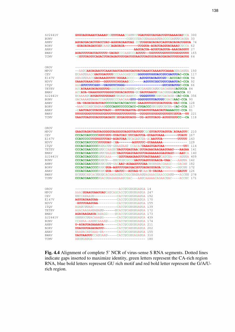
138
SJ2441V GUGUAGUAAAUCAAAAUCGUUUUAAACCAUUCCUGAUUGUUAUGAUUGUUAAAAUAUCCA 300
BUNV ---------------------------CGUCCCUACGUAAAAAUUAGCCCGAUUUCAGUA 33
ANBV AGUAUGACAUUUAUCUAU-AGGUAUAAUUAG CCUUGGUAUAUAUCAGUUAUAUAUUGGUA 74
BORV -GUAUAUAGAUCGUCAAUCAGAUAUA-------UUGGUA-AUAUUAGGUAUAAAUCUCCA 62
ANAV -------------------------------AAGUACUA-AUGUUAGUUA-AAAUAGGUU 27
BWAV AUAUUUUUAUUAUUUUU-UAUAUCCCAAUCCCAUUUU--UGUUUUUGUUUUUUGUGUUUU 165
TCMV -CGUUUAUGUCAGACUUAUAGAUUGUGAUUGUAAUUUAGUGUAUAUGAUAUUUGUGAUUG 64
OROV ------------------------------------------------------------
MPOV --AAUCAAUAGAAUUCAAAUAAUUAGUAUGAUUAUUAAAUCAAAAUUCAAAAGUAGAUUA 150
CEV UCAAUUAACCGAUUGAUUUUCCCAAGGAUCCCCGGGUGUUGGUAUCGUCGAUUUAC-CCA 117
E147V AUAGUUAACCGAUAAAGUUUUCUGGAAGCCC---AGUGUUAUAGUCGUCGACCCAC-CCA 94
NDVV UAAAUUAAACUGU--GGUUUUCUGGAAGCCC-----AGUUUCGGCUGUCGAAUUAC-CCA 92
ITQV AGAGUUCUUCGGU--UAUGUUCUGGG--------------------GUCGUAUUGC-CCA 66
TETEV ACCAUAAAUAUAUGGUUGAGGCUGUACAUUUG—UCGAAUUCAUUCGACAUUAUACUCCA 84
BMAV ACCAUA—UAAAUGGUUGGGGCUGUACAUUUG—UCGAUUUAAUUCGACGUUAUACACCA 83
SJ2441V UCAAAAUCAUGAUUGUGUAAUCUGAUCAAUCC--UGGGUUUUCGUCGACAGU-UAC-CCA 356
BUNV GACAAAAUUAAACCGAUUUUCCCAACAAAGUU—GGGUGUUUUAUUGUCGACGAAC-CCA 90
ANBV -UA-UAUAUAUAUUAUCCCCCACCACCACCCC—GAAGUUUGUCGUAUUGUA-UAC-CCA 128
BORV -AAACCCAUCUAUAAUCCCCAGUCCCCCCACC—CUGACCCGUCGUUCUCUA-UAC-CCA 117
ANAV -CAAUUAGCGUAUAUUAGU--GUUUAUAAUUA—GUGAUUGUAAAUAUUAAAACCC-CCA 81
BWAV UUUGUGUGUUUUUGUGUUUUUUUGUUUUUUUG--UGUGUUGUGUGUGUUGUCGUUA--UU 221
TCMV UAAUUUAGUGUAUGUGAUAUUCGUGAUGUAUG--UG-AUUUUAUG-AUUGUGUUCC--CA 118
OROV ------------------------------------------------------------
MPOV GAAGUAGAUUAGUAGGGGGUAGGGGUAGGUUAUUGUCGCGUUAUUUAGUUACAUAAGUUC 210
CEV CCCACCAUCCCCUGUCGUU—UUAUUAUCUUCGAUUUA-GUAAUUAAA-------UUAUU 167
E147V CCAUCCCCUUGUGGUUUAU—AGAUUAACUCAGAUCUA-ACAAUUUA--------UUUUU 143
NDVV CCCACCAUCCCCUUUGUAU—CGA-------AGUUUUC-GUAAAAAA------------- 129
ITQV CCCACCAGCCCCUGUGCUU—GUAAUAAU CCACACUAAAGUGAUUAA----------UUU 114
TETEV CCCACCAACCCCGUUCCAAGUCUAGUUGAGUAACGUUAGAAUAAGAAGUAAU---AAGAA 141
BMAV CCCACCAACCCCGUUCUAAGUCUAGUUGAGUAAUGUUAGAAAAAGGAGUAGU---AAUCA 140
SJ2441V CCCACCAGCCCCGUUGAAA---CGGUUAAAGAAGUUUAGUAAAAUCAUCUA----AUUUA 409
BUNV CCCACCAACCCCUGUCU---UUCUGUCGCCCGAUUUAGUUGUAAUA—UAAC---AAUUA 142
ANBV CCCACCAACCCCUGUCGUU—GACAAU-AUAAGUUUUAACUGUAUACGAAGC---GACAU 182
BORV CCCACCAACCCCUGUCGUA—AGUUUUGAUGACGUUUAUAUGUAUACUCUAC---GUCUU 172
ANAV CCCACCAACCCCUGUCGUA--GAUUC--AUUAG—UCAACU-UAUAA--------GAUUU 126
BWAV UCUUUCAUCACUUGUCACAAGAGUACCGGCUAUAAUUAGAAACGAACCGAUU---ACCUU 278
TCMV CCCACCAACCCCUGACGUAGAAUAAUCGAC---AAUCAAAAACAGAACUAC----ACCUC 171
OROV -----------------------ACCUCGUGUGAUGA 14
MPOV AAACGUAAUUAAUUAUCAUCAGCACCUCGUGUGAUGA 247
CEV UUCCAUAAGU------------CACCUCGUGUGAUGA 192
E147V AGUUAUAAGUAA----------CACCUCGUGUGAUGA 170
NDVV -GUUUUAAGUAA----------CACCUCGUGUGAUGA 155
ITQV AGAUCUUAAC------------CACCUCGUGUGAUGA 139
TETEV AGACAAGAAUAUAUU------UCACCUCAUGUGAUGA 172
BMAV AGAUAAGAAUACAUAGG----UCACCUCGUGUGAUGA 173
SJ2441V GUUUGCUUACAAAUG-------CACCUCGUGUGAUGA 439
BUNV CCAUAA-AAUUCAAAAU----CCACCUCGUGUGAUGA 174
ANBV U-AUAUUAGAAAAUA-------CACCUCGUGUGAUGA 211
BORV UUAUGUUAGAUAUUU-------CACCUCAUGUGAUGA 202
ANAV UAAUACAUGAAA-UA-------CACCUCGUGUGAUGA 155
BWAV UAUUAAUUUCCAUGAAU-----CACCUCGUGAGAUGA 310
TCMV AUGUGAUGA---------------------------- 180
Fig. 4.4 Alignment of complete 5’ NCR of virus-sense S RNA segments. Dotted lines
indicate gaps inserted to maximize identity, green letters represent the CA-rich region
RNA, blue bold letters represent GU rich motif and red bold letter represent the G/A/U-
rich region.

139
BWAV
61 CUGUAUAUAUUUUAUUUUAUAUUUUAU 87
103 UUGUAUAUAUUUUUAUUAUUUUUUAUA 129
151 UGUUUUUUGUGUUUUUUGU 170
171 GUGUUUUUGUGUUUUUUUG 189
TCMV
27 UUGUGAUUGUAAUUUAGUGUAUAUGAUAU 56
57 UUGUGAUUGUAAUUUAGUGUAUGUGAUAU 85
Fig. 4.5 Presence of repeated sequences in 5’ NCR of virus-sense RNA. Red letters
indicate the duplicated sequences

140
4.2.5 N nucleotide and protein comparison
Viruses in the Bunyamwera, California, Group C and Simbu serogroups were reported to
have the same length of N protein, 233, 235, 234 and 233 amino acids respectively. As
seen in Table 4.2, the same number of amino acids was also observed in the N proteins of
viruses within the same serogroup; ANAV and TCMV (245), ANBV and BORV (247),
GAMV and SJ2441 (238), NDV and E147V (233), and TETEV and BMAV (258). The N
protein of Tete serogroup viruses was the longest reported to date among
orthobunyaviruses. Amino acid sequence alignment of the N proteins of these viruses
(Figures 4.6) revealed that the extensions of the N protein sequence of BMAV and TETEV
were at the amino terminus. However truncation of the N protein at the carboxyl terminus
was also observed for these viruses. Furthermore, 8 amino acids were apparently inserted
after amino acid 23 of their amino terminus. In agreement with previous reports, amino
acid sequence alignments of this study also revealed the presence of some conserved
motifs in the N protein of these viruses.
The nucleotide and predicted amino acid sequences of the N protein coding region of
complete and partial S sequences of the viruses obtained in this study, together with the
published sequences for representatives of other serogroups, (BUNV, CEV, ITQV and
OROV) were compared (Table 4.3). Except for ANAV and TCMV in the Anopheles A
serogroup, whose S segments have only 59% nucleotide sequence identity and 53% amino
acid identity between the N proteins, the S segments and encoded N protein of the viruses
within the same serogroup were found to have nucleotide and amino acid sequence identity
greater than 70%. N sequence identity between viruses in different serogroups was as low
as 24% for amino acid sequences and 5% for nucleotide sequences, which were observed
between TCMV with TETEV and BMAV, and between ANBV with GAMV and OROV
(Table 4.3). This low value could be due to the more serogroups (12 serogroups) used in
these comparisons compared to only four in the previous comparisons. However, greater
sequence identity was also observed between CAPV with viruses in Guama serogroup
(BERV and GMAV), with 80% and 77%, and 73% and 74% for amino acid and nucleotide
identity respectively, while BERV and GMAV exhibited 73% identity for both nucleotide
and amino acid sequences, indicating that BERV and GMAV are closer to CAPV than to
each other. BWAV was found to exhibit high sequence identity with CEV with nucleotide
and amino acid sequence identity of 70% and 68% respectively.

141

142
Caraparu_BeAn3994 ---------------MIELIFKD--------VASFTDNTFDPEAAFLRFE 27
Itaqui_BeAn12797 ---------------MIELIFKD--------VPSFTDNTFDPEAAFLRFE 27
Vinces ---------------MIELIFKD--------EPSFTDNTFDPEAAFLRFE 27
Murutucu_BeAn974 ---------------MIELIFKD--------VASFTDNTFDPEAAYLRFE 27
Restan_TRVL51144 ---------------MIELIFKD--------VASFTDNTFDPEAAYLRFE 27
Marituba_BeAn ---------------MIELVFKD--------VASFTDNTFDPEAAYVRFK 27
Nepuyo_TRVL18462 ---------------MIELVFKD--------EASFTDNTFDPEAAYVRFK 27
Gumbo_LimboFE-371H ---------------MIELVFKD--------ETSFADNTFNPEAAFVAFK 27
Madrid_BT4075 ---------------MSELEFKD--------VPSFADNTFNPEAQYVTFI 27
Aino --------------MANQFIFQD--------VPQRNLATFNPEVGYVAFI 28
Akabane --------------MANQFIFND--------VPQRNAATFNPDAGYVAFI 28
Oropouche ---------------MSEFLFND--------VPQRTTSTFDPEAAYVAFE 27
Maguari ---------------MIELEFND--------VAANTSSTFDPEIAYVNFK 27
Cache_Valley ---------------MIELEFND--------VAANTSSTFDPEIAYVNFK 27
Batai ---------------MIELEFND--------VAANTSSTFDPEVAYINFK 27
Bunyamwera ---------------MIELEFND--------VAANTSSTFDPEVAYINFK 27
Main_Drain ---------------MIELEFHD--------VAANSSSTFDPEVAYASFK 27
Kairi ---------------MSEIEFHD--------VTANTSSTFDPEAGYAAFK 27
California_encephalitis ---------------MSDLVFYD--------VASTGANGFDPDAGYVDFC 27
Tahyna ---------------MSDLVFYD--------VASTGANGFDPDAGYVDFC 27
La_Crosse ---------------MSDLVFYD--------VASTGANGFDPDAGYMDFC 27
Jameston_Canyon ---------------MGDLVFYD--------VASTGANGFDPDAGFVAFM 27
Keystone ---------------MGDLVFYD--------VASTGANGFDPDAGYVAFM 27
BWAV ---------------MSDLIFYD--------VDSVNANEFDPDTGYLDFK 27
E147V ---------------MSEIAFYD--------VQSTAQNGFDPDQQYLAFK 27
NDV ---------------MSELVFYD--------VEPTAQNGFDPDKQYVAFK 27
CAPV ------------MGEVANFEFGER-------LIAEGRNPFVPDVAYARFI 31
ANA_BV ------------MASQVDFAFED--------TGNITQSDFIPDVGYTAFC 30
BORV ------------MSSTVDFAFED--------TGNVTQSDFVPDVGYTTFC 30
TCMV -------------MSGIDFIYDD--------PGRIQQSDFVPREEYAVFC 29
Anopheles_A -------------MSSMDYMFDD--------PGHIQIINYIPEEEYMAFC 29
GAMV -------------MSDSLYVFDD--------DSTASSSTYDPTQEYNVFI 29
SJ2441V -------------MSNDLYVFDD--------DTSASSSSYDPKREYDKFI 29
MPOV -------------MSDLVLTFSD--------SDDVSRSTYNPGEEYDTFV 29
TETEV MPKGGKSDPEPSINVVASAAFEDDEIDYHFTAGDDGGAPFNPMTAYKRFM 50
BMAV MSKVKRGESEPSISVAASAAFAEDEIDYHFSAGDDGGAPFNPMAAYKEFM 50
: : : * : *
Caraparu_BeAn3994 SAYGSQLVSAVVRVFYINAAKVKAYLSRSSELLVFLTLGGVNLAVLNTHF 77
Itaqui_BeAn12797 SAYGRQLVSAVVRVFYINAAKVKAYLSRSSELLVFLTLGGVNLAVLNTHF 77
Vinces SVYGQQLVAAVVRVFYINAAKVKAYLRRSRELLVFLTLGGVNLAVLNTHF 77
Murutucu_BeAn974 SDYGSQLESAIVRVFYINAAKVKAYLRRSSELLVSLTLGGVNLAVLNTHF 77
Restan_TRVL51144 SAYGSQLESAIVRVFYINAAKVKAYLRRSSELLVSLTLGGVNLAVLNTHF 77
Marituba_BeAn PAYGSELESSIVRVFYINAAKVKAYLRRSSELLVLLTLGGVNLAVLNTHF 77
Nepuyo_TRVL18462 PAYGSELESAIVRVFYINAAKVKAYLRRSSELLVSLTLGGVNLAVLNTHF 77
Gumbo_LimboFE-371H PTHGSELESSTVRIFYINAAKVKAYLRRSAEQLVSLTLGGVNLAVLNTHF 77
Madrid_BT4075 PRNGRVLEATIVRIFYINAAKVKAYLHRSSEQLVLLTLGGVNLAVLNTHF 77
Aino AKHGAQLNFDTVRFFFLNQKKAKMVLSKTAQPSVDLTFGGIKFTLVNNHF 78
Akabane SKYGQQLNFTVARVFFLNQKKAKMVLHKTPQPSVDLTFAGVKFTVVNNHF 78
Oropouche ARYGQVLNAGVVRVFFLNQKKAKDVLRKTSRPMVDLTFGGVQFAMVNNHF 77
Maguari RIHTTGLSYDHIRVLYIKGREIKTSLTKRSEWEVTLNLGGWKVAVFNTNF 77
Cache_Valley RIHTTGLSYDHIRIFYIKGREIKTSLTKRSEWEVTLNLGGWKVTVFNTNF 77
Batai RIYTTGLSYDNIRIFYIKGREIKTSLSKRSEWEVTLNLGGWKVTVFNTNF 77
Bunyamwera RLYTTGLSYDHIRIFYIKGREIKTSLAKRSEWEVTLNLGGWKVTVFNTNF 77
Main_Drain RVHTTGLSYDHIRIFYIKGREIKTSLSKRSEWEVTLNLGGWKVAVFNTNF 77
Kairi RRHTTGLNYDHIRIFFLNGKKAKDTLSKRSETTITLNFGGWKIPVVNTHF 77
California_encephalitis AKHGESINLAAVRIFFLNAAKAKAALSRKPERKANPKFGEWQVEVVNNHF 77
Tahyna IKHGEAINLHSVRIFFLNAAKAKAALARKPERKASPKFGEWQVEVVNYHF 77
La_Crosse VKNAESLNLAAVRIFFLNAAKAKAALSRKPERKANPKFGEWQVEVINNHF 77
Jameston_Canyon ADHGESINLSAVRIFFLNAAKAKAALARKPERKATPKFGEWQVEIVNNHF 77
Keystone ANHGESISLSTVRIFFLNAAKAKAALTRKPERKATPKFGEWQVEIVNNHF 77
BWAV NNYPGALNTNTARTFFLNAAKAKNVLRNKPDKKVNPKFGNWEVEVVNNHF 77
E147V AAAGAGLNIVSARIFFLNARKAKDQLSRRPEPKINLKFGTWSVEVVNNHF 77
NDV ASAGAGLNIVSARIFFLNARKAKDQLARRPEPKVGLKFGTWQVEVVNNHF 77
CAPV REHVPMLTPGNIRIFFLRAYDAKQKLKTTTARTANLKFGTATFTVKNNHN 81
ANA_BV LGKTAHLSLENIKIFFLNAGKLKQQMKTCSKTKIKAKFGTLEIELVNTLN 80
BORV IGRSNQLSLENIKIFFVNAGKLKMQMKSSLKPRIKAKFGTLEIEMVNTLN 80
TCMV SKFALVLILQNIRIFFLNAGKLKAALKLSPKRKVKAKFGTLEVEITNTHN 79
Anopheles_A AKYQDQLTVENIRIFFLNAGKFKKMLRTNPKLKVQVKFGTLQLEIINTHN 79
GAMV STHGDQLTPANIKCFFLHARTAKARLAKDPAERRTLTFGSWKVEVVNNHY 79
SJ2441V STYGQQLTPANIKCFLLHARTAKARLSKDPAERRTLTFGTWKVEVVNNHY 79
MPOV DTYREHLTVDNIRIFFLKAAEAKAHMAKVNNEVVEVHFGTLVLPLVNNHK 79
TETEV EVHGKELNLPNIKVFFLKARQAKEIMRSKAKSEMTFTFGSLTLTFKNTHH 100
BMAV ETHGKDLTVTNIRVFFLKARQAKEIMRSKAKSEMTFTFGSLTLTFKNTHH 100
: : : :. * : :. . . *

143
Caraparu_BeAn3994 PGTQSIPLPDYGLTLHRISGYLARWALYLIRTNPESDIAL-VRSTFVVPL 126
Itaqui_BeAn12797 PGTQSIPLPDYGLTLHRISGYLTRWALYLIKTNPESDIAL-VRSTFVVPL 126
Vinces PGTQSIPLPDYGLTIHRISGYFSRWALYLIATNPESDIAL-VRSTFVVPL 126
Murutucu_BeAn974 PGTQSIPLPDYGLTIHRLSGYLSRWALYLIRPSPESEIGL-VRSTFIVPL 126
Restan_TRVL51144 PGTQSIPLPDYGLTLHRLSGYLARWALYLIRPSPESDIGL-VRSTFIVPL 126
Marituba_BeAn PGTQSIPLPDYGLTIHRLSGYLSRWALYLIYIIPESEIDL-VRTTFIVPL 126
Nepuyo_TRVL18462 PGTQSIPLPDYGLTIHRLSGYLARWALYLICPIPDSDIDL-VRTTFIVPL 126
Gumbo_LimboFE-371H PGTQSIPLPDYGLTIHRLSGYLARWALYLICPSPDSSIGL-VRATFVVPL 126
Madrid_BT4075 PGNQSIPLPDYGLTIHRVSGYLARWALYLIAPSPYSDRDI-VRTTFIVPI 126
Aino PQYTANPVPDTALTLHRLSGYLAKWVADQCKTN-QIKLAE-AMEKIVMPL 126
Akabane PQYTANPVSDTAFTLHRISGYLARWVAEQCKAN-QIKFAE-AAATIVMPL 126
Oropouche PQFQSNPVPDNGLTLHRLSGYLARWAFTQMRS--PIKQAE-FRATVVVPL 124
Maguari PGNRNSPVPDDGLTLHRLSGFLARYLLEKILKVSDPEKLI-IKSKIINPL 126
Cache_Valley PGNRNSPVPDDGLTLHRLSGYLARYLLEKILKVSDPEKVI-IKSKIINPL 126
Batai PGNRNSPVPDDGLTLHRLSGFLARYLLEKILKVSDPEKLI-IKSKIVNPL 126
Bunyamwera PGNRNSPVPDDGLTLHRLSGYLARYLLEKILKVSDPEKLI-IKSKIINPL 126
Main_Drain PGNRNSPVPDDGLTLHRLSGFLARYLLEKILKVSEPEKLL-IKSKIINPL 126
Kairi LENRNMSVPDDGLTLHRVSGYLARYLLDRVYSAGEPEKLK-IKTTIINPI 126
California_encephalitis PANRNNPIGNNDLTIHRISGYLARWVLEQYKENEDESQRELVKTTVINPI 127
Tahyna PGNRNNPIDNNDLTIHRLYGYLARWVLEQFKENEDAAQRELIKTTVINPI 127
La_Crosse PGNRNNPIGNNDLTIHRLSGYLARWVLDQYNENDDESQHELIRTTIINPI 127
Jameston_Canyon SGNRNNPIGNNDLTIHRLSGYLARWVLEHFNSDDDESQRELIRSTIINPI 127
Keystone PGNRNNPIGNNDLTLHRISGYLARWVLEHFGEGEDESQKELIKSTVINPI 127
BWAV PGNRNNPIGKDDLTLHRISGYLARWILEEYKR-DDESEKEIIESTVINPI 126
E147V QGNRDNVISDTDLTIHRLSGYIARFILDQYLAGNTVVQSG-IQLQIVNPI 126
NDV QGNRDNPIGDSDLTMHRLSGYIARYILDQYLAGNSVAQAG-IQLQIINPI 126
CAPV ERNANIEVEAEDLTLHRISGFLAKFVRESMVDETASKMIR---DTIVNPI 128
ANA_BV RSLGQVSLQPNDVTLHRASAYLARKALELYREGQADFQAA-MRDQFVMPL 129
BORV KALGQISLQPTDLTLHRASAFLARRALEMYHGGAADFQAA-MRDQFVMPL 129
TCMV RAFADSKLEKGDITLHRLSGFLAKKILDIYNAGSFDLQTQ-IKAEIIIPL 128
Anopheles_A PANREIQLSSSDLTLHRVSAYLARKSLRLWEALTLDQKEE-FKLKIIMPL 128
GAMV RRATPVTVQDHDNTLHRISGYLALYCLQTSSQNKQNFER--IKSVVINPI 127
SJ2441V RRAAPITVQDHDLTLHRLSGYIALYCLQASVENAQSFER--IRSTVINPI 129
MPOV PGRAQREVADSDLTLHRVSGYLAKFVLELYKKLRKSSEREAITSRIINPI 129
TETEV PSNRHLAVEQDDLTINRVTGFMAHAILLTHRDPKHKDAVE---KTIINPI 147
BMAV PSNRHLTVDQDDLTINRATGFMAYAILLTHREPKSKDAVE---KTIINPI 147
: :*::* .::: .: *:
Caraparu_BeAn3994 AEENGITWR--DGVEMYLAFLPGAEMFMSTFEFYPLTIGMYRVRRWSMDP 174
Itaqui_BeAn12797 AEENGITWR--DGVEMYLAFLPGAEMFMSTFEFYPLTIGMYRVRRWSMDP 174
Vinces AEENGITWR--DGVEMYLAFLPGAEMFMSTFEFYPLTIGMYRVRRWSMDP 174
Murutucu_BeAn974 AEENGITWS--DGVGMYLAFLPGAEMFMSTFTFYPLTIGMYRVRRWSMDP 174
Restan_TRVL51144 AEENGITWS--DGVGMYLAFLPGAEMFMSTFAFYPLTIGMYRVRRWSMDP 174
Marituba_BeAn AEENGITWT--DGVAMYLAFLPGAEMFMSTFKFFPLTIGMYRVRRWSMDP 174
Nepuyo_TRVL18462 AEENGITWT--DGVAMYLAFLPGAEMFMSTFTFFPLTIGMYRVRRWSMDP 174
Gumbo_LimboFE-371H AEENGITWC--DGVAMYLAFLPGAEMFMSTFAFFPLTIGMYRVRRWTMDP 174
Madrid_BT4075 AEKNGITWC--DGVTMYLAFLPGSEMFMSTFRFFPLTIGMYKVQRWGMDP 174
Aino AEVKGCTWT--EGLTMYLGFAPGAEMFLETFEFYPLVIDMHRVLKDGMDV 174
Akabane AEVKGCTWS--DGYAMYLGFAPGAEMFLETFEFYPLVIDMHRVIKDGMDV 174
Oropouche AEVKGCTWN--DGDAMYLGFAAGAEMFLQTFTFFPLVIEMHRVLKDGMDV 172
Maguari AEKNGITWA--DGEEVYLSFFPGSEMFLGTFKFYPLAIGIYKVQKKEMEP 174
Cache_Valley AEKNGITWS--DGEEVYLSFFPGSEMFLGTFKFYPLAIGIYKVQRKEMEP 174
Batai AEKNGITWA--DGEEVYLSFFPGSEMFLGTFRFYPLAIGIYKVQRKEMEP 174
Bunyamwera AEKNGITWS--DGEEVYLSFFPGSEMFLGTFRFYPLAIGIYKVQRKEMEP 174
Main_Drain AEKNGITWA--DGEEVYLSFFPGSEMFLGIFKFYPLAIGIYKVQRKEMEP 174
Kairi AASHGITWD--DGEEVYLSFFPGSEMYLTTFKFYPLAIGIYKVQRKLMDP 174
California_encephalitis AESNGIRWE--NGAEIYLAFFPGTEMFLETFKFYPLTIGIYRVKNGMMDS 175
Tahyna AESNGIRWD--NGAEIYLAFFPGTEMFLETFNFYPLTIGIYRVKQGMMDP 175
La_Crosse AESNGVGWD--SGPEIYLSFFPGTEMFLETFKFYPLTIGIHRVKQGMMDP 175
Jameston_Canyon AESNGIHWN--NGPEIYLSFFPGTEMFLEIFKFYPLTIGIYRVKHGMMDP 175
Keystone AESNGIRWG--NGVEIYLSFFPGTEMFLELFKFYPLTIGIYRVKHGMMDA 175
BWAV AESNGIRWS--DGAEIYLSFFPGTEMFLEPFKFYPLAIGIYRVKHKMMDA 174
E147V AESNGIKWS--AGAEIYLSFFPGTEMFLEKFNFYPLAIGIYRVKRGMMDA 174
NDV AESNGIKWS--AGAEVYLSFFPGTEMFLEKFNFYPLAIGIYRVKKGMMEA 174
CAPV AESLGITWE--NGDDVYLSFFPGTEMFLDTFHMLPLAIGIYRVQQKQMKA 176
ANA_BV AEVAGVQFKPEVPPELYIGFAPGAEFFMKLFKCYPLAIAVVRVKKGQMQP 179
BORV AEVSGVQFKPEVPPELYIGFAPGAEFFMKLFKCYPLAIAVVRVKKGQMQP 179
TCMV AEVAGVSWANSTP-EIYLSFCPGSEFFLADFKFYPLAIGIARVKKDLMKP 177
Anopheles_A AEKAGVTWDNAGSVEMYLGFAPGAEFFLKDFKFYPLAIGIARVKKGLMSP 178
GAMV SESMGINWQ--HGAMIYLSTLPGAEMFLTEFQLFPLAFAVVRVKKNLASP 175
SJ2441V SESMGINWQ--HGAMMYLSTLPGAEMFLTEFQLLPLAFAVVRVKKNLASP 175
MPOV SAKMGFQWS--VGPEIYLSTLPGTEMFLDAFKMYPLAFILLRVKRGEIKI 177
TETEV ADSKGVTWK--ERGKCISCFFPGTEMFMLEFKFFPLAVGLARCHKEKMDT 195
BMAV AESKGVTWK--EGANIYLSFFPGTEMFMLEFRFFPLAVGLARCHKEKMDT 195
: * : :*:*::: * **.. : : . .
Caraparu_BeAn3994 NCLEKAIWQRYM-GIGADQWITKETVAILTRFEDVEKLDWARTAFTSAARE 224
Itaqui_BeAn12797 NCLEKAIWQRYM-GIGADQWITKETVAILTRFEDVEKLDWARTAFTSAARE 224

144
Vinces NCLEKAIWQRYM-GIGADQWITKETVAILTRFDAVEKLDWARTAFTSAARE 224
Murutucu_BeAn974 NCLEKAIWQRYM-GIGADQWITKETVAILTRFEDVEKLDWAKTAFTSSARE 224
Restan_TRVL51144 NCLEKAIWQRYM-GIGADQWITKETLAILTRFEDVEKLDWAKTAFTSAARE 224
Marituba_BeAn NCLEKAIWQRYM-GIGADDWITKETVTILARFEDVEKLDWARTAFTSSARE 224
Nepuyo_TRVL18462 NCLEKAIWQRYM-GIGADDWITKETVTILARFEDVEKLDWARTAFTSSARE 224
Gumbo_LimboFE-371H QCLEKAIWQRYM-GIGADEWITKETVSILARFEDVEKLDWAKTAFTSAARE 224
Madrid_BT4075 KCLEKAIWQRYA-GIGADTWIQKEYADINARFSVVESLDWARTAFTSTARE 224
Aino NFMRKVLRQRYG-TLTAEQWMTQKIDAVRAAFNAVGQLSWAKSGFSPAARA 224
Akabane NFMRKVLRQRYG-QLTAEEWMTSKLDAVKAAFSSVAQISWAKSGFSPAARA 224
Oropouche NFMKKVLRQRYG-QKTAEQWMREEIVAVRAAFEAVGTLAWARTGFSPAGRD 222
Maguari KYLEKTMRQRYM-GLEAATWTVSKVNEVQAALTVVSGLGWKKTNVSAAARE 224
Cache_Valley KYLEKTMRQRYM-GLEASTWTISKVNEVQAALTVVSGLGWKKTNVSAAARE 224
Batai KYLEKTMRQRYM-GLEASTWTVSKLNEVQSALTVVSGLGWKKTNVSSAARE 224
Bunyamwera KYLEKTMRQRYM-GLEASTWTVSKLNEVQSALTVVSGLGWKKTNVSSAARE 224
Main_Drain KYLEKTMRQKYM-NMDAATWTVTQVSEVQAALTVVSGLGWKKTNVSAAARE 224
Kairi KYLEKTMRQRYM-NLDASQWTQKHFSDVNSALTVVAGLGWKKANVSIAAKD 224
California_encephalitis QYLKKALRQRYG-SLTAEKWMSQKTGMIAKSLKEVEQLKWGRGGLSDTART 225
Tahyna QYLKKALRQRYG-SLTADKWMSQKTTAIAKSLKDVEQLKWGRGGLSDTART 225
La_Crosse QYLKKALRQRYG-TLTADKWMSQKVAAIAKSLKDVEQLKWGKGGLSDTAKT 225
Jameston_Canyon QYLKKALRQRYG-TLTAEKWMAQKTVLIAKSLKDVEQLKWGRGGLSDAART 225
Keystone QYLKKALRQRYG-TLTADKWMAQKTSMITKSLKDVEQLKWGKGGLSDTARA 225
BWAV QFLKKALRQRYG-KMTAEKWMSTKVKTIGEAVRNVEKLKWGKGGLSDAARN 224
E147V QYLKKALRQRYG-NLTADQWMQSKSDDVLRAIAVLEKLQWGKSGLSEAARQ 224
NDV QFLKKSLRQRYG-QMTADQWMQTKSDDVMRAVAVLEKLSWGRSGLSEAARQ 224
CAPV EFLKKHLRQQYG-GLAASQWMSERKEDVKLALTLISRLPWGKAGLSAAARN 226
ANA_BV EFLAKSLRQRYA-GAPPAEWMSTNAAAIKAATAMVEKYPMMKVASRPHIEK 229
BORV EFSAKCLRQRFG-GAPPAEWMTTNAPAIKIATAMVEKFPMMRTASRPHVEK 229
TCMV EFLAKLLRQRYG-NMTPGEWMKTHSVSVQSAISEIEKYPLMKVNSRPHVEE 227
Anopheles_A DFLSKTLRQRYG-GIPPADWMAIHKSAVRTAVEFVDKFPLMRSNASSHVEQ 228
GAMV ETVKKVLRQRYD-GRLPGEWMNSELEKVKTAITKVEALHKTFVGVTARMIE 225
SJ2441V ETVKKVLRQRYE-GRLPGEWMNSELEKVKAAITRVESLHKTFVGVTARMIE 225
MPOV DMAKKAMRQRYG-DKEASTWLEEEVDTVKSAIKTVEKLKPTLTGLAASMTK 227
TETEV EYLKKPMRQMLTDGTKAQVWLGAKIEEIRKAYKVCMNLKFVKAGFSEAARE 246
BMAV EYLKKPMRQMLTDGTKAQVWLGAKIEEIRKAYKVCMNLKFVKAGFSEAARE 246
. * :*:: . * . : .
Caraparu_BeAn3994 FVSRFGIRLA-------- 234
Itaqui_BeAn12797 FVSRFGIRLA-------- 234
Vinces FVIQFGIRLA-------- 234
Murutucu_BeAn974 FVSRFGIRLA-------- 234
Restan_TRVL51144 FVSSFGIRLA-------- 234
Marituba_BeAn FVSQFGIKLA-------- 234
Nepuyo_TRVL18462 FVSQFGIKLA-------- 234
Gumbo_LimboFE-371H FVSKFGIRLA-------- 234
Madrid_BT4075 FVAQFGIKLA-------- 234
Aino FLAQFGINI--------- 233
Akabane FLAQFGIQI--------- 233
Oropouche FLRQFGIGI--------- 231
Maguari FLAKFGINM--------- 233
Cache_Valley FLAKFGISM--------- 233
Batai FLAKFGISM--------- 233
Bunyamwera FLAKFGISM--------- 233
Main_Drain FLAKFGINM--------- 233
Kairi FLNKFGINI--------- 233
California_encephalitis FLQKFGIKLP-------- 235
Tahyna FLQKFGIRLP-------- 235
La_Crosse FLQKFGIRLP-------- 235
Jameston_Canyon FLIKFGVKLP-------- 235
Keystone FLAKFGVRLP-------- 235
BWAV FLKKFDIAMI-------- 234
E147V FLGKFGILI--------- 233
NDV FLGRFGIVI--------- 233
CAPV FLADFGITF--------- 235
ANA_BV LLSELGLSAPTIQQALTK 247
BORV LLSELGLSTPTIQRALTK 247
TCMV FLKSLGLSAHYINGALRG 245
Anopheles_A FLVDLGLSRATVASMKR- 245
GAMV FFTALGINLDMRR----- 238
SJ2441V FFSALGINLDLRR----- 238
MPOV FLQELGISK--------- 236
TETEV FLKEFGLDQDKN------ 258
BMAV FLKEFGLDQDKN------ 258
: :*
Fig. 4.6 Alignment of complete N protein of the viruses. * indicate 100% conserved; brown letters indicate
conserved motif; dotted lines indicate gaps inserted to maximize identity; : denotes more than 90%
conserved; . denotes more than 50% conserved.

145
4.2.6 NSs protein comparison
NSs amino acid sequences of NDV, E147V, BWAV, GAMV, SJ4441V and MPOV were
aligned with the published NSs sequences of BUNV, CEV, ITQV and OROV (Fig. 4.7).
This alignment showed that the NSs proteins vary in size especially at the carboxy
terminus. The largest NSs protein was observed in SJ2441V (137 amino acids) in Gamboa
serogroup and the smallest (79 amino acids) in MPOV in Turlock serogroup. These are the
largest and the smallest NSs protein reported to date. As observed with GROV NSs
protein, the relative truncation of MPOV was at both termini. In agreement with previous
results (Dunn et al., 1994; Bowen et al., 1995), NSs proteins reported in this study were
more divergent than the corresponding N proteins with no conserved amino acid motif
observed. The conserved LPS motif at the carboxy terminus in most of NSs protein of
Bunyamwera and California serogroup viruses was only observed in BWAV NSs protein.
This motif was also absent in ITQV, OROV and GROV NSs proteins (Figure 4.7). The
sequence identity of the NSs protein among viruses within the same serogroup was more
than 80% and between serogroups were as low as 4% (between ITQV and BWAV) (Table
4.4). In contrast to viruses in Bunyamwera, California, Group C and Simbu serogroup,
only viruses in Gamboa serogroup were found to have two lengths of NSs protein; 130
(GAMV) and 137 (SJ2441V) amino acids respectively. This could be due to the limited
number of viruses used for each serogroup in this study.
4.2.7 Phylogenetic analysis of S segments
To establish genetic relationships among the studied viruses with other orthobunyaviruses,
phylogenetic trees were constructed from the alignments of complete N amino acid
sequences of ANAV, ANBV, BMAV, BORV, BWAV, CAPV, E147V, GAMV, MPOV,
TCMV, NDV, TETEV and SJ2441V (Figure 4.8A) using NJ analysis, and partial N ORF
of ANAV, ANBV, BMAV, BORV, BWAV, CAPV, E147V, GAMV, MPOV, TCMV,
NDV, TETEV and SJ2441V, BERV, GMAV, PLSV and PATV using NJ and MP analysis
methods (Figure 4.8B and 4.8C). For NJ analysis, a distance matrix was calculated from
the aligned sequences using the Kimura two parameter formulas (Kimura, 1980) and NJ
tree was computed. The reliability of the inferred tree was tested by bootstrap analyses
using 100 replicates data sets generated from the original sequence alignment (Felsenstein,
1993) and a bootstrap consensus tree was generated. For MP analysis, phylogenetic tree

146
NDVNSs ------MMSSQLPKMDLILISSMWHLKLQLEQGLTLFPLGSSSSMPGKPKINSLVDQSRRLVLNLE 60
E147VNSs ------MMSSLLPKMDLILINNTWHLKLLQEQGLILFPLGSSSSMPGKPKINSLVDQSQRLTLNLA 60
BWANSs ------MMSTRLMPMSLTLIQDTWILKITIQGHSIQIPLGHSSSMPQRPRMCSAINLTRRSILNLE 60
CEVNSs ------MMSHPQVQMDLILMQGMWTSVLNMENQLTLLQLGSSSSMPQRPRLLSRVSQRGKLILNLA 60
BUNVNSs ------MMSLLTPAVLLTQRSHTLTLSVSTPLGLVMTTYESSTLKDARLKLVSQKEVNGKLHLTLG 60
GAMVNSs -------MIRRQALALMIQRRNTMSLLVLTGTSLLQQTLNVSSCMHVQPRLVWQRTRRNAERLLLD 59
SJ2441VNSs MIFTYSTMIRRQALALMIQRENMINLLVLTGSNLLQQTLNVFSCMHARPRLVFQRTRRNAERLLLE 66
OROVNSs -------MYHNGLHLHLIRRQHMWHLKLDTDKCSMLVLLESSSSTKRRPKMSYVRHRGPWLTLLLV 59
ITQVNSs -------MYPHSQTIHLTLKLLSLDSKVHTAGNWFPPSYESSTSTRRRSKLTSVGQVNSLFFLLLV 59
MPOVNSs ----------MSVEVLITQERNTIRLWIHTENISQWITLESSSSRLQRPKLTWRRLTTRSLKFILE 56
:: : : : : :: : *
NDVNSs HGRWKWSITIFKETGTILSATQISQCIDSQDI---------------------------------- 92
E147VNSs HGRWRWSITIFKETGTMLSATRILQSTDCQDI---------------------------------- 92
BWANSs TGRWRLSIIIFLETGTTQLVKMILPSTEFQDI---------------------------------- 92
CEVNSs SGRWRLSIIIFQQTGTIQLVTTILPSTASQDTLPDGS----------------------------- 97
BUNVNSs AGRLLYIIRIFLATGTTQFLTMVLPSTASVDSLPGTYLRRC------------------------- 101
GAMVNSs LGRLRLLITITEEPLRLQCRTMIIPCTAFPDILPCTACRPAPRISRTLSESSQSSSTRSLSPWGST 125
SJ2441VNSs LGRLRLLITIIDEPLQLRCRTMILPCTAFPDILPCTVCRPVWRMRSLSSESDQQSSTRSLSPWGST 125
OROVNSs GSNLQWLITISHSSSRIQCRTTVLPCTVCQDT---------------------------------- 91
ITQVNSs VLTLPYLTHISQEPSPFHCLTTDSRCTAYRVTSPDGPCI--------------------------- 98
MPOVNSs HSSSRWLITISLVEPSVRLRTLT--------------------------------------------79
*
NDVNSs -----
E147VNSs -----
BWANSs -----
CEVNSs -----
BUNVNSs -----
GAMVNSs GSMEQ 130
SJ2441VNSs GSMEQ 130
OROVNSs -----
ITQVNSs -----
MPOVNSs -----
Fig. 4.7 Alignment of NSs protein. * indicate 100% conserved; dotted lines indicate gaps
inserted to maximize identity; red bold letters indicate LPS conserved motif : denotes more
than 90% conserved.

147

148
A. Phylogenetic tree using Neighbor-joining based on complete amino acid N sequence
ANA_BVBORV
TCMVAnopheles_A
100
65CAPV
TETEVBMAV100CaraparuItaquiVinces
90
MurutucuRestan
94
99MaritubaNepuyo
67
100Gumbo
65
Madrid
94
AinoAkabaneOropouche
95
100
100MaguariCache_ValleyBataiBunyamwera
86
78Main_Drain
74
Guaroa
100
Kairi
57
72
100Jameston_CanyonKeystoneMEL
55
California_encephalitisTahynaLaCSSH
91
99
76
74
TVT
82
BWAV
95
E147VNDV
57
100
39
100
51
58
100
50
GAMVSJ2441VMPOV
100
0.1
GROUP C
CALIFORNIA
SIMBU
BUNYAMWERA
GAMBOA
ANOPHELES A
ANOPHELES B
TETE
TURLOCK
BWAMBA
NYANDO
CAPIM

149
B. . Phylogenetic tree using Neighbor-joining based on partial amino acid N sequence.
CaraparuItaquiMurutucu
100
Restan
48
Vinces
43
MaritubaNepuyoGumbo
96
53
35Madrid
98
MaguariCache_ValleyBataiBunyamwera
57
99Main_Drain
69
Kairi
96
Guaroa
68
100
100AinoAkabane
Oropouche
94
53
100TETEVBMAV
29
100La_CrosseTahynaCalifornia_encephalitis
56
Jameston_CanyonKeystone
58
44BWAV
100
E147VNDV
87
100PATV
90
CAPVBERVGMAV
54
31
100
6
20
ANBVBORV
TCMV
100
Anopheles_A
52
PLSV
89
49
51GAMVSJ2441V
MPOV
100
71
0.1
MINATITLAN
GROUPC
CALIFORNIA
SIMBU
BUNYAMWERA
GAMBOA
ANOPHELES B
TETE
TURLOCK
GUAMA
CAPIM
ANOPHELES A
PATOIS
NYANDO
BWAMBA

150
C. Phylogenetic tree using Maximum Parsimony method (Mega 2.1 software) based on
partial amino acid N sequence.
La Crosse
Tahyna
California encephalitis
Jameston Canyon
Keystone
BWAV
E147V
NDV
PATV
GMAV
CAPV
BERV
Guaroa
Kairi
Main Drain
Maguari
Cache Valley
Batai
Bunyamwera
Aino
Akabane
Oropouche
Madrid
Gumbo
Marituba
Nepuyo
Murutucu
Restan
Vinces
Caraparu
Itaqui
MPOV
GAMV
SJ2441V
ANA BV
BORV
TCMV
Anopheles A
TETEV
BMAV
PLSV
61
100
87
82
54
56
99
100
100
100
93
100
100
98
66
93
92
100
99
56
53
97
65
96
93
92
62
52
Fig. 4.8 Phylogeny of the studied viruses based on complete N ORF amino acid sequence of ANAV, ANBV,
BWAV, BORV, E147V, GAMV, MBOV, NDV, SJ2441V and TETEV, and partial N ORF of BERV,
GMAV and PLSV. Viruses in the Bunyamwera, California, Group C and Simbu serogroups were used as a
control group. The sequences were aligned using the ClustalX (NCBI) and ClustalW program (EMBL-EBL).
Phylogenetic analyses were generated by Neighbor-joining (NJ) based on complete N amino acid sequences
(A) and partial N sequences (B) and Maximum Parsimony (MP) based on partial N sequences (C). Numbers
adjacent to each branch represent the percentage bootstrap support calculated for 100 replicates. All the trees
yielding almost identical topologies, except with TCMV. Blue letters indicate the phylogeny results that not
correlated with serological grouping and red letters indicate serogroups used in this study. Scale bar represent
10% amino acid sequence divergence. Accession numbers of the control viruses are listed in 2.2.11.
BUNYAMWERA
CALIFORNIA
SIMBU
GROUP C
BWAMBA
NYANDO
PATOIS
GUAMA
CAPIM
TURLOCK
GAMBOA
ANOPHELES B
ANOPHELES A
TETE
MINATITLAN

151
was obtained by the heuristic search using equal weighting of all changes.Weighting
schemes of 3:1 and 10:1 were used and gaps were treated alternatively as missing data. The
reliability of the inferred tree was tested by bootstrap test performed on 100 replicates data
sets generated using heuristic search option and a bootstrap consensus tree was computed.
Representative members of Bunyamwera, California, Group C and Simbu serogroups were
included in this analysis to root the trees. The results indicated that the overall
phylogenetic relationships determined using NJ and MP methods were similar except with
one difference: the NJ plot based on complete N sequence and MP plot based on partial
sequence predict TCMV in the same clade as ANAV, while the NJ plot based on partial
sequence, separated these viruses into different clades. Previous serological classification
grouped BERV and GMAV in the Guama serogroup and CAPV in Capim serogroup.
However, phylogenetic analysis based on partial amino acid sequence of CAPV, BERV
and GMAV N proteins grouped BERV in the same clade as CAPV but separate from
GMAV, indicating that BERV was closer to CAPV than GMAV. With the exception of
BERV and TCMV viruses, all viruses from the same serogroup were placed in the same
clade in NJ and MP derived trees.
4.2.8 Partial M and L sequences
During the attempts to get the reverse-transcription PCR product for S segments, primers
S13+ and S13- were also used, which were designed based on conserved sequences of 13
terminal nucleotides of the S, M and L segments of the published sequences of
Bunyamwera, California, Group C and Simbu serogroups viruses. Partial sequences of the
L and M segments of PATV, and of the L segment of CAPV were obtained (Figure. 4.9).
For PATV L segment, both primers were found to hybridise at the internal sequences. Only
the forward primer hybridised at the 3’ end of virus-sense M RNA segment of PATV and
L segment of CAPV, while the reverse primer hybridised at internal sequences (Figure
4.10). The lengths of M and L segments obtained for PATV were 505 nucleotides
(representing 151 amino acids) and 922 nucleotides (representing 307 amino acids)
respectively. For CAPV L segment, a sequence of 742 nucleotides (representing 237 amino
acids) was obtained. The sequences were put in the BLAST program (NCBI) and
compared with the published sequences of representative virus of each serogroup; BUNV
for Bunyamwera, LACV for California, ITQV for Group C and OROV for Simbu.
However the comparisons were very limited due to the limited sequences of M and L

152
A. PATV M sequence
1 AGTAGTGTGCTCCACAATAAAGATTTCAAAGGAAATATCAAAAAGATACACCATGTTAAC
M L T
61 TAAAATGCTGCTGTTTGCACTGCTTACAGTATGTGCATCAGTACCCTTGGATAAGTGCTT
K M L L F A L L T V C A S V P L D K C F
121 CAGTGGAGGCATAGTTATAAAAAAGCTTCAAACTGATTATGGCCTCCCCCATATGTGTCT
S G G I V I K K L Q T D Y G L P H M C L
181 AAGAGACGATATAAGCATGATAAAAACTGATTCGGTTGCAGTTCAAGGTACTTCAAATGA
R D D I S M I K T D S V A V Q G T S N D
241 TGAGAAGACAATATTCACATCAAGTATCACAAGGAAATTGCTTGTGACGGATTGGAAAAA
E K T I F T S S I T R K L L V T D W K N
301 CTGTAGGCCTGAAAAAATGATAGGTGGTCCTATCATGTTGCTATCTGTAGATGACAGAGG
C R P E K M I G G P I M L L S V D D R G
361 TCACTTGAAATCAGAAGAGTATGTGTGTCAAAATGATTGTGAGATAAAATTGGAGAAAGA
H L K S E E Y V C Q N D C E I K L E K E
421 ATCAGGATTGATAATATTTGAAACAGCATCACTAAATTACTATCAAGTGAGTGGTACCAC
S G L I I F E T A S L N Y Y Q V S G T T
481 CATCAGTTCTGGCTGGTTTAAATCA
I S S G W F K S

153
B. PATV L sequence 1 AGTAGTGTGCCCCTCAGCAAGTATTCAATCTAAGAAATCAACTGTGGTGTTTTCAACAAT
V V C P S A S I Q S K K S T V V F S T I
61 TTGTCTACATAAGGATTCAAAGGATGTATTGAAATGCGGGGCATTGTATAAAACATACAA
C L H K D S K D V L K C G A L Y K T Y K
121 AATAGGAGACCAATACATATCTATATCTAAAGCTATTCGTTTAGATAAAGAGAGATGCCA
I G D Q Y I S I S K A I R L D K E R C Q
181 GAGGTTAGTCACAGCTCCTGGAATTTTCATGTTAACCACACTATTGTTGAAAGGGGATAC
R L V T A P G I F M L T T L L L K G D T
241 AGAAATAGACTTAGATGAGATCATGGCTTTCTCATTTTTCACCTCACTATCTATAACAAA
E I D L D E I M A F S F F T S L S I T K
301 AAGTATGCTCTCACTAACAGAGCCATCTAGGTATATGATAATGAACTCGTTGGCAGTGAG
S M L S L T E P S R Y M I M N S L A V S
361 CAGCCATGTCCGAGAATATATAGCCGAAAAGTTTTCTCCATATACAAAAACCTTATTCTC
S H V R E Y I A E K F S P Y T K T L F S
421 AGTTTATATGACAGAATTGATCCACAAAGGCTGCATGTCAGCAAACAACCAACGGTCTAA
V Y M T E L I H K G C M S A N N Q R S K
481 GATATCTGTGAAAAATGTTTTCCTCAATGAATATGAAATAACTCAAAAAGGGGTTTCAGA
I S V K N V F L N E Y E I T Q K G V S E
541 AGATCGCGATCTAGAATCAATCTGGTTTCCAGGACATGTAAATCTCAAGGAATATATCAA
D R D L E S I W F P G H V N L K E Y I N
601 TCAAGTGTATCTCCCCTTTTACTTCAATGCTAAAGGGTTACATAATAAGCATCATGTTAT
Q V Y L P F Y F N A K G L H N K H H V M
661 GATAGACTTAGCTAAGACAGTTTTAGAAATTGAACTAGAACAAAGAGAGCAACTGCCCAA
I D L A K T V L E I E L E Q R E Q L P N
721 CCCATGGGGCAAAGATTTCCAGAAACAATCTGTGAACCTAGAAATATTGATCTATTCGAT
P W G K D F Q K Q S V N L E I L I Y S I
781 AGCAAAGATGCTGAACAATGATACATCTAAGCATAATCATCTAAGAAGCAGGATTGAGAA
A K M L N N D T S K H N H L R S R I E N
841 TAGGAACAACTTCAAGAGGTCACTGGCAAGCATATCGACATTCACTAGCTCGAAGTCATG
R N N F K R S L A S I S T F T S S K S C
901 CATAAAAGTGGGGGATTTCTCG
I K V G D F S

154
C. CAPV L sequence 1 AGTAGTGTACCCTGACATAGTATTTAGCTATTTAGAAGGGTACACTCAAAAATGGCAACG
M A T
61 CAAATTGGAGAATTAGTTAGGCAGTTTGCTGCAAGAGTTAGGGCATTAAACCCAAATCAA
Q I G E L V R Q F A A R V R A L N P N Q
121 CCAGAGTTAGGGAGAGATCTTCTGTCTGAAATAACTGTAGCTCGACATAATTATTTTGCC
P E L G R D L L S E I T V A R H N Y F A
181 CAAGAGTTTTGCGAGAGCATCGGATTAGAATATAGAAACGATGTACCAGCTCTGAATATA
Q E F C E S I G L E Y R N D V P A L N I
241 GTAATGGAGATGAGACCGGACTTCGATCCTATGACAATTAAGGTGCCGGAAATCACACCA
V M E M R P D F D P M T I K V P E I T P
301 GACAACTATTACAGAGACGGGGACAAGATCTATATTATAGATTTTAAAGTATCGGTTAGT
D N Y Y R D G D K I Y I I D F K V S V S
361 GACGAATCTGCTAATCATACATTCAAGAAATACAATACTTTGTTCGGCGATGTCTTTGAT
D E S A N H T F K K Y N T L F G D V F D
421 AAGTTAGGGATTGAATTCGAGGTAGTCATTATTAGGATGGACCCCACAAGAGGAGAGATA
K L G I E F E V V I I R M D P T R G E I
481 TTGAAGGGTTGGGACAAAATGGTCGAAAGAGTCTCAGAAACTAGAGAGATAACCAGAGAT
L K G W D K M V E R V S E T R E I T R D
541 GTGACCAAGCAGAAGCCAAGTATACATTTCATATGGTCTGGGCACAATTCAAAAGAAGTT
V T K Q K P S I H F I W S G H N S K E V
601 ACAGGCAACACTCAGAAAATATTGAGGCTCTCAAAATGTCTACAAAGAATAAAGGACACC
T G N T Q K I L R L S K C L Q R I K D T
661 GACCAATTCAGCACAGCTTTTAGGAAAATAGGTGCACTAATGGATTTCAGTGAAAACATA
D Q F S T A F R K I G A L M D F S E N I
721 GCAGGCTACGAGAATTTTTGCCTGAAGCTAAAAGCAGACGCG
A G Y E N F C L K L K A D A
Fig. 4.9 Nucleotide and amino acid sequences of positive-sense of partial M (A)
and L segments (B) of PATV and partial L of CAPV (C). The nucleotides were translated
to amino acid using DNAMAN version 4.0 program (Lynnon Biosoft)

155
A
922 nt
742 nt
B
505 nt
Figure 4.10 Position of L sequence obtained for CAPV and PATV (A), and M sequence
obtained for PATV (B). Red letters indicate position relative to BUN sequences
L
M
PATV
CAPV
742
PATV 505
1675 2603
1
1

156
available. For instance, there are no sequences of L and only partial sequences of M
segment available for Group C virus and this M sequence was found to be outside the
comparison area of PATV. In comparison with L sequences of BUNV, LACV and OROV,
the identities of PATV are 65%, 62% and 64% respectively for nucleotides, and 64%, 60%
and 61% respectively for amino acids, while for CAPV, the identity values are 43%, 46%
and 47% respectively for nucleotides, and 28%, 26% and 30% respectively for amino
acids. The nucleotide and amino acid sequence identity of PATV M segment compared to
BUNV, LACV and OROV is 30%, 43% and 24% respectively for nucleotides, and 37%,
36% and 33% respectively for amino acids. The L protein sequences alignment of PATV
revealed 20 deletion of amino acid at the position of amino acid 664 relative to BUNV.
4.3 Discussion
Even though Dunn et al. (1994), who designed BUN S+/S- successfully obtained the
complete sequences of the S segment of seven viruses in Bunyamwera serogroup and
Nunes et al. (2005) managed to get complete sequences of 13 S segments of viruses in the
Group C serogroup, other researchers like Bowen et al. (1995) failed to use these primers
to amplify six of the viruses in California serogroup [CEV, JCV, Jerry Slough (JSV),
Melao (MELV), Keystone (KEYV) and TVTV]. An attempt to get complete S segment
sequences for Garissa virus with these primers during the hemorrhagic fever outbreak in
Kenya and Somalia (Bowen et al., 2001) also failed although partial S segment sequences
obtained using BUNCAL1 and BUNCAL2 primers revealed its closeness to BUNV (>98%
identity). From the sequences obtained from G-tailed cDNAs of California serogroup
viruses, variations in the terminal sequences of 3’ and 5’end were detected. In this study,
repeated attempts to amplify the complete S segment of some viruses using primers BUN
S+/S- were also unsuccessful except with the viruses in Nyando serogroup, although
different sources of RNA such as virus infected cell, virion RNA and nucleocapsid RNA
were used as a template for cDNA synthesis. Variable annealing temperatures were also
applied but yet failed to get the expected products. Perhaps some viruses do not share the
same consensus 3’/5’termini sequences of the published orthobunyaviruses. This is further
suggested by the data of Ushijima et al. (1981), who showed by direct RNA sequencing
that the terminal RNA sequences of Pahayokee virus were different to those of published
orthobunyaviruses (UCAUCAAAUGAAGAU). Sequence analysis of partial S sequences

157
of PATV, which were obtained using designed internal primer S400F and terminal primer
S13-, revealed that nucleotides 14 and 15 of the 3’ end of the positive-sense RNA were
different to the published S sequences (CA instead of AG- refer to Figure 4.1). This could
explain why I could not get any product with some of the studied viruses using primers
BUN S+/S-.
Attempts to get the product using 3’/5’ RACE and RCA-RACE, modified 3’/5’ RACE and
reverse-transcription PCR on ligated RNA using the internal primers that were designed
based on the partial sequences obtained also failed. One possible explanation would be that
ribonucleases present in some of the preparation could have degraded the RNA. Although
ribonuclease inhibitor was added to the reaction during the reverse-transcription process,
degradation could occur during the RNA extraction process. Polidoros et al. (2006)
suggested that the failure to get the product with 3’/5’ RACE could be due to the use of a
universal primer corresponding to the anchor sequence was present in all cDNAs and the
presence of many truncated RNAs. Combination of internal primers that were designed
based on some conserved motifs of the published sequences also did not yield any product
with some of the viruses, suggesting that not all the viruses contain these motifs.
The other major task in this study was to design the universal primers that are conserved
for all the viruses and could be used to amplify the S segment of viruses in all 18
serogroups of Orthobunyavirus genus. Besides that, cross-contamination of the preparation
with nucleic acid of other viruses especially BUNV was also one of the major problems in
this study since all the experiments such as RNA extraction, reagent preparation and gel
running were conducted in the same laboratory. Therefore, beside separating the place for
RNA extraction, reagent preparation and running gel, filtered tip and pipettors treated with
RNase AWAY (Molecular BioProducts, Inc) were also used during some of the
preparations to prevent this contamination.
This study represents the first report on the complete S segment sequences of viruses in
Anopheles A, Anopheles B, Nyando, Bwamba, Turlock, Tete and Gamboa serogroups,
partial S sequences of viruses in Patois, Guama, Capim and Minatitlan serogroups, partial
L sequences of PATV and CAPV, and partial M sequences of PATV. These partial
sequences will be useful as a template to obtain a complete sequence in the future.
Analysis for AUG-initiated ORFs (FRAMES analysis) and alignment of N with NSs

158
initiation codon revealed that some viruses lacked on NSs protein initiation codon. This is
the first report on the presence of naturally occurring orthobunyaviruses that do not have
NSs ORF, i.e. those in Anopheles A, Anopheles B, Tete and Capim serogroups. Whether
the NSs function has been replaced by the N protein or by proteins encoded on the other
segments is not known and therefore needs to be further investigated. It is interesting that
most of these viruses were found to have longer N proteins compared to viruses with an
NSs ORF. Similar to GROV NSs protein, a shortening of NSs protein of MPOV was also
observed at both ends. It is possible that this is a homoplastic event in the evolution of
orthobunyavirus that by having short or without NSs protein, to free the S segment from
the constraints involved in encoding N and NSs proteins in the same sequence.
Analysis of the second ORF potentially to encode NSs protein using FRAMES programme
of the DNAMAN software has detected the presence of different initiation codons at the
same relative spacing as BUNV and CEV in BMA and CAPV, and at the same relative
spacing as OROV and ITQV in ANAV, TCMV, ANBV, BORV and TETEV, but they
were considered too short to encode for NSs protein. In TCMV, an ORF starts with TTG
(Figure 4.3) with the length of 61 amino acids was observed. Whether they are the ORFs
for NSs protein need to be further investigated. It is possible that ATG initiation codon of
these viruses had been lost or mutated over time due to certain selective pressures.
Sequence alignments of the nucleotide and amino acid sequences of N protein obtained
here with the published sequences of viruses in Bunyamwera, California, Group C and
Simbu serogroups have identified some conserved regions. These regions might contain
possible functional domains which might play a role in N protein interaction with L protein
and virus RNA or epitopes for CF antibodies to react.
Previous serological classification has placed TCMV in the same serogroup as ANAV
from the Anopheles A serogroup and phylogenetic analysis using NJ plot based on
complete S sequence and MP plot based on partial N sequence supported this
classification. However based on published data, their nucleotide and amino acid sequence
identities were found outside the range to be placed in the same serogroup, and
phylogenetic analysis using NJ method based on partial N sequence have placed this virus
in different clade from ANAV. However a similar result has been reported before with the
viruses in the Simbu serogroup, where the N amino acid sequence identity between

159
Mermet virus (MERV) and Douglas virus (DOUV), and between Buttonwillow virus
(BUTV) and AKAV were as low as 59%, suggesting that greater sequence divergence was
observed in Simbu serogroup viruses (Saeed et al., 2001). Serological classification has
placed BERV in the same serogroup as GMAV in Guama serogroup. Based on the
comparison of partial amino acid sequences of the N protein and phylogenetic analysis
using NJ and MP methods, BERV was found to be closer to CAPV compared to GMAV.
However sequence identities between these three viruses were found to be more than 70%,
suggesting that these viruses originated from the same ancestor; therefore, it is
recommended that they be placed into one group. However, comparison of complete N
sequences of BERV, CAPV and GMAV are needed to confirm this. It is also possible that
TCMV and BERV are reassortant viruses that have different S segment but similar M
segment as ANAV and GMAV respectively, and were placed in their current serogroups
based on PRNT or HI assays which recognise epitopes encoded by M RNA gene products.
Therefore, sequence analyses of M segment of ANAV, TCMV, BERV, CAPV and GMAV
are necessary to confirm this speculation. The sequence comparison result of BWAV was
in agreement with previous serology classification, which also identified BWAV as
‘bridging’ virus between California and Bwamba serogroups (Calisher, 1988). Previously,
GAMV has been identified as a bridging virus between Gamboa and Capim serogroup
(Calisher, 1988). However, sequence comparison result has showed that the sequence
identity for GAMV and CAPV was only 35%. Based on the published data, this value was
too low to place this virus as a bridging virus between these two serogroups.
The last ten amino acid residues including the LPS motif at the carboxy terminal of NSs
protein, which were conserved in Bunyamwera and California serogroup viruses was only
detected in BWAV. However this motif was also absent in GROV, Group C and Simbu
serogroup viruses (Fig. 4.5). This domain is reported to be involved in the inhibition of
cellular RNA polymerase II-driven transcription through the interaction with MED8
component of mediator, a protein complex that is necessary for the host mRNA production
(Leonard et al., 2006). Absence of the domain was shown to reduce the ability of the virus
to inhibit host protein expression and the interferon response. This suggests that this motif
is not conserved in all orthobunyaviruses and it is possible that these LPS minus viruses
use a different binding motif or interact with a different cellular partner to inhibit host
protein expression and interferon response.

160
5 Interferon response and Shutoff of Host Protein Synthesis
caused by NSs Minus Orthobunyaviruses
5.1 Introduction
Sequencing data in Chapter 4 revealed that viruses in 3 serogroups (Anopheles A,
Anoheles B and Tete) as well as CAPV do not show any evidence of having an NSs ORF.
As mentioned in Chapter 1, the NSs proteins of BUNV in Bunyamwera serogroup and
LACV in California serogroup are necessary to cause the shutoff of host protein synthesis
in mammalian cells and also act as interferon antagonists (Bridgen et al., 2001; Weber et
al., 2002; Blakqori and Weber, 2005). In this chapter, I analysed the proteins expressed by
the S segments of these NSs-minus viruses, and studied the shutoff of host protein
metabolism and induction of the interferon response in infected cells. CAPV was omitted
in this study because its S sequence was only obtained after these experiments had been
conducted.
5.2 Results
5.2.1 In vitro transcription and translation (TnT)
This TnT system allows coupled in vitro transcription and translation of protein coding
regions cloned downstream of the T7, SP6 or T3 RNA polymerase promoter in a
eukaryotic environment. The S segment cDNAs of the viruses were cloned into
pBluescript2 KS plus plasmid (Strategene) under the control of a T7 RNA polymerase
promoter. The constructs were used to program protein synthesis in TnT systems
(Promega) using wheat or rabbit reticulocyte lysates in the presence of [35
S] methionine.
The products were then separated by SDS-PAGE. Plasmid without an insert was used as a
control.
In vitro TnT reactions using both rabbit reticulocyte and wheat germ lysate systems
showed that only N protein was expressed from the S segment of these viruses; in the
control BUNV S segment, both N and NSs were translated (Figure 5.1). The N protein
bands of these NSs minus viruses were shown to be larger than BUNV and BUNdelNSs N

161
Fig. 5.1 Proteins expressed from orthobunyavirus S segments in the in vitro TnT system
using wheat germ lysate and rabbit reticulocyte lysate system. The products were
fractionated by polyacrylamide gel electrophoresis [NUPAGE 6-12 % gradient gel
(Invitrogen)].
N protein
NSs protein
15 kDa
10 kDa
20 kDa
25 kDa
A. Wheat germ lysate ANAV TCMV ANBV BORV TETEV BMAV BUNV BUN Empty
delNSs plasmid
A. Rabbit reticulocyte lysate ANAV TCMV ANBV BORV TETEV BMAV BUNV BUN Empty
delNSs plasmid
N protein
NSs protein
10 kDa
15 kDa
20 kDa
25 kDa

162
protein bands. In the rabbit retyculocyte lysate protein gel, smaller products less abundant
than BUNV NSs band were detected. However, they were not obviously seen in the other
gel using wheat germ lysate system. Globin bands of 15–16 kDa were also observed in
some of the lanes using this system but not in the wheat germ system. In the wheat germ
system, a band of 16 kDa was observed in the BUNdelNSs lane but not in the other
system. A non-specific band of more than 26 kDa was seen in the control plasmid of both
gels, which was clearly observed in wheat germ system. How this band appeared is
unknown.
5.2.2 Shutoff of host protein synthesis
Two assay systems, direct protein radio-labelling of infected cells and a luciferase
expressing plasmid based assay were used in this study to examine the shutoff caused by
the different viruses.
5.2.2.1 Shutoff in A549 infected cells
In these cells, a drastic shutoff was only observed in cells infected with TCMV and
BUNV, but the level of shutoff caused by TCMV was slightly slower compared to BUNV,
where the shutoff was almost complete at 24 h pi. In comparison to shutoff in mock
infected cells, a slight shutoff was observed in cell infected with ANAV, ANBV, BORV,
TETEV, BMAV and BUNdelNSs virus (Figure 5.2).
5.2.2.2 Shutoff in Hep2 infected cells
No shutoff was observed in Hep2 cells infected with any of the virus including BUNV
(Fig. 5.3). The N protein bands were only observed in TCMV, BORV, BUNV and
BUNdelNSs-infected cells with the highest amount in BUNV-infected cells. No obvious
viral protein bands were observed in ANAV, ANBV, TETEV and BMAV-infected cells.
5.2.2.3 Shutoff in Hep2/V cells infected with the viruses
In these infected cells, similar result was observed as in infected Hep2 cells where no
shutoff was observed. Unlike in Hep2-infected cells, the N proteins expressed by TCMV,

163
Fig. 5.2 Shutoff of host protein synthesis in infected A549 cells at 18 h pi. The cells were
infected with 1 PFU/cell virus and labeled for 2 h with 30 µCi [35
S]methionine. Infected
cell lysates were harvested and analysed by 15% SDS-PAGE gel.
Mock ANAV TCMV ANBV BORV TETEV BMAV BUNV BUNdelNSs
N protein

164
Fig. 5.3 Shutoff of host protein synthesis in infected Hep2 cells at 18 h pi. The cells were
infected with 1 PFU/cell virus and labeled for 2 h with 30 µCi [35
S]methionine. Infected
cell lysates were harvested and analysed by 15% SDS-PAGE gel.
ANAV TCMV ANBV BORV TETEV BMAV BUNV BUN Mock
delNSs
N protein

165
BORV and BUNdelNSs in Hep2/V-infected cells were almost similar in amount to that in
BUNV-infected cells (Fig. 5.4). Similar to Hep2-infected cells, no viral protein bands were
observed in cells infected with ANAV, ANBV, TETEV and BMAV. Thus, the growth of
these viruses was either inhibited or delayed in these cells.
5.2.3 Shutoff of protein synthesis using a luciferase reporter based assay
5.2.3.1 Shutoff in infected BHK-21 cells
Transfection of cells with plasmids expressing luciferase gene has been used to investigate
the role of influenza A NS1 and rotavirus NSP3 proteins in shutoff of host protein
synthesis (Salvatore et al., 2002; Vende et al., 2000). A similar assay to examine the
shutoff caused by BUNV using a luciferase reporter plasmid (pRL-SV40 or phRL-CMV)
to investigate the effect of NSs on host protein synthesis in infected BHK-21 cells was first
established by Hart (2004). These two plasmids contain the luciferase gene isolated from
Renilla reniformis and were obtained from Promega. pRL-SV40 expresses Renilla
luciferase under the control of the SV40 early enhancer/promoter while phRL-CMV
expresses Renilla luciferase under the control of the CMV immediate early
enhancer/promoter. pRL-SV40 was recommended for use in mammalian cells while phRL-
CMV was recommended for used in insect cells due to a report of its efficient expression
in insect cell lines (Saraiva et al., 2000). BHK-21 cells were transfected with pRL-SV 40.
After 5 h incubation at 37oC, the cells were infected with 1 PFU/cell of virus and the
luciferase readings were taken after 12 and 24 h pi. There was a decrease in luciferase
activity observed in most of the virus infected cells after 12 and 24 h pi compared to mock
infected cells. The most reduction of luciferase activity was observed in BUNV-infected
cells, followed by ANAV, ANBV and TCMV-infected cells, which were lower than in
BUNdelNSs infected cells (Figure 5.5A). The luciferase activities in cells infected with
TETEV, BMAV and BORV were found to be higher than in BUNdelNSs-infected cells,
indicating that less shutoff of host protein synthesis occurred. For a better comparison
between different experiments, Hart (2004) recommended to calculate the luciferase
activities by dividing the luciferase activities in the virus-infected cell with those
corresponding mock infected cells. The highest percentage reduction was observed in
BUNV-infected cells after 12 and 24 h pi (almost 50% reduction in comparison to

166
Fig. 5.4. Shutoff of host protein synthesis in infected Hep2/V cells
at 18 h pi. The cells were infected with 1 PFU/cell virus and labeled for 2 h with 30 µCi
[35
S]methionine. Infected cell lysates were harvested and analysed by 15% SDS-PAGE
gel.
ANAV TCMV ANBV BORV TETEV BMAV BUNV BUN Mock
delNSs
N protein

167
B.
Fig. 5.5 Shutoff of protein synthesis in infected BHK-21 cells at 12 and 24 h pi from
expression of pRL-SV40. The cells were transfected with 0.1 µg/dish pRL-SV40 using
Lipofectamine 2000 diluted in OptiMEM. After 5 h incubation at 37oC, the cells were
infected with 1 PFU/cell virus and the cells were lysed with passive lysis buffer after 12
and 24 h pi. The samples were assayed using manual luminometer and Dual Luciferase
assay system (Promega). Results are shown as the luciferase readings from infected cells
without manipulation (A) and percentage luciferase activity of infected cells compared to
the corresponding mock infected cells (B). Results are the average readings of 3 samples.
Inhibition of shutoff in BHK infected cells
0
2000
4000
6000
8000
10000
Virus
Rela
tiv
e lig
ht
un
its
12 h pi
24 h pi
Mock BUNdelNSsBUNV ANAV TCMV ANBV BORV TETEV BMAV
Inhibition of shutoff compared to
Mock-infected cells
0
20
40
60
80
100
Virus
% o
f m
ock
infe
cte
d c
ells
12 h pi
24 h pi
BUNdelNSs BUNV ANAV TCMV ANBV BORV TETEV BMAV

168
BUNdelNSs-infected cells), followed by cells infected with ANAV, ANBV and TCMV,
BUNdelNSs, BMAV, TETEV and BORV (Figure 5.5B).
5.2.3.2 Shutoff in C6/36 infected cells
In this study, the cells were transfected with phRL-CMV and the luciferase reading was
taken at 18 h pi. Previous studies using radiolabeled protein of infected cells and luciferase
based assays have showed no shutoff observed in cells infected with BUNV and
BUNdelNSs virus (Elliott & Wilkie, 1986; Scallan & Elliott, 1992; Kohl et al., 2004, Hart,
2004). However, luciferase based assay used in this study revealed that in comparison to
mock infected cells, no shutoff was only observed in C6/36 cells infected with BUNdelNSs
virus, whereas shutoff less than 50% compared to the mock-infected cells was still
observed in the cells infected with BUNV, ANAV, TCMV, ANBV, BORV, TETEV,
BMAV (Figure 5.6).
5.2.4 Induction of IFN- α/β in infected cells
It has been shown previously that the NSs protein of BUNV and LACV acts as an IFN
antagonist that blocks the transcriptional activation of IFN- α/β. Therefore, the ability of
the NSs minus viruses to induce IFN- α/β was investigated by using two assay systems;
IFN promoter induction assay and RT-PCR to detect the presence of IFN- α/β mRNA in
infected cells.
5.2.4.1 Reporter gene assay
This assay was conducted to determine the ability of the NSs minus viruses to transactivate
the IFN- β promoter using a bioassay based on transfected reporter plasmids. The reporter
plasmid expressing the firefly luciferase gene under the control of the IFN- β promoter
(p125Luc) was used in this study. Confluent A549 cells were transfected with p125Luc
plasmid and infected with 1 PFU/cell of virus after 5 h incubation. Luciferase readings in
infected cell lysates were taken after 18 h pi. Wt BUNV, BUNdelNSs and mock- infected
cells were used as a control. The most activation of IFN- β promoter was observed in
TETEV-infected cells, followed by BORV, BUNdelNSs and ANBV-infected cells with
more than 10-fold increase of luciferase activity compared to those of mock-infected cells

169
Fig. 5.6 Inhibition of protein synthesis in infected C6/36 cells from the expression of
phRL-CMV at 18 h pi. The cells were transfected with 0.1 µg/dish phRL-CMV using
Lipofectamine 2000 diluted in OptiMEM. After 5 h incubation at 37oC, the cells were
infected with 1 PFU/cell virus and the cells were lysed with passive lysis buffer at 18 h pi.
The samples were assayed using manual luminometer and Dual Luciferase assay system
(Promega). Results are shown as the percentage luciferase activity of infected cells
compared to the corresponding mock infected cells and are the average readings of 2
samples.
Inhibition of protein synthesis in infected C6/36 cells compared to
MOCK-infected cells
0
20
40
60
80
100
ANAV TCMV ANBV BORV TETEV BMAV BUNV BUNdelNSS
Virus
% o
f M
ock
in
fect
ed c
ells

170
(Figure 5.7). A slight activation, less than 5-fold increase in luciferase activity compared to
mock-infected cells, was observed in ANAV, BMAV and TCMV-infected cells, while
BUNV was almost unable to activate IFN- β promoter.
5.2.4.2 IFN-specific RT-PCR
It has been shown previously that NSs protein of BUNV interferes with the production of
IFN by blocking transcription of the IFN gene (Weber at al., 2002). To investigate the
presence of IFN-β mRNA in infected cells, RT-PCR using primers IFN-βF and IFN-βR
(Table 2.1) on total RNA extracted from infected 293 cells was carried out. Controls
included RNA from mock infected cells, PCR without preceding RT and a reaction without
template, as well as RT-PCR using actin specific primers. A clear positive band was
observed using RNA extracted from cells infected with ANBV, BORV, TETEV and
BUNdelNSs virus, while a very faint band was observed with RNA from ANAV and
BMAV-infected cells (Figure 5.8A). Only TCMV gave a negative IFN- β mRNA signal
similar to BUNV and mock-infected cells. These positive bands were not observed in all
PCR without preceding RT reactions (Fig.5.8C), suggesting that there was no
contamination with residual genomic DNA in the RNA preparations. No band was
observed in RT-PCR reaction using water as template (Figure 5.8A), indicating that no
cross-contamination occurred in these preparations. RT-PCR to detect the presence of
cellular γ-actin mRNA was used to examined whether all the preparations contained
similar amounts of the RNA. The band for cellular γ-actin mRNA was almost the same
intensity in all the samples, indicating that they contained the same amount of RNA
(Fig.5.8B). These results demonstrated that with the exception of TCMV, IFN- β mRNA
was synthesized in cells infected by all the NSs minus viruses.
5.3 Discussion
Sequencing data for viruses in four serogroups of orthobunyaviruses (Bunyamwera,
California, Simbu and Group C) revealed the presence of an NSs protein in their S
segments encoded in an overlapping ORF with the N protein (Elliott et al., 1989; Dunn et
al., 1994; Bowen et al., 1995; Saeed et al., 2001; Nunes et al., 2005,). The roles of the NSs
protein in causing shutoff of host protein synthesis in mammalian cells and as an interferon
antagonist were studied previously using mutant viruses of BUN (BUNdellNSs) and LAC

171
IFN-ββββ PROMOTER
0
5
10
15
20
25
30
35
40
Virus
fold
acti
vati
on
Fig. 5.7. Activation of IFN-β promoter. Human A549 cells were transfected with the
reporter plasmid p-125Luc containing the firefly luciferase gene under the control of IFN-β
promoter. After 5 h incubation, the cells were infected with 1 PFU/cell viruses or left
uninfected (mock) and luciferase activities were taken 18 h later. The samples were
assayed using manual luminometer and single Luciferase assay system (Promega). Results
are the average reading of 3 samples.
Mock ANAV TCMV ANBV BORV BMAV TETEV BUNdelNSs BUNV

172
A IFN β-mRNA]
dH2O DNA marker ANAV TCMV ANBV BORV TETEV BMAV BUNdelNSs BUNV Mock
IFN β
500 bp

173
(rLACVdelNSs) that do not express NSs (Bridgen et al., 2001; Weber et al., 2002;
Blakqori and Weber., 2005). Here, I report the presence of naturally occurring viruses
lacking the NSs protein, and have analysed the proteins expressed by their S segment using
in vitro transcription-translation kit and their ability to cause shutoff of host protein
metabolism and to induce the interferon response in mammalian cells.
5.3.1 In vitro Transcription and Translation
Sequencing and FRAMES analysis data in Chapter 4 revealed that viruses in three
serogroups (Anopheles A, Anopheles B, Tete) were without NSs protein as no potential
second ORF to encode NSs protein was present in their S segments. In vitro TnT of these
viruses supported this finding, where no NSs protein was obviously expressed in both TnT
protein gels using wheat germ and rabbit reticulocyte lysate expression systems. Less
abundant and smaller bands than the NSs product of BUNV were observed in the protein
gel using rabbit reticulocyte lysate system, however, these could be artifacts of this in vitro
system since these bands were absent in the wheat germ lysate system. Globin bands of
15–16 kDa were obviously observed in some of the lanes in rabbit reticulocyte lysate
system but these bands were not detected in the other gel system, suggesting that these
could be also an artifact of this rabbit reticulocyte system. However the reason why these
bands were only obvious in certain lanes is not known.
5.3.2 Shutoff of host Protein synthesis
The shutoff of host protein synthesis induced by bunyaviruses appears to be caused by
multiple factors. It has been shown previously that NSs protein of orthobunyaviruses is
responsible in contributing to host cell shutoff in mammalian cells (Bridgen et al., 2001;
Blakqori and Weber, 2005). In the nucleus, NSs inhibits the transcriptional activity of
RNAP II by interfering with CTD phosphorylation (Thomas et al., 2004) and in the
cytoplasm, degradation of cellular capped mRNAs by cap-snatching of L protein further
contributes to the depletion of mRNAs (Raju and Kolakofsky, 1988).
In agreement with the shutoff observed by direct radio-labelling of infected Vero cells,
shutoff was also observed in BHK-21 cells infected with ANAV, TCMV, ANBV and
BORV using the luciferase based assay. However, in contrast to the direct radio-labelling

174
B γ-actin mRNA
γ-actin
marker ANAV TCMV ANBV BORV TETEV BMAV BUNdelNSs BUNV Mock
500 bp

175
Fig.5.8 Detection of IFN-β and cellular γ-actin mRNA. Total RNA was extracted from 293
infected cells at 18 h pi, treated with DNase, reversed-transcribed using hexanucleotide
primers and amplified with specific primers for IFN-β mRNA (A), cellular γ-actin mRNA
(B), PCR without preceding RT step (C). Products were analysed by agarose gel
electrophoresis.
DNA Marker ANAV TCMV ANBV BORV TETEV BMAV BUNdelNSs BUNV MOCK H2O
500 bp

176
experiment where no shutoff was observed in cells infected with TETEV and BMAV, a
slight shutoff was seen with luciferase based assay, suggesting that this reporter assay is
more sensitive. Furthermore, this assay was found to be more reliable and could be
measured quantitatively (Hart, 2004). Compared to the mock-infected cells, reduction of
luciferase activities was still observed in cells infected with all of these NSs minus viruses,
but not to the same extent as in BUNV-infected cells. These reductions could be because
the cells’ growth, division and metabolic pathway were impaired following virus infection
but were not affected in mock-infected cells (Hart, 2004).
No shutoff was observed in Hep2 or Hep2/V infected with any virus including BUNV. N
protein bands were clearly seen in both cell types infected with TCMV, BORV, BUNV
and BUNdelNSs, with the same intensity in infected Hep2 cells but less abundant in
infected Hep2/V cells compared to BUNV-infected cells. No viral proteins were observed
in either Hep2 or Hep2/V cells infected with ANAV, ANBV, TETEV and BMAV,
suggesting that the growth of these viruses was limited in these cells as previously
observed with the inability of Vero-E6 cells to support the replication of RSV (Young et
al., 2003). This indicates that some other factors such as host constraints may be involved
in limiting or delaying the replication of these viruses.
In infected A549 cells, a rapid shutoff almost similar to BUNV-infected cells was also
observed in cells infected with TCMV which was found does not have NSs protein in its S
segment (Figure 5.1). The mechanism of how it caused this shutoff is unknown. This
suggests that TCMV uses different strategies other than NSs protein to induce this shutoff.
A slight shutoff similar to BUNdelNSs was observed in cells infected with ANAV, ANBV,
BORV, TETEV and BMAV.
Although BUNdelNSs and these viruses are without NSs protein, slight shutoff was still
detected in their infected cells. This could be due to the degradation of cellular mRNA
during the cap-snatching process (Raju and Kolakofsky, 1988). However the effect of cap-
snatching on shutoff of host protein synthesis was found to be low compared to the shutoff
caused by NSs protein as observed in wt BUNV and LACV which encode NSs protein in
their S segment.

177
Previous studies showed that orthobunyaviruses do not cause shutoff of host protein
synthesis in Aedes albopictus C6/36 cells where infection is non-cytolytic and leads to long
term viral persistence (Elliott and Wilkie, 1986; Hacker et al., 1989; Scallan and Elliott,
1992; Kohl et.al., 2004; Hart, 2004). In this study using the luciferase based assay, no
apparent shutoff was observed with any of the viruses, including BUNV, supporting the
previous result that the NSs protein is only functional in mammalian cells but not in C6/36
cells. This may contribute to the persistent infection in insect cells (Thomas et al., 2004).
5.3.3 Production of Interferon in infected cells
The NSs protein of BUNV has been shown to block IFN gene expression by targeting
components of mammalian RNAP II (Thomas et al., 2004) and is linked to the interaction
with MED8, a protein complex necessary for mRNA production (Leonard et al., 2006).
Therefore, the presence of NSs protein in the virus was found to be of advantage for virus
multiplication in cells and animals with a functional IFN-ß system (Weber et al., 2002).
The IFN specific RT-PCR and IFN reporter gene assay results revealed that TCMV which
does not encodes NSs protein in its S segment is still capable of counteracting IFN
production similar to wt BUNV. In contrast to the previous result where all the
bunyaviruses used NSs protein to counteract the IFN production, TCMV which does not
have NSs protein might used different strategies to block the induction of IFN- ß mRNA in
293 and activation of IFN- ß promoter in A549 infected cells. In addition, TCMV was
isolated from human with mild symptoms, indicating that this virus has the ability to
counteract the IFN production in human and subsequently caused disease in this host. A
weak signal of IFN- ß mRNA and little activation of IFN- ß promoter were observed in
ANAV and BMAV-infected cells. These signals were not observed in PCR without
preceding RT, indicating that they did not arise from contaminating residual genomic
DNA.
These results led to the conclusion that beside the host constraints, capability to counteract
interferon production and shutoff mechanism are required for the virus to grow efficiently
in these mammalian cells.

178
6 General Discussion and Future Works
Previous serological classification has divided members of the Orthobunyavirus genus into
18 serogroups. Viruses in only four of these serogroups (Bunyamwera, California, Group
C and Simbu) have been studied intensively. Therefore this study was conducted with the
aim of acquiring new information about viruses in these unstudied serogroups. To achieve
this, I first had to grow and adapt the viruses in different cell culture systems, then observe
their plaque sizes, analyze their protein profiles and compare them with other
orthobunyaviruses, and investigate the ability of these viruses to cause shutoff of host
protein synthesis. This study also aimed to sequence the S segment of these viruses and
compare them with the other orthobunyavirus sequences. Finally, I aimed to investigate the
ability of the viruses that lacked an NSs protein to induce IFN and shutoff host protein
synthesis. Significant achievements and conclusions that can be drawn from this study are
discussed in the sections below.
6.1 Virus growth, protein synthesis and ability of the viruses to cause
shutoff of host protein synthesis
All the viruses studied except BOTV were successfully grown in Vero-E6, BHK-21 and
LLCMK2 cells. The failure to grow BOTV might be due to it already dead or it was unable
to replicate in the mammalian cells used in this study.
In general, viruses in the same serogroup were found to produce similar plaque sizes.
Whether plaque size is related to virulence could be verified by inoculating the virus into
an animal host. However a previous study showed that large and small plaque variants of
GROV were similar with respect to pathogenicity in suckling mice (Tauraso, 1969). This
led to the conclusion that plaque size of orthobunyaviruses is not related to their virulence.
Although it has been shown that BUNdelNSs has a smaller plaque size compared to wt
BUNV (Bridgen et al., 2001), in general, the presence of NSs protein did not correlate with
plaque size. For instance, NDV and SJ2441V, which were shown by sequencing and
protein profiles of infected cells to encode an NSs protein, produced a smaller plaque

179
compared to BUNdelNSs. Therefore, besides the presence of NSs protein, other factors
such as host restriction and adaptation of the virus to particular cells might possibly be
involved in determining these plaque-size differences.
Analysis of virus infected cells by metabolic labeling revealed that all of the viruses have
the typical orthobunyavirus protein profile and migration pattern, with L, Gc and N clearly
identified. This result confirmed the previous serological classification that grouped these
viruses into Orthobunyavirus genus.
Previously, the NSs proteins of BUNV and LACV were shown to play a role in causing
shutoff of host protein synthesis in infected mammalian cells. In this study, no shutoff was
observed in cells infected with TETEV, BMAV, PLSV, MNTV, PGAV and KETV and
shutoff similar to BUNdelNSs infected cells was observed in cells infected with ACAV,
ANAV, TCMV, ANBV, BORV, E147V, NDV, OLIV, KOOV, GAMV, SJ2441V, TURV,
MPOV, SJ2441V, BERV and GMAV. Shutoff at a level almost similar to BUNV-infected
cells was only observed in ACAV, BAKV, BWAV, CAPV, PAHV, PATV and WONV-
infected cells. Radio-labelled proteins in infected Vero cells, sequencing of the S segment
and in vitro TnT results of ANAV, ANBV, BORV, BMAV, TCMV and TETEV further
confirmed that these viruses lack an NSs protein. However in CAPV-infected cells, a
drastic shutoff was observed but no NSs protein was detected in the radio-labelled protein
gel and S segment sequences. The mechanism of shutoff involved by this virus is unclear
and needs to be further studied. Furthermore, sequencing of S segment of E147V, NDV,
GAMV, SJ2441V and MPOV revealed that these viruses encode NSs protein in their S
segment but the shutoff in their infected cells was at the same level as BUNdelNSs.
Therefore, it is very difficult to relate the shutoff caused by the virus with the presence of
NSs protein.
6.2 Sequence analysis of the S RNA segments of orthobunyaviruses
Complete S RNA sequences of the Anopheles A (ANAV and TCMV), Anopheles B
(ANBV and BORV), Nyando (NDV and EREV), Bwamba (BWAV), Turlock (MPOV),
Tete (TETEV and BMAV) and Gamboa (SJ2441V) serogroups, and partial S RNA
sequences of Gamboa (GAMV), Patois (PATV), Guama (GMAV and BERV, Capim
(CAPV) and Minatitlan (PLSV) serogroups were obtained in this study. No S sequence

180
was obtained for ACAV, BAKV, KETV, MNTV, OLIV, KOOV, PAHV, PGAV, TURV
and WONV.
Failure to get complete sequences for some of the viruses using primers BUN S+/S- (Dunn
et al., 1994), that were thought to be conserved to all orthobunyaviruses, could be due to
that not all orthobunyaviruses share these apparent consensus sequences.
An attempt to get partial sequence using internal primers that were designed based on
conserved sequences found by aligning available S sequences also failed on some viruses,
indicating that not all viruses contained these conserved sequences. Therefore, designing
degenerate primers that could be used to amplify orthobunyaviruses in all the serogroups
could be of great advantage.
Attempts to obtain the the 3’ and 5’ ends using 3’/5’ RACE, modified 3’/5’ RACE and
RT-PCR on ligated RNA were also unsuccessful. This could be due to the presence of
ribonucleases in some preparation that resulted in the truncation of viral RNA (Polidoros et
al., 2006). Furthermore, the use of a universal primer corresponding to the anchor sequence
present in all cDNAs resulted in a high background of nonspecific products. Therefore,
strategies to eliminate these problems are required in the future work.
Sequence analyses of complete S segments of these viruses have identified ANAV,
TCMV, ANBV, BORV, TETEV, BMAV and CAPV as naturally occurring
orthobunyaviruses not having an NSs protein. Most of these viruses except CAPV were
found to have longer N proteins compared to those reported of having NSs protein with the
biggest N protein detected in TETEV and BMAV. It is possible that these viruses do not
encode an NSs protein in their S segment, that their NSs initiation codon disappeared
during evolution, their NSs protein is very short or that they used different initiation
codons to encode their NSs protein.
Similar to OROV and GROV, the LPS motif that is thought to be important for interaction
of NSs with MED8 (Leonard et al., 2006), is absent in most of studied viruses except in
BWAV. Therefore, studies on the mechanism or the motif used by these viruses to inhibit
host protein synthesis and the interferon response would be of great interest.

181
The conserved CA rich motif in the 5’NCR of genomic-sense RNA of Bunyamwera,
California and Group C serogroups viruses, which was thought to play an important role in
transcriptional termination, was also present in all the studied viruses except in BWAV and
MPOV. This suggests that BWAV and MPOV might use different motif to terminate their
transcription process. However these viruses were found to have an extensive
GAU/AU/GU-rich motif in this region. ANBV and BORV were found to have two copies
of the CA motif. Repeated sequences in 5’ NCR of genomic-sense RNA, as previously
observed in LUMV (Dunn et al., 1994) were also detected in BWAV and TCMV.
Sequence comparisons and phylogenetic analyses used in this study were in agreement
with previous serological classification of these viruses, except for TCMV and BERV.
TCMV together with ANAV were serologically placed in the Anopheles A serogroup, but
their nucleotide and amino acid sequences identity was only 59% and 53% respectively,
Phylogenetic analyses using NJ plot based on complete N sequence and MP plot based on
partial N sequence supported the previous serological classification which placed this virus
in the same clade as ANAV, but the NJ plot based on partial N sequence has placed TCMV
as an intermediate virus between ANAV and ANBV. However similar results were also
obtained with viruses in Simbu serogroup (Saeed et al., 2001) and Group C serogroup
(Nunes et al., 2005), suggesting that viruses in these serogroups have greater sequence
divergence compared to the other serogroups. BERV was previously placed in Guama
serogroup, however partial S sequence and phylogenetic analysis of this virus revealed that
it was closer to CAPV than GMAV. Due to high sequence identity obtained between
BERV, CAPV and GMAV (>75%), these viruses are suggested to originate from the same
ancestor and therefore it is recommended to classify them into one group. It is also possible
that TCMV and BERV are bridging viruses between the two serogroups or reassortant
viruses that have different S segment but similar M segment as ANAV and GMAV
respectively. They were placed in their current serogroups based on PRNT or HI assays
which recognise epitopes encoded by M RNA determination rather than those encoded by
S RNA. However, complete sequences of their M segments are necessary to confirm this
speculation.
High sequence identity was observed between BWAV and CEV (68% identity), therefore
supporting serological classification that identified BWAV as a bridging virus between
California and Bwamba serogroups (Calisher, 1988). In contrast with GAMV which was

182
previously identified as a bridging virus between Gamboa and Capim serogroups (Calisher,
1988), the sequence identity of these viruses was only 35%, which is thought too low to
classify this virus as a bridging virus between these two serogroups.
Complete S segment sequence and phylogenetic analysis of NDV and MPOV by Yandoko
et al. (2007) have placed these viruses into Bunyamwera serogroup, with more than 85% N
sequence identity, which is in contrast to the previous serological classification. However,
the S segment sequence results obtained in this study for NDV and MPOV were in
agreement with previous serological classification which placed NDV in Nyando
serogroup and MPOV in Turlock serogroup. Furthermore, NDV was found to have N
sequence identity of more than 80% with EREV, which is also in Nyando serogroup.
Unfortunately, no S segment sequence for TURV was obtained. It is possible that the
viruses analysed by Yandoko et al have been misdiagnosed or are reassortant viruses,
which have the S segment close to BUNV but M segment similar to Nyando and Turlock
serogroups respectively. Furthermore, all the 10 viruses used in the Yandoko et al study,
which were classified previously by serological methods into the Bunyamwera, Nyando
and Simbu serogroups, all fell into Bunyamwera serogroup, strongly suggesting that cross-
contamination had occurred in their samples.
6.3 Ability of NSs minus viruses in inhibiting the production of
interferon and in causing the shutoff of host protein synthesis.
Shutoff in BHK-21 infected cells based on luciferase based assay was in agreement with
shutoff observed by direct radio-labelling of infected Vero cells, where shutoff caused by
ANAV, TCMV and ANBV was almost similar to the shutoff caused by BUNdelNSs and a
slower shutoff was seen with cells infected with BORV, TETEV and BMAV. However,
radio-labelling of infected cells showed no shutoff in TETEV and BMAV-infected cells,
therefore suggesting that luciferase based assay is more sensitive than radiolabelled protein
assay. No shutoff was caused by any of the viruses in Hep2 and Hep2/V infected cells. The
growth of ANAV, ANBV, TETEV and BMAV seemed to be very slow or almost restricted
in Hep2 and Hep2/V cells because no viral proteins were observed in either cell type.
In infected A549 cells, rapid shutoff was observed following infection with TCMV which
does not have an NSs in its S segment, suggesting that TCMV might use a different
mechanism to induce shutoff.

183
Luciferase reporter plasmid based assays in infected C6/36 cells revealed no significant
shutoff caused by any of the viruses, suggesting that NSs protein might not function in this
cell line.
The specific RT-PCR for IFN- ß mRNA in infected 293 cells and IFN reporter gene assay
to detect the activation of IFN- ß promoter in infected A549 cells revealed that TCMV was
still capable of counteracting IFN production similar to wt BUNV. TCMV was isolated
from a human with mild symptoms, suggesting that it is capable of circumventing the
human IFN response. The mechanism used by this virus to cause shutoff and subvert the
IFN response requires further investigation.
The other NSs minus viruses (ANAV, ANBV, TETEV and BMAV) were found to be
capable in inducing IFN in infected cells. However, very faint bands of IFN- ß mRNA and
little activation of IFN- ß promoter were detected in ANAV and BMAV-infected cells. The
ability of these viruses to cause infection in the host need to be further investigated.

184
References
Aigner, T. (2002). Apoptosis, necrosis, or whatever: how to find out what really happens?
The Journal of pathology 198, 1-4.
Akashi, H. & Bishop, D. H. (1983). Comparison of the sequences and coding of La Crosse
and snowshoe hare bunyavirus S RNA species. Journal of virology 45, 1155-1158.
Akashi, H., Gay, M., Ihara, T. & Bishop, D. H. (1984). Localized conserved regions of the
S RNA gene products of bunyaviruses are revealed by sequence analyses of the
Simbu serogroup Aino virus. Virus research 1, 51-63.
Akashi, H., Kaku, Y., Kong, X. & Pang, H. (1997). Antigenic and genetic comparisons of
Japanese and Australian Simbu serogroup viruses: evidence for the recovery of
natural virus reassortants. Virus research 50, 205-213.
Andrejeva, J., Young, D. F., Goodbourn, S. & Randall, R. E. (2002). Degradation of
STAT1 and STAT2 by the V proteins of simian virus 5 and human parainfluenza
virus type 2, respectively: consequences for virus replication in the presence of
alpha/beta and gamma interferons. Journal of virology 76, 2159-2167.
Barr, J. N., Rodgers, J. W. & Wertz, G. W. (2006). Identification of the Bunyamwera
bunyavirus transcription termination signal. The Journal of general virology 87,
189-198.
Barr, J. N. & Wertz, G. W. (2004). Bunyamwera bunyavirus RNA synthesis requires
cooperation of 3'- and 5'-terminal sequences. Journal of virology 78, 1129-1138.
Barr, J. N. & Wertz, G. W. (2005). Role of the conserved nucleotide mismatch within 3'-
and 5'-terminal regions of Bunyamwera virus in signaling transcription. Journal of
virology 79, 3586-3594.
Beaty, B. J. & Bishop, D. H. (1988). Bunyavirus-vector interactions. Virus research 10,
289-301.
Beaty, B. J., Sundin, D. R., Chandler, L. J. & Bishop, D. H. (1985). Evolution of
bunyaviruses by genome reassortment in dually infected mosquitoes (Aedes
triseriatus). Science 230, 548-550.
Beaty, B. J. C., C.H. (1991). Bunyaviridae-Natural history. Curr Top Microbiol Immunol
169, 27-78.
Bellocq, C. & Kolakofsky, D. (1987). Translational requirement for La Crosse virus S-
mRNA synthesis: a possible mechanism. Journal of virology 61, 3960-3967.
Bishop, D. H., Calisher, C. H., Casals, J., Chumakov, M. P., Gaidamovich, S. Y.,
Hannoun, C., Lvov, D. K., Marshall, I. D., Oker-Blom, N., Pettersson, R. F.,
Porterfield, J. S., Russell, P. K., Shope, R. E. & Westaway, E. G. (1980).
Bunyaviridae. Intervirology 14, 125-143.

185
Bishop, D. H., Gay, M. E. & Matsuoko, Y. (1983). Nonviral heterogeneous sequences are
present at the 5' ends of one species of snowshoe hare bunyavirus S complementary
RNA. Nucleic Acids Res 11, 6409-6418.
Bishop, D. H. L. (1996). Biology and Molecular Biology of Bunyaviruses: In: The
Bunyaviridae (R.M. Elliott, ed.), pp.19-53. Plenum Press, New York.
Bishop, D. H. L. & Shope, R.E. (1979). Bunyaviridae. Compr Virology 14, 1-156.
Blakqori, G., Kochs, G., Haller, O. & Weber, F. (2003). Functional L polymerase of La
Crosse virus allows in vivo reconstitution of recombinant nucleocapsids. The
Journal of general virology 84, 1207-1214.
Blakqori, G. & Weber, F. (2005). Efficient cDNA-based rescue of La Crosse bunyaviruses
expressing or lacking the nonstructural protein NSs. Journal of virology 79, 10420-
10428.
Borucki, M. K., Kempf, B. J., Blitvich, B. J., Blair, C. D. & Beaty, B. J. (2002). La Crosse
virus: replication in vertebrate and invertebrate hosts. Microbes and infection /
Institut Pasteur 4, 341-350.
Bouley, M. (1991). Bunyaviridae: Genome Organisation and Replication Strategies.
Advances in Virus Research 40, 235-275.
Bouloy, M. & Hannoun, C. (1976). Studies on lumbo virus replication. I. RNA-dependent
RNA polymerase associated with virions. Virology 69, 258-264.
Bouloy, M., Pardigon, N., Vialat, P., Gerbaud, S. & Girard, M. (1990). Characterization of
the 5' and 3' ends of viral messenger RNAs isolated from BHK21 cells infected
with Germiston virus (Bunyavirus). Virology 175, 50-58.
Bowen, M. D., Jackson, A. O., Bruns, T. D., Hacker, D. L. & Hardy, J. L. (1995).
Determination and comparative analysis of the small RNA genomic sequences of
California encephalitis, Jamestown Canyon, Jerry Slough, Melao, Keystone and
Trivittatus viruses (Bunyaviridae, genus Bunyavirus, California serogroup). The
Journal of general virology 76 (Pt 3), 559-572.
Bowen, M. D., Trappier, S. G., Sanchez, A. J., Meyer, R. F., Goldsmith, C. S., Zaki, S. R.,
Dunster, L. M., Peters, C. J., Ksiazek, T. G. & Nichol, S. T. (2001). A reassortant
bunyavirus isolated from acute hemorrhagic fever cases in Kenya and Somalia.
Virology 291, 185-190.
Bridgen, A., Weber, F., Fazakerley, J. K. & Elliott, R. M. (2001). Bunyamwera bunyavirus
nonstructural protein NSs is a nonessential gene product that contributes to viral
pathogenesis. Proceedings of the National Academy of Sciences of the United
States of America 98, 664-669.
Briese, T., Rambaut, A. & Lipkin, W. I. (2004). Analysis of the medium (M) segment
sequence of Guaroa virus and its comparison to other orthobunyaviruses. The
Journal of general virology 85, 3071-3077.
Brockus, C. L. & Grimstad, P. R. (1999). Sequence analysis of the medium (M) segment of
Cache Valley virus, with comparison to other Bunyaviridae. Virus genes 19, 73-83.

186
Bupp, K., Stillmock, K. & Gonzalez-Scarano, F. (1996). Analysis of the intracellular
transport properties of recombinant La Crosse virus glycoproteins. Virology 220,
485-490.
Calisher, C. H. (1983). Taxonomy, classification, and geographic distribution of California
serogroup bunyaviruses. Progress in clinical and biological research 123, 1-16.
Calisher, C. H. (1988). Evolutionary significance of the taxonomic data regarding
bunyaviruses of the family Bunyaviridae. Intervirology 29, 268-276.
Calisher, C. H. (1996). History, Classification, and Taxonomy of viruses in the Family
Bunyaviridae: In: The Bunyaviridae (R.M. Elliott, ed.), pp.1-18. Plenum Press,
New York.
Campbell, G. L., Mataczynski, J. D., Reisdorf, E. S., Powell, J. W., Martin, D. A.,
Lambert, A. J., Haupt, T. E., Davis, J. P. & Lanciotti, R. S. (2006). Second human
case of Cache Valley virus disease. Emerging infectious diseases 12, 854-856.
Campbell, W. P. & Huang, C. (1999). Sequence comparisons of medium RNA segment
among 15 California serogroup viruses. Virus research 61, 137-144.
Casal, J. & Whitman, L. (1961). Group C. A new serological group of hitherto undescribed
arthropod-borne viruses. Immunological studies. The American journal of tropical
medicine and hygiene 10, 250-258.
Casals, J. (1963). Relationships among Arthropod-Borne Animal Viruses Determined by
Cross-Challenge Tests. The American journal of tropical medicine and hygiene 12,
587-596.
Casals, J. & Whitman, L. (1960). A new antigenic group of arthropod-borne viruses: the
Bunyamwera group. The American journal of tropical medicine and hygiene 9, 73-
77.
Cash, P. (1982). Inhibition of La Crosse virus replication by monensin, monovalent
ionophore. The Journal of general virology 59, 193-196.
Chandler, L. J., Hogge, G., Endres, M., Jacoby, D. R., Nathanson, N. & Beaty, B. J.
(1991). Reassortment of La Crosse and Tahyna bunyaviruses in Aedes triseriatus
mosquitoes. Virus research 20, 181-191.
Cheng, L. L., Schultz, K. T., Yuill, T. M. & Israel, B. A. (2000). Identification and
localization of conserved antigenic epitopes on the G2 proteins of California
serogroup Bunyaviruses. Viral immunology 13, 201-213.
Childs, J., Shope, R. E., Fish, D., Meslin, F. X., Peters, C. J., Johnson, K., Debess, E.,
Dennis, D. & Jenkins, S. (1998). Emerging zoonoses. Emerging infectious diseases
4, 453-454.
Colon-Ramos, D. A., Irusta, P. M., Gan, E. C., Olson, M. R., Song, J., Morimoto, R. I.,
Elliott, R. M., Lombard, M., Hollingsworth, R., Hardwick, J. M., Smith, G. K. &
Kornbluth, S. (2003). Inhibition of translation and induction of apoptosis by
Bunyaviral nonstructural proteins bearing sequence similarity to reaper. Molecular
biology of the cell 14, 4162-4172.

187
Cunningham, C. & Szilagyi, J. F. (1987). Viral RNAs synthesized in cells infected with
Germiston Bunyavirus. Virology 157, 431-439.
Doherty, R. L., Carley, J. G., Mackerras, M. J. & Marks, E. N. (1963). Studies of
arthropod-borne virus infections in Queensland. III. Isolation and characterization
of virus strains from wild-caught mosquitoes in North Queensland. The Australian
journal of experimental biology and medical science 41, 17-39.
Douglas, R. G., Jr., Couch, R. B., Baxter, B. D. & Gough, M. (1974). Attenuation of
rhinovirus type 15: relation of illness to plaque size. Infection and immunity 9, 519-
523.
Dunn, E. F., Pritlove, D. C. & Elliott, R. M. (1994). The S RNA genome segments of
Batai, Cache Valley, Guaroa, Kairi, Lumbo, Main Drain and Northway
bunyaviruses: sequence determination and analysis. The Journal of general
virology 75 ( Pt 3), 597-608.
Dunn, E. F., Pritlove, D. C., Jin, H. & Elliott, R. M. (1995). Transcription of a recombinant
bunyavirus RNA template by transiently expressed bunyavirus proteins. Virology
211, 133-143.
Edwards, J. F., Livingston, C. W., Chung, S. I. & Collisson, E. C. (1989). Ovine
arthrogryposis and central nervous system malformations associated with in utero
Cache Valley virus infection: spontaneous disease. Vet Pathol 26, 33-39.
El Said, L. H., Vorndam, V., Gentsch, J. R., Clewley, J. P., Calisher, C. H., Klimas, R. A.,
Thompson, W. H., Grayson, M., Trent, D. W. & Bishop, D. H. (1979). A
comparison of La Crosse virus isolated obtained from different ecological niches
and an analysis of the structural components of California encephalitis serogroup
viruses and other bunyaviruses. The American journal of tropical medicine and
hygiene 28, 364-386.
Elliott, L. H., Kiley, M. P. & McCormick, J. B. (1984). Hantaan virus: identification of
virion proteins. The Journal of general virology 65 ( Pt 8), 1285-1293.
Elliott, R. M. (1985). Identification of nonstructural proteins encoded by viruses of the
Bunyamwera serogroup (family Bunyaviridae). Virology 143, 119-126.
Elliott, R. M. (1989). Nucleotide sequence analysis of the large (L) genomic RNA segment
of Bunyamwera virus, the prototype of the family Bunyaviridae. Virology 173, 426-
436.
Elliott, R. M. (1990). Molecular biology of the Bunyaviridae. The Journal of general
virology 71 (Pt 3), 501-522.
Elliott, R. M., Schmaljohn, C.S. and Collett, M.S. (1991). Bunyaviridae genome structure
and gene expression. Curr Top Microbiol Immunol 169, 91-142.
Elliott, R. M. (1996). The Bunyaviridae: Concluding Remarks and Future Prospects. In:
The Bunyaviridae (R.M. Elliott, ed.), pp.295-332. Plenum Press, New York.
Elliott, R.M. (1997). Emerging viruses: The Bunyaviridae. Molecular medicine 3
(Number 9), 572-577.

188
Elliott, R. M., Bouloy, M., Calisher, C.H., Goldbach, R., Moyer, J.T., Nichol,
S.T.,Pettersson, R., Plyusnin, A and Schmaljohn, C.S. (2000). Bunyaviridae. In
Virus Taxonomy. Seventh report of the International Committee on Taxonomy of
Viruses. San Diego: Academic Press.
Elliott, R. M. (2001). The continuing threat of bunyaviruses and hantaviruses. In Sixtieth
Symposium of the Society for General Microbiology, pp. 53-65. Edited by W. L. I.
G.L.Smith, J.W. McCauley & D.J. Rowlands. Heriot-Watt University: Cambridge
University Press.
Elliott, R. M. & Wilkie, M. L. (1986). Persistent infection of Aedes albopictus C6/36 cells
by Bunyamwera virus. Virology 150, 21-32.
Endres, M. J., Griot, C., Gonzalez-Scarano, F. & Nathanson, N. (1991). Neuroattenuation
of an avirulent bunyavirus variant maps to the L RNA segment. Journal of virology
65, 5465-5470.
Eshita, Y. & Bishop, D. H. (1984). The complete sequence of the M RNA of snowshoe
hare bunyavirus reveals the presence of internal hydrophobic domains in the viral
glycoprotein. Virology 137, 227-240.
Eshita, Y., Ericson, B., Romanowski, V. & Bishop, D. H. (1985). Analyses of the mRNA
transcription processes of snowshoe hare bunyavirus S and M RNA species.
Journal of virology 55, 681-689.
Fazakerley, J. K., Gonzalez-Scarano, F., Strickler, J., Dietzschold, B., Karush, F. &
Nathanson, N. (1988). Organization of the middle RNA segment of snowshoe hare
Bunyavirus. Virology 167, 422-432.
Felsenstein, J. (1993). PHYLIP (Phylogeny Inference Package) Version 3.5C. In
Department of Genetic: University of Washington.
Fenner, F. (1975). International Committee on Taxonomy of Viruses: official names for
viral families. Acta Virol 19, 93.
Frugulhetti, I. C. & Rebello, M. A. (1989). Na+ and K+ concentration and regulation of
protein synthesis in L-A9 and Aedes albopictus cells infected with Marituba virus
(Bunyaviridae). The Journal of general virology 70 ( Pt 12), 3493-3499.
Fuller, F. & Bishop, D. H. (1982). Identification of virus-coded nonstructural polypeptides
in bunyavirus-infected cells. Journal of virology 41, 643-648.
Gavrilovskaya, I. N., Brown, E. J., Ginsberg, M. H. & Mackow, E. R. (1999). Cellular
entry of hantaviruses which cause hemorrhagic fever with renal syndrome is
mediated by beta3 integrins. Journal of virology 73, 3951-3959.
Gentsch, J., Bishop, D. H. & Obijeski, J. F. (1977). The virus particle nucleic acids and
proteins of four bunyaviruses. The Journal of general virology 34, 257-268.
Giachetti, C. & Holland, J. J. (1989). Vesicular stomatitis virus and its defective interfering
particles exhibit in vitro transcriptional and replicative competition for purified L-
NS polymerase molecules. Virology 170, 264-267.

189
Gonzalez-Scarano, F., Janssen, R. S., Najjar, J. A., Pobjecky, N. & Nathanson, N. (1985).
An avirulent G1 glycoprotein variant of La Crosse bunyavirus with defective fusion
function. Journal of virology 54, 757-763.
Grady, L. J., Sanders, M. L. & Campbell, W. P. (1987). The sequence of the M RNA of an
isolate of La Crosse virus. The Journal of general virology 68 (Pt 12), 3057-3071.
Graham, F. L., Smiley, J., Russell, W. C. & Nairn, R. (1977). Characteristics of a human
cell line transformed by DNA from human adenovirus type 5. The Journal of
general virology 36, 59-74.
Haaster, C. C. & Bishop, D. H. (1980). Analyses of the 3' terminal sequences of snowshoe
hare and La Crosse Bunyaviruses. Virology 105, 564-574.
Hacker, D., Raju, R. & Kolakofsky, D. (1989). La Crosse virus nucleocapsid protein
controls its own synthesis in mosquito cells by encapsidating its mRNA. Journal of
virology 63, 5166-5174.
Hacker, D., Rochat, S. & Kolakofsky, D. (1990). Anti-mRNAs in La Crosse bunyavirus-
infected cells. Journal of virology 64, 5051-5057.
Hacker, J. K. & Hardy, J. L. (1997). Adsorptive endocytosis of California encephalitis
virus into mosquito and mammalian cells: a role for G1. Virology 235, 40-47.
Haller, O., Kochs, G. & Weber, F. (2006). The interferon response circuit: induction and
suppression by pathogenic viruses. Virology 344, 119-130.
Hammon, W. M., Reeves, W. C. & Sather, G. (1952). California encephalitis virus, a
newly described agent. II. Isolations and attempts to identify and characterize the
agent. J Immunol 69, 493-510.
Hart, T. (2004). The role of Bunyamwera nonstructural protein (NSs) in shutoff of host cell
protein synthesis. PhD thesis. In: Division of Virology, IBLS. University of
Glasgow, Glasgow
Holland, J. & Domingo, E. (1998). Origin and evolution of viruses. Virus genes 16, 13-21.
Holland, J., Spindler, K., Horodyski, F., Grabau, E., Nichol, S. & VandePol, S. (1982).
Rapid evolution of RNA genomes. Science 215, 1577-1585.
Hunt, A. R. & Calisher, C. H. (1979). Relationships of bunyamwera group viruses by
neutralization. The American journal of tropical medicine and hygiene 28, 740-749.
Igarashi, A. (1978). Isolation of a Singh's Aedes albopictus cell clone sensitive to Dengue
and Chikungunya viruses. The Journal of general virology 40, 531-544.
Ikegami, T., Won, S., Peters, C. J. & Makino, S. (2006). Rescue of infectious rift valley
fever virus entirely from cDNA, analysis of virus lacking the NSs gene, and
expression of a foreign gene. Journal of virology 80, 2933-2940.

190
Inaba, Y., Kurogi, H. & Omori, T. (1975). Letter: Akabane disease: epizootic abortion,
premature birth, stillbirth and congenital arthrogryposis-hydranencephaly in cattle, sheep
and goats caused by Akabane virus. Aust Vet J 51, 584-585.
Iroegbu, C. U. & Pringle, C. R. (1981). Genetic interactions among viruses of the
Bunyamwera complex. Journal of virology 37, 383-394.
Jin, H. & Elliott, R. M. (1991). Expression of functional Bunyamwera virus L protein by
recombinant vaccinia viruses. Journal of virology 65, 4182-4189.
Jin, H. & Elliott, R. M. (1993). Characterization of Bunyamwera virus S RNA that is
transcribed and replicated by the L protein expressed from recombinant vaccinia
virus. Journal of virology 67, 1396-1404.
Karabatsos, N., (ed) (1985). International catalogue of arbovirus including certain other
viruses of vertebrates, 3rd
edition. American Society of Tropical Medicine and
Hygiene, San Antonio, Texas, USA.
Kappus, K. D., Monath, T. P., Kaminski, R. M. & Calisher, C. H. (1983). Reported
encephalitis associated with California serogroup virus infections in the United
States, 1963-1981. Progress in clinical and biological research 123, 31-41.
Kascsak, R. J. & Lyons, M. J. (1978). Bunyamwera virus. II. The generation and nature of
defective interfering particles. Virology 89, 539-546.
Kimura, M. (1980). A simple method for estimating evolutionary rates of base
substitutions through comparative studies of nucleotide sequences. Journal of
molecular evolution 16, 111-120.
King, P. & Goodbourn, S. (1994). The beta-interferon promoter responds to priming
through multiple independent regulatory elements. The Journal of biological
chemistry 269, 30609-30615.
Kingsford, L. (1991). Antigenic variance. Curr Top Microbiol Immunol 169, 183-215.
Kingsford, L. & Hill, D. W. (1983). The effect of proteolytic cleavage of La Crosse virus
G1 glycoprotein on antibody neutralization. The Journal of general virology 64 (Pt
10), 2147-2156.
Klimas, R. A., Thompson, W. H., Calisher, C. H., Clark, G. G., Grimstad, P. R. & Bishop,
D. H. (1981). Genotypic varieties of La Crosse virus isolated from different
geographic regions of the continental United States and evidence for a naturally
occurring intertypic recombinant La Crosse virus. Am J Epidemiol 114, 112-131.
Klimas, R. A., Ushijima, H., Clerx-Van Haaster, C. M. & Bishop, D. H. (1981).
Radioimmune assays and molecular studies that place Anopheles B and Turlock
serogroup viruses in the Bunyavirus genus (Bunyaviridae). The American journal
of tropical medicine and hygiene 30, 876-887.
Kohl, A., Clayton, R. F., Weber, F., Bridgen, A., Randall, R. E. & Elliott, R. M. (2003).
Bunyamwera virus nonstructural protein NSs counteracts interferon regulatory
factor 3-mediated induction of early cell death. Journal of virology 77, 7999-8008.

191
Kohl, A., Dunn, E. F., Lowen, A. C. & Elliott, R. M. (2004). Complementarity, sequence
and structural elements within the 3' and 5' non-coding regions of the Bunyamwera
orthobunyavirus S segment determine promoter strength. The Journal of general
virology 85, 3269-3278.
Kohl, A., Lowen, A. C., Leonard, V. H. & Elliott, R. M. (2006). Genetic elements
regulating packaging of the Bunyamwera orthobunyavirus genome. The Journal of
general virology 87, 177-187.
Kolakofsky, D. & Hacker, D. (1991). Bunyavirus RNA synthesis: genome transcription
and replication. Curr Top Microbiol Immunol 169, 143-159.
Kumar, S., Tamura, K., Jakobsen, I.B., and Nei, M. (2001) MEGA2: Molecular
Evolutionary Genetics Analysis software, Version 2.1. Arizona State University, Arizona,
USA.
Lappin, D. F., Nakitare, G. W., Palfreyman, J. W. & Elliott, R. M. (1994). Localization of
Bunyamwera bunyavirus G1 glycoprotein to the Golgi requires association with G2
but not with NSm. The Journal of general virology 75 (Pt 12), 3441-3451.
Lazdins, I. & Holmes, I. H. (1979). Protein synthesis in Bunyamwera virus-infected cells.
The Journal of general virology 44, 123-133.
Lees, J. F., Pringle, C. R. & Elliott, R. M. (1986). Nucleotide sequence of the Bunyamwera
virus M RNA segment: conservation of structural features in the Bunyavirus
glycoprotein gene product. Virology 148, 1-14.
Leonard, V. H., Kohl, A., Hart, T. J. & Elliott, R. M. (2006). Interaction of Bunyamwera
Orthobunyavirus NSs protein with mediator protein MED8: a mechanism for
inhibiting the interferon response. Journal of virology 80, 9667-9675.
Leonard, V. H., Kohl, A., Osborne, J. C., McLees, A. & Elliott, R. M. (2005). Homotypic
interaction of Bunyamwera virus nucleocapsid protein. Journal of virology 79,
13166-13172.
Leppert, M., Rittenhouse, L., Perrault, J., Summers, D. F. & Kolakofsky, D. (1979). Plus
and minus strand leader RNAs in negative strand virus-infected cells. Cell 18, 735-
747.
Li, Z., Yu, M., Zhang, H., Wang, H. Y. & Wang, L. F. (2005). Improved rapid
amplification of cDNA ends (RACE) for mapping both the 5' and 3' terminal
sequences of paramyxovirus genomes. Journal of virological methods 130, 154-
156.
Liu, W. J., Wang, X. J., Mokhonov, V. V., Shi, P. Y., Randall, R. & Khromykh, A. A.
(2005). Inhibition of interferon signaling by the New York 99 strain and Kunjin
subtype of West Nile virus involves blockage of STAT1 and STAT2 activation by
nonstructural proteins. Journal of virology 79, 1934-1942.
Lowen, A. C., Boyd, A., Fazakerley, J. K. & Elliott, R. M. (2005). Attenuation of
bunyavirus replication by rearrangement of viral coding and noncoding sequences.
Journal of virology 79, 6940-6946.

192
Lowen, A. C. & Elliott, R. M. (2005). Mutational analyses of the nonconserved sequences
in the Bunyamwera Orthobunyavirus S segment untranslated regions. Journal of
virology 79, 12861-12870.
Ludwig, G. V., Israel, B. A., Christensen, B. M., Yuill, T. M. & Schultz, K. T. (1991).
Role of La Crosse virus glycoproteins in attachment of virus to host cells. Virology
181, 564-571.
Lyles, D. S. (2000). Cytopathogenesis and inhibition of host gene expression by RNA
viruses. Microbiol Mol Biol Rev 64, 709-724.
Lyons, M. J. & Heyduk, J. (1973). Aspects of the developmental morphology of California
encephalitis virus in cultured vertebrae and arthropod cells and in mouse brain.
Virology 54, 37-52.
Macpherson, I. & Stoker, M. (1962). Polyoma transformation of hamster cell clones--an
investigation of genetic factors affecting cell competence. Virology 16, 147-151.
Marriott, A. C., el-Ghorr, A. A. & Nuttall, P. A. (1992). Dugbe Nairovirus M RNA:
nucleotide sequence and coding strategy. Virology 190, 606-615.
Matsuoka, Y., Chen, S. Y. & Compans, R. W. (1991). Bunyavirus protein transport and
assembly. Curr Top Microbiol Immunol 169, 161-179.
Matumoto, M. (1966). Multiplication of measles virus in cell cultures. Bacteriological
reviews 30, 152-176.
McConnell, S., Livingston, C., Jr., Calisher, C. H. & Crandell, R. A. (1987). Isolations of
Cache Valley virus in Texas, 1981. Veterinary microbiology 13, 11-18.
McPhee, D. A. & Della-Porta, A. J. (1988). Biochemical and serological comparisons of
Australian bunyaviruses belonging to the Simbu serogroup. The Journal of general
virology 69 ( Pt 5), 1007-1017.
McPhee, D. A. & Westaway, E. G. (1981). Proteins and glycoproteins specified by
bunyamwera virus and by Belmont virus, a possible bunyavirus, in mammalian
cells. The Journal of general virology 54, 149-159.
Munoz-Jordan, J. L., Sanchez-Burgos, G. G., Laurent-Rolle, M. & Garcia-Sastre, A.
(2003). Inhibition of interferon signaling by dengue virus. Proceedings of the
National Academy of Sciences of the United States of America 100, 14333-14338.
Murphy, F. A., Harrison, A. K. & Whitfield, S. G. (1973). Bunyaviridae: morphologic and
morphogenetic similarities of Bunyamwera serologic supergroup viruses and
several other arthropod-borne viruses. Intervirology 1, 297-316.
Najjar, J. A., Gentsch, J. R., Nathanson, N. & Gonzalez-Scarano, F. (1985). Epitopes of the
G1 glycoprotein of La Crosse virus form overlapping clusters within a single
antigenic site. Virology 144, 426-432.
Nakitare, G. W. & Elliott, R. M. (1993). Expression of the Bunyamwera virus M genome
segment and intracellular localization of NSm. Virology 195, 511-520.

193
Newton, S. E., Short, N. J. & Dalgarno, L. (1981). Bunyamwera virus replication in
cultured Aedes albopictus (mosquito) cells: establishment of a persistent viral
infection. Journal of virology 38, 1015-1024.
Nichol, S. T. (2001). Bunyaviruses. In: Field Virology (D.M. Knipe and P.M. Howley,
eds.), Vol.2, pp.1603-1668. Lippincott, Williams & Wilkins.
Nunes, M. R., Travassos da Rosa, A. P., Weaver, S. C., Tesh, R. B. & Vasconcelos, P. F.
(2005). Molecular epidemiology of group C viruses (Bunyaviridae,
Orthobunyavirus) isolated in the Americas. Journal of virology 79, 10561-10570.
Obijeski, J. F., Bishop, D. H., Palmer, E. L. & Murphy, F. A. (1976). Segmented genome
and nucleocapsid of La Crosse virus. Journal of virology 20, 664-675.
Obijeski, J. F., McCauley, J. & Skehel, J. J. (1980). Nucleotide sequences at the terminal
of La Crosse virus RNAs. Nucleic Acids Res 8, 2431-2438.
Obijeski, J. F. & Murphy, F. A. (1977). Bunyaviridae: recent biochemical developments.
The Journal of general virology 37, 1-14.
Osborne, J. C. & Elliott, R. M. (2000). RNA binding properties of bunyamwera virus
nucleocapsid protein and selective binding to an element in the 5' terminus of the
negative-sense S segment. Journal of virology 74, 9946-9952.
Pardigon, N., Vialat, P., Gerbaud, S., Girard, M. & Bouloy, M. (1988). Nucleotide
sequence of the M segment of Germiston virus: comparison of the M gene product
of several bunyaviruses. Virus research 11, 73-85.
Patel, A. H. & Elliott, R. M. (1992). Characterization of Bunyamwera virus defective
interfering particles. The Journal of general virology 73 ( Pt 2), 389-396.
Patterson, J. L., Holloway, B. & Kolakofsky, D. (1984). La Crosse virions contain a
primer-stimulated RNA polymerase and a methylated cap-dependent endonuclease.
Journal of virology 52, 215-222.
Pekosz, A., Griot, C., Nathanson, N. & Gonzalez-Scarano, F. (1995). Tropism of
bunyaviruses: evidence for a G1 glycoprotein-mediated entry pathway common to
the California serogroup. Virology 214, 339-348.
Pennington, T. H., Pringle, C. R. & McCrae, M. A. (1977). Bunyamwera virus-induced
polypeptide synthesis. Journal of virology 24, 397-400.
Pettersson, R. F. (1974). Effect of Uukuniemi virus infection on host cell macromolecule
synthesis. Medical biology 52, 90-97.
Pinheiro, F. P., Travassos da Rosa, A. P., Travassos da Rosa, J. F., Ishak, R., Freitas, R. B.,
Gomes, M. L., LeDuc, J. W. & Oliva, O. F. (1981). Oropouche virus. I. A review
of clinical, epidemiological, and ecological findings. The American journal of
tropical medicine and hygiene 30, 149-160.
Plassmeyer, M. L., Soldan, S. S., Stachelek, K. M., Martin-Garcia, J. & Gonzalez-Scarano,
F. (2005). California serogroup Gc (G1) glycoprotein is the principal determinant
of pH-dependent cell fusion and entry. Virology 338, 121-132.

194
Pobjecky, N., Smith, J. & Gonzalez-Scarano, F. (1986). Biological studies of the fusion
function of California serogroup Bunyaviruses. Microb Pathog 1, 491-501.
Polidoros, A. N., Pasentsis, K. & Tsaftaris, A. S. (2006). Rolling circle amplification-
RACE: a method for simultaneous isolation of 5' and 3' cDNA ends from amplified
cDNA templates. BioTechniques 41, 35-36, 38, 40 passim.
Pollitt, E., Zhao, J., Muscat, P. & Elliott, R. M. (2006). Characterization of Maguari
orthobunyavirus mutants suggests the nonstructural protein NSm is not essential for
growth in tissue culture. Virology 348, 224-232.
Pringle, C. R. (1991). The Bunyaviridae and their genetic-An Overview. Curr Top
Microbiol Immunol 169, 3-25.
Pringle, C. R. (1996). Genetic and genome segment reassortment. In The Bunyaviridae
(R.M. Elliott, ed.), pp.189-226. Plenum Press, New York.
Raju, R. & Kolakofsky, D. (1988). La Crosse virus infection of mammalian cells induces
mRNA instability. Journal of virology 62, 27-32.
Roberts, A., Rossier, C., Kolakofsky, D., Nathanson, N. & Gonzalez-Scarano, F. (1995).
Completion of the La Crosse virus genome sequence and genetic comparisons of
the L proteins of the Bunyaviridae. Virology 206, 742-745.
Rossier, C., Patterson, J. & Kolakofsky, D. (1986). La Crosse virus small genome mRNA
is made in the cytoplasm. Journal of virology 58, 647-650.
Rossier, C., Raju, R. & Kolakofsky, D. (1988). LaCrosse virus gene expression in
mammalian and mosquito cells. Virology 165, 539-548.
Saeed, M. F., Li, L., Wang, H., Weaver, S. C. & Barrett, A. D. (2001). Phylogeny of the
Simbu serogroup of the genus Bunyavirus. The Journal of general virology 82,
2173-2181.
Saeed, M. F., Wang, H., Nunes, M., Vasconcelos, P. F., Weaver, S. C., Shope, R. E.,
Watts, D. M., Tesh, R. B. & Barrett, A. D. (2000). Nucleotide sequences and
phylogeny of the nucleocapsid gene of Oropouche virus. The Journal of general
virology 81, 743-748.
Saitou, N. & Nei, M. (1987). The neighbor-joining method: a new method for
reconstructing phylogenetic trees. Molecular biology and evolution 4, 406-425.
Salvatore, M., Basler, C. F., Parisien, J. P., Horvath, C. M., Bourmakina, S., Zheng, H.,
Muster, T., Palese, P. & Garcia-Sastre, A. (2002). Effects of influenza A virus NS1
protein on protein expression: the NS1 protein enhances translation and is not
required for shutoff of host protein synthesis. Journal of virology 76, 1206-1212.
Saraiva, E., Fampa, P., Cedeno, V., Bergoin, M., Mialhe, E. & Miller, L. H. (2000).
Expression of heterologous promoters in Lutzomyia longipalpis and Phlebotomus
papatasi (Diptera: Psychodidae) cell lines. Journal of medical entomology 37, 802-
806.

195
Scallan, M. F. & Elliott, R. M. (1992). Defective RNAs in mosquito cells persistently
infected with Bunyamwera virus. The Journal of general virology 73 (Pt 1), 53-60.
Schmaljohn, C. S. & Dalrymple, J. M. (1983). Analysis of Hantaan virus RNA: evidence
for a new genus of bunyaviridae. Virology 131, 482-491.
Schmaljohn, C. S., and Hooper, J. W. (2001). Bunyaviridae: The viruses and their
replication. In: Field Virology (D.M. Knipe and P.M. Howley, eds.), Vol.2,
pp.1581-1602. Lippincott, Williams & Wilkins.
Sexton, D. J., Rollin, P. E., Breitschwerdt, E. B., Corey, G. R., Myers, S. A., Dumais, M.
R., Bowen, M. D., Goldsmith, C. S., Zaki, S. R., Nichol, S. T., Peters, C. J. &
Ksiazek, T. G. (1997). Life-threatening Cache Valley virus infection. The New
England journal of medicine 336, 547-549.
Shi, X., Kohl, A., Leonard, V. H., Li, P., McLees, A. & Elliott, R. M. (2006). Requirement
of the N-terminal region of orthobunyavirus nonstructural protein NSm for virus
assembly and morphogenesis. Journal of virology 80, 8089-8099.
Shi, X., Lappin, D. F. & Elliott, R. M. (2004). Mapping the Golgi targeting and retention
signal of Bunyamwera virus glycoproteins. Journal of virology 78, 10793-10802.
Short, N. J., Meek, A. D. & Dalgarno, L. (1982). Seven infection-specific polypeptides in
BHK cells infected with Bunyamwera virus. Journal of virology 43, 840-843.
Singh, K. V., Saad, N. & el-Zein, A. (1970). Sensitivity of the plaque technique for the
study of selected vaccine strains and a virulent strain of Newcastle disease virus.
Applied microbiology 20, 638-640.
Smith, J. F. & Pifat, D. Y. (1982). Morphogenesis of sandfly viruses (Bunyaviridae
family). Virology 121, 61-81.
Soldan, S. S., Plassmeyer, M. L., Matukonis, M. K. & Gonzalez-Scarano, F. (2005). La
Crosse virus nonstructural protein NSs counteracts the effects of short interfering
RNA. Journal of virology 79, 234-244.
Spiegel, M., Pichlmair, A., Martinez-Sobrido, L., Cros, J., Garcia-Sastre, A., Haller, O. &
Weber, F. (2005). Inhibition of Beta interferon induction by severe acute
respiratory syndrome coronavirus suggests a two-step model for activation of
interferon regulatory factor 3. Journal of virology 79, 2079-2086.
Streitenfeld, H., Boyd, A., Fazakerley, J. K., Bridgen, A., Elliott, R. M. & Weber, F.
(2003). Activation of PKR by Bunyamwera virus is independent of the viral
interferon antagonist NSs. Journal of virology 77, 5507-5511.
Suzich, J. A. & Collett, M. S. (1988). Rift Valley fever virus M segment: cell-free
transcription and translation of virus-complementary RNA. Virology 164, 478-486.
Tauraso, N. M. (1969). Identification of two plaque variants of Guaroa virus. Archiv fur
die gesamte Virusforschung 28, 212-218.

196
Thomas, D., Blakqori, G., Wagner, V., Banholzer, M., Kessler, N., Elliott, R. M., Haller,
O. & Weber, F. (2004). Inhibition of RNA polymerase II phosphorylation by a viral
interferon antagonist. The Journal of biological chemistry 279, 31471-31477.
Thompson, W. H. & Beaty, B. J. (1977). Venereal transmission of La Crosse (California
encephalitis) arbovirus in Aedes triseriatus mosquitoes. Science 196, 530-531.
Ushijima, H., Clerx-van Haaster, M. & Bishop, D. H. (1981). Analyses of Patois group
Bunyaviruses: evidence for naturally occurring recombinant Bunyaviruses and
existence of immune precipitable and nonprecipitable nonvirion proteins induced in
Burnyavirus-infected cells. Virology 110, 318-332.
Ushijima, H., Klimas, R., Kim, S., Cash, P. & Bishop, D. H. (1980). Characterization of
the viral ribonucleic acids and structural polypeptides of Anopheles A,
Bunyamwera, Group C, California, Capim, Guama, Patois, and Simbu
bunyaviruses. The American journal of tropical medicine and hygiene 29, 1441-
1452.
Vasconcelos, P. F. C., Travassos da Rosa, A. P. A., Rodrigues, S. G., Degallier, N. &
Travassos da Rosa, J. F. S. (2001). Inadequate management of natural ecosystem in
the Brazilian Amazon region results in the emergence and reemergence of
arbovirus. Cad Saude Publica 17.
Vende, P., Piron, M., Castagne, N. & Poncet, D. (2000). Efficient translation of rotavirus
mRNA requires simultaneous interaction of NSP3 with the eukaryotic translation
initiation factor eIF4G and the mRNA 3' end. Journal of virology 74, 7064-7071.
Vezza, A. C., Repik, P. M., Cash, P. & Bishop, D. H. (1979). In vivo transcription and
protein synthesis capabilities of bunyaviruses: wild-type snowshoe hare virus and
its temperature-sensitive group I, group II, and group I/II mutants. Journal of
virology 31, 426-436.
Wang, H., Beasley, D. W., Li, L., Holbrook, M. R. & Barrett, A. D. (2001). Nucleotide
sequence and deduced amino acid sequence of the medium RNA segment of
Oropouche, a Simbu serogroup virus: comparison with the middle RNA of
Bunyamwera and California serogroup viruses. Virus research 73, 153-162.
Wang, M., Pennock, D. G., Spik, K. W. & Schmaljohn, C. S. (1993). Epitope mapping
studies with neutralizing and non-neutralizing monoclonal antibodies to the G1 and
G2 envelope glycoproteins of Hantaan virus. Virology 197, 757-766.
Watret, G. E., Pringle, C. R. & Elliott, R. M. (1985). Synthesis of bunyavirus-specific
proteins in a continuous cell line (XTC-2) derived from Xenopus laevis. The
Journal of general virology 66 (Pt 3), 473-482.
Weber, F., Bridgen, A., Fazakerley, J. K., Streitenfeld, H., Kessler, N., Randall, R. E. &
Elliott, R. M. (2002). Bunyamwera bunyavirus nonstructural protein NSs
counteracts the induction of alpha/beta interferon. Journal of virology 76, 7949-
7955.
Weber, F., Dunn, E. F., Bridgen, A. & Elliott, R. M. (2001). The Bunyamwera virus
nonstructural protein NSs inhibits viral RNA synthesis in a minireplicon system.
Virology 281, 67-74.

197
White, A. B. (1975). Structural polypeptides of California encephalitis virus: BFS-283.
Archives of virology 49, 281-290.
Whitman, L. & Shope, R. E. (1962). The California complex of arthropod-borne viruses
and its relationship to the Bunyamwera group through Guaroa virus. The American
journal of tropical medicine and hygiene 11, 691-696.
Yanase, T., Yoshida, K., Ohashi, S., Kato, T. & Tsuda, T. (2003). Sequence analysis of the
medium RNA segment of three Simbu serogroup viruses, Akabane, Aino, and
Peaton viruses. Virus research 93, 63-69.
Yandoko, E. N., Gribaldo, S., Finance, C., Le Faou, A. & Rihn, B. H. (2007). Molecular
characterization of African orthobunyaviruses. The Journal of general virology 88,
1761-1766.
Young, D. F., Andrejeva, L., Livingstone, A., Goodbourn, S., Lamb, R. A., Collins, P. L.,
Elliott, R. M. & Randall, R. E. (2003). Virus replication in engineered human cells
that do not respond to interferons. Journal of virology 77, 2174-2181.
Zeller, H. G., Karabatsos, N., Calisher, C. H., Digoutte, J. P., Cropp, C. B., Murphy, F. A.
& Shope, R. E. (1989). Electron microscopic and antigenic studies of
uncharacterized viruses. II. Evidence suggesting the placement of viruses in the
family Bunyaviridae. Archives of virology 108, 211-227.

198
Appendix
Analyses of the S Segments of Divergent Orthobunyaviruses
XIII International Conference on Negative Strand Viruses, June 17th
-22nd
,2006,
Salamanca, Spain