sequence and structure classification of kinasesprodata.swmed.edu/lab/kinase_jmb02.pdfable kinase...
TRANSCRIPT
Sequence and Structure Classification of Kinases
Sara Cheek1, Hong Zhang1 and Nick V. Grishin1,2*
1Department of BiochemistryUniversity of TexasSouthwestern Medical Center5323 Harry Hines Blvd., DallasTX 75390, USA
2Howard Hughes MedicalInstitute, University of TexasSouthwestern Medical Center5323 Harry Hines Blvd., DallasTX 75390, USA
Kinases are a ubiquitous group of enzymes that catalyze the phosphoryltransfer reaction from a phosphate donor (usually ATP) to a receptor sub-strate. Although all kinases catalyze essentially the same phosphoryltransfer reaction, they display remarkable diversity in their substratespecificity, structure, and the pathways in which they participate. Inorder to learn the relationship between structural fold and functionalspecificities in kinases, we have done a comprehensive survey of all avail-able kinase sequences (.17,000) and classified them into 30 distinctfamilies based on sequence similarities. Of these families, 19, coveringnearly 98% of all sequences, fall into seven general structural folds forwhich three-dimensional structures are known. These fold groups includesome of the most widespread protein folds, such as Rossmann fold,ferredoxin fold, ribonuclease H fold, and TIM b/a-barrel. On the basis ofthis classification system, we examined the shared substrate binding andcatalytic mechanisms as well as variations of these mechanisms in thesame fold groups. Cases of convergent evolution of identical kinaseactivities occurring in different folds are discussed.
q 2002 Elsevier Science Ltd. All rights reserved
Keywords: protein classification; fold; homology; enzymes; genomes*Corresponding author
Introduction
Kinases are a ubiquitous group of enzymes thatparticipate in a variety of cellular pathways. Bydefinition, the common name kinase is applied toenzymes that catalyze the transfer of the terminalphosphate group from ATP to an acceptor, whichcan be a small molecule, lipid, or protein substrate.The cellular and physiological roles of kinases arediverse. Many kinases participate in signal trans-duction pathways, in which these enzymes areessential components. Other kinases are involvedcentrally in the metabolism of carbohydrates,lipids, nucleotides, amino acid residues, vitamins,and cofactors. Some kinases have roles in variousother processes, such as gene regulation, musclecontraction, and antibiotic resistance. Because oftheir universal roles in cellular processes, kinasesare among the best studied enzymes at the struc-tural, biochemical, and cellular level. Despite thefact that all kinases catalyze essentially the samephosphoryl transfer reaction, they display remark-
able diversity in their structures, substrate speci-ficity, and in the number of pathways in whichthey participate.
In order to investigate the relationship betweenstructural fold and functional specificities inkinases, we carried out a comprehensive analysisof available kinase structures and sequences. Wehave classified all available kinase sequences intostructure/sequence families with evolutionaryimplications. We predicted a number of hypotheti-cal proteins in the database that may possesskinase activity. Furthermore, we performed alarge-scale structural prediction for kinases withunknown structures. Such classification of proteinfamilies can be quite helpful in understandingrelationships between protein sequence, structure,and function. Functional information aboutproteins is often inferred following the identifi-cation of homologs based on the observation thatevolutionary relatives commonly have similar orrelated functions. Possible biochemical roles for anuncharacterized protein are suggested by the func-tion of its homologs. This information can then beutilized to aid the experimental determination of aprotein’s role at the biochemical and cellular level.Evolutionary relatives are often identified bysequence analysis. However, even sequencesimilarity searches with powerful profile-basedtools such as PSI-BLAST1 and HMMER2 tend to
0022-2836/02/$ - see front matter q 2002 Elsevier Science Ltd. All rights reserved
E-mail address of the corresponding author:[email protected]
Abbreviations used: TRP, transient receptor potential;PEPCK, phosphoenolpyruvate carboxykinase; P-loop,phosphate binding loop; HPPK, 7,8-dihydro-6-hydroxymethylpterin-pyrophosphokinase.
doi:10.1016/S0022-2836(02)00538-7 available online at http://www.idealibrary.com onBw
J. Mol. Biol. (2002) 320, 855–881

Table 1. Classification of kinase activities by family and group
Group Family and PFAM/COG members EC no. and kinase activitiesRepresentativePDB or gi
Group 1: protein S/T-Y kinase/lipid Protein S/T-Y kinase/lipid kinase/ 2.7.1.32 Choline kinase PDB: 1cdkkinase/atypical protein kinase/ atypical protein kinase: COG0478, 2.7.1.37 Protein kinaseSAICAR synthase/ATP-grasp, COG2112, PF00069, PF00454, PF01163, 2.7.1.38 Phosphorylase kinase9799 sequences PF01633, 9600 sequences 2.7.1.39 Homoserine kinase
2.7.1.67 1-Phosphatidylinositol4-kinase2.7.1.70 Protamine kinase2.7.1.72 Streptomycin 6-kinase2.7.1.82 Ethanolamine kinase2.7.1.87 Streptomycin 300-kinase2.7.1.95 Kanamycin kinase2.7.1.100 5-Methylthioribosekinase2.7.1.103 Viomycin kinase2.7.1.112 Protein-tyrosine kinase2.7.1.116 [Isocitrate dehydrogen-ase (NADPþ)] kinase2.7.1.117 [Myosin light-chain]kinase2.7.1.119 Hygromycin-B kinase2.7.1.123 Calcium/calmodulin-dependent protein kinase2.7.1.125 Rhodopsin kinase2.7.1.126 [Beta-adrenergic-receptor] kinase2.7.1.129 [Myosin heavy chain]kinase2.7.1.135 [Tau protein] kinase2.7.1.137 1-Phosphatidylinositol3-kinase2.7.1.141 [RNA-polymerase]-sub-unit kinase
SAICAR synthase: PF01504,82 sequences
2.7.1.68 1-Phosphatidylinositol-4-phosphate kinase
PDB: 1bo1
ATP-grasp: PF01326, 117 sequences 2.7.1.133 1D-Myo-inositol-trisphosphate 6-kinase
PDB: 1dik
2.7.1.139 1D-Myo-inositol-trisphosphate 5-kinase2.7.9.1 Pyruvate, phosphatedikinase2.7.9.2 Pyruvate, water dikinase
Group 2: Rossmann-like, P-loop kinases: COG0645, COG1618, 2.7.1.12 Gluconokinase PDB: 1qf93777 sequences COG1663, COG1936, COG2019, 2.7.1.19 Phosphoribulokinase
PF00265, PF00406, PF00485, PF00625, 2.7.1.21 Thymidine kinasePF00693, PF01121, PF01202, PF01583,PF01591, PF01712, PF02223, PF02224,
2.7.1.22 Ribosylnicotinamidekinase
PF02283, 1756 sequences 2.7.1.24 Dephospho-CoA kinase2.7.1.25 Adenylylsulfate kinase2.7.1.33 Pantothenate kinase2.7.1.37 Protein kinase (bacterial)2.7.1.48 Uridine kinase2.7.1.71 Shikimate kinase2.7.1.74 Deoxycytidine kinase2.7.1.76 Deoxyadenosine kinase2.7.1.78 Polynucleotide50-hydroxyl-kinase2.7.1.105 6-Phosphofructo-2-kinase2.7.1.113 Deoxyguanosinekinase2.7.1.130 Tetraacyldisaccharide40-kinase2.7.4.2 Phosphomevalonatekinase2.7.4.3 Adenylate kinase2.7.4.4 Nucleoside-phosphatekinase2.7.4.8 Guanylate kinase
(continued)
856 Classification of Kinases
Table 1 Continued
Group Family and PFAM/COG members EC no. and kinase activitiesRepresentativePDB or gi
2.7.4.9 Thymidylate kinase2.7.4.10 Nucleoside-tri-phosphate-adenylate kinase2.7.4.13 (Deoxy)nucleoside-phosphate kinase2.7.4.14 Cytidylate kinase2.7.4.- Uridylate kinase
Phosphoenolpyruvate carboxykinase:COG1493, PF01293, PF00821, 212
2.7.1.37 Protein kinase (HPrkinase/phosphatase)
PDB: 1aq2
sequences 4.1.1.32 Phosphoenolpyruvatecarboxykinase (GTP)4.1.1.49 Phosphoenolpyruvatecarboxykinase (ATP)
Phosphoglycerate kinase: PF00162, 2.7.2.3 Phosphoglycerate kinase PDB: 13pk271 sequences 2.7.2.10 Phosphoglycerate kinase
(GTP)
Aspartokinase: PF00696, 420 sequences 2.7.2.2 Carbamate kinase PDB: 1b7b2.7.2.4 Aspartate kinase2.7.2.8 Acetylglutamate kinase2.7.2.11 Glutamate 5-kinase2.7.4.- Uridylate kinase
Phosphofructokinase-like: PF00365, 2.7.1.11 6-phosphofructokinase PDB: 4pfkPF00781, PF01219, PF01513, 2.7.1.23 NAD(þ) kinase451 sequences 2.7.1.56 1-Phosphofructokinase
2.7.1.90 Diphosphate-fructose-6-phosphate 1-phosphotransferase2.7.1.107 Diacylglycerol kinase
Ribokinase-like: PF00294, PF01256, 2.7.1.2 Glucokinase PDB: 1rkdPF02110, 517 sequences 2.7.1.3 Ketohexokinase
2.7.1.4 Fructokinase2.7.1.11 6-Phosphofructokinase2.7.1.15 Ribokinase2.7.1.20 Adenosine kinase2.7.1.35 Pyridoxal kinase2.7.1.45 2-Dehydro-3-deoxy-gluconokinase2.7.1.49 Hydroxymethyl-pyrimidine kinase2.7.1.50 Hydroxyethylthiazolekinase2.7.1.56 1-Phosphofructokinase2.7.1.73 Inosine kinase2.7.1.144 Tagatose-6-phosphatekinase2.7.1.146 ADP-dependentphosphofructokinase2.7.1.147 ADP-dependentglucokinase2.7.4.7 Phosphomethylpyrimi-dine kinase
L-2-Haloacid dehalogenase, 2 sequences 2.7.1.39 Homoserine kinase PDB: 1j97
Thiamin pyrophosphokinase, 148sequences
2.7.6.2 Thiamin pyrophospho-kinase
PDB: 1ig0
Group 3: ferredoxin-like foldkinases, 1798 sequences
Nucleoside-diphosphate kinase:PF00334, 200 sequences
2.7.4.6 Nucleoside-diphosphatekinase
PDB: 2bef
HPPK: PF01288, 70 sequences 2.7.6.3 2-Amino-4-hydroxy-6-hydroxymethyldihydropteridinepyrophosphokinase
PDB: 1eqo
Guanido kinases: PF00217, 2.7.3.1 Guanidoacetate kinase PDB: 1bg0151 sequences 2.7.3.2 Creatine kinase
2.7.3.3 Arginine kinase2.7.3.5 Lombricine kinase
Histidine kinase: PF00512, COG2172,1377 sequences
2.7.1.37 Protein kinase (histidinekinase)
PDB: 1i59
(continued)
Classification of Kinases 857
Table 1 Continued
Group Family and PFAM/COG members EC no. and kinase activitiesRepresentativePDB or gi
2.7.1.99 [Pyruvate dehydrogen-ase(lipoamide)] kinase2.7.1.115 [3-Methyl-2-oxobutano-ate dehydrogenase (lipoamide)]kinase
Group 4: ribonuclease H-like, COG0837, PF00349, PF00370, PF00871 2.7.1.1 Hexokinase PDB: 1dgk723 sequences 2.7.1.2 Glucokinase
2.7.1.4 Fructokinase2.7.1.5 Rhamnulokinase2.7.1.12 Gluconokinase2.7.1.16 l-Ribulokinase2.7.1.17 Xylulokinase2.7.1.27 Erythritol kinase2.7.1.30 Glycerol kinase2.7.1.47 D-Ribulokinase2.7.1.51 L-Fuculokinase2.7.1.53 L-Xylulokinase2.7.1.55 Allose kinase2.7.1.58 2-Dehydro-3-deoxy-galactonokinase2.7.1.59 N-Acetylglucosaminekinase2.7.1.60 N-Acylmannosaminekinase2.7.1.63 Polyphosphate-glucosephosphotransferase2.7.1.85 Beta-glucoside kinase2.7.2.1 Acetate kinase2.7.2.7 Butyrate kinase2.7.2.14 Branched-chain-fatty-acid kinase
Group 5: TIM beta/alpha-barrelkinase, 231 sequences
PF00224 2.7.1.40 Pyruvate kinase PDB: 1a49
Group 6: GHMP kinase, 382sequences
COG1685, COG1907, PF00288, PF01971 2.7.1.6 Galactokinase PDB: 1h72
2.7.1.36 Mevalonate kinase2.7.1.39 Homoserine kinase2.7.1.71 Shikimate kinase2.7.4.2 Phosphomevalonatekinase2.7.1.148 4-(Cytidine 50-di-phospho)-2-C-methyl-D-erythritol kinase
Group 7: AIR synthetase-like, 251sequences
PF00586 2.7.4.16 Thiamine-phosphatekinase
PDB: 1cli
2.7.9.3 Selenide, water dikinase
Group 8: integral membranekinases, 63 sequences
Dolichol kinase: PF01879, 24 sequences 2.7.1.108 Dolichol kinase gil6323655[349..513]
Undecaprenol kinase, 39 sequences 2.7.1.66 Undecaprenol kinase gil1705428
Group 9: polyphosphate kinase,63 sequences
PF02503 2.7.4.1 Polyphosphate kinase gil7465499[48..730]
Group 10: riboflavin kinase,69 sequences
PF01687 2.7.1.26 Riboflavin kinase gil6320442
Group 11: inositol 1,4,5-trispho-sphate 3-kinase, 57 sequences
2.7.1.127 1D-Myo-inositol-trisphosphate 3-kinase
gil10176869
Group 12: inositol 1,3,4,5,6-penta-kisphosphate 2-kinase, 2 sequences
gil6320521
(continued)
858 Classification of Kinases

miss more distant homologs. In some cases,comparative analysis of protein structural foldsallows for the inference of biochemical and biologi-cal functional properties. Structure analysismethods are able to detect evolutionary relation-ships that sequence similarity searches miss,because protein structure conservation persistsafter sequence similarity disappears. However,similarity of fold alone does not necessarily indi-cate a common ancestor. Furthermore, structuralinformation is much less readily available thansequence information. Thus, the most effectiveroute to the identification of homologs and theprediction of protein function is provided by theintegration of sequence and structure data.
Currently, several protein classification schemessuch as SCOP,3 CATH,4 and Pfam5 have beendeveloped for the purpose of cataloging all proteinsequences and structures. Here, we present theclassification of a single group of proteins thatcatalyze a phosphoryl transfer reaction, and wesubsequently examine the relationships betweenthe fold and biochemistry within this group. Theavailability of thousands of kinase sequences andhundreds of kinase structures coupled with awealth of biochemical data make kinases an idealgroup of enzymes for such structural/functionalclassification and analysis. We have analyzed over17,000 kinase sequences and found that ,98% ofthem fall into seven general fold groups withknown structures. We describe the common struc-tural features of each group and the familiestherein, emphasizing the shared catalysis and sub-strate-binding mechanisms as well as variationswithin the same fold groups. In particular, we tryto address the questions of how different kinasestructural folds accomplish the same requiredsteps in the common phosphoryl transfer reactionand, in some cases, even recognize exactly thesame substrate, and conversely, how kinases of the
same fold recognize substrates of very differentstructures.
Results
Our kinase classification is summarized in Table1. For each group and family therein, the Pfam/COG members are listed as well as the kinaseactivities and a corresponding representative PDBor gi accession number. The total number ofsequences in each family and group is specified aswell. The EC (Enzyme Commission) activitiesshown in bold have solved structures. It should benoted that the activity lists are not exhaustive, asthey include only kinase activities with designatedEC numbers. Of the 184 enzymes listed in the ECsystem over our chosen range (EC2.7.1.- throughEC2.7.4.-), 112 activities were placed in our kinasefamilies. Sequences for 70 of the remaining kinaseactivities from our chosen EC range have not beenidentified and thus could not be included in ouranalysis. The two remaining kinase activities wereexcluded intentionally (see Methods).
Overall, 17,310 sequences were analyzed andclassified into 30 families. Sequences in each familyare supported by statistically significant sequencesimilarities, indicating that they are homologs.Some of these families unify several Pfam/COGmembers. In the case of the P-loop kinase family,18 Pfam/COG members are found to containstatistically significant links and therefore belongto the same protein family. There are nine families,each containing between two and 148 sequences,which are not present in Pfam or COG (Table 1).These kinase families are assembled into foldgroups on the basis of similarity of structural fold.Within a fold group, the core of the nucleotide-binding domain of each family has the same archi-tecture, and the topology is either identical or
Table 1 Continued
Group Family and PFAM/COG members EC no. and kinase activitiesRepresentativePDB or gi
Group 13: tagatose 6-phosphatekinase, 8 sequences
2.7.1.101 Tagatose kinase gil1168382
Group 14: pantothenate kinase,6 sequences
2.7.1.33 Pantothenate kinase gil4191500
Group 15: glycerate kinase,20 sequences
2.7.1.31 Glycerate kinase gil2495546
Group 16: putative glycerate kinase,18 sequences
COG2379 2.7.1.31 Glycerate kinase gil1907334
Group 17: dihydroxyacetone kinase,43 sequences
2.7.1.29 Glycerone kinase gil7387627
The EC (Enzyme Commission) activities shown in bold have solved structures.
Classification of Kinases 859

Figure 1 (Legend opposite)
related by circular permutation. Homologybetween families in a fold group is not implied.The structural features of the nucleotide-bindingdomain of each group are described in the foldgroup descriptions below.
Most kinase sequences (,98%) belong tofamilies with known structures and could beplaced in one of the seven known fold groups.The seven fold groups that describe kinasestructures are all either of the a þ b or the a/bclass in SCOP,3 with approximately half of thefamilies in these seven groups belonging to eachof these two classes. Because the completegenomes of many model organisms have beensequenced, we believe that the sequences formost, if not all genes encoding the remaining 70kinase activities are already known, but remain tobe annotated or experimentally characterized. It islikely that most of these kinases would fall intoone of the existing families or fold groups of thecurrent classification scheme.
Fold group descriptions
Group 1: S/T-Y protein kinase-like/SAICARsynthase/ATP-grasp/atypical protein kinase
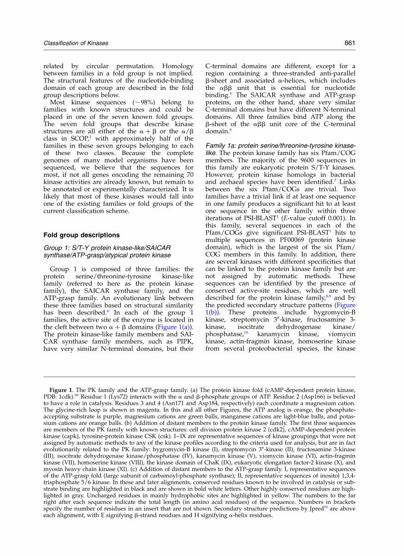
Group 1 is composed of three families: theprotein serine/threonine-tyrosine kinase-likefamily (referred to here as the protein kinasefamily), the SAICAR synthase family, and theATP-grasp family. An evolutionary link betweenthese three families based on structural similarityhas been described.6 In each of the group 1families, the active site of the enzyme is located inthe cleft between two a þ b domains (Figure 1(a)).The protein kinase-like family members and SAI-CAR synthase family members, such as PIPK,have very similar N-terminal domains, but their
C-terminal domains are different, except for aregion containing a three-stranded anti-parallelb-sheet and associated a-helices, which includesthe abb unit that is essential for nucleotidebinding.6 The SAICAR synthase and ATP-graspproteins, on the other hand, share very similarC-terminal domains but have different N-terminaldomains. All three families bind ATP along theb-sheet of the abb unit core of the C-terminaldomain.6
Family 1a: protein serine/threonine-tyrosine kinase-like. The protein kinase family has six Pfam/COGmembers. The majority of the 9600 sequences inthis family are eukaryotic protein S/T-Y kinases.However, protein kinase homologs in bacterialand archaeal species have been identified.7 Linksbetween the six Pfam/COGs are trivial. Twofamilies have a trivial link if at least one sequencein one family produces a significant hit to at leastone sequence in the other family within threeiterations of PSI-BLAST1 (E-value cutoff 0.001). Inthis family, several sequences in each of thePfam/COGs give significant PSI-BLAST1 hits tomultiple sequences in PF00069 (protein kinasedomain), which is the largest of the six Pfam/COG members in this family. In addition, thereare several kinases with different specificities thatcan be linked to the protein kinase family but arenot assigned by automatic methods. Thesesequences can be identified by the presence ofconserved active-site residues, which are welldescribed for the protein kinase family,8,9 and bythe predicted secondary structure patterns (Figure1(b)). These proteins include hygromycin-Bkinase, streptomycin 300-kinase, fructosamine 3-kinase, isocitrate dehydrogenase kinase/phosphatase,10 kanamycin kinase, viomycinkinase, actin-fragmin kinase, homoserine kinasefrom several proteobacterial species, the kinase
Figure 1. The PK family and the ATP-grasp family. (a) The protein kinase fold (cAMP-dependent protein kinase,PDB: 1cdk).89 Residue 1 (Lys72) interacts with the a and b-phosphate groups of ATP. Residue 2 (Asp166) is believedto have a role in catalysis. Residues 3 and 4 (Asn171 and Asp184, respectively) each coordinate a magnesium cation.The glycine-rich loop is shown in magenta. In this and all other Figures, the ATP analog is orange, the phosphate-accepting substrate is purple, magnesium cations are green balls, manganese cations are light-blue balls, and potas-sium cations are orange balls. (b) Addition of distant members to the protein kinase family. The first three sequencesare members of the PK family with known structures: cell division protein kinase 2 (cdk2), cAMP-dependent proteinkinase (capk), tyrosine-protein kinase CSK (csk). I–IX are representative sequences of kinase groupings that were notassigned by automatic methods to any of the kinase profiles according to the criteria used for analysis, but are in factevolutionarily related to the PK family: hygromycin-B kinase (I), streptomycin 300-kinase (II), fructosamine 3-kinase(III), isocitrate dehydrogenase kinase/phosphatase (IV), kanamycin kinase (V), viomycin kinase (VI), actin-fragminkinase (VII), homoserine kinase (VIII), the kinase domain of ChaK (IX), eukaryotic elongation factor-2 kinase (X), andmyosin heavy chain kinase (XI). (c) Addition of distant members to the ATP-grasp family. I, representative sequencesof the ATP-grasp fold (large subunit of carbamoyl-phosphate synthase); II, representative sequences of inositol 1,3,4-trisphosphate 5/6 kinase. In these and later alignments, conserved residues known to be involved in catalysis or sub-strate binding are highlighted in black and are shown in bold white letters. Other highly conserved residues are high-lighted in gray. Uncharged residues in mainly hydrophobic sites are highlighted in yellow. The numbers to the farright after each sequence indicate the total length (in amino acid residues) of the sequence. Numbers in bracketsspecify the number of residues in an insert that are not shown. Secondary structure predictions by Jpred70 are aboveeach alignment, with E signifying b-strand residues and H signifying a-helix residues.
Classification of Kinases 861
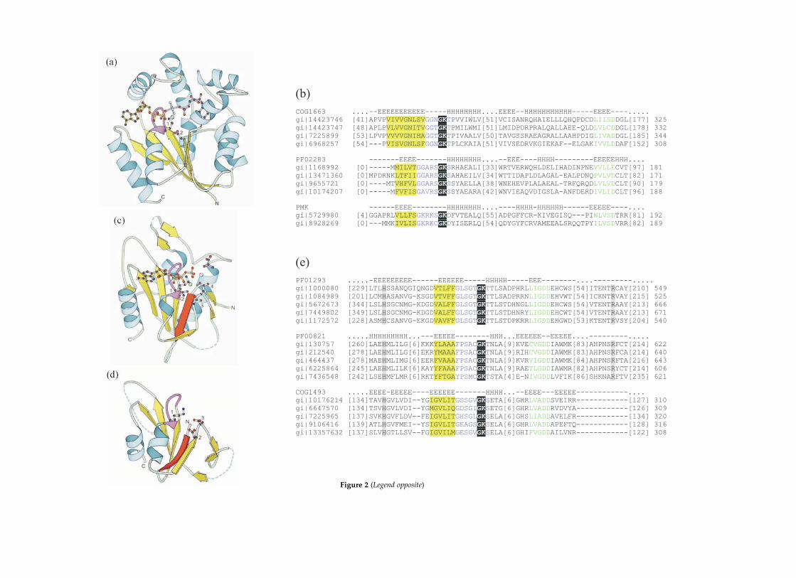
Figure 2 (Legend opposite)
domain of ChaK (a transient receptor potential(TRP) channel), eukaryotic elongation factor-2kinase, and myosin heavy chain kinase. Solvedstructures for kanamycin kinase,11 actin-fragminkinase,12 and the kinase domain of ChaK13 haveconfirmed that these three kinases are indeedstructurally similar to protein kinases. Multiplealignment of inositol 1,4,5-trisphosphate 3-kinase(group 11) sequences indicates that thesesequences may belong to the protein kinase-likefamily, although such a prediction cannot bemade with confidence.
The kinase domain of ChaK, eukaryoticelongation factor-2 kinase, and myosin heavychain kinase are also known as a-kinases oratypical kinases. These kinases do not have signifi-cant sequence similarity to classical proteinkinases;14 thus, the structural similarity betweenthe a-kinases and classical protein kinases wasunexpected.
Protein kinases are among the most thoroughlystudied protein families. This family has severalhighly conserved active-site residues. A conservedlysine residue from the N-terminal domain inter-acts with the a and b-phosphate groups of ATP.The aspartate residue and the asparagine residuein a highly conserved DXXXXN motif play a rolein catalysis and in coordinating a secondary mag-nesium divalent cation, respectively. The primaryMg2þ is coordinated by the conserved aspartateresidue of the DFG motif (Figure 1(b)). Proteinkinases also have a glycine-rich loop (GXGXXGXV)that interacts with the phosphate groups of theATP.
The mechanism of protein kinases was histori-cally thought to be acid–base catalysis. However,recent studies have questioned this hypothesisand led to the proposal that phosphoryl transfer isaccomplished by the simultaneous transfer of aproton from the substrate hydroxyl group to anoxygen atom of the g-phosphate group.15 The con-served aspartate residue (of the DXXXXN motif)
that was once thought to act as the base catalyst isnow suggested to have a role in stabilization ofthe protonated form of the transferred phosphate.
Family 1b: SAICAR synthase (as defined inSCOP3). The only kinase member of this family istype IIb phosphatidylinositol phosphate kinase(PIPK). Since the N-terminal domains of PIPKand protein kinases are very similar, the glycine-rich phosphate-binding loop and the conservedlysine residue located at the N-terminal domainare preserved in both structures. Additionally,PIPK has two conserved aspartate residues,Asp278 and Asp369, which can be aligned withthe catalytic aspartate residue of the DXXXXNmotif, and the Mg2þ-coordinating aspartateresidue of the DFG motif in protein kinases,respectively.6,16 The roles of these residues arelikely to be the same as those of their proteinkinase counterparts.
Family 1c: ATP-grasp. The ATP-grasp folddescribes several different ATP-hydrolyzingenzymes.17 However, there is only one kinasePfam member of this family (PF01326: pyruvatephosphate dikinase, PEP/pyruvate-bindingdomain). In this family, ATP is held between twoanti-parallel b-sheets. Hence, the term ATP-graspis used to describe this fold in SCOP.3 The mech-anism of the phosphotransfer reaction in pyruvatephosphate dikinase involves the reversible phos-phorylation of a histidine residue.18,19 In thisenzyme, the binding sites of the nucleotide andthe pyruvate are in distant locations on theprotein, and the shuttling of the phosphorylatedhistidine residue between the two active sites isaccomplished by a dramatic swivel of thephospho-histidine domain.20 Inspection of themultiple alignment of inositol 1,3,4-trisphosphate5/6-kinase sequences has indicated that thiskinase also belongs to the ATP-grasp family.Figure 1(c) shows a sequence alignment of the
Figure 2. The P-loop kinase family and the PEPCK family. (a) UMP/CMP kinase (PDB: 1qf9)90 of the P-loop kinasefamily. Residues 1 and 2 are the conserved Lys19 of the Walker A motif and the conserved Asp89 of the Walker Bmotif, respectively. Lys19 coordinates the b and g phosphate groups of ATP, while Asp89 coordinates the magnesiumcation. The P-loop is shown in magenta. (b) The alignment of representative sequences of each of the two Pfam/COGmembers with non-trivial links to the P-loop kinase family. Phosphomevalonate kinase sequences (PMK) containingthe Walker A and Walker B motifs are shown. (c) Phosphoenolpyruvate carboxykinase (PDB: 1aq2)30 of the PEPCKfamily. Residues 1 and 2 are Lys254 and Thr255 of the Walker A motif, respectively. Residues 3 and 4 are the pair ofconserved Asp residues (Asp268 and Asp269) in the PEPCK Walker B motif. Lys254 coordinates the b and g phosphategroups of ATP, while Thr255, Asp268, and Asp269 coordinate the magnesium cation. The anti-parallel b-strand isshown in red (see the text), and the P-loop is shown in magenta. Most of the N-terminal domain and some elementsof the C-terminal domain were removed for clarity. (d) HPr kinase/phosphatase (PDB: 1jb1)29 of the PEPCK family.Residue 1 is Lys161 of the Walker A motif and is involved in coordination of the b and g phosphate groups of ATP.Residues 2 and 3 are the pair of conserved Asp residues (Asp178 and Asp179) in the Walker B motif. Asp178 andAsp179 coordinate the magnesium cation. The anti-parallel b-strand is shown in red (see the text), and the P-loop isshown in magenta. Only the core of the C-terminal domain is shown. (e) The alignment of three Pfam members ofthe PEPCK family: PF01293 (phosphoenolpyruvate carboxykinase-ATP), PF00821 (phosphoenolpyruvate carboxy-kinase-GTP), and COG1493 (HPr kinase/phosphatase). The conserved histidine and arginine residues are believed tobe involved in substrate binding and are catalytically important in PEPCK.28 In these alignments, blue and green letterssignify the Walker A and the Walker B motifs, respectively.
Classification of Kinases 863

Figure 3 (Legend opposite)

large subunit of Escherichia coli, human, and yeastcarbomoyl-phosphate synthases (a representativeof the ATP-grasp fold) with inositol 1,3,4-trisphos-phate 5/6-kinases.
Group 2: Rossmann-like
The second kinase fold group contains eightRossmann-like families. The common structuralfeature of these families is that the architecture oftheir nucleotide-binding domain core is threelayers (a/b/a) composed of ba repeats, with thecentral b-sheet mostly parallel. There is always achange in direction of strand order in the middleof the sheet,21 resulting in the most common strandorder of 321456, although modifications of this top-ology are frequent. The total number of strandsand the strand order of the b-sheet may differbetween the families in this group. There is a widerange of insertions or additional domains that areassociated mostly with phosphoryl-acceptor sub-strate binding, accounting for the extremelydiverse substrate specificities in this group ofkinases. In addition to sharing a common fold ofthe nucleotide-binding domain, the families in theRossmann-like group have similar nucleotide-binding patterns. In each family, the nucleotidebinds at the C-terminal end of the b-sheet of thecore, with the phosphate tail always located at theN-terminal end of one or more a-helices (Figures2 and 3). More thorough descriptions of nucleo-tide-binding specifics for the families within thisgroup are provided below.
Family 2a: P-loop kinases. The largest family ingroup 2 is the P-loop-containing kinases, whichunifies 18 Pfam/COG members. Of these Pfam/COG members, 16 have trivial PSI-BLAST1 links.Alignments identifying the Walker A and WalkerB motifs for the remaining two members areshown in Figure 2(b). Additionally, a smallnumber of phosphomevalonate kinase sequencesfrom animals can be assigned to this family.22
Alignments identifying the conserved diagnosticmotifs for these phosphomevalonate kinasesequences are shown in Figure 2(b).
The P-loop kinases contain one three-layered(a/b/a) domain. For the majority of the membersof this family, the central parallel b-sheet is five-stranded with strand order 23145. Nucleotidebinding in this family is distinguished by the pre-sence of the conserved Walker A (GXXXXGKT/S)
and Walker B (ZZZZD, where Z is any hydro-phobic residue) motifs.23 The Walker A motifforms a phosphate-binding loop (P-loop) and isfound in a variety of different proteins that bindnucleotides.24 In this family of kinases, the P-loopis located at the end of the first b-strand andincludes the first half-turn of the followinga-helix. The conserved lysine residue of the WalkerA motif binds to and orients oxygen atoms of the band g-phosphate groups of ATP. The essentialmagnesium cation is coordinated directly by thehydroxyl group of the conserved threonine/serineof the Walker A motif and indirectly by the con-served aspartate residue of the Walker B motif.Figure 2(a) illustrates the Walker A and Walker Bmotifs, and metal-coordinating residues in UMP/CMP kinase.
The Walker A and Walker B motifs are commonin nucleotide-binding proteins. In SCOP,3 theP-loop-containing nucleotide triphosphate hydro-lases fold currently contains 82 proteins in 15families. Of these proteins, 13 are kinases; thus,,84% of P-loop-containing proteins with knownstructures are not kinases. The non-kinase, P-loop-containing proteins are mostly NTPases, which arelikely to have come before the kinases, since theycatalyze simpler reactions and are involved inmore fundamental biological processes.
A variety of catalytic mechanisms are utilized bythe kinases in this family. For example, an iso-random Bi–Bi mechanism has been suggested foradenylate kinase.25 A mechanism involving thesynchronous shift of a proton to the transferredphosphate group (similar to that proposed forprotein kinases) has been suggested for theUMP/CMP-kinase reaction.26 If correct, such amechanism could apply to the other nucleotide-phosphorylating kinases in this family. However,the phosphorylation of some metabolites appearsto require a base catalyst.27 This would apply tocertain members of the P-loop kinase familyincluding, for example, phosphoribulokinase andshikimate kinase.
Family 2b: phosphoenolpyruvate carboxykinase.The phosphoenolpyruvate carboxykinase (PEPCK)family consists of three Pfam/COG members:PF01293 (phosphoenolpyruvate carboxykinase-ATP), PF00821 (phosphoenolpyruvate carboxy-kinase-GTP), and COG1493 (HPr kinase/phospha-tase). Members of this family are distinguishedby their shared nucleotide-binding region,
Figure 3. (a) Phosphofructokinase (PDB: 4pfk).91 (b) The two Pfam members containing diacylglycerol kinase in thephosphofructokinase-like family. Representative diacylglycerol kinase sequences from PF00781 (diacylglycerol kinasecatalytic domain, presumed) and PF01219 (prokaryotic diacylglycerol kinase) are shown. (c) Ribokinase (PDB:1rkd).37 (d) Addition of distant members of the ribokinase-like family. I, representative sequences of ribokinase-likefamily: ribokinase (rk), fructokinase (fk), adenosine kinase (ak), gluconate kinase (gk); II, representative sequences ofarchaeal phosphofructokinase/glucokinase: phosphofructokinase (pfk), glucokinase (glk), related hypotheticalproteins (hyp). The underlined residues are conserved motifs in the nucleotide-binding pocket (nb) and the substrate-binding pocket (sub).92
Classification of Kinases 865
characterized by the topology of the b-sheet andthe placement of the Walker A motif and an aty-pical Walker B motif within the nucleotide-bind-ing fold. Solved structures of representatives fromPF01293 and COG1493 illustrate this shared top-ology (Figure 2(c) and (d)). Although no structurehas yet been solved for a representative ofPF00821, members of this family contain thecharacteristic Walker A and Walker B motifs.Similar predicted secondary structure distri-butions and conserved potential catalytic residuesindicate that proteins from PF00821 and PF01293share similar fold and active site architecture(Figure 2(e)).
PEPCK contains two a/b domains. The nucleo-tide-binding fold is located in the C-terminaldomain and is composed of the six-strandedmixed b-sheet and the surrounding a-helices.28
The PEPCK family proteins contain the typicalWalker A motif and a deviant Walker B motif(Figure 2(e)). Figure 2(a) and (c) illustrate the phos-phate-binding loops of a P-loop kinase andPEPCK, respectively. Note the similar structures ofthe Walker A motifs (in magenta) and the differentspatial locations of the Walker B aspartate residuesbetween the two proteins. The topology of thenucleotide-binding fold of PEPCK differs fromthat in P-loop kinases. The central b-sheet of thePEPCK nucleotide-binding C-terminal domain is amixed six-stranded sheet with strand order312456, and strands 1 and 5 are anti-parallel to therest of the sheet,28 whereas the central b-sheet ofP-loop kinases consists typically of five parallelb-strands of strand order 21345. Furthermore,while the b-strand preceding the Walker A motifand the b-strand preceding the Walker B motif areneighboring structural elements in space in P-loopkinases, PEPCK has an anti-parallel b-strand thatlies between the two b-strands associated with theWalker A and Walker B motifs. This strand iscolored red in Figure 2(c) and (d). Thus, whetherPEPCK is evolutionarily related to P-loop kinasesis still a subject of debate.
The structure of the C-terminal segment of theother kinase in PEPCK family, HPr kinase/phos-phatase (HPrK/P), has been solved.29 The fold ofthis segment of HPrK/P is similar to theC-terminal domain of PEPCK. In HPrK/P, theb-sheet associated with nucleotide binding has atleast five strands (strand order 31245, with strand1 anti-parallel to the rest of the sheet). A sixthstrand in this sheet seems likely, although itspresence is uncertain, due to poor electron densityin the crystal structure in this region.29 Thissixth strand would make the topology of thisb-sheet in HPrK/P identical with that of thecorresponding b-sheet in PEPCK. Additionally,the placement of the Walker A and Walker Bmotifs is similar in both HPrK/P and PEPCK(Figure 2(e)). In both kinases, the Walker A motifis located on a ba loop following b-strand 2. TheWalker B motif is found at the C-terminal end ofb-strand 3.
Two divalent metal cations are present in theactive site of PEPCK (Figure 2(c)). The magnesiumcation is coordinated by the threonine hydroxylgroup of the Walker A motif and indirectly by theconserved pair of aspartate residues in the WalkerB motif. The manganese cation is coordinated bythe side-chain nitrogen atoms of a histidine residueand a lysine residue in addition to the secondaspartate residue of the Walker B motif. The mag-nesium cation interacts with oxygen atoms of theb and g-phosphoryl groups of ATP while themanganese cation associates with an oxygen atomof the g-phosphoryl group and hypotheticallywith the enolate oxygen atom of pyruvateduring catalysis.30 PEPCK is different from otherkinases, in that this enzyme catalyzes both thedecarboxylation and phosphorylation of its sub-strate, oxaloacetate, to form phosphoenolpyruvate.However, the details of this mechanism areunknown.
Family 2c: phosphoglycerate kinase. Phospho-glycerate kinase is composed of two a/b/adomains. Both domains adopt a Rossmann-likefold and each contains a six-stranded parallelb-sheet. The N-terminal domain b-sheet hasstrand order 342156, while the C-terminal domainb-sheet has strand order 321456. The active site ofthis enzyme is located in the cleft between thesetwo domains.31 In this enzyme, the C-terminaldomain binds ATP, while the N-terminal domainbinds the 3-phosphoglycerate substrate. Thenucleotide binds at the edge of the b-sheet in theC-terminal domain and is roughly perpendicularto the b-strands. There are no sequence or struc-tural motifs resembling the Walker A or Walker Bmotifs in phosphoglycerate kinase.
The primary factor contributing to catalysis inphosphoglycerate kinase is transition state stabiliz-ation. In this enzyme, all three peripheral oxygenatoms of the transferred phosphate group arestabilized by interactions with protein residues orthe required divalent cation in the transition state.However, only two of these oxygen atoms arestabilized when the phosphate group is fullybonded to either the phosphate donor (ATP) oracceptor (phosphoglycerate substrate).32
Family 2d: aspartokinase. The only member ofthis family with a solved structure is carbamatekinase from Enterococcus faecalis33 and Pyrococcusfuriosus.34 Carbamate kinase and aspartokinase(along with acetylglutamate kinase, glutamate5-kinase, and uridylate kinase) belong to the samePfam profile (PF00696: amino acid kinase family).Therefore, the structure of carbamate kinase canserve as a prototype for other members of thefamily. The nucleotide-binding domain in carba-mate kinase is composed of three layers (a/b/a),including a b-sheet with Rossmann-liketopology.33 The bound nucleotide is located at theedge of the b-sheet strands and is approximatelyperpendicular to the direction of the b-strands, as
866 Classification of Kinases

is shown in the complex structure of the carba-mate kinase-like carbamoyl phosphate synthetasefrom P. furiosus.34
Family 2e: phosphofructokinase-like. The phos-phofructokinase-like family contains four Pfammembers. Links between three of these membersare trivial via PSI-BLAST.1 The link betweenanother Pfam member, PF01219 (prokaryoticdiacylglycerol kinase), to the phosphofructo-kinase-like family was established throughmultiple sequence alignment analysis and second-ary structure predictions (Figure 3(b)).
The fold of phosphofructokinase consists oftwo a/b/a domains. The nucleotide-bindingN-terminal domain has a seven-stranded mixedb-sheet of strand order 3214567, where strands 3and 7 are anti-parallel to the rest of the sheet(Figure 3(a)). The active site is located in the cleftbetween the two domains.35 The ATP moiety,which is positioned above the b-sheet of theN-terminal domain and is approximately parallelwith the b-strands, sits between an a-helix and along loop segment (Figure 3(a)).
Family 2f: ribokinase-like. The ribokinase-likefamily contains several carbohydrate kinases.All three Pfam members in this familyhave trivial PSI-BLAST1 links. An additionalgrouping of archaeal phosphofructokinase/gluco-kinase sequences can be placed in the ribokinase-like family. An alignment for this assignment isshown in Figure 3(d). The solved structure ofglucokinase from Thermococcus litoralis shows thatthe structure of this archaeal ADP-dependentkinase is indeed similar to that of the othermembers of the ribokinase-like family.36
The core of the ribokinase-like fold is a three-layered domain (a/b/a) with a central eight-stranded b-sheet. The strand order of this sheet is21345678, with strand 7 anti-parallel to the rest ofthe b-sheet. Ribokinase has an additional sub-domain composed of a four-stranded b-sheet. Inaddition to acting as a “substrate lid”, this b-sheetforms the dimerization surface.37 In ribokinase, thenucleotide-binding site is found along a shallowgroove in the core of the sole a/b/a domain.37
The ATP moiety lies at the edge of the b-sheet andis roughly perpendicular to the adjacent b-strands(Figure 3(c)).
Family 2g: L-2-haloacid dehalogenase (HAD-like).The HAD-like family contains only two kinasesequence members, which are the bifunctionalhomoserine kinase/phosphoserine phosphatase(ThrH) from Pseudomonas aeruginosa.38 PSI-BLAST1
establishes a link between these sequences andphosphoserine phosphatase, which has the HAD-like fold.39,40 The core of the HAD-like fold is asix-stranded parallel b-sheet of strand order321456 with an additional four-helix bundlesubdomain.41 The nucleotide-binding site in aHAD-like enzyme is suggested to be located in
the cleft between the two domains of the fold;41
however, the details of the orientation of the ATPmoiety are unknown.
HAD-like family enzymes typically utilize a con-served aspartate residue for nucleophilic catalysisin a reaction that includes a covalent intermediateinvolving the phosphate group and the aspartateresidue.42,43 This mechanism could be used for thekinase phosphotransfer reaction. Alignment of thehomoserine kinase isozyme with phosphoserinephosphatase suggests that Asp7 (gi’4138297)is the residue that is phosphorylated for theformation of the covalent intermediate in thehomoserine kinase isozyme.
Family 2h: thiamin pyrophosphokinase. Thiaminpyrophosphokinase (TPK) is a two-domainprotein. The ATP-binding C-terminal domain hasRossmann-like topology and is composed of threelayers (a/b/a) with a central six-stranded parallelb-sheet of strand order 432156, while theN-terminal domain consists of a four-strandedb-sheet and a six-stranded b-sheet that form aflattened sandwich (“jelly-roll topology”).44 TPK isa homodimer with the active site located in a cleftat the interface of the component monomers.Thus, active-site residues are contributed by theN-terminal domain of one monomer and theC-terminal domain of the opposing monomer.44
The precise orientation of the nucleotide in theactive site has not been determined.
Group 3: ferredoxin-like fold kinases
Group 3 is composed of kinases whose nucleo-tide-binding domain core resembles the ferre-doxin-like fold. The four families in this group areas follows: nucleoside-diphosphate (NDP) kinase,7,8-dihydro-6-hydroxymethylpterin-pyrophospho-kinase (HPPK), guanido kinases, and histidinekinase. The ferredoxin fold (also known as thea 2 b plait fold or the a þ b sandwich) is charac-terized by its babbab unit with the four-strandedanti-parallel b-sheet having strand order 2314 andtwo helices on one side of the sheet. One exceptionis the histidine kinase family, in which the core ofthe nucleotide-binding domain is related to theferredoxin-like topology by a circular permutation(see below). An interesting feature of this group isthat the mode of nucleotide binding differs signifi-cantly between each of the families, in terms ofstructural elements utilized in nucleotide–proteininteractions, and in the orientation and position ofthe nucleotide relative to the ferredoxin-like core.Figure 4 illustrates the orientation of the boundnucleotide in NDP kinase, HPPK, arginine kinase,and histidine kinase.
Family 3a: NDP kinase. In addition to the ferre-doxin-like core of NDP kinase, this enzyme hasseveral other secondary structural elements,including the Kpn loop, an a-helix hairpin, and aC-terminal extension (Figure 4(a)). The Kpn loop
Classification of Kinases 867
is a small, compact structural element containingan interesting combination of helical structures: aturn of 310 helix, a turn of polyproline II left-handed helix, and a turn of standard a-helix.45
The Kpn loop and the a-helix hairpin constitute anucleotide-binding site that is unique to NDPkinase (Figure 4(a)). In terms of the orientationand position of this substrate, the nucleotide liesat the edge of the b-sheet that defines the ferre-doxin-like core, with the adenine base more dis-tant from the sheet and the phosphate tailextending towards the sheet.46
Catalysis in NDP kinase occurs by a ping-pongmechanism in which the phosphoryl group istransferred first from the nucleoside triphosphateto the enzyme, involving the formation of a
covalent intermediate of the phosphate with ahistidine residue in the active site, followed by thetransfer of the phosphate group to the nucleosidediphosphate substrate.
Family 3b: HPPK. In addition to the ferredoxin-like core of HPPK, this enzyme has a pair ofb-strands and a pair of a-helices at the C-terminalend of the protein.47,48 In HPPK, ATP is bound in theregion between the a2–b4 connecting loop and ashort C-terminal b-strand that is not part of the fer-redoxin-like core (Figure 4(b)). The ATP moiety liesat the edge of the b-sheet of the ferredoxin-like coreand is curled such that the adenine base and ribosesugar angle away from the b-sheet, while the tri-phosphate tail points towards the b-sheet. The
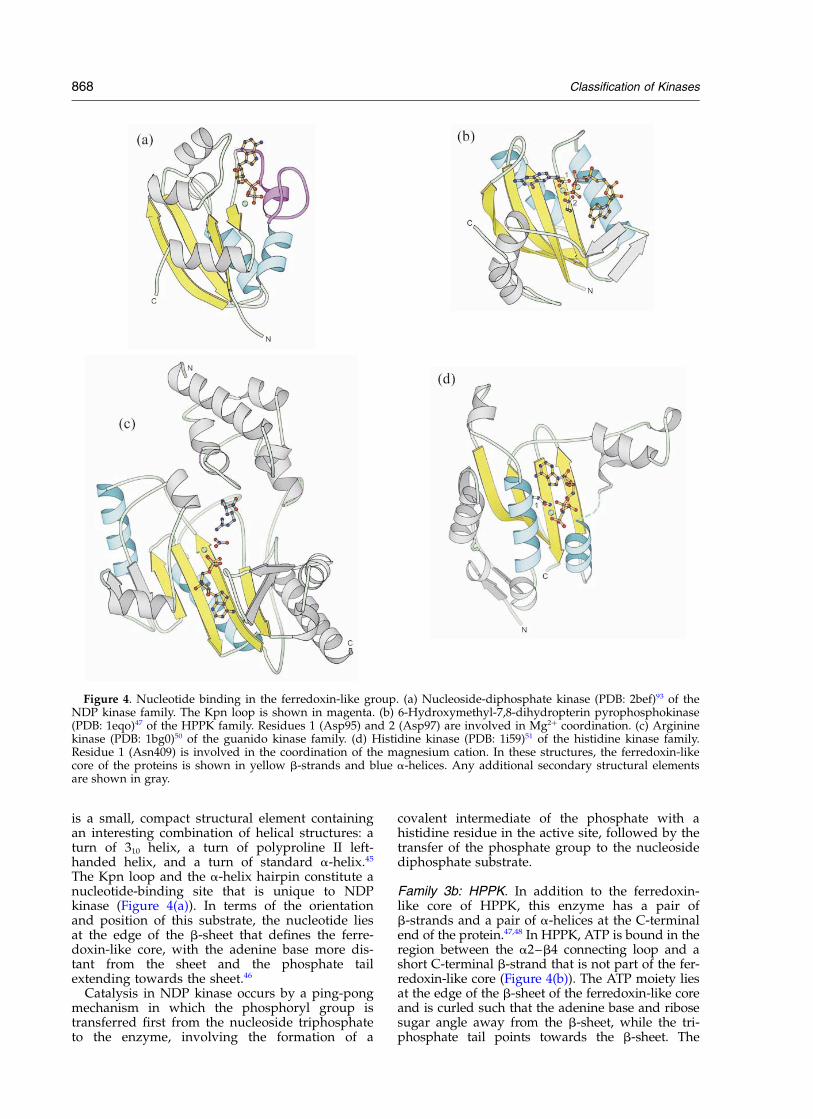
Figure 4. Nucleotide binding in the ferredoxin-like group. (a) Nucleoside-diphosphate kinase (PDB: 2bef)93 of theNDP kinase family. The Kpn loop is shown in magenta. (b) 6-Hydroxymethyl-7,8-dihydropterin pyrophosphokinase(PDB: 1eqo)47 of the HPPK family. Residues 1 (Asp95) and 2 (Asp97) are involved in Mg2þ coordination. (c) Argininekinase (PDB: 1bg0)50 of the guanido kinase family. (d) Histidine kinase (PDB: 1i59)51 of the histidine kinase family.Residue 1 (Asn409) is involved in the coordination of the magnesium cation. In these structures, the ferredoxin-likecore of the proteins is shown in yellow b-strands and blue a-helices. Any additional secondary structural elementsare shown in gray.
868 Classification of Kinases

adenine base and the ribose sugar residue lie inthe same plane as the b-sheet. However, the tri-phosphate tail reaches over the b-sheet on theopposite side as the a-helices associated with theferredoxin-like core.47,48
HPPK utilizes two magnesium cations in its activesite, each of which is coordinated by two aspartateresidues. The mechanism of HPPK has not beenelucidated, but it is presumed to be a direct in-linetransfer of the pyrophosphate group. Suggestionsfor the mechanism include roles for the two mag-nesium cations and acid–base catalysis with a watermolecule acting as the general base.47
Family 3c: guanido kinases. The fold of theguanido kinase family consists of two domains.The smaller N-terminal domain is composedentirely of a-helices, and the nucleotide-bindingC-terminal domain is composed of an eight-stranded b-sheet of strand order 23451687, whichis flanked by seven a-helices.49,50 The middle fourstrands of the b-sheet and associated a-heliceshave ferredoxin-like fold topology. Figure 4(c)illustrates the ferredoxin-like topology within thisfold.
In arginine kinase, ATP binding is accomplishedby interactions with five arginine residues and amagnesium cation. The ATP moiety lies in theplane above the b-sheet, rather than at the edge ofthe b-sheet, and is oriented approximately parallelwith the b-strands (Figure 4(c)). The nucleotide ispositioned above the center two strands of thefour-stranded section that resembles the ferre-doxin-like fold. In this enzyme, the bound nucleo-tide and the a-helices that compose theferredoxin-like topology lie on opposite sides ofthe b-sheet.50
Arginine kinase catalyzes an associative in-linephosphotransfer reaction. The primary factor incatalysis appears to be substrate alignment by posi-tioning reaction components in close proximity andpromoting proper alignment of orbitals, althoughacid–base catalysis, polarization, and transitionstate stabilization may contribute to the reaction.50
Family 3d: histidine kinases. There are two Pfam/COG members of this family. The link betweenthe members is trivial via PSI-BLAST.1 Histidinekinases (HKs) catalyze a trans-autophosphoryl-ation reaction in the two-component system ofsignal transduction. The fold of the ATP-domain(the catalytic domain) is an a/b sandwich com-posed of a five-stranded b-sheet and foura-helices (Figure 4(d)).51,52 The core of this foldhas a tertiary structure similar to that of the ferre-doxin fold. The HK fold has topology abbabbwith strand order 3421, which can be related tothe ferredoxin-like topology by a circular permu-tation consisting of cutting the loop between thefirst and second b-strands and connecting thenatural termini.
There are two classes of HKs, which can be dif-ferentiated by their domain organization.53 EnvZ
is a member of class I in which the H-domain (thedomain that contains the histidine phosphorylationsite) directly precedes the ATP domain. CheA is amember of class II in which the H domain andATP domain are separated by at least one otherdomain. The ATP domains of HKs are structurallysimilar to the ATP-binding domains of the GHLfamily (DNA gyrase/Hsp90/MutL).51,52 There iscurrently some debate as to whether both HKclasses or only class II bind ATP in the same con-formation as the GHL family.51,52 In both classes,there are four conserved motifs that contribute tothe nucleotide-binding pocket; namely, the N, G1,F, and G2 boxes.54 The required magnesium cationis coordinated by direct interactions with an aspar-agine residue and indirectly by a histidine residueand an arginine residue.51
In HK, the ATP moiety lies above the b-sheetthat is part of the ferredoxin-like core (Figure4(d)). The nucleotide is above two of the b-strandsand is found at the edge of the sheet rather thanthe center of it. In HK, the adenine base is nearerto the b-sheet than the triphosphate tail, whichangles away from the b-strands. The nucleotideand the a-helices associated with the ferredoxin-like core are located on the same side of theb-sheet.51
HK, as noted above, operates via the two-component system. Here, one HK monomerphosphorylates a histidine residue in the othermonomer of the homodimer, which results in ahigh-energy phosphoryl group. The regulatorydomain of the cognate response regulator (RR)then catalyzes the reaction that transfers the phos-phoryl group from the histidine residue to anaspartate residue in the RR. The mechanism issomewhat similar to that of NDP kinase, in thatboth involve the formation of a high-energy phos-phohistidine residue. However, NDP kinasephosphorylates a histidine residue in its own activesite, while HK phosphorylates a histidine residuein another HK monomer.
Group 4: ribonuclease H-like kinases
There are four Pfam/COG members in thisgroup. Three of these members have trivial linksvia PSI-BLAST.1 The fourth member (PF00871: theacetokinase family, which contains acetate kinaseand butyrate kinase) was predicted to be a memberof this family by Buss et al.55 The recently solvedstructure of acetate kinase shows that this enzymedoes in fact adopt the ribonuclease H-like fold.56
Multiple alignment of 2-dehydro-3-deoxygalacto-nokinase sequences indicates that this kinaseactivity belongs to the ribonuclease H-like group(Figure 5(b)). The ribonuclease H-like group con-tains the ASKHA (acetate and sugar kinase/hsc70/actin) superfamily, the structures of whichare characterized by duplicate domains of the ribo-nuclease H-like fold. The ribonuclease H-like foldis composed of three layers (a/b/a) (Figure 5(a)).The five-stranded mixed b-sheet has strand order
Classification of Kinases 869
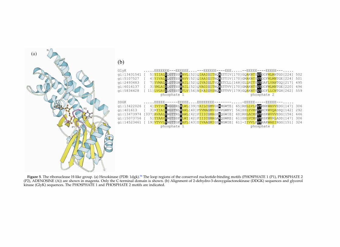
Figure 5. The ribonuclease H-like group. (a) Hexokinase (PDB: 1dgk).58 The loop regions of the conserved nucleotide-binding motifs (PHOSPHATE 1 (P1), PHOSPHATE 2(P2), ADENOSINE (A)) are shown in magenta. Only the C-terminal domain is shown. (b) Alignment of 2-dehydro-3-deoxygalactonokinase (DDGK) sequences and glycerolkinase (GlyK) sequences. The PHOSPHATE 1 and PHOSPHATE 2 motifs are indicated.

32145, with strand 2 anti-parallel to the rest of thesheet. The topology of the core of this fold is bbbababa. Nucleotide binding and divalent metalcoordination are achieved by interactions of ATPwith several motifs conserved within the ASKHAsuperfamily.57 These conserved motifs include theADENOSINE motif that interacts with ribosyl andthe a-phosphoryl group of ATP, the PHOSPHATE1 motif that interacts with Mg2þ through coordi-nated water molecules, and the PHOSPHATE 2motif that interacts with the b and g-phosphorylgroups of ATP (Figure 5(a)). A modeled active siteof hexokinase predicts that the required divalentmetal cation is not liganded to the protein directly,but is positioned by coordinated water molecules.58
The mechanism of kinases in this group is pre-sumed to be acid–base catalysis. In hexokinase, anaspartate residue is the putative catalytic base thatdeprotonates the 6-hydroxyl group of glucose.58,59
Group 5: TIM b/a-barrel fold kinases
Group 5 is described by the TIM b/a-barrel fold,which consists of an eightfold repeat of ba unitsthat form a closed barrel. The barrel is composedof an inner layer of eight parallel b-strands ofstrand order 12345678 and an outer layer of eighta-helices (Figure 6(a)). This fold characterizesmany different enzyme families that have
extremely low levels of sequence similarity andcatalyze unrelated reactions.60,61 The active site ofall TIM b/a-barrel enzymes is located at theC-terminal end of the parallel b-strands. The onlykinase known to adopt this fold is pyruvate kinase,which has an additional domain inserted at theC-terminal end of b-strand 3.62 The nucleotide-binding pattern of pyruvate kinase is thought tobe novel.63 The triphosphate tail of the ATP moietyis held by hydrogen bond interactions with twoarginine residues, an asparagine residue, a lysineresidue, and three metal cations (two Mg2þ andone Kþ). The adenine ring sits in a pocket that isbounded by a histidine residue, a proline residue,and a tyrosine residue. One distinctive feature ofthe pyruvate kinase active site is the coordinationof each of the g-phosphoryl peripheral oxygenatoms to a different inorganic cofactor.63 One ofthe magnesium cations is coordinated by the car-boxylate groups of an aspartate residue and aglutamate residue. The other magnesium cation isnot liganded to the protein directly. The potassiumcation is coordinated by the carbonyl group of athreonine residue, the hydroxyl group of a serineresidue, the carboxylate group of an aspartateresidue, and the carboxyamide group of an aspara-gine residue. The coordination of the metalcofactors is detailed in Figure 6(a). The reactioncatalyzed by pyruvate kinase is presumed to be
Figure 6. (a) Metal cofactor coordination and nucleotide orientation in the TIM b/a-barrel kinase family (pyruvatekinase, PDB: 1a49).63 Residues 1 (Asn74), 2 (Ser76), and 3 (Asp112) coordinate the potassium cation. Residues 4(Glu271) and 5 (Asp295) coordinate one of the magnesium cations. The C-terminal subdomain was removed for clarity.(b) Homoserine kinase (PDB: 1h72)67 of the GHMP kinase group. The novel P-loop is shown in magenta.
Classification of Kinases 871
direct in-line phosphotransfer via acid–basecatalysis, although the group(s) responsible for theacid–base catalysis has not been identified. Recentwork suggests the possibility that a proton relaythrough a series of conserved residues may beresponsible for acid–base catalysis.63
Group 6: GHMP kinases
The members of the GHMP kinase superfamilyconstitute group 6. The GHMP kinase superfamilywas named after its original four members: galac-tokinase, homoserine kinase, mevalonate kinase,and phosphomevalonate kinase.64 The crystalstructure of two members of this family, homo-serine kinase and mevalonate kinase, have beensolved.65,66 The fold of this group consists of twoa þ b domains, with the active site in the cleftbetween the two domains (Figure 6(b)). TheN-terminal domain contains two b-sheets andfour a-helices. The C-terminal domain has a ferre-doxin-like core with four additional a-helices. Thenucleotide-binding site resides mostly with theN-terminal domain. Nucleotide binding is accom-plished by a novel P-loop with a conserved PXXX-GSSAA motif. The structure of homoserine kinaserevealed the presence of an unusual left-handedbaba unit in the N-terminal domain. The secondba loop in this unit contains the novel phosphatebinding loop (Figure 6(b)).65 Notably, theorientation of the ATP is different in the GHMPphosphate-binding loop than in the classicalWalker A P-loop. A glutamate residue acts tocoordinate the essential magnesium cation. Inhomoserine kinase, it has been suggested that thehomoserine hydroxyl group is deprotonated notby a catalytic base, but by interaction with theg-phosphate group in a mechanism similar to thatproposed for protein kinases.67 There are fourPfam/COG members of this group. The linksbetween the four members are trivial via PSI-BLAST.1
Group 7: AIR synthetase (PurM)-like
Two kinases, thiamine-phosphate kinase andselenide, water dikinase, belong to this group.Although the structures of these enzymes havenot been solved, the known structure of thehomologous aminoimidazole ribonucleotidesynthetase68 (AIR synthetase, PurM) can serve as aprototype for this group. AIR synthetase has twoa/b domains. The N-terminal domain contains amixed four-stranded b-sheet with four a-heliceson one side of the sheet. The C-terminal domainhas a mixed six-stranded b-sheet flanked by sevena-helices. Four of the b-strands and two of thea-helices in the C-terminal domain adopt a tertiarystructure and topology that resembles the ferre-doxin-like fold. The crystal structure indicates thatthis enzyme exists as a dimer, with the active sitelikely to be located in a cleft between the twosubunits.68 A sulfate ion is bound in this cleft and
could indicate a phosphate-binding site, althoughnot necessarily the ATP-binding site, since bothsubstrates of AIR synthetase contain a phosphategroup.68 Mutagenesis and affinity-labeling studiesof this enzyme suggest that the ATP-binding siteis located close to the N terminus of the enzyme.69
Thus, it appears that nucleotide-binding is accom-plished predominantly by the N-terminal domainof one subunit in the dimer, while the C-terminaldomain of the opposing subunit binds the secondsubstrate. A similar situation may be seen in thekinase members of this family. However, as nosubstrate/product complex structure is availablefor any member of this group, the exact nucleo-tide-binding mode is not clear.
Groups 8–17: kinases with unknown structures
The remaining groups, which account for only2% of the sequences in our analysis, containkinases with unsolved structures. For each group,Jpred70 was used to generate secondary structuralpredictions. On the basis of these predictions, themajority of these groups are expected to be of thea/b or a þ b protein classes in SCOP,3 like theirknown-structure kinase counterparts, which areall a/b or a þ b proteins. The exceptions are thetwo families in group 8 (dolichol kinase and unde-caprenol kinase), which are both predicted to becomposed almost entirely of a-helices. This is notunexpected, because these are both integralmembrane proteins.
For some of these groups, it is tempting topredict their fold on the basis of similar substratespecificities. For example, one might conjecturethat inositol 1,4,5-trisphosphate 3-kinase (group11) and inositol 1,3,4,5,6-pentakisphosphate2-kinase (group 12) are likely to be members ofgroup 1, which includes many of the other kinaseparticipants in the inositol phosphate metabolismpathway. Similarly, dihydroxyacetone kinase(group 17) may belong to the ribonuclease H-likegroup, which includes glycerol kinase. However,such links are difficult to establish without theexpected presence of specific conserved motifsand confirmed active-site residues. Furthermore,functional links such as these can be misleading.One such example is pantothenate kinase. Group14 contains eukaryotic pantothenate kinasesequences. The solved structure of prokaryoticpantothenate kinase identifies the enzyme as amember of the P-loop kinase family.71 However,due to the lack of sequence identity between theprokaryotic and eukaryotic versions of thisprotein,72 in conjunction with dissimilar secondarystructural predictions, eukaryotic pantothenatekinase is expected to adopt a fold distinct fromthat of its prokaryotic counterpart. Additionally,two distinct families of glycerate kinase sequencesare found in groups 15 and 16. Although theseproteins have been predicted to have the samebiochemical activity, sequence similarity betweenthe members of the two groups is not detected.
872 Classification of Kinases

Group 15 glycerate kinases are from bacterialspecies, primarily of the firmicutes group and ofthe gamma subdivision of the proteobacterialgroup. Group 16 contains predicted glyceratekinase sequences from eukaryotes and archaealspecies in addition to several bacterial species.Most group 16 bacteria are from the alpha sub-division of the proteobacterial group.
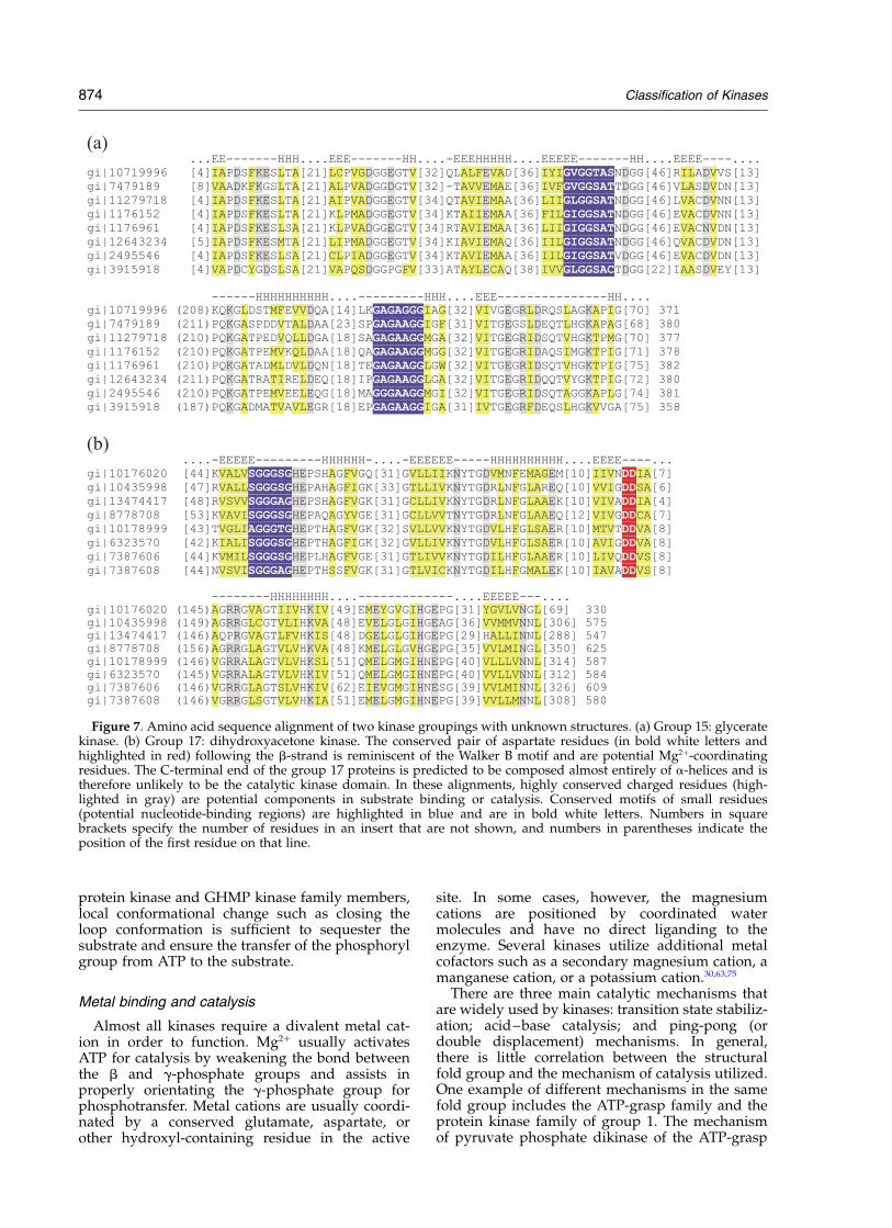
Potential active-site components for some ofthese groups can be predicted by studyingmultiple alignments of the sequences. Conservedpatterns containing several glycine residues andother small residues are typical of nucleotide-binding motifs. For example, the highly conservedVPVPGGA motif in tagatose 6-phosphate kinase(group 13) might be involved in ATP binding (e.g.gi’1168382, residues 176–182). Furthermore, thismotif is found in the loop between a predictedb-strand and a-helix. Nucleotide-binding residueslocated on ba loops are a theme in kinases. Con-served, positively charged amino acid residues,especially lysine and arginine, are common inbinding and orienting of the ATP triphosphatetail. Mg2þ coordination is often accomplished witha conserved aspartate residue or with another con-served hydroxyl-containing group in the activesite. Other conserved, charged amino acid residuescould be potential catalytic or substrate-bindingresidues. Multiple alignments identifying potentialactive-site components from two of the kinasegroupings with unknown structures (glyceratekinase and dihydroxyacetone kinase) are shown inFigure 7.
Discussion
Common structural mechanisms sharedamong kinases
Although all kinases catalyze a similar phos-phoryl transfer reaction, they adopt a wide varietyof structural folds. Our current survey cataloged30 distinct sequence/structure families of kinases.Of these families, 19, covering 98% of over 17,000sequences, were further grouped into sevengeneral fold groups. This classification systemcombined with a wealth of kinase structural andbiochemical data enables us to address the ques-tion of how these different structural folds accom-plish the same chemistry. The common structuralfeatures that influence phosphoryl transfer bykinases have been reviewed.73 Briefly, all phospho-transfer reactions contain the following threeprincipal components: binding and orienting thephosphate donor (ATP); binding and orienting thephosphate acceptor (substrate); and catalysis ofthe chemical reaction.
Nucleotide binding
Several distinct modes of nucleotide bindinghave emerged. One recurring theme is that the
nucleotide binds at the C-terminal end of b-strandsand N terminus of a-helices. This is observed in allRossmann-like fold families and in the GHMPfamily. In three of these families, P-loop kinase,PEPCK, and GHMP, the connecting loop betweenthe b-strand and a-helix are extended, formingthe so-called phosphate-binding loop (P-loop) thatwraps around the triphosphate tail of the boundnucleotide. The glycine-rich nature of these loopsenables them to adopt conformations such thatseveral main-chain amide groups are all pointedtowards the bound nucleotide. Together with thepositive dipole of the a-helix and some positivelycharged lysine or arginine side-chains, a stronganion hole is created for the binding of thenucleotide.
The glycine-rich phosphate-binding loops areobserved in families of the protein kinase-like foldand ribonuclease H-like fold groups. However, inthese cases, the loops are mostly b-hairpin loopsthat connect two anti-parallel b-strands. No majorcontribution from helix dipoles is involved innucleotide binding in these kinase families. How-ever, several b-hairpin loops, such as in the case ofhexokinase (ribonuclease H-like), may congregate atthe active site with their main-chain amide groupsinteracting with the phosphate group. In the abovetwo distinct nucleotide-binding modes, the proteinmain-chain interacting with the nucleotide is promi-nent. This is probably one of the reasons why, forexample, in all Rossmann-fold kinase families, theATP moiety binds at roughly the same place.
Nucleotide binding in the ferredoxin-like groupand TIM-barrel group are completely different. Nocommonly shared local structural motif is observedacross families. In general, kinases in these twofold groups use mainly positively charged proteinside-chains to interact with the nucleotide phos-phate groups. As discussed before, nucleotidebinding differs greatly between the families of theferredoxin-like group, probably as a result of alack of interactions with protein backbones.
Binding of phosphoryl acceptor substrates
Binding and orientation of the phosphate-accept-ing substrate in kinases depends on interactionsbetween the substrate and strategically placedactive-site residues. The details of such interactionsare, of course, contingent upon the specific activityof the kinase in question. Extra structural motifsor domains in addition to the nucleotide-bindingcore are usually necessary for the recognition ofthe substrate. Since the size and structure of kinasesubstrates varies drastically from a small moleculeof a few atoms to a whole protein, the substrate-binding motifs also vary significantly. One com-mon phenomenon associated with the substratebinding is induced conformational changes. Insome cases, such as pyruvate phosphate dikinaseand phosphoglycerate kinase, drastic domainmovements occur in order to bring the substrateto the active site.20,74 While in other cases, such as
Classification of Kinases 873
protein kinase and GHMP kinase family members,local conformational change such as closing theloop conformation is sufficient to sequester thesubstrate and ensure the transfer of the phosphorylgroup from ATP to the substrate.
Metal binding and catalysis
Almost all kinases require a divalent metal cat-ion in order to function. Mg2þ usually activatesATP for catalysis by weakening the bond betweenthe b and g-phosphate groups and assists inproperly orientating the g-phosphate group forphosphotransfer. Metal cations are usually coordi-nated by a conserved glutamate, aspartate, orother hydroxyl-containing residue in the active
site. In some cases, however, the magnesiumcations are positioned by coordinated watermolecules and have no direct liganding to theenzyme. Several kinases utilize additional metalcofactors such as a secondary magnesium cation, amanganese cation, or a potassium cation.30,63,75
There are three main catalytic mechanisms thatare widely used by kinases: transition state stabiliz-ation; acid–base catalysis; and ping-pong (ordouble displacement) mechanisms. In general,there is little correlation between the structuralfold group and the mechanism of catalysis utilized.One example of different mechanisms in the samefold group includes the ATP-grasp family and theprotein kinase family of group 1. The mechanismof pyruvate phosphate dikinase of the ATP-grasp
Figure 7. Amino acid sequence alignment of two kinase groupings with unknown structures. (a) Group 15: glyceratekinase. (b) Group 17: dihydroxyacetone kinase. The conserved pair of aspartate residues (in bold white letters andhighlighted in red) following the b-strand is reminiscent of the Walker B motif and are potential Mg2þ-coordinatingresidues. The C-terminal end of the group 17 proteins is predicted to be composed almost entirely of a-helices and istherefore unlikely to be the catalytic kinase domain. In these alignments, highly conserved charged residues (high-lighted in gray) are potential components in substrate binding or catalysis. Conserved motifs of small residues(potential nucleotide-binding regions) are highlighted in blue and are in bold white letters. Numbers in squarebrackets specify the number of residues in an insert that are not shown, and numbers in parentheses indicate theposition of the first residue on that line.
874 Classification of Kinases
family involves the reversible phosphorylation of ahistidine residue.18,19 Although the mechanism uti-lized by protein kinases is currently a matter ofdebate, the phosphotransfer reaction in this familyis thought to proceed either via a proposed simul-taneous transfer mechanism,15 which falls into thecategory of transition-state stabilization, or viaacid–base catalysis. A second example includesthe families of the ferredoxin-like fold group.Nucleoside diphosphate kinase (NDP) and histi-dine kinase both utilize ping-pong mechanisms.Although the possibility of acid–base catalysiscannot be eliminated in arginine kinase of theguanido kinase family, the primary factor in themechanism of this enzyme appears to be tran-sition-state stabilization and precise substrateorientation in the active site.50
Within most kinase families, all enzymes usuallyutilize the same type of catalytic mechanism. Allprotein kinases, for example, are thought to havesimilarly catalyzed reactions. However, there areexceptions to this tendency. In the GHMP family,for example, the homoserine kinase mechanism isproposed to proceed via a simultaneous transferreaction (transition-state stabilization),67 while thearchaeal shikimate kinase reaction may follow aping-pong mechanism.76 In a second example,reactions catalyzed by P-loop family kinases mayproceed via different mechanisms. Acid–basecatalysis is the probable mechanism of P-loopenzymes such as phosphoribulokinase andshikimate kinase for the production of phosphoryl-ated metabolites,27 while UMP/CMP kinase of thesame family has been suggested to utilize a simul-taneous transfer mechanism similar to that pro-posed for protein kinases (transition-statestabilization).26
Same activity, different fold
There are several cases of the same kinaseactivity that exists in unrelated fold families,reflecting convergent evolution of the same func-tion from different ancestors. For example,Galperin et al. have identified analogous enzymesin fructokinase, 6-phosphofructokinase, andgluconokinase.77 Comparisons of homoserinekinase, phosphomevalonate kinase, shikimatekinase, glucokinase, 1-phosphofructokinase,uridylate kinase, and pantothenate kinase fromdifferent folds are summarized below.
Homoserine kinase is thus far the only activity tobe found in three distinct fold groups (the proteinkinase family, the HAD-like family, and theGHMP family). The protein kinase family containshomoserine kinases from several proteobacterialspecies (eubacteria). The GHMP kinase familyincludes homoserine kinases from the majority ofeubacteria, from archaebacteria, and fromeukaryotes. The HAD-like family currently containsonly one homoserine kinase isozyme, which is thebifunctional ThrH from Pseudomonas aeruginosa.38 Itis interesting to note that the same mechanism of cat-
alysis has been proposed for both the protein kinasefamily and the GHMP family (the “synchronousshift” mechanism). The existence of a HAD-likehomoserine kinase/phosphoserine phosphatase,however, demonstrates that nature has accom-plished this same activity via a completely differentmechanism. Catalysis in the HAD-like homoserinekinase isozyme is expected to proceed via a phos-phoaspartate intermediate similar to the mechanismemployed by the other members of the HAD-likefamily. Another surprising feature is homoserinekinase’s use of the protein kinase fold, which is gen-erally utilized for the accommodation of very largesubstrates.
Phosphomevalonate kinase (PMK) andshikimate kinase (SK) are each found in both theP-loop kinase family and the GHMP kinasefamily.22,76 PMK from higher eukaryotes (includinghuman, pig, and fruit fly) belong to the P-loopkinase family. All other identified PMKsequences (such as those of Saccharomyces cerevisiae,Staphylococcus aureus, and Streptococcus pneumoniae )are found in the GHMP kinase family. A structurehas not been solved for PMK from either foldfamily. SK also contributes members to each ofthese two families. The P-loop kinase family includesprimarily eubacterial SK, while the GHMP kinasefamily contains archaebacterial SK.76 Currently, theonly SK structure that has been solved is that ofErwina chryanthemi (eubacteria), which belongs tothe P-loop kinase family.78
Glucokinase is found in both the ribokinase-likefamily of the Rossmann-like fold group and theribonuclease H-like fold group. Glucokinase fromarchaeal species such as Thermococcus litoralis andPyrococcus furiosus belong to the ribokinase-likefamily. The ribonuclease H-like family containsglucokinases from eukaryote, eubacterial, and afew archaebacterial species. Unlike the ATP-depen-dent ribonuclease H-like glucokinases, the archaealglucokinases of the ribokinase-like family are ADP-dependent. The modes of nucleotide binding differbetween these two families.
1-Phosphofructokinase can be found in boththe phosphofructokinase-like and ribokinase-likefamilies. The sequences found in the ribokinase-like family are from bacterial species, while onlyhuman sequences with this activity are found inthe phosphofructokinase-like family. Both of thesefamilies belong to the Rossmann-like group, andboth families are believed to utilize acid–basecatalysis in their reactions. Thus, the core of thefolds and the mechanisms of phosphotransfer areexpected to be similar for 1-phosphofructokinasesfrom each family. However, because the nucleo-tide-binding patterns are somewhat differentbetween these two families, the precise locationand orientation of the bound ATP will most likelydiffer between human and bacterial 1-phospho-fructokinase.
Uridylate kinase is found in two differentRossmann-like families. Uridylate kinase fromLeishmania major and S. cerevisiae belong to the
Classification of Kinases 875

P-loop kinase family. The structure of yeasturidylate kinase has been solved.79 Uridylatekinase sequences in the aspartokinase family arepredominantly from bacterial and archaebacterialspecies. Again, since both families are in theRossmann-like group, the core of the uridylatekinase structures will be similar, while specificitiesof nucleotide binding will differ between theuridylate kinase representatives in the twofamilies.
Pantothenate kinase should provide anotherinteresting example. Prokaryotic pantothenatekinase is known to belong to the P-loop kinasefamily.71 However, eukaryotic pantothenate kinase,for which there is currently no solved structure, isnot similar to its prokaryotic counterpart in eitherprimary sequence72 or in predictions of secondarystructure elements. Thus, the prokaryotic andeukaryotic versions of this enzyme are likely to dif-fer in fold, in mode of nucleotide binding, and incatalytic mechanism.
The examples above describe cases in whichnature has developed the same activity in multipleways. The opposite situation, in which the samestructural fold is used for many different substratespecificities, is readily observable as well.Examples of families in which one structural foldaccounts for kinase activity on many different sub-strates include the P-loop kinase family, theribokinase-like family, and the ribonuclease H-likefamily.
Correlation of structural fold with placement incellular pathway
One of the few generalities that can be made inthe correlation between structural fold and cellularpathway is that the protein kinase fold is dedicatedpredominantly to cellular signaling. The vastmajority of the members of the protein kinasefamily participate in signal transduction. Further-more, the Rossmann-like fold in kinases is appar-ently utilized exclusively in metabolic pathways.However, the types of metabolic pathways thatthe kinases participate in vary between each of thefamilies of the Rossmann-like group. For example,most kinases in the ribokinase-like family areinvolved in carbohydrate metabolism, although afew do participate in the metabolism of nucleotidesor vitamins and cofactors. The aspartokinasefamily, however, has many members that partici-pate in amino acid metabolism, in addition to afew that are involved in energy metabolism ornucleotide metabolism. P-loop kinases representthe Rossmann-like family whose members partici-pate in the widest variety of metabolic pathwaytypes. While the largest fraction of P-loop kinasefamily members participate in nucleotidemetabolism, a substantial number function in themetabolism of lipids, carbohydrates, amino acidresidues, and multiple other types of molecules.As a whole, group 2 (Rossmann-like) kinases areinvolved in the entire scope of metabolic pathway
types, including carbohydrate, lipid, amino acid,nucleotide, cofactor, vitamin, and energymetabolism.
Kinases of the ferredoxin-like fold group haveevident partialities in terms of pathway type.Members of the guanido kinases family functionsolely in amino acid metabolism, and histidinekinases are signaling enzymes. The other twofamilies in this group each contain only one kinasemember: nucleoside-diphosphate kinase functionsin nucleotide metabolism while HPPK participatesin vitamin metabolism pathways.
Although GHMP kinases participate in severaldifferent metabolic pathways, such as carbo-hydrate, amino acid, and lipid metabolism, theirrole in the isoprenoid biosynthesis pathways isparticularly prominent. Notably, members of theGHMP kinase superfamily (mevalonate kinase,phosphomevalonate kinase, and mevalonate pyro-phosphate decarboxylase) catalyze three consecu-tive steps in the early mevalonate pathway. Oneother GHMP kinase, 4-(cytidine 50-diphospho)-2-C-methyl-D-erythritol kinase, participates in therecently characterized non-mevalonate isoprenoidbiosynthesis pathway.80 The essentiality of theseenzymes has identified them as potential anti-bacterial drug targets.81 – 83
Trends in other groups are less evident. Mem-bers of the ribonuclease H-like group participatemainly in carbohydrate metabolism, although asignificant number of kinases are involved inother types of metabolic pathways as well.Notably, ATP-grasp and TIM b/a barrel, two ofthe most widespread protein folds, are adoptedfor only four and one kinase activities, respectively.However, each family includes participants inthree different types of metabolic pathways (lipid,carbohydrate, and energy metabolism for ATP-grasp and nucleotide, carbohydrate, and energymetabolism for TIM b/a-barrel kinases). This mayreflect the overall functional diversity of both folds.
Distribution of kinases across genomes andfold families
The distribution of kinases from the genomes ofseveral representative species is shown in Table 2.The protein kinase (PK) family of group 1 is by farthe largest family in terms of number of sequencesfrom the non-redundant database. The PK familycontains over half of all the kinase sequences inour analysis. In the selected representatives ofeukaryotic species (Homo sapiens, Drosophila melano-gaster, Caenorhabditis elegans, S. cerevisiae ),sequences belonging to the PK family account forbetween 65% and 85% of all identified kinases inthese genomes. In prokaryotes, however, the two-component system (histidine kinase) is the pre-dominant method of signal transduction. This isreflected by the small number of proteins in thePK family from the genomes of E. coli andMethanococcus jannaschii. The non-signaling kinasegroups, which include all families except the
876 Classification of Kinases
protein kinase family and the histidine kinasefamily, contain predominantly metabolic kinases.For these families, the distribution of kinasesequences is approximately equal for five of thesix representative species (Table 2). Although thepercentage of metabolic kinases differs betweenthe organisms, the actual numbers of theseenzymes in each of the representative genomesare rather similar, ranging from 59 in S. cerevisiaeto 100 in human. Notably, there are only 31 identi-fied kinase genes in M. jannaschii. This may be dueto the smaller genome size of M. jannaschii, aswell as unique aspects of archaeal metabolisms. Itneeds to be pointed out that the number of kinaseslisted here is far from complete. There are stillmany “hypothetical” or non-annotated genes ineach of the complete genomes, and genes codingfor ,70 known kinase activities have not beenidentified in any organisms.
In conclusion, we have, for the first time, per-formed a comprehensive survey of all availablekinase sequences, regarding their evolutionaryrelationships, structural fold diversity, and specificactivities. Our sequence and structural classifi-cation of all kinase groups should be beneficial toboth experimental and computational biologists.With this classification system, the function andmechanism of poorly studied or newly discoveredkinases may be deduced from proteins in thesame family. The common enzymatic and struc-tural mechanisms of kinases may be generalizedfor different families or fold groups based on theframework of the current classification system.The insights gained from this analysis will furtherour understanding of protein structure–functionrelationships in general and the evolution ofvarious kinases in particular.
Methods
Initial groupings of all kinase sequences
A list of all Pfam profiles5 from version 5.4 and COGsfrom version 284 that describe catalytic kinase domainswas constructed. For cases in which a COG’s contentswere contained completely within a Pfam profile, theCOG was removed from the list to avoid redundancy.The reduced list contained 44 Pfam profiles and 12 COGsequence sets. One COG from version 385 was later
added, forming a total of 57 kinase profiles (44 fromPfam and 13 from COG). The hmmbuild and hmmcali-brate programs of the HMMER2 package2 were used toconstruct profiles for the 13 COG sequence sets. Thehmmsearch program of the HMMER2 package2 wasthen used to assign sequences from the non-redundant(NR) protein database at the National Center for Biotech-nology Information (NIH, Bethesda, MD) to the kinaseprofiles (E-value cutoff 0.1). Additionally, the GREFDprogram of the SEALS package86 was used to extract allsequences for which the definition line contained thepattern “kinase”. Three iterations of PSI-BLAST1 wererun for each kinase sequence that was not alreadyassigned to a profile. Any of these sequences that pro-duced hits (E-value cutoff 0.001) to sequences alreadyassigned were subsequently placed in those profiles.The remaining unassigned kinase sequences (sequenceswith the word kinase in the definition line that were notplaced in the Pfam/COG kinase profiles) were then fil-tered manually to remove fragments, non-kinase entries(e.g. kinase inhibitors), and non-catalytic entries (e.g.regulatory subunits). Thus, after manual filtering anyremaining entries are considered “true” catalytic kinasesequences that cannot be assigned to existing kinase pro-files by automatic methods with the criteria describedabove. These remaining sequences were clustered bysequence similarity using the GROUPER program (scorecutoff 50, single linkage) of the SEALS package.86
Multiple sequence alignments and secondary structurepredictions were performed on these groups. Furthersequence similarity searches with PSI-BLAST1 were car-ried out with somewhat relaxed thresholds and theirresults were inspected manually. As a result, some ofthese initially unassigned groupings could be mergedinto existing profiles based on the presence of conservedcatalytic residues and other distinguishing motifs, aswell as matching secondary structure distributions (seeexamples in Results), while others were placed intonovel groupings.
Homologous sequence family classification
After all the kinase sequences had been assigned toPfam/COG profiles or to novel groupings, PSI-BLAST1
was used to detect possible evolutionary links betweenthese Pfam/COG profiles. Sequences from differentPfam/COG profiles with statistically significant simi-larities were identified and assembled into families. Inother words, sequences from each Pfam/COG profile inthe same family usually produce significant PSI-BLAST1
hits to each other. Therefore, homology is inferred to allsequences in the same family. In most cases, findingthese links was trivial. A trivial link is one that is
Table 2. Distribution of kinases in genomes of various organisms
Number of kinase sequences
Protein kinase family Non-signaling kinase families All kinases % All identified proteins in genome
Homo sapiens 391 100 492 1.5Drosophila melanogaster 273 91 365 2.5Caenorhabditis elegans 473 76 549 2.7Saccharomyces cerevisiae 126 59 187 3.0Escherichia coli 1 76 107 2.5Methanococcus jannaschii 2 29 31 1.8
The non-signaling kinase families include all families except the protein kinase family and the histidine kinase family.
Classification of Kinases 877

established by three iterations of PSI-BLAST1 withE-value cutoff 0.001. For orphan Pfam/COG profiles orsequence groupings, multiple alignments were con-structed in order to reveal conserved active-site motifs.Secondary structure predictions with Jpred70 and manualinspection of PSI-BLAST1 search results were performed.In some cases, these orphan groupings can be placed intoexisting families with confidence (see Results). Otherswere assigned as novel kinase families.
For the purposes of this study, we chose to examinethose enzymes in EC2.7.1.- (phosphotransferases withan alcohol group as acceptor), EC2.7.2- (phospho-transferases with a carboxyl group as acceptor),EC2.7.3.- (phosphotransferases with a nitrogenousgroup as acceptor), and EC2.7.4.- (phosphotransferaseswith a phosphate group as acceptor) of the EC system.Once the groupings had been made on the basis of theprofile and sequence similarity searches, a handful ofother activities that are not a part of this range fell neatlyinto the pre-existing groups. These enzymes were addedto our analysis. These added activities include 4.1.1.32(phosphoenolpyruvate carboxykinase-GTP), 4.1.1.49(phosphoenolpyruvate carboxykinase-ATP), 2.7.9.1 (pyr-uvate, phosphate dikinase), 2.7.9.2 (pyruvate, waterdikinase), 2.7.9.3 (selenide, water dikinase), 2.7.6.2(thiamin pyrophosphokinase), and 2.7.6.3 (7,8-dihydro-6-hydroxymethylpterin-pyrophosphokinase). It shouldbe noted that many of the activities between 2.7.1.- and2.7.4.- do not have identified sequences and thereforecould not be included in the groupings. Kinase activitiesutilizing different phosphate donors, such as 2.7.2.10(phosphoglycerate kinase (GTP)), were included in ourclassification if they were found to belong to a pre-existing kinase group. Kinases that do not hydrolyzeATP and do not belong to existing kinase groups wereexcluded intentionally. These excluded kinase activitiesinclude 2.7.1.69 (protein-N(PI)-phosphohistidine-sugarphosphotransferase) and 2.7.3.9 (phosphoenolpyruvate-protein phosphatase).
Fold group classification
A total of 30 kinase families, containing as few as twosequences and as many as 9600 sequences, were formed.Of these kinase families, 19 contained at least one mem-ber with a solved structure. These 19 families could beassembled into seven fold groups on the basis of simi-larities of structural fold. Families in the same foldgroup share structurally similar nucleotide-bindingdomains that are of the same architecture and topology(or related by circular permutation) for at least the coreof the domain. The remaining ten groups are composedof the 11 families that contain no members with solvedstructures.
Distribution of kinase sequences in thecompleted genomes
The distribution of kinase sequences in the completedgenomes of several representative species was investi-gated (Table 2). For each representative genome, thenumber of kinase sequences in each of the families wasdetermined. The hmmsearch program (HMMER22) wasused to assign sequences from the selected genomes tothe 57 kinase profiles (E-value cutoff 0.1). In each family,any assigned sequences for which the word kinase wasnot found in the definition line were identified. BLAST1
was run for each of these potential non-kinase sequences
in order to identify homologs. Any sequences for whichBLAST results did not indicate that the protein was akinase were removed from the profiles. Additionally,the GREFD program (SEALS86) was used to extract allentries with the word kinase in the definition line, andPSI-BLAST1 was used to place any unassigned kinasesequences into the profiles.
The genomes of H. sapiens†, S. cerevisiae (NC_001133,NC_001134, NC_001135, NC_001136, NC_001137,NC_001138, NC_001139, NC_001140, NC_001141,NC_001142, NC_001143, NC_001144, NC_001145,NC_001146, NC_001147, and NC_001148), E. coli(NC_000913), and M. jannaschii (NC_000909) wereobtained from the NCBI (NIH, Bethesda, MD) site.
The D. melanogaster genome (release 2) was down-loaded from the Berkeley Drosophila Genome Projectsite‡.
The C. elegans genome (wormpep70) was downloadedfrom the Sanger Institute site§.
Multiple alignments were generated using the T-COF-FEE program87 and adjusted manually. Ribbon diagramsof the proteins were drawn using the program MOLSC-RIPT.88
Supplementary Material
Supplementary Material is available by anonymousftp at ftp://iole.swmed.edu/pub/kinase/
Acknowledgments
This work was supported, in part, by National Insti-tutes of Health grant GM63689 (to H.Z.) and the WelchFoundation grant I-1505 (to N.V.G.).
References
1. Altschul, S. F., Madden, T. L., Schaffer, A. A., Zhang,J., Zhang, Z., Miller, W. & Lipman, D. J. (1997).Gapped BLAST and PSI-BLAST: a new generation ofprotein database search programs. Nucl. Acids Res.25, 3389–3402.
2. Eddy, S. R. (1998). Profile hidden Markov models.Bioinformatics, 14, 755–763.
3. Murzin, A. G., Brenner, S. E., Hubbard, T. & Chothia,C. (1995). SCOP: a structural classification of proteinsdatabase for the investigation of sequences andstructures. J. Mol. Biol. 247, 536–540.
4. Orengo, C. A., Michie, A. D., Jones, S., Jones, D. T.,Swindells, M. B. & Thornton, J. M. (1997). CATH—ahierarchic classification of protein domain structures.Structure, 5, 1093–1108.
5. Bateman, A., Birney, E., Durbin, R., Eddy, S. R.,Howe, K. L. & Sonnhammer, E. L. (2000). The Pfamprotein families database. Nucl. Acids Res. 28,263–266.
† Downloaded from ftp://ftp.ncbi.nih.gov/refseq/H_sapiens/H_sapiens/protein/
‡ http://www.fruitfly.org/§ http://www.sanger.ac.uk/Projects/C_elegans/
wormpep/
878 Classification of Kinases
6. Grishin, N. V. (1999). Phosphatidylinositol phosphatekinase: a link between protein kinase and gluta-thione synthase folds. J. Mol. Biol. 291, 239–247.
7. Leonard, C. J., Aravind, L. & Koonin, E. V. (1998).Novel families of putative protein kinases in bacteriaand archaea: evolution of the “eukaryotic” proteinkinase superfamily. Genome Res. 8, 1038–1047.
8. Goldsmith, E. J. & Cobb, M. H. (1994). Proteinkinases. Curr. Opin. Struct. Biol. 4, 833–840.
9. Hanks, S. K. & Hunter, T. (1995). Protein kinases 6.The eukaryotic protein kinase superfamily: kinase(catalytic) domain structure and classification.FASEB J. 9, 576–596.
10. Oudot, C., Cortay, J. C., Blanchet, C., Laporte, D. C.,Di Pietro, A., Cozzone, A. J. & Jault, J. M. (2001).The catalytic triad of isocitrate dehydrogenasekinase/phosphatase from E. coli and its relationshipwith that found in eukaryotic protein kinases.Biochemistry, 40, 3047–3055.
11. Burk, D. L., Hon, W. C., Leung, A. K. & Berghuis,A. M. (2001). Structural analyses of nucleotide bind-ing to an aminoglycoside phosphotransferase.Biochemistry, 40, 8756–8764.
12. Steinbacher, S., Hof, P., Eichinger, L., Schleicher, M.,Gettemans, J., Vandekerckhove, J. et al. (1999). Thecrystal structure of the Physarum polycephalum actin-fragmin kinase: an atypical protein kinase with aspecialized substrate-binding domain. EMBO J. 18,2923–2929.
13. Yamaguchi, H., Matsushita, M., Nairn, A. C. &Kuriyan, J. (2001). Crystal structure of the atypicalprotein kinase domain of a TRP channel with phos-photransferase activity. Mol. Cell, 7, 1047–1057.
14. Ryazanov, A. G., Ward, M. D., Mendola, C. E., Pavur,K. S., Dorovkov, M. V., Wiedmann, M. et al. (1997).Identification of a new class of protein kinases rep-resented by eukaryotic elongation factor-2 kinase.Proc. Natl Acad. Sci. USA, 94, 4884–4889.
15. Hart, J. C., Hillier, I. H., Burton, N. A. & Sheppard,D. W. (1998). An alternative role for the conservedAsp residue in phosphoryl transfer reactions. J. Am.Chem. Soc. 120, 13535–13536.
16. Rao, V. D., Misra, S., Boronenkov, I. V., Anderson,R. A. & Hurley, J. H. (1998). Structure of type IIbphosphatidylinositol phosphate kinase: a proteinkinase fold flattened for interfacial phosphorylation.Cell, 94, 829–839.
17. Murzin, A. G. (1996). Structural classification ofproteins: new superfamilies. Curr. Opin. Struct. Biol.6, 386–394.
18. Spronk, A. M., Yoshida, H. & Wood, H. G. (1976).Isolation of 3-phosphohistidine from phosphorylatedpyruvate, phosphate dikinase. Proc. Natl Acad. Sci.USA, 73, 4415–4419.
19. Goss, N. H., Evans, C. T. & Wood, H. G. (1980).Pyruvate phosphate dikinase: sequence of thehistidyl peptide, the pyrophosphoryl and phos-phoryl carrier. Biochemistry, 19, 5805–5809.
20. Herzberg, O., Chen, C. C., Kapadia, G., McGuire, M.,Carroll, L. J., Noh, S. J. & Dunaway-Mariano, D.(1996). Swiveling-domain mechanism for enzymaticphosphotransfer between remote reaction sites. Proc.Natl Acad. Sci. USA, 93, 2652–2657.
21. Rossmann, M. G., Moras, D. & Olsen, K. W. (1974).Chemical and biological evolution of nucleotide-binding protein. Nature, 250, 194–199.
22. Smit, A. & Mushegian, A. (2000). Biosynthesis ofisoprenoids via mevalonate in Archaea: the lost path-way. Genome Res. 10, 1468–1484.
23. Walker, J. E., Saraste, M., Runswick, M. J. & Gay, N. J.(1982). Distantly related sequences in the a- andb-subunits of ATP synthase, myosin, kinases andother ATP-requiring enzymes and a common nucleo-tide binding fold. EMBO J. 1, 945–951.
24. Saraste, M., Sibbald, P. R. & Wittinghofer, A. (1990).The P-loop-a common motif in ATP- and GTP-bind-ing proteins. Trends Biochem. Sci. 15, 430–434.
25. Sheng, X. R., Li, X. & Pan, X. M. (1999). An iso-ran-dom Bi Bi mechanism for adenylate kinase. J. Biol.Chem. 274, 22238–22242.
26. Hutter, M. C. & Helms, V. (2000). Phosphoryl transferby a concerted reaction mechanism in UMP/CMP-kinase. Protein Sci. 9, 2225–2231.
27. Miziorko, H. M. (2000). Phosphoribulokinase:current perspectives on the structure/function basisfor regulation and catalysis. Advan. Enzymol. Relat.Areas Mol. Biol. 74, 95–127.
28. Matte, A., Goldie, H., Sweet, R. M. & Delbaere, L. T.(1996). Crystal structure of Escherichia coli phospho-enolpyruvate carboxykinase: a new structural familywith the P-loop nucleoside triphosphate hydrolasefold. J. Mol. Biol. 256, 126–143.
29. Fieulaine, S., Morera, S., Poncet, S., Monedero, V.,Gueguen-Chaignon, V., Galinier, A. et al. (2001).X-ray structure of HPr kinase: a bacterial proteinkinase with a P-loop nucleotide-binding domain.EMBO J. 20, 3917–3927.
30. Tari, L. W., Matte, A., Goldie, H. & Delbaere, L. T.(1997). Mg2þ–Mn2þ clusters in enzyme-catalyzedphosphoryl-transfer reactions. Nature Struct. Biol. 4,990–994.
31. Watson, H. C., Walker, N. P., Shaw, P. J., Bryant, T. N.,Wendell, P. L., Fothergill, L. A. et al. (1982). Sequenceand structure of yeast phosphoglycerate kinase.EMBO J. 1, 1635–1640.
32. Bernstein, B. E. & Hol, W. G. (1998). Crystal struc-tures of substrates and products bound to the phos-phoglycerate kinase active site reveal the catalyticmechanism. Biochemistry, 37, 4429–4436.
33. Marina, A., Alzari, P. M., Bravo, J., Uriarte, M.,Barcelona, B., Fita, I. & Rubio, V. (1999). Carbamatekinase: new structural machinery for making carba-moyl phosphate, the common precursor of pyrimi-dines and arginine. Protein Sci. 8, 934–940.
34. Ramon-Maiques, S., Marina, A., Uriarte, M., Fita, I. &Rubio, V. (2000). The 1.5 A resolution crystal struc-ture of the carbamate kinase-like carbamoyl phos-phate synthetase from the hyperthermophilicArchaeon Pyrococcus furiosus, bound to ADP, con-firms that this thermostable enzyme is a carbamatekinase, and provides insight into substrate bindingand stability in carbamate kinases. J. Mol. Biol. 299,463–476.
35. Shirakihara, Y. & Evans, P. R. (1988). Crystal struc-ture of the complex of phosphofructokinase fromEscherichia coli with its reaction products. J. Mol.Biol. 204, 973–994.
36. Ito, S., Fushinobu, S., Yoshioka, I., Koga, S., Matsu-zawa, H. & Wakagi, T. (2001). Structural basis forthe ADP-specificity of a novel glucokinase from ahyperthermophilic archaeon. Structure, 9, 205–214.
37. Sigrell, J. A., Cameron, A. D., Jones, T. A. & Mow-bray, S. L. (1998). Structure of Escherichia coli riboki-nase in complex with ribose and dinucleotidedetermined to 1.8 A resolution: insights into a newfamily of kinase structures. Structure, 6, 183–193.
38. Patte, J. C., Clepet, C., Bally, M., Borne, F., Mejean, V.& Foglino, M. (1999). ThrH, a homoserine kinase
Classification of Kinases 879
isozyme with in vivo phosphoserine phosphataseactivity in Pseudomonas aeruginosa. Microbiology, 145,845–853.
39. Cho, H., Wang, W., Kim, R., Yokota, H., Damo, S.,Kim, S. H. et al. (2001). BeF3
2 acts as a phosphate ana-log in proteins phosphorylated on aspartate: struc-ture of a BeF3
2 complex with phosphoserinephosphatase. Proc. Natl Acad. Sci. USA, 98,8525–8530.
40. Wang, W., Kim, R., Jancarik, J., Yokota, H. & Kim,S. H. (2001). Crystal structure of phosphoserinephosphatase from Methanococcus jannaschii, ahyperthermophile, at 1.8 A resolution. Structure, 9,65–71.
41. Hisano, T., Hata, Y., Fujii, T., Liu, J. Q., Kurihara, T.,Esaki, N. & Soda, K. (1996). Crystal structure ofL-2-haloacid dehalogenase from Pseudomonas sp.YL. An a/b hydrolase structure that is differentfrom the a/b hydrolase fold. J. Biol. Chem. 271,20322–20330.
42. Collet, J. F., Stroobant, V. & Van Schaftingen, E.(1999). Mechanistic studies of phosphoserine phos-phatase, an enzyme related to P-type ATPases.J. Biol. Chem. 274, 33985–33990.
43. Morais, M. C., Zhang, W., Baker, A. S., Zhang, G.,Dunaway-Mariano, D. & Allen, K. N. (2000). Thecrystal structure of Bacillus cereus phosphonoacetal-dehyde hydrolase: insight into catalysis of phos-phorus bond cleavage and catalytic diversificationwithin the HAD enzyme superfamily. Biochemistry,39, 10385–10396.
44. Baker, L. J., Dorocke, J. A., Harris, R. A. & Timm, D. E.(2001). The crystal structure of yeast thiamin pyro-phosphokinase. Structure, 9, 539–546.
45. Janin, J., Dumas, C., Morera, S., Xu, Y., Meyer, P.,Chiadmi, M. & Cherfils, J. (2000). Three-dimensionalstructure of nucleoside diphosphate kinase.J. Bioenerg. Biomembr. 32, 215–225.
46. Morera, S., Lascu, I., Dumas, C., LeBras, G., Briozzo,P., Veron, M. & Janin, J. (1994). Adenosine 50-dipho-sphate binding and the active site of nucleosidediphosphate kinase. Biochemistry, 33, 459–467.
47. Blaszczyk, J., Shi, G., Yan, H. & Ji, X. (2000). Catalyticcenter assembly of HPPK as revealed by the crystalstructure of a ternary complex at 1.25 A resolution.Structure, 8, 1049–1058.
48. Stammers, D. K., Achari, A., Somers, D. O., Bryant,P. K., Rosemond, J., Scott, D. L. & Champness, J. N.(1999). 2.0 A X-ray structure of the ternary complexof 7,8-dihydro-6-hydroxymethylpterinpyrophospho-kinase from Escherichia coli with ATP and a substrateanalogue. FEBS Letters, 456, 49–53.
49. Fritz-Wolf, K., Schnyder, T., Wallimann, T. & Kabsch,W. (1996). Structure of mitochondrial creatine kinase.Nature, 381, 341–345.
50. Zhou, G., Somasundaram, T., Blanc, E., Parthasar-athy, G., Ellington, W. R. & Chapman, M. S. (1998).Transition state structure of arginine kinase: impli-cations for catalysis of bimolecular reactions. Proc.Natl Acad. Sci. USA, 95, 8449–8454.
51. Bilwes, A. M., Quezada, C. M., Croal, L. R., Crane,B. R. & Simon, M. I. (2001). Nucleotide binding bythe histidine kinase CheA. Nature Struct. Biol. 8,353–360.
52. Tanaka, T., Saha, S. K., Tomomori, C., Ishima, R., Liu,D., Tong, K. I., Park, H. et al. (1998). NMR structureof the histidine kinase domain of the E. coli osmosen-sor EnvZ. Nature, 396, 88–92.
53. Dutta, R., Qin, L. & Inouye, M. (1999). Histidinekinases: diversity of domain organization. Mol.Microbiol. 34, 633–640.
54. Robinson, V. L., Buckler, D. R. & Stock, A. M. (2000).A tale of two components: a novel kinase and a regu-latory switch. Nature Struct. Biol. 7, 626–633.
55. Buss, K. A., Ingram-Smith, C., Ferry, J. G., Sanders,D. A. & Hasson, M. S. (1997). Crystallization of acet-ate kinase from Methanosarcina thermophila and pre-diction of its fold. Protein Sci. 6, 2659–2662.
56. Buss, K. A., Cooper, D. R., Ingram-Smith, C., Ferry,J. G., Sanders, D. A. & Hasson, M. S. (2001). Urki-nase: structure of acetate kinase, a member of theASKHA superfamily of phosphotransferases.J. Bacteriol. 183, 680–686.
57. Bork, P., Sander, C. & Valencia, A. (1992). An ATPasedomain common to prokaryotic cell cycle proteins,sugar kinases, actin, and hsp70 heat shock proteins.Proc. Natl Acad. Sci. USA, 89, 7290–7294.
58. Aleshin, A. E., Kirby, C., Liu, X., Bourenkov, G. P.,Bartunik, H. D., Fromm, H. J. & Honzatko, R. B.(2000). Crystal structures of mutant monomeric hex-okinase I reveal multiple ADP binding sites and con-formational changes relevant to allosteric regulation.J. Mol. Biol. 296, 1001–1015.
59. Arora, K. K., Filburn, C. R. & Pedersen, P. L. (1991).Glucose phosphorylation. Site-directed mutationswhich impair the catalytic function of hexokinase.J. Biol. Chem. 266, 5359–5362.
60. Wierenga, R. K. (2001). The TIM-barrel fold: a versa-tile framework for efficient enzymes. FEBS Letters,492, 193–198.
61. Hegyi, H. & Gerstein, M. (1999). The relationshipbetween protein structure and function: a compre-hensive survey with application to the yeast genome.J. Mol. Biol. 288, 147–164.
62. Larsen, T. M., Laughlin, L. T., Holden, H. M., Ray-ment, I. & Reed, G. H. (1994). Structure of rabbitmuscle pyruvate kinase complexed with Mn2þ, Kþ,and pyruvate. Biochemistry, 33, 6301–6309.
63. Larsen, T. M., Benning, M. M., Rayment, I. & Reed,G. H. (1998). Structure of the bis(Mg2þ)-ATP-oxalatecomplex of the rabbit muscle pyruvate kinase at2.1 A resolution: ATP binding over a barrel. Biochem-istry, 37, 6247–6255.
64. Bork, P., Sander, C. & Valencia, A. (1993). Convergentevolution of similar enzymatic function on differentprotein folds: the hexokinase, ribokinase, and galac-tokinase families of sugar kinases. Protein Sci. 2,31–40.
65. Zhou, T., Daugherty, M., Grishin, N. V., Osterman,A. L. & Zhang, H. (2000). Structure and mechanismof homoserine kinase: prototype for the GHMPkinase superfamily. Structure, 8, 1247–1257.
66. Yang, D., Shipman, L. W., Roessner, C. A., Scott, A. I.& Sacchettini, J. C. (2002). Structure of the Methano-coccus jannaschii mevalonate kinase—a member ofthe GHMP kinase superfamily. J. Biol. Chem. 277,9462–9467.
67. Krishna, S. S., Zhou, T., Daugherty, M., Osterman, A.& Zhang, H. (2001). Structural basis for the catalysisand substrate specificity of homoserine kinase.Biochemistry, 40, 10810–10818.
68. Li, C., Kappock, T. J., Stubbe, J., Weaver, T. M. &Ealick, S. E. (1999). X-ray crystal structure of amino-imidazole ribonucleotide synthetase (PurM), fromthe Escherichia coli purine biosynthetic pathway at2.5 A resolution. Structure, 7, 1155–1166.
880 Classification of Kinases
69. Mueller, E. J., Oh, S., Kavalerchik, E., Kappock, T. J.,Meyer, E., Li, C., Ealick, S. E. & Stubbe, J. (1999).Investigation of the ATP binding site of Escherichiacoli aminoimidazole ribonucleotide synthetase usingaffinity labeling and site-directed mutagenesis.Biochemistry, 38, 9831–9839.
70. Cuff, J. A., Clamp, M. E., Siddiqui, A. S., Finlay, M. &Barton, G. J. (1998). JPred: a consensus secondarystructure prediction server. Bioinformatics, 14,892–893.
71. Yun, M., Park, C. G., Kim, J. Y., Rock, C. O.,Jackowski, S. & Park, H. W. (2000). Structural basisfor the feedback regulation of Escherichia colipantothenate kinase by coenzyme A. J. Biol. Chem.275, 28093–28099.
72. Calder, R. B., Williams, R. S., Ramaswamy, G., Rock,C. O., Campbell, E., Unkles, S. E. et al. (1999). Cloningand characterization of a eukaryotic pantothenatekinase gene (panK) from Aspergillus nidulans. J. Biol.Chem. 274, 2014–2020.
73. Matte, A., Tari, L. W. & Delbaere, L. T. (1998). Howdo kinases transfer phosphoryl groups? Structure, 6,413–419.
74. Bernstein, B. E., Michels, P. A. & Hol, W. G. (1997).Synergistic effects of substrate-induced confor-mational changes in phosphoglycerate kinase acti-vation. Nature, 385, 275–278.
75. Machius, M., Chuang, J. L., Wynn, R. M., Tomchick,D. R. & Chuang, D. T. (2001). Structure of rat BCKDkinase: nucleotide-induced domain communicationin a mitochondrial protein kinase. Proc. Natl Acad.Sci. USA, 98, 11218–11223.
76. Daugherty, M., Vonstein, V., Overbeek, R. &Osterman, A. (2001). Archaeal shikimate kinase, anew member of the GHMP-kinase family. J. Bacteriol.183, 292–300.
77. Galperin, M. Y., Walker, D. R. & Koonin, E. V. (1998).Analogous enzymes: independent inventions inenzyme evolution. Genome Res. 8, 779–790.
78. Krell, T., Coggins, J. R. & Lapthorn, A. J. (1998). Thethree-dimensional structure of shikimate kinase.J. Mol. Biol. 278, 983–997.
79. Muller-Dieckmann, H. J. & Schulz, G. E. (1994). Thestructure of uridylate kinase with its substrates,showing the transition state geometry. J. Mol. Biol.236, 361–367.
80. Luttgen, H., Rohdich, F., Herz, S., Wungsintaweekul,J., Hecht, S., Schuhr, C. A. et al. (2000). Biosynthesisof terpenoids: YchB protein of Escherichia coli phos-phorylates the 2-hydroxy group of 4-diphospho-cytidyl-2C-methyl-D-erythritol. Proc. Natl Acad. Sci.USA, 97, 1062–1067.
81. Jomaa, H., Wiesner, J., Sanderbrand, S., Altincicek, B.,Weidemeyer, C., Hintz, M. et al. (1999). Inhibitors of
the nonmevalonate pathway of isoprenoid biosyn-thesis as antimalarial drugs. Science, 285, 1573–1576.
82. Lichtenthaler, H. K., Zeidler, J., Schwender, J. & Mul-ler, C. (2000). The non-mevalonate isoprenoid bio-synthesis of plants as a test system for newherbicides and drugs against pathogenic bacteriaand the malaria parasite. Z. Naturforsch. 55, 305–313.
83. Wilding, E. I., Brown, J. R., Bryant, A. P., Chalker,A. F., Holmes, D. J., Ingraham, K. A. et al. (2000).Identification, evolution, and essentiality of themevalonate pathway for isopentenyl diphosphatebiosynthesis in gram-positive cocci. J. Bacteriol. 182,4319–4327.
84. Tatusov, R. L., Galperin, M. Y., Natale, D. A. &Koonin, E. V. (2000). The COG database: a tool forgenome-scale analysis of protein functions andevolution. Nucl. Acids Res. 28, 33–36.
85. Tatusov, R. L., Natale, D. A., Garkavtsev, I. V.,Tatusova, T. A., Shankavaram, U. T., Rao, B. S. et al.(2001). The COG database: new developments inphylogenetic classification of proteins from completegenomes. Nucl. Acids Res. 29, 22–28.
86. Walker, D. R. & Koonin, E. V. (1997). SEALS: a systemfor easy analysis of lots of sequences. Proc. Conf.Intell. Syst. Mol. Biol. 5, 333–339.
87. Notredame, C., Higgins, D. G. & Heringa, J. (2000).T-Coffee: a novel method for fast and accurate mul-tiple sequence alignment. J. Mol. Biol. 302, 205–217.
88. Kraulis, P. J. (1991). MOLSCRIPT: a program to pro-duce both detailed and schematic plots of proteinstructures. J. Appl. Crystallog. 24, 946–950.
89. Bossemeyer, D., Engh, R. A., Kinzel, V., Ponstingl, H.& Huber, R. (1993). Phosphotransferase and sub-strate binding mechanism of the cAMP-dependentprotein kinase catalytic subunit from porcine heartas deduced from the 2.0 A structure of the complexwith Mn2þ adenylyl imidodiphosphate and inhibitorpeptide PKI(5-24). EMBO J. 12, 849–859.
90. Schlichting, I. & Reinstein, J. (1999). pH influencesfluoride coordination number of the AlFx phos-phoryl transfer transition state analog. Nature Struct.Biol. 6, 721–723.
91. Evans, P. R., Farrants, G. W. & Hudson, P. J. (1981).Phosphofructokinase: structure and control. Philos.Trans. Roy. Soc. ser. B, 293, 53–62.
92. Carret, C., Delbecq, S., Labesse, G., Carcy, B., Precigout,E., Moubri, K. et al. (1999). Characterization and mol-ecular cloning of an adenosine kinase from Babesiacanis rossi. Eur. J. Biochem. 265, 1015–1021.
93. Xu, Y. W., Morera, S., Janin, J. & Cherfils, J. (1997).AlF3 mimics the transition state of protein phos-phorylation in the crystal structure of nucleosidediphosphate kinase and MgADP. Proc. Natl Acad.Sci. USA, 94, 3579–3583.
Edited by G. Thornton
(Received 11 February 2002; received in revised form 10 May 2002; accepted 21 May 2002)
Classification of Kinases 881