sequence requirements for binding of rep68 to the adeno
TRANSCRIPT

JOURNAL OF VIROLOGY, Mar. 1996, p. 1542–1553 Vol. 70, No. 30022-538X/96/$04.0010Copyright q 1996, American Society for Microbiology
Sequence Requirements for Binding of Rep68 to the Adeno-AssociatedVirus Terminal Repeats
JOHN H. RYAN, SERGEI ZOLOTUKHIN, AND NICHOLAS MUZYCZKA*
Department of Molecular Genetics and Microbiology, College of Medicine, University of Florida, Gainesville, Florida 32610
Received 26 October 1995/Accepted 29 November 1995
We have used reciprocal competition binding experiments with mutant substrates and chemical modificationinterference assays to precisely define the sequences within the adeno-associated virus (AAV) terminal repeat(TR) that are involved in site-specific binding to the AAV Rep protein. Mutagenesis experiments were done witha 43-bp oligonucleotide which contained the Rep binding element (RBE) within the A stem of the TR.Experiments in which two adjacent base pairs of the RBE were substituted simultaneously with nucleotidesthat produced transversions identified a 22-bp sequence (CAGTGAGCGAGCGAGCGCGCAG) in which sub-stitutions measurably affected the binding affinity. Although the 22-bp RBE contains the GAGC motifs thathave been found in all known Rep binding sites, our results suggest that the GAGC motifs alone are not theonly sequences specifically recognized by Rep. The effects of substitutions within the 22-bp sequence wererelatively symmetrical, with nucleotides at the periphery of the RBE having the least effect on binding affinityand those in the middle having the greatest effect. Dinucleotide mutations within 18 (GTGAGCGAGCGAGCGCGC) of the 22 bp were found to decrease the binding affinity by at least threefold. Dinucleotide mutationswithin a 10-bp core sequence (GCGAGCGAGC) were found to decrease binding affinity by more than 10-fold.Single-base substitutions within the 10-bp core sequence lowered the binding affinity by variable amounts (upto fivefold). The results of the mutagenesis analysis suggested that the A-stem RBE contains only a single Repbinding site rather than two or more independent sites. To confirm the results of the mutant analysis and todetermine the relative contribution of each base to binding, chemical modification experiments using dimethylsulfate and hydrazine were performed on both the linear A-stem sequence and the entire AAV TR in both theflip and flop hairpinned configurations. Interference assays on the linear A stem identified the 18-bp sequencedescribed above as essential for binding. G, C, and T residues on both strands contributed to binding, and theinterference pattern correlated well with the results of the mutagenesis experiments. Interference assays withcomplete hairpinned TR substrates also identified the 18-bp sequence as important for binding. However, theinterference patterns on the two strands within the RBE and the relative contributions of the individual basesto binding were clearly different between the hairpinned substrates and the linear A-stem binding element.Interference assays also allowed us to search for residues within the small internal palindromes of the TR (Band C) that contribute to binding. The largest effect was seen by modification of two T residues within thesequence CTTTG. This sequence was present in the same position relative to the terminal resolution site (trs)in both the flip and flop orientations of the TR. In addition, the interference pattern suggested that theremaining bases within the CTTTG motif as well as other bases within the B and C palindromes make contactswith the Rep protein, albeit with lower affinities. Regardless of whether the TR was in the flip or floporientation, most of the contact points were clustered in the small internal palindrome furthest away from thetrs. We also determined the relative binding affinity of linear substrates containing a complete RBE withhairpinned substrates and found that linear substrates bound Rep less efficiently. Our results were consistentwith our previous model that there are three distinct elements within the hairpinned AAV TR that contributeto binding affinity or to efficient nicking at the trs: the A-stem RBE, the secondary structure element whichconsists of the B and C palindromes, and the trs. The identification of the CTTTG motif within the B and Cpalindromes suggested that the interaction of Rep with the secondary structure element is both sequence andstructure dependent. In addition, the interaction of the Rep protein with the invariant CTTTG motif suggesteda mechanism by which the Rep protein discriminates between hairpinned and linear AAV termini during theterminal resolution process so that it preferentially nicks the hairpinned substrate.
The relatively small genome of adeno-associated virus(AAV) codes for four nonstructural polypeptides referred toby their apparent molecular sizes of 78, 68, 52, and 40 kDa (27,33). These four related overlapping proteins are encoded bythe viral rep gene and contain multiple, sometimes redundantfunctions necessary for the propagation of the virus. The two
smaller proteins, Rep52 and Rep40, are thought to be involvedin the accumulation of single-stranded AAV DNA and viruspackaging (8) but are not required for the accumulation ofreplicative intermediates (8, 25, 30). In addition, Rep52 hassome ability to repress heterologous promoters (16). The twolarger Rep proteins, Rep78 and Rep68, are essential in transfor AAVDNA replication (12), for transactivation of the AAVpromoters (17, 20, 34), and for the repression of viral andheterologous promoters (3, 11, 15, 16, 28, 34).Our previous biochemical studies of Rep68 and Rep78 have
shown that these two proteins are ATP-dependent site-specificand strand-specific endonucleases (14) that preferentially bind
* Corresponding author. Mailing address: Department of MolecularGenetics and Microbiology, College of Medicine, University of Flor-ida, P.O. Box 100266 JHMHSC, Gainesville, FL 32610. Phone: (904)392-8541. Fax: (904) 392-3133. Electronic mail address: [email protected].
1542

and cut the terminal resolution site (trs) in hairpinned AAVterminal repeats (TRs) during the process of terminal resolu-tion (14, 31, 32). During this process, the Rep protein is co-valently attached to the 59 end of the cut site via a tyrosineresidue (30). The enzyme also has an intrinsic DNA helicaseactivity which may participate in unwinding the TR duringterminal resolution or in initiating the synthesis of new progenystrands by strand displacement synthesis (14). In vitro replica-tion studies have shown that either Rep68 or Rep78 is capableof supporting AAV DNA replication (25). Finally, a recentreport by Wonderling et al. (36) demonstrates that the Repprotein is capable of unwinding RNA-DNA hybrids.To accomplish its role in DNA replication, the Rep enzyme
must be capable of discriminating between hairpinned andlinear AAV termini and preferentially cutting the hairpinnedsubstrate. It must also be capable of processing linear dimerintermediates. Furthermore, the enzyme must be oriented onthe TR in such a way that the correct strand is cut at the trs. Todetermine how this occurs, we have been mapping the essentialrecognition elements within the TR for binding and trs endo-nuclease activity. Our analysis suggested that there were atleast three elements of the AAV TR that were important forRep function at the TR (Fig. 1a): the sequence at the trs, thesecondary structure element composed of the B and C palin-dromes, and a linear Rep-binding element (RBE) proximal tothe B and C palindromes within the A stem of the TR (22, 31).Using homogeneously pure Rep68 and partially purifiedRep78, we identified the linear binding element within the Astem as an approximately 25-bp sequence which could bindRep protein in the absence of the B and C palindromes and thetrs (22) (Fig. 1a). Degenerate RBEs were also found inpBR322 and in the AAV p5 and p19 promoters (22).Comparison of these sequences suggested that a repeating
GAGC motif contained within the 25-bp A-stem sequence wasimportant for recognition (22). This possibility was supportedby the fact that mutations within the GAGC motifs eliminatedbinding to the linear A-stem sequence and reduced binding tothe complete hairpinned TR (23). In addition, the fact thatmutagenesis of some of the GAGC motifs did not change thepattern or number of bound Rep species suggested that theA-stem sequence contained only one Rep binding site. In con-trast, mutations within the trs did not appear to significantlyaffect Rep binding to the complete hairpinned TR or to sub-strates that were missing the B and C palindromes (Fig. 1a)(23, 31). The importance of the GAGC motifs also was sup-ported by the fact that Owens and colleagues had earliermapped four G residues (Fig. 1a) within the GAGC repeat asimportant for Rep binding to the hairpin TR by methylationinterference assays (26). Nevertheless, the p5 promoter bind-ing site contained only one perfect GAGC motif, suggestingthat additional sequences in the A-stem binding element mightbe important for binding (22). Moreover, binding to a frag-ment truncated at the DdeI site in the A stem (Fig. 1a), whichcontained most of the 25-bp binding element and all of theGAGC motifs as well as the trs, was up to 125-fold less efficientin binding to Rep than the complete hairpin TR (23). Thisfinding confirmed earlier comparisons between the completeduplex, linear TR and the hairpinned TR by us and others (1,13) and suggested that a portion of the B-C secondary struc-ture element contributed to the binding affinity for the AAVTR. We suggested that this difference in binding affinity was atleast in part responsible for the difference in site-specific nick-ing activity at the trs that we had seen between hairpinnedsubstrates containing the B and C palindromes and linearsubstrates containing only the A-stem sequences and the trs(22, 31).
Weitzman et al. (35), independently, also identified the lin-ear RBE in their studies of the human chromosome 19 targetsequence for AAV DNA integration and suggested that theGAGC (or GCTC) repeat was necessary for binding. Muta-tions within the G residues mapped by Owens et al. (26) elim-inated binding to the chromosome 19 target site (35). How-ever, the GAGC repeats themselves were not sufficient forbinding, suggesting that additional flanking sequences werenecessary. Chiorini et al. (9, 10) reported similar binding ac-tivity for a mutant Rep68 fusion protein that contained themaltose binding domain. Again, 18-bp oligonucleotides thatcontained the tetrameric GAGC repeats bound Rep poorly.This group concluded that a 28-bp sequence within the A stemof the TR that included the imperfect GAGC tetrameric re-peat was essential for binding but that high-affinity binding alsorequired flanking DNA of random sequence to stabilize bind-ing by the Rep protein (10) (Fig. 1a). In addition, Chiorini etal. (10) confirmed our reports (22, 31) that the Rep protein cutthe trs at significantly higher frequencies in the context of thehairpinned TR than in a linear substrate and that sequences inthe vicinity of the trs do not affect binding. However, they sawno difference in binding between the complete hairpinned TR
FIG. 1. Mutational strategy for defining the RBE. (a) Sequence of the AAVterminal repeat in the flip hairpin configuration. Boldface letters indicate basesthat were protected from DNase I (13) or copper phenanthralene (14a). In theregion of the DdeI site (vertical arrows), the underlined bases were not protectedfrom DNase I (13). Asterisks indicate the G residues identified by Owens et al.(26) and Weitzman et al. (35) by methylation interference assay as necessary forRep binding. The A-stem and B-C substrates were used previously by us (22, 23)to define sequences required for Rep binding. The bent line indicates the overlapregion in these two substrates that was used to ligate them to make the completehairpin TR substrate used in our previous studies. The boxed sequence indicatesthe 25-bp sequence suggested by us to contain most if not all of the linear A-stemRBE (23, 31). It includes the GAGC/GCTG repeats suggested by us (23) and byChiorini et al. (9, 10) and Weitzman et al. (35) as necessary for Rep binding. Thesequence within the box plus the additional 3 bp of A-stem sequence to the leftof the box constitutes the 28-bp sequence suggested by Chiorini et al. (10) to bethe minimal sequence that contains all of the bases required for specific basecontacts by Rep protein. For maximal binding affinity, Chiorini et al. (10) foundthat the oligonucleotide containing the 28-bp sequence had to be extended withheterologous sequence on the right to a total length of 46 bp. (b) Sequence of the43-bp synthetic oligonucleotide substrate (A43) used in this study for competi-tion gel shift assays to define the sequence of the A-stem RBE essential forbinding. Boxed region indicates the portion of the oligonucleotide that containsa sequence identical to the wild-type A-stem sequence. Flanking boldface nucle-otides outside the box are heterologous sequences. A series of oligonucleotideA43 substrates that contained either 2-bp transversions (mutants 1 to 13) orsingle-base transversions (mutants 1 to 10) was synthesized.
VOL. 70, 1996 BINDING OF Rep68 TO AAV TERMINAL REPEATS 1543
and linear substrates that contained only the A-stem sequencesand the trs. Chiorini et al. (10) concluded that the B-C portionof the hairpin made no contribution to binding affinity butstimulated the trs endonuclease activity. They suggested thatearlier reports of lower-affinity binding by A-stem substrateswere due either to the absence of a complete binding site or tothe lack of sufficient nonspecific flanking DNA (10).The A-stem RBE is clearly central to the function of the
large Rep proteins. Its presence in the AAV TR (9, 10, 22, 23,31), the p5 and p19 promoters (22), heterologous promoters(2), and the chromosome 19 integration site (35) suggests thatthe RBE is involved in viral DNA replication, transcription,and proviral integration. In this study, we use two differentapproaches, competition binding experiments with mutatedA-stem substrates and chemical modification interference as-says, to define in detail the sequence within the A stem that isnecessary for sequence-specific binding by Rep68. In addition,we use chemical modification interference assays to identifysequences within the B and C palindromes that are necessaryfor Rep binding. The most important sequence appears to bea CTTTG motif that occurs in the same position with respectto the trs in both the flip and flop orientations of the TR. Wesuggest that this motif and possibly other contact points foundwithin the B and C palindromes probably explain the differ-ence in binding affinity and most if not all of the difference innicking activity that we see between the hairpinned and linearTR substrates. In addition, the A-stem RBE and the CTTTGmotif together could explain how the Rep protein is orientedon the TR with respect to the trs. Finally, our results areconsistent with our previous model of a tripartite origin forAAV DNA replication that consists of the A-stem RBE, the Band C palindromes, and the trs.
MATERIALS AND METHODSRep protein purification. Rep68 was extracted from recombinant baculovirus-
infected SF9 cells and purified by chromatography as described previously (25).Alternatively, Rep68 was purified by affinity chromatography as described pre-viously for Rep78 (22). The protein concentration was determined to be 0.3 or0.05 mg/ml, respectively, with the Bradford reagent (Bio-Rad), using gammaglobulin as the standard. Both types of preparations were homogeneously pure,as judged by silver staining after sodium dodecyl sulfate-acrylamide gel electro-phoresis. Binding activity was found to be stable when the enzyme was stored at2808C. To enhance the binding activity, the enzyme was treated, upon thawing,with Tween 20 (polyoxyethylenesorbitan monolaurate). This was done by mixingthe enzyme with an equal volume of 1% Tween 20–0.03 M NaCl–20% glycer-ol–50 mM HEPES (N-2-hydroxyethylpiperazine-N9-2-ethanesulfonic acid)-NaOH (pH 7.9)–2.5 mM dithiothreitol–1.5 mM MgCl2 and incubating the mix-ture for 2 h on ice prior to the binding assay.DNA substrates. (i) A43 substrate. Synthetic oligonucleotides were purified
and annealed as described previously (23). The substrates built from the A43oligonucleotides are shown in Fig. 1b. For each substrate, the concentrations ofthe gel-purified A-stem strands were determined by A260, and then equalamounts of the two strands were annealed. An aliquot of each double-strandedannealed product was labeled at its 59 ends with [g-32P]ATP and T4 polynucle-otide kinase and electrophoresed on a polyacrylamide gel to determine theextent of annealing. The remainder of the annealed product was then used as theunlabeled competitor. The labeled wild-type substrate was made by phosphory-lating the 59 ends of a known quantity of the double-stranded wild-type A43substrate with [g-32P]ATP and T4 polynucleotide kinase.(ii) A-D substrate. The A-D substrate for the chemical modification interfer-
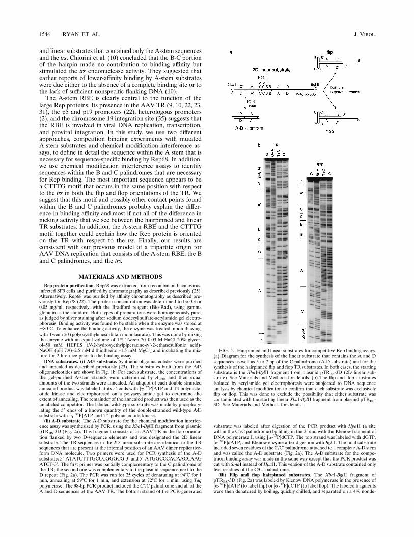
ence assay was synthesized by PCR, using the XbaI-BglII fragment from plasmidpTRBS-3D (Fig. 2a). This fragment consists of an AAV TR in the flop orienta-tion flanked by two D-sequence elements and was designated the 2D linearsubstrate. The TR sequences in the 2D linear substrate are identical to the TRsequences that are present at the internal position of an AAV dimer replicative-form DNA molecule. Two primers were used for PCR synthesis of the A-Dsubstrate: 59-ATATCTTTGCCCGGGCG-39 and 59-ATGGCCCACAACCAAGATCT-39. The first primer was partially complementary to the C palindrome ofthe TR; the second one was complementary to the plasmid sequence next to theD repeat (Fig. 2a). The PCR was run for 25 cycles of denaturing at 948C for 1min, annealing at 598C for 1 min, and extension at 728C for 1 min, using Taqpolymerase. The 98-bp PCR product included the C9/C palindrome and all of theA and D sequences of the AAV TR. The bottom strand of the PCR-generated
substrate was labeled after digestion of the PCR product with HpaII (a sitewithin the C9/C palindrome) by filling in the 39 end with the Klenow fragment ofDNA polymerase I, using [a-32P]dCTP. The top strand was labeled with dGTP,[a-32P]dATP, and Klenow enzyme after digestion with BglII. The final substrateincluded seven residues of the C/C9 palindrome attached to a complete A-D stemand was called the A-D substrate (Fig. 2a). The A-D substrate for the compe-tition binding assay was made in the same way except that the PCR product wascut with SmaI instead of HpaII. This version of the A-D substrate contained onlyfive residues of the C/C9 palindrome.(iii) Flip and flop hairpinned substrates. The XbaI-BglII fragment of
pTRBS-3D (Fig. 2a) was labeled by Klenow DNA polymerase in the presence of[a-32P]dATP (to label flip) or [a-32P]dCTP (to label flop). The labeled fragmentswere then denatured by boiling, quickly chilled, and separated on a 4% nonde-
FIG. 2. Hairpinned and linear substrates for competitive Rep binding assays.(a) Diagram for the synthesis of the linear substrate that contains the A and Dsequences as well as 5 to 7 bp of the C palindrome (A-D substrate) and for thesynthesis of the hairpinned flip and flop TR substrates. In both cases, the startingsubstrate is the XbaI-BglII fragment from plasmid pTRBS-3D (2D linear sub-strate). See Materials and Methods for details. (b) The flip and flop substratesisolated by acrylamide gel electrophoresis were subjected to DNA sequenceanalysis by chemical modification to confirm that each substrate was exclusivelyflip or flop. This was done to exclude the possibility that either substrate wascontaminated with the starting linear XbaI-BglII fragment from plasmid pTRBS-3D. See Materials and Methods for details.
1544 RYAN ET AL. J. VIROL.

naturing acrylamide gel in 13 Tris-borate-EDTA at 7.5 V/cm for 8 h. Underthese conditions, the three possible products migrated in the following order:flip, flop, renatured double-stranded linear. Following electrophoresis, the flipand flop bands were eluted from the gel.To ensure that equal molar amounts of unlabeled competitor DNA were used
in the competition assay between flip and flop hairpin, 2D linear, A43, and A-Dsubstrates, the concentration of each substrate was determined by A260. Theconcentrations were confirmed by comparing the intensities of ethidium bromidestaining of aliquots of each substrate. Finally, an aliquot of each substrate was 39labeled with [a-32P]dCTP and Klenow polymerase to determine whetherequimolar amounts of substrates contained equivalent numbers of 39 ends. Thelatter test was omitted for the A43 substrate.Plasmids. Plasmid pTRBR was constructed as follows. The 1,270-bp adenovi-
rus type 5 BglII fragment was inserted into the unique BglII site of the previouslydescribed vector dl3-94 (24). The resulting plasmid was then partially digestedwith BglII and completely digested with PstI, and a 1,440-bp fragment whichcontained the left AAV TR and the adenovirus stuffer fragment was isolated. Ina separate series of reactions, a 50-bp synthetic fragment coding for the simianvirus 40 (SV40) early polyadenylation signal and containing BamHI- and BglII-compatible ends was ligated to dl3-94 BglII linear DNA. The ligation mixture wasthen cut with PstI to isolate the 225-bp fragment which consisted of the left TRfrom dl3-94 joined through a BglII-BamHI junction to the synthetic poly(A)fragment. The 225-bp fragment and the 1,440-bp fragment were then ligated toeach other, and a 1,640-bp fragment was isolated from the ligation mixture afterit was digested with PstI. The resulting 1,640-bp fragment was then inserted intothe PstI site of pBR322 and called pTRBR.To construct pTRBS-3D, 293 cells were cotransfected with pTRBR and pIM45
(20) and infected with adenovirus type 5 ts149 at a multiplicity of infection of 10.At 36 h posttransfection, Hirt DNA was isolated and digested with DpnI. A DNAadapter that consisted of the AAV TR sequence from the trs to the end of theD sequence (nucleotides 125 to 145) and an XbaI sticky end was synthesized. Theadapter was ligated to the Hirt DNA. The products of the ligation reaction wereseparated on a 1% agarose gel, and the DNA band corresponding to the mono-mer duplex form was eluted and purified. The 59 ends of this fragment werekinase treated, and the DNA was cloned into the XbaI site of plasmid pBS(1)(Stratagene). Several independent clones were sequenced; one was found thatcontained the right AAV TR from pTRBR joined to the synthetic D sequence onone side and the SV40 poly(A) site on the other and contained an intact trs at theligation junction. This plasmid was p418wt. The BglII-PstI partial digest fragmentfrom pTRBR which contains the left AAV TR and the adenovirus stuffer frag-ment was then ligated to the BglII-XbaI fragment from p418wt which containsthe SV40 poly(A) site and the 2D TR sequence. The ligation products weredigested with PstI and XbaI and cloned into the polylinker site of plasmidpBS(1) that had been digested with PstI and XbaI. The resulting plasmid,pTRBS-3D, contains, from left to right: the left AAV TR, the adenovirus stufferfragment, the SV40 poly(A) site, and the right AAV TR joined to an additionalD sequence. The XbaI-BglII fragment containing the right AAV TR is illustratedin Fig. 2a.EMS competition assay. The electrophoretic mobility shift (EMS) assay was
performed as described earlier (23). Binding conditions were chosen so that 10%or less of the starting substrate was bound. To determine the ratio of dissociationconstants (Kds) of the wild-type and mutant A43 substrates, we used a compe-tition binding analysis at high substrate concentrations (Fig. 3). For example, theEMS assay shown in Fig. 3A illustrates the binding of 0.75 pmol of Rep68 to 0.25pmol of labeled wild-type A43 substrate in the absence of competitor or in thepresence of either unlabeled wild-type A43 substrate or unlabeled mutant A43substrate. The bound and free DNA fractions in each experiment were measuredby excising gel slices of each lane that encompassed all of the protein-DNAcomplexes and the free species, respectively, and then counting each gel slicewith a scintillation counter. The fraction of bound wild-type A43 substrate wasthen plotted as a function of picomoles of cold competitor (Fig. 3B). The ratioof the Kds of the wild-type and mutant RBEs was calculated by dividing theamount of homologous wild-type competitor by the amount of heterologousmutant competitor that was required to reduce the fraction of labeled wild-typeA43 substrate bound to 50% of the starting value in the absence of any com-petitor.Chemical modification interference assay. Chemical modifications of the A-D
substrate and the flip and flop hairpinned substrates were performed as de-scribed previously (7), with slight modifications. For the G reaction, about 107
cpm of labeled A-D substrate was dissolved in 10 ml of Tris-EDTA (TE) bufferand diluted with 200 ml of dimethyl sulfate (DMS) reaction buffer (50 mMsodium cacodylate [pH 8.0]–1 mM EDTA [pH 8.0]). After addition of 1 ml ofDMS, the reaction was allowed to proceed at room temperature for 5 min andthen quenched by adding 40 ml of 1.5 M sodium acetate (pH 7.0), 1 mM2-mercaptoethanol, 1 ml of tRNA (10 mg/ml), and 600 ml of 100% ethanol. TheDNA was precipitated twice with ethanol, rinsed, dried, and dissolved in TE. Forthe C1T reaction, the same amount of DNA substrate dissolved in 25 ml of H2Owas mixed with 15 ml of hydrazine, and the reaction was carried out at roomtemperature. After 30 min of incubation, the reaction was stopped by the addi-tion of 160 ml of hydrazine stop buffer (0.3 M sodium acetate [pH 7.0], 0.1 mMEDTA, 25 mg of tRNA per ml) and precipitated with 700 ml of ethanol. TheDNA was purified as described above and dissolved in TE. The DMS- and
hydrazine-modified DNA substrates were then used in a standard binding reac-tion with Rep68 as described previously (23). Time course reactions were doneto determine the time required to bind and shift approximately 50% of thestarting substrate, and an incubation time of 15 min was chosen. The bound andunbound fractions were separated on a 4% nondenaturing polyacrylamide gel for1.5 h at 12.5 V/cm. After electrophoresis, the gel was autoradiographed and thebands corresponding to free and bound DNA were eluted by electroelution ontoDEAE paper, eluted, extracted with phenol-chloroform, precipitated with eth-anol, rinsed, and dried. The DNA was then subjected to piperidine cleavage (19),purified, and dissolved in water. Samples containing equal amounts of radioac-tivity were then separated on an 8% sequencing gel. A reference sequence ladderwas generated by treating an aliquot of the labeled DNA substrate with formicacid and piperidine. The exposed autoradiographs were processed on a densi-tometer (UltroScan; LKB), and arbitrary intensity values were derived for eachband of the sequencing gel.
RESULTS
Defining the A-stem RBE. As mentioned earlier, previousstudies have demonstrated the ability of Rep protein to bind asubsequence of the A palindrome in the absence of secondarystructure (9, 10, 22, 23). To define the A-stem binding elementprecisely, we performed competition binding assays betweenwild-type and mutant A-stem substrates. The wild-type sub-strate was a synthetic 43-bp double-stranded DNA molecule(Fig. 1b, A43) that contained 30 bp of A stem flanked by 8 and5 bp of heterologous DNA on the B-C and trs proximal sides,respectively. On the B-C proximal side, the 30 bp of A-stemsequence contained all of the sequences shown to be protectedfrom DNase I digestion by Im and Muzyczka (13) as well as anadditional 2 bp that had not been protected. In addition, theA-stem substrate was longer on both sides than the maximumregion of DNase I protection seen by Chiorini et al. (10) and
FIG. 3. Competition binding assay used to define the RBE in the A stem. (A)Example of the competition binding assay. For the EMS assay, the standard10-ml reaction mixture contained 0.75 pmol of Rep68, 0.25 pmol of 32P-labeledA43 wild-type (WT) substrate, and 0 to 4 pmol of unlabeled homologous (WT Astem) or heterologous (A stem mutant #7) competitor DNA. Mutant 7 con-tained a dinucleotide GC-to-TA transversion at the position shown in Fig. 1b.(B) The fraction of bound labeled substrate was plotted as a function of theamount of competitor DNA added. Squares, homologous wild-type competitor;triangles, mutant 7 competitor.
VOL. 70, 1996 BINDING OF Rep68 TO AAV TERMINAL REPEATS 1545

contained all but 3 of the 28 bp that these workers suggestedcontained the RBE. These 3 bp on the B-C proximal side weresubstituted with heterologous DNA. The wild-type substratealso contained 4 bp on the B-C proximal side within the DdeIsite that had been shown by Chiorini et al. (10) to be necessaryfor maximum binding but that had not been present in previ-ous linear A-stem substrates used by us, either in the syntheticA-stem substrate (22, 23) or in the DdeI fragment (13, 31)(compare Fig. 1a and b). The wild-type A-stem binding sub-strate, therefore, contained all of the A sequences that hadbeen implicated in binding Rep (9, 10, 13, 22, 23, 31) andcontained sufficient flanking heterologous DNA as suggestedby Chiorini et al. (10). We then constructed a series of 13mutants in which two consecutive base pairs were substitutedwith nucleotides that produced transversions (Fig. 1b). Forexample, in mutant 1, the dinucleotide CT was substituted withAG.To determine the ratio of the Kds of the wild-type and
mutant substrates, each mutant substrate was compared withthe wild-type substrate as a competitor for binding to homo-geneously pure Rep68. This was done by using EMS assays tomeasure the amounts of wild-type and mutant competitors thatwere required to achieve a 50% reduction in the fraction ofbound 32P-labeled wild-type substrate under relatively highsubstrate concentrations. Under these conditions, most if notall of the enzyme is bound to substrate, and the ratio of wild-type to mutant competitor required to achieve some arbitrarylevel of competition is equal to the ratio of the Kds. Figure 3illustrates the competition binding experiment that was donefor mutant 7, a GC-to-TA transversion. The ratio of the wild-type Kd to mutant Kd for mutant 7 was calculated from thesedata to be 0.067. A ratio of 1 would have indicated no differ-ence in the dissociation constants; in this case, the wild-typesubstrate had a 15-fold-higher affinity for Rep than the mutant.Each mutant was individually compared with the wild type, andthe ratios of the Kds obtained were plotted as a function of theA-stem sequence that was mutated (Fig. 4). The data indicatedthat the A-stem RBE consists of a 22-bp sequence, CAGTGAGCGAGCGAGCGCGCAG. Each mutation within this 22-bpsequence increased the Kd for Rep by at least twofold. Muta-tions within an internal 18-bp sequence, GTGAGCGAGCGAGCGCGC, decreased binding affinity by at least threefold.Finally, mutations within a 10-bp core sequence, GCGAGC-GAGC, produced at least a 10-fold change in binding affinity.The symmetry of the effect of these mutations over the 22-bpregion suggested that the 22-bp sequence contained a singleRep binding site. If there had been two or more discrete Repbinding sites, we would have expected to find a region withinthe 22-bp sequence that had relatively little effect on the Kd.In our previous studies of Rep binding to the A stem, we had
used either the A-stem DdeI fragment (13, 31) or a syntheticA-stem substrate (22, 23) (Fig. 1a). As suggested by Chiorini etal. (10) and the results in Fig. 4, both of these substrates weremissing the 2 bp at the far left end of the 22-bp RBE. Theresults in Fig. 4 predicted that there would be about a twofolddifference in Kd between the A-stem substrate (or DdeI frag-ment) that we had used previously (13, 22, 23, 31) and the A43substrate. To test this, we compared the A-stem substrate withA43 by competition mobility shift assays. The results confirmedthat the A-stem substrate had a twofold-lower affinity for Repthan A43 (data not shown).To define the contribution of each base within the A-stem
core region, we synthesized a series of single-base-pair muta-tions within the 10-bp core region in which each mutant con-tained a substitution that produced a transversion (Fig. 1b).The effects of these single-base-pair mutations were measured
by competition binding gel shift experiments as describedabove, and the ratio of the wild-type to mutant Kds was plottedas a function of the mutated base (Fig. 5). Unlike the 2-bpmutations, the effects of the single-base-pair mutations werenot symmetrical. The two AT base pairs had no measurableeffect on binding, and the remaining base pairs producedchanges in the Kd that varied over a sixfold range. The differ-ence in Kd produced by the single-base-transversion mutants
FIG. 4. Ratio of the wild-type Kd to mutant Kd for the dinucleotide trans-version mutants within the RBE. For each dinucleotide transversion mutant(mutants 1 to 13 in Fig. 1b), a competition binding assay of the type shown in Fig.2 was used to determine the ratio of wild-type A43 to mutant A43 substraterequired to achieve a 50% reduction in the fraction of bound substrate. A ratioof 1 indicates no change in the Rep binding affinity. The 22-bp sequence thatshowed measurable changes in binding affinity is shown below the graph as aboxed sequence in the flip orientation of the hairpinned AAV TR. Boldfaceletters in the sequence are the bases previously shown to be protected fromDNase I (13) or copper phenanthralene (14a).
FIG. 5. Ratio of the wild-type Kd to mutant Kd for the single-nucleotidetransversion mutants within the 10-bp core region of the RBE. For each single-nucleotide-transversion mutant (mutants 1 to 10 in Fig. 1b), a competitionbinding assay of the type shown in Fig. 2 was used to determine the ratio ofwild-type A43 to mutant A43 substrate required to achieve a 50% reduction inthe fraction of bound substrate. A ratio of 1 indicates no change in the Repbinding affinity.
1546 RYAN ET AL. J. VIROL.

did not always accurately predict the effect of the 2-bp substi-tutions shown in Fig. 4. The predicted values differed from theactual values measured in the dinucleotide substitution exper-iment over a 1.5- to 6-fold range. We were not certain whatthese discrepancies meant. At the lower end, these differencesmay reflect errors in our measurements; at the high end, theymay suggest that mutation of multiple base pairs can producecompensatory or synergistic effects on binding affinity withinsome regions of the core sequence.Effects of chemical modifications on Rep binding to a linear
A-stem substrate. The competition analysis described aboveused substrates containing transversions on both strands of theRBE sequence. Therefore, it could not distinguish which res-idue on which strand of the DNA helix was actually a contactpoint for the bound Rep molecule. To overcome this limita-tion, we did interference binding assays using chemically mod-ified substrates. Two types of chemical modifications wereused, methylation of guanine residues at the N-7 position withDMS and modification of thymine and cytosine residues withhydrazine. The substrate used for this experiment contained allof the A and D sequences and 7 bp of the C palindrome. (Fig.2a, A-D substrate; see Materials and Methods.) The chemical
modifications were done under conditions that produced onaverage one modified base per substrate molecule. Bound andunbound substrate molecules were separated by EMS assay(not shown) and treated with piperidine to cleave at the mod-ified bases. The cleavage sites within the bound and unboundsubstrates were then compared on a DNA sequencing gel (Fig.6a). To get a quantitative measure of the effect of each base onbinding, the autoradiogram of Fig. 6 was subjected to opticaldensitometry and the relative intensity of each band was plot-ted in arbitrary units for both the bound and unbound sub-strates (Fig. 6c). The ratio of unbound to bound band inten-sities for each base was then calculated and plotted as afunction of the DNA sequence for both the trs-containingstrand (Fig. 6d) and the complementary strand (Fig. 6b). Anunbound-to-bound ratio of 1 indicated that a particular basemodification had no effect on Rep binding. Ratios greater than1 suggested that the particular base in question was overrep-resented in the unbound fraction and, therefore, made a spe-cific contact with the Rep protein. To get an approximateestimate of the variation in this kind of analysis, we examinedthe unbound-to-bound ratio in the D sequence, which all pre-vious data had shown was not involved in Rep binding (10, 13,
FIG. 6. DMS and hydrazine interference assays using the linear A-D substrate. (a) The A-D substrate (Fig. 2a) was labeled with 32P on the strand containing thetrs (bottom strand) or its complement (top strand), modified with either DMS (G lanes) or hydrazine (C1T lanes), and bound to Rep. Following cleavage at modifiedresidues, equal amounts of the bound (1) and unbound (2) fractions were compared. Bars at the side of each panel mark the regions where interference was seen.(b to d) Graphical representation of the DMS and hydrazine interference assay using the A-D substrate. In panel c, arbitrary numerical values for each residue werederived by laser densitometry scanning of the autoradiogram and plotted for each residue of the top or bottom strand. Shaded bars represent the amount of boundDNA substrate; clear bars indicate unbound substrate. Panels b and d represent the ratios of unbound to bound substrate (U/B) for modifications in the top and bottomstrands of the A-D substrate, respectively. A ratio close to 1 (horizontal line) indicates that modification of the particular base had little or no effect on Rep binding affinity.
VOL. 70, 1996 BINDING OF Rep68 TO AAV TERMINAL REPEATS 1547

23, 31). In the case of Fig. 6, we concluded that a ratio of 1.5or greater was likely to indicate that a particular base wasinvolved in Rep binding. Using this standard, we concludedthat the 18-base sequence GCGCGCTCGCTCGCTCAC wasrecognized by the Rep protein on the trs-containing strandand the 17-base sequence TGAGCGAGCGAGCGCGC wasbound on the complementary strand. These 17- and 18-bpsequences are essentially identical to the 18-bp sequence iden-tified by the dinucleotide mutagenesis analysis describedabove. We anticipated that the interference assays would beless sensitive than the dinucleotide mutagenesis analysis be-cause they were measuring on average the effect of single-basemodifications on only one strand rather than the effect of fourbase changes distributed on both strands. Thus, there was anexcellent correspondence between the results of the interfer-ence assays and the mutagenesis experiments (compare Fig. 4and 6b to d; see also Fig. 9).The DMS treatment used in these experiments was not
sufficient to produce significant methylation of A residues.Thus, we could not determine whether A residues in eitherstrand were involved in Rep binding. However, with the pos-sible exception of one G residue on the trs-containing strand,all of the remaining bases (G, C, and T) within the 18-bpregion clearly contributed to Rep binding. The four G residueson the trs-containing strand that were identified earlier bymethylation interference in a study by Owens et al. (26) (Fig.1a) are within the 18-bp region identified in this study. Finally,we note that a single G residue (underlined) on the trs-minusstrand (within the sequence GGCCAA) near the trs positionalso appeared to be involved in binding.Effects of chemical modifications on Rep binding to the
complete hairpinned TR. We and others had shown that trsendonuclease activity was higher on complete hairpinned sub-strates than on molecules that were missing the B and C pal-indromes (10, 23, 31). This finding implied that some contactoccurred between the bound Rep protein and a portion of theB and C palindromes. Furthermore, our DNase I protectionassays (13) (Fig. 1a) and the methylation interference experi-ments of Ashktorab and Srivastava (1) suggested that most ofthe contacts occurred within the small internal palindrome thatwas furthest away from the trs regardless of whether this wasthe B or C palindrome. Since these two palindromes areflipped during AAV DNA replication to produce two differentsequence configurations, the Rep protein was believed to rec-ognize only the secondary structure within the B-C region andnot a particular DNA sequence (4, 18, 22). To see if we coulddetect specific bases within the B-C region that make contactwith the Rep protein and to see if the contacts in the A-stembinding element are different in the presence of the B and Chairpins, we performed chemical modification interference ex-periments with both the flip and flop orientations of the hair-pinned TR. The two orientations of the hairpin were isolatedfrom a plasmid which contains the complete TR sequence plusan additional D sequence (Fig. 2a). Upon boiling and chilling,the hairpinned flip and flop orientations were separated fromeach other and from nonhairpinned starting material by gelelectrophoresis (not shown). Both the flip and flop hairpinnedsubstrates were then sequenced to confirm that each substratewas not contaminated with the starting linear duplex DNA andcontained only a hairpinned species. In addition, the sequenceconfirmed that each substrate contained the correct orienta-tion (Fig. 2b). Each substrate was then treated with DMS andhydrazine as described above to produce on average a singlemodification per molecule and then bound to Rep protein.Bound and unbound species were isolated and compared toidentify possible Rep contact points (Fig. 7).
As expected, modifications in the A-stem RBE interferedwith Rep binding to both the flip and flop substrates (Fig. 7b).Essentially the same 18- to 20-base region that was seen withthe linear A-stem substrates described above (A43 and A-Dsubstrates) was involved in binding Rep in both the flip andflop orientations (Fig. 7b; summarized in Fig. 9). However, theshape of the interference pattern appeared to be differentbetween the linear A-stem RBE and the same sequence in thecontext of the hairpinned substrate (compare Fig. 7b and 6b tod). In addition, the contribution of the trs-containing strand tobinding appeared to be lower in the hairpinned substrates thanin the linear A-D substrate. Finally, comparison of the flip andflop orientations (Fig. 7b) suggested that there might be somedifferences in the contributions of specific bases in the twoorientations, particularly bases in the trs-minus strand.Surprisingly, examination of the sequences in the B and C
palindromes also revealed specific bases that were involved inRep binding. This finding suggested that the secondary struc-ture of the B-C region was not the only feature recognized byRep. Particularly prominent was the contribution of theCTTTG motif that is present at the tip of the palindromefurthest away from the trs site. This motif and particularly thetwo underlined T residues clearly affected Rep binding in boththe flip and flop substrates. The CTTTG motif was interestingin that it is one of only two sequence motifs in the B and Cpalindromes that are identical in sequence and in position withrespect to the trs site in both the flip and flop orientations. Inaddition, other bases within the B and C palindromes alsoappeared to interfere with binding, albeit to a lesser extent.These bases were not the same in the two orientations. How-ever, in both kinds of substrates, all of the bases interactingwith Rep were clustered in or near the small internal palin-drome that was furthest away from the trs, that is, the B pal-indrome in the flip orientation and the C palindrome in theflop orientation (Fig. 7c and 9).Differential binding affinities of the hairpinned TR and lin-
ear A-stem substrates for Rep. We have pointed out before(23) that during AAV DNA replication, the Rep proteinshould theoretically encounter three kinds of substrates thatcontain an RBE. The first is a hairpinned end in the flip or flopconfiguration. This kind of substrate must be resolved to anopen duplex end for net DNA synthesis to occur. The secondkind of substrate is an end that has already been resolved. Inprinciple, there should be a mechanism for discriminatingagainst this substrate because nicking linear ends would wastetime and energy during DNA replication. In fact, we and oth-ers have shown that linear substrates that are not capable offorming the secondary structure element are nicked ineffi-ciently in vitro at the trs (10, 22, 31) and replicate poorly in vivo(6, 18, 29). Finally, Rep is also likely to encounter a thirdsubstrate which consists of a linear TR flanked by two Dsequences, which we refer to as a 2D linear substrate. 2D linearsubstrates are found in the middle of dimer replicative inter-mediates which are commonly formed during AAV DNA syn-thesis. An example of this kind of substrate is the linear XbaI-BglII fragment from pTRBR3D (Fig. 2a). To see if there was adifference in binding affinity for these three kinds of substrates,we compared the A43, A-D, flop, flip, and 2D linear substratesby competition gel shift assay for the ability to compete forbinding with flop hairpin DNA (Fig. 8). Essentially no differ-ence was seen in binding affinities of flop and flip hairpins(data not shown). A-D and A43 DNA, both of which contain acomplete RBE but are incapable of forming a secondary struc-ture element, competed to approximately the same extent withhomologous flop DNA. The four- to fivefold difference be-tween A-D and A43 DNA may be due to the fact that the A-D
1548 RYAN ET AL. J. VIROL.

substrate contains 5 bp of the C palindrome, which containstwo additional contact residues for Rep protein (Fig. 7 and 9).Both A43 and A-D DNA had significantly (approximately 170-fold for the A-D substrate) lower affinity for Rep than the flophairpin. This finding was consistent with our previous studiesusing substrates similar to A-D DNA (13, 23, 31). Finally, the2D linear substrate also had a lower affinity for the Rep proteinthan the flop hairpin, but the difference was considerably less,approximately 10-fold when a correction was made for the factthat the 2D substrate has two RBEs.
DISCUSSION
Mapping the A-stem RBE.We have used two approaches tomap in detail the bases within the A stem that affect Repbinding affinity, competition gel shift assays and chemical mod-ification interference assays. These two approaches comple-ment each other. The reciprocal competition analysis providesinformation about whether a particular set of base pairs con-tribute to binding affinity. The chemical modification interfer-ence assay can identify critical base residues on either DNAstrand and their relative effects on binding. Both methods arebase specific; that is, nonspecific contacts with the sugar orphosphate backbone of the substrate are not likely to be af-
fected. It is worth noting that chemical modifications of nucle-otide residues produce base moieties that are of a differenttype than transversion mutations. However, the results ob-tained from these two methods closely match in regard to thebases that are shown to be involved in binding by the Repprotein. Analysis of the A43 2-bp-substitution mutants identi-fied an unusually long 22-bp recognition sequence (plus orminus one base at either end) that was necessary for optimumbinding (Fig. 9a). Chemical modification experiments of theA-D substrate, which were expected to be inherently less sen-sitive because only one base is modified, identified an 18-bpsequence that is a subset of the larger 22-bp sequence (Fig. 9a).Analysis of the 2-bp-transversion mutants and chemical
modification interference assays both suggested that the RBEwas a single recognition site. Had there been two independentsites within the 22-bp sequence, we would have expected to seeregions that had relatively little effect on binding flanked byregions that had a larger effect. Instead, the effects of the 2-bp
FIG. 7. DMS and hydrazine interference assays using the flip and flop hair-pinned substrates. (a) The flip and flop hairpinned substrates (Fig. 2a) werelabeled with 32P, modified with either DMS (G lanes) or hydrazine (C1T lanes),and bound to Rep. Following cleavage at modified residues, equal amounts ofthe bound (1) and unbound (2) fractions were compared. Bars at the side ofeach panel mark the regions where interference was seen. (b) Graphical repre-sentation of the DMS and hydrazine interference assay within the A-stem regionsof the flip and flop substrates. The graph shows the ratios of unbound to boundsubstrate for modifications in the top or bottom strand of the flip (top graph) orflop (bottom graph) substrate. A ratio close to 1 (horizontal line) indicates thatmodification of the particular base had little or no effect on Rep binding affinity.(c) Ratios of unbound to bound substrate for modifications within the B and Csequences of the flip (top graph) and flop (bottom graph) hairpinned substrates.A comparison of the flip and flop sequences is shown in the middle.
VOL. 70, 1996 BINDING OF Rep68 TO AAV TERMINAL REPEATS 1549

mutations across the RBE were symmetrical across the se-quence, with mutations at the center having greater effects onbinding affinity than mutations at the ends of the RBE (Fig. 4).This was seen as well in the chemical modification interferenceassays using the A-D substrate. The ratio of unbound to boundsubstrate found in these experiments (Fig. 6b to d) was directlyproportional to the relative contribution of each residue tobinding affinity by Rep protein. The differences between Gmodifications and C and T modifications did not necessarilyshow the relative importance of purines versus pyrimidines butrather could reflect the different character of the modificationscaused by DMS or hydrazine. However, when G residues alonewere examined, it seemed clear that G residues in the middleof the RBE had a greater effect on binding affinity than thoseat the periphery. For the most part, the same was true when Cresidues were compared with other C residues, and the patternof interference within the string of G’s, as well as C’s, closelymatched the results for the 2-bp-transversion mutants (Fig. 4and 6b to d).The 22-bp RBE sequence mapped in this study is consistent
with most of the previous work from our laboratory and others(10, 22, 23, 26, 31, 35). The 22-bp sequence is virtually identicalin sequence and position to the site predicted by us (31) on thebasis of mutations within other regions of the A stem as well asour previous DNase I protection studies (Fig. 9a) (13). In theDNase I protection studies (13), 21 of the 22 bp of the RBEwere clearly protected; the remaining base pair, a CG that wasclosest to the B and C palindromes, might also have beenprotected but could not be clearly scored. The 22-bp RBE alsocontains the sites of the 5- and 7-bp substitution mutants thatreduced binding affinity in our previous studies (23), and it isconsistent with the 28-bp sequence suggested by the work ofChiorini et al. (10). Owens et al. (26) had identified four Gresidues by methylation interference assays on the trs-contain-ing strand (Fig. 9a) that appeared to be involved in Rep bind-ing, and Weitzman et al. (35) identified the same four Gresidues in their analysis of the chromosome 19 target site forAAV integration. Our results confirm their observations. Ash-ktorab and Srivastava (1) had identified a set of three G resi-
dues by methylation interference within the B palindrome ofthe flop hairpin that did not appear to be involved in Repbinding, and this finding was confirmed by our results (Fig. 9a).Chiorini et al. (10) demonstrated that fragments truncated atthe DdeI site, which would have 2 bp missing from the left end(B-C proximal) of the RBE, had reduced binding affinity.These 2 bp were missing in the DdeI fragment and the filled-in
FIG. 8. Competition binding assay comparing linear and hairpinned sub-strates. For the EMS assay, the standard 10-ml reaction mixture contained 0.075pmol of Rep68, 0.05 pmol of 32P-labeled flop hairpinned substrate, and 0 to 14pmol of unlabeled homologous flop substrate (squares) or heterologous com-petitor DNA (A-D substrate [circles], 2D linear substrate [triangles], or A43substrate [diamonds]). The fraction of bound labeled substrate was plotted as afunction of the amount of competitor DNA added. Competition with unlabeledflip hairpin substrate produced a competition curve identical to the flop curve(not shown). Only a portion of the competition curve is shown; competition withthe A43 substrate reached 62% at 14 pmol of competitor. From this, it wasextrapolated that 50% competition would require 21 pmol of A43 competitorDNA.
FIG. 9. Summary of information about sequences within the TR that affectRep binding affinity. (a) The complete hairpinned AAV TR is shown in the fliporientation; only the B and C sequences of the flop orientation are shown.Nucleotides protected from DNase I (13) cleavage are shown in boldface. Thesolid line box within the A stem indicates the 18-bp sequence that was shown toaffect binding affinity by mutagenesis and by DMS and hydrazine interference inthis study. The dotted-line extensions indicate two additional bases at either endwhose mutagenesis affected the binding affinity by approximately twofold; the22-bp sequence bounded by the dotted lines is likely to be the minimum RBE,plus or minus one base at each end. The left end of the solid box is the DdeIcleavage site. The solid lines below the box show the regions of DMS andhydrazine interference for the top and bottom strands of the A-stem bindingelement for the A-D substrate (A stem) and the flip and flop substrates. Asterisksindicate the G residues previously identified by Owens et al. (26) and Weitzmanet al. (35) by DMS interference assays with the AAV TR and the chromosome19 RBE, respectively. The 22-bp RBE defined in this study contains the GAGCor GCTC repeats identified earlier in the studies by Weitzman et al. (35),McCarty et al. (22, 23), and Chiorini et al. (9, 10). It is also the sequencepredicted earlier by Snyder et al. (31) to contain the RBE on the basis of differentarguments and is consistent with the 28-bp sequence suggested by Chiorini et al.(10), which contained 1 and 5 additional bp on the right and left, respectively.Within the secondary structure element, the box indicates the CTTTG elementthat is identical with respect to both position and sequence in both orientations.Bases which contribute to Rep binding affinity by interference assays are indi-cated by solid dots. The strongest contribution to binding appeared to come fromthe two circled T residues. The three open dots in the flop orientation indicateresidues that were found to contribute little if anything to binding in the study byAshktorab and Srivastava (1). Consistent with the previous suggestions by Im andMuzyczka (13) and Ashktorab and Srivastava (1), the residues within the B andC palindromes that are involved in Rep binding are clustered within the palin-drome that is further from the terminal resolution site (B in the flip substrate andC in the flop substrate). Arrows indicate the positions of SmaI cleavage in thetwo orientations. Consistent with the results of this study, cleavage of the fliphairpinned substrate with SmaI does not affect binding or trs endonucleaseactivity (13, 31). Also consistent with this study, mutations within the trs regionoutside the RBE do not appear to affect Rep binding affinity (10, 23, 31). (b)Comparison of the 22-bp RBE within the A stem of the TR with the other knownRBEs in pBR322 and the AAV p5 promoter (22) and in human chromosome 19(35). The boxed region indicates the 18-bp sequence consistent with the inter-ference data in this study; the shaded sequence indicates the 10-bp core regionwhich has the greatest effect on binding affinity by mutagenesis and interferenceanalysis.
1550 RYAN ET AL. J. VIROL.

synthetic A-stem substrate that we had used previously forbinding studies (Fig. 1) (13, 22, 23, 31). Consistent with theobservation of Chiorini et al. (10), we found that when these 2bp were mutagenized, there was approximately a twofold dropin binding affinity (Fig. 4). Direct comparison by competitiongel shift assay of our previously used A-stem substrate with theA43 substrate used in this study confirmed that there was atwofold difference in the binding affinities of these two sub-strates (data not shown). Finally, the RBE sequence showsstrong similarity to three other sequences that have beenshown to bind to Rep protein. The 22-bp sequence has a19-of-22-base identity to the chromosome 19 integration site(35) and is identical at 15 and 11 positions to the pBR322 siteand the p5 promoter site, respectively (22) (Fig. 9b).The 22-bp RBE sequence contains a set of four imperfect
GAGC motifs which we and others (1, 10, 22, 23, 35) hadspeculated might be the important feature of this element forRep binding. Our analysis suggests that the sequence recog-nized by Rep may be more complex and may be affected byother nearby sequences. First, the central 10 bp of the RBE,which appeared to have the most pronounced effect on bindingwhen we analyzed the 2-bp-transversion mutants, consisted ofa portion of the GAGC motifs, GCGAGCGAGC (Fig. 4 and9b). Furthermore, it was clear that bases outside the GAGCmotifs have an effect on binding. This would explain in partwhy an oligonucleotide that consists only of the GAGC motifsis not sufficient to bind Rep protein (35) or binds poorly (10).Chiorini et al. (10) have suggested that additional nonspecificcontacts are necessary in addition to the GAGCmotifs for Repbinding. Our results did not exclude this possibility but diddemonstrate that additional specific base contacts that do notinvolve the GAGC repeats are necessary for optimum Repbinding. In addition, the results of the chemical interferenceassay using the A-D substrate suggested that the center of theinteraction curve of the bottom strand appeared to be shiftedleftward toward the BC palindromes, which could reflect dif-ferential binding of Rep to the two DNA strands (Fig. 6 to d).Differential binding to the two strands appeared to be evenmore pronounced in the hairpinned substrates (Fig. 7b). Therewere also several indications that there was some flexibility insequence recognition by the Rep protein. For example, exam-ination of the chemical interference pattern on the trs-contain-ing strand of the A-D substrate (Fig. 6d) indicated that the twoT residues in the 10-bp core region of the RBE contributedsignificantly to binding affinity. Modification of either T pro-duced approximately a fourfold difference in the ratio of un-bound to bound substrate. In contrast, when the AT base pairscontaining these two T residues were individually substitutedwith CG residues, no effect was seen on binding affinity (Fig.5). This result suggested that the AT-to-CG transversion mu-tations produced compensatory interactions on one or bothstrands. Finally, the A-D substrate and the hairpinned sub-strates exhibited different patterns of interference (compareFig. 6b to d and 7b), and there were a number of differences aswell between the two hairpinned substrates, flip and flop (Fig.7b). These differences may be due to the specific and slightlydifferent base contacts made by Rep protein within the B andC palindromes of the flip and flop hairpins (Fig. 7c and 9a) andto the fact that none of these contact sites are available in theA-D substrate.Specific base contacts in the B and C palindromes. From
previous work, it could be argued that contacts between theRep protein and the B and C palindrome sequences were notlikely to be base specific. The argument was that since Rep iscapable of binding and resolving two orientations of the B andC palindromes, flip and flop, and the sequences of these two
orientations are not the same (4), it is more likely that Repprotein recognizes the secondary structure of the B and Cpalindromes rather than a specific base sequence. Berns andhis colleagues (18) were the first to propose this idea and testit directly. When they substituted either an 8-bp or a 12-bpsymmetrical linker for a 9-bp SmaI fragment from the C pal-indrome (Fig. 9a), the ability of the mutant to replicate in vivowas nearly normal (5, 6, 18). A deletion of the SmaI fragmentor a nonsymmetrical substitution was defective for DNA rep-lication (6, 29). They concluded that the secondary structure ofthe C palindrome was more important than the actual se-quence.In contrast, the results reported here suggest that the se-
quences of the internal palindromes, B and C, may have a rolein Rep binding and DNA replication. Our chemical modifica-tion experiments indicated that strong base contacts occurredbetween Rep and two T residues (underlined) in a CTTTGmotif that is present in the same position of the hairpin struc-ture with respect to the trs and the RBE in both the flip andflop orientations (Fig. 7c and 9a). These two T residues af-fected the unbound-to-bound ratio by four- to sixfold, an effectthat was comparable to that seen with bases in the core of theRBE. Modification of the remaining bases of the CTTTGmotif also appeared to affect binding affinity, particularly in theflop orientation (Fig. 7c). In addition, several other baseswithin the B and C palindromes of both the flip and floporientations appeared to be making base contacts with Repprotein. These additional contacts were not in the same posi-tion or sequence with respect to the trs and RBE. We note alsothat most of the contact sites revealed by chemical modifica-tion were in the internal palindrome (B or C) that was furtheraway from the trs, regardless of whether the orientation wasflip or flop. This observation confirmed previous data frommethylation interference and DNase I protection studies (1,13). It was also consistent with our previous finding that thelower end of the C palindrome could be removed by SmaIdigestion from the flip hairpin substrate without affecting Repbinding or nicking in vitro (Fig. 9a) (13, 31).Taken together, our results imply that binding by Rep pro-
tein to the hairpinned TR is inherently asymmetrical. First, theRBE is a nonpalindromic sequence; thus, Rep is likely to bindto it in a particular orientation. Second, Rep makes contactspredominately with only one of the two internal palindromes,which again implies an asymmetric alignment of Rep with thehairpinned TR. These two elements are, therefore, likely toensure that Rep is correctly aligned on the TR to nick only thecorrect strand at the trs.The observation of specific base contacts within the B and C
palindromes is not incompatible with the results of the substi-tution mutants studied by Berns and colleagues (5, 6, 18). Allof the substitution mutants used by Berns and colleagues re-tained at least one of the T residues that is present in thewild-type sequence. Furthermore, the mutations reported bythis group would have affected terminal resolution of only theflip, not the flop, hairpin. Thus, a significant difference inreplication capacity might not have been apparent. In addition,Berns and his colleagues (5, 6) found that the wild-type se-quence was preferred over the substitution mutants undersome conditions, suggesting that sequence substitutions withinthe C palindrome did affect the efficiency of DNA replication.Finally, we and others have shown that regardless of whetherthe wild-type CTTTGmotif is present, there is still a significantdifference in the abilities of hairpinned and linear TRs to benicked in vitro (10, 22, 31). Thus, the fact that the secondarystructure of the B and C palindromes places the contact sites in
VOL. 70, 1996 BINDING OF Rep68 TO AAV TERMINAL REPEATS 1551

this region in a particular spatial orientation for Rep interac-tion clearly plays a role in Rep function at the trs.Contact points in the trs region. When we examined the
DMS interference pattern obtained with A-D substrate, wefound approximately a fourfold interference at a single G res-idue on the strand opposite to the trs, 3 bp away from the cutsite (Fig. 6b to d). The significance of this observation was notclear. DMS interference assays on the hairpinned substratesdid not show contacts at this position (Fig. 7b, flip). In addi-tion, we and others have reported that mutants in this regionthat are missing this G residue have approximately the samebinding affinity for Rep (10, 23). Nevertheless, these bindingexperiments might well have missed a fourfold effect. More-over, it seems clear that mutations in the trs region can abolishnicking activity, suggesting that a specific sequence is recog-nized by Rep in the trs region prior to nicking (31). The Gresidue that emerged from the A-D substrate analysis may beone of these specific trs contact sites, but a more systematicanalysis of mutations in the trs region will be needed to deter-mine the significance of this G residue.Differential binding affinities of Rep to hairpinned and lin-
ear substrates.We reported previously that there was a signif-icant difference in binding affinity between linear substratesthat contained the RBE and hairpinned substrates that con-tained the B and C palindromes in addition to the RBE (13, 23,31). We estimated the difference in Kd to be greater than125-fold. Recently, Chiorini et al. (10) have suggested thatthere is no difference in binding affinity between linear andhairpinned substrates and suggested that the difference inbinding affinity that we observed might be due to the fact thatwe used a linear substrate (the DdeI fragment or the A-stemsubstrate) that was missing a portion of the RBE or possiblywas not long enough to provide nonspecific contacts neededfor optimal Rep binding. As already mentioned, we found inthis study that both the DdeI fragment and the A-stem sub-strate used in our previous studies were indeed missing tworesidues of the RBE. However, our results suggested that thedifference in binding affinity due to these two base pairs wasonly twofold. We also compared the affinity of Rep for linearand hairpinned substrates directly (Fig. 8) and found that thedifference in binding affinity between the hairpinned flip sub-strate and the A-D substrate containing a complete RBE wasapproximately 170-fold. This result was consistent with theresults of the chemical modification interference experimentswhich demonstrated that there were specific contacts betweenRep and bases within the B and C palindromes. These contactsites were not present in the A-D substrate and probably ac-counted for at least some of the difference in binding affinity.Finally, we have presented evidence before that substitutionmutants within the RBE that consisted of 5 or 7 bp had agreater effect on binding to the linear A-stem substrate than tohairpinned substrate (23). This also suggested a difference inbinding affinity between linear and hairpinned substrates. Weconcluded that there is a substantial difference in binding af-finity between linear substrates containing the RBE and sub-strates which contain the RBE and the B and C palindromes ina hairpinned configuration.We are not certain why there is a discrepancy between our
results with linear and hairpinned substrates and those of Chi-orini et al. (9, 10, 13, 23, 31). Possibly it is due to the fact thatmany of the studies by Chiorini et al. (10) were done with amutant Rep68 protein that is fused at its N terminus to aportion of the maltose-binding protein of Escherichia coli (9).We and others have shown that sequences within the N termi-nus of Rep are essential for binding to the TR (21, 26, 37).Thus, the 37-kDa maltose-binding domain of the chimeric Rep
protein might change the binding characteristics of Rep. Re-gardless of the reason for the discrepancy, it is worth notingthat there is general agreement that Rep protein nicks linearsubstrates at a much lower frequency than hairpinned sub-strates (10, 23, 31). Thus, Rep has a mechanism for discrimi-nating between these two kinds of terminal sequences and cutsone preferentially. Our results suggest that much of the dis-crimination occurs at the level of binding affinity for the twokinds of substrates.We also compared the binding affinities of flip hairpinned
substrates and the 2D linear substrate. The 2D linear substratecontains two RBE sites, two trs regions, and a single copy of theB and C palindromes. Essentially, the 2D linear substrate con-tains the kind of TR structure that is present within dimermolecules produced during AAV DNA replication. We ex-pected that binding to the 2D linear substrate would occur atabout the same level as binding to the A-D substrate. Surpris-ingly, the 2D linear substrate bound Rep much more efficientlythan A-D substrate even when a correction was made for thefact that 2D linear DNA contains two RBE sites per molecule.The difference in binding affinity between a hairpinned TR anda 2D linear substrate was approximately 10-fold. It is worthnoting that monomer and dimer duplex replicative forms ac-cumulate in vivo at approximately a 5:1 ratio during AAVDNA replication. Thus, the difference in binding affinity be-tween 2D linear DNA and hairpin DNA could account for therelative accumulation of monomer and dimer DNA duringAAV DNA replication.
ACKNOWLEDGMENTS
This work was supported by grants PO1 CA2814607, RO1GM3572302, and HL/DK 50257 from the National Institutes of Healthto N.M. and by Public Health Service training grant (T32 AI25530) toJ.H.R.
REFERENCES
1. Ashktorab, H., and A. Srivastava. 1989. Identification of nuclear proteinsthat specifically interact with adeno-associated virus type 2 inverted terminalrepeat hairpin DNA. J. Virol. 63:3034–3039.
2. Batchu, R. B., R. M. Kotin, and P. L. Hermonat. 1994. The regulatory repprotein of adeno-associated virus binds to sequences within the c-H-raspromoter. Cancer Lett. 86:23–31.
3. Beaton, A., P. Palumbo, and K. I. Berns. 1989. Expression from the adeno-associated virus p5 and p19 promoters is negatively regulated in trans by theRep protein. J. Virol. 63:4450–4454.
4. Berns, K. I., W. W. Hauswirth, K. H. Fife, and E. Lusby. 1979. Adeno-associated virus DNA replication. Cold Spring Harbor Symp. Quant. Biol.43(Pt. 2):781–787.
5. Bohenzky, R. A., and K. I. Berns. 1989. Interactions between the termini ofadeno-associated virus DNA. J. Mol. Biol. 206:91–100.
6. Bohenzky, R. A., R. B. LeFebvre, and K. I. Berns. 1988. Sequence andsymmetry requirements within the internal palindromic sequences of theadeno-associated virus terminal repeat. Virology 166:316–327.
7. Brunelle, A., and R. F. Schleif. 1987. Missing contact probing of DNA-protein interactions. Proc. Natl. Acad. Sci. USA 84:6673–6676.
8. Chejanovsky, N., and B. J. Carter. 1989. Mutagenesis of an AUG codon inthe adeno-associated virus rep gene: effects on viral DNA replication. Vi-rology 173:120–128.
9. Chiorini, J. A., M. D. Weitzman, R. A. Owens, E. Urcelay, B. Safer, and R. M.Kotin. 1994. Biologically active Rep proteins of adeno-associated virus type2 produced as fusion proteins in Escherichia coli. J. Virol. 68:797–804.
10. Chiorini, J. A, S. M. Wiener, R. A. Owens, S. R. M. Kyostio, R. M. Kotin, andB. Safer. 1994. Sequence requirements for stable binding and function ofRep68 on the adeno-associated virus type 2 inverted terminal repeats. J.Virol. 68:7448–7457.
11. Hermonat, P. L. 1994. Down-regulation of the human c-fos and c-mycproto-oncogene promoters by adeno-associated virus Rep78. Cancer Lett.81:129–136.
12. Hermonat, P. L., M. A. Labow, R. Wright, K. I. Berns, and N. Muzyczka.1984. Genetics of adeno-associated virus: isolation and preliminary charac-terization of adeno-associated virus type 2 mutants. J. Virol. 51:329–339.
13. Im, D. S., and N. Muzyczka. 1989. Factors that bind to adeno-associatedvirus terminal repeats. J. Virol. 63:3095–3104.
1552 RYAN ET AL. J. VIROL.

14. Im, D. S., and N. Muzyczka. 1990. The AAV origin binding protein Rep68is an ATP-dependent site-specific endonuclease with DNA helicase activity.Cell 61:447–457.
14a.Im, D. S., and N. Muzyczka. Unpublished observation.15. Kyostio, S. R., R. A. Owens, M. D. Weitzman, B. A. Antoni, N. Chejanovsky,
and B. J. Carter. 1994. Analysis of adeno-associated virus (AAV) wild-typeand mutant Rep proteins for their abilities to negatively regulate AAV p5and p19 mRNA levels. J. Virol. 68:2947–2957.
16. Labow, M. A., L. H. Graf, Jr., and K. I. Berns. 1987. Adeno-associated virusgene expression inhibits cellular transformation by heterologous genes. Mol.Cell. Biol. 7:1320–1325.
17. Labow, M. A., P. L. Hermonat, and K. I. Berns. 1986. Positive and negativeautoregulation of the adeno-associated virus type 2 genome. J. Virol. 60:251–258.
18. LeFebvre, R. B., S. Riva, and K. I. Berns. 1984. Conformation takes prece-dence over sequence in adeno-associated virus DNA replication. Mol. Cell.Biol. 4:1416–1419.
19. Maxam, A. M., and W. Gilbert. 1980. Sequencing end-labeled DNA withbase-specific chemical cleavages. Methods Enzymol. 65:499–560.
20. McCarty, D. M., M. Christensen, and N. Muzyczka. 1991. Sequences re-quired for coordinate induction of adeno-associated virus p19 and p40 pro-moters by Rep protein. J. Virol. 65:2936–2945.
21. McCarty, D. M., T. H. Ni, and N. Muzyczka. 1992. Analysis of mutations inadeno-associated virus Rep protein in vivo and in vitro. J. Virol. 66:4050–4057.
22. McCarty, D. M., D. J. Pereira, I. Zolotukhin, X. Zhou, J. H. Ryan, and N.Muzyczka. 1994. Identification of linear DNA sequences that specificallybind the adeno-associated virus Rep protein. J. Virol. 68:4988–4997.
23. McCarty, D. M., J. H. Ryan, S. Zolotukhin, X. Zhou, and N. Muzyczka. 1994.Interaction of the adeno-associated virus Rep protein with a sequence withinthe A palindrome of the viral terminal repeat. J. Virol. 68:4998–5006.
24. McLaughlin, S. K., P. Collis, P. L. Hermonat, and N. Muzyczka. 1988.Adeno-associated virus general transduction vectors: analysis of proviralstructures. J. Virol. 62:1963–1973.
25. Ni, T. H., X. Zhou, D. M. McCarty, I. Zolotukhin, and N. Muzyczka. 1994.In vitro replication of adeno-associated virus DNA. J. Virol. 68:1128–1138.
26. Owens, R. A., M. D. Weitzman, S. R. Kyostio, and B. J. Carter. 1993.Identification of a DNA-binding domain in the amino terminus of adeno-associated virus Rep proteins. J. Virol. 67:997–1005.
27. Redemann, B. E., E. Mendelson, and B. J. Carter. 1989. Adeno-associatedvirus Rep protein synthesis during productive infection. J. Virol. 63:873–882.
28. Rittner, K., R. Heilbronn, J. A. Kleinschmidt, and G. Sczakiel. 1992. Adeno-associated virus type 2-mediated inhibition of human immunodeficiencyvirus type 1 (HIV-1) replication: involvement of p78rep/p68rep and theHIV-1 long terminal repeat. J. Gen. Virol. 73:2977–2981.
29. Samulski, R. J., A. Srivastava, K. I. Berns, and N. Muzyczka. 1983. Rescueof adeno-associated virus from recombinant plasmids: gene correction withinthe terminal repeats of AAV. Cell 33:135–143.
30. Snyder, R. O., D. S. Im, and N. Muzyczka. 1990. Evidence for covalentattachment of the adeno-associated virus (AAV) Rep protein to the ends ofthe AAV genome. J. Virol. 64:6204–6213.
31. Snyder, R. O., D. S. Im, T. Ni, X. Xiao, R. J. Samulski, and N. Muzyczka.1993. Features of the adeno-associated virus origin involved in substraterecognition by the viral Rep protein. J. Virol. 67:6096–6104.
32. Snyder, R. O., R. J. Samulski, and N. Muzyczka. 1990. In vitro resolution ofcovalently joined AAV chromosome ends. Cell 60:105–113.
33. Srivastava, A., E. W. Lusby, and K. I. Berns. 1983. Nucleotide sequence andorganization of the adeno-associated virus 2 genome. J. Virol. 45:555–564.
34. Tratschin, J. D., J. Tal, and B. J. Carter. 1986. Negative and positiveregulation in trans of gene expression from adeno-associated virus vectors inmammalian cells by a viral rep gene product. Mol. Cell. Biol. 6:2884–2894.
35. Weitzman, M. D., S. R. Kyostio, R. M. Kotin, and R. A. Owens. 1994.Adeno-associated virus (AAV) Rep proteins mediate complex formationbetween AAV DNA and its integration site in human DNA. Proc. Natl.Acad. Sci. USA 91:5808–5812.
36. Wonderling, R. S., S. R. Kyostio, and R. A. Owens. 1995. A maltose-bindingprotein/adeno-associated virus Rep68 fusion protein has DNA-RNA heli-case and ATPase activities. J. Virol. 69:3542–3548.
37. Yang, Q., and J. P. Trempe. 1993. Analysis of the terminal repeat bindingabilities of mutant adeno-associated virus replication proteins. J. Virol. 67:4442–4447.
VOL. 70, 1996 BINDING OF Rep68 TO AAV TERMINAL REPEATS 1553