seroconversion and abundance of igg antibodies against s1
TRANSCRIPT
Research ArticleSeroconversion and Abundance of IgG Antibodies againstS1-RBD of SARS-CoV-2 and Neutralizing Activity in theChilean Population
R. González-Stegmaier,1 K. Cereceda,1 J. L. Briones,2 C. Beltran-Pávez,3,4
A. Oyarzún-Arrau,3,4 S. Riquelme-Barrios,3,4 C. Selman,5,6 F. Yarad,5 M. Mahave,7
C. Caglevic,8 R. Morales,9 A. Aguirre,1 F. Valiente-Echeverría,3,4 R. Soto-Rifo,3,4
H. Marsiglia,10 R. Gazitua ,2 and F. Villarroel-Espindola 1,8
1Translational Medicine Laboratory, Instituto Oncológico Fundación Arturo López Pérez, Santiago, Chile2Haematology Department, Instituto Oncológico Fundación Arturo López Pérez, Santiago, Chile3Laboratory of Molecular and Cellular Virology, Virology Program, Institute of Biomedical Sciences, Faculty of Medicine,Universidad de Chile, Chile4HIV/AIDS Work Group, Faculty of Medicine, Universidad de Chile, Chile5Diagnostic Units, Instituto Oncológico Fundación Arturo López Pérez, Santiago, Chile6Biobank, Instituto Oncológico Fundación Arturo López Pérez, Santiago, Chile7Medical Oncology Department, Instituto Oncológico Fundación Arturo López Pérez, Santiago, Chile8Cancer Research Department, Instituto Oncológico Fundación Arturo López Pérez, Santiago, Chile9Internal Medicine Department, Instituto Oncológico Fundación Arturo López Pérez, Santiago, Chile10Radiotherapy Department, Instituto Oncológico Fundación Arturo López Pérez, Santiago, Chile
Correspondence should be addressed to R. Gazitua; [email protected] F. Villarroel-Espindola; [email protected]
Received 20 December 2020; Revised 3 January 2021; Accepted 24 January 2021; Published 23 February 2021
Academic Editor: Baohui Xu
Copyright © 2021 R. González-Stegmaier et al. This is an open access article distributed under the Creative Commons AttributionLicense, which permits unrestricted use, distribution, and reproduction in any medium, provided the original work isproperly cited.
COVID-19 is a pandemic caused by SARS-CoV-2. In Chile, half a million people have been infected and more than 16,000 havedied from COVID-19. As part of the clinical trial NCT04384588, we quantified IgG against S1-RBD of SARS-CoV-2 (anti-RBD)in recovered people in Santiago and evaluated their suitability as COVID-19 convalescent plasma donors. ELISA and aluminescent SARS-CoV-2 pseudotype were used for IgG and neutralizing antibody quantification. 72.9% of the convalescentpopulation (468 of 639) showed seroconversion (5-55μg/mL anti-RBD IgG) and were suitable candidates for plasma donation.Analysis by gender, age, and days after symptom offset did not show significant differences. Neutralizing activity correlated withan increased concentration of anti-RBD IgG (p < 0:0001) and showed a high variability between donors. We confirmed that themajority of the Chilean patients have developed anti-SARS-CoV-2 antibodies. The quantification of anti-RBD IgG inconvalescent plasma donors is necessary to increase the detection of neutralizing antibodies.
1. Introduction
Coronavirus disease 2019 (COVID-19) is caused by severeacute respiratory syndrome coronavirus 2 (SARS-CoV-2). Itemerged in December 2019 in Wuhan, China [1, 2], and
was subsequently declared a worldwide pandemic by theWorld Health Organization (WHO) [3]. Currently, Chilehas reported half a million infected people and more than16,000 deaths caused by COVID-19. The government andthe private sector have implemented several programs to
HindawiJournal of Immunology ResearchVolume 2021, Article ID 6680337, 11 pageshttps://doi.org/10.1155/2021/6680337
mitigate this pandemic, including PCR and rapid testingacross the country, while some institutions have started trialsfor convalescent plasma. However, these efforts still do notseem to be enough to control this sanitary emergency.
The measurement of serum antibodies has provided cru-cial data for understanding key aspects of the infection [4]. Itis widely accepted that IgM provides the first line of defenseduring viral infections, before generating an adaptiveresponse with a high-affinity IgG production, which isimportant for long-term immunity and immunologicalmemory [5]. In patients infected with SARS-CoV-2, immu-noglobulin M (IgM) antibodies are detectable around 7 dayspostinfection and IgG antibodies usually take two weeks todevelop [6–9]. The clinical features of COVID-19 varywidely, ranging from asymptomatic to mild or severe formswith multiorgan dysfunction, and most COVID-19 data werederived from hospitalized patients [8, 10–13].
Accurate quantitative measurement of anti-SARS-CoV-2antibody responses is essential for public health interventionsand therapeutic applications [14, 15]. The SARS-CoV-2shows two structural proteins as major immunogens, thespike (S) and nucleocapsid (N) proteins, with the RBDdomain of the S protein being responsible for the infectionof respiratory epithelial cells via interaction with the cellsurface receptor angiotensin converting enzyme 2 (ACE2).Antibodies against this segment have been reported as antivi-ral based on their neutralizing activity in plasma [7, 13, 16–18]. In this study, we quantified the abundance of IgGantibodies against the S1-RBD fragment of SARS-CoV-2 inpeople who recovered from COVID-19 in Santiago, Chile.After analyzing 639 convalescent participants, 72.9% showedanti-RBD IgG seroconversion with a concentration range inserum between 5 and 55μg/mL, and no significant correla-tion with gender, age, and days after symptom offset wereobserved between groups. Further analysis showed thatserum samples with antibodies above 20.9μg/mL showed astatistically increased proportion of neutralizing antibodies(Nab) against SARS-CoV-2 during convalescence. Regardingearly seroconversion, 14 of 26 cases within the first two weeksof severe COVID-19 showed positive levels of anti-RBD IgGwith a mean of 21.2μg/mL, and 7 cases were above the 75thpercentile. Our results confirmed a heterogeneous but spe-cific IgG seroconversion after SARS-CoV-2 infection withinthe population from the metropolitan area of Chile. The pro-duction of antibodies against the RBD of spike were shown tobe as early as the first 14 days after symptoms started, andthose can persist in the blood for almost two months anddisplay protective neutralizing activity during active diseaseand convalescence.
2. Materials and Methods
2.1. Patients and Samples. This research consisted of 690samples distributed in 3 groups: 639 convalescents, 26 activeCOVID-19 cases, and 25 healthy never-exposed people as anegative control (supplementary Table 1). There were asecond cohort of 72 convalescent plasma donors for theanalysis of neutralizing antibodies and a third cohort of 100unrelated cases (positive and negative COVID-19) for
ELISA validation. Recovered volunteers and convalescentplasma donors met all criteria for the clinical trialNCT04384588 and signed the specific consent letter. Allvolunteer patients who recovered from COVID-19 wereasymptomatic for at least 21 days and showed a negativeresult for SARS-CoV-2 by real-time polymerase chainreaction (PCR) test. Volunteers with active COVID-19consented at the moment of hospitalization and met thefollowing inclusion criteria: (a) patients over 18 years oldand (b) COVID-19 diagnosis at enrolment confirmed witha positive SARS-CoV-2 PCR in a nasopharyngeal swab.
All SARS-CoV-2-related samples used in this researchwere collected between July and August 2020, in Santiago,Chile.
Healthy group samples were considered as plasma from aregular blood donation obtained before October 2019 (never-exposed cases) and as a single blood sample drawn in April2020 from asymptomatic healthy volunteers with a negativeSARS-CoV-2 PCR result (contemporary cases).
For the active cases, treatments given before blood collec-tion are described in supplemental Table 2.
2.2. Quantitative IgG ELISA Anti-RBD S1 SARS-CoV-2.Anti-SARS-CoV-2 IgG ELISA test was developed and validated inthe Translational Medicine Laboratory of Fundación ArturoLópez Pérez (Cereceda et al., submitted); a comparative table(Table S3) is included as a supplementary data. Briefly,MaxiSorp™ 96-well microplates (439454, NUNC, ThermoFisher Scientific) were coated with 50 ng of S1-RBD proteinof SARS-CoV-2 (RB.230-30162-100, RayBiotech) in 0.1Mcarbonate buffer pH9 and incubated overnight at 4°C. Thecoating solution was removed and each well was washedonce with cold phosphate-buffered saline solution (PBS).An uncoated surface was blocked for 4 hours at roomtemperature using 400μL of blocking solution containing5% skim milk in PBS pH7 and supplemented with 0.1%bovine serum albumin (BSA), 0.1% nonimmune donkeyserum (017-000-121, Jackson ImmunoResearch), and 0.1%Tween-20. Later, the solution was eliminated and the plateswere air dried before storing and freezing at -20°C. Frozenplates were shown to be stable for up to 45 days based onthe measurement of the interwell variation coefficient ofblanks and positive controls. Before testing the unknownsamples, the plates were thawed at room temperature andthe excess blocking solution was removed using a washingsolution (0.1% Tween-20 in PBS). Serum samples,controls, and calibrators were diluted fresh at 1 : 320 usinga solution containing 0.1% BSA in PBS supplemented with0.1% nonimmune donkey serum and 0.1% Tween-20. Thecalibration curve was prepared by serial dilution at afactor of 10 of a commercial chimeric mouse scFv fusedwith human IgG1 Fc anti-SARS-CoV-2-S1-RBD (CSB-YP3324GMY1, Cusabio). 100μL of each dilution wasseeded in duplicate and incubated for 1 hour at roomtemperature (20 ± 2°C). Then, all liquids were removedand the plate rinsed 5 times with 250μL of washingsolution with 2 sec shaking and 10 sec soaking each timein an automatic microplate washer.
2 Journal of Immunology Research
Later, each well was incubated at 20°C for 1 h with 100μLof 20 ng/mL solution of peroxidase-conjugated AffiniPureDonkey Anti-Human IgG, Fcγ fragment specific (709-035-098, Jackson ImmunoResearch). Finally, the assay was devel-oped using 3,3′,5,5′-tetramethyl-benzidine substrate (T0440,Sigma-Aldrich) and stopped after 15 minutes with 2M sulfu-ric acid; then, the absorbance was measured at 450nm in aCytation 5® plate reader (BioTek). For interpretation, wehave the following: (1) qualitative absorbance ratio betweenthe unknown sample and a calibrator (positive ratio > 1:1;negative ratio < 0:9; undetermined1:1 < ratio > 0:9) and (2)quantitative interpolation from a standard calibration curve.Blank, controls, calibrators, and curves were performedwithin the respective 5-10% coefficient variation (CV).
Sera previously titered using an IVD ELISA test (Euro-immun) were used as internal positive controls (ratio above2.5, 7.5%CV), as a calibrator (ratio 1:0 ± 0:05, <5%CV), andas internal negative controls (ratio below 0.8, 5%CV). Addi-tionally, a calibration curve between 0.0 and 250.0 ng/mL ofspecific IgG was used, and it included as a reference mate-rial a commercial chimeric mouse scFv fused with humanIgG1 Fc anti-SARS-CoV-2-S1-RBD (CSB-YP3324GMY1,Cusabio).
2.2.1. Neutralization Assay. Samples were diluted in DMEM,and 50μL of dilution was added to each well in triplicate.Three to 5 pg of a luciferase-coding SARS-CoV-2 pseudotypewas prepared freshly, mixed 1 : 1 with a diluted sample, andincubated for 1 hour at 37°C. DMEM alone was used as apositive control (100% infectivity). Then, 1 × 10⁴ of HEK-ACE2 cells were incubated with each sample mix and cul-tured for 48h before measuring the firefly-luciferase activity.Neutralizing assays were validated considering the followingpass/fail criteria: (1) The average relative light units (RLU) ofthe pseudovirus control wells are ≥10 times the average RLUof the negative control wells (HEK293T cells). (2) The coef-ficient of variation (CV) between RLU in the pseudotypecontrol wells is ≤30%. (3) The percentage difference for trip-licate wells is ≤30% for sample dilutions that yield at least40% neutralization. (4) Positive control neutralization curvecrosses the 50% neutralization cut-off 0-1 times. (5) Finally,sigmoid curves and estimation of infectious dose 80 (ID80)were obtained using a 4-parameter nonlinear regressioncurve fit [19].
2.3. Statistical Analyses. Statistical analysis was performedusing GraphPad Prism software, version 8.0. Qualitativeand quantitative correlation was assessed using Pearson’scorrelation coefficient. Fisher’s contingency test was used toassess the IgG levels and neutralization activity. Differencesin mean values between groups were analyzed by theMann–Whitney test. All values were depicted as a geometricmean with 95% CI. One-way ANOVA and Tukey’s multi-ple comparison test were performed for statistical analysisbetween various variables. The critical value for statisticalsignificance was established as p ≤ 0:05. Values markedwith asterisks mean the following: ∗p < 0:05, ∗∗p < 0:01,∗∗∗p < 0:001, and ∗∗∗∗p < 0:0001.
3. Results
3.1. Objective Metrics for Stratification and Plasma DonorSelection Based on Anti-RBD IgG Levels. For this research,we developed and validated a qualitative ELISA for the detec-tion of a human IgG anti-S1-RBD fragment of SARS-CoV-2(anti-RBD IgG). Previously, a qualitative ELISA from ourlaboratory showed 99% sensitivity and 85% specificity, anda percentage of agreement of 92.1 compared to the IVDELISA from Euroimmun (Cereceda et al., 2020, submitted).For all our analyses, each plate used was coated with 50 ngof antigen in a proportion of 1.8 pmol of S1-RBD fragmentper cubic centimeter of active surface. Using a commercialchimeric mouse scFv fused with human IgG1 Fc anti-SARS-CoV-2-S1-RBD (CSB-YP3324GMY1, Cusabio) as ref-erence material, our improved ELISA showed a linearitybetween 6.25 and 50μg/mL, a limit of detection (LoD) of6 ng/mL, and 40ng/mL as limit of quantification (LoQ) forthe specific IgG. This assay showed for the qualitative andquantitative analyses a direct proportionality and correlationwith Pearson’s r coefficient of 0.9916 with a 95% CI from0.9523 to 0.9985 and apvalue < 0.0001, allowing both metricsfor sample analysis (Supplemental Figure S1). Using 100samples previously tested for anti-SARS-CoV-2 IgG, theassay showed 99% specificity and 98.5% sensitivity, and acorrelation of 92%. Based on the covariance observed in alocal group never exposed to SARS-CoV-2 (mean2.4μg/mL), the cut-off for positivity was estimated usingthe mean plus 3 standard deviation (mean + 3SD) toconsider 99.73% of the negative population in a normaldistribution (Supplemental Figure S1), and the calculatedvalue was 6.6μg/mL of anti-RBD IgG in serum.
3.2. Anti-RBD IgG Seroconversion in Individuals Exposed toSARS-CoV-2 in Santiago, Chile. 639 serum samples were col-lected in Santiago, Chile, during July and August 2020, andwere analyzed to estimate the seroconversion and quantifica-tion of specific anti-RBD IgG. This cohort included convales-cent cases between 21 and 123 days after their symptomswere offset, with a median of 34 days (SupplementalTable 1). All patients recovered from mild to moderateCOVID-19 without special medical requirements. Overall,the infection with SARS-CoV-2 promoted the specificproduction of antibodies in infected people, and theirconcentrations in blood were substantially and statisticallyhigher than asymptomatic never-exposed people (p < 0:0001)(Figure 1(a)). In fact, the segregation of positive and negativecases using a quantitative cut-off showed a positivity of72.9% (466 of 639), with a geometric mean of 14.84μg/mLand a calculated 75th percentile (P75) of 20.9μg/mL of anti-RBD IgG in serum for that group; most of the positive casesshowed a range between 5 and 20μg/mL (Figure 1(b)). Theconvalescent population included in this study had a medianage of 35 years old; when the levels of IgG were analyzed byparticipant’s age, they showed a high dispersion with nosignificant differences between groups, except between thetwo extreme age groups (18-24 versus 55-plus years old)where higher levels of anti-RBD were observed for olderpeople (p = 0:015) (Figure 1(c)). Regarding the time after
3Journal of Immunology Research

Healthy Recovered
p value < 0.0001
80
60
40
20
Ant
i-RBD
lgG
(𝜇g/
ml) ⁎⁎⁎⁎
(a)
612
Negative
PositiveAbove P75
17
4154
95
141
174
99
200
150
100
50
Num
ber o
f acc
umul
ated
case
s
00-5 5-10 10-15 15-20 20-25 25-30
Serum lgG anti-RBD (𝜇g/ml)30-35 35-40 40 plus
(b)
Ant
i-RBD
lgG
(𝜇g/
ml)
0
18-24 25-34
p value < 0.0150
35-44
Age (years)
45-54 55 plus
20
40
60
(c)
Ant
i-RBD
lgG
(𝜇g/
ml)
21-35
0
20
40
60
36-49 50-63
Days after end of symptoms64-123
(d)
Figure 1: Analysis of anti-RBD IgG in individuals exposed to SARS-CoV-2 in the metropolitan region of Chile. (a) Anti-RBD IgG levels inparticipants, namely, healthy controls as the baseline group (N = 25), and participants who recovered from COVID-19 with different days ofconvalescence (N = 639). (b) Histogram for accumulated frequency and antibody concentration range. Positive and negative ELISA resultsare indicated. Darker bars represent the 75th percentile (P75) for the studied cohort. (c) Anti-RBD levels based on age for positive (bluebars) and negative (black bars) cases. (d) Levels of IgG after symptom offset in the convalescent group and positive qualitative ELISA. (a–d) In all cases, the dashed line corresponds to the healthy controls.
4 Journal of Immunology Research

symptoms were offset, the global average concentration ofanti-RBD IgG did not show any statistical differencesbetween groups (Figure 1(d)). When the population wassegregated by gender, no statistical differences were observed(Figures 2(a) and 2(b)); however, a trend of higher levels ofspecific IgG were observed for older men compared with theyounger group (p = 0:0461) (Figure 2(b)). The amounts ofanti-RBD antibodies were not influenced by the time aftersymptoms ended and did not show any statistical differencesbetween men or women groups (Figures 2(c) and 2(d)).
Regarding the 75th percentile (P75) by gender, menshowed statistically higher levels of IgG than women whenP75 was estimated for each independent gender group (highmale vs. high female, p = 0:0030), and specifically 67 of 268women (25%) and 48 of 198 men (24.2%) were above theglobal P75 with an average concentration of specific IgG of26.5μg/mL and 29.6μg/mL, respectively (SupplementalFigure S2).
3.3. Anti-RBD IgG Kinetic and Neutralization Activity inConvalescent Plasma. To evaluate the persistence of antibod-ies against SARS-CoV-2 after infection, six cases were underfollow-up and evaluated for up to 50 days of convalescence,and the blood collection started 28 days after their symptomsfinished. In addition, the neutralization activity in serum wasmeasured as the 80% Inhibitory Dilution (ID) usingpseudoviral particles [19]. The cases showed anti-RBDIgG levels below and above the measured P75(concentration = 20:9 μg/mL) but significantly higher thanthe control group, and a broad range of neutralizingID80 between 83 and 12,400 (Figures 3(a) and 3(b)). Indi-viduals with a similar IgG concentration exhibited dra-matic differences in the ability to neutralize the virusin vitro, up to 100-fold higher than others, and the startingamount of IgG did not reflect the observed decaying curvefor the specific IgG or neutralizing activity (Figures 3(b)and 3(c)). Overall, the quantified concentration of anti-RBD IgG correlated with the presence of SARS-CoV-2NAb (Pearson’s r = 0:51, p = 0:001) (data not shown). Fur-ther analysis considering 72 unrelated cases showed thatserum samples with antibodies below P75 had statisticallyvery low levels of neutralization (p < 0:0001) (Figure 3(d))and, when the patients were selected based on IgG abovethat cut-off, those showed a significantly increased propor-tion of NAb (p < 0:0001) (Figure 3(e)).
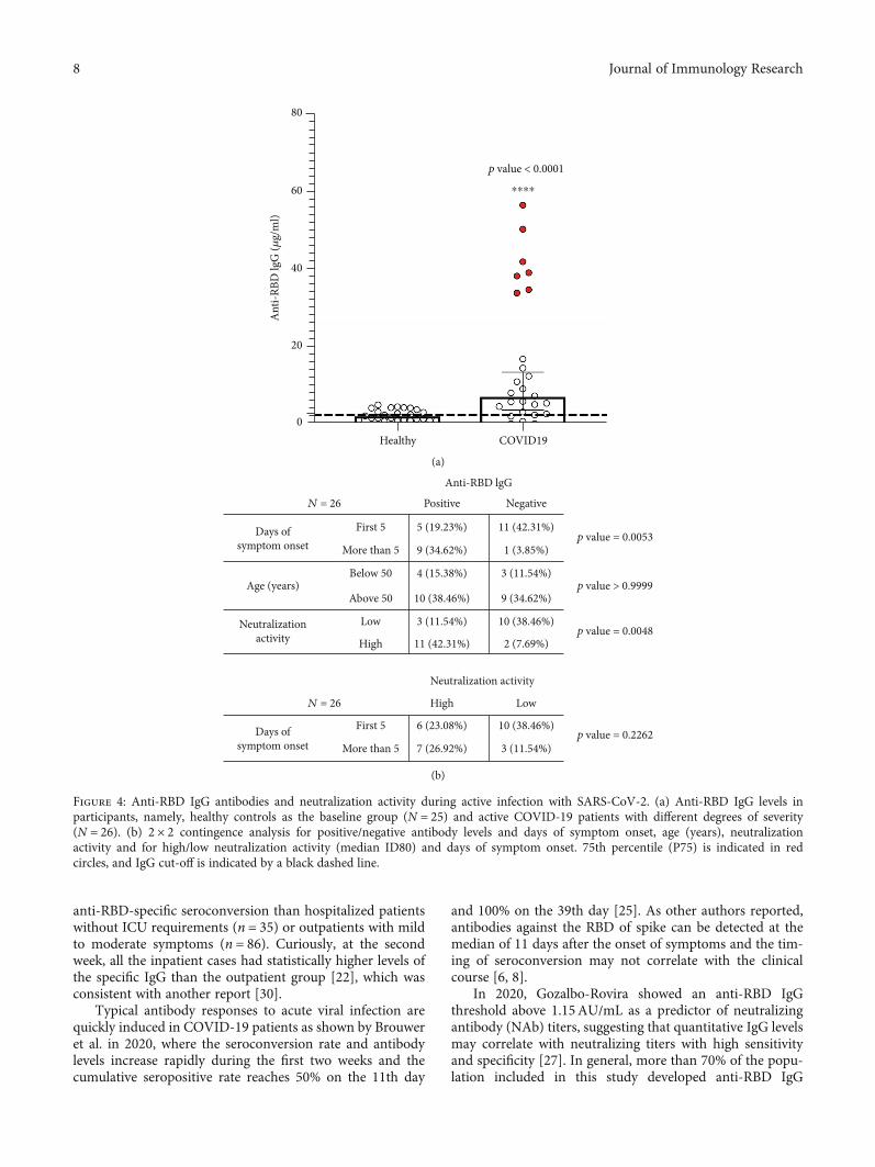
3.4. Early Seroconversion in Individuals with an ActiveInfection of SARS-CoV-2. We analyzed samples from 26patients who were categorized as severe COVID-19 caseswithin the first 14 days of their diagnosis of an infection withSARS-CoV-2 (Table 1). This group showed 53.8% positivity(14 of 26) with a mean concentration of 21.2μg/mL of anti-RBD IgG, and 7 out of the 26 patients (27%) had antibodiesabove the 75th percentile (20.9μg/mL) (Figure 4(a), redcircle). We did not observe correlations in IgG levels andprevious medication to the blood drawing (Table S2), andonly one case received hydroxychloroquine as treatment ofa systemic lupus erythematosus before the COVID-19diagnosis. A contingency analysis showed a correlation
between days of disease and seroconversion (p = 0:0053),increasing the frequency of positivity after 5 or more daysafter the symptoms started. However, 5 of 26 cases (19.2%)developed antibodies against RBD within the first 5 days(Figure 4(b)). No correlation between age at diagnosis andearly seroconversion was observed (p > 0:9999)(Figure 4(b)). As we expected, the presence of anti-RBDantibodies correlated with the ability to neutralize SARS-CoV-2 in vitro (p = 0:0048), but the magnitude of themeasured neutralizing activity in serum from patients witha severe COVID-19 was independent of the days aftersymptoms started (p = 0:2262) (Figure 4(b)).
4. Discussion
Early results from the Hubei province of China showed apolyclonal IgG prevalence against SARS-CoV-2 of 89.8%(95% CI 88.2-91.3%) in 1,740 COVID-19 convalescents[20], and the severity of the symptoms during COVID-19have been related to the duration of humoral response, whereasymptomatics or individuals with a mild illness have showna quicker reduction in antibody titers than more severe cases[21, 22]. In the current study, 50% of the convalescent popu-lation was considered within the range between 18 and 68years old with a median age of 35, which represents the mainworkforce in Chile. The estimated seroconversion was 72.9%,which did not show significant differences at the level of gen-der, age, and days after symptom offset; however, the serumconcentration of IgG anti-SARS-CoV-2 varied dramaticallybetween cases, and it was not reflected in the measured neu-tralizing activity. Our observations were consistent with thepreviously reported seroconversion rates after four weeks ofsymptoms [20, 22, 23]. It is interesting that the Chilean con-valescent population of this study showed similar resultscompared to a group of 49 recovered COVID-19 patientsrecruited in Wuhan, China, between February and March2020 [24]. In 2020, Li et al. reported that 28 days after theonset of symptoms, 90% of the group (18-55 years old)showed an S-RBD-specific IgG titer above 1 : 160 and 78%had a titer of 1 : 640 or higher [24], which means that 90%of that group seroconverted after infection, and the S-RBD-specific and N-specific IgG antibodies increased after 4 weeksfrom the onset of symptoms, with no significant correlationto age, sex, or ABO blood type [24]. It is important to con-sider that an IgG response against the spike RBD domainhas been associated with a patient’s improved survival inde-pendently of other factors such as sex or age, supportingthe concept that these antibodies are a significant contributorto the protective effect of humoral immunity in COVID-19[25]. Additionally, we observed a significantly higher amountof anti-RBD IgG in older men, and it was previously shownin a preprint article where the authors suggested that beingmale, an older adult, and being hospitalized with COVID-19 were each associated with having greater neutralizing anti-body titers and IgGs against the S1-domain, the S1-RBDfragment, or the full spike protein [26].
Several authors have reported a rapid seroconversionduring the first weeks of symptoms [23, 25, 27]; we observed53.8% of IgG seroconversion in severe COVID-19 patients
5Journal of Immunology Research

between 4 and 9 days after a positive PCR test. A very earlystudy reported for a small group (17) showed an anti-RBD-specific seroconversion rate of 64.71% for IgG within the 4-10 days after illness onset; however, after 15 days of disease,the seroconversion rate reached 100% (169 cases) for thethree immunoglobulins [28]. Another study which consid-ered 28 mild and 7 severe COVID-19 cases showed that
IgG in serum was detected in 1 of the 35 cases at the first weekafter the date of illness onset; however, at the third week,68.9% (n = 20/29) of the patients seroconverted in mild cases,while it was 100% (n = 7/7) in severe cases [29]. Recently, itwas reported from 166 studied cases that patients withinthe first week of symptoms with a very severe case ofCOVID-19 (n = 45) had a quicker and more significant
18-240
20
40
60
25-34 35-44Age (years)
45-54 55 plus
Ant
i-RBD
lgG
(𝜇g/
ml)
Female
(a)
18-240
20
40
60
25-34 35-44Age (years)
45-54 55 plus
Ant
i-RBD
lgG
(𝜇g/
ml)
p value = 0.0461
Male
⁎
(b)
Ant
i-RBD
lgG
(𝜇g/
ml)
60
40
20
021-35 36-49 50-63
Days after end of symptoms64-133
(c)
Ant
i-RBD
lgG
(𝜇g/
ml)
60
40
20
021-35 36-49 50-63
Days after end of symptoms64-133
(d)
Figure 2: Stratified anti-RBD IgG levels by gender, age, and days of convalescence. Measured IgG distribution for positive (blue) and negative(black) quantitative ELISA. Cases were segregated as female (N = 361) (a, c) and male (N = 278) (b, d) based on age and days after symptomoffset. In all cases, the dashed line corresponds to the control group.
6 Journal of Immunology Research

280
10
20
30
40
50
35 42Days after end of symptoms
49 56
Healthy
Ant
i-RBD
lgG
(𝜇g/
ml)
Patient 1Patient 2
Patient 3
Patient 4Patient 5
Patient 6
(a)
Patient 1Patient 2
Patient 3
Patient 4Patient 5
Patient 6
280
500
1000
1500
Neu
tral
izat
ion
titer
s
2000
5000
10000
15000
35 42Days after end of symptoms
49 56
(b)
15000
10000
5000
50
40
Ant
i-RBD
lgG
(𝜇g/
ml)
Neu
tral
izat
ion
titer
s
30
20
10
028 35 42 49
Days after end of symptoms
(c)
Anti-RBD lgG (𝜇g/ml)
Neu
tral
izat
ion
titer
s
Low0
1000
2000
3000
4000
5000p value < 0.0001
⁎⁎⁎⁎
High
(d)
Anti-RBD lgG
LowLowNeutralization
activity
High
High
37 (51.39%) 23 (31.94%)
0 (0%) 12 (16.67%)
37 35
60
12
72
Total
Total
p value < 0.0001
(e)
Figure 3: Convalescent plasma donors and IgG antiviral levels. Anti-RBD IgG concentration (a) and neutralizing activity (b) weredetermined at different days of convalescence. In (c), representative cases with a similar IgG concentration and decreasing neutralizingantibodies. (d) Neutralization activity and IgG levels; average neutralizing activity was represented for stratified high (N = 20) and low(N = 52) IgG anti-RBD cases based on the 75th percentile. The results are presented as the mean ± SEM, and statistical differences fromthe Mann–Whitney test are shown. (e) 2×2 contingence analysis for high/low antibody levels (P75) and high/low neutralizing activity(median ID80). 75th percentile (P75) and IgG cut-off are indicated in red and black dotted lines, respectively.
7Journal of Immunology Research
anti-RBD-specific seroconversion than hospitalized patientswithout ICU requirements (n = 35) or outpatients with mildto moderate symptoms (n = 86). Curiously, at the secondweek, all the inpatient cases had statistically higher levels ofthe specific IgG than the outpatient group [22], which wasconsistent with another report [30].
Typical antibody responses to acute viral infection arequickly induced in COVID-19 patients as shown by Brouweret al. in 2020, where the seroconversion rate and antibodylevels increase rapidly during the first two weeks and thecumulative seropositive rate reaches 50% on the 11th day
and 100% on the 39th day [25]. As other authors reported,antibodies against the RBD of spike can be detected at themedian of 11 days after the onset of symptoms and the tim-ing of seroconversion may not correlate with the clinicalcourse [6, 8].
In 2020, Gozalbo-Rovira showed an anti-RBD IgGthreshold above 1.15AU/mL as a predictor of neutralizingantibody (NAb) titers, suggesting that quantitative IgG levelsmay correlate with neutralizing titers with high sensitivityand specificity [27]. In general, more than 70% of the popu-lation included in this study developed anti-RBD IgG
Healthy0
20
40
60
80
p value < 0.0001⁎⁎⁎⁎
COVID19
Ant
i-RBD
lgG
(𝜇g/
ml)
(a)
Anti-RBD lgG
Neutralization activity
N = 26 High Low
Days ofsymptom onset
N = 26
Days ofsymptom onset
First 5
First 5
5 (19.23%)
6 (23.08%)
7 (26.92%)
10 (38.46%)
3 (11.54%)
9 (34.62%)
4 (15.38%)
10 (38.46%)
3 (11.54%)
11 (42.31%)
11 (42.31%)p value = 0.0053
p value > 0.9999
p value = 0.0048
p value = 0.2262
1 (3.85%)
3 (11.54%)
9 (34.62%)
10 (38.46%)
2 (7.69%)
More than 5
More than 5
Age (years)Below 50
Above 50
Neutralizationactivity
Low
High
Positive Negative
(b)
Figure 4: Anti-RBD IgG antibodies and neutralization activity during active infection with SARS-CoV-2. (a) Anti-RBD IgG levels inparticipants, namely, healthy controls as the baseline group (N = 25) and active COVID-19 patients with different degrees of severity(N = 26). (b) 2 × 2 contingence analysis for positive/negative antibody levels and days of symptom onset, age (years), neutralizationactivity and for high/low neutralization activity (median ID80) and days of symptom onset. 75th percentile (P75) is indicated in redcircles, and IgG cut-off is indicated by a black dashed line.
8 Journal of Immunology Research
antibodies, and 24.9% of the positive cases were above20.9μg/mL, suggesting a heterogeneous seroconversion buta sustained production of the specific IgG up to 100 days aftersymptom offset, independenly of age and gender. All conva-lescent participants showed neutralizing activity with a vari-ability up to 100 times between cases. However, high levels ofspecific IgG, even though above P75, did not guarantee a sig-nificant antiviral or neutralizing activity in plasma. Theseresults were concordant with the results shown by Wang in2020, where IgG levels (anti-S or anti-N) exhibited a moder-ate correlation with neutralization titers in plasma (Pearson’sr = 0:5393 and r = 0:6709, respectively) [32]. A theoreticalmodeling report suggested that the probability of detectingIgG could reach a maximum of around 25-27 days afterCOVID-19 symptoms end, with a predicted positivitybetween 98% and 100% of individuals for anti-SARS-CoV-2IgG [26]. The report also predicted that the probability ofdetecting NAb in plasma may rapidly rise to near 100%around 29 days after symptoms end without significant dif-ferences between mild/moderate and severe/clinical cases[33]. Previously, a trial using COVID-19 convalescentplasma was halted prematurely because 80% (53 of 66) ofsymptomatic participants for only 10 days at the time ofenrolment showed seroconversion [27]. In addition, theauthors reported SARS-CoV-2 neutralizing activity in 79%of the plasma receptors before transfusion (44 of 56) withmedian titers comparable to the donors (1 : 160 vs. 1 : 160, p= 0:40). Our results have confirmed that most of the Chileanpopulation which recovered from COVID-19 developedanti-SARS-CoV-2 antibodies even only a few days afterinfection as shown in patients with critical conditions. Thequantification of anti-RBD IgG within the local populationis necessary as an objective criterion to estimate the neutral-izing activity as a protective biomarker within the infectedpopulation with SARS-CoV-2. It may be also consideredwhen the laboratory measurement of neutralizing antibod-ies is limited. As a limitation of this study, the reducednumber of cases for some analysis do not allow us to con-sider our cohort as representative of a larger population.We did not include cases with active disease classified asmild or moderate COVID-19, so we cannot estimate if thereis or is not an association between symptomatology andearly seroconversion.
5. Conclusions
In general, more than 70% of the population included in thisstudy developed anti-RBD antibodies with a variable neutral-izing activity, suggesting a heterogeneous seroconversionafter recovering from COVID-19 with a sustained produc-tion of specific IgG up to 100 days, independent of age andgender.
Data Availability
The clinical data used to support the findings of this study areavailable from the corresponding authors upon request.
Ethical Approval
This study is part of the clinical trial NCT04384588 (clinical-trials.gov). This research was reviewed and approved by theInstitutional Scientific and Ethical Committee of InstitutoOncológico Fundación Arturo López Pérez (Santiago, Chile).This study complied with all the regulatory authorizationsfrom the Medical Direction together with all the legal andethical requirements of Chilean law.
Conflicts of Interest
The authors declare that they have no conflicts of interest.
Authors’ Contributions
Conceptualization was performed by RGS, FVE, and RG.Methodology was selected by RGS, FVE, KC, JLB, CB, AO,SR, FV-E, and RSR. Validation was performed by RGS,FVE, CS, CB, and RSR. Formal analysis was performed byRGS, FVE, CB, and RSR. Investigation was performed byRGS, KC, FVE, CB, AO, SR, and JLB. Resources wereacquired by RG, JLB, CS, CC, RM, FY, MM, AA, AO, SR,CB, JLB, and CS. Data curation was performed by CS, JLB,and RG. Writing the original draft was performed by RGSand FVE. Writing, reviewing, and editing were performedby RGS, FVE, RG, JLB, CS, CC, RM, FY, MM, AA, CB, JLB,CS, RSR, FV-E, CB, and HM. Visualization was performedby RGS and FVE. Supervision was performed by FVE, RG,CS, FV-E, and RSR. Project administration was performedby FVE, RG, CS, and CC. Funding acquisition was performedby FVE, RG, CC, and HM. All authors have read andapproved the final manuscript. R. González-Stegmaier andK. Cereceda have contributed equally to this work.
Acknowledgments
This study was supported by “Fondo de Adopción tecnoló-gica SIEmpre” sponsored by SOFOFA (Sociedad de FomentoFabril); CPC Chile (Confederación de la Producción y deComercio); Ministerio de Ciencia y Tecnología, Conoci-miento e Innovación, Chile; and Fundación Arturo LópezPérez (FALP), Santiago, Chile. Additionally, the ELISA testdesign was partially supported by Chile Conicyt FondecytPostdoctoral Grant 3170356 (RGS), and the neutralizingantibody assay design was supported by ANID Chile FON-DECYT grant Nos. 1190156 (RS-R) and 1180798 (FV-E),CONICYT grant Nos. 21190771 (AA-A) and 21160818(SR-B), and Proyecto de Internacionalización grant No.UCH-1566 (CB-P).
Supplementary Materials
Supplementary Figure 1: qualitative and quantitative ELISAperformance. In (a), a histogram is shown with the calculatedabsorbance ratio in a qualitative analysis (positive, ratioabove 1.1 as dotted line) and its corresponding anti-RBDIgG concentration range. Data are shown as mean± 95C.I. (b) Correlation between qualitative and quantitativeELISA analysis; average values for absorbance ratio and
9Journal of Immunology Research
concentration are represented as continuous variables. Sup-plementary Figure 2: high and low levels of anti-RBD IgG inrecovered COVID-19 patients. High (n = 116) and low(n = 350) levels were estimated as above or below the 75thpercentile for the global cohort (a) or separated by gender(b). In both cases, the dashed line corresponds to the groupof healthy individuals. Asterisks indicate significantdifferences between groups and analyzed with the Mann–Whitney test. ∗∗∗∗p < 0:0001. (Supplementary Materials)
References
[1] N. Zhu, D. Zhang, W. Wang et al., “A novel coronavirus frompatients with pneumonia in China, 2019,” The New EnglandJournal of Medicine, vol. 382, no. 8, pp. 727–733, 2020.
[2] C. Wang, P. W. Horby, F. G. Hayden, and G. F. Gao, “A novelcoronavirus outbreak of global health concern,” Lancet,vol. 395, no. 10223, pp. 470–473, 2020.
[3] A. L. Phelan, R. Katz, and L. O. Gostin, “The novel coronavirusoriginating in Wuhan, China,” Journal of the American Medi-cal Association, vol. 323, no. 8, pp. 709-710, 2020.
[4] J. S. Weitz, S. J. Beckett, A. R. Coenen et al., “Modeling shieldimmunity to reduce COVID-19 epidemic spread,” NatureMedicine, vol. 26, no. 6, pp. 849–854, 2020.
[5] R. Racine and G. M. Winslow, “IgM in microbial infections:taken for granted?,” Immunology Letters, vol. 125, no. 2,pp. 79–85, 2009.
[6] R. Wölfel, V. M. Corman, W. Guggemos et al., “Virologicalassessment of hospitalized patients with COVID-2019,”Nature, vol. 581, no. 7809, pp. 465–469, 2020.
[7] P. Zhou, X. L. Yang, X. G.Wang et al., “A pneumonia outbreakassociated with a new coronavirus of probable bat origin,”Nature, vol. 579, no. 7798, pp. 270–273, 2020.
[8] J. Zhao, Q. Yuan, H. Wang et al., “Antibody responses toSARS-CoV-2 in patients with novel coronavirus disease2019,” Clinical Infectious Diseases, vol. 71, no. 16, pp. 2027–2034, 2020.
[9] F. Xiang, X. Wang, X. He et al., “Antibody detection anddynamic characteristics in patients with coronavirus disease2019,” Clinical Infectious Diseases, vol. 71, no. 8, pp. 1930–1934, 2020.
[10] W. J. Guan, Z. Y. Ni, Y. Hu et al., “Clinical characteristics ofcoronavirus disease 2019 in China,” The New England Journalof Medicine, vol. 382, no. 18, pp. 1708–1720, 2020.
[11] T. Singhal, “A review of coronavirus disease-2019 (COVID-19),” Indian Journal of Pediatrics, vol. 87, no. 4, pp. 281–286,2020.
[12] Q. X. Long, B. Z. Liu, H. J. Deng et al., “Antibody responses toSARS-CoV-2 in patients with COVID-19,” Nature Medicine,vol. 26, no. 6, pp. 845–848, 2020.
[13] K. K.-W. To, O. T.-Y. Tsang, W.-S. Leung et al., “Temporalprofiles of viral load in posterior oropharyngeal saliva samplesand serum antibody responses during infection by SARS-CoV-2: an observational cohort study,” The Lancet Infectious Dis-eases, vol. 20, no. 5, pp. 565–574, 2020.
[14] F. Krammer and V. Simon, “Serology assays to manageCOVID-19,” Science, vol. 368, no. 6495, pp. 1060-1061, 2020.
[15] O. F. Norheim, “Protecting the population with immuneindividuals,” Nature Medicine, vol. 26, no. 6, pp. 823-824,2020.
[16] F. Amanat, D. Stadlbauer, S. Strohmeier et al., “A serologicalassay to detect SARS-CoV-2 seroconversion in humans,”Nature Medicine, vol. 26, no. 7, pp. 1033–1036, 2020.
[17] N. M. A. Okba, M. A. Müller, W. Li et al., “Severe acute respi-ratory syndrome coronavirus 2-specific antibody responses incoronavirus disease patients,” Emerging Infectious Diseases,vol. 26, no. 7, pp. 1478–1488, 2020.
[18] B. Meyer, C. Drosten, andM. A. Müller, “Serological assays foremerging coronaviruses: challenges and pitfalls,” VirusResearch, vol. 194, pp. 175–183, 2014.
[19] C. Beltrán-Pávez, S. Riquelme-Barrios, A. Oyarzún-Arrauet al., “Insights into neutralizing antibodies responses in indi-viduals exposed to SARS-CoV-2 in Chile. Accepted, ScienceAdvances abe6855,” 2020.
[20] L. Guo, L. Ren, S. Yang et al., “Profiling early humoral responseto diagnose novel coronavirus disease (COVID-19),” ClinicalInfectious Diseases, vol. 71, no. 15, pp. 778–785, 2020.
[21] D. F. Gudbjartsson, G. L. Norddahl, P. Melsted et al.,“Humoral immune response to SARS-CoV-2 in Iceland,”The New England Journal of Medicine, vol. 383, no. 18,pp. 1724–1734, 2020.
[22] K. Röltgen, A. E. Powell, O. F. Wirz et al., “Defining the fea-tures and duration of antibody responses to SARS-CoV-2infection associated with disease severity and outcome,” Sci-ence Immunology, vol. 5, no. 54, article eabe0240, 2020.
[23] M. Secchi, E. Bazzigaluppi, C. Brigatti et al., “COVID-19 sur-vival associates with the immunoglobulin response to theSARS-CoV-2 spike receptor binding domain,” The Journal ofClinical Investigation, vol. 130, no. 12, pp. 6366–6378, 2020.
[24] L. Li, X. Tong, H. Chen et al., “Characteristics and serologicalpatterns of COVID-19 convalescent plasma donors: optimaldonors and timing of donation,” Transfusion, vol. 60, no. 8,pp. 1765–1772, 2020.
[25] P. J. M. T. Brouwer, G. Caniels, K. van der Straten et al.,“Potent neutralizing antibodies from COVID-19 patientsdefine multiple targets of vulnerability,” Science, vol. 369,no. 6504, pp. 643–650, 2020.
[26] S. L. Klein, A. Pekosz, H. S. Park et al., “Sex, age, and hospital-ization drive antibody responses in a COVID-19 convalescentplasma donor population,” The Journal of Clinical Investiga-tion, vol. 130, no. 11, pp. 6141–6150, 2020.
[27] R. Gozalbo-Rovira, E. Gimenez, V. Latorre et al., “SARS-CoV-2 antibodies, serum inflammatory biomarkers and clin-ical severity of hospitalized COVID-19 patients,” Journal ofClinical Virology : the official publication of the Pan Ameri-can Society for Clinical Virology, vol. 131, article 104611,2020.
[28] H. Ma, W. Zeng, H. He et al., “Serum IgA, IgM, and IgGresponses in COVID-19,” Cellular & Molecular Immunology,vol. 17, no. 7, pp. 773–775, 2020.
[29] J. Sun, X. Tang, R. Bai et al., “The kinetics of viral load andantibodies to SARS-CoV-2,” Clinical Microbiology and Infec-tion : the official publication of the European Society of ClinicalMicrobiology and Infectious Diseases, vol. 26, no. 12,pp. 1690.e1–1690.e4, 2020.
[30] E. Marklund, S. Leach, H. Axelsson et al., “Serum-IgGresponses to SARS-CoV-2 after mild and severe COVID-19infection and analysis of IgG non-responders,” PLoS One,vol. 15, no. 10, article e0241104, 2020.
[31] A. Padoan, L. Sciacovelli, D. Basso et al., “IgA-Ab response tospike glycoprotein of SARS-CoV-2 in patients with COVID-19:
10 Journal of Immunology Research

a longitudinal study,” Clinica Chimica Acta, vol. 507, pp. 164–166, 2020.
[32] Y. Wang, L. Zhang, L. Sang et al., “Kinetics of viral load andantibody response in relation to COVID-19 severity,” TheJournal of Clinical Investigation, vol. 130, no. 10, pp. 5235–5244, 2020.
[33] B. Borremans, A. Gamble, K. C. Prager et al., “Quantifyingantibody kinetics and RNA detection during early-phaseSARS-CoV-2 infection by time since symptom onset,” eLife,vol. 9, article e60122, 2020.
[34] K. Cereceda, R. González-Stegmaier, J. L. Briones et al., Sero-logical Profile of Specific Antibodies against Dominant Antigensof SARS-CoV-2 in Chilean COVID-19 Patients, Unpublished,Submitted PlosOne, 2020.
11Journal of Immunology Research