seroprevalence of brucellosis among animal...
TRANSCRIPT
SEROPREVALENCE OF BRUCELLOSIS AMONG ANIMAL
HANDLERS AND ANALYSIS OF RISK FACTORS
DISSERTATION SUBMITTED FOR
M.D. (BRANCH IV)
MICROBIOLOGY
TIRUNELVELI MEDICAL COLLEGE
TIRUNELVELI
THE TAMILNADU DR. M.G.R. MEDICAL UNIVERSITY,
CHENNAI, TAMILNADU,
APRIL – 2013.

CERTIFICATE
This is to certify that the Dissertation “SEROPREVALENCE OF
BRUCELLOSIS AMONG ANIMAL HANDLERS AND ANALYSIS
OF RISK FACTORS” presented herein by Dr. S.NIRMALADEVI is
an original work done in the Department of Microbiology, Tirunelveli
Medical College Hospital, Tirunelveli for the award of Degree of M.D.
(Branch IV) Microbiology under my guidance and supervision during the
academic period of 2010 - 2013.
The DEAN
Tirunelveli Medical College,
Tirunelveli - 627011.

Department Of Microbiology,
Tirunelveli Medical College,
Tirunelveli-11
CERTIFICATE
This is to certify that the dissertation entitled,
‘‘SEROPREVALENCE OF BRUCELLOSIS AMONG ANIMAL
HANDLERS AND ANALYSIS OF RISK FACTORS” by
Dr.S.NIRMALADEVI, Post graduate in Microbiology (2010-2013), is a
bonafide research work carried out under our direct supervision and
guidance and is submitted to The Tamilnadu Dr. M.G.R. Medical
University, Chennai, for M.D. Degree Examination in Microbiology,
Branch IV, to be held in April 2013.
Dr.S.Poonkodi@ Lakshmi Dr. N. Palaniappan
Professor and Guide Professor and Head,
Department of Microbiology, Department of Microbiology,
Tirunelveli Medical College, Tirunelveli Medical College,
Tirunelveli-11. Tirunelveli-11.


'

DECLARATION
I solemnly declare that the dissertation titled
“SEROPREVALENCE OF BRUCELLOSIS AMONG ANIMAL
HANDLERS AND ANALYSIS OF RISK FACTORS” is done by me
at Tirunelveli Medical College Hospital, Tirunelveli.
The dissertation is submitted to The Tamilnadu Dr. M.G.R.Medical
University towards the partial fulfilment of requirements for the award of
M.D. Degree (Branch IV) in Microbiology.
Place: Tirunelveli Dr. S.Nirmala Devi,
Date: Postgraduate Student,
M.D Microbiology,
Department of Microbiology,
Tirunelveli Medical College,
Tirunelveli.

ACKNOWLEDGEMENT
I sincerely express my heartful gratitude to the Dean, Tirunelveli
Medical College, Tirunelveli for all the facilities provided for the study.
I take this opportunity to express my profound gratitude to
Dr.N.Palaniappan,M.D., Professor and Head, Department of
Microbiology, Tirunelveli Medical College, whose kindness, guidance
and constant encouragement enabled me to complete this study.
I am deeply indebted to Dr.S.Poongodi@Lakshmi,M.D., Professor,
Department of Microbiology, Tirunelveli Medical College, who helped
me to sharpen my critical perceptions by offering most helpful
suggestions and corrective comments.
I am very grateful to Dr.C.Revathy, M.D., Professor, Department of
Microbiology, Tirunelveli Medical College, for the constant support
rendered throughout the period of study and encouragement in every
stage of this work.
I wish to thank Dr. V.Ramesh babu, M.D., Professor, Department
of Microbiology, Tirunelveli Medical College, for his valuable guidance
for the study.
I am highly obliged to Dr.B.Cinthujah, M.D., Senior
Asssistant Professor, Dr. G.Velvizhi, M.D., Dr. G.Sucila Thangam,
M.D, Dr V.P Amudha M.D., Dr I.M Regitha M.D., Assistant
Professors, Department of Microbiology, Tirunelveli Medical College,

for their evincing keen interest, encouragement, and corrective comments
during the research period.
I wish to thank Dr.M.A.AshikaBegum,M.D., and
DR.T.Jeyamurugan ,M.D., Senior Assistant Professors, Department of
Microbiology, Tirunelveli Medical College for their help and
encouragement at the initial stage of study.
Special thanks are due to my colleagues Dr.G.Manjula,
Dr.T.Sucitha, Dr.A.Anupriya and Dr.Chitra for never hesitating to
lend a helping hand throughout the study.
I would also wish to thank my junior post-graduate colleagues,
Dr.S.Suganya, Dr.K.Girija, Dr.J.Senthilkumar, Dr.J.K.Jeyabharathi,
Dr.J.Jeyadeepana, Dr.V.G.Sridevi, Dr.R.Nagalakshmi,
Dr.C.Meenakshi, and Dr.A.Uma maheswari for their help and support.
Thanks are due to the, Messer V.Parthasarathy, V.Chandran,
S.Pannerselvam, S.Santhi, S.Venkateshwari. M.Mali, S.Arifal Beevi,
S.Abul Kalam, Kavitha, Vadakasi, Jeya, Sindhu, Manivannan,
K.Umayavel, Sreelakshmi and other supporting staffs for their services
rendered.
I thank my beloved sister, Dr.S.Chitra devi, M.V.S.c, Assistant
professor TANUVAS, for her valuable guidance and support for this
study.

I thank Dr.Shankara kumar, Assistant Director, Department of
Animal Husbandary, Tirunelveli district, for his immense support in
collecting samples.
I thank Mr. Arumugam, who helped me in the statistical analysis
of the data.
I am indebted to My parents and Brother not only for their moral
support but also for tolerating my dereliction of duty during the period of
study.
And of course, I thank the Almighty for His presence throughout
my work. Without the Grace of God nothing would have been possible.
ABBREVIATIONS
WHO - WORLD HEALTH ORGANIZATION
STAT - STANDARD TUBE AGGLUTINATION TEST
RBPT - ROSE BENGAL PLATE TEST
MAT - MICROAGGLUTINATION TEST
2-ME - MERCAPTOETHANOL AGGLUTINATION TEST
ELISA - ENZYME LINKED IMMUNO SORBENT ASSAY
PCR - POLYMERASECHAIN REACTION
PUO - PYREXIA OF UNKNOWN ORIGIN
LPS - LIPOPOLY SACCHARIDES
S-LPS - SMOOTH LIPOPOLYSACCHARIDE
CSF - CEREBRO SPINAL FLUID

CONTENTS
PAGE NO.
1.
2.
3.
4.
5.
6.
7.
8.
INTRODUCTION
AIM AND OBJECTIVE
REVIEW OF LITERATURE
MATERIALS AND METHODS
RESULTS
DISCUSSION
SUMMARY
CONCLUSION
BIBLIOGRAPHY
ANNEXURE-I
ANNEXURE-II
1
21
22
41
48
69
77
79

1
Seroprevalence of brucellosis among animal handlers and analysis of
risk factors
1.Introduction
Brucellosis is a re-emerging infectious disease, by Brucella spp
and usually transmitted to humans from infected animals. It is a most
important source of disease in humans and live stocks. The clinical
manifestations in humans differs from an acute febrile illness to a
chronic, low grade ill defined disease. In each year 5,00,000 new cases
are reported worldwide, but according to the WHO these numbers
greatly underestimate the true incidence, because the clinical picture of
human brucellosis is extremely variable and misdiagnosed by physicians.
However, the actual incidence seems to be twenty five times higher than
the reported incidence. Brucella is considered a biological weapon in the
category B pathogen, inhalation of only a few organisms is sufficient to
cause infection.
1.1 History
Since, the man started to domesticating the livestocks brucellosis
has emerged as a disease.1 The disease was characterized with fever
which has remissions and intermissions. It was known by several names
in relation with places where it was more common viz Malta,
Mediterranean, Gibraltar and undulant fever. 2

2
In 1895, J.A. Marston have given a correct description of the
disease and he named it as “Mediterranean” or “Gartnic remittent fever”.
The cause of the disease was obscure until 1886. Sir David Bruce a
British army Medical Officer in 1887 demonstrated a plenty of
microorganism from a spleen of military personals who had infected and
died of Gibraltar fever. Bruce reproduced the disease in chimpanzees
with the isolates, and named this organism as “ Micrococcus melitensis.”3
In the year 1897, Huges named the fever as undulant fever. Bang
isolated the bacilli from a aborted products of animals and named that
Bacillus abortus. 4 On June 14, 1905, Zammit isolated the bacilli from
infected goat’s urine and milk . Zammit also developed an agglutination
test to detect agglutinin in the milk. Further work rendered it evident that
goat was the natural host of Micrococcus melitensis and that infection
was communicated to man by consumption of raw milk.5
Alice C. Evans (1918) of Washington D.C., drew attention to the
similarity between Micrococcus melitensis and Bacillus abortus, the
causative agent of Malta fever and infectious miscarriages in cattles.6 In
1920, the generic name Brucella was proposed by Meyer and Shaw for
this microorganisms in honour of Bruce.7 Subsequently a 3rd species,
Brucella suis was identified as a cause of epizootic abortion of swine. 4 th
species, Brucella canis was identified as the cause of abortion in beagles.
Brucella ovis was identified as the causative agent of contagious
3
epididymitis of rams and Brucella neotomae was isolated from desert
wood rats. Out of these the most important species that infect human
beings are B. melitensis, B. abortus, B. suis and B. canis.2
1.2 Epidemiology
1.2.1 Global scenario
Brucellosis continues to be of great health significance and
economic importance in many countries. Countries like the Arabian Gulf,
the Indian subcontinent, Mediterranean basin and parts of Mexico as well
as central and South America are especially endemic for human
brucellosis. In endemic areas, the reported incidence of human brucellosis
varies from <0.01 to > 200 in one lakh population.1 The world burden of
human brucellosis remains to be > five lakh infections per year.8 In
1990, Egypt and many Arabian countries reported a incidence of > 90
thousand cases of human brucellosis per year.9 Every year, in USA 100 -
200 brucella cases are reported. The incidence and prevalence of
brucellosis varies from country to country. In endemic areas only few
cases are reported it reflects the poor surveillance and under reporting.10
1.2.2 Indian scenario
In India, brucellosis is a major emerging veterinary and civic health
concern. The existence of brucellosis was noted in the 20th century itself
from then it has been reported from almost all states of India but the
situation varies greatly between states.11 India is an agricultural country
4
and more than 3/4 th of the population resides in rural areas having close
contact with wild and domestic animals. Thus, the Indian population
stands at a significant risk of acquiring zoonotic diseases as well as
brucellosis. On the other hand, the reports regarding the distribution of
brucellosis are often incomplete. This may be due to the fact that poor
diagnostic services, decreased awareness and no exchange of data
among health and veterinary authorities.1
1.3 Agent
B.melitensis, B.abortus, B.canis and B.suis are the causative agent
of human brucellosis. The species level identification of the bacteria are
essential because the severity of disease in humans is mainly dependent
on the type and source of the infection. Human brucellosis commonly
caused by B.melitensis and B. abortus among this B.melitensis produce
severe disease. The other species in this genus also cause disease in
humans.12
1.4 Morphology
Brucella are small gram negative coccobacilli. They are shorter and
slender have straight axis. The length vary from 0.6 - 1.5μm and breadth
is 0.5 - 0.7μm. They are arranged singly, pairs, short chains or in small
groups. 2
5
Brucella are gram negative, weakly acid fast, non-motile and non-
sporing. Resists the decolourisation by acid and alkali and used in
staining of infected tissues. A modification of Ziehl Neelsen method may
be used, though it is not absolutely specific for Brucella.2
1.5 Genome
Brucella genome has 2 circular chromosomes of 2.1 Mb and 1.5
Mb except B.suis biovar 3, which contains one chromosome of 3.1 Mb.
The replicons code for important metabolic and replicative functions.13
Natural plasmids have not been detected in Brucella, although
transformation has been effected by wide host range plasmids after
conjugative transfer or electroporation.14
1.6 Antigenic determinants
The important antigens of Brucella includes the
lipopolysaccharide (LPS) complex and two related polysaccharides.
There are at least two antigenic determinants A (abortus) and M
(melitensis ) which have been identified as the O chain of the LPS
complexes. Strains of B. abortus, B. melitensis and B. suis can be A, M
or A and M antigen positive. S-LPS is the immune predominant and is
one of the important virulence marker. Plenty of periplasmic, outer or
inner, cytoplasmic proteins are identified.1

6
1.7 Cultural Characteristics
Brucella is one of the difficult organisms to cultivate. They are
aerobic bacteria and fail to grow in strict anaerobic conditions. Br.
abortus is capnophilic in nature. This should be incubated an atmosphere
of 5% to 10% CO2
at 35°C to 37°C and colonies usually appear within 4
to 5 days, but cultures should be kept for one month before they are
declared as negative. 2
Brucella species are usually isolated from blood or bone marrow
cultures during the acute illness. Eduardo Gotuzzo et al15
reported that
culture from bone marrow yields better results than blood cultures. The
prior use of antimicrobials will reduce the blood culture positivity but
will not affect the bone marrow culture. We can isolate the organisms
from samples like CSF, lymph, urine and liver of infected patients.
The biphasic culture method by Castaneda is recommended for the
culture of blood and other body fluids.16
Solid media recommended for
isolation of Brucella are trypticase soy agar, serum dextrose agar,
tryptose agar, Douglas agar and Brucella agar. The aqueous phase
consists of the same basal medium without agar. 2
Brucella strains grow on chocolate agar, blood agar and Brucella
agar or some infusion base agar. 5% heated horse or rabbit serum

7
enhances growth on all media. The optimum pH range is 6.6 to 7.4.
Temperature range is 20°C-40°C optimum being 37°C.2
On subculture to solid media the colonies appear after 4-5 days and
are 0.5 to 1 mm in diameter, raised convex, circular, moist, translucent
and easily emulsifiable. On further incubation they increase in size to 4-6
mm 2 .
In synthetic media, Pantothenate and erythritol improves the
growth of brucella and tryptophan and cystine are highly toxic to Br.
abortus17
.
1.8 Biochemical Reaction
Brucella produces catalase and many species of Brucella are
oxidase positive except Br. ovis and B. neotomae. Many species reduces
nitrate and it is variable in B.ovis and B. suis biogroup. Almost all
species, except B. ovis and some strains of B.melitensis and B. abortus
hydrolyze urea rapidly. B. ovis may hydrolyze urea weakly or not at
all.2
B. abortus produces moderate amount of H2S for 4-8 days except
the Danish variety, which does not produce H2S. The American strains of
B. suis and B. neotomae also produce H2S, while B. melitensis, B. canis
and B. ovis do not produce H2S. Brucella are negative for MR, VP and do
not produce Indole.2

8
The commonly used method to identify Brucella from humans is
the dye inhibition test. Dilutions of basic fuchsin and thionin are put up.
B. abortus should grow only on basic fuchsin, B. suis only on thionin and
B. melitensis on both2.
The CO2 requirement of freshly isolated B.abortus for growth helps
in differentiating them. The use of monospecific sera against the species
helps in differentiating them from each other.
1.9 Susceptibility to Physical and Chemical Agents
Brucella are destroyed by heat at 60°C in 10 minutes and by 1%
phenol in 15 minutes. They are killed by pasteurization. They remain
viable for 10 days in refrigerated milk, 30 days in ice cream, 120 days in
butter and for varying period in cheese depending on its pH. They are
sensitive to direct sunlight and acid and tend to die in butter milk.
Brucella will survive in bovine feaces for four months, in uterine
exudate for at least 200 days and in liquid manuer for upto 2 ½ years if
the temperature is kept near 0°C. Formaldehyde is the most effective of
the commonly available disinfectants. 2
1.10 Susceptibility to antimicrobial agents
Most Brucella strains are sensitive to gentamicin, kanamycin
tobramycin, amikacin and streptomycin. In vitro they are insensitive to

9
β-lactam antibiotics except monolactum and clavalunate potentiated
amoxicillin. Sensitivity to tetracyclines is almost universal. 2
1.11 Pathogenesis
Brucella enters into the human tissue by various routes like
abrasions in the skin, alimentary canal, respiratory route.18 At or near the
site of entry the bacteria are likely to be engulfed by either mononuclear
or polymorpho nuclear cells. After their entry into the macrophages, they
are transported into various organs of the body.
Within the phagocytic cells, these organism resists the phagosome,
lysosome fusion and undergo replication19. As a result of this, the
infected cells are destroyed by the pathogen if the process is not checked
by the immune system.
1.12 Host immune response
Infection with Brucella usually results in the induction of both
humoral and T-cell mediated immune response.
1.13 High risk groups
Veterinarians, abattoir worker, cattle ranchers, dairy farmers, meat
sellers, meat inspectors, shepherds, goatherds, hunters, lab workers,
travellers and persons consuming raw dairy products are particularly at
risk.

10
1.14 Transmission
The mode of transmission and prevalence of brucellosis depend on
many factors like dietary practice, customs in the society, socioeconomic
status, climate, animal husbandry practices and environmental sanitation.
The species of Brucella that are infective to humans and livestock
reservoir includes, B. melitensis in goat and sheep, B. abortus in cattle,
B. suis in pigs, and B. canis in dogs. The major source for transmission of
disease in general population is the consumption of unpasteurized milk
and its products which contains high amount of organism1.
Though animal muscles contain low bacterial load, eating of
improperly cooked meat can leads to brucellosis. Handling of live
cultures and travel to endemic area also increases the risk.1
Recently other routes of transmission have been identified like,
infection through breast milk,20 sexual transmission,21 blood transfusion,
bone marrow transplantation, infection contracted by an obstetrician
during the delivery of a transplacentally infected baby21 and accidental
inoculation with animal vaccines B. abortus strain S19 and B. melitensis
Rev-1.
1.15 Clinical spectrum
Human brucellosis is a systemic infection, presents with wide
spectrum of clinical manifestations. It usually presents as a fever without
any apparent focus of infection. But in few cases focal forms are seen

11
which might affects many organs and systems of the body with the
skeletal forms being more common, cardiac and neurological forms being
more severe. The case fatality rate may be up to 2% and usually results
from endocarditis. 2
The onset of the disease may be either acute or insidious.
The infective dose is low. It has a incubation period of 10-21 days to
several months. Acute brucellosis mostly presents as a undulant pattern of
fever with profuse sweating, body pain, loss of appetite and weight loss
and fatigue. Liver, spleen and lymphnodes become enlarged.22
1.16 Complications
Osteoarticular, alimentary canal and hematologic forms are the
common complications but the cardiac, neurologic forms are more
severe but infrequent.45
1.16.1 Skeletal complications
Although monoarticular septic arthritis occur, 30 to 40% of
patients have reactive symmetrical polyarthritis involving the knee, hip,
shoulder, sacroiliac and sternoclavicular joints. Cultures of synovial fluid
are positive in about 50% of cases.24
Brucella osteomyelitis is rare.25
Bursitis and tendinitis due to Brucella have also been reported but is very
rare.

12
1.16.2 Gastrointestinal system
Gastrointestinal manifestations of Brucella infection are generally
mild and may include nausea, vomiting, constipation, acute abdominal
pain and diarrhea. Hepatic and spleenic enlargement may be seen in 15-
20 % of cases and abscesses may develop in the liver and spleen. Mild
jaundice is seen in few cases.25
1.16.3 Haematologic complications
The heamatologic manifestations of brucellosis include anemia.
leucopenia, thrombocytopenia and clotting disorders.26 Granulomas are
found in the bone marrow in up to 75% of cases, but they are small and
indistinct.
1.16.4 Cardiovascular system
Cardiovascular complication of Brucellosis include endocarditis,
myocarditis, pericarditis, aortic root abscess, mycotic aneurysms,
thrombophlebitis with pulmonary aneurysms and pulmonary embolism.
Brucella endocarditis usually involve the valves. It is reported to be the
most common cause in fatal cases of human brucellosis.25
1.16.5 Central nervous system
Neurobrucellosis is uncommon but serious and includes meningo
encephalitis, multiple cerebral or cerebellar abscesses, ruptured mycotic
13
aneurysms, myelitis, GB syndrome, cranial nerve lesion, hemiplegia,
sciatica, myositis and rhabdomyolysis.25
1.16.6 Genito urinary system
The genitourinary infections due to Brucella include epididymo-
orchitis, prostatitis, seminal vesiculitis, dysmenorrhoea, tubo-ovarian
abscess, salpingitis, cervicitis and acute pyelonephritis. Brucella has been
isolated from urine in up to 50% of cases of genitourinary Brucella
infection.25
1.16.7 Ocular Complication
A variety of ocular complications have been reported in patients
with brucellosis. Uveitis is generally a late manifestation consisting of
chronic iridocyclitis, nummular keratitis, multifocal choroidits and optic
neuritis25.
1.16.8 Skin Manifestation
Skin manifestation of Brucella are uncommon. They include
maculopapular eruption, purpura and petechiae, multiple cutaneous and
subcutaneous abscess, discharging sinuses, superficial trombophlebitis,
erythema nodosum and pemphigus.25

14
1.16.8 Respiratory System
Respiratory illness due to Brucella include sore throat, tonsillitis,
dry cough, hilar and paratracheal lymphadenopathy, pneumonia, solitary
or multiple pulmonary nodular lung abscesses and empyema.25
1.17 Chronic Brucellosis
Since the onset of symptoms of brucellosis is insidious, not easy
to differentiate between acute and chronic form of the disease. Most
patients with chronic brucellosis have persisting foci of infection, such as
suppurative lesions in bone, liver or spleen. Some patients who have had
brucellosis will continue to have symptoms like malaise, lassitude and
depression in the absence of objective evidence of infection.27
1.18 Laboratory diagnosis
The clinical presentation of brucellosis mimics some infectious
and noninfectious conditions. So, the diagnosis of the disease is very
difficult and frequently delayed or missed. Early diagnosis of the disease
and inclusion of proper antimicrobial treatment is vital for patients,
especially to prevent the development of complications and appearance of
relapses.

15
1.18.1 Culture
Definitive diagnosis of brucellosis is made when Brucella are
recovered from blood, bone marrow, lymph nodes, cerebrospinal fluid,
urine, pus from abscesses and rarely from sputum, breast milk, vaginal
discharges and seminal fluids. Failure to isolate the bacteria is a
drawback of blood culture due to the fact for isolation, the bacteria needs
extended incubation period, special media, and frequent subcultures are
due to the fastidious, slow growing nature of the bacteria. Though bone
marrow cultures are said to be the gold standard one, but results have not
been generally reproducible. In acute stage of the disease blood culture
has increased sensitivity. Inspite of this, this test is not done routinely in
rural areas, where the disease is endemic.28
1.18.2 Serology
The limitations of a blood culture makes serology the most useful
way to diagnose. Detection of antibodies directed against the S-LPS,
internal cytosolic proteins are the important antigens which are used in
the serological diagnosis of brucellosis. IgM agglutinins start to appear in
the blood after 7 days of disease followed by IgG. This IgG agglutinins
will persist for longer periods even after the disease has been cured.29
Many serological tests have been used for the diagnosis of human
brucellosis.The test that are presently available are standard tube
agglutination test (STAT), Antihuman globulin test, Rose bengal plate

16
test (RBPT), complement fixation, Enzyme linked immunosorbent assay
(ELISA) and Immuno fluorescent antibody assay. STAT, Coombs test,
ELISA are the widely used method for the detection of Brucella
agglutinins.29
1.18.2.1 Antigen detection
Antigen detection which is the suitable alternative to blood culture
but this test have not been standardized yet.30
1.18.2.2 Antibody detection
a. Rose Bengal Plate Test (RBPT)
This is a simple and rapid plate agglutination test and has a higher
degree of sensitivity for the diagnosis brucellosis irrespective of the stage
of disease. This high sensitivity, rapid and easy to perform makes the test
ideal for screening the patients for human brucellosis.1
b. Standard tube agglutination test (STAT)
STAT is the accepted test for obtaining quantitative information
about the immune response against specific brucellar antigens. This test
estimates the quantity of agglutinating antibodies (IgG and IgM). When
suitable clinical manifestations are present a presumptive diagnosis of
brucellosis is made serologically as a titre of 1in 160 and above. Where in
endemic areas, a titre of 1in 320 dilution makes this test more specific
one.1
17
Even though this test being the more standard one, it is time-
consuming hence, unsuitable as a primary test for laboratories with large
specimen workloads.31 In some sera a blocking factor may interfere with
agglutination at low serum dilutions (Prozone phenomenon) this may be
due to the presence of IgA or other non-agglutinating antibody. Another
drawback is, the diagnosis of brucellosis cannot be established on the
antibody titer by these classic test alone, because healthy persons engaged
with animal husbandry practices may show significant titers
of Brucella agglutinins.32 The other disadvantage is cross reactions
between Brucella and other bacteria such as Vibrio cholerae,
Pseudomonas maltophilia, Francisella tularensis, Yersinia enterocolitica
and Escherichia coli O: 157. This reactions can result in false positives
in the serologic tests for brucellosis. 2
c. Mercaptoethanol (ME) agglutination test
This test determines the nature of immunoglobulins responsible for
the agglutination in the STAT. In STAT, agglutination may be due to
IgG, IgM or both. 2-mercaptoethanol destroys the agglutinating activity
of IgM and therefore agglutination in this test is indicative of IgG. This
2-ME dissolves the disulphide links of IgM pentamer, thus interfers with
its agglutinating capacity while not affecting IgG antibodies.33

18
d. Microagglutination test (MAT)
Microagglutination test is a simpler and more efficient test than
STAT. It can be performed more rapidly and employs less serum and
antigen. It has been found to be more sensitive and specific than STAT.
Since the MAT is simpler to perform than STAT and can potentially be
automated. 34
e. Anti human globulin test (Coombs test)
This test detects the incomplete antibodies. Nowadays, the Coombs
test is rarely performed in routine clinical laboratories because the
procedure is too complex, time consuming and labor intensive, requires
skilled persons to perfom the test. 1
f. Complement Fixation Test:
This test is mainly used in the diagnosis of chronic brucellosis. 3 It
detects the complement fixing antibodies. It is the IgG antibody in
brucellosis, which readily fixes complement. IgG appears late in the
disease and hence this test is more useful in diagnosing chronic
brucellosis. 33
g. Enzyme Linked ImmunoSorbent Assay (ELISA)
ELISA which is a more sensitive and specific test with increased
performance than other conventional tests. It gives a profile of Brucella
specific IgA, IgM and IgG agglutinins in case of acute and chronic
brucellosis.35
19
ELISA gives high sensitivity and specificity when compared to
STAT. 31and it is used for the diagnosis of chronic and complicated cases.
In endemic areas this test has significant diagnostic advantage than other
conventional methods.35 ELISA can be used to study the subclass of Ig,
so that the role of each Ig in the different phases and evolutional forms of
brucellosis can be ascertained.29 It detects the antibodies against many
bacterial antigenic structure like S-LPS or cytoplasmic protein
antigens.36 Applying a combination of IgM and IgG ELISA testing could
be of value for the definitive diagnosis of brucellosis in developing
countries, where diagnostic capabilities for culture, including automated
culture systems and Polymerase chain reaction are poor.31
h. Immunocapture agglutination test
The Immunocapture agglutination test has high sensitivity and
specificity that detects the antibodies against especially for S-LPS. 37
1.18.3 Genomic detection
Molecular diagnosis by polymerase chain reaction (PCR) has been
used for confirmation and differentiation of Brucella. This methods are
very useful for follow up testing of unusual phenotypes. PCR is a very
expensive test and not used routinely as a diagnostic method.1
1.18.4 Newer rapid tests
The latex agglutination test and lateral flow immunoassay are the
newer tests available for the detection of Brucella IgG and IgM. It is a

20
rapid and simple test with increased specificity and sensitivity and is a
ideal test can be used in remote area.38
In this back ground, the present study was conducted to assess the
seroprevalence of brucellosis among animal handlers and to analyse the
risk factors associated with this infection in Tirunelveli district of
Tamil nadu.

21
2. AIMS AND OBJECTIVES
1. To detect the seroprevalence of brucellosis among animal handlers in
Tirunelveli district of Tamil nadu.
2. To analyse the risk factors associated with the seropositivity of
Brucella infection.
22
3. REVIEW OF LITERATURE
Brucellosis is one of the world’s major zoonotic disease. It has a
variable trend in United states and the European countries but has a
predominant presence in Asia and in developing countries. It is a
unrestrained public health crisis in developing countries like India, here it
is a common but often a ignored disease and it is also a disease of
considerable economic and social importance. The prevalence of human
brucellosis is difficult to estimate since many times the disease has been
misdiagnosed or undiagnosed because of their inapparent or protean
manifestations. Nowadays a wide battery of serological tests are available
for the diagnosis of human brucellosis.
3.1 Prevalence
3.1.1 Global prevalence
Brucellosis is an important global problem. It is a reemerging
zoonotic disease cause severe economic loss and infection to humans.
Most of the developing countries the burden of the disease is increased by
the absence of national surveillance programme and inadequate
laboratory facilities. Some countries, conventionally considered to be
endemic-e.g: Israel, France and most of Latin America but now
controlled the brucellosis.1 But, in central Asia the new foci of infection
been emerged. The circumstances in Syria has been fast declining.

23
In Arabian countries, the prevalence of human brucellosis is higher as a
result of increased live stock production units.
In 1999, Mohammed A Al Sekait 39 done a seroepidemiological
survey in Saudi Arabia and the results revealed that the seroprevalence
was 15.0%. Seropositivity of brucellosis from various regions of
Mediterranean basin varied from 8% in Jordan 40 to 12% in Lebanon and
Kuwait.41,42 Even higher seroprevalence rates have been reported in Sub-
Saharan countries, with 18% in Uganda 43 and 13% in Nigeria.
A Seroepidemiological study conducted in Kars district of Turkey
for period of 2 years from Jan- 2004 to Dec-2006 revealed that the
seroprevalence was 17.88%. 44 In a study conducted by Apan et al 2007 45
in Middle Anatolia, Turkey, the seroprevalence of brucellosis was
determined as 3.2%.
Kose et al in 2006 46 reported the seroprevalence of 2.9 to 8.5%
in rural and suburban communities in West Anatolia, Turkey. Another
study performed in Middle Anatolia by Cetinkaya et al 2005 47 revealed
that the seropositivity was 4.8%.
A Serological survey conducted by Cadmus S.I.B et al 2004 48
showed that the overall seroprevalence of brucellosis in Ibadan ,
Southwestern Nigeria was 31.82%.
Hajia et al 49 had done a study to estimate the antibody levels of
Brucellosis and showed the prevalence rate to be 3.28% in Hamedan,
24
Western Iran. Bokaei et al 50 studied the prevalence rate of brucellosis in
Birjand, Iran. It was 37 in one lakh population
A study conducted on the seroprevalence of brucellosis in 184
suspected cases 51 for period of 2004 to 2009 showed that 5.4% were
seropositive for Brucella agglutinins in their serum.
A study done in Pyrexia of Unknown Origin patients of Makurdi
in 2004 52 revealed that among a total of 1040 serum samples screened,
the overall seroprevalence was 7.6%.
3.1.2 India
India is an agricultural country and majority of people in our
country are engaged in agriculture related activities like seasonal
agriculture labours, dairy product selling and animal meat selling. Several
studies and publications from India revealed that human brucellosis is a
common disease. Studies conducted from various centers of India have
reported seroprevalence ranging from 0.8 to 6.8% in patients with PUO.53
A prospective study conducted in North India by Handa R et al 54
observed that 3.3% patients with PUO had acute brucellosis while 6.6%
had serological evidence of previous Brucella infection. 14% of the
asymptomatic, 'at risk' individuals screened were seropositive for
Brucella infection.
Kadri SM et al 55 in 2000 observed that out of a 3,532 hospitalised
patients for PUO 28 (0.8%) were found seropositive for brucellosis .

25
A study done by Appanavar SB et al 56 in North India revealed
that the seroprevalence of brucellosis in PUO cases were 9.94% of
which 45% had acute infection and the remaining 55% had chronic
infection.
Moti Yohannes et al 57 in 2011 had done a seroepidemiological
survey of human brucellosis in and around Luthiana among 241 high
risk persons with and without pyrexia of unknown origin and the
seroprevalence documented was 26.6%. In Orissa it was 6.8% 58 and in
Andhra Pradesh it was 11.51%.
A study conducted by Vaishnavi C et a l 59 reported that out of
292 serum samples of blood donors, 16.8% had positive Brucella
antibodies of this 0.36% had high titres of antibodies.
A Seroprevalence study conducted in Davangere, Karnataka60
reported that the overall seroprevalence of brucellosis was 3.3%. It was
2.4% in general population and 11.1% in veterinary staff. In study from
Kerala in 2005 61 reported that the overall seroprevalence of brucellosis
was 1.6%. Among the general population the prevalence rate was 2.45%
and in veterinary students it was 1.14%.
In Gujarat62 8.5% prevalence of 8.5%, in Belgaum 63
seroprevalence of 8.5% in publics and 5.8 to 14.3% in veterinary persons.
In another study in 2006, reported a prevalence rate of 1.8% in Bijapur.53

26
Panjarathinam et al 64 reported the seropositivity of 6.5% in
aborted women. The high incidence of spontaneous abortion may be due
to the fact that this organism infects the chorio amniotic tissue
Persistence of infection in animal reservoir, low physician
awareness, poor availability of diagnostic facilities, and the non existence
of regional data bases contribute towards the perpetuation of this zoonosis
in India, while it has been eradicated from most developed countries.
3.2 Age
Brucellosis affects all age groups.
In a study, Fatima et al 65 observed that those who were in the age
of 51 to 60 yrs had increasd seropositivity followed by of 41 to 50 yrs. As
brucellosis is an occupational disease, individuals in this age were at a
greater risk because of prolonged years of exposure.
In a study by Ramos TRR in 2006 66 reported that there was a
significant association between age group and seropositivity for this
disease, with individuals above 40 years of age more predisposed to being
infected, which is similar to the findings of Bigler et al 67 and Feliciano
et al reported that there is increased frequency of seropositivity with
advancing age, probably due to a longer period of exposure to the
organism.
A study from Karnataka, in 2007 68 noted that the highest
prevalence of 45.36% was found in 41to50 yrs of age followed by

27
32.98% in the age of 31 to 40 yrs. 14.43% were in the age of 51 to 60 yrs.
7.21% cases in the age of 21 to 30 yrs. This study reported that a wide
variation in the age group of seropositives.
Jama’ayah MZ et al in 201151 found in their study that persons
were in the age of 20 to 45 yrs had higher seroprevalence. 70% were in
the age of > 40 yrs and 2% were < 20 yrs of age.
Mahmoud N Abo-Shehada et al in 1996 69observed in their study
that seroprevalence of brucellosis increased with age. Among 26.3 % of
seropositive veterinary surgeons the highest prevalence was noted in
34-43 years of age .
Metri Basavaraj C et al in 201160 reported that in veterinary
surgeons, brucellosis was more common among in the age group of 31-
40 years followed by persons more than 41 years. Randhawa et al 70 have
reported that individuals in the age of thirty (14.2%) and forty years
(7.0%) had the highest prevalence among high risk persons. More
prevalence in this age group may be due to increased activities with
regards to their occupation, thereby increasing the risk of acquiring the
infection.
Ali et al 71 reported that those were in the age of 25 to 35 yrs had
higher soprevalence. Kadri et al 55 also observed that persons of 21 to 30
yrs had the higher (43%) seropositivity.

28
A seroepidemiological study done in 1999 39 revealed that
seropositivity of brucellosis increases with age advances. The lower
seroprevalence rates found in paediatrics may be due to the fact that
adults had the much exposure to animals. A study by Abu-Shehada et al69
and Araj GF et al 41 reported an increase in frequency of seropositivity
when the age advances.
Ofukwu et al in 2004 52 in their study reported that higher
seroprevalence (38%) were in the age of 21 to 40 yrs and 32.9% in the
age of 41to 60 yrs. 13.9% and 15.2% were in the age group of 0- 20
years and in the age group of 60 and above years. There was no
significant association in the age specific prevalence rates.
3.3 Sex
Brucella affects both males and females equally. There is no sex
wise discrimination between the two sexes provided they are equally
exposed to the potential risk factors. But most of the studies reported that
males are most commonly affected than females which is perhaps
justified by the greater presence of men in the veterinary profession,
slaughterhouses and dairy farms.
A study from Malasyia 51 showed that seropositivity was common
in (90%) males than (10%) females.
Moti Yohannes et al 57 observed in their study that all seropositive
cases were men. Various other studies conducted by other researchers

29
also found that males were more commonly affected than females. This is
due to the fact that males are employed in field of animal husbandry or
are exposed to infected animals and their secretions.
Kapoor et al in 198 72 and Hussein et al in 2005 73 reported higher
seropositivity among females.
A study from Nigeria,52 reported that out of 79 Brucella positive
cases, 45.6% were male and 54.4% were females. This results were in
concordance with the findings of Falade and Junaidu et al. 74
Aseroepidemiological survey of brucellosis among animal handlers
38 revealed that seropositivity and gender was not significantly
associated.75 A study by Al seikait 39 also reported that there was no
significant difference between the sexes.
A study by Ramos TRR in 2005 66 found that among the 26
serology positive individuals 3 (2.0%) were females and 23 (4.7%) were
males. There was no significant association between sex and
seropositivity in the individuals studied.
3.4 Education
Low literacy is one of the risk factor for brucellosis because they
are not aware of the zoonotic diseases and they don’t follow any hygienic
practices after handling the animals and their products. Karimi et al76
reported that significant association between low literacy and
seropositivity.

30
Sumer et al 77 and Fatima et al 65 found that no association
between the literacy and seropositivity.
3.5 Residential Background
A study conducted in Lahore 65 reported that out of the 78
seropositive individuals , 22.6% were from urban localities and 20.3%
from rural areas. The residential background was not significantly
associated with seropositivity. Baba’s et al 78 also reported the similar
findings.
Nabi et al 79 reported that 84.2% of seropositives reside in rural
area and 15.8% from urban areas. The increased prevalence in rural
people may be due to the fact that they are involved in cattle rearing and
handling.68
In a study from different regions of Saudi Arabia,39 the
seropositivity was higher ( 26.2%) in those from rural areas compared
with (9.5%) in urban areas. This may be due to the fact that ingestion of
raw milk and their products from infected animals as well as handling of
aborted fetuses, placenta and uterus and vaginal secretions may be
considered sources of infection for the high positivity of brucellosis
among the rural population.
31
3.6 SocioEconomic status
Al sekait 39 noted the highest (24.4%) seroprevalence among the
persons of low socioeconomic status when compared to the persons
(6.3%) of high socioeconomic status.
3.7 Occupation
Meky et al reported that workers in occupations dealing with
animals had a 2.4-times higher risk of acquiring the infection than those
in occupation not dealing with animals.
Brucella are detected in the abortive products and carcasses of
infected animals and their secretions thus brucellosis becomes an
occupational disease for veterinary persons, slaughterhouse workers and
farmers.66
Brucellosis is an occupational hazard to veterinary professionals.
They are exposed to all risks except their awareness about the zoonotic
disease. These persons usually acquire the disease by cuts in the skin and
conjunctival splashes or through inadvertent injection of animal vaccine
and during the removal of infected uterine products.1
A study conducted in high risk group individuals observed that the
seroprevalence of brucellosis was 41.2% in Live stock inspectors, 30.9%
in veterinary assistants, 12.37% in veterinary doctors, 6.18% in
supervisors and in sweepers, 2.06%, 1.03% in shepherd and butchers
respectively.68

32
In study by Thakur 80 reported the overall seroprevalence of 4.97%
among animal handlers of which veterinary surgeons (17.39%) had the
higher seroprevalence.
Mrunalini et al 81 reported seroprevalence of 25.24% in
veterinarians, 23.3% in para-veterinarians, 12.62% in farmers, 11.65% in
shepherds and 6.8% in other occupational groups.
A study conducted in the year of 2003 82 among animal handlers
found that out of 225, 5.33% were seropositive. Among them 14.63% of
veterinary doctors and 4.51 % of dairy farms workers were
seropositive.
A seroprevalence study on persons of high risk occupation
revealed that the veterinarians (50%) had the highest percentages of
seroprevalence followed by farmers who had a seroprevalence of 23.9 %
and butchers had 9.2% prevalence rate.41 In Saudi Arabia, 7.14% of
veterinary surgeons and 2 .67 % of butchers were positive for Brucella
agglutinins.
Dairy workers are exposed continuously to the pathogenic agent
while milking the animals because their hands get contaminated.82
Eritrea, Omer et al reported 83 that the highest prevalence among
dairy farm workers 7.1% and owners, followed by 4.5% in veterinary
staffs. Soman and Kothari reported that 4% of dairy workers were

33
positive for Brucella antibodies. Sohaila et al reported the seroprevalence
of 6.1% among dairy workers.
Meat handlers, abattoires are in direct contact with carcasses of
infected animals and raw meat are at greater risk. They are infected
through abrasions and cuts in mucous membrane and splashes in
conjunctival mucosa.
Cadmus S.I.B et al 2006 48 found that the overall seroprevalence of
brucellosis was 31.82%. Of this 63.63% were butchers. Butchers are
frequently infected might be due to the fact that they do not use PPE
and exposed to infective material like blood, vaginal secretion, retained
uterine products and urine from infected animals.
A study by Kumar et al among abattoir workers 84 in which, blood
collectors had the highest (99.77%) seropositivity followed by, 68.96%
among animal handlers, 68.00% in butchers, 57.14% in sweepers and
28.57% were among the veterinary surgeons.
A study conducted in 2004 52 revealed that the seroprevalence of
43.8% in abattoir workers/ butchers. This was similar to the reports by
Falade 1974, Ocholi 1993, Edu, 2005 the prevalence was 28 to 57%
among abattoire workers .
A study on seroepidemiological survey of brucellosis antibodies
from various occupational groups in Saudi Arabia 39 stated that the

34
seroprevalence of was higher in occupationally exposed group such as
meat sellers, veterinary professional and farmers.
In Pakistan, Masoumi et al85 carried a study among abbatoire
workers and found the seroprevalence to be 8.33%. In Algeria, the
seroprevalence was found to be 37.6% among high risk persons.
Shepherds also at the risk of acquiring the disease due to the
widespread infection of Brucella melitensis in sheep.
Sonmez et a observed the seroprevalence of brucellosis to be 6.2%
among farmers in East Anatolia of Turkey. MH Salari et al in 1997 75
reported a seroprevalence of 3.75% among animal farmers of Yazd
province of central Iran. The poor hygienic practice favours the
transmission of infection among farmers
A seoprevalence study done in high risk group showed that
significantly higher seropositivity of 8.2% among high risk groups.64
Asanda 86 reported that seropositivity was increased in persons
who are involved in livestock and their product activities than those in
other productive activity.39 A study of Detection of Brucella abortus
antibodies in animal handlers in Pune by S Mudaliar et al 2003 82 found
that out of 225 animal handlers screened, 15.33% were positive for
Brucella antibodies.

35
3.8 Duration of work
A study from Northern Jordan observed that the seropositivity of
brucellosis was significantly high in persons those who are
occupationally exposed to animals for more than 22 yrs when compared
to people having work of less than 22 years.69
Karimi et al 76 and Sohaila et al reported strong correlation
between the seropositivity and duration of exposure to animals.
3.9 Other Risk factors
Brucella infection is transmitted to humans through the
consumption of raw milk or unheattreated dairy products.
Hasanjani Roushan etal 2 and Al-Fadhli et ai Observed that
consumption of fresh cheese, unpasturised milk as the risk factor.39
Ali et al 71and Al Sekait 39 reported contact with animals, raising
animals in the vicinity of residence and drinking unpasteurised milk as
risk factors.
3.10 Vaccination
Animal vaccines are being used with increasing frequency to
protect the health of live stocks. However, the persons who are handling
the vaccines get exposed by means of inadvertent inoculation or by other
routes. During the vaccination of animals accidental human inoculations
can occur. Studies reported that there are few reports of adverse events in
persons associated with the use of animal vaccine.

36
The attenuated strain of B. abortus S19 is the vaccine commonly
used to prevent animal brucellosis. Nowadays rough mutant strain RB51,
replaces the S19 vaccine because it is less virulent and will not interfere
with serological tests. Though they are less virulent, both are infectious to
humans. Human get infected through conjunctival splashes, cuts in skin
and mucous membrane or via infectious aerosols.
J. C.Wallach 87 conducted a survey to evaluate the consequences
of exposure to the vaccine strain Br. abortus S19 from employees of
vaccine-manufacturing plants in Argentina and the results revealed that
active infection was observed in 30% of individuals, out of which 5
(23.8 %) were given a history of accidental exposure to the vaccine.
Ashford DA et al 88 reported in their study that 81% of
individuals got a accidental exposure to RB51 vaccine by needle stick
injury, 15% by conjunctival splash, and 4% by cuts in skin and
mucous membrane among the persons who handled the live cultures or
participated in the vaccination of animals.
A seroepidemiological study conducted by Moti Yohannes et al 57
reported that among the veterinarians, seroprevalence of brucellosis was
significantly high in those who participated in vaccination of animals
against brucellosis than those who did not.
37
3.11 Diagnosis
The heterogenous type of clinical symptoms, paucity of clinical
signs and the presence of subclincal and atypical infections makes the
clinical diagnosis difficult. Isolation of bacteria and the serological
evidence of infection are vital to confirm the clinical diagnosis.
In acute stage of disease, IgM agglutinins starts to appear within
a few days of infection and the peak level is reached after one month.
IgG antibodies may be detected after 10 days and it attains the peak level
8 weeks after the infection. In acute stage of the disease The IgM level
always exceeds the IgG antibody level. There is sustained production of
IgG agglutinins in subacute and chronic stage of brucellosis.1
A natural survey conducted in Saudi Arabia to asses the
seroprevalence of brucellosis and the results revealed that out of 23,613
persons participated in the study , 3558 (15.0%) gave a positive reaction
with STAT.39
While screening 292 blood donars in Chandigarh by slide
agglutination method for brucella only one ample was positive and one
was inconclusive. Of the above 292 samples 273 were screened for the
presence of brucella antibodies by STAT. Only one sample was found to
be positive at 1in160 dilution.59
Ofukwu et al 52 conducted a study among hospitalized patients in
North central Nigeria, a total of 1040 samples were screened using the

38
RBPT and STAT. Of this 79 (7.6%) were seropositive by RBPT. The
RBPT positive samples were further tested by STAT and 57 showed
agglutination. The sensitivity of both these test are same.
The ELISA the advantage of identifying different classes of
antibodies in comparison to other agglutination methods. This situation
has an effect on the sensitivity, specificity and ultimately applicability of
the method. This tests are relatively costlier tests in comparison to
agglutination tests that requires equipment and experience.
The study conducted by Fatima et al in 2008 at Lahore, Pakistan65
reported that the seroprevalence of Brucellosis among abattoir workers
estimated using ELISA technique was 21.7%.
Kostoula et al 89 reported that when compared to the STAT, ELISA
is most sensitive in the diagnosis of human brucellosis, because it
detects IgM, IgA and IgG.
Ariza et al29 reported that ELISA was a most sensitive and specific
technique than SAT for the diagnosis of human brucellosis. Gazapo et
al 90 stated that IgM and IgG ELISA can be used for epidemiological
investigations.
Agasthaya et al 68 reported that out of 618 samples screened,
2.26%.2.26% and 15.69% were positive by STAT, RBPT and ELISA
respectively. However, the overall prevalence was 15.69% by indirect
ELISA. This ELISA has high sensitivity and specificity.
39
Hassan JS et al reported that ELISA was the most appropriate test
when compared with STAT because ELISA detects more IgG positive
cases than STAT.
Orduna et al 37 conducted a study and the results revealed that the
sensitivity of Immuncapture agglutination test, Coombs test in STAT in
the diagnosis of brucellosis was found to be 95.1 %, 91.5 % and 65.8 %
respectively.
In a comparative study conducted by Prado et al 91 Brucella capt,
STAT and Coombs anti-Brucella test were compared with Ig G, Ig A and
Ig M ELISA tests.The sensitivity and specificity of Brucellacapt and
Coombs anti-brucella were similar to one another. In the follow-up of
the treatment, the antibody titers determined via these tests were close to
ELISA and it was concluded that they were well correlated.
Mantur B et al 31 conducted a study in a 92 patients having
presumptive diagnosis of brucellosis and the diagnosis was confirmed by
blood cuture, STAT, 2-ME and ELISA. Blood culture was positive in 31
(33.6%), STAT in 23 (25%) cases, and 2-ME was positive in 21( 22.8%)
whereas ELISA IgM and IgG together were found positive in 56 (60.9%)
cases. The sensitivity and specificity for ELISA were found to be 100%
and 71.31% respectively. A statistically significant difference was noticed
in the performance of ELISA over traditional agglutination tests.

40
A comparative study conducted by Araj et al 35argued that the
ELISA method should be preferred because in chronic and complicated
cases, STAT and Rose Bengal tests might miss a serious portion of
positive cases
A comparative study of laboratory diagnostic tests of human
brucellosis by Sathyanarayan MS et al 92 found that among the cases of
PUO, none of them grew any isolate of Brucella in blood cultures. Seven
were seropositive by agglutination tests and 13 of the 42 cases yielded
positive results with ELISA .Serological tests are more sensitive as
compared to blood culture. ELISA is the most sensitive test .
Vaso Taleski 93 conducted a study to analyse the various
diagnostic methods of human brucellosis from patients at different stages
of the disease showed the sensitivity of culture was 17.7%, RBPT 96%,
STAT 84%, Coombs 86%, cELISA 98%, ELISA 98%, and - Specificity
of: culture 100%, RBPT 97%, SAT 100%, Coombs 100%, cELISA 98%,
ELISA 100%. The test results revealed that ELISA is the best serological
test for diagnosis of human brucellosis.
41
4. MATERIALS AND METHODS
The present study was conducted at Tirunelveli Medical
College and Hospital, Tirunelveli, Tamilnadu from September 2011 to
August 2012 to detect the seroprevalence of brucellosis by measuring
the Brucella IgG antibody by ELISA.
4.1 Materials
4.1.1 Study population
a. Animal handlers.
A total of 130 blood samples from veterinary surgeons, veterinary
hospital workers and farmers of Tirunelveli district were collected.
b. Control group
A total of 130 blood samples were collected from Doctors, Post
graduate students, lab technicians and clerical staffs of Tertiary care
hospital of Tirunelveli.
Thus a total of 260 individuals participated in this study.
4.1.2 Ethical clearance
As this study involved collection of blood from human beings,
ethical committee clearance was obtained before the commencement of
the study.

42
4.1.3 Informed consent
Informed consent was obtained from all the persons who were
involved in this study before blood collection.
4.1.4 Questionnaire
A questionnaire was used to obtain information from the study.
(Annexure-I )
4.1.5 Sample collection and serum separation
About 3-5 ml of venous blood was collected from all the persons
who involved in this study with aseptic precautions in sterile collection
tubes and labelled properly. They were allowed to clot at room
temperature for 30 minutes.
4.1.6 Storage of serum
Serum samples were stored at -20°C in deep freezer until testing.
Repeated freezing and thawing was avoided.
4.1.7 Bio medical waste management
As the materials handled were highly infectious, proper biomedical
waste management according to the regional guidelines was followed
throughout the study.
4.2 ELISA kit
ELISA kit for Brucella IgG estimation was purchased from Vircell,
Granada, Spain.

43
4.2.1 Principle
This ELISA method works on the principle that antibodies in the
sample reacts with the antigen adsorbed on the polystyrene surface.
Those unbound immunoglobulins are washed off. To this antigen –
antibody complex, an enzyme-labelled anti-human globulin binds. Finally
the substrate binds to the entire complex forming blue coloured soluble
product which turns into yellow after adding the 0.5M sulphuric acid.
4.2.2 Contents of the kit
Vircell brucella plate
96 wells plate coated with LPS antigen of B. abortus,strain S-99.
Serum diluent
Phosphate buffer containing protein stabilizers and proclin.
IgG positive control
Positive control serum.
IgG cut off control
Cut off control serum
IgG negative control
Negative control serum .
IgG conjugate
Anti-human IgG peroxidase conjugate dilution in an orange-
coloured proclin-containing buffer.

44
TMB substrate solution
Substrate solution containing TetraMethyl Benzidine (TMB).
Stop reagent
0.5 M H2SO4.
Wash buffer
20x washing solution, a phosphate buffer containing TweenR-20.
and proclin.
4.2.3 Storage of kit
All the kit contents were stored at 2-8ºC.
4.3 Estimation of Brucella IgG Antibody by ELISA
4.3.1Requirements
Kit contents
Serum samples
5 μl and 100 μl micropipettes
100 μl 8 channel micropipette
Micro tips, adhesive slips
ELISA washer
Thermostablised incubator or water bath
Spectrophotometer with a 450 nm measuring filter and a 620 nm
reference filter
Distilled water
Disposable gloves, absorbent papers, absorbent pad
Disinfectant solution, black cover.

45
4.3.2 Preparation of the washing buffer
Distilled water was added to 50 ml of 20x washing solution and
made upto 1 litre. The diluted wash buffer was stored at 2-8ºC.
4.3.3 Pre-requisites
Incubator was set with 37±1ºC.
All the reagents were brought to room temperature before use.
The kit reagents were mixed thoroughly.
4.3.4 Procedure
Plates were removed from the package.
The samples were pre diluted in a separate tube, by adding 200 μl
of serum diluent with 10 μl of sample. It was mixed
homogenously with the pipette.
First four wells were used for the controls - one for negative
control, two for cut off serum and one for positive control. 100 μl
of serum diluent was added to all the four wells and 5 μl of
corresponding control sera were added.
105 μl of each diluted sample was dispensed into the appropriate
wells. The plate was sealed and incubated at 37±1º C for 45 mints.
The plate was washed with the diluted washing buffer for five
times with the help of ELISA washer. Then the plate was blotted
dry by tapping firmly onto absorbent paper.
In each well, 100 μl of IgG conjugate solution was added.

46
The ELISA plate was sealed and incubated at 37±1ºC for 30 mints.
The seal was removed and the plate was washed with the diluted
washing buffer for five times with the help of ELISA washer.
Then the plate was blotted dry by tapping firmly onto absorbent
paper.
Then 100 μl of TMB substrate solution was added in all wells and
incubated at RT for 20 mints and protected from light by keeping
the plate in the dark environment.
In each well, 50 μl of 0.5M H2SO4 was added and the it was read
with a help of ELISA reader with spectrophotometer at 450/620
nm within 1 hour of stopping the reaction.
4.3.5 Validation protocol
CONTROL OD
Positive control > 0.9
Negative control < 0.55
Cut off control < 0.7 x (O.D. Positive control)
>1.5 x (O.D. Negative control)

47
4.3.6 Interpretation of results
The mean O.D. for cut off serum was calculated.
Antibody index = (sample O.D./ cut off serum mean O.D.) x 10
Index Interpretation
< 9 Negative
9-11 Equivocal
> 11 Positive
Samples with equivocal results were repeated
Samples with index < 9 were considered negative for IgG
specific antibodies against Brucella.
Samples with index > 11 were considered as positive for IgG
specific antibodies against Brucella.


48
5. RESULTS
5.1 Study population
5.1.1 Animal handlers
A total of 130 animal handlers were included in this study. They
were veterinary surgeons and veterinary hospital workers who were
working in and around Tirunelveli district and farmers who were residing
in the same area. The study period was from September 2011 to August
2012.
5.1.2 Control group
A total of 130 non animal handlers were included in this study as
controls. They were Doctors, Post graduate students, Laboratory
technicians and Clerical staffs of Tertiary care hospital, Tirunelveli.
5.2 Statistical Analysis
The animal handlers were divided into two groups according to
their Brucella IgG ELISA positive and negative results. They were
matched according to their age and gender. The continuous variables
between the two groups were compared by Student’s unpaired ‘t’ test and
the categorical variables were compared by χ 2 (Chi-square) test. The
above statistical procedures were performed by the statistical package
IBM SPSS statistics -20. The P values <0.05 determined the significance
in two tailed tests.

49
Table-1 Occupation wise distribution of animal handlers.
S.No Occupation No. of
participants Percentage
1. Veterinary surgeons 80 61.5
2. Veterinary hospital
workers
45 35.6
3. Farmers 5 3.9
Total 130 100
The above table shows, among the 130 study population, 80 (61.5%)
were veterinary surgeons, 45 (34.6%) were veterinary hospital workers
and 5 (3.8%) were farmers.(Fig-1)

50
Table-2 Occupation wise distribution of control group
S.No Occupation No. of
participants Percentage
1. Doctors 34 26.1
2. Post graduate
students
42 32.3
3. Lab.Technicians 21 16.1
4 Clerical staffs 33 25.4
Total 130 100
Table -2 shows, out of 130 participants in the control group,
34(26.1%) were Doctors, 42(32.3%) were post graduate students , 21
(16.1%) were lab technicians and 33 (25.4%) were clerical staffs. (Fig-2)

Fig.
Fig
Assista
1 Occupa
g.2 Occup
35.6%
Veterina
16.1%
nt professors
ation wise
pation wis
ary surgeons
25.4%
Post grad
51
e distribut
se distribu
3.9%
Veterinary
duate student
tion of an
ution of co
hospital work
3
s Lab.Tech
nimal han
ontrol gro
61.5%
kers Farme
26.1%
32.3%
hnicians C
ndlers
oup
%
ers
Clerical staffs

52
Table- 3.Age and Sex wise distribution of animal handlers
.Age
( years)
Male Female Total
No % No % No %
25-34 13 11.3 6 40 19 6.1
35-44 35 30.4 8 53.3 43 17.7
45-54 56 48.7 0 0 56 43.1
55-64 11 9.6 1 6.7 12 33.1
Total 115 100 15 100 130 100
Among the 130 participants,115 (88.5%) were males and 15
(11.5%) were females. Of this 13 (11.3%) males and 6 (40%) females
were in the age group of 25-34 years, 35 (30.4%) males and 8 (53.3%)
females were in 35-44 years, 56 (48.7%) males and none of the females
were in the age group of 45- 54 and 11 (9.6%) males, one (6.7%)
female was in the age group of 55- 64 years. The mean age of the animal
handlers was 44.5±8.3 years More males were in the age group of 45-54
years. (Fig-3)

53
Table-4 Age and Sex wise distribution of control group.
Age
( years)
Male Female Total
No % No % No %
25-34 22 31.0 36 61.0 58 44.6
35-44 33 46.5 17 28.8 50 38.5
45-54 14 19.7 6 10.2 20 15.4
55-64 2 2.8 0 0 2 1.5
Total
71
(54.6%) 100
59
(45.4%) 100 130 100
Among the 130 participants,71(54.6%) were males and 59 (45.4%)
were females. Of this 22 (31%) males and 36 (61%) females were in the
age group of 25-34 years, 33 (46.5%) males and 17 (28.8%) females
were in 35-44 years, 14 (19.7%) males and 6 (10.2%) females were in the
age group of 45- 54 and 2 (2.8%) males and none of the females were
in the age group of 55- 64 years. (Fig-4)

Fig, 3
Fig.
0
10
20
30
40
50
60
Percen
tage
A
0
10
20
30
40
50
60
70
Percen
tage
3. Age and
4 Age an
25‐34
11.3
40
Age and Se
25‐34
31
61
Age and S
d Sex wise
d Sex wis
35‐44
30.4
53
Ag
ex wise di
35‐44
46.5
28.8
Age
Sex wise d
54
e distribu
se distribu
45‐5
48.73.3
ge in years
istribution
45‐54
19.7
10
in years
distributio
ution of a
ution of
54 5
7
9
0
n of anim
55‐
2.80.2
on of con
nimal han
control g
55‐64
9.6 6.7
mal handle
‐64
8 0
ntrol group
ndlers
roup
ers
M
Fe
p
Male
Fem
ale
emale
e
ale

55
Table-5 Brucella IgG positives and negatives among the animal
handlers and control group.
Brucella IgG
ELISA Test
Animal handlers Control group
No % No %
Positive 19 14.6 0 0
Negative 111 85.4 130 100
Total 130 100 130 100
The above table describes out of 130 study group, 19 (14.6%)
were positive for Brucella IgG antibody and 111 (85.4%) were negative
for IgG antibody. Among the 130 control group none were positive for
Brucella IgG antibody. Seropositivity between animal handers and
control group (non-animal handlers) was staistically significant ( P<
0.0001). (Fig-5)

56
Table- 6. Gender wise distrbution of seropositives and seronegatives
among animal handlers.
Brucella IgG ELISA Male Female
No % No %
Seropositive n =19 18 94.7 1 5.3
Seronegative n=111 97 87.4 14 12.6
The above table shows, out of the 19 seropositive individuals 18
(94.7%) were males and one (5.3%) was female. Among the 111
seronegatives 97 (87.4%) were males and 14 (12.6% ) were females.
The results revealed that there was no significant association of
seropositivity between males and females. (P>0.05) (Fig-6)

Fig. 5
Fig. 6
0
20
40
60
80
100
120Pe
rcen
tage
0
20
40
60
80
100
120
140
160
180
200
Percen
tage
5 Brucella
6 Gender seron
Anima
94.
87.
Ma
a IgG poshandlers
wise distrnegatives a
14.6
85.4
al handlers
Stud
7
4
le
Gen
57
sitives andand cont
rbution aamong an
dy Population
51
Fem
nder
d negativetrol group
mong sernimal han
0
100
Control grou
n
5.32.6
male
es amongp
ropositivendlers.
p
Seron
Serop
g the anim
es and
Negative
Positive
egative n=111
ositive n =19
mal
1
58
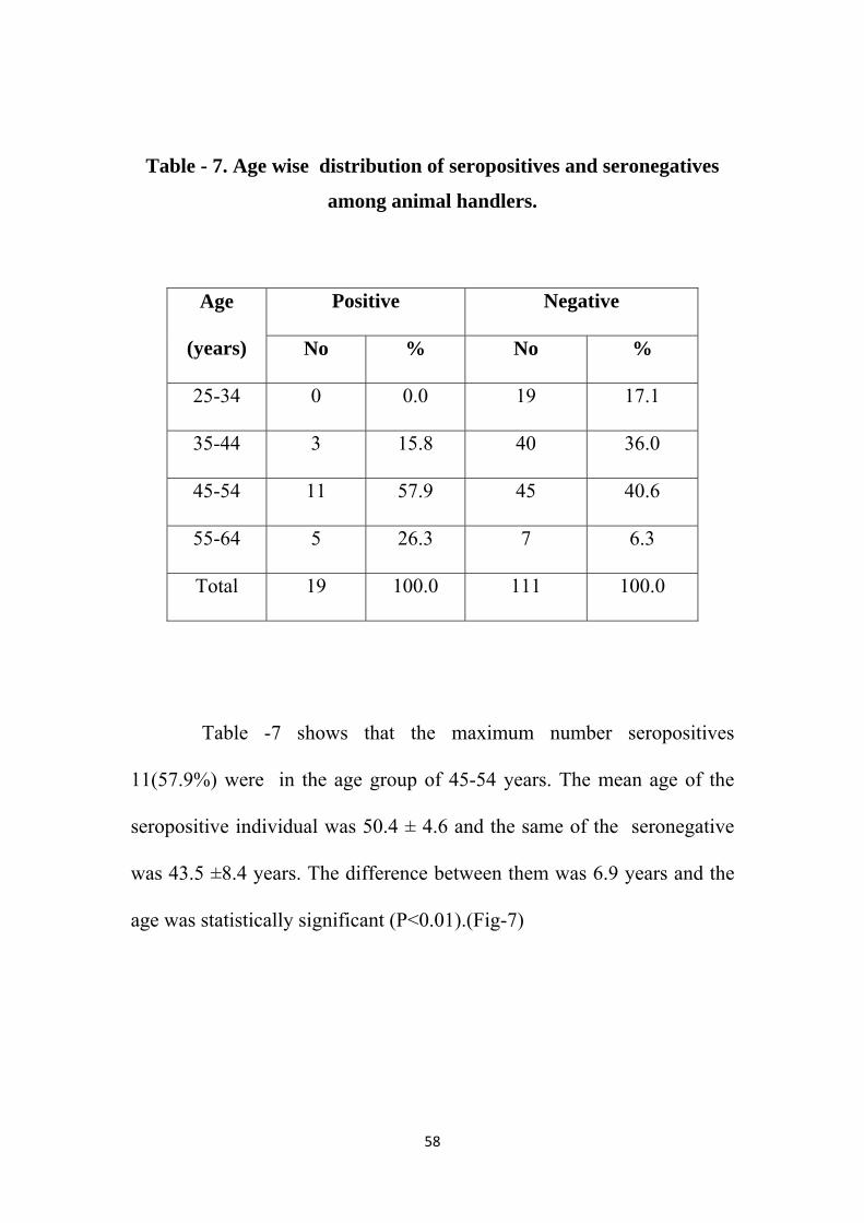
Table - 7. Age wise distribution of seropositives and seronegatives
among animal handlers.
Age
(years)
Positive Negative
No % No %
25-34 0 0.0 19 17.1
35-44 3 15.8 40 36.0
45-54 11 57.9 45 40.6
55-64 5 26.3 7 6.3
Total 19 100.0 111 100.0
Table -7 shows that the maximum number seropositives
11(57.9%) were in the age group of 45-54 years. The mean age of the
seropositive individual was 50.4 ± 4.6 and the same of the seronegative
was 43.5 ±8.4 years. The difference between them was 6.9 years and the
age was statistically significant (P<0.01).(Fig-7)

59
Table-8. Occupation wise distribution of seropositives and
seronegatives among animal handlers.
Occupation Seropositive Seronegative
No % No %
Veterinary Surgeon 11 57.9 69 62.2
Vet. Hosp. Workers 8 42.1 37 33.3
Farmers 0 0.0 5 4.5
Total 19 100 111 100
The above table describes, among the 19 seropositives 11 (57.9%)
were veterinary Surgeons and 8 (42.1%) were veterinary hospital
workers. None of the farmers were positive for IgG agglutinins.
Among the 111( 85.3%) seronegatives 69 (62.2%) were veterinary
surgeons, 37 (33.3%) were veterinary hospital workers and 5 (4.5%) were
farmers. The test results revealed that there was no significant association
of seropositivity between the veterinary surgeons and veterinary hospital
workers. (P>0.05).(Fig-8)

Fig
Fig
g.7 Age w
g.8 Occup
0
10
20
30
40
50
60
Percen
tage
0
10
20
30
40
50
60
70
V
Percen
tage
wise distri
pation wis
25‐34
Veterinary Sur
57.962
ibution ofanim
se distribuamong
35‐44
Age i
rgeon Vet.
2.2
O
60
f seroposimal hand
ution of sanimal h
45‐54
n years
Hosp. Worke
42.1
33.3
Occupation
itives and dlers
eropositivhandlers
55‐64
rs Fa
0
seronega
ves and se
4
rmers
04.5
atives amo
eronegati
Seropositi
Seronegat
ong
ives
ve
tive
Seropositive
Seronegative
e
61
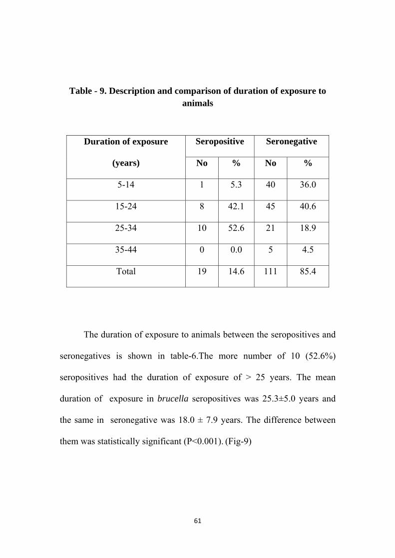
Table - 9. Description and comparison of duration of exposure to animals
Duration of exposure
(years)
Seropositive Seronegative
No % No %
5-14 1 5.3 40 36.0
15-24 8 42.1 45 40.6
25-34 10 52.6 21 18.9
35-44 0 0.0 5 4.5
Total 19 14.6 111 85.4
The duration of exposure to animals between the seropositives and
seronegatives is shown in table-6.The more number of 10 (52.6%)
seropositives had the duration of exposure of > 25 years. The mean
duration of exposure in brucella seropositives was 25.3±5.0 years and
the same in seronegative was 18.0 ± 7.9 years. The difference between
them was statistically significant (P<0.001). (Fig-9)

62
Table- 10. Handling of animal vaccine for Brucella among animal
handlers.
Handling of Brucella
vaccine
Positive Negative Total
No % No % No
Yes (Handled) 13 68.4 8 7.2 21
No ( did Not handle) 6 31.6 103 92.8 109
Total 19 100 111 100 130
Table-10 describes, among the130 animal handlers 21 had handled
the brucella vaccine for animals and 109 had not handled the vaccine.
Out of the 21 vaccine handled persons13 were seropositive and 8 were
seronegative for Brucella IgG antibody.
Among the 19 seropositives, 13 (68.4%) had handled the vaccine
and 6 (31.6%) had not handled the vaccine. Out of 111 seronegatives
only 8 (7.2%) had handled the vaccine and 103 (92.8%) had not handled
the vaccine. Thus the association between handling of vaccine and
seropositivity is statistically significant (P<0.0001). (Fig-10)

Fig
P
g. 9 Descr
Fig. 10 H
0
10
20
30
40
50
60
Percen
tage
0
20
40
60
80
100
120
140
Percen
tage
ription an
Handling
5.3
36
5‐14
6
Yes (H
d compar
of animal
42.1 40.6
15‐24
Duration of
68.4
7.2
Handled)
Handling o
63
rison of d
l vaccine handlers
52.6
18
25‐3
exposure in y
No
of Brucella va
uration o
for Bruces
0
8.9
4 3
years
31.6
92.8
o ( did Not hand
ccine
of exposur
ella amon
0
4.5
35‐44
le)
re to anim
ng animal
Seropositi
Seronegat
Seronegative
Seropositive
mals
ve
tive
e

64
Table- 11. Consumption of raw dairy products among animal handlers.
Raw dairy
consumption
Positive Negative Total
No % No % No
Yes 11 57.9 25 22.5 36
No 8 42.1 86 77.5 94
Total 19 100 111 100 130
Among the total of 130 study group, 36 had history of consumption
of raw dairy products and 94 had no such history.
Out of the total 19 brucella IgG positives, 11(57.9%) had
consumed the raw dairy products and 8 (42.1%) had not consumed the
products. Among the111 seronegative individuals, 25 (22.5%) had the
history of consumption and 86 (77.5%) had not consumed the raw dairy
products. The test results revealed that there was significant association
between seropositivity and consumption of raw dairy products. (Fig-11)

65
Table- 12. Awareness of brucellosis among animal handlers.
Awareness
of brucellosis
Seropositive Seronegative Total
No % No % No
Yes 11 57.8% 95 85.6 106
No 8 42.2 16 14.4 24
Total 19 100 111 100 130
Table -12 shows, among the 130 animal handlers 106 were aware
of the disease and 24 were not aware of the disease. Among those who
were aware of the disease 11 were seropositive and 95 were negative for
brucella IgG antibody. Out of 24 persons who were not aware of the
disease 8 were positive and 16 were negative.
Among the total 19 IgG positive individuals, 11 (57.8%) were
aware of the disease and 8 (42.2%) were not aware of the disease. Out of
the 111 seronegatives 95 (85.6%) were aware of brucellosis and 16
(14.4%) were not aware of it. There was significant (P<0.001) association
between awareness of disease and decreased seropositivity.

Fig
P
g. 11 Con
Fig.12
0
10
20
30
40
50
60
70
80
90Pe
rcen
tage
0
10
20
30
40
50
60
70
80
90
Percen
tage
nsumption
2 Awarene
57.9
Sero
Yes
57.8
A
n of raw d
ess of br
42.1
positive
Raw
85.6
Awareness of
66
dairy prod
rucellosis
w dairy consum
N
42.2
f brucellosis
ducts amo
among a
22.5
Seron
mption
No
2
14.4
ong anim
animal ha
77.5
negative
mal handle
andlers
Y
Seropositi
Seronegat
ers.
Yes
No
ve
tive

67
Table-13. Socio demographic profile of seropositives.
Table-13 summarises the important sociodemographic profile of
the total 19 Brucella IgG positive animal handlers and their significance
in association with the disease.
S.No Variable Total ELISA positive
P value No %
1. Age group in years 25-34 19 0 0
35-44 43 3 69.8 45-54 56 11 19.6 55-64 1 5 20.0
2. Sex P>0.05 Male 115 18 15.6
Female 15 1 6.7 3. Duration of animal exposure
P<0.001 5-14 41 1 2.4
15-24 53 8 15.1 25-34 31 10 32.2 35-44 5 0 0
4. Handling of brucella vaccine for animals P<0.0001 Yes (Handled) 21 13 61.9
No (Not handled) 109 6 5.5 5. Awareness of disease
P<0.001 Yes 106 11 10.4 No 24 8 3.3
6. Raw dairy products consumption P<0.001 Yes 36 11 30.5
No 94 8 8.5
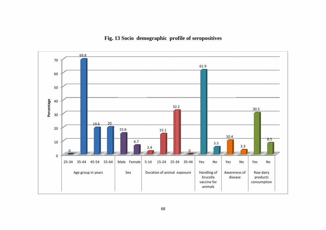
0
10
20
30
40
50
60
70
Percen
tage
25‐34 35‐44
Age group
0
69.8
Fig. 13
45‐54 55‐64 M
p in years
19.6 20
3 Socio demo
Male Female 5‐
Sex D
15.6
6.7
2
68
ographic pro
‐14 15‐24 25‐3
Duration of animal e
2.4
15.1
32
ofile of seropo
34 35‐44 Yes
exposure Handbru
vaccani
.2
0
61.9
ositives
No Yes
dling of ucella cine for imals
Awarendisea
5.5
10.4
No Yes
ness of ase
Raw daproduc
consump
3.3
30.5
No
iry cts ption
8.5
69
6. DISCUSSION
6.1 Study population
130 animal handlers ( high risk group) and 130 controls were
included in the present study to detect the seroprevalence of brucellosis.
In the high risk group, 80 ( 61.5%) veterinary surgeons,
45(35.6%) veterinary hospital workers and 5 (3.9%) farmers of
Tirunelveli district , were tested for seroprevalence of brucellosis. It
consisted of more males (88.5%) than females (11.5%) and many (56)
participants were in the age group of 45-54 years.
In the control group, 34 (26.1%) Doctors, post graduate students,
21(16.1%) lab technicians and 33(25.4%) clerical staff were included.
This group comprised of more males ( 54.6%) than females (45.4%). 58
participants were in the age group of 25 – 34 years and 50 were in the age
group of 35-44 years .
6.2 Seroprevalence
The seroprevalence of brucellosis was found to be 14.6 % among
these animal handlers which is suggestive increased exposure to
Brucella species. In the control group the seroprevalence was found to be
nil. This could be due to the fact that the control group had no
occupational contact with animals and also because of an urban life style
and safe hygienic measures like consumption of pasturised dairy
products. On the other hand, the animal handlers may have developed
70
increase in titre of humoral antibodies as a result of repeated exposure to
the agent.
The disease produced by Brucella results in protean clinical
manifestation and low mortality. However, the morbidity of brucellosis
is high because the disease process is long and incapacitating. In this
study, all of the screened individuals were asymptomatic. The Brucella
antibodies detected in asymptomatic individuals may be due to a history
of repeated exposure to antigenic stimuli or inactive brucellosis.
The ELISA test which was used to detect IgG antibodies in the
present study is very sensitive. This method can be used for the
detection of immunoglobulin isotypes and gives better interpretation
regarding the clinical situation and overcome the false negative and
false positives which may arise in agglutination tests. More over this test
is suited for screening large number of samples.
These antibodies seems to persist for a longer period or until they
maintain contact with infected animals. This finding was similar to the
studies among high risks group persons by Agasthya et al (15.69%) in
Karnataka, 68 Bhat et al (5.8 to 14.3% ) in Belgum 63 and Alsekait (15%)
in Saudi Arabia.66 Reports from various developing countries in
Mediterranean basin revealed that the seroprevalence ranged from 8% in
Jordan 40 to 12 % in Lebanon and Kuwait,41,42 18% in Uganda and 13% 43
in Nigeria.
71
In comparison to other studies by Fatima Mukhtar (21.7%) in
Pakistan,65 Rana et al (27.7%), Kumar et al (28.57%) 84 and Cadmus
S.I.B et al (31.82%)48 in Ibadan, Southwestern Nigeria, the
seroprevalence in the present study was low. This could be due to the
difference in geographical location, variation in existence of disease
among animals and personal hygienic practice and social habits.
But the seroprevalence was found to be high when compared to
few Indian studies done by Thakur and Thalipal (4.97%) in Uttaranchal80
and Ajay et al (1.14%) in Kerala.61 This could be because of intense
agricultural activities, cattle rearing, dairy farming and close contact with
animals in the study area.
6.3 Sex
In the present study the seropositivity was found to be higher in
males (94.7%) than females (5.3%). This is in concordance with the
study done by A.S. Agasthya et al in Karnataka,68 Jama ayah MZ et al
in Malaysia51 and Moti Yohannes et al in Ludhiana 57 where prevalence
was more common among males than females. Whereas Kapoor et al 72
and Hussein et al 73 reported in their study that increased seropositivity
in females when compared to males. However in the present study
seropositivity between male and females was not statistically significant,
which may be due to less number of females in the occupationally
exposed study group.

72
6.4 Age
The seroprevalence of Brucellosis among animal handlers in the
present study was highest in 45-54 (57.9%) years of age.Agasthya et al
68 reported highest prevalence of 45.36% among 41-50 years age group
and Fatima et al 65 observed that the highest number of seropositives
were in the age of 51 to 60 years . Ramos et al,66 Jama’ayah MZ et al 51
reported that, animal handlers those who were in the age of 40 and above
years had the highest prevalence. Whereas Kadri et al 55 reported that
those were in the age of 21 to 30 years had increased (43%)
seropositivity.
As brucellosis is an occupational disease, animal handlers > 40
years are at a greater risk of acquiring the disease due to prolonged
duration of exposure to live stocks. The present study showed there was
significant association between age group and seropositivity of disease.
6.5 Occupation
In the present study , 57.9% of veterinary surgeons and 42.1% of
veterinary hospital workers were positive for brucella antibody. This is in
concordance with the study done by Araj GF et al 41 who reported that
veterinary surgeons had the highest prevalence (50%) among the
occupationally exposed group. Agasthya et al 68 also observed that 59.8%
of veterinary professionals and 37.1% of veterinary hospital workers had
positive serology for Brucella antibody. The high prevalence rate among

73
this group may be due to the fact that brucellosis is an occupational
hazard to the veterinary professionals. Even though they are aware of the
disease, they acquire brucellosis by cuts in the skin or splashes into the
conjunctiva while handling the animal products and during intravaginal
or intrauterine manipulations in infected animals or by accidental
inoculation of animal vaccines.
Cadmus S.I.B et al 48 reported that butchers (63.63%) had the
highest seroprevalence among the high risk groups. A study among
abattoir workers in which, blood collectors had the highest (99.77%)
seropositivity followed by 68.00% in butchers.84 Abattoire workers and
meat handlers who are in direct contact with carcasses of infected
animals and raw meat have more chances of getting infection.
Farmers usually reside in close proximity with their livestocks.
They get infection by contact with infected animals and also by handling
the infected placenta, its membranes and their discharges. During the
burial of the products of conception their hands and forearms get
contaminated. However in this study seropositivity was not detected
among farmers. It may be due to the fact that only few farmers were
included in this study and also they were not exposed to the infected
animals and their products.
74
6.6 Duration of work
In the present study 52.6% of seropositive individuals had worked
in this profession for more than 25 years and 47.4% of them had worked
less than 25 years. Similar results were obtained by Abo-Shehada etal69
where the seropositivity of brucellosis was significantly high among
those people who had worked for more than 22 years of duration.
Karimi et al76 and Sohaila et al reported strong correlation between the
seropositivity of brucellosis and duration of work.
This is because many veterinary professionals have a long duration
of practice in this field of veterinary medicine which brings them in close
contact with animals. There is a strong correlation between duration of
work and seropositivity in the present study.
6.7 Handling of animal vaccine for Brucella
In the present study 68.4% of seropositives had participated in
vaccination of animals. There is a strong association between the
participation in vaccination of animals and seropositivity. This is similar
to the study by Moti Yohannes A et al57 and Ashford DA et al 88 who
reported significantly increased seroprevalence of brucellosis among the
veterinarians who participated in vaccination of animals than those who
did not.
The animal vaccine is prepared from the attenuated strain B.
abortus S19 and is most commonly used to prevent animal brucellosis.

75
The high rate of seroprevalence among vaccine handlers would have been
due to accidentally acquiring the infection through conjunctival splashes,
cuts in skin and mucous membrane and infectious aerosol. 88
6.8 Consumption of raw dairy products
In the present study 52.9% of seropositive individuals had
consumed the raw dairy products . This was similar to the findings by
Baba et al, 78 Hasanjani Roushan et al and Al-Fadhli et al observed that
consumption of unpasturised dairy products was significantly associated
with high seropositivity.There was significant association between
seropositivity and consumption of unpasturised dairy products.1
Raw milk may also transmit other diseases to humans like bovine
tuberculosis, salmonellosis and campylobacteriosis. Thus pasteurization
of milk is an integerated control measure for multiple pathogens.
6.9 Awareness of brucellosis
In the present study out of the the 130 animal handlers, 106 were
aware of the disease and 24 were not aware of the disease. 85.6 % of
seronegative individuals were aware of brucelloisis All the veterinary
surgeons and some of veterinary hospital worker were aware of
brucellosis. There was significant association between awareness of
disease and low seropositivity. This low seropositivity in persons who
were aware of the disease may be due to the practice of proper hand
hygienic measures and using personal protective equipments while
76
handling livestocks and their products.65 Moti Yohannes et al 57 reported
significant association between awareness of brucellosis and positive
serology among high risk groups.
Brucella is one of the agent likely to be used as a biological
weapon due to the fact that transmission through a spray is possible. It is
a highly contagious bacteria and less than 100 organisms would be
sufficient to produce infection in humans.
There is no vaccine for prevention brucellosis in humans. Human
brucellosis should be controlled by vaccination of animals. Animal
owners should be educated about the importance of vaccination of
animals. Mass vaccination of cattles along with other measures such as
screening and quarantine of infected live stocks can efficiently control the
brucellosis in livestocks, thereby eventually reducing the transmission of
infection to human population. Awareness about the preventive
measures like barrier protection methods, hand hygiene should be
created among the high risk persons in order to reduce the transmission
of infection from animals to humans.1
77
7. SUMMARY
The present study was aimed at assessing the seroprevalence of
brucellosis in Tirunelveli district of Tamil nadu and to analyse the risk
factors associated with this disease.
130 controls and 130 animal handlers were included in the study.
Blood samples were collected and brucella IgG antibodies were tested by
the method of ELISA and the results were analysed.
More number of participants were males in both groups.
Among animal handlers most of them were in the age group of 45-54
years.
Seroprevalence in the control group was nil.
The seroprevalence among animal handlers was 14.6%
Gender was not significantly associated with seropositivity. It may be
due to less number of female veterinary professionals.
Maximum number of seropositive cases occured in the 45-54 age
group. The mean age of the seropositive individual was 50.4 ± 4.6.
All the animal handlers included in this study were at risk
irrespective of their nature of work.
Duration of exposure to animals and handling the animals was a
significant risk factor. Those who had the exposure of more than 25
years were at great risk. The mean duration of exposure for
seropositivity was 25.3±5.0.
78
Handling the animal vaccine for brucella was a significant risk factor.
B.abortus RB 51 is a live vaccine and the handlers get infected by
inadvertent inoculation, conjunctival splashes and through aerosols.
Consumption of raw dairy products was significantly associated with
the seropositivity of brucellosis.
Awareness about brucellosis and its route of transmission had an
impact on with seroprevalence. The prevalence was low among the
persons who were aware of the disease.
The lack of human vaccine and effective control measures make it
necessary for the veterinary health personnel, animal husbandary
workers and health care providers to take protective measures.
79
8. CONCLUSION
The present study showed a significantly higher seroprevalence among
animal handlers compared to the controlgroup.
Occupationally exposed persons to animal were at greater risk of
acquiring brucellosis.
Higher age groups, long duration of exposure to animals, handling
and participation in vaccination of animals and consumption of raw
dairy products were significantly associated with the increased
seroprevalence.
Awareness of brucellosis was significantly associated with the
decreased seroprevalence.
Gender and nature of work among the animal handlers did not show
any significant association.
The results of this study emphasizes that contact with animals and
ingestion of contaminated animal products are important methods of
transmission of brucellosis.
To prevent the occupation related brucellosis the veterinary health
professionals, animal husbandary workers and laboratory personnel
should wear PPE and follow adequate hand hygienic measures.
There is no human vaccine to prevent brucellosis in man. Since the
disease has been seen as a major occupational hazard to the animal
handlers, the successful way for the control of disease in human

80
beings is by proper elimination of the infected animals and
vaccinating the non infected animals.
Reporting the existence of brucellosis to health authorities and
veterinary professionals is essential and knowledge about the
prevalence of the disease can be used to prioritize a disease control
strategy and to alert the health authorities.

REFERENCES
1. Mantur BG, Amarnath SK. Brucellosis in India - a Review. J
Biosci. 2008 ;33(4):539-47.
2. Murray PR,Corbel MJ. Brucella. In: Topley and Wilson’s Microbial
infections, Bacteriology.10th ed. Borrielo SP, Murray PR, Funke
G:eds. Washington: Arnold Publications: 2005. p1719- 1751.
3. Dalrymple – Champneys W. Undulant fever – a neglected problem.
Lancet 1950; 1: 429-435 and 478-485.
4. Smith DT, Conant NF, Overman JR. Brucella. In: Zinsser’s
Microbiology; 13th
ed. New York, USA: Appliton-Century-Croft
Publishers: 1960. pp640-49.
5. Horrocks WH, kennedy JC. Goats asameans of propagaon of
Mediterranean Fever, Report of the commission on Mediterranean
Fever, part IV. London: Harron and Sons.
6. Bang B. The etiology of epizootic abortion. Jour. Comp. Patho. 1897;
10: 125-149.
7. Meyer P, Queipo-Ortuno MI, Colmenero JD. Strategy for optimizing
DNA amplification in peripheral blood PCR assay used for diagnosis
of human brucellosis. Jour. Clin. Micro. 1998; 36: 2443-2446.

8. Pappas G, Papadimitriou P, akritidis N, Christou L, Tsianos E V The
new global map of human brucellosis; Lancet. Infect. Dis. 2006; 6:
91–99
9. Awad R Human brucellosis in the Gaza Strip, Palestine; East.
Mediterr. Health J. 1998; 4: 225–233
10. McDermott J J, Arimi S M. Brucellosis in sub-Saharan Africa:
epidemiology, control and impact. Vet. Microbiol. 2002; 90: 111–134
11. Sehgal S and Bhatia R Zoonoses in India. J. Commun. Dis. 1990; 22:
227–235
12. Maurin M. Brucellosis at the down of 21st century. Med Mal Infect
2005; 35:6-16.
13. Bitlak EJ, Maugard C,Charachon SM, Servent A, Perrin A,
O'Callaghan D. Study of the organization of the genomes of
Escherichia coli, Brucella melitensis and Agrobacterium tumefaciens
by insertion of a unique restriction site. Microbiology. 1995;141:2425-
32.
14. Rigby CE, Fraser ADE. Plasmid transfer and plasmid-mediated
genetic exchange in Brucella abortus. Can J Vet Res 1989;53:326-30.
15. Gotuzzo E, Carillo C, Guerra J, Llosa L. An evaluation of diagnostic
methods for brucellosis - the value of the marrow culture. J. Infect.
Dis. 1986; 153(1): 127-125.
16. Castaneda MR. A practical method for routine blood cultures in
brucellosis. Prac. Soc. Expt. Biol. Med. 1947; 64: 114-115.
17. Schuhardt V, Rode LJ, Oglebsy G. The toxicity of certain amino acids
for Brucella. J Bact. 1949; 58(5): 665-674.
18. Buchanan TM, Hendricks SL, Patton CM, Feldman RA. Brucellosis in
the United and States, an abattoir-associated disease. Part III.
Epidemiology evidence for acquired immunity. Medicine (Baltimore).
1974; 53:427–439.
19. Harmon BG, Adams LG, Frey M. Survival of rough and smooth
strains of Brucella abortus in bovine mammary gland macrophages.
Am J Vet Res. 1988;49:1092–1097.
20. Tikare NV, Mantur BG, Bidari LH. Brucellar meningitis in an infant-
Evidence for human breast milk transmission. J Trop Pediatr. 2008;
54: 272-4.
21. Lim HS, Min YS, Lee HS. Investigation of a series of brucellosis
cases in Gyeongsangbuk do during 2003-2004. J Prev Med PubHealth
2005; 38: 482-8.
22. Al-Eissa YA. Unusual suppurative complications of brucellosis in
children. Acta Paediatr. 1993;82:987-92.
23. Al-Kasab S, Al-Fagih MR, Al-Yousef S. Brucella infective
endocarditis: successful combined medical and surgical therapy. J
Thorac Cardiovasc Surg. 1988;95:862-7.
24. Mantur BG., Mangalgi SS. and Pattanshetty OB. Brucella arthritis – A
case report. IndJou Med Micro.1995;13: 45-47
25. Madkour MM. Brucellosis. In: Fauci AS., Braunwald E., Isselbacher
KJ. et al.eds Harrison’s Principle’s of Internal Medicine. 14th
ed. Vol
1. Mc.Graw-Hill: 1998. 969-971.
26. Al-Eissa YA, Assuhaimi SA, Al-Fawaz IM, Higgy KE, Al-Nasser
MN, Al-Mobaireek KF. Pancytopenia in children with brucellosis:
clinical manifestations and bone marrow findings. Acta Haematol
1993:89:132-6
27. Young EJ. Brucella species. In: Mandell GL., Bennett JE. And Dolin
R. eds Mandell, Douglus and Bennett’s Principles and Practice of
infectious diseases. 4th
ed. Vol. 2. Churchill Livingstone; 1995. 2053-
2060
28. Serra J, Vinas M. Laboratory diagnosis of brucellosis in a rural
endemic area in northeastern Spain. Int. Microbiol. 2004; 7:53-58
29. Ariza J, Pellicer T, Pallares R, Foz A, Gudiol F. Specifi c antibody
profi le in human Brucellosis. Clin Infect Dis 1992; 14:131-40.

30. Al-Shamahy H A, Wright S G. Enzyme-linked immunosorbent assay
for Brucella antigen detection in human sera; J. Med. Microbiol. 1998;
47: 169–172
31. Mantur B, Parande A, Amarnath S, Patil G, Walvekar R, Desai A et
al. ELISA versus conventional methods of diagnosing endemic
brucellosis. Am J Trop Med Hyg. 2010 ;83(2):314-8.
32. Zourbas J, Masse L, Roussey A, David C, Maurin J, Torte J. Sampling
survey on brucellosis among farmers and their families in IIIe-et-
Vilaine (Brittany) Int J Epidemiol. 1977;6:335–343.
33. Kerr WR, McCaughey WJ, Coghlan JD. et al. Techniques and
interpretations in the serological diagnosis of brucellosis in man. Jour.
Med. Micro. 1968; 1: 181-193.
34. Polt SS and Schaefar J. A microagglutination test for human Brucella
canis antibodies. Am. Jour. Clin. Path. 1982; 77:740-744.
35. Araj GF, Lulu AR, Mustafa MY, Khateeb MI. Evaluation of ELISA in
the diagnosis of acute and chronic brucellosis in human beings. J Hyg
(Lond). 1986 ;97(3):457-69.
36. Baldi PC, Miguel SE, Fossati CA, Wallach JC (1996a) Serological
followup of human brucellosis by measuring IgG antibodies to
lipopolysaccharide and cytoplasmic proteins of Brucella species. Clin
Infect Dis.(1996a);122:446– 455.

37. Ordun˜a A, Almaraz A, Prado A, Gutie´rrez MP, Garcı´a-Pascual A,
Duen˜as A et al. Evaluation of an immunocapture-agglutination test
(Brucellacapt) for serodiagnosis of human brucellosis. J Clin
Microbiol 38:4000– 4005.
38. Abdoel T H, Smits H L. Rapid latex agglutination test for the
serodiagnosis of human brucellosis; Diagn. Microbiol. Infect. Dis.
2007; 57: 123–128.
39. Al Sekait AM. Sero epidemiological survey of brucellosis antibodies
in Saudi Arabia.
40. Dajani YH, Masoud AA, Barakat HF. Epidemiology and diagnosis of
human brucellosis in Jordan. J Trop Med Hyg. 1989; 92:209-14.
41. Araj GF, Azzam RA. Seroprevalence of antibodies among persons in
high-risk occupation in Lebanon. Epidemiol Infect. 1996; 117:281-8.
42. Lulu AR, Arja GF, Khateeb MY, Yusuf AR, Ferech FF. Human
brucellosis in Kuwait. QJ Med 1998; 66:39-44.
43. Ndyabahinduka DG, Chu IH. Brucellosis in Uganda. Int J Zoonoses.
1984;11:59-64.
44. Otlu S, Sahi M, Atabay H I, Unver A. Serological Investigations of
Brucellosis in Cattle, Farmers and Veterinarians in the Kars District of
Turkey. Acta Vet. Brno 2008; 77: 117-121.

45. Apan TZ, Yildrim M, Istanblluoglu E. Seroprevalence of brucellosis
in human, sheep and cattle population in Kirikkale (Turkey).
TurkJ.Vet.Anim Sci.2007;31:251-55.
46. Kose S, Smits HL, Abdoel TH, Ozbel Y. Prevalence of Brucella
antibodies in rural and suburban communities in three provinces of
Turkey: need for improved diagnosis and prevention. J Infect. 2006
Nov;53(5):308-14.
47. Cetinkaya Z, Aktepe OC, Ciftci IH, Demirel R. Seroprevalence of
human brucellosis in a rural area of Western Anatolia, Turkey. J
Health Popul Nutr. 2005 ;23(2):137-41.
48. Cadmus SIB, Ijagbone IF,Oputa HE, Adesokan HK, Stack JA.
Serological Survey of Brucellosis in Livestock Animals and Workers
in Ibadan, Nigeria. Afr. J. Biomed. Res. 9: 163 – 168.
49. Hajia M, Rahbar M, Hosseini A. Brucellosis antibody level of
hospitalized patients in Hamedan,Western Iran. Shiraz Med.J.2007;8:
1-5
50. Bokaei S, Sharifi L, Alizadeh H. Epidemiological survey of
brucellosis in human and animals in Birjand, East of Iran.
J.Anim.Vet.Adv.2008; 7: 460-63.

51. Jama’ayah MZ Heu JY, Norazah A. Seroprevalance of brucellosis
among suspected cases in Malaysia. Malaysian J Pathol 2011; 33(1) :
31 – 34.
52. Ofukwu AR, Yohann, CA ,Abuh. H.A. Brucella infection among
hospital patients in Makurdi, North Central Nigeria.
53. Mantur BG, Biradar MS, Bidri RC, Mulimani MS, Veerappa K,
Kariholu P, et al. Protean clinical manifestations and diagnostic
challenges of human brucellosis in adults: 16 years' experience in an
endemic area. J Med Microbiol. 2006;55:897-903.
54. Handa, R, Singh S, Singh N. Wali J P. Brucellosis in north India
Results of a prospective study. J Commun Dis.1998; 30: 85-87.
55. Kadri SM, Rukhsana A, Laharwal, MA, Tanvir M. Seroprevalence of
brucellosis in Kashmir (India) among patients with pyrexia of
unknown origin. J Indian Med Assoc 2000;98:170-1
56. Appannanavar SB, Sharma K, Verma S, Sharma M. Seroprevalence
of Brucellosis: A 10-year experience at a tertiary care center in north
India. Indian J Pathol Microbiol. 2012;55:271-219 .
57. Yohannes M, Gill JPS. Seroepidemiological survey of human
brucellosis in and around Ludhiana, India.J Emerg Health
Threats. 2011; 4: 7361
58. Mohanty TN, Panda SN, Das BR, Pradhan SK, Pradhan RK. Sero-
incidence of brucellosis among dairy farm workers in Orissa. Indian
Vet J. 2000;77:568-70.
59. Vaishnavi C, Kumar S. Investigation for background prevalence
of Brucella agglutinins among blood donors. Indian J Med Microbiol.
2007;25:302-4
60. Basavaraj CM, Mahesh CB, Jyothi P, Lava R, Basavarajappa,
Hanumanthappa A R et al. Seroprevalence of brucellosis in
Davangere, Karnataka. J of Clin and Diag Res. 2011 ; 5(1):41-44
61. Ajaykumar VJ, Nanu E. Seropositive of Brucellosis in Human beings.
Ind. J. Pub. Helth. 2005 ; 49(1): 22-24.
62. Panjarathinam R and Jhala C I. Brucellosis in Gujarat state. Indian J.
Pathol. Microbiol.1986; 29: 53–60
63. Bhat K, Hemashettar, BM, Jain R, Anrade AT, Patil CS. Latex
agglutination test for antigen detection in human brucellosis. Indian J
Med Microbiol. 1997;15:210-25.
64. Panjarathinam R. Anti-brucella agglutinins in aborted women. J
Gynecol Obstet Biol Reprod.1984;13(4):433-6
65. Mukhtar F, Kokab F. Brucella serology in abattoir workers J Ayub
Med Coll Abbottabad. 2008;20(3):57-61.

66. Ramos TR, Pinheiro Junior JW, Sobrinho M P A, Santana VL, Guerra
NR, De Melo LE, Mota RA. Epidemiological aspects of an infection
by Brucella abortus in risk occupational groups in the microregion of
Araguaína, Tocantins. Braz J Infect Dis. 2008 ;12(2):133-8.
67. Bigler W.J., et al. Trends of brucellosis in Florida: An epidemiological
review. Am J Epidemiol. 1997; 105:245-251.
68. Agasthya AS, Isloor S, Prabhudas K. Brucellosis in high risk group
individuals. Indian J Med Microbiol. 2007;25:28-31
69. Abo-Shehada MN, Odeh JS, Abu-Essud M, Abuharfeil N.
Seroprevalence of brucellosis among high risk people in northern
Jordan. Int J Epidemiol. 1996 ;25(2):450-4
70. Randhawa AS, Dhillon SS. Seroprevalence of brucellosis in humans
and animals of Punjab. Ind J Pub Health 1974; 18: 15-9.
71. Ali A, Bahadar S, Ikhwan, Jehangir, Irshad. Study on the Clinico-
epidemiological and therapeutic aspects of human brucellosis in
NWFP. J Life Sci In.t 2007;1:350–486.
72. Kapoor PK, Sharma SN, Rao KL. Seroprevalence of brucellosis in
goats and human being in Bikaner, (Rajasthan). Ind J Comp Microbiol
Immunol Infect Dis. 1985; 6:96-101

73. Hussein AAA, Sayed ASM, Feki MAE. Seroepidemiological study on
human brucellosis in assiut governorate. Egypt J Immunol.
2005;12:49-56.
74. Falade, S. Brucella agglutination antibodies in the Sera of persons
dwelling in Ibadan and the Surrounding districts. Nigerian Veterinary
Journals.1974;3:21-23
75. Salari MH. Seroepidemiological Survey of Brucellosis Among Animal
Farmers of Yazd Province. Iranian J. Publ. Health. 2002; 31: 29-3.
76. Karimi A, Alborzi A, Rasooli M, Kadivar MR, Nateghian AR.
Prevalence of antibody to Brucella species in butchers, slaughterers
and others. East Mediterr Health J 2003;9:178-81.
77. Sumer H, Sumer Z, Alim A, Nur N, Ozdemir L. Seroprevalence of
brucellosis in an elderly population in Mid-Anatolia, Turkey. J Health
Popul Nutr 2003;21:158-61.
78. Baba MM, Sarkindared SE, Brisibe F. Serological evidence of
brucellosis among predisposed patients with pyrexia of unknown
origin in the north eastern Nigeria. Cent Eur J Public Health
2001;9:158–61.
79. Nabi G, Mir NA. Brucellosis in pediatric patients: a review of114
cases from Asir region. Ann Saudi Med 1992;12:286-8.
80. Thakur SD, Thapliyal DC. Seroprevalence of brucellosis in man. J
Commun Dis. 2002;34:106-9.
81. Mrunalini N, Eddy RMS, Ramasastry P, Rao MR. Seroepidemiology
of human brucellosis in Andhra Pradesh. Indian Vet J. 2004;81:744-7.
82. Mudaliar S, Bhore A, Pandit D. Detection of antibodies to Brucella
abortus in animal handlers. Indian J Med Sci. 2003;57:181-6
83. Omer MK, Assefaw T, Skjerve E, Tekleghiorghis T, Woldehiwet Z.
Prevalence of antibodies to Brucella spp. and risk factors related to
high-risk occupational groups in Eritrea. Epidemiol Infect.
2002;129:85-91.
84. Kumar P, Singh DK, Barbuddhe SB. Sero-prevalence of brucellosis
among abattoir personnel of Delhi. J Commun Dis 1997;29:131-7.
85. Masoumi J P, Sheikh M A, Ahmad R, Naeem M, Ahmad M, Hussain
I. Seroprevalence of Brucellosis in sheep, goats and man in Lahore
area. Ind J Dairy Science. 1992; 14: 298-9.
86. Asanda, NN. and Agbede SA. Brucellosis in Cattle and Human in
Adamawa State. Sokoto Journal of Veterinary Science. 2001; 3:34 –
38.
87. Wallach JC, Miguel SE, Baldi PC, Guarnera E, Goldbaum FA, Fossati
CA. Urban outbreak of a Brucella melitensis infection in an Argentine
family: clinical and diagnostic aspects. FEMS Immunol Med
Microbiol. 1994 ;8(1):49-56.
88. Ashford DA, di Pietra J, Lingappa J, Woods C, Noll H, Neville B et
al. Adverse events in humans associated with accidental exposure to
the livestock brucellosis vaccine RB51 Vaccine. 2004;22:3435-9.
89. Kostoula A, Bobogianni H, Virioni G ,Tabatabai LB, Deyoe BL.
Detection of Brucella IgG, IgM and IgA antibodies with ELISA
method in patients with Brucellosis. Clin Microbiol Infect, 2001; 7:
108.
90. Gazapo E, Lahoz GJ, Subiza J, Santos JM. Changes in IgM and IgG
antibody concentrations in brucellosis over time: Importance for
diagnosis and follow up. J Infect Dis.1989; 159: 219-225.
91. Prado A, Gutierrez P, Duenas A. Serological follow-up of brucella
patients using an immunocapture-agglutination test (Brucellacapt),
Coombs anti-Brucella and LPS-ELISA tests.Clin Microbiol and
Infect. 2001;7:394
92. Sathyanarayan MS , Suresh D , Suresh BS , Krishna S , Mariraj J ,
Surekha Ya , A comparative study of agglutination tests, blood culture
& ELISA in the laboratory diagnosis of human brucellosis. Int J Biol
Med Res. 2011; 2(2): 569-572.

93. Taleski V. An Overview of Introducing Various Laboratory Tests for
Diagnosis of Human Brucellosis in the Republic of Macedonia. Maced
J Med Sci. 2010; 3(3):239-245.

Annexure-I
DATA SHEET FOR COLLECTION OF SOCIO-DEMOGRAPHIC PROFILE
FOR P.G DISSERTATION WORK ON
“Seroprevalence of brucellosis among animal handlers and
analysis of risk factors”
1. General information
Name : Age: Sex: M / F
Address : Occupation:
Residential Background : Urban / Semi urban / Rural
Socio Economic Status : Upper/ Middle/ Low.
Educational Status : Illiterate/ Elementary/Higher secon /Graduate
H/o handling animals : Yes / No
H/o consumption of : Raw milk/raw dairy products
2. Occupational History
i. Occupation : Animal Keeper/ Veterinarian/Livestock Inspector Butchers/ Abattoir Worker/ Vet.Hospital Worker Meat Seller/ Milk handlers/ Others.
ii. Type of animal handling : Cattle/ Sheep/Pet animals/ others
iii. Duration of contact with animal :
iv. H/o handling animal product : Yes/No
iv. H/o handling animal blood : Yes/No
v. H/o handling animal vaccine for Brucella : Yes/No
vi Awareness of brucellosis : Yes/No

3.Lab investigations
i. Sample : Blood for serum
ii. Investigation : IgG ELISA for Brucella
4. Report
Name : Age/ Sex :
Address : Sample : Blood
Received On :
Reported On :
Test Method Sample OD Reference OD Value
Brucella IgG Indirect ELISA (Vircel)
< 9 = Negative 9-11 = Equivocal >11 = Positive
Interpretation:
Signature

S.No Age Sex OccupationDuration of exposure to animals
Handling brucella vaccine
Handling of animal products
Raw dairy consumption
Awareness IgG ELISA Result
1 45 M Vet.Surgeon 22 No yes No Yes 1.69 Negative2 51 M Vet.Surgeon 26 No yes No Yes 0.738 Negative3 46 M Vet.Surgeon 25 No yes No Yes 9.5 Negative4 45 M Vet.Surgeon 22 Yes yes Yes Yes 22.6 Positive5 44 M Vet.Surgeon 20 No yes No Yes 1.5 Negative6 50 M Vet.Surgeon 23 No yes No Yes 2.707 Negative7 48 M Vet.Surgeon 24 Yes yes No Yes 17.32 Positive8 46 M Vet.Surgeon 25 No yes No Yes 0.707 Negative9 46 M Vet.Surgeon 23 No yes No Yes 1.2 Negative10 48 M Vet.Surgeon 24 No yes Yes Yes 18.8 Positive11 42 M Vet.Surgeon 20 No yes No Yes 4.183 Negative12 42 M Vet.Surgeon 20 No yes No Yes 0.81 Negative13 43 M Vet.Surgeon 20 No yes No Yes 3 Negative14 29 M Vet.Surgeon 10 No yes No Yes 1.661 Negative15 45 M Vet.Surgeon 23 No yes No Yes 0.83 Negative16 45 M Vet.Surgeon 5 No yes No Yes 0.738 Negative17 51 M Vet.Surgeon 27 Yes yes No Yes 19.5 Positive18 48 M Vet.Surgeon 25 No yes No Yes 0.692 Negative19 50 M Vet.Surgeon 23 No yes No Yes 1.107 Negative20 50 M Vet.Surgeon 27 No yes No Yes 23.4 Positive21 52 M Vet.Surgeon 29 No yes No Yes 5.07 Negative22 38 F Vet.Surgeon 14 No yes No Yes 0.923 Negative23 37 M Vet.Surgeon 15 No yes No Yes 0.8 Negative24 51 M Vet.Surgeon 30 Yes yes No Yes 29.33 Positive25 27 F Vet.Surgeon 9 No yes No Yes 4.266 Negative26 40 F Vet.Surgeon 14 No yes No Yes 0.8 Negative27 45 M Vet.Surgeon 23 No yes No Yes 0.92 Negative28 38 F Vet.Surgeon 15 No yes No Yes 1.733 Negative
MASTER CHARTSOCIO‐DEMOGRAPHIC DATA AND TEST RESULTS
ANNEXURE II

29 31 M Vet.Surgeon 12 No yes No Yes 1.066 Negative30 42 M Vet.Surgeon 19 No yes No Yes 1.146 Negative31 53 M Vet.Surgeon 28 No yes No Yes 5.74 Negative32 51 M Vet.Surgeon 30 Yes yes Yes Yes 8.82 Negative33 29 M Vet.Surgeon 10 No yes No Yes 0.81 Negative34 48 M Vet.Surgeon 23 Yes yes Yes Yes 0.72 Negative35 27 F Vet.Surgeon 6 No yes No Yes 1.1 Negative36 32 M Vet.Surgeon 15 No yes Yes Yes 1.09 Negative37 30 M Vet.Surgeon 10 No yes No Yes 0.92 Negative38 39 M Vet.Surgeon 15 No yes No Yes 0.97 Negative39 34 F Vet.Surgeon 12 No yes Yes Yes 0.58 Negative40 53 M Vet.Surgeon 30 Yes yes No Yes 13.1 Positive41 32 M Vet.Surgeon 10 No yes No Yes 2.21 Negative42 43 M Vet.Surgeon 20 Yes yes Yes Yes 22.36 Positive43 40 M Vet.Surgeon 18 Yes yes No Yes 2.86 Negative44 42 F Vet.Surgeon 22 No yes No Yes 1.92 Negative45 57 M Vet.Surgeon 32 Yes yes No Yes 28.36 Positive46 38 M Vet.Surgeon 19 No yes No Yes 0.84 Negative47 51 M Vet.Surgeon 25 Yes yes No Yes 16.09 Positive48 34 M Vet.Surgeon 10 No yes No Yes 0.65 Negative49 42 F Vet.Surgeon 15 No yes No Yes 8.5 Negative50 26 F Vet.Surgeon 6 No yes Yes Yes 0.826 Negative51 42 M Vet.Surgeon 18 Yes yes No Yes 0.9 Negative52 38 M Vet.Surgeon 14 No yes No Yes 0.85 Negative53 30 M Vet.Surgeon 8 No yes Yes Yes 1.1 Negative54 34 F Vet.Surgeon 14 No yes No Yes 0.89 Negative55 37 F Vet.Surgeon 14 No yes No Yes 0.96 Negative56 30 F Vet.Surgeon 5 No yes Yes Yes 1.96 Negative57 38 M Vet.Surgeon 20 No yes Yes Yes 0.826 Negative58 42 M Vet.Surgeon 8 Yes yes No Yes 1.173 Negative59 42 M Vet.Surgeon 15 No yes Yes Yes 2.4 Negative60 40 M Vet.Surgeon 20 No yes No Yes 0.93 Negative61 42 M Vet.Surgeon 15 Yes yes Yes Yes 3.04 Negative62 41 M Vet.Surgeon 18 Yes yes No Yes 4.58 Negative63 43 M Vet.Surgeon 17 No yes No Yes 1.573 Negative64 37 M Vet.Surgeon 14 No yes Yes Yes 1.13 Negative

65 51 M Vet.Surgeon 26 No yes No Yes 0.973 Negative66 42 M Vet.Surgeon 30 No yes No Yes 5.06 Negative67 44 M Vet.Surgeon 12 No yes Yes Yes 0.68 Negative68 27 M Vet.Surgeon 8 No yes No Yes 0.82 Negative69 47 M Vet.Surgeon 23 No yes No Yes 3.38 Negative70 44 M Vet.Surgeon 23 Yes yes Yes Yes 21.81 Positive71 44 M Vet.Surgeon 22 No yes No Yes 0.6 Negative72 36 M Vet.Surgeon 11 No yes No Yes 2 Negative73 45 M Vet.Surgeon 22 No yes No Yes 1.63 Negative74 53 M Vet.Surgeon 26 No yes No Yes 2.87 Negative75 47 M Vet.Surgeon 23 No yes No Yes 4.27 Negative76 43 M Vet.Surgeon 20 No yes Yes Yes 2.17 Negative77 32 M Vet.Surgeon 13 No yes No Yes 2.914 Negative78 27 M Vet.Surgeon 8 No yes No Yes 0.6 Negative79 27 M Vet.Surgeon 8 No yes No Yes 0.82 Negative80 65 M Farmer 10 No No No Yes 0.6 Negative81 33 M Farmer 12 No No Yes No 0.51 Negative82 37 M Farmer 5 No No No No 0.8 Negative83 47 M Farmer 15 No No No No 0.77 Negative84 35 M Farmer 20 No No Yes Yes 1.72 Negative85 42 M Vet.Surgeon 17 No Yes No No 1.13 Negative86 52 M Vet.Hosp work 27 No Yes Yes No 11.7 Positive87 58 M Vet.Hosp work 30 No Yes No No 0.75 Negative88 45 M Vet.Hosp work 15 No Yes No No 3.6 Negative89 43 M Vet.Hosp work 18 No Yes Yes Yes 2 Negative90 54 M Vet.Hosp work 24 No Yes No No 4.9 Negative91 54 M Vet.Hosp work 35 No Yes No No 3.7 Negative92 47 M Vet.Hosp work 19 No Yes Yes Yes 3.6 Negative93 54 M Vet.Hosp work 14 No Yes No No 0.6 Negative94 56 M Vet.Hosp work 30 No Yes No Yes 0.51 Negative95 43 F Vet.Hosp work 20 No Yes No Yes 0.75 Negative96 55 M Vet.Hosp work 35 No Yes Yes Yes 1.07 Negative97 54 M Vet.Hosp work 26 No Yes No Yes o.7 Negative98 49 M Vet.Hosp work 33 Yes Yes No Yes 5.32 Negative99 54 M Vet.Hosp work 22 No Yes Yes Yes 0.8 Negative100 58 M Vet.Hosp work 22 No Yes No No 0.77 Negative

101 57 M Vet.Hosp work 14 No Yes Yes Yes 0.7 Negative102 50 M Vet.Hosp work 35 No Yes No Yes 0.9 Negative103 56 F Vet.Hosp work 30 No Yes Yes No 22.5 Positive104 41 M Vet.Hosp work 11 No Yes No Yes 2.2 Negative105 53 M Vet.Hosp work 15 No Yes No Yes 1.4 Negative106 45 M Vet.Hosp work 5 No No No No 0.8 Negative107 52 M Vet.Hosp work 27 No Yes No Yes 0.9 Negative108 56 M Vet.Hosp work 20 yes Yes Yes No 15.8 Positive109 45 M Vet.Hosp work 5 No Yes No No 3.977 Negative110 40 M Vet.Hosp work 5 No Yes No Yes 1.5 Negative111 50 M Vet.Hosp work 6 No Yes Yes No 3.76 Negative112 42 M Vet.Hosp work 12 No Yes No Yes 1.3 Negative113 52 M Vet.Hosp work 25 No Yes No Yes 1.6 Negative114 52 M Vet.Hosp work 15 No Yes Yes Yes 1 Negative115 53 M Vet.Hosp work 27 No Yes No Yes 5.7 Negative116 57 M Vet.Hosp work 30 No Yes Yes No 11.5 Positive117 56 M Vet.Hosp work 36 No Yes Yes Yes 5.46 Negative118 49 M Vet.Hosp work 35 No Yes No No 2.4 Negative119 45 M Vet.Hosp work 25 No Yes No Yes 0.7 Negative120 46 M Vet.Hosp work 20 Yes Yes Yes No 14 Positive121 43 F Vet.Hosp work 5 No No No Yes 1.8 Negative122 48 M Vet.Hosp work 15 No Yes Yes No 1.4 Negative123 43 M Vet.Hosp work 12 No Yes No Yes 0.5 Negative124 53 M Vet.Hosp work 13 No Yes No No 1.08 Negative125 53 M Vet.Hosp work 27 No Yes No Yes 3.4 Negative126 54 M Vet.Hosp work 28 No Yes Yes Yes 1.05 Negative127 50 M Vet.Hosp work 30 No Yes Yes No 21.4 Positive128 44 M Vet.Hosp work 12 Yes Yes Yes No 17 Positive129 55 M Vet.Hosp work 24 Yes Yes No No 26.3 Positive130 51 M Vet.Hosp work 20 No Yes No Yes 6.11 Negative
Abbreviations M ‐ Male, F ‐ Female Vet.Hosp work ‐ Veterinary Hospital workerVet.Surgeon ‐ Veterinary Surgeon

S.No Age Sex OccupationHandling of animals
Duration of exposure to animals
Handling brucella vaccine
Raw dairy Awareness IgG ELISA Result
1 35 M Doctors No Nil Nil No Yes 1.69 Negative2 37 M Doctors No Nil Nil No Yes 0.738 Negative3 39 M Doctors No Nil Nil No Yes 1.32 Negative4 41 M Doctors No Nil Nil No Yes 0.8 Negative5 36 F Doctors No Nil Nil No Yes 1.5 Negative6 37 M Doctors No Nil Nil No Yes 2.707 Negative7 36 M Doctors No Nil Nil Yes Yes 1.5 Negative8 35 M Doctors No Nil Nil No Yes 0.707 Negative9 35 F Doctors No Nil Nil No Yes 3.6 Negative10 38 M Doctors No Nil Nil No Yes 1.2 Negative11 39 M Doctors No Nil Nil No Yes 4.183 Negative12 41 M Doctors No Nil Nil No Yes 0.81 Negative13 40 M Doctors No Nil Nil No Yes 2.5 Negative14 35 F Doctors No Nil Nil No Yes 1.78 Negative15 36 M Doctors No Nil Nil Yes Yes 0.752 Negative16 38 M Doctors No Nil Nil No Yes 0.869 Negative17 38 M Doctors No Nil Nil No Yes 0.56 Negative18 38 F Doctors No Nil Nil No Yes 0.34 Negative19 53 M Doctors No Nil Nil No Yes 1.27 Negative20 47 M Doctors No Nil Nil No Yes 4.23 Negative21 52 M Doctors No Nil Nil No Yes 4.96 Negative22 37 F Doctors No Nil Nil No Yes 0.759 Negative23 37 M Doctors No Nil Nil No Yes 0.82 Negative24 56 M Doctors No Nil Nil Yes Yes 3.56 Negative25 36 F Doctors No Nil Nil No Yes 4.12 Negative26 39 F Doctors No Nil Nil No Yes 0.8 Negative27 42 M Doctors No Nil Nil No Yes 0.96 Negative28 49 F Doctors No Nil Nil No Yes 1.23 Negative29 45 M Doctors No Nil Nil No Yes 1.012 Negative
ANNEXURE IIMASTER CHART
SOCIO‐DEMOGRAPHIC DATA AND TEST RESULTS

30 46 F Doctors No Nil Nil No Yes 1.06 Negative31 44 M Doctors No Nil Nil No Yes 4.01 Negative32 48 M Doctors No Nil Nil No Yes 3.65 Negative33 45 F Doctors No Nil Nil No Yes 0.81 Negative34 49 M Doctors No Nil Nil No Yes 0.75 Negative35 34 F Students No Nil Nil No Yes 1.023 Negative36 26 F Students No Nil Nil Yes Yes 1.09 Negative37 32 F Students No Nil Nil No Yes 0.756 Negative38 27 F Students No Nil Nil No Yes 0.845 Negative39 31 F Students No Nil Nil No Yes 0.562 Negative40 36 M Students No Nil Nil No Yes 4.36 Negative41 30 F Students No Nil Nil No Yes 2.65 Negative42 25 F Students No Nil Nil No Yes 1.95 Negative43 37 F Students No Nil Nil No Yes 1.65 Negative44 40 F Students No Nil Nil No Yes 1.32 Negative45 37 M Students No Nil Nil No Yes 1.96 Negative46 27 F Students No Nil Nil No Yes 0.84 Negative47 35 F Students No Nil Nil No Yes 2.15 Negative48 38 F Students No Nil Nil No Yes 0.692 Negative49 42 F Students No Nil Nil No Yes 0.98 Negative50 29 F Students No Nil Nil No Yes 0.826 Negative51 33 M Students No Nil Nil No Yes 1.2 Negative52 32 F Students No Nil Nil No Yes 1.65 Negative53 28 F Students No Nil Nil No Yes 1.2 Negative54 33 M Students No Nil Nil No Yes 0.96 Negative55 26 M Students No Nil Nil No Yes 0.925 Negative56 25 F Students No Nil Nil No Yes 1.45 Negative57 28 F Students No Nil Nil Yes Yes 0.85 Negative58 32 F Students No Nil Nil No Yes 1.026 Negative59 28 M Students No Nil Nil No Yes 1.89 Negative60 28 F Students No Nil Nil No Yes 0.963 Negative61 31 F Students No Nil Nil No Yes 3.04 Negative62 29 F Students No Nil Nil No Yes 3.65 Negative63 29 M Students No Nil Nil No Yes 1.256 Negative64 28 F Students No Nil Nil No Yes 1.09 Negative

65 29 F Students No Nil Nil No Yes 2.61 Negative66 30 M Students No Nil Nil No Yes 3.65 Negative67 26 F Students No Nil Nil No Yes 0.965 Negative68 28 F Students No Nil Nil No Yes 1.86 Negative69 31 F Students No Nil Nil No Yes 1.396 Negative70 32 M Students No Nil Nil Yes Yes 2.95 Negative71 34 F Students No Nil Nil No Yes 3.2 Negative72 27 F Students No Nil Nil No Yes 1.956 Negative73 28 F Students No Nil Nil No Yes 1.65 Negative74 27 F Students No Nil Nil No Yes 2.65 Negative75 31 M Students No Nil Nil No Yes 3.86 Negative76 26 F Students No Nil Nil No Yes 2.56 Negative77 39 M Lab tehnicians No Nil Nil No Yes 2.69 Negative78 35 M Lab tehnicians No Nil Nil No Yes 0.768 Negative79 42 M Lab tehnicians No Nil Nil No Yes 0.82 Negative80 27 M Lab tehnicians No Nil Nil No Yes 0.6 Negative81 31 M Lab tehnicians No Nil Nil No No 0.59 Negative82 32 F Lab tehnicians No Nil Nil No No 0.81 Negative83 29 M Lab tehnicians No Nil Nil No No 0.82 Negative84 31 M Lab tehnicians No Nil Nil No No 1.68 Negative85 29 F Lab tehnicians No Nil Nil No No 1.69 Negative86 32 M Lab tehnicians No Nil Nil No No 1.25 Negative87 31 F Lab tehnicians No Nil Nil No No 0.862 Negative88 29 M Lab tehnicians No Nil Nil No No 3.56 Negative89 28 M Lab tehnicians No Nil Nil Yes Yes 2 Negative90 42 F Lab tehnicians No Nil Nil No No 1.32 Negative91 37 M Lab tehnicians No Nil Nil No No 3.36 Negative92 38 F Lab tehnicians No Nil Nil Yes No 2.5 Negative93 35 M Lab tehnicians No Nil Nil No No 1.01 Negative94 41 M Lab tehnicians No Nil Nil No No 0.86 Negative95 29 F Lab tehnicians No Nil Nil No No 0.12 Negative96 38 M Lab tehnicians No Nil Nil No No 2.01 Negative97 37 M Lab tehnicians No Nil Nil No No 1.07 Negative98 50 F Clerical staffs No Nil Nil No No 4.32 Negative99 46 M Clerical staffs No Nil Nil No No 0.8 Negative

100 37 M Clerical staffs No Nil Nil No No 0.96 Negative101 29 M Clerical staffs No Nil Nil No No 0.845 Negative102 48 M Clerical staffs No Nil Nil No No 0.925 Negative103 46 F Clerical staffs No Nil Nil No No 4.2 Negative104 25 M Clerical staffs No Nil Nil No No 2.65 Negative105 27 M Clerical staffs No Nil Nil No No 1.98 Negative106 47 F Clerical staffs No Nil Nil No No 2.65 Negative107 39 M Clerical staffs No Nil Nil No No 3.6 Negative108 38 M Clerical staffs No Nil Nil No No 1.65 Negative109 42 M Clerical staffs No Nil Nil No No 3.654 Negative110 32 F Clerical staffs No Nil Nil No Yes 1.12 Negative111 28 M Clerical staffs No Nil Nil No No 3.48 Negative112 26 M Clerical staffs No Nil Nil No No 1.65 Negative113 33 F Clerical staffs No Nil Nil No No 1.6 Negative114 34 M Clerical staffs No Nil Nil Yes No 1 Negative115 34 F Clerical staffs No Nil Nil No No 2.5 Negative116 34 F Clerical staffs No Nil Nil No No 2.65 Negative117 32 M Clerical staffs No Nil Nil No No 2.45 Negative118 35 M Clerical staffs No Nil Nil No No 3.254 Negative119 31 F Clerical staffs No Nil Nil No No 0.845 Negative120 37 M Clerical staffs No Nil Nil No No 4.1 Negative121 37 F Clerical staffs No Nil Nil No No 2.96 Negative122 45 M Clerical staffs No Nil Nil No No 1.52 Negative123 48 M Clerical staffs No Nil Nil No Yes 0.658 Negative124 39 F Clerical staffs No Nil Nil No No 1.64 Negative125 47 M Clerical staffs No Nil Nil No No 3.584 Negative126 56 M Clerical staffs No Nil Nil No No 2.54 Negative127 45 M Clerical staffs No Nil Nil Yes No 1.365 Negative128 49 M Clerical staffs No Nil Nil No No 1.346 Negative129 37 F Clerical staffs No Nil Nil No No 1.355 Negative130 50 M Clerical staffs No Nil Nil No No 3.265 Negative
Abbreviations M ‐ Male, F ‐ Female