seruminter- -inhibitoractivatestheyestyrosinekinase andyap ... fileseruminter-...
TRANSCRIPT

Serum Inter-�-inhibitor Activates the Yes Tyrosine Kinaseand YAP/TEAD Transcriptional Complex in Mouse EmbryonicStem Cells*
Received for publication, May 9, 2014, and in revised form, October 6, 2014 Published, JBC Papers in Press, October 9, 2014, DOI 10.1074/jbc.M114.580076
Sara Pijuan-Galitó‡, Christoffer Tamm‡, and Cecilia Annerén‡§1
From the ‡Department of Medical Biochemistry and Microbiology, Uppsala University, SE-75 123 Uppsala, Sweden and §GEHealthcare Bio-Sciences AB, SE-751 84 Uppsala, Sweden
Background: Serum contributes to embryonic stem (ES) cell maintenance of pluripotency and activates Yes.Results: The serum protein Inter-�-inhibitor (I�I) was found to activate the Yes/YAP/TEAD transcription factor pathway andinduce expression of Oct3/4 and Nanog in ES cells.Conclusion: I�I activates key pluripotency pathways.Significance: I�I may play a significant role in ES cell maintenance and self-renewal.
We have previously demonstrated that the Src family kinaseYes, the Yes-associated protein (YAP) and TEA domain TEAD2transcription factor pathway are activated by leukemia inhibi-tory factor (LIF) and contribute to mouse embryonic stem (mES)cell maintenance of pluripotency and self-renewal. In addition,we have shown that fetal bovine serum (FBS) induces Yes auto-phosphorylation and activation. In the present study we confirmthat serum also activates TEAD-dependent transcription in atime- and dose-dependent manner and we identify Inter-�-in-hibitor (I�I) as a component in serum capable of activating theYes/YAP/TEAD pathway by inducing Yes auto-phosphoryla-tion, YAP nuclear localization and TEAD-dependent transcrip-tion. The cleaved heavy chain 2 (HC2) sub-component of I�I, isdemonstrated to be responsible for this effect. Moreover, I�I isalso shown to efficiently increase expression of TEAD-down-stream target genes including well-known stem cell factorsNanog and Oct 3/4. I�I is not produced by the ES cells per se butis added to the cells via the cell culture medium containingserum or serum-derived components such as bovine serumalbumin (BSA). In conclusion, we describe a novel function ofI�I in activating key pluripotency pathways associated with EScell maintenance and self-renewal.
Pluripotent stem (PS)2 cells, i.e. embryonic stem (ES) cellsand induced pluripotent stem (iPS) cells, can self-renew indef-initely in culture while retaining the potential to differentiateinto any cell type in an organism. To successfully maintain plu-ripotency and self-renewal, PS cells depend on different signals:Mouse PS (mPS) cells respond to the cytokine leukemia inhib-itory factor (LIF) and either serum or bone morphogenic pro-
teins (BMPs) (reviewed in Ref. 1), while human PS (hPS) cellsneed fibroblast growth factor (FGF) and transforming growthfactor (TGF-�, Activin A) (2, 3). The extracellular matrix hasalso been proven to be important for PS cells, in particular hPScells, which depend on either feeder cells or extracellularmatrix-derived coating for attachment and subsequent survivalin culture. The extracellular environment has been linked toboth maintenance and directed differentiation (4 – 8).
LIF has been described to activate several intracellular path-ways in mES cells, namely the JAK/STAT3, MAPK, PI3K, andSrc-family pathways (reviewed in Ref. 9). Downstream of thesepathways an intricate transcriptional network decides the stemcell fate. Some of these transcriptional factors have been desig-nated as stem-cell markers, e.g. Nanog, Oct3/4, Tfe3, Sox2, andEsrrb (10). In addition, the YAP-TEAD transcription factorcomplex has been reported to be important for mES cell self-renewal and maintenance of pluripotency (11, 12). We haveshown that LIF signaling through the LIF-receptor activatesYes, which in turn induces nuclear translocation of YAP.Nuclear YAP forms a complex with members of the TEADtranscription factor family (11, 13, 14). Yes, YAP, and TEAD2are highly expressed in self-renewing mES cells and are down-regulated when cells are induced to differentiate. In addition,the Yes kinase has been shown to suppress differentiation andblock embryoid body maturation when overexpressed in mEScells (15). Moreover, TEAD2 can directly associate with theOct3/4 promoter and activation of the Yes pathway induces,whereas suppression inhibits, Oct3/4 and Nanog promoteractivities. In addition to LIF, we have previously demonstratedthat Yes can be activated by fetal bovine serum (FBS) but thespecific factor(s) in serum responsible for this effect was notidentified (13).
In the present study, we have successfully isolated and iden-tified Inter-�-inhibitor (I�I) as one of the components in serumcapable of activating the Yes-YAP-TEAD pathway in mES cells.The I�I protein family is a group of protein-glycosaminogly-can-protein complexes that are present in plasma at high con-centrations ranging from 0.6 to 1.2 mg/ml in humans (16). Theyconsist of alternate combinations of heavy chains (HC1-HC5)
* This work was supported by the Swedish Research Council, the Jaenssons’Foundations, and the Faculty of Medicine, Uppsala University.
1 To whom correspondence should be addressed: Dept. of Medical Biochem-istry and Microbiology, Husargatan 3 Box 582, SE-75123 Uppsala, Sweden.Tel.: (�46)-076-891-15-38; Fax: (46)-018-471-46-73; E-mail: [email protected].
2 The abbreviations used are: PS, pluripotent stem; YAP, Yes-associated pro-tein; LIF, leukemia inhibitory factor; ES, embryonic stem; iPS, induced PS;I�I, inter-�-inhibitor; ECM, extracellular matrix; HC, heavy chain.
THE JOURNAL OF BIOLOGICAL CHEMISTRY VOL. 289, NO. 48, pp. 33492–33502, November 28, 2014© 2014 by The American Society for Biochemistry and Molecular Biology, Inc. Published in the U.S.A.
33492 JOURNAL OF BIOLOGICAL CHEMISTRY VOLUME 289 • NUMBER 48 • NOVEMBER 28, 2014
by guest on March 21, 2019
http://ww
w.jbc.org/
Dow
nloaded from

and a light chain called bikunin linked together by a chondroi-tin 4-sulfate chain, or as unassembled proteins. I�I is the mostabundant family member in human serum and consists of theHC1, HC2, and Bk domains. It is mainly produced by the liverand is considered to be inactive until it reaches the target tissuewhere it is cleaved by TNF-stimulated gene 6 protein (TSG-6),which in turn forms a transient covalent bond with the HCs andtransfers them to hyaluronan (HA), a major constituent of theextracellular matrix (ECM) (17).
Up until recently all medium for culturing mouse and humanPS cells have included serum or derivatives thereof, such asKnock-Out Serum Replacement (KOSR) or bovine serum albu-min (BSA), and thereby also contain I�I proteins. We here showthat I�I purified from human plasma, as well as cleaved HC2,but not HC1, activate the Yes/YAP/TEAD2 pathway andinduce expression of the pluripotent stem cell transcription fac-tors Nanog and TEAD target genes Oct3/4, Cyr61, and CTGF.
EXPERIMENTAL PROCEDURES
Materials and Cell Lines—Cell lines: Feeder-independentE14 and E14/T (constitutively expressing polyoma large T)mES cells, H181 human ES (hES) cells, K02C human iPS (hiPS)cells, mouse embryonic fibroblasts (MEFs) generated by theUppsala University Transgenic Facility (Uppsala University),human feeder cell line from human foreskin fibroblasts (HFFs)purchased from ATCC (ATCC-CRL-2429), human kidney cellline (HEKBlue) and human liver tumor cell line (HepG2). Pri-mary antibodies: anti-HC2 (H-90) sc-99107 (Santa Cruz Bio-technology), anti-Oct3/4 (POUF5F1, cl. 7F9.2 (Millipore), anti-YAP #4912 (Cell Signaling), anti-YAP sc-101199 (Santa CruzBiotechnology), anti-� actin ab8229 (Abcam), anti-Yes 610376(BD Biosciences), anti-Phospho-Src family (Tyr-416) #2101S(Cell Signaling), anti-P-Ser127-YAP #13008 (Cell Signaling),anti-P-Tyr357-YAP ab62751 (Abcam), and anti-P-Tyr100#9411 (Cell Signaling). Secondary antibodies: Alexa Fluor 555goat anti-rabbit (Invitrogen), Alexa Fluor 488 goat anti-mouse(Invitrogen), Alexa Fluor goat anti-rabbit 680 (Millipore), andIRDye 800CW goat anti-mouse (LI-COR Biosciences). Otherreagents: Hoechst 33342 (Invitrogen), Odyssey blocking buffer(LI-COR Biosciences), SsoFast EvaGreen Supermix (Bio-Rad),Corning Matrigel hES-qualified (Corning), gelatin 2% (Sigma),protease inhibitory mixture (PIC, Sigma), sodium orthovana-date (Na3VO4, Sigma), PD0325901 inhibitor and CHIR99021inhibitor (both from Selleckchem), LIF (Millipore). Luciferaseand �-gal assay reagents: D-Luciferin (Biothema), 2-nitrophenil�-D-galactopyranoside (Sigma), and adenosine triphosphate(Sigma).
Cell Culture—The E14 and E14/T mES cell lines were cul-tured on 0,1% gelatin in 10% serum in the absence of feeder cellsas previously described (18). Unless stated otherwise, mES cellswere plated and grown overnight at 37 °C and 5% CO2 until70 – 80% confluence and then serum-starved for 4 –24 h in min-imal GMEM-based medium (serum-free medium, here-incalled “No FBS”) before treatment with different factors. ThemES cells were also cultured in 2i medium (19), a serum-freeN2B27 medium supplemented with the MEK inhibitorPD0325901 (1 �M) and GSK3 inhibitor CHIR99021 (3 �M), and1,000 units/ml LIF as described in Ref. 20. The hES cell line
H181 and the hiPS cell line K02C were cultured on Matrigelcoating (Corning) in mTESR-1 medium (StemCell Technolo-gies) (21). Mouse embryonic fibroblasts (MEFs), human fore-skin fibroblasts (HFFs), human kidney cells HEKBlue, andhuman liver carcinoma cell line HepG2 were expanded inATCC-formulated Iscove’s Modified Dulbecco’s medium sup-plemented with 10% FBS.
Serum Fractionation and Identification of TEAD-activatingFractions—FBS was treated with a mild acetonitrile (ACN) pre-cipitation to separate smaller proteins from its carriers as pre-viously described (22). The supernatant was diluted into 2 M
ammonium sulfate containing binding buffer for a modifiedBlue-Sepharose chromatography, described previously (23)and eluted using 2 M NaCl (Eluate 1) and 1 M arginine (Eluate 2).The eluted fractions were concentrated and dialyzed againstphosphate-buffered saline (PBS) using Vivaspin 6 columns (GEHealthcare) and then tested for induction of TEAD activityusing luciferase assay (described below). Eluate 1 was furtherpurified using Heparin-Sepharose 6 Fast Flow (GE Healthcare)and eluted stepwise with an NaCl gradient. The eluted fractionswere again dialyzed against PBS and tested for their ability toinduce TEAD-dependent transcription.
Purification of Human I�I—The isolation of I�I and theheavy chains HC1 and HC2 was performed as described previ-ously (24). Briefly, a side fraction from the commercial produc-tion of factor IX (Pharmacia-Upjohn, Stockholm, Sweden) wassubjected to gel filtration on a HiPrep 26/60 Sephacryl S-400HR column using the ÄKTA system (both from GE Health-care), generating more than 95% pure I�I. For the release of theheavy chains, 2 M NaOH treatment followed by anion exchangechromatography (MonoQ 5/50 GL; GE Healthcare) was per-formed as described before (25). The fractions were run on an8% acrylamide SDS-PAGE gel and stained with Coomassie Bril-liant Blue followed by mass spectrometry analysis (MS-MALDI-TOF) for assessment of purity. Unless specified dif-ferently, protein concentrations were determined by UVmeasurements and the absorbance coefficients for the proteinmoieties of I�I, HC1, and HC2 were obtained from Bloom et al.(24). The protein solutions were concentrated and dialyzedagainst PBS on Vivaspin 20 columns and stored at �20 °C.
SDS-PAGE and MS-MALDI-TOF Analysis—Protein samplesfrom the purification were loaded on 8% acrylamide gels. Thegels were subsequently stained with Coomassie Blue stainingand the selected bands were excised and subjected toMS-MALDI TOF analysis (Mass Spectrometry platform,Uppsala University).
Luciferase and �-Galactosidase Assays—Luciferase expres-sion constructs (final concentration 1–2 �g DNA/cm2) wereintroduced into mES cells by transfection with Lipofectamine2000 (LP2000, Invitrogen) according to the manufacturer’s rec-ommendations. Briefly, cells were incubated in Opti-MEM(Invitrogen), plasmids and LP2000 mixture at 37 °C for 4 h. Thetransfection was stopped with 10:1 V/V of serum-free medium.The constructs used were pCS GT-IIC-luciferase (GTIIC) (26),pPyCAGIP Nanog, a pGL3-basic vector carrying the 1 kbupstream region of mouse Nanog (27), and the pCMV �-galreference plasmid containing a bacterial �-galactosidase gene.The cells were serum-starved for up to 18 h after transfection
I�I Activates the Yes/YAP/TEAD Pathway in Mouse Embryonic Stem Cells
NOVEMBER 28, 2014 • VOLUME 289 • NUMBER 48 JOURNAL OF BIOLOGICAL CHEMISTRY 33493
by guest on March 21, 2019
http://ww
w.jbc.org/
Dow
nloaded from

and exposed for 6 up to 18 h to the different factors. The differ-ent samples were assayed for luciferase and �-galactosidaseactivities in a microplate luminometer and photometer reader(Wallac VICTOR 1420 Multilabel Counter; Perkin Elmer).
Immunocytochemistry (ICC)—Cells were fixed with cold 4%paraformaldehyde (Sigma) for 15 min and then blocked withPBS 0.5% BSA and 0.3% Triton-X100 (Sigma). The cells wereincubated at 4 °C overnight with primary antibodies anti-YAP(1:300) and anti-Oct3/4 (1:300), and subsequently incubatedwith Alexa Fluor secondary antibodies and co-stained withHoechst (DAPI). Coverslips were mounted using fluoromount(Sigma), and analyzed with inverted confocal fluorescentmicroscopy (LSM700, Zeis). YAP nuclear localization wasmeasured using ImageJ freeware (NIH). Shortly, DAPI stainingwas used to determine the limits of nuclear localization andOct3/4 staining confirmed pluripotent stem cell colonies. Rel-ative YAP nuclear staining was calculated by dividing YAPnuclear staining relative to area divided by cytoplasmic stainingrelative to area.
Immunoprecipitation and Western Blotting—Cells wereincubated 15 min with 100 �M Na3VO4 and harvested in lysisbuffer as previously described (13). The lysates were sonicated,and total protein concentration was measured using BCA Pro-tein Assay kit (Pierce). 10 �g anti-Yes antibody (BD Biosci-ences) or anti-YAP antibody (Santa Cruz Biotechnology) werecoupled to Protein A Dynabeads according to the manufactu-rer’s recommendations using the Immunoprecipitation Dyna-beads Protein A kit (Life Technologies) and then incubatedend-over-end with 1 mg of protein extract overnight at 4 °C.The bound proteins were eluted in blotting loading buffer con-taining SDS and �-mercaptoethanol. Immunoprecipitatedfractions and total lysates were run on 7.5% acrylamide gelsunder denaturing conditions, and the proteins were transferredto an Immobilon-FL membrane (Millipore). The membraneswere blotted with anti-phospho-Src family (Tyr-416, 1:500),anti-phopsho tyrosine (P-Tyr100, 1:500), total YAP (1:250),�actin (1:5000), total Yes (1:1000), P-Ser127 YAP (1:1000), orP-Tyr357 YAP (1:500). The membranes were then incubatedwith Alexa Fluor anti-rabbit 680 (1:1000) and IRDye 800CWanti-mouse (1:1000). Immunosignals were imaged using anOdyssey fluorescent imaging scanner and band intensity quan-tification was analyzed using Odyssey 2.1 software (LI-CORBiosciences).
qPCR and RT-PCR—Total RNA was extracted and purifiedwith Qiagen RNeasy Mini kit (Qiagen) according to the man-ufacturer’s instructions. The cDNA was reverse-transcribedfrom 1 �g of total RNA using iScript (Bio-Rad). First-strandcDNAs were amplified by Quantitative real-time PCR using theMiniopticon Real-Time PCR Detection System (Bio-Rad), Sso-Fast Evagreen Supermix and specific primers. The average Ctvalue for each gene was normalized against GAPDH or 18 Sribosomal RNA (18 S) gene expression and the comparative Ctvalue (fold change) was calculated using 2���C(t). Transcriptlevel comparison was based on primer efficiency estimatedfrom five-point dilution curves and used for comparative Ctcomputation according to the PFAFFL method (28). For RT-PCR the first-strand cDNAs were amplified using the SsoFastEvagreen Supermix (Bio-Rad) and specific primers. The PCR
products were electrophoresed on a 2% (w/v) agarose gel withGelRed (Biotum) staining, and photographed using the UVPBioImaging system (Bio-Rad). The PCR primers used are:mouse HC1 (forward: aggctgacgttcgggctcgt, reverse: ggcccaga-gcctctccacatga), mouse HC2 (forward: agtgatcttgtggaactggcc-cca, reverse: acagtgtccgtctcctctccgg), mouse TSG-6 (forward:cgtccacggctttgtaggaagatac, reverse: agcctccagccgtgacggat),mouse Oct3/4 (forward: gatgctgtgagcccaaggcaag, reverse: ggc-tcctgatcaacagcatcac), mouse Nanog (forward: ctttcacctattaagg-tgcttgc, reverse: tggcat cggttcatcatggtac), mouse Cyr61 (for-ward: cagctc actgaagaggcttcct, reverse: gcgtgcagagggttgaaaa),mouse CTGF (forward: cttctgcgatttcggctcc, reverse: tacaccgac-ccaccgaaga), mouse 18 S (forward: agtccctgccctttgtacaca, re-verse: gatccga- gggcctcactaaac), human HC1 (forward: gcctgg-tgttatcctgagcc, reverse: cggggttcagcgtaatgttc), human HC2(forward: ctcacctaaagcccacggac, reverse: ggcttctcagggtcctttcc),human TSG-6 (forward: aagcacggtctggcaaatacaagc, reverse:atccatccagcagcacagacatga), human GAPDH (forward: gaaggtg-aaggtcggagtc, reverse: gaagatggtgatgggatttc).
Statistical Analysis—Experiments were performed at least inthree independent experiments, and data are presented asmean � S.E. When applicable, statistical analysis was doneusing One-way ANOVA with Dunnett’s test on GraphPadPrism version 5.00d for Mac (GraphPad Software, San Diego,CA), * � p � 0.05, ** � p � 0.001, and *** � p � 0.001.
RESULTS
FBS Activates TEAD-dependent Transcription in a Dose andTime-dependent Manner—We have previously shown that FBSinduces auto-phosphorylation activity of the Src-family mem-ber Yes (13). To determine whether FBS also activates TEAD-dependent transcription, mES cells were transfected with areporter gene construct expressing the firefly luciferase genedriven by the human chorionic somatomammotropin (CS) pro-moter with multiple (24) copies of the TEAD-binding GTIICenhanson (pGTIIC-Luc), as previously described (11). TheTEAD-binding enhanson region binds all four TEAD familytranscription factors (TEAD1-TEAD4) and therefore measuresinduction of TEAD family-dependent transcription (26). In aprevious report, we demonstrated only minimal luciferaseactivity in mES cells transfected with the control luciferase con-struct containing only the CS promoter (11). After transfection,the mES cells were serum and LIF-starved, stimulated withincreasing concentrations of FBS and harvested at differenttime points. The TEAD-dependent luciferase expression wassubsequently analyzed. Total protein values were used to nor-malize luciferase levels. As shown in Fig. 1, A and B, FBS signif-icantly activates TEAD-dependent transcription in a dose andtime-dependent manner. The highest TEAD activity wasachieved with 5% FBS and for 6-h exposure time, and thereforeall subsequent experiments to assess activation of TEAD-de-pendent transcription were performed using these conditions,unless stated otherwise.
BMP4 Has No Effect on TEAD-dependent Transcription—BMP4 can replace FBS in mES cell cultures (29). It has also beenshown that YAP binds to SMAD1 in response to BMP signalingand contributes to mES cell maintenance of self-renewal andpluripotency by inducing Id gene transcription (30). Therefore,
I�I Activates the Yes/YAP/TEAD Pathway in Mouse Embryonic Stem Cells
33494 JOURNAL OF BIOLOGICAL CHEMISTRY VOLUME 289 • NUMBER 48 • NOVEMBER 28, 2014
by guest on March 21, 2019
http://ww
w.jbc.org/
Dow
nloaded from

we considered BMP4 as a potential component of serum capa-ble of activating TEAD transcription. However, BMP4 did notinduce TEAD-dependent transcription. Moreover, the addi-tion of the BMP inhibitor Noggin did not inhibit the capacity ofFBS to induce TEAD, suggesting that another factor present inserum activates TEAD-dependent transcription (Fig. 1C).
Different Commercial Growth Factors Do Not Affect TEAD-dependent Transcription—To assess whether the TEAD activ-ity could be induced by other growth factors present in serum,we tested a panel of commercially available growth factors fortheir ability to affect TEAD family-dependent transcription, inserum-starved cells. However, no noticeable effect on TEAD-dependent transcription was detected after stimulation withthe different factors, except for EGF, which caused a smallyet insignificant increase in TEAD dependent-transcription(Fig. 1D).
I�I Is a Component in Serum Able to Increase TEAD Activityin mES Cells—Because none of the tested growth factorsaffected TEAD-dependent transcription, a reverse approachwas initiated with the aim of isolating and identifying one ormore components in serum that can induce TEAD-depen-dent transcription. Thus, FBS was separated by chromatogra-
phy as described under “Experimental Procedures,” and theeluted fractions were tested for their ability to induce TEAD-dependent transcription. Mouse ES cells co-transfected withpGTIIC-Luc and pCMV �-gal constructs were then serum- andLIF-starved and subsequently stimulated with the eluted frac-tions. The luciferase activity was normalized to the �-gal tran-scription level to remove transfection rate variations betweensamples and overall activation induced by the complex mix ofgrowth factors, cytokines, and other molecules present in FBS.The fractions were also separated with SDS-PAGE and stainedwith Coomassie Blue to assess protein content. Fig. 2A showsthe stained gels and the respective fraction’s ability to induceTEAD-dependent transcription. Eluate 1 from the first Blue-Sepharose chromatography step was found to induce the high-est level of TEAD activation, and was therefore used for furtherseparation by heparin chromatography. The eluted fractionswere again analyzed for TEAD-dependent activation asdescribed above (Fig. 2B).
By correlating band intensity with luciferase activity we iden-tified two protein bands that were stronger in the fractions withhigher luciferase activity and hypothesized that any of thesebands may contain a protein that is able to activate TEAD (Box
FIGURE 1. FBS activates TEAD-dependent transcription in mES cells in a dose- and time-dependent manner while different commercial growth factorshave no effect on TEAD activity. TEAD-dependent transcription was measured as luciferase activity of mES cells transfected with pGTIIC-Luc construct andwas normalized to total protein content. Cells were LIF- and serum-starved for 24 h and exposed to: A, increasing concentration of FBS for 2 h (white bars) or 6 h(black bars); B, 5% FBS for different time periods; C, 5% FBS, 50 ng/ml Noggin, and/or 100 ng/ml BMP-4 for 6 h; or D, 5% FBS, 10 ng/ml PDGF-AA, 10 ng/mlPDGF-BB, 50 ng/ml VEGF, 10 ng/ml SDF, 5 ng/ml TGF-�, 100 ng/ml BMP-7, 10 ng/ml FGF1, 10 ng/ml FGF2, or 10 ng/ml FGF8� for 6 h. All bars representmean � S.E.
I�I Activates the Yes/YAP/TEAD Pathway in Mouse Embryonic Stem Cells
NOVEMBER 28, 2014 • VOLUME 289 • NUMBER 48 JOURNAL OF BIOLOGICAL CHEMISTRY 33495
by guest on March 21, 2019
http://ww
w.jbc.org/
Dow
nloaded from

1 and 2 in Fig. 2B). The bands were therefore excised and sub-jected to MS-MALDI-TOF analysis. The protein band in Box 1was identified as Inter-�-inhibitor heavy chain 2 polypeptide B(HC2, Bos taurus) and the protein band in box 2 as �-2-macro-globulin (Bos taurus) (Fig. 2C). Because of its key function in
oocyte formation, its presence in amniotic fluid and its recentlyreported capacity to induce cell signaling (31–33), I�I-HC2 wasconsidered to be the most likely candidate for activating TEAD,and was therefore selected to be studied in more detail.
I�I Induces TEAD-dependent Transcription in a Dose-depen-dent Manner—To assess whether I�I can activate TEAD-de-pendent transcription, human I�I was purified from a side-fraction of human plasma, as described in Ref. 24. Moreover,the two heavy chains, HC1 and HC2, were released by alkalinetreatment and purified with a MonoQ chromatography (24).Separation of the purified proteins on an SDS-PAGE gel andMS-MALDI-TOF analysis confirmed more than 95% purity(Fig. 3A).
Purified human I�I was then tested for its ability to induceTEAD-dependent transcription as described above. As shownin Fig. 3B, purified I�I did indeed significantly activate TEAD ina dose-dependent manner. Moreover, the �-galactosidaseactivity in I�I exposed cells was maintained at a similar level asthe serum-starved cells, while the luciferase TEAD-dependentactivity increased, suggesting a specific effect on TEAD ratherthan a general effect on transcription or cell proliferation. Incontrast, FBS increased both �-galactosidase and TEAD-de-pendent transcription (results not shown).
I�I and the Cleaved I�I-HC2 Globular Domain InduceTEAD-dependent Transcription and Nanog Promoter Acti-vation—I�I is known to be cleaved in vivo by TSG-6 and thecleaved heavy chains are then covalently bound to hyaluronan,where they modify the properties of the extracellular matrix.Moreover, the released heavy chains have been shown to induceeffects of their own, such as inhibiting the migration propertiesof a malignant glioma cell line in vitro (33, 34). Therefore, puri-fied HC1 and HC2 domains were independently assessed fortheir ability to activate TEAD family-dependent transcription.HC2 activated TEAD-dependent transcription in a similarmanner as the whole I�I protein, while HC1 did not induce anyeffect on TEAD-dependent transcription (Fig. 3C).
We have previously shown that the Yes/YAP/TEAD pathwaycan activate the Nanog-promoter via induction of Oct 3/4 (11).Thus, Nanog-promoter activity was assessed by co-transfectingmES cells with a Nanog-promoter luciferase reporter constructand the pCMV �-gal construct. Cells were then serum-starvedand exposed to FBS, I�I or HC2 for 18 h. FBS, I�I, and HC2 allinduced significant Nanog-promoter activity (Fig. 3D). More-over, 24 h exposure to FBS, LIF, or I�I in serum-free mediumincreased Nanog mRNA levels as assessed by qPCR (Fig. 3E).
I�I Induces Transcription of TEAD Target Genes Cyr61,CTGF, and Oct3/4 —To further investigate the effect of I�I onTEAD-dependent transcription, mES cells were exposed toFBS, LIF, and I�I for 24 h and subsequently subjected to qPCRanalysis for CTGF, Cyr61, and Oct3/4, which are all reportedtarget genes for the YAP/TEAD complex. CTGF’s promoterregion contains several TEAD-binding elements (35–37),Cyr61 has been shown to be up-regulated by YAP/TAZ in sev-eral microarray studies (35–37) and Oct3/4 has been reportedto be a direct target of the YAP/TEAD complex (11, 12). I�Iconsistently and significantly increased the expression of allthree target genes, similarly to FBS (Fig. 3, F, G, and H).
FIGURE 2. Fractionation of serum and identification of serum proteinscapable of TEAD activation in mES cells. A, FBS was treated with 20% ace-tonitrile and subjected to a Blue-Sepharose chromatography. The differentfractions were analyzed by SDS-PAGE and Coomassie Blue staining andadded to LIF and serum-starved mES cell cultures for 6 h. Induction of TEAD-dependent luciferase activity was then measured as compared withuntreated control. 5% FBS was used as positive control. B, Eluate 1, inducedthe highest luciferase activity and was therefore selected for further fraction-ation by heparin chromatography. The different eluted fractions were againsubjected to SDS-PAGE and Coomassie Blue staining and tested for their abil-ity to induce TEAD-dependent transcription. Bars represent mean � S.E. Theluciferase activity was compared with the protein patterns of the differentfractions and two bands (boxes 1 and 2) were identified as possible TEAD-activating proteins. C, bands in box 1 and 2 were then excised, and the proteinidentity was determined by MS-MALDI-TOF analysis.
I�I Activates the Yes/YAP/TEAD Pathway in Mouse Embryonic Stem Cells
33496 JOURNAL OF BIOLOGICAL CHEMISTRY VOLUME 289 • NUMBER 48 • NOVEMBER 28, 2014
by guest on March 21, 2019
http://ww
w.jbc.org/
Dow
nloaded from

I�I Activates Yes and YAP—Upon activation, YAP translo-cates to the nucleus where it can form a complex with membersof the TEAD family and induce or enhance transcription ofvarious genes (14). Thus, to further investigate the effect of I�Ion the YAP-TEAD pathway, the YAP nuclear localization inresponse to different factors was assessed. Increased relativenuclear staining was measured in mES cells treated with LIF,I�I, HC2, and hyaluronan, as compared with serum-starvedmES cell colonies; while bFGF and HC1 did not exhibit anysignificant effect (Fig. 4, A and B). Interestingly, the 2i inhibitormixture containing CHIR99021 and PD0325901, which isknown to stimulate stem cell factors and has been described toaccomplish lower grades of spontaneous differentiation in cul-ture, also increased YAP nuclear localization, further linkingYAP nuclear localization to stem cell maintenance and self-renewal (19, 38).
To further investigate whether I�I can activate the Src familykinase Yes in ES cells, mES cells were serum starved followed by
stimulation with 100 �g/ml I�I for 40 min. Cell lysates weresubjected to immunoprecipitation with anti-Yes antibody andsubsequent Western blot analysis for total and tyrosine 416-phosphorylated Yes. The latter recognizes the auto-phosphor-ylation site in the activation loop of the kinase domain of all theSrc family of non-receptor tyrosine kinases, which is necessaryfor full catalytic activity. Our results show that I�I indeedinduces an early increase in Yes auto-phosphorylation, suggest-ing that I�I at least partly signals via activation of Yes (Fig. 4C).
Phosphorylation of YAP on serine 127 induces binding to14-3-3 and subsequent cytoplasmic retention (reviewed in Ref.39). In contrast, tyrosine 357 phosphorylation leads to YAPactivation and translocation to the nucleus where it forms atranscriptionally active complex with members of the TEADfamily (14, 40). In order to examine the effect of I�I-stimulationon YAP phosphorylation, mES cells were serum starved for 5 hand subsequently exposed to FBS or I�I for 2 h. YAP phosphor-ylation was assessed by Western blotting of YAP-immunopre-
FIGURE 3. I�I and HC2 induce TEAD-dependent transcription, Nanog promoter activity, and mRNA expression of Nanog and TEAD target genes. A,SDS-PAGE and Coomassie Blue staining of human I�I, HC1 and HC2 purified from human plasma. B, TEAD-dependent transcription in serum-starved E14 mEScells treated with FBS and increasing concentrations of I�I for 6 h. C, TEAD-dependent transcription in mES cells in response to FBS, I�I, HC1, and HC2 for 18 h.D, Nanog-promoter activity in E14 mES cell line in response to I�I and HC2 for 18 h. E, qPCR analysis of Nanog (F) Oct3/4 (G), CTGF and (H) Cyr61 levels inserum-starved E14 mES cells treated with LIF, FBS, and I�I for 24 h. All bars show mean � S.E. mRNA levels relative to 18 S C(t) values and normalized to theserum-starved negative control.
I�I Activates the Yes/YAP/TEAD Pathway in Mouse Embryonic Stem Cells
NOVEMBER 28, 2014 • VOLUME 289 • NUMBER 48 JOURNAL OF BIOLOGICAL CHEMISTRY 33497
by guest on March 21, 2019
http://ww
w.jbc.org/
Dow
nloaded from

cipitated and total cell lysate. As shown in Fig. 4D, I�I induceda significant increase in tyrosine 357 phosphorylated YAP con-current with decreased levels of serine 127 phosphorylatedYAP, providing additional evidence that I�I induces activationand nuclear translocation of YAP.
I�I Is Present in ES Cell Medium and Is Transferred to theECM by TSG-6 —ES cells grown in serum-containing mediumare exposed to high I�I concentrations, but there are also newserum-free medium available for successful ES culture that arebased on serum-derived components such as KOSR and BSA.
FIGURE 4. I�I induces YAP nuclear localization and Yes tyrosine phosphorylation. A, representative images of cell densities for the YAP nuclear localizationassay, E14 mES cell line colonies were immunostained for Oct3/4 (green), YAP (red), and DAPI (blue) and analyzed using confocal microscopy. B, relative YAPnuclear localization as assessed using immunocytochemistry staining and ImageJ fluorescence quantification in LIF and serum-starved E14 mES cells treatedwith different factors for up to 40 min. Bars represent YAP nuclear staining relative to the serum-starved control (No FBS). C, Yes tyrosine auto-phosphorylationin response to I�I. Lysates were prepared from LIF- and serum-starved E14/T mES cells treated with 100 �g/ml I�I for 40 min and subjected to anti-Yesimmunoprecipitation and Western blotting analysis for auto-phosphorylated Yes, total Yes, and �-actin. D, YAP tyrosine and serine phosphorylation inresponse to FBS and I�I for 2 h. YAP immunoprecipitates and total lysate were blotted for total YAP, P-Tyr100, P-tyr357 YAP, P-Ser127 YAP, and �-actin. Westernblot bands were quantified using Odyssey software (LI-COR), bars represent mean � S.E.
I�I Activates the Yes/YAP/TEAD Pathway in Mouse Embryonic Stem Cells
33498 JOURNAL OF BIOLOGICAL CHEMISTRY VOLUME 289 • NUMBER 48 • NOVEMBER 28, 2014
by guest on March 21, 2019
http://ww
w.jbc.org/
Dow
nloaded from
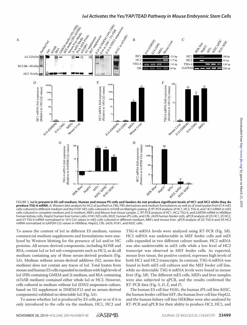
To assess the content of I�I in different ES medium, variouscommercial medium supplements and formulations were ana-lyzed by Western blotting for the presence of I�I and/or HCproteins. All serum-derived components, including KOSR andBSA, contain I�I or I�I sub-components such as HC2, as do allmedium containing any of those serum-derived products (Fig.5A). Medium without serum-derived additives (N2, serum-freemedium) does not contain any traces of I�I. Total lysates frommouse and human ES cells expanded in medium with high levels ofI�I (FBS containing GMEM and 2i medium, and BSA containingmTeSR medium) contained either whole I�I or HC2. However,cells cultured in medium without I�I (ESN2 suspension culture,based on N2 supplement in DMEM/F12 and no serum-derivedcomponents) exhibited no detectable I�I (Fig. 5A).
To assess whether I�I is produced by ES cells per se or if it isonly introduced to the cells via the medium, HC1, HC2 and
TSG-6 mRNA levels were analyzed using RT-PCR (Fig. 5B).HC1 mRNA was undetectable in MEF feeder cells and mEScells expanded in two different culture medium. HC2 mRNAwas also undetectable in mES cells while a low level of HC2transcript was observed in MEF feeder cells. As expected,mouse liver tissue, the positive control, expresses high levels ofboth HC1 and HC2 transcripts. In contrast, TSG-6 mRNA wasfound in both mES cell cultures and the MEF feeder cell line,while no detectable TSG-6 mRNA levels were found in mouseliver (Fig. 5B). The different mES cells, MEFs and liver sampleswere also subjected to qPCR, and the results confirmed theRT-PCR data (Fig. 5, D, E, and F).
The human ES cell line H181, the human iPS cell line K02C,the human feeder cell line hFF, the human liver cell line HepG2,and the human kidney cell line HEKBlue were also analyzed byRT-PCR and qPCR for their ability to produce HC2, HC1, and
FIGURE 5. I�I is present in ES cell medium. Human and mouse PS cells and feeders do not produce significant levels of HC1 and HC2 while they doproduce TSG-6 mRNA. A, Western blot analysis for HC2 of purified I�I, FBS, FBS-derivatives and medium formulations as well as of total lysates from E14 mEScells cultured in different medium and the H181 hES cells cultured in mTeSR on Matrigel coating. B, RT-PCR analysis of HC1, HC2, TSG-6, and 18 S mRNA in mEScells cultured in complete medium and 2i medium, MEFs and Mouse liver tissue sample. C, RT-PCR analysis of HC1, HC2, TSG-6, and GAPDH mRNA in HEKBluehuman kidney cells, HepG2 human liver tumor cells, H181 hES cells, K02C human iPS cells, and CRL-2429 human feeder cells. qPCR analysis of (D) HC1, (E) HC2,and (F) TSG-6 mRNA normalized to 18 S C(t) values in mES cells cultured in different medium, MEFs and mouse liver. qPCR analysis of (G) TSG-6 and (H) HC2mRNA normalized to GAPDH C(t) values in HEKBlue, HepG2, CRL-2429, H181, and K02C cells.
I�I Activates the Yes/YAP/TEAD Pathway in Mouse Embryonic Stem Cells
NOVEMBER 28, 2014 • VOLUME 289 • NUMBER 48 JOURNAL OF BIOLOGICAL CHEMISTRY 33499
by guest on March 21, 2019
http://ww
w.jbc.org/
Dow
nloaded from

TSG-6 (Fig. 5, C, G, and H). As expected, neither the humankidney negative control cells, the human ES cells nor the humaniPS cells express any HC2 mRNA, nor did the hFF feeder cellline. However, as expected the human liver cell line expresseshigh levels of both HC1 and HC2, while no TSG-6 mRNA wasdetected. Similar to mES cells, the human PS cell lines and thehuman feeder cells express high levels of TSG-6 mRNA (Fig. 5,C and G), further supporting the idea that I�I is supplemented,and that the cells capacity to produce TSG-6 confers them withthe ability to transfer the HCs to the hyaluronan chains presentin their ECM.
DISCUSSION
Serum supports attachment, survival, and proliferation ofmany cell types, including pluripotent stem cells. Many cyto-kines and extracellular matrix proteins have been identified inserum as key factors for the induction of proliferation andattachment, but many others remain to be studied for theireffect on cell culture. Even though stem cell research is movingtoward serum-free conditions, novel medium that aredescribed as serum-free usually still contain traces of serum-derived components through supplements such as BSA, andtherefore contain I�I proteins, as suggested by our results. Inthis study, we establish that I�I, a major component of serum,induces a signaling pathway known to activate the key stem cellfactors Oct3/4 and Nanog.
We have previously shown that LIF activates the Src familykinase Yes, the Yes-associated protein (YAP) and the family ofTEAD transcription factor, and that TEAD in its turn canenhance transcription of other stem cell factors such as Oct3/4and Nanog. For the first time we show that an ECM protein canalso activate this pathways in a similar manner. I�I has tradi-tionally been described as a structural component of the ECMand the HCs are the only proteins that have been demonstratedto bind covalently to HA (41). I�I has been described to benecessary for the cumulus formation of the ovary (31) and toplay an important role in tissue repair (34, 42, 43). Moreover, anisoform of I�I has been reported to be produced by the amni-otic membrane, and the hyaluronan-HC (HC�HA) complex ispresent in amniotic fluid, giving evidence of TSG-6 mediatedtransfer of HCs to hyaluronan in amniotic tissue (32, 44). Thesedata suggest that the HCs play a role during embryonic devel-opment and may also be important for ES cell maintenance.Neither mouse nor human embryonic stem cells express I�Iproteins as assessed by HC1 and HC2 mRNA expression. How-ever, they express TSG-6, supporting the hypothesis that I�I isintroduced to the cells through the medium and that the HCsare transferred to the stem cell’s ECM and hyaluronan throughTSG-6-mediated transfer.
I�I also induces signaling events that promote self-renewal ofmES cells, such as Yes-activating tyrosine auto-phosphoryla-tion, YAP activation by tyrosine phosphorylation and nuclearlocalization, TEAD family-dependent transcription, Nanog-promoter activity and mRNA production of the TEAD targetgenes Oct3/4, Cyr61, and CTGF.
Human serum contains an amount of I�I ranging from 0.6 –1.2 mg/ml (16). Thus, if estimating a concentration of 1 mg/mlin bovine serum; 5% FBS would account for 50 �g/ml. Never-
theless, a higher I�I concentration, 100 �g/ml, was required insome experiments to achieve significant TEAD response. Thereason may be that serum contains other components that addto the TEAD-activating effect, or it could be a side-effect ofcompromised I�I stability since we have observed that purifiedI�I exhibits a tendency to aggregate and partly degrade overtime.
The HC1 and HC2 domains are quite conserved betweendifferent species, and the mouse, human and bovine HC pro-teins share 80 – 85% amino acid identity. This concurs with ourdata showing that both bovine and human I�I induce similareffects in mouse ES cells. Interestingly, HC2, but not HC1 acti-vates YAP nuclear localization, TEAD-dependent transcriptionand Nanog promoter activity. HC1 and HC2 share only 39%identity, despite the fact that they both have the same structuraldomains, a vault protein I�I domain, and a von Willebrandfactor type A domain. Most studies so far have used the entireI�I molecule, making it difficult to distinguish the effect of thedifferent HCs. However, a few studies have demonstrated dif-ferent effects of HC1 and HC2. Tamra et al. showed that HC2acts as a natural inhibitor of brain tumor invasion, and thataddition of conditioned medium from cells expressing HC2 orforced expression of HC2 in the glioma cell line inhibits inva-sion and proliferation in vitro by inhibiting the PI3K/Akt path-way (33). Another study by Sanggaard et al. suggests that HC2 isnecessary for the TSG-6-mediated transfer to take place, possi-bly explaining both the inability of HC1 alone to induce TEADfamily and the need for higher concentrations of HC2 to inducea robust effect on TEAD activity (45). Interestingly, Zhang et al.studied the presence of HCs coupled to hyaluronan in amnioticmembrane, and found that whereas the amniotic membraneproduces HC1, HC2, and HC3, only HC1�HA complex wasfound in physiological amniotic membrane extracts (44), whichcould account for the anti-inflammatory, anti-scarring, andanti-angiogenic effects that this particular HCŸHA complexyields (46).
Up until now, I�I has mostly been studied as a structuralcomponent of the ECM and only a limited number of recentstudies have shown the ability of I�I to promote specific cellresponses. Moreover, most of these studies have focused onother components in the complex, such as hyaluronan,fibronectin, and vitronectin (31, 34, 42, 46, 47). Therefore themechanism through which I�I can signal to the cells is still notknown. It could be related to its binder hyaluronan, as there areseveral articles that have shown CD44 binding to hyaluronan tobe able to induce cell signaling, for example through Akt phos-phorylation (48). There is also the possibility of an integrin-mediated signaling, using I�I’s ability to bind to vitronectin(34), as integrin-mediated attachment has been also beendescribed to induce cell signaling such as PI3K/Akt phosphor-ylation (49). Moreover, recent articles have linked YAP to cel-lular responses induced by extracellular matrix stiffness, show-ing that hES cells, for example, have increased YAP nuclearlocalization when seeded on stiffer gels (50, 51). I�I may there-fore not signal to the cells directly, but modify the hyaluronannetwork, changing the mechanical properties of the surround-ing matrix of the cells, which in its turn could result in a cellularresponse such as YAP nuclear localization.
I�I Activates the Yes/YAP/TEAD Pathway in Mouse Embryonic Stem Cells
33500 JOURNAL OF BIOLOGICAL CHEMISTRY VOLUME 289 • NUMBER 48 • NOVEMBER 28, 2014
by guest on March 21, 2019
http://ww
w.jbc.org/
Dow
nloaded from

In conclusion, our results show, for the first time that I�I is acomponent of the ES cell ECM network and has the capacity toactivate intracellular signal transduction pathways linked to keyregulatory transcription factors, and could therefore play animportant role in ES cell maintenance. Since I�I is present in allmES and hES cell cultures, either via supplements in themedium or the matrix/feeders the cells grow on, it is intriguingto speculate that I�I may be an unacknowledged crucial ECMcomponent making up the stem cell niche and that it may needto be supplemented to the medium in a purified or recombinantform when moving toward completely defined, xeno-free con-ditions. We are currently investigating the mode of action forI�I induced intracellular signaling as well as its biological func-tion in human and mouse ES and iPS cell cultures.
Acknowledgments—We thank the following individuals who kindlyprovided reagents and cell lines: Dr. I. Chambers and Dr. A. Smithfrom the University of Edinburgh and University of Cambridge,respectively (E14/T cells, pPyCAGIP, and pPyCAGIP Nanog vec-tor), Dr. N. Eberhardt from Mayo Clinic (the pCS and pGTIICconstructs), Dr. K. Shiota from University of Tokyo (Nanog-luciferaseconstruct), Dr. E. Fries from Uppsala University (purified human I�I,HCs and fraction of human serum), I.M. Lööf from Pharmacia-Upjohn (side fraction from the commercial production of factor IX),Dr. P. Heldin from Uppsala University (Hyaluronan), Dr. O. Hovattafrom Karolinska Institute (H181 human ES cell line), Dr. J. Schusterfrom Uppsala University (K02C human induced pluripotent stem cellline), Dr. Jing-Ping Li from Uppsala Universitert (mouse liver tissuesample), Dr. Femke Heindrycks from Uppsala University (HepG2 cellline) and Dr. Peder Fredlund Fuchs from Uppsala University (HEKBlue cell line). We thank Dr. Jing-Ping and Dr. Erik Fries for fruitfuldiscussions and support and Dr. Lena Kjellén and Dr. PaulO’Callaghan for critical reviewing of the manuscript.
REFERENCES1. Chambers, I., and Smith, A. (2004) Self-renewal of teratocarcinoma and
embryonic stem cells. Oncogene 23, 7150 –71602. Xiao, L., Yuan, X., and Sharkis, S. J. (2006) Activin A maintains self-re-
newal and regulates fibroblast growth factor, Wnt, and bone morphogenicprotein pathways in human embryonic stem cells. Stem Cells 24,1476 –1486
3. Vallier, L., Alexander, M., and Pedersen, R. A. (2005) Activin/Nodal andFGF pathways cooperate to maintain pluripotency of human embryonicstem cells. J. Cell Sci. 118, 4495– 4509
4. Miyazaki, T., Futaki, S., Suemori, H., Taniguchi, Y., Yamada, M., Kawasaki,M., Hayashi, M., Kumagai, H., Nakatsuji, N., Sekiguchi, K., and Kawase, E.(2012) Laminin E8 fragments support efficient adhesion and expansion ofdissociated human pluripotent stem cells. Nature Commun. 3, 1236
5. Braam, S. R., Zeinstra, L., Litjens, S., Ward-van Oostwaard, D., van denBrink, S., van Laake, L., Lebrin, F., Kats, P., Hochstenbach, R., Passier, R.,Sonnenberg, A., and Mummery, C. L. (2008) Recombinant vitronectin is afunctionally defined substrate that supports human embryonic stem cellself-renewal via �v�5 integrin. Stem Cells 26, 2257–2265
6. Melkoumian, Z., Weber, J. L., Weber, D. M., Fadeev, A. G., Zhou, Y.,Dolley-Sonneville, P., Yang, J., Qiu, L., Priest, C. A., Shogbon, C., Martin,A. W., Nelson, J., West, P., Beltzer, J. P., Pal, S., and Brandenberger, R.(2010) Synthetic peptide-acrylate surfaces for long-term self-renewal andcardiomyocyte differentiation of human embryonic stem cells. NatureBiotechnology 28, 606 – 610
7. Ilic, D. (2006) Culture of human embryonic stem cells and the extracellu-lar matrix microenvironment. Regenerative Med. 1, 95–101
8. Stojkovic, P., Lako, M., Przyborski, S., Stewart, R., Armstrong, L., Evans, J.,
Zhang, X., and Stojkovic, M. (2005) Human-serum matrix supports un-differentiated growth of human embryonic stem cells. Stem Cells 23,895–902
9. Annerén, C. (2008) Tyrosine kinase signalling in embryonic stem cells.Clin. Sci. 115, 43–55
10. Betschinger, J., Nichols, J., Dietmann, S., Corrin, P. D., Paddison, P. J., andSmith, A. (2013) Exit from pluripotency is gated by intracellular redistri-bution of the bHLH transcription factor Tfe3. Cell 153, 335–347
11. Tamm, C., Bower, N., and Annerén, C. (2011) Regulation of mouse em-bryonic stem cell self-renewal by a Yes-YAP-TEAD2 signaling pathwaydownstream of LIF. J. Cell Sci. 124, 1136 –1144
12. Lian, I., Kim, J., Okazawa, H., Zhao, J., Zhao, B., Yu, J., Chinnaiyan, A.,Israel, M. A., Goldstein, L. S., Abujarour, R., Ding, S., and Guan, K. L.(2010) The role of YAP transcription coactivator in regulating stem cellself-renewal and differentiation. Genes Dev. 24, 1106 –1118
13. Annerén, C., Cowan, C. A., and Melton, D. A. (2004) The Src family oftyrosine kinases is important for embryonic stem cell self-renewal. J. Biol.Chem. 279, 31590 –31598
14. Vassilev, A., Kaneko, K. J., Shu, H., Zhao, Y., and DePamphilis, M. L. (2001)TEAD/TEF transcription factors utilize the activation domain of YAP65, aSrc/Yes-associated protein localized in the cytoplasm. Genes Dev. 15,1229 –1241
15. Zhang, X., Meyn, M. A., 3rd, and Smithgall, T. E. (2014) c-Yes tyrosinekinase is a potent suppressor of ES cell differentiation and antagonizes theactions of its closest phylogenetic relative, c-Src. ACS chemical biology 9,139 –146
16. Josic, D., Brown, M. K., Huang, F., Lim, Y. P., Rucevic, M., Clifton, J. G., andHixson, D. C. (2006) Proteomic characterization of inter-� inhibitor pro-teins from human plasma. Proteomics 6, 2874 –2885
17. Jessen, T. E., and Ødum, L. (2003) Role of tumour necrosis factor stimu-lated gene 6 (TSG-6) in the coupling of inter-�-trypsin inhibitor to hya-luronan in human follicular fluid. Reproduction 125, 27–31
18. Smith, A. (1991) Culture and differentiation of embryonic stem cells.Methods in Cell Science 13, 89 –94
19. Ying, Q. L., Wray, J., Nichols, J., Batlle-Morera, L., Doble, B., Woodgett, J.,Cohen, P., and Smith, A. (2008) The ground state of embryonic stem cellself-renewal. Nature 453, 519 –523
20. Li, P., Tong, C., Mehrian-Shai, R., Jia, L., Wu, N., Yan, Y., Maxson, R. E.,Schulze, E. N., Song, H., Hsieh, C. L., Pera, M. F., and Ying, Q. L. (2008)Germline competent embryonic stem cells derived from rat blastocysts.Cell 135, 1299 –1310
21. Ludwig, T. E., Bergendahl, V., Levenstein, M. E., Yu, J., Probasco, M. D.,and Thomson, J. A. (2006) Feeder-independent culture of human embry-onic stem cells. Nature Methods 3, 637– 646
22. Lei, T., He, Q. Y., Wang, Y. L., Si, L. S., and Chiu, J. F. (2008) Heparinchromatography to deplete high-abundance proteins for serum proteom-ics. Clin. Chim. Acta 388, 173–178
23. Arakawa, T., Tsumoto, K., Ejima, D., Kita, Y., Yonezawa, Y., and Toku-naga, M. (2007) Induced binding of proteins by ammonium sulfate inaffinity and ion-exchange column chromatography. J. Biochem. Biophys.Methods 70, 493– 498
24. Blom, A. M., Mörgelin, M., Oyen, M., Jarvet, J., and Fries, E. (1999) Struc-tural characterization of inter-�-inhibitor. Evidence for an extendedshape. J. Biol. Chem. 274, 298 –304
25. Enghild, J. J., Thøgersen, I. B., Pizzo, S. V., and Salvesen, G. (1989) Analysisof inter-�-trypsin inhibitor and a novel trypsin inhibitor, pre-�-trypsininhibitor, from human plasma. Polypeptide chain stoichiometry and as-sembly by glycan. J. Biol. Chem. 264, 15975–15981
26. Jiang, S. W., and Eberhardt, N. L. (1995) Involvement of a protein distinctfrom transcription enhancer factor-1 (TEF-1) in mediating human chori-onic somatomammotropin gene enhancer function through the GT-IICenhanson in choriocarcinoma and COS cells. J. Biol. Chem. 270,13906 –13915
27. Hattori, N., Imao, Y., Nishino, K., Hattori, N., Ohgane, J., Yagi, S., Tanaka,S., and Shiota, K. (2007) Epigenetic regulation of Nanog gene in embryonicstem and trophoblast stem cells. Genes Cells 12, 387–396
28. Pfaffl, M. W. (2001) A new mathematical model for relative quantificationin real-time RT-PCR. Nucleic Acids Res. 29, e45
I�I Activates the Yes/YAP/TEAD Pathway in Mouse Embryonic Stem Cells
NOVEMBER 28, 2014 • VOLUME 289 • NUMBER 48 JOURNAL OF BIOLOGICAL CHEMISTRY 33501
by guest on March 21, 2019
http://ww
w.jbc.org/
Dow
nloaded from

29. Ying, Q. L., Nichols, J., Chambers, I., and Smith, A. (2003) BMP inductionof Id proteins suppresses differentiation and sustains embryonic stem cellself-renewal in collaboration with STAT3. Cell 115, 281–292
30. Varelas, X., Sakuma, R., Samavarchi-Tehrani, P., Peerani, R., Rao, B. M.,Dembowy, J., Yaffe, M. B., Zandstra, P. W., and Wrana, J. L. (2008) TAZcontrols Smad nucleocytoplasmic shuttling and regulates human embry-onic stem-cell self-renewal. Nature Cell Biology 10, 837– 848
31. Hess, K. A., Chen, L., and Larsen, W. J. (1999) Inter-�-inhibitor binding tohyaluronan in the cumulus extracellular matrix is required for optimalovulation and development of mouse oocytes. Biol. Reprod. 61, 436 – 443
32. He, H., Li, W., Tseng, D. Y., Zhang, S., Chen, S. Y., Day, A. J., and Tseng,S. C. (2009) Biochemical characterization and function of complexesformed by hyaluronan and the heavy chains of inter-�-inhibitor (HC*HA)purified from extracts of human amniotic membrane. J. Biol. Chem. 284,20136 –20146
33. Werbowetski-Ogilvie, T. E., Agar, N. Y., Waldkircher de Oliveira, R. M.,Faury, D., Antel, J. P., Jabado, N., and Del Maestro, R. F. (2006) Isolation ofa natural inhibitor of human malignant glial cell invasion: inter �-trypsininhibitor heavy chain 2. Cancer Res. 66, 1464 –1472
34. Adair, J. E., Stober, V., Sobhany, M., Zhuo, L., Roberts, J. D., Negishi, M.,Kimata, K., and Garantziotis, S. (2009) Inter-�-trypsin inhibitor promotesbronchial epithelial repair after injury through vitronectin binding. J. Biol.Chem. 284, 16922–16930
35. Zhao, B., Ye, X., Yu, J., Li, L., Li, W., Li, S., Yu, J., Lin, J. D., Wang, C. Y.,Chinnaiyan, A. M., Lai, Z. C., and Guan, K. L. (2008) TEAD mediatesYAP-dependent gene induction and growth control. Genes Dev. 22,1962–1971
36. Lai, D., Ho, K. C., Hao, Y., and Yang, X. (2011) Taxol resistance in breastcancer cells is mediated by the hippo pathway component TAZ and itsdownstream transcriptional targets Cyr61 and CTGF. Cancer Res. 71,2728 –2738
37. Zhang, H., Liu, C. Y., Zha, Z. Y., Zhao, B., Yao, J., Zhao, S., Xiong, Y., Lei,Q. Y., and Guan, K. L. (2009) TEAD transcription factors mediate thefunction of TAZ in cell growth and epithelial-mesenchymal transition.J. Biol. Chem. 284, 13355–13362
38. Tamm, C., Pijuan Galitó, S., and Anneren, C. (2013) A comparative studyof protocols for mouse embryonic stem cell culturing. PloS one 8, e81156
39. Zhao, B., Li, L., Lei, Q., and Guan, K. L. (2010) The Hippo-YAP pathway inorgan size control and tumorigenesis: an updated version. Genes Dev. 24,862– 874
40. Levy, D., Adamovich, Y., Reuven, N., and Shaul, Y. (2008) Yap1 phosphor-ylation by c-Abl is a critical step in selective activation of proapoptoticgenes in response to DNA damage. Mol. Cell 29, 350 –361
41. Zhao, M., Yoneda, M., Ohashi, Y., Kurono, S., Iwata, H., Ohnuki, Y., andKimata, K. (1995) Evidence for the covalent binding of SHAP, heavychains of inter-�-trypsin inhibitor, to hyaluronan. J. Biol. Chem. 270,
26657–2666342. Garantziotis, S., Zudaire, E., Trempus, C. S., Hollingsworth, J. W., Jiang,
D., Lancaster, L. H., Richardson, E., Zhuo, L., Cuttitta, F., Brown, K. K.,Noble, P. W., Kimata, K., and Schwartz, D. A. (2008) Serum inter-�-tryp-sin inhibitor and matrix hyaluronan promote angiogenesis in fibrotic lunginjury. Am. J. Respir. Crit. Care Med. 178, 939 –947
43. Garantziotis, S., Hollingsworth, J. W., Ghanayem, R. B., Timberlake, S.,Zhuo, L., Kimata, K., and Schwartz, D. A. (2007) Inter-�-trypsin inhibitorattenuates complement activation and complement-induced lung injury.J. Immunol. 179, 4187– 4192
44. Zhang, S., He, H., Day, A. J., and Tseng, S. C. (2012) Constitutive expres-sion of inter-�-inhibitor (I�I) family proteins and tumor necrosis factor-stimulated gene-6 (TSG-6) by human amniotic membrane epithelial andstromal cells supporting formation of the heavy chain-hyaluronan (HC-HA) complex. J. Biol. Chem. 287, 12433–12444
45. Sanggaard, K. W., Sonne-Schmidt, C. S., Krogager, T. P., Lorentzen, K. A.,Wisniewski, H. G., Thøgersen, I. B., and Enghild, J. J. (2008) The transfer ofheavy chains from bikunin proteins to hyaluronan requires both TSG-6and HC2. J. Biol. Chem. 283, 18530 –18537
46. Shay, E., He, H., Sakurai, S., and Tseng, S. C. (2011) Inhibition of angio-genesis by HC.HA, a complex of hyaluronan and the heavy chain of inter-�-inhibitor, purified from human amniotic membrane. Invest. Ophthal-mol. Vis. Sci. 52, 2669 –2678
47. Tan, K. T., Baildam, A. D., Juma, A., Milner, C. M., Day, A. J., and Bayat, A.(2011) Hyaluronan, TSG-6, and inter-�-inhibitor in periprosthetic breastcapsules: reduced levels of free hyaluronan and TSG-6 expression in con-tracted capsules. Aesthet. Surg. J. 31, 47–55
48. Shimabukuro, Y., Terashima, H., Takedachi, M., Maeda, K., Nakamura,T., Sawada, K., Kobashi, M., Awata, T., Oohara, H., Kawahara, T.,Iwayama, T., Hashikawa, T., Yanagita, M., Yamada, S., and Murakami, S.(2011) Fibroblast growth factor-2 stimulates directed migration of peri-odontal ligament cells via PI3K/AKT signaling and CD44/hyaluronan in-teraction. J. Cell. Physiol. 226, 809 – 821
49. Jin, Y. J., Park, I., Hong, I. K., Byun, H. J., Choi, J., Kim, Y. M., and Lee, H.(2011) Fibronectin and vitronectin induce AP-1-mediated matrix metal-loproteinase-9 expression through integrin �(5)�(1)/�(v)�(3)-dependentAkt, ERK and JNK signaling pathways in human umbilical vein endothelialcells. Cell. Signal. 23, 125–134
50. Musah, S., Morin, S. A., Wrighton, P. J., Zwick, D. B., Jin, S., and Kiessling,L. L. (2012) Glycosaminoglycan-binding hydrogels enable mechanicalcontrol of human pluripotent stem cell self-renewal. ACS nano 6,10168 –10177
51. Dupont, S., Morsut, L., Aragona, M., Enzo, E., Giulitti, S., Cordenonsi, M.,Zanconato, F., Le Digabel, J., Forcato, M., Bicciato, S., Elvassore, N., andPiccolo, S. (2011) Role of YAP/TAZ in mechanotransduction. Nature 474,179 –183
I�I Activates the Yes/YAP/TEAD Pathway in Mouse Embryonic Stem Cells
33502 JOURNAL OF BIOLOGICAL CHEMISTRY VOLUME 289 • NUMBER 48 • NOVEMBER 28, 2014
by guest on March 21, 2019
http://ww
w.jbc.org/
Dow
nloaded from

Sara Pijuan-Galitó, Christoffer Tamm and Cecilia AnnerénTranscriptional Complex in Mouse Embryonic Stem Cells
-inhibitor Activates the Yes Tyrosine Kinase and YAP/TEADαSerum Inter-
doi: 10.1074/jbc.M114.580076 originally published online October 9, 20142014, 289:33492-33502.J. Biol. Chem.
10.1074/jbc.M114.580076Access the most updated version of this article at doi:
Alerts:
When a correction for this article is posted•
When this article is cited•
to choose from all of JBC's e-mail alertsClick here
http://www.jbc.org/content/289/48/33492.full.html#ref-list-1
This article cites 51 references, 22 of which can be accessed free at
by guest on March 21, 2019
http://ww
w.jbc.org/
Dow
nloaded from