session number 216 certification review: cardiovascular
TRANSCRIPT
Session Number 216
CERTIFICATION REVIEW: Cardiovascular Part 2
Barbara Pope, RN, MSN, CCRN, PCCN, CCNS [email protected]
Critical Care Clinical Educator Albert Einstein Healthcare Network
Philadelphia, PA
Content Description
This session reviews the pathophysiology, presentation, diagnosis, and collaborative care of chronic and acute decompensated heart failure. It also discusses cardiomyopathy , valvular diseases and cardiac inflammatory disease as they relate to heart failure. Finally, it will look at dysrhythmias and the care of the patient with a temporary pacemaker. These topics will be discussed with an emphasis on possible questions that may be asked on these subjects in the CCRN, PCCN, and CMC examinations. There will be time allotted for sample questions. Learning Objectives At the end of this session, the participant will be able to:
1. Describe the clinical presentation, diagnosis and collaborative management of the patient
with chronic and acute decompensated heart failure. 2. Describe the presentations and collaborative care of cardiomyopathy, valvular disease,
and cardiac inflammatory disease. 3. Discuss the identification of and interventions for patients experiencing cardiac
dysrhythmia and those requiring a temporary pacemaker
REFERENCES NOTE: Please refer to outline for references pertaining to this session.

Certification Review: Cardiovascular Part 2
Approximately 20% of the CCRN exam, 36% of the PCCN exam and 43% of the CMC exam will focus on cardiovascular disease.
CCRN, PCCN and CMC Acute Coronary Syndrome Interventional cardiology Cardiac surgery Heart Failure Acute pulmonary edema Dysrhythmias Conduction defects Cardiomyopathies Structural heart defects Cardiogenic shock Hypovolemic shock (in multisystem on PCCN; discussed here)
CCRN, PCCN and CMC Acute peripheral vascular insufficiency/ peripheral vascular surgery Hypertensive crisis Ruptured or dissecting aneurysm CCRN and CMC only Cardiac trauma PCCN and CMC only Acute inflammatory disease Cardiac tamponade Pulmonary hypertension (in pulmonary on PCCN. Discussed in pulmonary session)
Note for PCCN candidates: This presentation includes discussions of pulmonary artery catheter measurements, administration of vasoactive medications, and advanced mechanical devices such as intra-aortic balloon pump and ventricular assist devices. These topics will not be tested in the PCCN exam.
I. Heart failure A. Definitions
1. ACC/AHA Practice Guidelines A complex clinical syndrome that can result form any structural or functional cardiac disorder that impairs the ability of the ventricle to fill with or eject blood. 2. The inability of the heart to maintain a continuous flow of blood commensurate with the metabolic needs of tissues and organs, or to do so only by means of increased filling pressures.
B. Determination of severity of heart failure
New York Heart Association Functional Classification System Class I Minimal
No limitations Normal daily activity does not initiate symptoms of fatigue, dyspnea, palpitations or angina

New York Heart Association Functional Classification System Class II Mild
Slightly limited physical activity Comfortable at rest Normal daily activities initiate onset of symptoms
Class III Moderate
Markedly limited physical activity Comfortable at rest Less than normal activity initiates symptoms
Class IV Severe
Any type of activity initiates symptoms Symptoms may occur at rest
C. Cardinal symptoms of heart failure Dyspnea Fatigue Fluid retention
D. Causes Most common causes
CAD Hypertension Dilated cardiomyopathy
Other causes Valvular disease Diabetes mellitus Hyperlipidemia Smoking PVD
RHD Mediastinal irradiation Sleep apnea Illicit drug use
E. Physiology of HF Effects of HF on cardiac output
Heart rate Preload Afterload
Contractility Neurohormonal mechanisms in heart failure Sympathetic nervous stimulation Renin-angiotensin-aldosterone system activity
Meant to be compensatory Results in:
Structural changes Chamber dilation or hypertrophy (cardiac remodeling) Damaged myocytes More spherical shape
} Components of stroke volume

Decreased functioning F. Diagnostic tests
Diagnosis made mainly through history and physical findings Chest X-Ray: Enlarged cardiac silhouette; Kerley B lines, pleural effusion ECG: Q waves, IVCD, atrial fibrillation, ventricular ectopy, atrial enlargement, ventricular hypertrophy Both nonspecific. Two-dimensional echocardiogram with Doppler flow studies: abnormal wall motion, chamber dilation, hypertrophy, valve dysfunction, decreased ejection fraction. Most useful test
G. Labs CBC, electrolytes, including Ca++, Mg+ and Phosphorus, BUN, creatinine, liver enzymes, thyroid testing B-type natriuretic peptide (BNP) levels
Elevated in proportion to severity of HF - ≥100 pg/ml Lower levels rule out HF as a cause of dyspnea - ≤ 80 pg/ml Levels correlate with NYHA classifications of heart failure
H. Treatment Drug therapy
Diuretics Loop – Furosmide (Lasix), bumetanide (Bumex)
Monitor K-+, Mg+, BUN, Cr, hypotension, I&O, daily
weights Administer IV push slowly – 20 mg/min to avoid ototoxicity
Potassium-sparing – spironolactone (Aldactone) Use in combination with loop diuretics; watch for hyperkalemia Contraindicated in patients with SPB < 90 mmHg, cardiogenic shock
Thiazide – Metolazone (Zarolyolyn) Enhace diuresis when used with furosemide or bumetanide
ACE inhibitors - captopril (Capoten), enalapril (Vasotec) Inhibits RAA system; monitor K+, BUN, creatinine, hypotension, dry cough, angioedema
ARBs - losartan (Cozaar), valsartan (Diovan) Not as effective as ACEI, less risk of cough, angioedema. Monitor as with ACEI
Beta-blockers - bisoprolol (Zebeta) , carvedilol (Coreg), and metoprolol (Lopressor) only βB recommended in AHA/ACC guidelines
Titrate slowly; contraindicated in hypotension, bradycardia, 2nd & 3rd degree AVB, asthma
Digitalis glycosides Positive inotrope, increases cardiac contractility; may also down-regulate sympathetic activity. Indicated for patients who remain symptomatic or who are being titrated on βB. Monitor dysrhythmia, esp. heart block, N/V, visual disturbances, dig toxicity.
I. Management of Patient with HF Control of hypertension, diabetes, hyperlipidemia Smoking cessation; restrict/eliminate alcohol use Healthy diet/ low sodium diet, 2-4 gm Fluid restriction, 64 oz/day; daily weight
Report weight gain of 3-4 lb over several days. Flu and pneumonia vaccination Supervised exercise
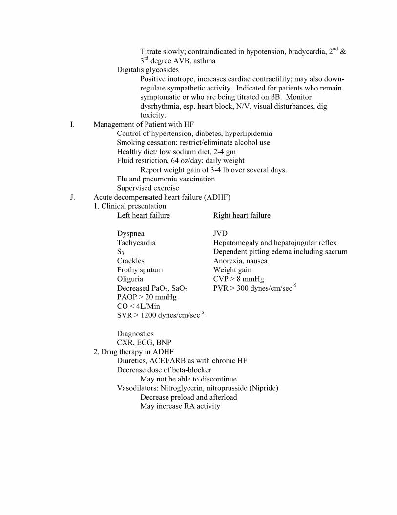
J. Acute decompensated heart failure (ADHF) 1. Clinical presentation
Left heart failure
Right heart failure
Dyspnea Tachycardia S3 Crackles Frothy sputum Oliguria Decreased PaO2, SaO2 PAOP > 20 mmHg CO < 4L/Min SVR > 1200 dynes/cm/sec-5
JVD Hepatomegaly and hepatojugular reflex Dependent pitting edema including sacrum Anorexia, nausea Weight gain CVP > 8 mmHg PVR > 300 dynes/cm/sec-5
Diagnostics CXR, ECG, BNP
2. Drug therapy in ADHF Diuretics, ACEI/ARB as with chronic HF Decrease dose of beta-blocker
May not be able to discontinue Vasodilators: Nitroglycerin, nitroprusside (Nipride)
Decrease preload and afterload May increase RA activity

Use with ACEI or ARB to prevent Decrease myocardial oxygen consumption Do not cause dysrhythmias. Nitroglycerin
Venous dilator and preload reducer. Decreases coronary vascular resistance. Afterload reducer at doses >30 mcg/min;. May cause headache and abdominal pain
Nitroprusside Venous and arterial dilator at all doses. Effective in acute mitral or aortic regurgitation. May cause coronary steal – vasodilation in nonischemic areas that can shunt blood from areas of ischemia. Should have arterial line and/or PA catheter. Monitor for cyanide toxicity.
Morphine If given, should be after nitroglycerin treatment. No evidence of efficacy; second-line treatment
Neurohormonal: nesiritide (Natrecor) Reduces preload and afterload Improves CO/CI Diuretic and natriuretic Decreased levels of aldosterone and norepinephrine Fast onset of action
Decreases PAOP within 15 minutes No atrial or ventricular arrhythmias as with positive inotropes Does not require ICU admission, arterial line, PA catheter. Does not appear to increase mortality. Major side effect is hypotension.
Turn off or decrease rate of infusion. Reposition patient on side, administer fluids. Restart nesiritide at rate 30% lower when BP stable
Recent concern that it may cause decreased renal function Inotropes: Dobutamine (Dobutrex), milrinone (Primacor)
Increase CO by increasing cardiac contractility Increases heart rate Increases myocardial oxygen demand Associated with increased mortality Contraindicated in hypertrophic cardiomyopathy, aortic stenosis Should only be used in shock or poor perfusion Milrinone does not compete with beta blockade
3. Oxygen

CPAP or BiPAP Noninvasive increase in intrathoracic positive pressure decreases preload
Mechanical ventilation if NIPPV is not effective and in cardiogenic shock 4. Other treatment modalities
ICD Sudden death often cause of mortality in HF. Implantation of ICD may decrease mortality. Increase in hospital admission for HF, possibly due to pacemaker function of IVCD causing ventricular dyssynchrony. Used in conjunction with ventricular antidysrhythmics such as Amiodarone
Cardiac resynchronization IVCD results in dyssynchronous ventricular contractions. Worsens systolic HF, interferes with diastolic function. May worsen MR. Biventricular pacemaker synchronized to patient’s sinus rhythm and programmed to stimulate the right ventricle with a conventional lead and the left ventricle through a specially designed coronary sinus lead. Biventricular pacing and ICD capabilities in most cases. Improves NYHA class, exercise tolerance, LVEF, and quality of life
Ultrafiltration Removal of fluid via ultrafiltration mode of CRRT Administered over 8 hours Maximum fluid removal 500 ml/hr Well tolerated
5. Nursing interventions Patient assessment Q 1-2 hours: VS, LOC Head-to-toe assessments Neuro: decreased mentation can indicate poor perfusion to brain Cardiac: Heart sounds: S3, gallop, murmur, distant
Peripheral pulses, edema Skin temperature, color, moisture
Nailbeds: color, capillary refill Hemodynamic parameters: CO, PAP, PAOP, SVR
Baseline, after initiation of therapy to determine effectiveness Pulmonary: Crackles, wheezes, tachypnea, frothy sputum
GI: N/V, appetite, bowel sounds

GU: urine output: oliguria, anuria. Appearance: concentrated, dilute Daily weights, I&O
Monitor electrolytes Assess effectiveness of interventions Patient/family education
Medication Tests Therapies ICU routine
Discharge teaching Activity level Diet, including sodium and fluid restriction Discharge medications Weight monitoring
Certification Questions 1. A patient is admitted with complaints of chest pain accompanied by nausea and vomiting. The skin is cool and clammy. The following are noted:
VS: BP 140/90; HR 120; RR 26 CO 3.5 L/min CVP 10 mmHg CI 2.1 L/min/M3 PAOP 20 mmHg Presence of S3 12 lead ECG shows acute changes in V2, V3, and V4
For this patient, the goals of therapy would be to: A. Decrease preload and afterload C. Increase contractility and preload B. Increase preload and afterload D. Decrease contractility and afterload 2. A patient with acute decompensated heart failure is receiving a continuous infusion of nesiritide (Natrecor) at 0.1 mcg/kg/min. Currently, the patient has the following vital signs: HR 116 bpm, sinus tachycardia with premature atrial contractions, BP 78/48 mmHg, RR 28/min, and SpO2 90% on 40% BiPAP mask. Immediate interventions by the nurse would include which of the following?
A. Discontinue the nesiritide (Natrecor) infusion B. Place the patient in a supine position C. Administer a 250 ml normal saline fluid bolus D. Continue the nesiritide (Natrecor) infusion and administer 40 mg furosemide (Lasix) intravenously
3. After implantation of a biventricular pacemaker for end-stage heart failure, ECG signs that the pacemaker is not functioning properly would include:
A. A-V interval less than 0.20 sec B. Widening of the QRS C. T-wave inversion D. More than one P wave for each QRS
4. A Patient admitted with shortness of breath demonstrates the following findings: temperature 36.8oC, HR 120/min sinus tachycardia, BP 130/76 mmHg, RR 36/min with SpO2 91%. Breath sounds reveal inspiratory crackles and rhonchi in all lung fields, The chest X-ray report states that there are Kerley B lines, enlargement of the peribronchial hilar spaces, and enlarged cardiac silhouette. These findings are consistent with which of the following?
A. Pericardial tamponade B. Pulmonary edema C. Pneumonia D. Acute inferior wall MI with right ventricular failure
5. When performing patient teaching with a heart failure patient, the nurse should instruct the patient to immediately contact the HCP for which of the following?
A. Weight gain greater than 2 kg in 24 hours B. Development of cough C. Leg edema D. Increased fatigue and exercise intolerance for 24 hours
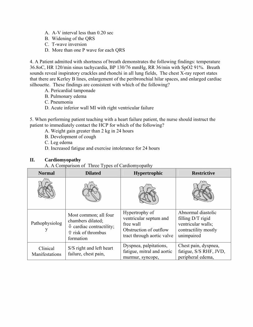
II. Cardiomyopathy A. A Comparison of Three Types of Cardiomyopathy
Normal Dilated Hypertrophic Restrictive
Pathophysiology
Most common; all four chambers dilated; cardiac contractility; risk of thrombus formation
Hypertrophy of ventricular septum and free wall Obstruction of outflow tract through aortic valve
Abnormal diastolic filling D/T rigid ventricular walls; contractility mostly unimpaired
Clinical Manifestations
S/S right and left heart failure, chest pain,
Dyspnea, palpitations, fatigue, mitral and aortic murmur, syncope,
Chest pain, dyspnea, fatigue, S/S RHF, JVD, peripheral edema,
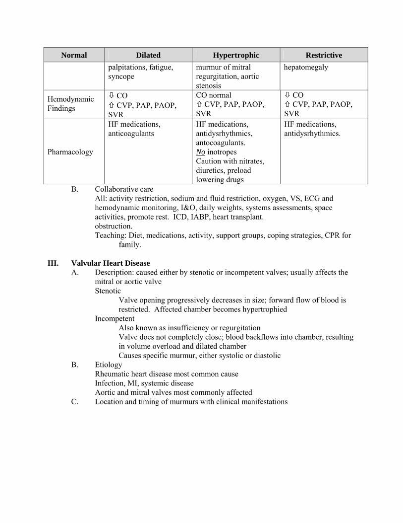
Normal Dilated Hypertrophic Restrictive
palpitations, fatigue, syncope
murmur of mitral regurgitation, aortic stenosis
hepatomegaly
Hemodynamic Findings
CO CVP, PAP, PAOP, SVR
CO normal CVP, PAP, PAOP, SVR
CO CVP, PAP, PAOP, SVR
Pharmacology
HF medications, anticoagulants
HF medications, antidysrhythmics, antocoagulants. No inotropes Caution with nitrates, diuretics, preload lowering drugs
HF medications, antidysrhythmics.
B. Collaborative care All: activity restriction, sodium and fluid restriction, oxygen, VS, ECG and hemodynamic monitoring, I&O, daily weights, systems assessments, space activities, promote rest. ICD, IABP, heart transplant. obstruction. Teaching: Diet, medications, activity, support groups, coping strategies, CPR for
family.
III. Valvular Heart Disease A. Description: caused either by stenotic or incompetent valves; usually affects the
mitral or aortic valve Stenotic
Valve opening progressively decreases in size; forward flow of blood is restricted. Affected chamber becomes hypertrophied
Incompetent Also known as insufficiency or regurgitation Valve does not completely close; blood backflows into chamber, resulting in volume overload and dilated chamber Causes specific murmur, either systolic or diastolic
B. Etiology Rheumatic heart disease most common cause
Infection, MI, systemic disease Aortic and mitral valves most commonly affected
C. Location and timing of murmurs with clinical manifestations
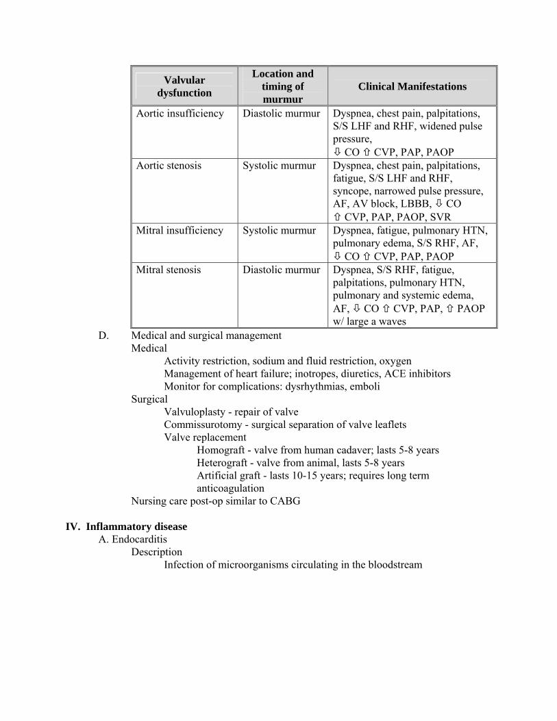
Valvular dysfunction
Location and timing of murmur
Clinical Manifestations
Aortic insufficiency Diastolic murmur Dyspnea, chest pain, palpitations, S/S LHF and RHF, widened pulse pressure, CO CVP, PAP, PAOP
Aortic stenosis Systolic murmur Dyspnea, chest pain, palpitations, fatigue, S/S LHF and RHF, syncope, narrowed pulse pressure, AF, AV block, LBBB, CO CVP, PAP, PAOP, SVR
Mitral insufficiency
Systolic murmur
Dyspnea, fatigue, pulmonary HTN, pulmonary edema, S/S RHF, AF, CO CVP, PAP, PAOP
Mitral stenosis Diastolic murmur
Dyspnea, S/S RHF, fatigue, palpitations, pulmonary HTN, pulmonary and systemic edema, AF, CO CVP, PAP, PAOP w/ large a waves
D. Medical and surgical management Medical
Activity restriction, sodium and fluid restriction, oxygen Management of heart failure; inotropes, diuretics, ACE inhibitors Monitor for complications: dysrhythmias, emboli
Surgical Valvuloplasty - repair of valve Commissurotomy - surgical separation of valve leaflets Valve replacement
Homograft - valve from human cadaver; lasts 5-8 years Heterograft - valve from animal, lasts 5-8 years Artificial graft - lasts 10-15 years; requires long term anticoagulation
Nursing care post-op similar to CABG IV. Inflammatory disease
A. Endocarditis Description
Infection of microorganisms circulating in the bloodstream

Bacteria carried through system and deposited onto endocardial surfaces, especially valve leaflets
B. Infectious process Bacteria grow on leaflets –Vegetation Lesions or vegetations form on valves Valves have irregular or “cauliflower” appearance Leaflets damaged and dysfunctional Life-threatening May require valve surgery Can grow to involve the:
Cordae tendinae Papillary muscles Conduction system
C. Causative organisms Streptococcus (most common) Staphylococcus Gram negative bacilli (E. coli, Klebsiella) Fungi (Candida, Histoplasma)
D. Affected valves Mitral most commonly affected Aortic second most affected
E. Populations at risk Rarely occurs in people with normal hearts IV drug abuse Risk increased with preexisting cardiac conditions
Prosthetic valves History of previous endocarditis Damaged or abnormal heart valves due to: Rheumatic fever Congenital heart disease Congenital valve defects
F. Presenting symptoms Dependent on: Valve involved Organism Duration of time Extent of vegetative growth Fever Chills Night sweats
Fatigue Headache Musculoskeletal complaints
Cough Weight loss General malaise Weakness
New murmur Heart failure Positive blood cultures Anemia
G. Treatment
Blood cultures to identify organism Administration of antibiotics or antifungals Oxygen if indicated HF treatment if present May require valve replacement surgery
V. Dysrhythmias A. Defined
Any cardiac rhythm other than sinus rhythm at a normal rate Due to MI, ischemia, hypoxemia, electrolyte imbalance, acid-base imbalance
B. Clinical Manifestations Anxiety, weakness, dizziness, syncope, chest pain, dyspnea, change in level of consciousness, palpitations, S3 heart sound, crackles. Signs and symptoms of decreased cardiac output:
Tachycardia, hypotension, tachypnea, cool, clammy skin, oliguria/anuria, restlessness, confusion.
C. Collaborative Management Monitor cardiac rhythm for dysrhythmias Obtain 12-lead ECG with onset of dysrhythmia Administer oxygen if indicated Maintain patent IV access Treat etiology, follow ACLS algorithms - http://www.acls.net Antidysrhythmic therapy
Asystole, PEA: Transcutaneaous pacemaker (TCP), epinephrine, atropine Pulseless VT, VF: Shock, epinephrine, vasopressin in place of one dose of epi Sinus tach, SVT, AF, atrial flutter: adenosine, amiodarone Sinus brady, Wenckebach, 3rd degree AVB: atropine, TCP VT with pulse: amiodarone
Defibrillation or cardioversion Provide emotional support to patient and family Patient/family education
D. Pacemaker therapy for dysrhythmias 1. Indications: 2nd degree AVB, type II, 3rd degree AVB, atrial fibrillation with
slow ventricular response; symptomatic bradycardias 2. Types of temporary pacemakers
Transcutaneous (TCP) Pacing through chest; electrodes on skin surface Usually used in emergent situations Not as effective as other forms of temporary pacing
Transvenous Pacing electrode advanced through central access into RV
Epicardial Pacing electrodes sewn to epicardium during cardiac surgery
3. Most frequently used modes of temporary pacing and pacing codes Three categories, each represented by a letter.
First letter refers to chamber that is paced Second letter represents chamber being sensed Third letter is chamber being triggered and/or inhibited in response to the sensing
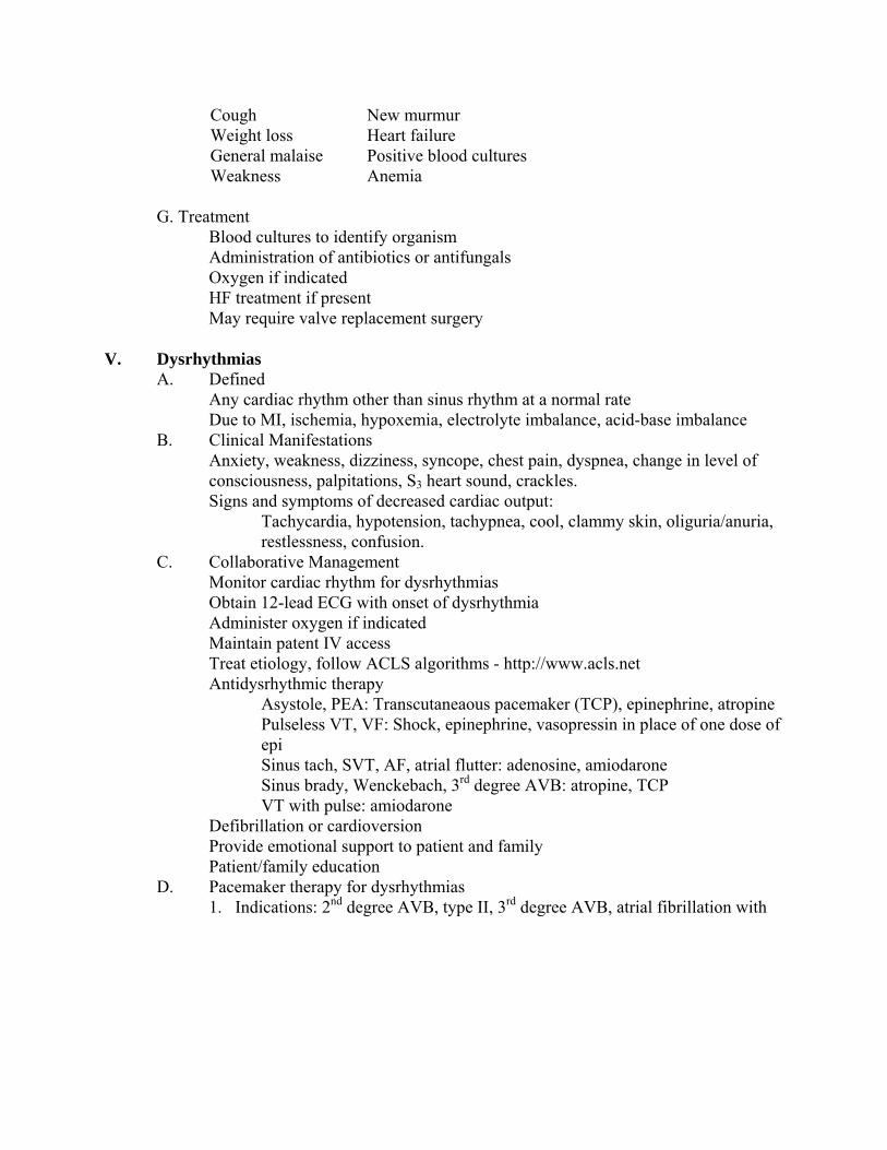
Five categories for permanent pacemakers Ventricular pacing – VVI
Ventricle paced, sensed and inhibited One pacemaker spike, just before QRS; appears wide
May not see with every beat; depends on rate. If rate if faster that set rate, pacemaker will not fire.
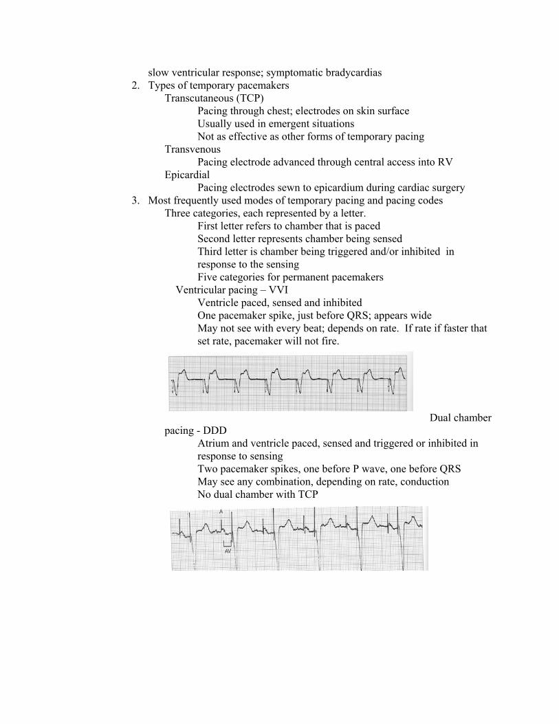
Dual chamber pacing - DDD
Atrium and ventricle paced, sensed and triggered or inhibited in response to sensing
Two pacemaker spikes, one before P wave, one before QRS May see any combination, depending on rate, conduction No dual chamber with TCP

TCP Does not use letter system; only two types of settings Demand (Synchronous)
Paces only when patient’s HR falls below set rate Appearance similar to VVI
Fixed (Asynchronous) Unable to obtain capture Pacer unable to sense intrinsic activity Artifact prevents sensing Danger of pacemaker spike falling on T wave and causing ventricular tachycardia or ventricular fibrillation
4. Pacemaker settings Rate control
Regulates impulses per minute; usually set between 60-80 bpm. AV pacemaker, rate controls both Higher if being used for overdrive suppression of tachyarrhythmias. Ordered by physician
Output dial Regulates amount of electrical current delivered to initiate depolarization and contraction Measured in milliamperes (mA).
Threshold (capture) Point at which depolarization occurs Pacemaker spike followed by P wave (atrial pacing) or QRS (ventricular pacing) Separate output dials for atrial and ventricular pacing.
Sensitivity control Regulates ability of the pacemaker to detect the heart’s intrinsic electrical activity. Measured in millivolts (mV) Pacemaker has a sense indicator, usually a light, which will indicate each time the pacemaker senses electrical activity. To increase sensitivity, the dial is turned down No sensitivity on TCP.
AV interval control Used in AV pacing. Regulates the time interval between the atrial and ventricular pacing stimuli Sets PR interval; usually equal to PR of .20.

5. Initiating pacing Set heart rate and AV control per physician order Determine threshold Set mA 2-3 times higher than threshold to ensure capture
TCP may increase mA by 10% after capture obtained Set sensitivity control so that pacemaker senses the heart’s intrinsic electrical activity (R wave)
Ensure that sensitivity not so low that it also senses lower amplitude electrical signals such as the T wave
Will consider it to be an R wave and double-count rate Will not pace when HR is below set rate
No sensitivity on TCP TCP Preparation
Explain to patient and family TCP will be uncomfortable May require analgesia
Thoroughly wash and dry skin Use skin preparation
Pad placement Anterior-posterior preferred Anterior-anterior may need to be used
6. Nursing care Report settings as part of shift change report Check connections Temporatry/epicardial
Wear gloves when handling pacing wires to prevent microshock and VF
When not in use, cover leads with gauze and secure to patient with tape
Secure temporary pacemaker to patient’s waist with strap/telemetry pouch.
Suspend from IV pole with twill tape if patient is on bedrest.
Have extra batteries and pacemaker on hand. Perform site care, inspect site to prevent/identify infection
TCP Assess integrity of TCP pads Assess skin integrity Be sure TCP is plugged in
7. Pacemaker malfunction

Failure to capture
Pacemaker spikes not followed by complex
All: May be due to mA too low, low battery Transvernous/epicardial: fibrin at tip of lead, fractured lead wire, movement away from ventricular wall Increase mA, replace battery, notify physician
Failure to sense Pacemaker does not sense patient’s intrinsic rhythm; paces inappropriately Pacemaker spikes throughout strip, do not correlate with complexes
Transvenous/epicardial: Due to sensitivity set to low
Correct by increasing sensitivity To increase, turn sensitivity down
TCP: No sensitivity setting Change to fixed mode setting
Certification Questions 1. Which of the following medications is administered to prevent sudden death associated with dilated cardiomyopathy?
A. Warfarin (Coumadin) to prevent clot formation B. Calcium channel blockers to control tachycardia C. Nitrates to improve coronary artery perfusion D. Digoxin to reduce atrial dysrhythmias and improve contractility
2. Chest pain associated with aortic stenosis can be caused by:

A. Decreased stroke volume B. Disproportionate oxygen supply versus demand C. Prolapsed valve leaflets D. Decreased contractility 3. Which of the following medications may worsen symptoms of heart failure associated with hypertrophic cardiomyopathy?
A. Calcium channel blockers B. Beta-blockers C. Nitroglycerin D. Amiodarone
4. Which of the following medication regimens would be most appropriate to relieve chest pain in a patient with a diagnosis of myocarditis?
A. Nitroglycerin 1/150 grains sublingual B. Furosemide 40 mg IV C. Ibuprofen 800 mg PO D. Morphine sulfate 2 mg IV
5. In a coronary care unit in which medications are not permitted to be left at the bedside and the crash cart with monitor/transcutaneous pacemaker/defibrillator is outside the central nurses’ station, an intubated patient on mechanical ventilation develops third-degree AV block at a rate of 35 beats/min with signs of poor tissue perfusion. The most appropriate initial intervention for the nurse assigned to this patient would be to
A. Initiate transcutaneous pacing B. Administer atropine 0.5 mg IV C. Initiate an infusion of epinephrine at 5 mcg/min D. Initiate an infusion of dopamine at 5 mcg/kg/min
6. The nurse should perform which of the following interventions for a patient with chest pain, hypotension and tachycardia at a rate of 180 bpm?
A. Administer amiodarone 150 mg IV over 10 minutes B. Administer adenosine 6 mg rapid IV push C. Perform synchronized cardioversion D. Defibrillate with 300 joules
7. A patient who suffered complete heart block after an anterior wall infarction receives a temporary pacemaker. The following rhythm develops:
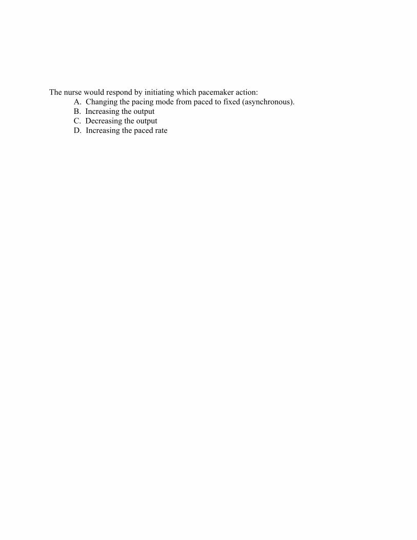
The nurse would respond by initiating which pacemaker action: A. Changing the pacing mode from paced to fixed (asynchronous). B. Increasing the output C. Decreasing the output D. Increasing the paced rate
References Aehlert, B. (2005). ECGs Made Easy, 3nd ed. St. Louis: Mosby. Ahrens, T.S., Prentice, D., and Kleinpell, R.M. (2010). Critical Care Nursing Certification:
Preparation, Review and Practice Exams, 6th edition. New York: McGraw-Hill Alspach, J.G. (Ed.). (2006) American Association of Critical Care Nurses Core Curriculum for
Critical Care Nursing (6th ed.). Phila: W.B. Saunders Company. Alspach, J.G. (Ed.). (2008). AACN Certification and Core Review for High Acuity and Critical
Care. St. Louis: Saunders Elsevier Bradley, D.J.; Bradley, E.A.; Baughman, K.I.; Berger, R.D.; Calkins, H.; Goodman, S.N.; Kass, D.A.; Powe, N.R. (2003). Cardiac Resynchronization and Death from Progressive Heart Failure: A Meta-analysis of Randomized Controlled Trials. Journal of the American Medical Association. 289(6). pp. 730-740. Branham, K. (2003). Management of Decompensated Heart Failure. AACN Clinical Issues. 14(4). 498.500. Brorsen, A.J. and Rogelet, K.R. (2009). Adult CCRN Certification Review. Massachusetts:
Jones and Bartlett Publishers. Brorsen, A.J. and Rogelet, K.R. (2009). PCCN Certification Review. Massachusetts: Jones and
Bartlett Publishers. Burger, A.J., Burger, M. R., and Aronson, D. (2002). New Therapies for theTreatment of Congestive Heart Failure. Drugs of Today. 38 (1): 31-48. Cianci, P.; Lonergan-Thomas, H; Slaughter, M.; Silver, M.A. (2003). Current and Potential Applications of Left Ventricular Assist Devices. The Journal of Cardiovascular Nursing. 18(1); 17-22. Chulay, M. and Burns, S. (2007) AACN Essentials of Progressive Care Nursing. New York:
McGraw-Hill. Dennison, R.F. (2007). Pass CCRN! (3nd ed) St. Louis: Mosby Elsevier

Hunt, S.A., Baker, D.W., Chin, M.H., Cinquegrani, M.P., Feldman, A.M., Frances, G.S., Ganiats, T.G., Goldstein, S., Gregoratos, G., Jessup, M.I., Noble, R.J., Packer, M., Silver, M.A., and Stevenson, L.W. (2001). ACC/AHA Guidelines for the Evaluation and Management of Chronic Heart Failure in the Adult: a Report of the American College of Cardiology/American Heart Association Task Force on Practice Guidelines. American College of Cardiology website: http://www.ACC.org/clinical/guidelines/failure/hf_index.htm Kaplan CCRN Certification for Adult, Pediatirc, and Neonataol Critical Care Nurses, 2009
Edition (2009). Harwani, S.C., Contributing Editor. New York: Kaplan Publishing. Lewis, D. A., Gurran, N. R., Abraham, W. T., and Akers, W. S. (2003). Effect of nesiritide
versus milrinone in the treatment of acute decompensated heart failure. American Journal of Health-System Pharmacists. 60 (Supp 4). S16-S20.
Mehra, M. R. (2006). Optimizing outcomes in the patient with acute decompensated heart
failure. American Heart Journal. 151(3). 571-579. Nicholas, M. (2004). Heart failure: pathophysiology, treatment and nursing care. Nursing
Standard 11(19) 46-54 Nohria, A., Lewis, E., and Stevenson, L.W. (2002). Medical Management of Advanced Heart Failure. Journal of the American Medical Association. 287 (3). 628-39. Prahash, A, and Lynch, T. (2004) B-Type Natriuretic Peptide: a Diagnostic, Prognostic, and Therapeutic Tool in Heart Failure. American Journal of Critical Care 13 (1). 46-53.
Southworth, M.R. (2003) Treatment Options for Acute Decompensated Heart Failure. American Society of Health-System Pharmacists, Inc. 60 suppl 4 Sole, M.L.; Klein, D.G.; and Moseley, M.J. (2009). Introduction to Critical Care Nursing, 5th
Edition. St. Louis: Elsevier Saunders Springhouse Review for Critical Care Nursing Certification, 4th Edition. (2007). Ambler, Pa: Lippincott, Williams, and Wilkins. Urden, L. D.; Stacy, K. M.; and Lough, M. E. (2010) Critical Care Nursing: Diagnosis
and Management, 6th ed. St. Louis: Mosby Elsevier.