seven new vernocuminosides from the stem bark of vernonia cumingiana benth
TRANSCRIPT
Carbohydrate Research 345 (2010) 1156–1162
Contents lists available at ScienceDirect
Carbohydrate Research
journal homepage: www.elsevier .com/locate /carres
Seven new vernocuminosides from the stem bark of Vernonia cumingiana Benth
Jing Liu, Shuanggang Ma, Shishan Yu *, Haining Lv, Yong Li, Xianfu Wu, Yuanyan LiuKey Laboratory of Bioactive Substances and Resources Utilization of Chinese Herbal Medicine, Ministry of Education & Institute of Materia Medica,Chinese Academy of Medical Sciences and Peking Union Medical College, No.1 Xiannongtan Street, Beijing 100050, People’s Republic of China
a r t i c l e i n f o a b s t r a c t
Article history:Received 23 December 2009Received in revised form 22 March 2010Accepted 30 March 2010Available online 2 April 2010
Keywords:Vernonia cumingianaStigmastaneCircular dichroismAnti-inflammatory activity
0008-6215/$ - see front matter � 2010 Elsevier Ltd. Adoi:10.1016/j.carres.2010.03.039
* Corresponding author. Tel.: +86 10 6316 5324; faE-mail address: [email protected] (S. Yu).
Seven new stigmastane-type steroidal glycosides, vernocuminosides H–N, have been isolated from thestem bark of Vernonia cumingiana Benth. The structures of these compounds were determined by NMRspectrometric methods including HSQC, HMBC, 1H–1H COSY, and NOE, as well as circular dichroismexperiments. Anti-inflammatory activities of vernocuminosides I and K–N were determined.
� 2010 Elsevier Ltd. All rights reserved.
1. Introduction
Vernonia cumingiana Benth belongs to the family Compositaeand has been used for curing rheumatic arthritis, lumbocrural pain,fracture, and malaria in China.1 Studies on this genus showed itsrich content of sesquiterpene lactones and stigmastane-type glyco-sides with extensive biological activities including cytotoxic, anti-bacterial, antimalarial effects and others.2 In our previous paper,from the stem bark of the species, seven D7,9(11) stigmastane-typeglycosides with linear side chains containing C-21 in the form of acarboxyl group were isolated.3 During our further investigations,we found other seven new D 7,9(11) stigmastane-type glycosides,vernocuminosides H–N (1–7). Among them, the side chains ofcompounds 1–4 were in the form of highly oxygenated d-lactones,and the linear side chains of 5–7 possessed different oxygenatedforms of C-21. In this paper, we describe the isolation and struc-tural elucidation of compounds 1–7 (vernocuminosides H–N) andanti-inflammatory activities of compounds 2 and 4–7.
2. Results and discussion
During our further studies on the ethanolic extract of stem barkof V. cumingiana, seven vernocuminosides H–N (compounds 1–7,Fig. 1) were isolated as white amorphous powders and showed po-sitive Liebermann–Burchard and Molisch reactions. All UV spectradisplayed the same absorption bands at 235, 242, and 250 nm asthose for vernocuminosides A–F.3 All IR spectra indicated the pres-
ll rights reserved.
x: +86 10 6301 7757.
ence of a hydroxyl group (3331–3433 cm�1) and a double bond(1642–1650 cm�1); moreover, the IR spectra of compounds 1–4also revealed the existence of a carbonyl group (1700–1710 cm�1).
The molecular formula of compound 1 was determined asC35H54O9 with 9 degrees of unsaturation on the basis of its posi-tive-ion HRESIMS (m/z 641.3650 [M+Na]+, calcd for C35H54O9Na:641.3666). The 1H NMR spectrum of 1 presented the angular sig-nals at d 0.59 (3H, s, CH3-18), 0.86 (3H, s, CH3-19), two olefinic pro-ton signals at d 5.33 (1H, br s, H-7), 5.44 (1H, br d, J = 6.0 Hz, H-11),and the distinctive H-3 multiplet at d 3.64 (Table 1), as well as a setof proton signals for the sugar moiety (Table 2) with its anomericproton at d 4.33 (1H, d, J = 7.6 Hz, H-10 of b-Glc). The 13C NMR spec-trum exhibited 35 signals (Tables 3 and 4) including 6 for a hexoseunit and 29 for the aglycone moiety, which revealed a conjugateddiene consisting of two trisubstituted carbon signals [d 119.8 (C-11), 121.4 (C-7)] and two tetrasubstituted ones [d 137.5 (C-8),145.1 (C-9)], which were further confirmed by the cross peaks fromd 5.44 (H-11) to d 137.5 (C-8), 37.1 (C-10), 40.7 (C-12), and 43.4 (C-13); from d 0.86 (CH3-19) to d 145.1 (C-9); from both d 1.97 (1H, brd, J = 17.2, H-12) and 2.19 (1H, dd, J = 17.6, 6.8 Hz, H-12) to d 145.1(C-9) and d 119.8 (C-11) in the HMBC spectrum (Fig. 2). The above-mentioned data proved that compound 1 was a D7,9(11) stigmas-tane-type glycoside. Excluding the 7 degrees of unsaturation dueto the D7,9(11) stigmastane-type steroidal skeleton and the sugarunit, there remained 2 for the C10H17O3 side chain which includedthree methyl, two methene, three methenyl, and two quaternarycarbons containing a carboxyl group (d 177.7) as determined fromthe DEPT spectrum. For the side chain, the proton signals at d 0.92(3H, d, J = 6.8 Hz, CH3-26), 0.95 (3H, d, J = 6.8 Hz, CH3-27), 1.94 (1H,m, H-25), together with two secondary methyl resonances at d 17.3

Table 11H NMR chemical shifts of aglycon moieties for compounds 1, 2, and 5 in CD3OD (400 MHz), 3 (600 MHz), and 4, 6, and 7 (500 MHz) in pyridine-d5
a
Aglycone moiety 1 2 3 4 5 6 7
1 1.24; 1.93 1.27; 1.92 1.18; 1.92 1.16; 1.81 1.31; 1.95 1.27; 1.88 1.20; 1.842 1.54; 1.94 1.54; 2.00 1.84; 2.13 1.77; 2.17 1.60; 1.93 1.81; 2.11 1.77; 2.133 3.64 (m) 3.65 (m) 3.85 (m) 3.89 (m) 3.66 (m) 3.96 (m) 3.93 (m)4 1.28; 1.82 1.29; 1.87 1.61; 2.15 1.64; 2.18 1.30; 1.86 1.40; 2.01 1.39; 1.995 1.34 1.34 1.32 1.32 1.33 1.32 1.276 1.86 1.92 1.84 1.83 1.86 1.70 1.777 5.33 (br s) 5.35 (br s) 5.33 (br s) 5.31 (br s) 5.34 (br s) 5.39 (br s) 5.34 (br s)
11 5.44 (br d, 6.0) 5.44 (br d, 6.4) 5.39 (br d, 6.0) 5.37 (br s) 5.43 (br d, 5.6) 5.49 (br s) 5.46 (br s)12 1.97 (br d, 17.2)
2.19 (dd, 17.6, 6.8)1.98 (br d, 17.6)2.16 (dd, 17.2, 6.8)
2.10 (br d, 17.4)2.44 (dd, 17.4, 6.6)
2.19 2.06 (br d, 16.8)2.25 (dd, 16.8, 6.0)
2.48 2.28
14 2.16 (br s) 2.17 (br s) 2.21 (br s) 2.53 (br s) 2.13 (br s) 2.18 (br s) 2.74 (br s)15 1.40; 1.75 1.42; 1.59 1.40; 1.61 1.83; 2.09 1.35; 1.74 1.82; 2.47 2.07; 2.1416 1.43; 1.90 1.44; 1.88 1.41; 1.86 5.52 (m) 1.38; 1.99 4.80 (m) 4.57 (m)17 2.39 2.14 2.25 2.86 1.29 1.82 2.0718 0.59 (s) 0.55 (s) 0.72 (s) 0.64 (s) 0.48 (s) 1.07 (s) 0.66 (s)19 0.86 (s) 0.86 (s) 0.90 (s) 0.87 (s) 0.86 (s) 0.86 (s) 0.82 (s)20 2.34 (m) 2.42 (m) 2.71 (m) 2.96 (m) 1.31 (m) 2.43 (m) 1.88 (m)21 0.90 (d, 6.4) 4.04; 4.20 3.85; 4.1322 1.86; 1.92 1.81; 2.08 1.75; 2.19 1.99; 2.04 1.56; 1.99 2.11; 2.32 2.17; 2.4723 1.60 1.79; 2.05 1.76; 2.15 1.81; 2.36 1.35; 1.86 2.11 1.99; 2.2025 1.94 (m) 1.92 (m) 1.92 (m) 1.82 (m) 1.95 (m) 2.18 (m) 2.17 (m)26 0.92 (d, 6.8) 0.92 (d, 6.4) 0.98 (d, 6.6) 0.99 (d, 5.5) 0.90 (d, 6.4) 1.09 (d, 6.5) 1.09 (d, 6.5)27 0.95 (d, 6.8) 0.95 (d, 6.8) 1.07 (d, 6.6) 1.13 (d, 6.0) 0.93 (d, 7.2) 1.23 (d, 6.5) 1.21 (d, 6.5)28 3.88 (q, 6.4) 3.87 (q, 6.4) 4.22 4.25 3.93 (q, 6.4) 4.35 4.3729 1.11 (d, 6.8) 1.11 (d, 6.4) 1.31 (d, 6.0) 1.26 (d, 5.5) 1.14 (d, 6.4) 1.48 (d, 6.5) 1.50 (d, 5.5)16-CH3COO 2.04 (s)
a Overlapped signals are reported without designated multiplicities.
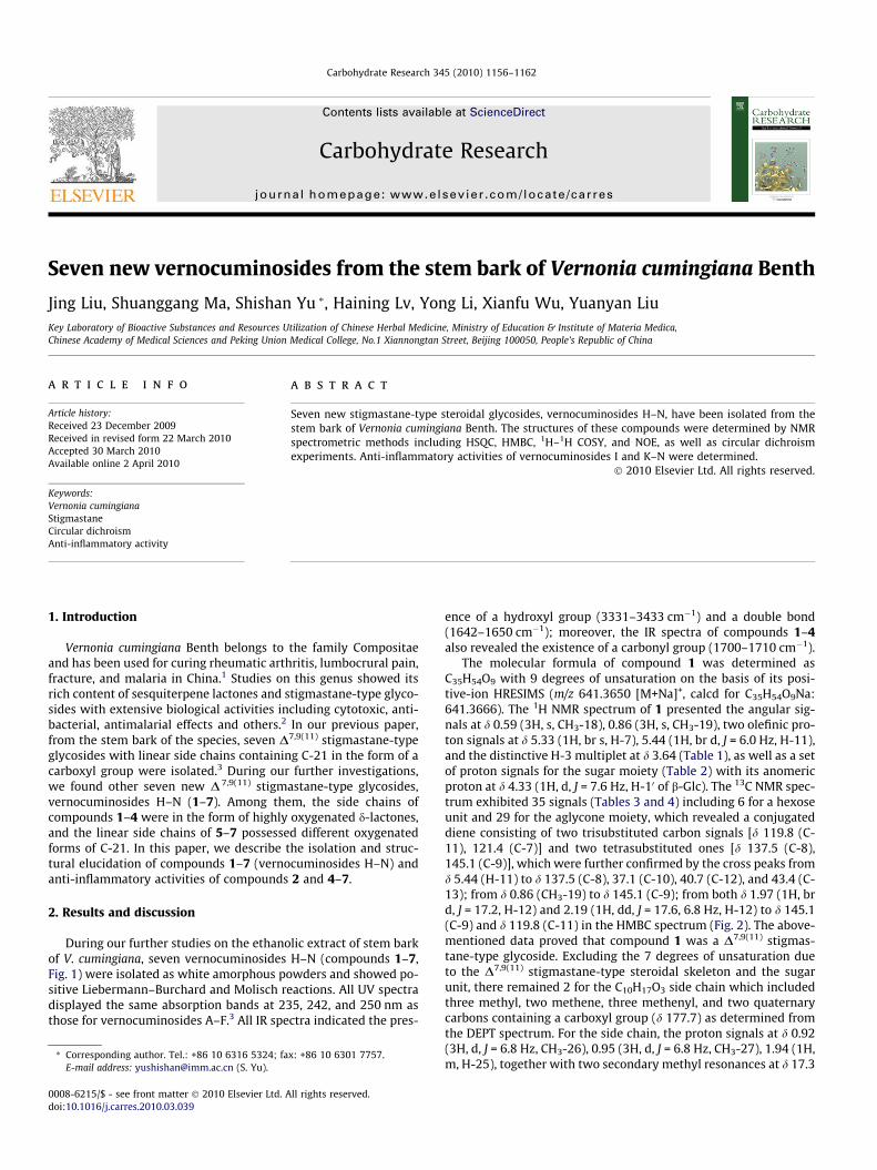
RO
GlcO
H3C
OGlc
OH
GlcO
HOH2C
OGlc
OH
512
37
9
11
14
17
20
OO
OH
21
24
R1
R6. R = β-OH
7. R = α-OH
1. R = β -D-Glc; R1 = H, H
2. R = β -D-Gal-(1 2)-β-D-Glc; R1 = H, H
3. R = β-D-Glc-(1 2)-β-D-Glc; R1 = H, H
4. R = β-D-Gal-(1 2)-β-D-Glc; R1 =β-CH3COO
Figure 1. The structures of compounds 1–7.
J. Liu et al. / Carbohydrate Research 345 (2010) 1156–1162 1157
(C-26), 18.0 (C-27), and a methenyl carbon signal at d 36.6 (C-25)revealed the presence of an isopropyl moiety. Meanwhile, the pro-ton signals at d 1.11 (3H, d, J = 6.8 Hz, CH3-29), 3.88 (1H, q,J = 6.4 Hz, H-28), as well as the secondary methyl carbon signalat d 18.0 (C-29), and the oxygenated methenyl resonance at d71.4 (C-28) indicated an ethyl moiety with its methene substitutedby a hydroxyl group. Both substituted moieties were determined tobe attached at C-24 (d 91.6) by the HMBC (Fig. 2) from d 0.92 (CH3-26), 0.95 (CH3-27), 1.94 (H-25), 1.11 (CH3-29), and 3.88 (H-28) to d91.6 (C-24). When compared with NMR data for the side chain ofvernocuminoside C,3 a downfield shift of C-24 (from d 77.0 to d89.7) and upfield shifts of C-20 (from d 49.9 to d 41.2), C-21 (fromd 178.9 to d 174.4), C-22 (from d 27.1 to d 23.1), and C-23 (from d31.3 to d 21.0) were observed. Combined analysis of the observa-tion and unsaturation of degrees indicated the cyclic side chainwith a d-lactone unit formed at C-21 and C-24 as in 1.
NOE experiments showed that NOEs were recorded for bothCH3-19 and H-20 by irradiation of CH3-18, and for CH3-18 by irra-diating H-20. Therefore, the natural aglycone of 1 was deduced as3b,24,28-trihydroxy-stigmasta-7,9(11)-dien-21,24-lactone.
The stereochemistry of C-24, as the d-position of d-lactone, wasassigned by use of circular dichroism (CD). Korver used the ap-proach of Legend and Bucourt and summarized the relation be-tween the sign of Cotton effect of any optically active d-lactoneand its absolute configuration.4 According to the literature,4,5 thereare two bands in the recorded CD spectrum of a d-lactone, whichhas been ascribed to the boat conformation (k <225 nm) and thehalf-chair conformation (k >230 nm). When there is a substituentat the a- and/or d-position of the d-lactone, 1,4-interations ariseand can force the molecule into the half-chair conformation. Actu-ally the half-chair conformation was more stable in the examplestudied with a substituent at the d-position.4 For compound 1,

Table 21H NMR chemical shifts of sugar moieties for compounds 1, 2, and 5 in CD3OD (400 MHz), 3 (600 MHz), and 4, 6, and 7 (500 MHz) in pyridine-d5
a
Aglycon moiety 1 2 3 4 5 6 7
C-3 b-D-Glc b-D-Glc b-D-Glc b-D-Glc b-D-Glc b-D-Glc b-D-Glc10 4.33 (d, 7.6) 4.48 (d, 7.6) 5.00 (d, 7.8) 5.04 (d, 7.5) 4.34 (d, 7.6) 5.01 (d, 8.0) 5.00 (d, 7.5)20 3.08 (t, 8.0) 3.34 (t, 8.0) 4.12 (t, 8.4) 4.10 (ov) 3.08 (t, 8.4) 4.02 (ov) 4.03(t, 7.0)30 3.29 (t, 8.4) 3.49 (t, 6.8) 4.34 (t, 8.4) 4.33 (ov) 3.29 (ov) 4.28 (ov) 4.22 (ov)40 3.21 (ov) 3.25 (ov) 4.28 (t, 8.4) 4.23 (ov) 3.25 (ov) 4.24 (ov) 4.20 (ov)50 3.21 (ov) 3.23 (ov) 3.94 (br m) 3.89 (br m) 3.24 (ov) 3.99 (ov) 3.98 (ov)60 3.59 (dd, 11.6, 4.8)
3.79 (br d, 11.6)3.60 (dd, 11.6, 5.2)3.79 (br d, 11.2)
4.42 (br d,12.6)4.51 (br d, 12.6)
4.33 (ov)4.50 (ov)
3.61 (br d, 11.2)3.80 (dd, 12.0, 3.2)
4.41 (ov)4.57 (br d, 11.5)
4.36 (ov)4.50 (br d, 10.5)
b-D-Gal b-D-Glc b-D-Gal100 4.43 (d, 8.0) 5.24 (d, 7.8) 5.14 (d, 7.5)200 3.56 (t, 7.6) 4.09 (t, 7.8) 4.59 (ov)300 3.46 (ov) 4.22 (ov) 4.17 (ov)400 3.80 (br d, 3.6) 4.19 (ov) 4.61 (ov)500 3.47 (ov) 3.88 (br m) 4.10 (ov)600 3.69 (2H, br d, 6.4) 4.33 (ov);
4.49 (br d, 12.6)4.44 (br s)4.57 (ov)
C-28 b-D-Glc b-D-Glc b-D-Glc1000 4.29 (d, 7.6) 4.96 (d, 7.0) 4.93 (d, 8.0)2000 3.13 (t, 8.4) 3.99 (ov) 3.98 (ov)3000 3.31 (t, 8.4) 4.20 (ov) 4.18 (ov)4000 3.25 (ov) 4.20 (ov) 4.27 (t, 8.5)5000 3.29 (ov) 3.92 (ov) 3.93 (ov)6000 3.61 (br d, 11.2)
3.80 (dd, 12.0, 3.2)4.35 (ov)4.49 (br d, 12.0)
4.41 (ov)4.57 (br d, 10.5)
a Overlapped signals are reported without designated multiplicities. Glc: glucopyranosyl; Gal: galactopyranosyl.
Table 313C NMR chemical shifts of 1a, 2a and 5a in CD3OD (100 MHz), 1b, 2b, 5b, 4, 6, and 7 in pyridine-d5 (125 MHz), and 3 in pyridine-d5 (150 MHz)
Aglycon moiety 1a 1b 2a 2b 3 4 5a 5b 6 7
1 36.0 35.0 36.0 35.0 35.1 34.9 36.0 35.1 35.0 34.92 30.6 30.1 30.6 30.1 30.2 30.1 30.6 30.2 30.2 30.13 78.9 77.1 79.9 78.5 78.6 78.4 78.9 77.1 77.2 77.04 35.0 34.5 35.1 34.6 34.6 34.5 35.0 34.6 34.5 34.55 40.5 39.2 40.6 39.4 39.4 39.3 40.5 39.3 39.3 39.26 31.0 30.2 31.0 30.2 30.3 30.1 31.1 30.2 30.1 30.27 121.4 120.6 121.6 121.0 121.1 121.5 121.4 120.7 120.4 120.88 137.5 136.5 137.3 136.2 136.3 135.7 137.5 136.5 136.4 136.09 145.1 144.0 145.4 144.3 144.4 144.3 145.3 144.3 144.3 144.3
10 37.1 36.1 37.1 36.2 36.3 36.1 37.1 36.2 36.2 36.211 119.8 119.1 119.5 118.7 118.8 118.1 119.5 118.7 119.1 118.512 40.7 40.0 41.2 40.3 40.4 40.5 43.5 42.5 42.5 41.713 43.4 42.7 43.6 42.8 42.9 43.4 43.6 42.6 42.5 43.714 52.8 52.0 52.6 51.8 51.9 49.0 52.9 51.9 50.4 49.415 23.5 22.8 23.7 23.0 23.1 33.0 24.3 23.5 36.7 36.016 27.4 27.0 26.6 26.4 26.5 76.8 29.6 28.8 72.2 74.517 49.3 48.8 50.4 49.7 49.8 56.0 57.6 56.7 57.3 62.618 12.0 11.9 12.3 12.0 12.1 13.5 11.7 11.6 13.4 13.519 19.9 19.5 20.0 19.6 19.7 19.6 19.9 19.6 19.6 19.520 41.8 41.2 41.7 41.2 41.2 39.6 38.4 37.5 39.3 42.021 177.7 174.4 178.4 175.1 175.3 175.3 18.9 18.7 61.5 63.522 23.6 23.1 23.7 23.1 23.2 23.5 30.6 29.9 23.3 21.823 21.7 21.0 23.0 21.7 21.8 24.3 31.1 30.7 29.9 30.024 91.6 89.7 91.1 89.5 89.6 89.1 78.6 76.7 77.1 76.825 36.6 35.8 36.2 36.2 35.1 36.1 34.4 33.5 34.2 33.826 17.3 17.2 17.2 17.1 17.2 16.7 17.9 17.8 17.9 17.927 18.0 18.4 17.6 17.5 17.6 17.4 18.1 18.0 18.1 18.228 71.4 70.2 71.8 70.7 70.8 70.5 79.3 81.3 80.9 80.729 18.0 17.9 17.9 18.4 18.5 18.3 14.8 16.3 15.7 15.716-CH3COO 21.216-CH3COO 170.6
1158 J. Liu et al. / Carbohydrate Research 345 (2010) 1156–1162
besides the d-lactone unit, the inherent D7,9(11) conjugated dienechromophore could interfere with the correct interpretation ofthe CD spectrum as it could produce a strong Cotton effect in thewavelength range under study. Thus, the CD differential spectrum(Fig. 3) obtained by subtracting the CD spectrum of vernocumino-side A from that of 1 was presented. As shown in Figure 3, the po-sitive Cotton effect at 219 nm for the boat conformation and thenegative Cotton effect at 242 nm for the half-chair conformation
were observed, in agreement with the literature.4,5 According tothe known relationship between the absolute configuration andthe sign of the Cotton effect,5 the absolute configuration of C-24was determined as R. The absolute configuration of C-28 in com-pound 1 was deduced as (28R) from the perspective of biogenesis.It could result from vernocuminoside C by loss of H2O due to theesterification reaction between C-21 and C-24 without involve-ment of the chiral carbon C-28. Thus, the absolute configuration

Table 413C NMR chemical shifts of sugars for 1a, 2a, and 5a in CD3OD (100 MHz), 1b, 2b, 5 b, 4, 6, and 7 in pyridine-d5 (125 MHz), and 3 in pyridine-d5 (150 MHz)�
Sugar moiety 1a 1b 2a 2b 3 4 5a 5b 6 7
C-3 b-D-Glc b-D-Glc b-D-Glc b-D-Glc b-D-Glc b-D-Glc b-D-Glc b-D-Glc b-D-Glc b-D-Glc10 102.3 102.3 101.4 101.3 101.5 101.3 102.3 102.3 102.3 102.320 75.1 75.3 83.7 85.0 84.7 85.1 75.1 75.4 75.3 75.330 78.1 78.5 77.7 77.9 77.9 77.9 78.0 78.5 78.6 78.440 71.7 71.7 71.5 71.4 71.5 71.4 71.8 71.8 71.7 71.650 77.9 78.5 77.7 78.2 78.9 78.2 77.9 77.1 78.5 78.560 62.8 62.9 62.7 62.6 62.7 62.6 62.8 62.8 62.9 62.7
b-D-Gal b-D-Gal b-D-Glc b-D-Gal100 106.2 107.4 106.6 107.4200 73.5 74.5 77.1 74.6300 74.7 75.0 78.0 75.0400 70.0 69.9 71.5 69.9500 77.0 77.2 78.2 77.2600 62.1 61.9 62.6 61.9C-28 b-D-Glc b-D-Glc b-D-Glc b-D-Glc1000 101.9 103.7 103.2 103.32000 75.0 74.9 74.9 75.03000 78.1 78.6 78.6 78.54000 71.8 71.9 71.5 71.75000 78.0 78.5 78.5 78.66000 62.8 62.8 62.6 62.9
� Glc: glucopyranosyl; Gal: galactopyranosyl.
O
OO
OH
OHO
HOHO
OH
Figure 2. Key HMBC (H?C) in compound 1.
J. Liu et al. / Carbohydrate Research 345 (2010) 1156–1162 1159
of C-28 in compound 1 and vernocuminoside C should be same,that is R.
NMR data for the sugar moiety (Tables 2 and 4) of 1 demon-strated the existence of a b-glucopyranosyl unit with its anomericproton at d 4.33 (1H, d, J = 7.6 Hz, H-10 of b-Glc). The linkage of ab-D-glucopyranosyl residue to C-3 was supported by the HMBC ob-served from d 4.33 (H-10) to d 78.9 (C-3) and from d 3.64 (H-3) to d102.3 (C-10 of b-Glc) (Fig. 2). The absolute configuration of glucosewas assigned as the D type. The DEPT, HSQC, HMBC, and 1H–1HCOSY spectra allowed the assignments of all proton and carbonsignals. Therefore, the structure of compound 1 was elucidated as3b,(24R),(28R)-trihydroxy-stigmasta-7,9(11)-dien-21,24-lactone-3-O-b-D-glucopyranoside, named vernocuminoside H.
Figure 3. Circular dichroism differential spectrum of compound
The positive HRESIMS of compound 2 gave an [M+Na]+ peak atm/z 803.4191 (calcd for C41H64O14Na: 803.4194), corresponding tothe molecular formula C41H64O14. The 1H and 13C NMR data (Tables1–4) of 2 were basically in agreement with those of 1 except for thepresence of an additional set of b-galactopyranose resonances withan anomeric proton signal at d 4.43 (1H, d, J = 8.0 Hz, H-100 of b-Gal)and a downfield shift of C-20 (from d 75.1 to d 83.7) of the b-gluco-pyranosyl unit due to glycosidation shift. The linkage positions andsequence for the sugar moieties were further supported by thelong-range correlations from d 4.48 (1H, d, J = 7.6 Hz, H-10 of b-Glc) to d 79.9 (C-3), from d 4.43 (H-100 of b-Gal) to d 83.7 (C-20 ofb-Glc), and from d 3.34 (H-20 of b-Glc) to d 106.2 (C-100 of b-Gal)in the HMBC spectrum. The absolute configurations of the glucoseand galactose were assigned as D. Additionally, the similar 13C NMRdata (from C-22 to C-29) for compounds 1 and 2, together withthe biogenesis consideration, indicated their same side-chainstructure. Thus, compound 2 was determined as 3b,(24R),(28R)-tri-hydroxy-stigmasta-7,9(11)-dien-21,24-lactone-3-O-b-D-galacto-pyranosyl-(1?2)-b-D-glucopyranoside, named vernocuminoside I.
Compound 3 possessed the molecular formula of C41H64O14
according to the positive-ion HRESIMS (m/z 803.4185 [M+Na]+,calcd 803.41988). The NMR data, especially the 13C NMR data(Tables 3 and 4) of 3 matched quite well with those for 2. Furtheranalysis indicated that there was a difference in the resonancescorresponding to the sugar residues. TLC and NMR data compari-son indicated the presence of D-glucose in 3. The connectivity forthe sugar residues was further confirmed by the HMBC from d
1 (subtracted by the CD spectrum of vernocuminoside A).

1160 J. Liu et al. / Carbohydrate Research 345 (2010) 1156–1162
5.00 (1H, d, J = 7.8 Hz, H-10 of b-Glc) to d 78.6 (C-3), from d 5.24 (1H,d, J = 7.8 Hz, H-100 of b-Glc) to d 84.7 (C-20 of b-Glc). Therefore, thestructure of compound 3 was assigned as 3b,(24R),(28R)-trihy-droxy-stigmasta-7,9(11)-dien-21,24-lactone-3-O-b-D-glucopyran-osyl-(1?2)-b-D-glucopyranoside, named vernocuminoside J.
The molecular formula of compound 4 was determined asC43H66O16 on the basis of its positive-ion HRESIMS (m/z 861.4206[M+Na]+, calcd for C43H66O16Na: 861.4249). The 13C NMR data (Ta-bles 3 and 4) of 4 closely resembled those of 2 except for an addi-tional acetoxy group [d 21.2, 170.6], downfield shifts of C-15 (d33.0), C-17 (d 56.0), and C-16 (d 76.8) in the form of an oxygenatedmethenyl with its proton signal at d 5.52 (1H, m, H-16). Careful anal-ysis revealed that the acetoxy group could be attached at C-16,which was further ascertained by the HMBC from both d 2.04 (3H,s, CH3COO-16) and d 5.52 (H-16) to d 170.6 (CH3COO-16) and fromboth d 1.83 (H-15) and d 2.86 (H-17) to d 76.8 (C-16). NOE experi-ments on compound 4 showed that there were NOEs recorded forboth CH3-19 and H-20 when irradiating CH3-18, for H-14 when irra-diating H-17, and for H-14 when irradiating H-16, which confirmedthe a-orientation of H-16. The similar 13C NMR data (from C-22 toC-29) for 1 and 4, combined with the perspective of biogenesis, re-vealed that both possessed the same steroidal side-chain structure.The linkage positions and sequence for sugar residues were furtherconfirmed by the HMBC spectrum that showed long-range correla-tions from d 5.04 (1H, d, J = 7.5 Hz, H-10 of b-Glc) to d 78.4 (C-3), fromd 5.14 (1H, d, J = 7.5 Hz, H-100 of b-Gal) to d 85.1 (C-20 of b-Glc), from d3.89 (1H, m, H-3) to d 101.3 (C-10 of b-Glc), and from d 4.10 (1H, H-20
of b-Glc) to d 107.4 (C-10 0 of b-Gal). The DEPT, HSQC, HMBC and1H–1H COSY experiments permitted assignment of all proton andcarbon signals. Thus, the structure of compound 4 was elucidatedas 3b,(24R),(28R)-trihydroxy-16b-acetoxy-stigmasta-7,9(11)-dien-21,24-lactone-3-O-b-D-galactopyranosyl-(1?2)-b-D-gluco-pyrano-side, named vernocuminoside K.
Compound 5 possessed the molecular formula C41H68O13
according to its positive-ion HRESIMS (m/z 791.4598 [M+Na]+,calcd for C41H68O13Na: 791.4558). The 13C NMR data (Tables 3and 4) of 5 matched well with those of vernocuminoside A,3 exceptfor the presence of an additional secondary methyl signal at d 18.9(C-21) and the disappearance of a carboxyl carbon resonance. TheHMBC from d 0.90 (3H, d, J = 6.4 Hz, CH3-21) to d 57.6 (C-17) andfrom d 1.56 (H-22) to d 18.9 (C-21) confirmed C-21 as a secondarymethyl group. The connectivities of both sugar moieties were fur-ther ascertained by long-range correlations from d 4.34 (1H, d,J = 7.6 Hz, H-10 of b-Glc) to d 78.9 (C-3), from d 4.29 (1H, d,J = 7.6 Hz, H-1000 of b-Glc) to d 79.3 (C-28), from d 3.66 (1H, m,H-3) to d 102.3 (C-10 of b-Glc), and from d 3.93 (1H, q, J = 6.4 Hz,H-28) to d 101.9 (C-1000 of b-Glc) in the HMBC spectrum. The abso-lute configuration of glucose was determined as D. Combinedanalysis of DEPT, HSQC, HMBC, and 1H–1H COSY spectra permittedassignment of all proton and carbon signals. Generally, the differ-ence between compound 5 and vernocuminoside A was the differ-ent forms of C-21, while the stereochemistry for other chiralcenters should be same from the perspective of biogenesis. There-fore, the structure of 5 was deduced as 3b,(24R),(28R)-trihydroxy-stigmamasta-7,9(11)-dien-21-yl-3,28-di-O-b-D-glucopyranoside,named vernocuminoside L.
The positive-ion HRESIMS of compound 6 gave an [M+Na]+ peakat m/z 823.4497 (calcd for C41H68O15Na: 823.4456), correspondingto the molecular formula C41H68O15. A comparison of the analysesof 1H and 13C NMR data for compound 6 (Tables 1–4) with thoseof vernocuminoside A,3 suggested that 6 possessed the sameD7,9(11) stigmastane-type steroidal skeleton and sugar residues.The evident differences included the presence of an oxygenatedmethene carbon signal at d 61.5 (C-21) and an oxygenated methe-nyl carbon resonance at d 72.2 (C-16) in 6 instead of the carbonylsignal at d 178.9 and methene carbon resonance at d 27.9 in vernoc-
uminoside A. The upfield shifts of C-20 (from d 49.7 to d 39.3), C-22(from d 26.8 to d 23.3), and C-23 (from d 31.5 to d 29.9), togetherwith the downfield shifts of C-15 (from d 23.1 to d 36.7) and C-17(from d 53.3 to d 57.3), demonstrated that C-21 was in the formof a primary alcohol, and C-16 was an oxygenated methenyl group,which was further confirmed by HMBC from d 2.43 (1H, m, H-20) tod 61.5 (C-21) and from d 1.82 (1H, m, H-17) and d 2.43 (1H, m, H-20)to d 72.2 (C-16). NOE experiments showed that NOEs were recordedfor both CH3-19 and H-20 by irradiation of CH3-18, for CH3-26, CH3-27, H-28, and H-1000 by irradiation of CH3-29, as well as for H-17 andH-14 by irradiation of H-16, which indicated the a-orientation ofH-16. The long-range correlations observed from d 5.01 (1H, d,J = 8.0 Hz, H-10 of b-Glc) to d 77.2 (C-3), from d 4.35 (1H, H-28) tod 103.2 (C-1000 of b-Glc), as well as from d 4.96 (1H, d, J = 7.0 Hz,H-1000 of b-Glc) to d 80.9 (C-28) in the HMBC spectrum confirmedthe respective linkage position of both b-glucopyranosyl units.From the above and the perspective of biogenesis, the difference be-tween the side chain of compound 6 and that of vernocuminoside Awas the different oxygenated forms of C-21. The stereochemistryfor C-24 and C-28 should be the same in both compounds. Inconclusion, the structure of compound 6 was established as3b,16b,(24R),(28R)-tetrahydroxy-stigmamasta-7,9(11)-dien-21-ol-3,28-di-O-b-D-glucopyranoside, named vernocuminoside M.
The molecular formula of compound 7 was established asC41H68O15 on the basis of the positive-ion HRESIMS (m/z801.4578 [M+H]+, calcd for C41H69O15: 801.4636). The 1H and 13CNMR data (Tables 1–4) of 7 were quite similar to those of 6 exceptfor the downfield shifts of C-16 (from d 72.2 to d 74.5) and C-17(from d 57.3 to d 62.6), implying the hydroxyl group at C-16 in 7could be in an opposite orientation to that of 6. The deductionagreed with the conclusion obtained by comparison of 13C NMRdata for 16a- and 16b-hydroxyl-substituted vernoniosides thatshowed downfield shifts of C-16 for those with a 16a-hydroxylgroup.6,7 The NOE experiments showed NOEs for H-17 by irradia-tion of H-14, as well as for H-25, CH3-26, CH3-27, H-28, and H-1000
by irradiation of CH3-29, for CH3-19 and H-20 by irradiation ofCH3-18, and for CH3-18 by irradiation of H-16, which proved thea-orientation of hydroxyl group at C-16. In the HMBC spectrum,correlations from d 5.00 (1H, d, J = 7.5 Hz, H-10 of b-Glc) to d 77.0(C-3), from d 4.93 (1H, d, J = 8.0 Hz, H-10 0 0 of b-Glc) to d 80.7 (C-28), and from d 4.37 (1H, H-28) to d 103.3 (C-10 0 0 of b-Glc) ascer-tained the connectivities of both b-glucopyranosyl moieties. Theabsolute configuration of glucose was determined as D. The HSQC,HMBC, and 1H–1H COSY spectra permitted the assignment of allproton and carbon signals. Thus, the structure of compound 7,combined with the biogenesis, was assigned as 3b, 16a,(24R),(28R)-tetrahydroxy-stigmamasta-7,9(11)-dien-21-ol-3,28-di-O-b-D-glucopyranoside, named vernocuminoside N.
Inhibition effect assay on PAF-induced release of b-glucuroni-dase from rat PMNs (in vitro) showed that compounds 4 and 7exhibited weak anti-inflammatory activities at a concentration of10�5 mol/L with inhibition ratios at 17.10% and 13.33%, respec-tively, using ginkgolide B as a positive control at 81.05%. These re-sults agreed with the observation from vernocuminosides A–G,which indicated that minor structural differences could affect theanti-inflammatory activities of these stigmastane-type glycosides.
3. Experimental
3.1. General methods
Optical rotations were determined on a Perkin–Elmer 241 auto-matic digital polarimeter. IR spectra were recorded on a Nicolet5700 FTIR spectrometer by a microscope transmission method.UV spectra were obtained on an Agilent 1100 series UV spectrom-eter. 1D NMR and 2D NMR experiments were performed on Inova

J. Liu et al. / Carbohydrate Research 345 (2010) 1156–1162 1161
400, 500, and SX-600 spectrometers. Chemical shifts are given in d(ppm) with solvent (pyridine-d5 or CD3OD) peaks as references.ESIMS were measured on an Agilent 1100 Series LC/MSD Trap massspectrometer. HRESIMSs were measured on a Bruker FTMS APEXIII7.0T mass spectrometer. GLC was performed on a TSQ7000 (Finni-gan, America) GC–MS instrument under the following conditions:capillary column, DB-5 (30 m � 0.25 mm � 0.25 lm); detection,FID; detector temperature, 280 �C; injection temperature, 250 �C;initial temperature was maintained at 100 �C for 2 min and thenraised to 280 �C at the rate of 10 �C/min; final temperature wasmaintained for 5 min; carrier, N2 gas. CD spectra were measuredon a JASCO J-815 spectropolarimeter with a 0.1-cm cell in CH3OHat room temperature. Silica gel (160–200, 200–300 mesh, QingdaoMarine Chemical Factory, Qingdao, China), macroporous resinD101 (26–60 mesh, Tianjin Haiguang Chemistry Company, Tianjin,China), polyamide (30–60 mesh, Jiangsu Linjiang Chemical Re-agents Factory, China), Sephadex LH-20 (Amersham PharmaciaBiotech AB, Sweden), and ODS (45–70 lm, E. Merck) were usedfor column chromatography. Silica gel GF254 plates for TLC werepurchased from Qingdao Marine Chemical Company, Qingdao,China. HPLC was carried out on Shimadazu LC-6AD with SPD-6Adetector, employing a preparative column (YMC-Pack ODS-A20 � 250 mm, 10 lm) and an analytical column (Diamonsil C184.6 � 150 mm, 5 lm). Solvents of analytical and spectroscopygrade were purchased from Beijing Chemical Company, China.
3.2. Plant material
The stem bark of V. cumingiana was collected from Guangxiprovince, China, in August of 2004, and identified by ProfessorSongji Wei (Guangxi College of Chinese Traditional Medicine). Avoucher specimen (No. 80210) was deposited in the herbarium ofInstitute of Materia Medica, Chinese Academy of Medical Sciences& Peking Union Medical College.
3.3. Extraction and isolation
The dried stem bark of V. cumingiana (4.1 kg) was extractedwith 95% EtOH under reflux. The ethanolic extract was evaporatedto almost dryness in vacuo, and the resulting mixture was sus-pended in water and successively partitioned with petroleumether, CHCl3, EtOAc, and n-BuOH,. The partial EtOAc extract(35.0 g) was directly chromatographed on a silica gel columneluted with a gradient of CHCl3–MeOH (100:1, 20:1, 10:1, 3:1,0:100, v/v) to yield five fractions (Frs. VA1–VA5). Fraction VA4
(25.7 g) was further chromatographed repeatedly on a SephadexLH-20 column with MeOH as an eluent to give the crude steroidalfraction (4.5 g), which was then chromatographed on a silica gelcolumn and Rp-18 silica gel column to give 10 fractions (VE1–VE10). Then compound 1 (9 mg) and 5 (14 mg) from Fr. VE9
(0.17 g) using MeOH–H2O (73:27, v/v) as eluent were isolated bypreparative HPLC, respectively. Compounds 2 (16 mg) and 3(1 mg) were obtained from Fr. VE7 (0.19 g) with MeOH–H2O(70:30, v/v) as mobile phase, and compound 4 (17 mg) was ob-tained from Fr. VE3 (0.70 g) using CH3CN–H2O (31:69, 0.05% HOAc,v/v) as the eluent on a preparative HPLC column.
A portion of the n-BuOH extract (170.0 g) was fractionated overmacroporous resin with H2O, 60% EtOH, and 95% EtOH to furnish 3fractions (Frs. VB1–VB3). Fr. VB2 (57.0 g) was chromatographed onpolyamide column eluted with gradient EtOH–H2O (from 0% to95%, v/v) to afford five fractions (Frs. VC1–VC5). Fr. VC1 (21.0 g)was then submitted to silica gel column chromatography to give10 fractions (Frs. VD1–VD10). Fr. VD8 (4.5 g) was further purifiedby Sephadex LH-20 and Rp-18 silica gel column chromatographyto give 9 subfractions (Frs. 1–9). Compound 6 (10 mg) from Frs. 2(0.10 g) and compound 7 (25 mg) from Frs. 3 (0.11 g) were isolated
on a preparative HPLC column using CH3CN–H2O (23:77 and25.5:74.5, v/v, respectively) as eluent.
3.3.1. Vernocuminoside H (1)White amorphous powder; ½a�20
D +4.0 (c 0.2, MeOH); UV(MeOH): kmax 235, 242, 250 nm; FTIR: mmax 3433, 2920, 2878,1700 (sh), 1685, 1564, 1443, 1406, 1365, 1318, 1271, 1226, 1110,1056, 1016, 945, 927, 896, 829, 791, 715, 652 cm�1; 1H and 13CNMR data (CD3OD and pyridine-d5): see Tables 1–4; ESIMS m/z:641 [M+Na]+; HRESIMS m/z: 641.3650 [M+Na]+ (calcd forC35H54O9Na: 641.3666).
3.3.2. Vernocuminoside I (2)White amorphous powder; ½a�20
D +13.8 (c 0.03, MeOH); UV(MeOH): kmax 235, 242, 250 nm; FTIR: mmax 3372, 2934, 2875,1703, 1443, 1359, 1317, 1267, 1164, 1071, 1028, 900, 844, 791,699, 657, 618 cm�1; 1H and 13C NMR data (CD3OD and pyridine-d5): see Tables 1–4; ESIMS m/z: 803 [M+Na]+, HRESIMS m/z:803.4191 [M+Na]+ (calcd for C41H64O14Na: 803.4194).
3.3.3. Vernocuminoside J (3)White amorphous powder; ½a�20
D +14.7 (c 0.04, MeOH); UV(MeOH): kmax 235, 242, 250 nm; FTIR: mmax 3361, 2920, 2851,1700 (sh), 1659, 1632, 1539, 1488, 1423, 1034, 879, 783 cm�1;1H and 13C NMR data (pyridine-d5): see Tables 1–4; ESIMS m/z:803 [M+Na]+, HRESIMS m/z: 803.4185 [M+Na]+ (calcd forC41H64O14Na: 803.4188).
3.3.4. Vernocuminoside K (4)White amorphous powder; ½a�20
D +45.8 (c 0.05, MeOH); UV(MeOH): kmax 235, 242, 250 nm; FTIR: mmax 3346, 2924, 2877,1709, 1563, 1413, 1377, 1249, 1076, 1049, 938, 900, 655, 618 cm�1;1H and 13C NMR data (pyridine-d5): see Tables 1–4; ESIMS m/z:861 [M+Na]+, HRESIMS m/z: 861.4206 [M+Na]+ (calcd forC43H66O16Na: 861.4249).
3.3.5. Vernocuminoside L (5)White amorphous powder; ½a�20
D +12.7 (c 0.06, MeOH); UV(MeOH): kmax 235, 242, 250 nm; FTIR: mmax 3376, 2922, 2877,1642, 1434, 1372, 1264, 1202, 1158, 1020, 897, 663 cm�1; 1Hand 13C NMR data (CD3OD and pyridine-d5): see Tables 1–4; ESIMSm/z: 791 [M+Na]+, HRESIMS m/z: 791.4598 [M+Na]+ (calcd forC41H68O13Na: 791.4558).
3.3.6. Vernocuminoside M (6)White amorphous powder; ½a�20
D +1.6 (c 0.03, MeOH); UV(MeOH): kmax 235, 242, 250 nm; FTIR: mmax 3375, 2927, 2880,1702, 1650, 1562, 1445, 1379, 1265, 1201, 1162, 1075, 1021,896, 803 cm�1; 1H and 13C NMR data (pyridine-d5): see Tables 1–4; ESIMS m/z: 823 [M+Na]+, HRESIMS m/z: 823.4497 [M+Na]+
(calcd for C41H68O15Na: 823.4456).
3.3.7. Vernocuminoside N (7)White amorphous powder; ½a�20
D +7.7 (c 0.05, MeOH); UV(MeOH): kmax 235, 242, 250 nm; FTIR: mmax 3332, 2930, 2873,1646, 1556, 1443, 1377, 1259, 1171, 1044, 899, 881, 800, 690,649, 615 cm�1; 1H and 13C NMR data (pyridine-d5): see Tables 1–4; ESIMS m/z: 823 [M+Na]+, HRESIMS m/z: 801.4578 [M+H]+ (calcdfor C41H69O15: 801.4636).
3.4. Determination of the absolute configurations of the sugarmoieties
Based on the reported procedure,8 each (2 mg) of compounds1–2 and 4–7 was dissolved in 2 N HCl (dioxane–H2O, 1: 1 v/v)and refluxed for 10 h. After removal of the HCl by evaporation

1162 J. Liu et al. / Carbohydrate Research 345 (2010) 1156–1162
and extraction with EtOAc, the H2O extract was again evaporatedand dried in vacuo to furnish a monosaccharide residue. The resi-due was dissolved in pyridine (1 mL) to which 2 mg L-cysteinemethyl ester hydrochloride was added. The mixture was kept at60 �C for 2 h, evaporated under an N2 stream, and dried in vacuo,then trimethylsilylated with N-trimethylsilylimidazole (0.2 mL)for 2 h. The mixture was partitioned between n-hexane and H2O(2 mL each), and the n-hexane extract was analyzed by GC–MS. Inthe acid hydrolysate of 1–2 and 4–7, D-galactose, and D-glucose wereverified by comparison with retention times of their derivatives andthose of corresponding control samples prepared in the sameway, which exhibited retention times at 24.32, and 24.09 min,respectively.
3.5. Anti-inflammation bioassays
According to the reported procedures,9,10 the anti-inflammatoryactivities of compounds 2 and 4–7 were assayed by measuring theinhibition of the platelet-activating factor (PAF)-induced release ofb-glucuronidase from rat poly-morphonuclear leukocytes (PMNs)in vitro. Briefly, test compounds were dissolved in DMSO at a con-centration of 0.1 mol/L and diluted with RPMI-1640 to 10�3 mol/Lwhen used. The suspension of rat poly-morphonuclear leukocytes(PMNs) (245 lL) at a density of 2.5 � 106 cells mL�1 and test sam-ples (2.5 lL) were incubated at 37 �C for 15 min, and for another5 min after the addition of 1 mM cytochalasin B (2.5 lL). Subse-quently 2.5 lL of 0.2 lM platelet-activating factor (PAF) wasadded. The reaction was terminated in an ice-bath 10 min later.The supernatant was obtained by centrifugation at 4000 rpm for5 min. Then 25 lL of supernatant and 2.5 mM phenolphthaleinglucuronic acid (25 lL) were incubated with 100 lL of 0.1 M HOAcbuffer (pH 4.6) at 37 �C, 5% CO2 for 18 h. The reaction was com-pleted on addition of 0.3 M NaOH (150 lL). The absorbance wasread at 550 nm and then the inhibitory ratio was calculated. Inhib-itory rate (IR) represents the inhibitory activities of compounds onthe bonding ability of PAF. The IR was calculated as IR(%) = (APAF � At)/(APAF � AC) � 100%, APAF, At and Ac refer to the cel-
lular level of PAF, test compounds, and control groups,respectively.
Acknowledgments
Financial support was provided by grants from the NationalScience Fund for Distinguished Young Scholars, Project No.30625040, and National Key Basic R&D (973) Project, ProjectNo. 2004CB518906, and the National Science and TechnologyProject of China, Project No. 2009ZX09311-004. We are thankfulto Professor Songji Wei (Guangxi College of Chinese TraditionalMedicine) for identifying the plant materials. We are gratefulto the Department of Instrumental Analysis and the Departmentof Pharmacology, Institute of Materia Medica, Chinese Academyof Medical Sciences and Peking Union Medical College formeasuring the IR, UV, NMR, MS spectra and for bioactivity test,respectively.
Supplementary data
Supplementary data (1D, 2D NMR, HRESIMS and IR data of com-pounds 1–7) associated with this article can be found, in the onlineversion, at doi:10.1016/j.carres.2010.03.039.
References
1. Lin, R.; Chen, Y. L.; Shi, Z.. In Flora of China (Zhongguo Zhiwu Zhi); Science Press:Beijing, 1985; Vol. 74. p 21.
2. Liu, Q. H.; Yang, J. S.; Suo, M. R. China J. Chin. Mater. Med. 2007, 32, 10–17.3. Liu, J.; Liu, Y. B.; Si, Y. K.; Yu, S. S.; Qu, J.; Xu, S.; Hu, Y. C.; Ma, S. G. Steroids 2009,
74, 51–61.4. Korver, O. Tetrahedron 1970, 26, 2391–2396.5. Wolf, H. Tetrahedron Lett. 1966, 42, 5151–5156.6. Jisaka, M.; Ohigashi, H.; Takegawa, K.; Hirota, M.; Irie, R.; Huffman, M. A.;
Koshimizu, K. Phytochemistry 1993, 34, 409–413.7. Igile, G.; Oleszek, W.; Jurzysta, M. J. Nat. Prod. 1995, 58, 1438–1443.8. Kinjo, J.; Araki, K.; Fukui, K.; Higuchi, H.; Ikeda, T.; Nohara, T.; Ida, Y.; Takemoto,
N.; Miyakoshi, M.; Shoji, J. Chem. Pharm. Bull. 1992, 40, 3269–3273.9. Kang, J.; Chen, R. Y.; Yu, D. Q. Planta Med. 2006, 72, 52–59.
10. Dai, S. J.; Mi, Z. M.; Ma, Z. B.; Li, S.; Chen, R. Y.; Yu, D. Q. Planta Med. 2004, 70,758–763.