sex-related liver injury due to alcohol involves activation of kupffer
TRANSCRIPT

Sex-related liver injury due toalcohol involves activation of
Kupffer cells by endotoxinRonald G Thurman PhD
Can J Gastroenterol Vol 14 Suppl D November 2000 129D
This mini-review was prepared from a presentation made at the 1998 World Congress of Gastroenterology, Vienna, Austria, September 6 to 11, 1998Laboratory of Hepatobiology and Toxicology, Department of Pharmacology, The University of North Carolina at Chapel Hill, North Carolina, USACorrespondence and reprints: Dr Ronald G Thurman, Department of Pharmacology, Mary Ellen Jones Building, The University of North Carolina at
Chapel Hill, Chapel Hill, North Carolina 27599-7365, USA. Telephone 919-966-4745, fax 919-966-1893, e-mail [email protected] for publication June 15, 1999. Accepted June 23, 1999
MINI-REVIEW
RG Thurman. Sex-related liver injury due to alcohol involves ac-tivation of Kupffer cells by endotoxin. Can J Gastroenterol2000;14(Suppl D):129D-135D. Females have a greater suscepti-bility to ethanol-induced liver injury than males. Females whodrink ethanol regularly and have been overweight for 10 years ormore are at greater risk for both hepatitis and cirrhosis than males,and females develop ethanol-induced liver injury more rapidly andwith less ethanol than males. Female rats on an enteral ethanol pro-tocol exhibit injury more quickly than males and have widespreadfatty changes over a larger portion of the liver lobule. Moreover,levels of plasma endotoxin, intracellular adhesion molecule-1, freeradical adducts, infiltrating neutrophils and nuclear factor kappa Bare doubled in female rat livers compared with male rat livers after en-teral ethanol treatment. Additionally, estrogen treatment in vivo in-creases the sensitivity of hepatic macrophages or Kupffer cells toendotoxin. Evidence has been presented that Kupffer cells are pivotalin the development of ethanol-induced liver injury. Destroying Kupf-fer cells with gadolinium chloride or decreasing bacterial endotoxinby sterilizing the gut with antibiotics inhibits early inflammation dueto ethanol. Similar results have been obtained with anti-tumour ne-crosis factor-alpha antibody. These data pointed to the hypothesisthat ethanol-induced liver injury involves elevations in circulatingendotoxin concentrations leading to activation of Kupffer cells,which causes a hypoxia-reoxygenation injury. This theory has beentested using pimonidazole, a 2-nitroimidazole marker, to quantify hy-poxia in downstream, pericentral regions of the hepatic lobule. Afterchronic enteral ethanol treatment, pimonidazole binding doubles.Enteral ethanol also increases free radicals detected with electronspin resonance. Radical adducts, with coupling constants such asalpha-hydroxyethyl radical, have been shown to arise from ethanol.Importantly, hypoxia and radical production detected in bile are alsodecreased by the destruction of Kupffer cells with gadolinium chlo-ride. These data support the hypothesis that Kupffer cells contributeto the vital sex differences in liver injury caused by ethanol.
Key Words: Endotoxin; Ethanol; Hepatic injury; Hypoxia-
reoxygenation; Kupffer cells; Rats; Sex
Le degré d’atteinte hépatique d’origineéthylique selon le sexe dépend de l’activationdes cellules de Kupffer par les endotoxinesRÉSUMÉ : Les femmes sont plus sujettes à l’atteinte hépatique d’origineéthylique que les hommes. En effet, celles qui boivent régulièrement et quifont de l’embonpoint depuis 10 ans et plus sont exposées à un plus grand ris-que d’hépatite et de cirrhose que les hommes, et chez elles, l’atteinte hépati-que d’origine éthylique s’installe plus rapidement et après l’ingestion dequantités moindres d’alcool. Des rates soumises à un protocole éthylique en-térique manifestent une atteinte hépatique plus rapidement que les mâles etprésentent des anomalies adipeuses réparties sur une portion plus importantedu lobule hépatique. De plus, les taux d’endotoxines plasmatiques, de molé-cule 1 de l’adhésion, d’adduits des radicaux libres, de neutrophiles infiltrantset de facteur kappa B nucléaire se trouvent multipliés par deux dans les foiesdes rates si on les compare aux rats soumis au même traitement. En outre, letraitement œstrogénique in vivo accroît la sensibilité des macrophages du foie(ou cellules de Kupffer) aux endotoxines. Selon les preuves accumulées, lescellules de Kupffer sont essentielles au développement de l’atteinte hépati-que d’origine éthylique. La destruction des cellules de Kupffer au moyen dechlorure de gadolinium ou la réduction des taux d’endotoxines bactériennesobtenue par la stérilisation du tractus intestinal au moyen d’antibiotiques in-hibe l’inflammation dès les premiers stades. Des résultats similaires ont étéenregistrés avec l’anticorps dirigé contre le facteur de nécrose tumorale al-pha. Ces données ont mené à l’hypothèse selon laquelle l’atteinte hépatiqued’origine éthylique suppose des élévations des taux circulants d’endotoxinesamenant une activation des cellules de Kupffer responsables des lésions d’hy-poxie-réoxygénation. Cette théorie a été testée avec le pimonidazole, unmarqueur du 2-nitro-imidazole, pour quantifier l’hypoxie dans des régions enaval et péricentrales du foie. Après le traitement à l’éthanol entérique, lafixation du pimonidazole double. L’éthanol fait aussi augmenter les taux deradicaux libres décelés par spectroscopie par résonance du spin électronique.Les adduits des radicaux, avec constantes de couplage, comme le radical del’alpha-hydroxyéthyle, sont dérivés de l’éthanol. Fait à noter, l’hypoxie et laproduction de radicaux décelés dans la bile sont aussi atténuées par la des-truction des cellules de Kupffer obtenue avec le chlorure de gadolinium. Cesdonnées appuient l’hypothèse selon laquelle les cellules de Kupffer contri-buent aux différences d’atteinte hépatique d’origine éthylique selon le sexe.

Mechanisms responsible for the hepatotoxicity of etha-nol have not been fully characterized despite years of
research. Chronic ethanol ingestion stimulates hepatic oxy-gen consumption and causes fatty liver, hepatomegaly, inflam-mation, fibrosis and cirrhosis. Recently, exciting evidence hasemerged implicating hepatic macrophages or Kupffer cells inseveral aspects of this pathophysiology.
Because alcoholics are susceptible to infection, interest inthe effect of ethanol on the reticuloendothelial system hasescalated (1). After consumption of ethanol, significant al-terations in host defense mechanisms occur, including al-terations in reticuloendothelial function as well as modifiedimmune, lymphocyte, granulocyte and platelet function (2).Recently, attention has turned toward the effect of ethanol onKupffer cells (3), which are activated by gut endotoxin or lipo-polysaccharide (LPS), and have been shown to be involved inethanol-induced liver damage (4). The hepatocyte haspreviously been the central focus of most studies on the effectsof ethanol on liver function. The ability of Kupffer cells toeliminate and detoxify various exogenous and endogenouscompounds (eg, LPS) is an important physiological regulatoryfunction. Recent studies have demonstrated that Kupffer cellsproduce key mediators that stimulate ethanol metabolism (5).
Studies were hampered by the lack of an appropriatemodel for the examination of the mechanism of ethanol-induced liver injury in laboratory animals until Tsukamotoet al (6) introduced the in vivo rat model of continuous en-teral ethanol administration. In addition to the steatosispresent in other models of ethanol injury, this model exhib-ited several characteristics that were similar, but not exactlyidentical, to human alcoholic liver disease, including in-flammation, pericentral necrosis and ultimately fibrosis.
It has been hypothesized that the cascade of events lead-ing to hepatotoxicity by ethanol is initiated by an increase incirculating LPS. We hypothesized that LPS initially acti-vates Kupffer cells to produce mediators – a necessary step inproducing a hepatic hypermetabolic state (eg, the ‘swiftincrease in alcohol metabolism’ [SIAM]) in parenchymalcells. Subsequently, hypoxia occurs in pericentral regions ofthe liver lobule, where toxic free radicals are formed on rein-troduction of oxygen, resulting in cell death (Figure 1). Thisarticle reviews new data in support of the proposal that Kupf-fer cells play a pivotal role in hepatotoxicity after ethanol ex-posure, examines sex differences and focuses predominantlyon new information obtained with an enteral ethanol deliv-ery system.
GUT FLORA AND ETHANOL-INDUCEDLIVER DAMAGE
Considerable recent evidence supports the theory that thegut and LPS participate in alcoholic liver injury. LPS is oneof the components of the outer wall of Gram-negative bacte-ria that is involved in sepsis, organ failure and lethal shock.Elevated levels of blood LPS circulating to the liver cancause hepatic tissue injury. Figure 1 illustrates our hypothesisthat ethanol alters gut microflora, resulting in an increase inGram-negative bacteria, which is the origin of LPS. Alterna-
tively, ethanol may alter the permeability of the gut to mac-romolecules, thus increasing the release of LPS from the gutinto the portal circulation.
DIETARY EFFECTS ONETHANOL-INDUCED INJURY
Because undernourishment is frequently a complication ofalcoholism, the effect of diet and ethanol on the gut flora isan important consideration (7). French and colleagues (7),using the enteral ethanol model, demonstrated that a diethigh in unsaturated fatty acids (ie, linoleic or linolenic) isnecessary for ethanol-induced liver injury. Rats fed a beeftallow diet show mild hepatic injury after chronic ethanolexposure and have normal gut bacterial flora (8). Alterna-tively, Gram-negative bacteria increase jejunal microflora inalcoholics (9). Furthermore, ethanol-induced liver injury us-ing enteral ethanol feeding is decreased when gut microfloralevels are diminished after treatment with lactobacillus (10)or antibiotics (11). Lactobacillus can suppress the develop-ment of a broad range of Gram-negative bacteria by produc-
130D Can J Gastroenterol Vol 14 Suppl D November 2000
Thurman
Figure 1) Working hypothesis depicting the mechanism by which ethanolelevates lipopolysaccharide (LPS) and stimulates Kupffer cells (KC).Ethanol alters gut bacteria, and an overgrowth of Gram-negative flora oc-curs. LPS is elevated in the blood and activates KC to produce receptors(CD14) and mediators such as macrophage inflammatory protein-2(MIP-2) or intercellular adhesion molecule-1 (ICAM-1). Neutrophils(polymorphonuclear [PMN]) are activated and tumour necrosis fac-tor-alpha (TNF-�) increases ICAM-1 expression on endothelial cells(EC). KC produce other chemokines such as prostaglandin E2 (PGE2)or alpha-hydroxyethyl radicals, which cause parenchymal cell (PC) in-jury. The inhibition of calcium channels with nimodipine, the destructionof LPS with antibiotics or the treatment with gadolinium chloride (GdCl3),a KC toxicant, blocks the pathophysiological effects of ethanol in vivo
ing low molecular weight substances, whereas antibioticsobliterate bacteria. Taken together, the results of these stud-ies are consistent with the hypothesis that gut microflora canbecome more virulent after exposure to ethanol.
ROLE OF GUT-DERIVED LPS INETHANOL-INDUCED INJURY
Under normal conditions, the gut mucosal layer allows smallamounts of antigens and other macromolecules to cross intothe blood. Acute and chronic treatment with ethanol increasesgut permeability to hemoglobin, horseradish peroxidase andpolyvinylpyrrolidone macromolecules (12). Additionally, acuteexposure to ethanol in vitro increases the permeability of theisolated small intestine to labelled LPS in a dose-dependentmanner (13). In alcoholics, permeability of the small intestineto labelled ethylenediamine-tetra-acetic acid is doubled (14).Physical chemical studies of interactions of lipids with mem-branes using electron spin resonance demonstrated that etha-nol increases membrane fluidity by altering the lipid andlipoprotein composition of the cell membrane. This alterationof membrane fluidity due to ethanol may result in enhancedtransport and absorption of macromolecules. Changes in mem-brane fluidity by ethanol were observed almost 20 years beforethose significant experiments that used the enteral ethanolanimal model, which demonstrated that dietary requirementsare involved in ethanol toxicity. However, it is still not cer-tain whether the dietary effects that prevent hepatic injuryoperate at the level of the gut mucosal barrier.
KUPFFER CELLS IN ETHANOL-INDUCEDLIVER INJURY IN VIVO
Several findings support the hypothesis that Kupffer cells areinvolved in hepatic injury caused by ethanol. First, Adachiet al (15) demonstrated that, when Kupffer cells treated withenteral ethanol in rats are inactivated by gadolinium chloridein vivo, serum enzyme levels, fatty changes, inflammation andnecrosis are significantly diminished. Additionally, ethanol af-fects Kupffer cell functions such as phagocytosis, bactericidalactivity and cytokine production (Figure 1) (16). Serum tu-mour necrosis factor-alpha (TNF-�) levels are elevated in al-coholics (17), supporting the theory that Kupffer cells areactivated in patients with alcoholic liver disease. In a recentstudy, plasma LPS, soluble TNF-� receptors and TNF-� weremeasured in patients who suffered from ethanol-induced cir-rhosis (18). The data from this study were the first to reportthe strong correlation between plasma LPS and TNF-� solu-ble receptors in alcoholic cirrhosis. TNF-� is produced exclu-sively by the monocyte-macrophage lineage, and Kupffercells are the primary population of this lineage (3). Lastly,Kupffer cells contain calcium ion channels, and chronic etha-nol treatment promotes the opening of these channels. Intra-cellular calcium ion concentrations are increased twofold inisolated Kupffer cells only 2 h (early) after ethanol treatment invivo. Moreover, Iimuro et al (19) recently reported that nimo-dipine, a calcium channel blocker, decreases ethanol-inducedinjury in the enteral ethanol model, suggesting that calciumchannels have a crucial role in Kupffer cell activation.
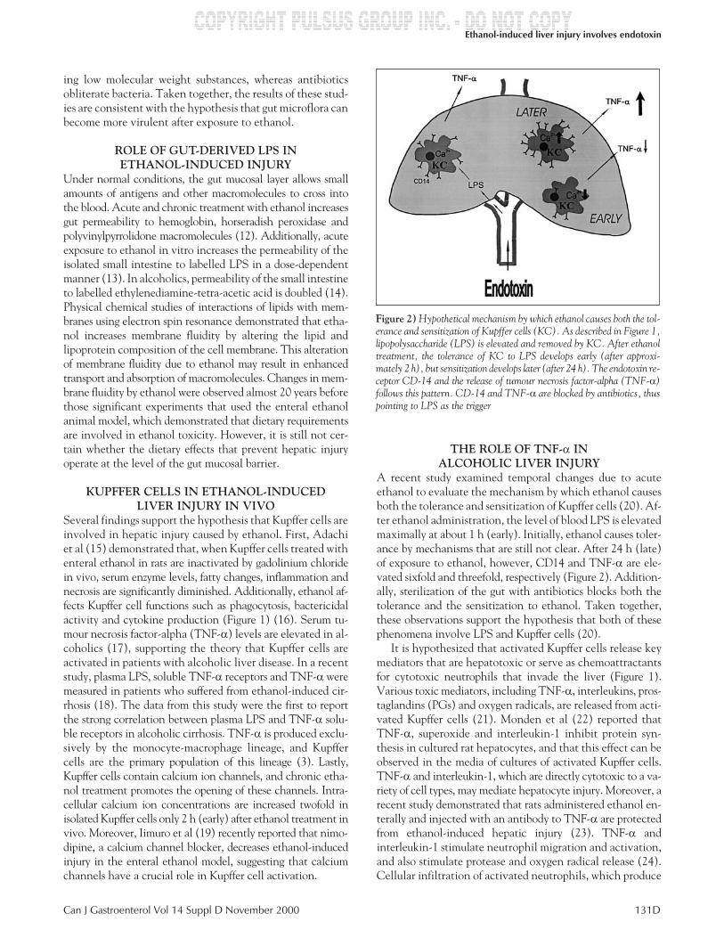
THE ROLE OF TNF-� INALCOHOLIC LIVER INJURY
A recent study examined temporal changes due to acuteethanol to evaluate the mechanism by which ethanol causesboth the tolerance and sensitization of Kupffer cells (20). Af-ter ethanol administration, the level of blood LPS is elevatedmaximally at about 1 h (early). Initially, ethanol causes toler-ance by mechanisms that are still not clear. After 24 h (late)of exposure to ethanol, however, CD14 and TNF-� are ele-vated sixfold and threefold, respectively (Figure 2). Addition-ally, sterilization of the gut with antibiotics blocks both thetolerance and the sensitization to ethanol. Taken together,these observations support the hypothesis that both of thesephenomena involve LPS and Kupffer cells (20).
It is hypothesized that activated Kupffer cells release keymediators that are hepatotoxic or serve as chemoattractantsfor cytotoxic neutrophils that invade the liver (Figure 1).Various toxic mediators, including TNF-�, interleukins, pros-taglandins (PGs) and oxygen radicals, are released from acti-vated Kupffer cells (21). Monden et al (22) reported thatTNF-�, superoxide and interleukin-1 inhibit protein syn-thesis in cultured rat hepatocytes, and that this effect can beobserved in the media of cultures of activated Kupffer cells.TNF-� and interleukin-1, which are directly cytotoxic to a va-riety of cell types, may mediate hepatocyte injury. Moreover, arecent study demonstrated that rats administered ethanol en-terally and injected with an antibody to TNF-� are protectedfrom ethanol-induced hepatic injury (23). TNF-� andinterleukin-1 stimulate neutrophil migration and activation,and also stimulate protease and oxygen radical release (24).Cellular infiltration of activated neutrophils, which produce
Can J Gastroenterol Vol 14 Suppl D November 2000 131D
Ethanol-induced liver injury involves endotoxin
Figure 2) Hypothetical mechanism by which ethanol causes both the tol-erance and sensitization of Kupffer cells (KC). As described in Figure 1,lipopolysaccharide (LPS) is elevated and removed by KC. After ethanoltreatment, the tolerance of KC to LPS develops early (after approxi-mately 2 h), but sensitization develops later (after 24 h). The endotoxin re-ceptor CD-14 and the release of tumour necrosis factor-alpha (TNF-�)follows this pattern. CD-14 and TNF-� are blocked by antibiotics, thuspointing to LPS as the trigger

oxygen radicals and secrete other toxic mediators, may in-crease the inflammatory response, leading to hepatocellularinjury and death. Indeed, inflammatory cell infiltration dueto enteral ethanol is diminished by gadolinium chloridetreatment. Disruption of the microcirculation caused by va-soactive mediators released from Kupffer cells and neutro-phils may amplify hypoxia and lead to a vicious cycle ofpathophysiology.
ETHANOL EXACERBATES THE EFFECT OF LPSPreviously, it has been reported that blood LPS concentra-tions are often elevated in alcoholics (25). In exciting experi-ments using the enteral ethanol model, levels of LPS in theblood begin to rise after about two weeks of enteral ethanoltreatment (26). LPS concentrations increase nearly fivefoldin the systemic circulation and are possibly higher in the por-tal circulation. Interestingly, blood LPS correlated (r=0.84)with pathology (necrosis, steatosis, inflammation, etc) (26).Bode et al (25), Remmer (27) and Adachi et al (11) have con-sistently been proponents of the theory that LPS plays a piv-otal role in ethanol-induced liver injury.
Acute exposure to ethanol also activates Kupffer cells.During acute exposure to ethanol, carbon uptake by the per-fused liver due to phagocytosis of particles by Kupffer cellsincreases about 25% (28). Carbon uptake is also increasedsignificantly, about 35% in rat livers treated with ethanol afew hours before liver perfusion. Similar results have beenobtained in vivo (29).
Few data linking Kupffer cell function to chronic ethanolexposure exist; however, a recent study has reported that oxy-gen radical production by Kupffer cells is elevated after chronicethanol ingestion (16). Others have reported that TNF-� re-lease and TNF-� mRNA expression are increased by ethanol,which is consistent with findings that TNF-� levels are in-creased in alcoholics (29). However, a number of studies haveshown that ethanol paradoxically inhibits Kupffer cell func-tion (30). Recently, ethanol treatment has been shown tocause tolerance after 2 h (early), followed by sensitization inKupffer cells after 24 h (late) (Figure 2). TNF-� productionfrom Kupffer cells stimulated with LPS decreases about fourfold2 h after ethanol treatment but is elevated about threefold24 h after ethanol treatment (20). Both tolerance and sensiti-zation are blocked by antibiotics, implicating LPS and ex-plaining this paradox.
KUPFFER CELLS PARTICIPATE INETHANOL-INDUCED HYPERMETABOLISM
Israel and colleagues (31) were the first to describe a hyper-metabolic state due to ethanol exposure. Moreover, Yukiand Thurman (32) showed that oxygen and ethanol uptakeincreased twofold 2 to 3 h after a single large dose of ethanolusing a perfused rat liver model. They also demonstrated thatthe hormone-mediated depletion of liver carbohydrate re-serves participates in this process, a phenomenon that hasbeen named ‘SIAM’.
Ethanol metabolism increases with a reduction in both gly-colysis and glycogen reserves during SIAM (32). The involve-
ment of Kupffer cells in carbohydrate metabolism has beendemonstrated (33). Moreover, oxygen and ethanol uptakewere almost doubled after ethanol treatment – a phenomenonblocked by gadolinium chloride (5). Thus, increases in respi-ration and ethanol metabolism observed after ethanol treat-ment are blocked by the inactivation of Kupffer cells.Specifically, Kupffer cells produce PGs, primarily PGD2 andPGE2, which enhance the production of glucose from endoge-nous hepatic glycogen by activating phosphorylase A (33).Additionally, conditioned media from isolated Kupffer cells ofethanol-treated rats, which contain elevated levels of PGE2,enhance parenchymal cell oxygen consumption (34). There-fore, regulation of SIAM clearly involves a Kupffer cell com-ponent, which appears to be due to production of PGE2.
ETHANOL PRODUCES HYPOXIA INPARENCHYMAL CELLS
In addition to hypermetabolism, high doses of ethanol changehepatic microcirculation by stimulating endothelin-1 produc-tion (35). Additionally, enteral ethanol administration causeshypoxia (36). Because ethanol also causes a compensatory in-crease in blood flow in the liver, which may elevate hepaticoxygen levels, it has been proposed that this increase ne-gates any effect of hypoxia due to hypermetabolism or mi-crocirculatory disturbances (37). Therefore, pimonidazole,a 2-nitroimidazole hypoxia marker used in radiobiology toassess local hypoxia in tumours, was studied (38). Pimonida-zole, which is reductively activated by nitroreductases, bindsto thiol residues on macromolecule proteins in the absence ofoxygen, and adducts can be detected immunochemically.Pericentral hypoxia occurs during SIAM and is blocked whenKupffer cells are destroyed with gadolinium chloride (5).
It has been reported that chronic ethanol treatment us-ing the enteral ethanol model also causes hypoxia by using2-nitroimidazole markers and other techniques (15). Thesedata provide direct evidence that ethanol increases tissuehypoxia in vivo (38). By employing the hypoxia marker pi-monidazole, hypoxia can be quantified in rats after a monthof ethanol feeding. Image analysis techniques have demon-strated that ethanol treatment for one or four weeks causes anaugmentation of pimonidazole binding in the liver from 18%(control group) to 32% to 35% (ethanol-treated group) (38).Thus, direct evidence has been obtained demonstratingthat hypoxia caused by ethanol treatment occurs in theclinically relevant enteral ethanol model or after acuteethanol treatment.
ROLE OF FREE RADICALS IN THE MECHANISMOF ETHANOL-INDUCED LIVER INJURY
Free radical production by ethanol has been implicated as a fac-tor in its hepatotoxicity. Although evidence of lipid radical for-mation due to ethanol treatment in vivo has been reported, freeradicals from ethanol alone been detected in living animalsonly recently (39). Ethanol-treated, alcohol dehydrogenase-deficient deermice exhibited an alpha-(4-pyridyl-1-oxide)-N-t-butylnitrone (POBN)/alpha-hydroxyethyl radicaladduct in bile after administration of ethanol and the spin
132D Can J Gastroenterol Vol 14 Suppl D November 2000
Thurman

trap POBN using electron paramagnetic resonance tech-nique and spin trapping (39).
Free radical formation most likely participates in the pro-gression of primary events in alcoholic liver disease. Rats ex-posed to ethanol using the Tsukamoto et al (6) model ofcontinuous enteral ethanol administration had a free radical inbile (15). This free radical signal decreased by over 50% whenKupffer cells were destroyed after treatment with gadoliniumchloride. Furthermore, bile from rats fed a control corn oil dietcontains low concentrations of radical adducts. The free radicalhas been identified as alpha-hydroxyethyl on the basis of the12-line spectrum obtained when 13C ethanol is used (40). Thus,ethanol-derived free radicals are detected in the bile of enteralethanol-fed rats after a high fat, ethanol-containing diet. Theprecise pathways responsible for the formation of free radicalsremain unclear. A possible candidate is oxygen radical produc-tion by the reduced nicotinamide adenine dinucleotidephosphate oxidase system in Kupffer cells, because the elec-tron paramagnetic resonance signal is reduced by gadoliniumchoride treatment. However, a reperfusion injury involvinghypoxia and free radical formation via the xanthine-xanthineoxidase system and the cytochrome P450 2E1 system shouldnot be excluded, especially because radicals in bile are expectedto originate from parenchymal cells.
ROLE OF SEXThree major independent risk factors for the development ofhepatitis and cirrhosis have been identified after evaluationof 1600 alcoholic patients: consuming ethanol, being over-weight for at least 10 years and being female. Sensitivity toethanol in females is summarized in Table 1. In general, thesestudies demonstrate that ethanol consumption potentiates in-flammatory responses, ethanol metabolism, hormone levels,hypoxia, free radicals and LPS in females. In rats, a study us-ing enteral ethanol feeding established that ethanol causesmore hepatic injury in females than in males (41). In thatstudy, parameters including serum aspartate transaminase,pathological score, neutrophil infiltration, levels of circu-lating LPS and intracellular adhesion molecule-1 expres-sion were evaluated. Interestingly, all parameters assessedwere increased by ethanol treatment approximately two-fold in females compared with males. The most dramatic his-tological change is the panlobular deposition of fat in femalelivers after ethanol feeding, compared with the well knownpericentral localization in males. Significantly more hepaticinfiltration of inflammatory cells is observed after ethanol ad-ministration in the female. It has recently been demonstratedthat the LPS receptor CD14 is elevated in Kupffer cells aftertreatment with estriol (42). Furthermore, LPS is higher afterethanol in female rats (41). Therefore, these data are consis-tent with the hypothesis that LPS and Kupffer cells are re-sponsible for the increased susceptibility of females to ethanol.
Tissue hypoxia has been quantified using the hypoxiamarker pimonidazole in male and female rats after enteral etha-nol feeding (43). In this study, hypoxia marker binding is two tothree times stronger in females than in males after a month ofethanol treatment. Furthermore, nuclear factor kappa B is
sensitive to oxidants and is increased seven to eight timesmore in females than in males. Because nuclear factor kappaB also increases adhesion molecule synthesis and inflamma-tory cytokine production, the above data may lead to themolecular mechanism of greater injury in females due toethanol.
The use of the rat enteral feeding model will enable fur-ther mechanistic studies to provide insight into the patho-physiology of important sex differences due to alcohol. Thesedata collectively demonstrate that females are more suscepti-ble than males to ethanol-induced liver injury.
ACKNOWLEDGEMENTS: The authors thank the National Insti-tute on Alcohol Abuse and Alcoholism for partial support of this work.
REFERENCES1. Adams HG, Jordan C. Infections in the alcoholic.
Med Clin North Am 1984;68:179-200.2. Tabakoff B, Hoffman PL, Lee JM, Saito T, Willard B, Leon-Jones F.
Differences in platelet enzyme activity between alcoholics andnonalcoholics. N Engl J Med 1988;318:134-9.
3. Decker T, Lohmann-Matthes ML, Karck U, Peters T, Decker K.Comparative study of cytotoxicity, tumor necrosis factor, and
Can J Gastroenterol Vol 14 Suppl D November 2000 133D
Ethanol-induced liver injury involves endotoxin
TABLE 1Some factors for the increased susceptibility of females toethanol-induced liver injury
FindingsSpeciesstudied Reference
Higher levels of circulating LPS Rat 41
Panlobular fat distribution in the liver Rat 41
Greater liver injury with less ethanol RatHuman
4144
Elevated intercellular adhesion molecule-1expression
Rat 41
More infiltrating neutrophils Rat 41
Rapid ethanol metabolism RatHuman
4145,46
Increased mortality by estrogen due to LPS Rat 42
Increased estrogen levels by ethanol Human 47
Increased ethanol-induced injury by fat Human 48
Decreased gastric alcohol dehydrogenase Human 49
Elevated CD14 and lipid-binding protein withethanol
Rat 20
Greater hypoxia and more free radicals Rat 50
Greater fibrosis Human 48
Injury due to ethanol blocked by ovariectomy,which is reversed by estrogen
Rat 51
Actin polymerization in Kupffer cells occurs onlywhen ethanol and LPS are combined
Rat 52
Suppressed P450-mediated metabolism by LPS Human 53
Lower cytokine-induced neutrophilchemoattractant after LPS
Rat 54
Decreased phagocytotic response to ethanol Rat 54
Higher phospholipase A2 activity inlymphocytes and neutrophils
Human 55
LPS lipopolysaccharide

prostaglandin release after stimulation of rat Kupffer cells, murineKupffer cells, and murine inflammatory liver macrophages.J Leukoc Biol 1989;45:139-46.
4. Nolan JP, Leibowitz A, Vladatin AL. Influence of alcohol on Kupffercell function and possible significance in liver injury. In: Liehr H,Green M, eds. The Reticuloendothelial System and Pathogenesis ofLiver Disease. Amsterdam: Elsevier, 1980:125-36.
5. Bradford BU, Misra UK, Thurman RG. Kupffer cells are required forthe swift increase in alcohol metabolism. Res Commun Subst Abuse1993;14:1-6.
6. Tsukamoto H, Reidelberger RD, French SW, Largman C. Long-termcannulation model for blood sampling and intragastric infusion in therat. Am J Physiol 1984;247:R595-9.
7. French SW. Nutrition in the pathogenesis of alcoholic liver disease.Alcohol Alcohol 1993;28:97-109.
8. Hentges DV, Maier BR, Burton GC, Flynn MA, Tsutakawa RK.Effect of a high-beef diet on the fecal bacterial flora of humans.Cancer Res 1977;37:568-71.
9. Bode JC, Bode C, Heidelbach R, Durr HK, Martini GA.Jejunal microflora in patients with chronic alcohol abuse.Hepatogastroenterology 1984;31:30-4.
10. Nanji AA, Khettry U, Sadrzadeh SM. Lactobacillus feeding reducesendotoxemia and severity of experimental alcoholic liver (disease).Proc Soc Exp Biol Med 1994;205:243-7.
11. Adachi Y, Moore LE, Bradford BU, Gao W, Thurman RG.Antibiotics prevent liver injury in rats following long-term exposureto ethanol. Gastroenterology 1995;108:218-24.
12. Bode JCH. Alcohol and the gastrointestinal tract. In: Frick HP,Harnack GA, Martini GA, Prader A, eds. Advances in InternalMedicine and Pediatrics. Heidelberg: Springer-Verlag, 1980:1-75.
13. Arai M. [Effect of ethanol on the intestinal uptake of endotoxin].Nippon Shokakibyo Gakkai Zasshi 1986;83:863.
14. Bjarnason I, Peters TJ. The leaky gut of alcoholism: possible route ofentry for toxic compounds. Lancet 1984;i:179-82.
15. Adachi Y, Bradford BU, Gao W, Bojes HK, Thurman RG.Inactivation of Kupffer cells prevents early alcohol-induced liverinjury. Hepatology 1994;20:453-60.
16. Yamada S, Mochida S, Ohno A, et al. Evidence for enhancedsecretory function of hepatic macrophages after long-term ethanolfeeding in rats. Liver 1991;11:220-4.
17. Stahnke LL, Hill DB, Allen JI. TNF� and IL-6 in alcoholic liverdisease. In: Wisse E, Knook DL, McCuskey RS, eds. Cells of theHepatic Sinusoid, 3rd edn. Leiden: Kupffer Cell Foundation,1991:472-5.
18. Hanck C, Rossol S, Böcker U, Tokus M, Singer MV. Presence ofplasma endotoxin is correlated with tumour necrosis factor receptorlevels and disease activity in alcoholic cirrhosis. Alcohol Alcohol1998;33:606-8.
19. Iimuro Y, Ikejima K, Rose ML, Bradford BU, Thurman RG.Nimodipine, a dihydropyridine-type calcium channel blocker,prevents alcoholic hepatitis due to chronic intragastric ethanolexposure in the rat. Hepatology 1996;24:391-7.
20. Enomoto N, Ikejima K, Bradford BU, et al. Alcohol causes bothtolerance and sensitization of rat Kupffer cells via mechanismsdependent on endotoxin. Gastroenterology 1998;115:443-51.
21. Martinez F, Abril ER, Earnest DL, Watson RR. Ethanol and cytokinesecretion. Alcohol 1992;9:455-8.
22. Monden K, Arii S, Itai S, et al. Enhancement andhepatocyte-modulating effect of chemical mediators and monokinesproduced by hepatic macrophages in rats with induced sepsis.Res Exp Med 1991;191:177-87.
23. Iimuro Y, Gallucci RM, Luster MI, Kono H, Thurman RG.Antibodies to tumor necrosis factor-� attenuate hepatic necrosis andinflammation due to chronic exposure to ethanol in the rat.Hepatology 1997;26:1530-7.
24. Thiele DL. Tumor necrosis factor, the acute phase response andthe pathogenesis of alcoholic liver disease. Hepatology1989;9:497-9.
25. Bode CH, Kugler V, Bode JC. Endotoxemia in patients with alcoholicand non-alcoholic cirrhosis and in subjects with no evidence ofchronic liver disease following acute alcohol excess. J Hepatol1987;4:8-14.
26. Nanji AA, Khettry U, Sadrzadeh SM, Yamanaka T. Severity of liverinjury in experimental alcoholic liver disease. Correlation with plasmaendotoxin, prostaglandin E2, leukotriene B4, and thromboxane B2.Am J Pathol 1993;142:367-73.
27. Remmer H. Die Wirkungen des Alkohols. Alkoholwirkungen1981;17:1-11.
28. D’Souza NB, Bagby GJ, Lang CH, Deaciuc IV, Spitzer JJ. Ethanolalters the metabolic response of isolated, perfused rat liver to aphagocytic stimulus. Alcohol Clin Exp Res 1993;17:147-54.
29. Earnest DL, Abril ER, Jolley CS, Martinez F. Ethanol anddiet-induced alterations in Kupffer cell function. Alcohol Alcohol1993;28:73-83.
30. Nelson S, Bagby GJ, Bainton BG, Summer WR. The effects of acuteand chronic alcoholism on tumor necrosis factor and theinflammatory response. J Infect Dis 1989;160:422-9.
31. Israel Y, Videla L, Bernstein J. Liver hypermetabolic state afterchronic ethanol consumption: Hormonal interrelations andpathogenic implications. Fed Proc 1975;34:2052-9.
32. Yuki T, Thurman RG. The swift increase in alcohol metabolism:Time course for the increase in hepatic oxygen uptake and theinvolvement of glycolysis. Biochem J 1980;186:119-26.
33. Casteleijn E, Kuiper J, Van Rooij HC, Kamps JA, Koster JF,Van Berkel TJ. Hormonal control of glycogenolysis in parenchymalliver cells by Kupffer and endothelial liver cells. J Biol Chem1988;263:2699-703.
34. Qu W, Zhong Z, Goto M, Thurman RG. Kupffer cell prostaglandin E2stimulates parenchymal cell O2 consumption: alcohol and cell-cellcommunication. Am J Physiol 1996;270:G574-80.
35. Hijioka T, Sato N, Matsumura T, et al. Ethanol-induced disturbanceof hepatic microcirculation and hepatic hypoxia. Biochem Pharmacol1991;11:1551-7.
36. French SW, Benson NC, Sun PS. Centrilobular liver necrosisinduced by hypoxia in chronic ethanol-fed rats. Hepatology1984;4:912-7.
37. Shaw S, Heller EA, Friedman HS, Lieber CS. Increased hepaticoxygenation following ethanol administration in the baboon.Proc Soc Exp Biol Med 1977;156:509-13.
38. Arteel GE, Iimuro Y, Yin M, Raleigh JA, Thurman RG. Chronicenteral ethanol treatment causes hypoxia in rat liver tissue in vivo.Hepatology 1997;25:920-6.
39. Knecht KT, Bradford BU, Mason RP, Thurman RG. In vivoformation of a free radical metabolite of ethanol. Mol Pharmacol1990;38:26-30.
40. Knecht KT, Adachi Y, Bradford BU, et al. Free radical adducts in thebile of rats treated chronically with intragastric alcohol: Inhibition bydestruction of Kupffer cells. Mol Pharmacol 1995;47:1028-34.
41. Iimuro Y, Frankenberg MV, Arteel GE, Bradford BU, Wall CA,Thurman RG. Female rats exhibit greater susceptibility to earlyalcohol-induced injury than males. Am J Physiol1997;272:G1186-94.
42. Ikejima K, Enomoto N, Iimuro Y, et al. Estrogen increasessensitivity of hepatic Kupffer cells to endotoxin. Am J Physiol1998;274:G669-76.
43. Iimuro Y, Kono H, Connor HD, et al. Increased susceptibility toalcoholic hepatitis in female rats is associated with elevated freeradical formation. Hepatology 1996;24:1249. (Abst)
44. Blume SB. Alcohol and other drug problems in women. In:Lowinson JH, Ruiz P, Millman RB, Langrod JG, eds. Substance Abuse:A Comprehensive Textbook, 2nd edn. Baltimore: Williams &Wilkins, 1992:794-807.
45. Holtzman JL, Gebhard RL, Eckfeldt JH, Mottonen LR, Finley DK,Eshelman FN. The effects of several weeks of ethanol consumption onethanol kinetics in normal men and women. Clin Pharmacol Ther1985;38:157-63.
46. Thomasson HR. Gender differences in alcohol metabolism:Physiological responses to ethanol. In: Galanter M, ed. RecentDevelopments in Alcoholism: Alcoholism and Women. New York:Plenum Press, 1995:163-79.
47. Gavaler JS. Alcohol effects on hormone levels in normalpostmenopausal women and in postmenopausal women withalcohol-induced cirrhosis. In: Galanter M, ed. Recent Developmentsin Alcoholism: Women and Alcoholism. New York: Plenum Press,1995:199-208.
48. Naveau S, Giraud V, Borotto E, Aubert A, Capron F, Chaput JC.Excess weight risk factor for alcoholic liver disease. Hepatology1997;25:108-11.
49. Frezza M, di Padova C, Pozzato G, Terpin M, Baraona E, Lieber CS.High blood alcohol levels in women. The role of decreasedgastric alcohol dehydrogenase activity and first-pass metabolism.N Engl J Med 1990;322:95-9.
134D Can J Gastroenterol Vol 14 Suppl D November 2000
Thurman

50. Kedderis GL, Argenbright LS, Miwa GT. Covalent interation of5-nitroimidazoles with DNA and protein in vitro: Mechanism ofreductive activation. Chem Res Toxicol 1989;2:146-9.
51. Yin M, Bradford BU, Thurman RG. Ovariectomy reducesalcohol-induced liver injury in rats. Hepatology1997;26:274A. (Abst)
52. Zhang P, Spitzer JA. Acute ethanol administrationmodulates leukocyte actin polymerization in endotoxic rats.Alcohol Clin Exp Res 1997;21:779-83.
53. Shedlofsky SI, Israel BC, Tosheva R, Blouin RA. Endotoxin depresseshepatic cytochrome P450-mediated drug metabolism in women.Br J Clin Pharmacol 1997;43:627-32.
54. Spitzer JA, Zhang P. Gender differences in neutrophil function andcytokine-induced neutrophil chemoattractant generation in endotoxicrats. Inflammation 1996;20:485-98.
55. Kuslys T, Vishwanath BS, Frey FJ, Frey BM. Differences inphospholipase A2 activity between males and females and AsianIndians and Caucasians. Eur J Clin Invest 1996;26:310-5.
Can J Gastroenterol Vol 14 Suppl D November 2000 135D
Ethanol-induced liver injury involves endotoxin

Submit your manuscripts athttp://www.hindawi.com
Stem CellsInternational
Hindawi Publishing Corporationhttp://www.hindawi.com Volume 2014
Hindawi Publishing Corporationhttp://www.hindawi.com Volume 2014
MEDIATORSINFLAMMATION
of
Hindawi Publishing Corporationhttp://www.hindawi.com Volume 2014
Behavioural Neurology
EndocrinologyInternational Journal of
Hindawi Publishing Corporationhttp://www.hindawi.com Volume 2014
Hindawi Publishing Corporationhttp://www.hindawi.com Volume 2014
Disease Markers
Hindawi Publishing Corporationhttp://www.hindawi.com Volume 2014
BioMed Research International
OncologyJournal of
Hindawi Publishing Corporationhttp://www.hindawi.com Volume 2014
Hindawi Publishing Corporationhttp://www.hindawi.com Volume 2014
Oxidative Medicine and Cellular Longevity
Hindawi Publishing Corporationhttp://www.hindawi.com Volume 2014
PPAR Research
The Scientific World JournalHindawi Publishing Corporation http://www.hindawi.com Volume 2014
Immunology ResearchHindawi Publishing Corporationhttp://www.hindawi.com Volume 2014
Journal of
ObesityJournal of
Hindawi Publishing Corporationhttp://www.hindawi.com Volume 2014
Hindawi Publishing Corporationhttp://www.hindawi.com Volume 2014
Computational and Mathematical Methods in Medicine
OphthalmologyJournal of
Hindawi Publishing Corporationhttp://www.hindawi.com Volume 2014
Diabetes ResearchJournal of
Hindawi Publishing Corporationhttp://www.hindawi.com Volume 2014
Hindawi Publishing Corporationhttp://www.hindawi.com Volume 2014
Research and TreatmentAIDS
Hindawi Publishing Corporationhttp://www.hindawi.com Volume 2014
Gastroenterology Research and Practice
Hindawi Publishing Corporationhttp://www.hindawi.com Volume 2014
Parkinson’s Disease
Evidence-Based Complementary and Alternative Medicine
Volume 2014Hindawi Publishing Corporationhttp://www.hindawi.com