shaken baby syndrome “silenced angels” - c.ymcdn.com · cerebral edema results from various...
TRANSCRIPT

Shaken Baby Syndrome“Silenced Angels”
Anesthesia ImplicationsCase Study
Marie A. De Francesco-Loukas, MSN, CRNP, SRNAVillanova University
Crozer-Chester Medical CenterNurse Anesthesia Program
Mentor: Ms. Bette M. Wildgust, MS, MSN, CRNA

Outline
• Overview of Shaken Baby Syndrome• Case Study: Pediatric Head Trauma• Anesthesia Implications• Problem-Based Learning with
Audience Participation• Questions & Answers

What Type of Cases will we See?
• Orthopedic• Neurosurgery• Ophthalmic• Trauma• General Surgery: Feeding Tubes etc.• Sedation for LP: r/o Meningitis

History
More than 1000 children die each year in the US as a result of child maltreatment (McClain, 1993)Of these at least 250 are victims of Shaken Baby Syndrome1996: First National Shaken Baby Conference

History
Although the SBS was not formally described until 1972 by John Caffey, it can be traced back at least 500 yearsNostradamus (1555) made a prophecy relating to subduralhematomas and their relationship to trauma and abuse

History Lesson
James Parkinson (1799): well known for describing “shaking palsy”Became of the first physician to publicly condemn child abuseHe addressed the issue of head trauma and its implications directly“Dropsy of the Head”: subduralhematoma

Caffey“Whiplash Shaken Infant
Syndrome”27 Cases 15 cases were found to be attributed to a nurse, Virginia Jaspers: Newsweek Magazine, 1956They tied this in to Nurse Jaspers’massive physical traits. (6’, 220 lbs)

Shaken Baby Syndrome
Reflects a subset of injuries caused by abusive head trauma (AHT) and is a part of Silver’s (1962) “Battered Child Syndrome”There is a pattern of abuse injuries associated with SBS with and without impact

Medical Components
Retinal HemorrhagesSubdural or Subarachnoid Hemorrhage
Associated FracturesOther External Physical FindingsIt is the often the absence of external signs of abuse that makes the early diagnosis of SBS so difficult

Incidence
Difficult to ascertainThe perpetrator is reluctant to provide an accurate historyMost children with injuries seen with SBS without obvious external trauma are presumed to have been shaken (Hadley et al., 1989)

Risk Factors
Perps described as having reversed nurturing needsThey are looking to be nurtured by their infantsWhen this does not happen, abuse can occur

Risk Factors
Ex-preemies or children with handicaps People who have admitted to shaking: a child have stated that they were not trying to harm the infant, but were trying to make the baby stop crying

Perpetrators Males (60-70%)Mother’s Boyfriend (34%)Baby Sitter (4-30%)Mother (6.5%)
Family Single ParentMaternal Age <18 yrsPoor SocioeconomicsMaternal Education <HSKnown to Social Service
Victim Age <1 yrMale InfantsPrematurity/LBWNo PNC

Physical Risk Factors of Infants
Small Body SizeLarge ratio of head (~ 25% of body wt.)Weak neck musclesLack of head controlUn-fused suturesHigh brain water contentLess myelination of nerve cellsLarge subarachnoid space

Seasonal Variations?
HMC: “Christmas”20025-6 babies with SBS under 8 week of age? Seasonal VariationsMore Common during Holidays?Stressors: financial, depression, burdens

Mechanisms of Injury
Infants are more susceptible to whiplash shaking injuries than older childrenRelatively large heads supported by weak neck muscles which increases their head movement during shakingTheir unmyelinated brain, soft sutures, open fontanelles, and relatively increased CSF result in a brain that is more vulnerable to injury

Clinical Presentation
Can be very non-specificThe infant may present in a coma, with a bulging fontanelle, or with subtle signs such as vomiting, irritability, seizures, poor feeding or failure to thriveSome are misdiagnosed as: meningitisA “bloody” Tap may be thought to be related to the tap itself rather than a sign of a subarachnoid bleed (also HSV)

Clinical Presentation
Some argue that retinal hemorrhages were pre-existing from birth trauma14-40% of newborns sustain retinal hemorrhages at birth (Budenz, 1974)These usually resolve in several days and certainly within 2-3 weeksAfter the neonatal period any finding of retinal hemorrhage should suggest abusive head trauma

Lab Data
May present with clotting dysfunction which is reflective of DIC secondary to intracranial traumaMild-moderate anemiaLP: will be bloodyLeukocytosis, Electrolyte Disturbances

Making the Diagnosis
CT is the method of choice for initial imaging. It readily identifies lesions requiring emergency surgeryMRI has shown to detect 50% more subdural hemorrhages than CT and can detect smaller injuries missed by the CTThe cost and availability of an MRI makes it more useful as a second study

Prognosis
Victims of SBS suffer significant morbidity and mortalityLudwig et al. (1984) showed a 15% mortality rate and a 50% morbidity rate in their review of 20 casesPermanent brain damage, hydrocephalus, developmental delay, blindness, deafness, paralysis and mental retardation have been noted

Long-Term Medical Burden
High incidence of mortality and morbiditySBS accounts for 10-12% of all child-maltreatment deaths25% of victims of SBS will dieOf those that survive 57% will have neurological complicationsIncluding: severe motor deficits, seizures, developmental delay, and blindness

Biomechanics of Abusive Head Trauma
Precise mechanisms of neurological injury remains unclearInjuries occur from rapid and repetitive flexion, extension, and rotation of the head and neck around a relatively stable torso with (Duhaime) or without impact (Dias)

Biomechanics
The injuries with SBS are not likely caused by falls from short distances, rough horseplay, swinging or rocking babyInfants’ brains have a higher water content and less myelination than the adult brain, it is more gelatinous and is easily compressed and distorted from shaking

Shaking Event

Shaking Event
Skeletal Findings
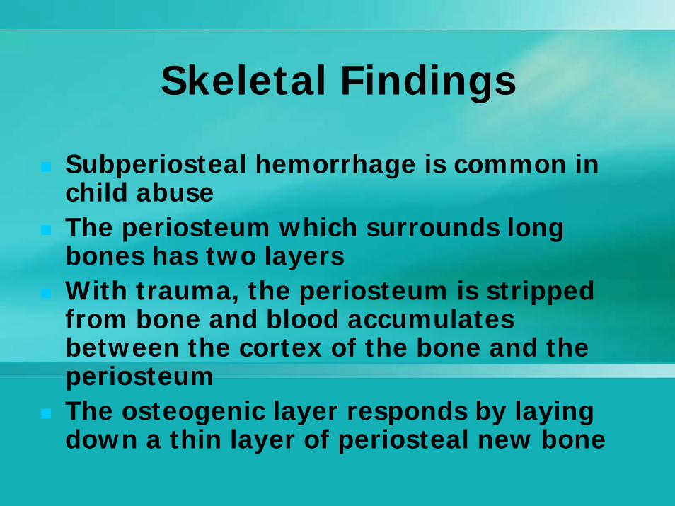
Subperiosteal hemorrhage is common in child abuseThe periosteum which surrounds long bones has two layersWith trauma, the periosteum is stripped from bone and blood accumulates between the cortex of the bone and the periosteumThe osteogenic layer responds by laying down a thin layer of periosteal new bone

PeriostealNew Bone
SubperiostealHemorrhage

Diaphyseal Fractures
Spiral fractures can occur with long bones (femur or humerus)In the non-mobile infant, this is highly suggestive of abuseIn a toddler learning to work: can be Toddler’s Fracture: non-abuseDistal clavicular fractures can result from traction of twisting forces applied to the arm

Spiral Fractures

Clavicular Fractures

Rib Fractures

Rib Fractures
Courtesy of Children’s Hospital, Boston: Child Protection Team

Skull and Scalp Injuries
Infants with significant impact injuries to the head frequently exhibit swelling of a portion of the scalp
The majority of skull fractures in abused children occur during infancy (80-85% under age 2), (Lazoritz, 2001)
Skull fractures do not heal by callus formation thus making the DX difficult

Skull Fractures

What are Patients with Skull Fractures at Risk For?
A. MeningitisB. Intracranial BleedsC. Mike Tyson SyndromeD. A&B

Example of Mike Tyson SyndromeDefinition: Volatile Temperament from
Repeated Head Trauma
Mike As Baby Mike Now

Leptomeningeal CystAKA “Growing Skull Fracture”
Extra-Axial Hemorrhage
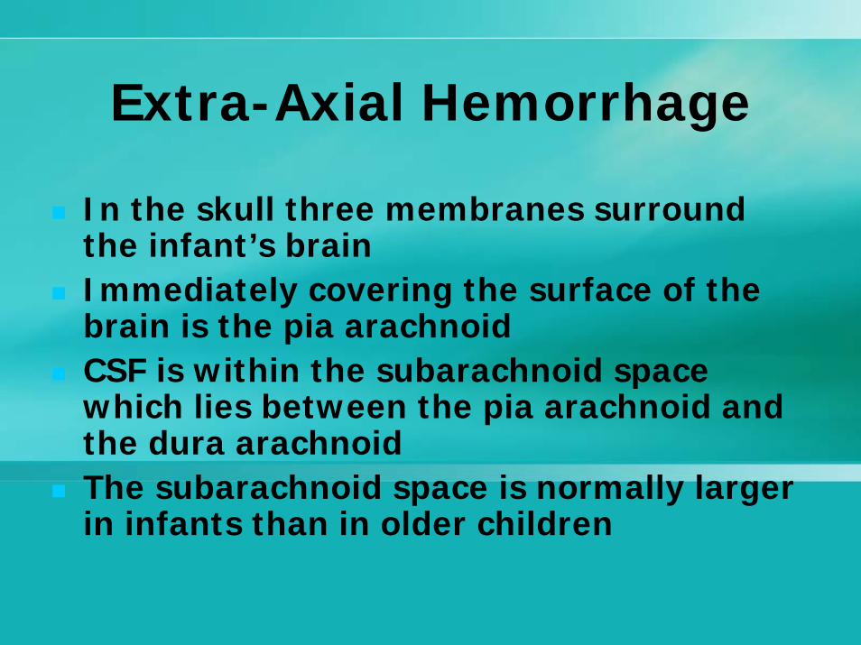
In the skull three membranes surround the infant’s brainImmediately covering the surface of the brain is the pia arachnoidCSF is within the subarachnoid space which lies between the pia arachnoid and the dura arachnoidThe subarachnoid space is normally larger in infants than in older children

Subdural Hemorrhage

Subdural Hematomas with SBSPost-Mortem View

CT Findings Subdural Bleed & Cerebral Edema

MRI Findings
Coronal View

Parenchymal Brain Injuries
Subarachnoid hemorrhage usually occurs in association with parenchymal brain injurySubarachnoid & epidural hemorrhage are both relatively uncommon with child abuseThey can, however, occur with associated parenchymal brain injuries with SBS
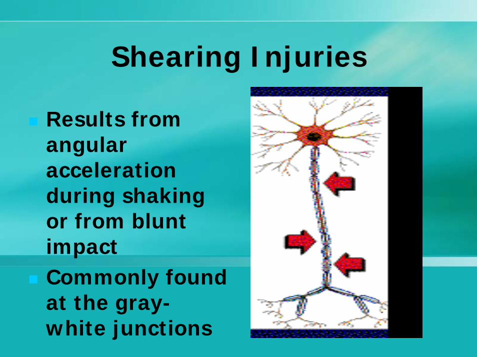
Shearing Injuries
Results from angular acceleration during shaking or from blunt impactCommonly found at the gray-white junctions

Cerebral Edema
Results from various mechanisms but is probably an indirect pathophysiological response to head trauma (Aldrick, 1992)There is also associated hypoxic-ischemic injury with SBS (apnea).This is caused by increased ICP/hypo-perfusionDiffuse cerebral edema from hypoxic-ischemic injury carries a grave prognosisMay produce a “Mass-Effect”Edema may be present on CT after 2-3 hours “or”not until 1-2 days (when they usually present in ER)

Cerebral Edema with SBSNo Space Between Ventricles

Ophthalmic Manifestations
Retinal hemorrhages are the most common ocular finding with SBSCannot be dated clinically Non-ophthalmologists have great difficulty in seeing RH with direct exam and un-dilated pupilsRH can cause loss of vision, but the most common cause of blindness with SBS is a direct bilateral injury to the visual pathways of the brain

Retinal Hemorrhages
Occurs in 50-100% (other studies 75-90%) in patients with SBSThere are different layers of the retina that may be affectedHemorrhages include: splinter, dot-blot, and large blot Large dome-like bleeds have been observed in the macular area

RH
Extremely common with birth (50% @ 1st DOL)Influenced by age of infant, type of birth, and parity of the motherAppears to be less frequent in premature infants (although they are at higher risk for abuse)By 3 weeks: RH are generally not r/t birth unless the birth was extremely traumatic

Iatrogenic Causes of RH
ECMOHeparinization, alterations in cerebral blood flow, major vessel ligation, mechanical ventilation, and hypoxia could contribute to RHAVM or Aneurysms could also cause RH

Other Causes of RH
Meningitis Accidental trauma “rarely” causes RHChest trauma can cause RHSome RH can be caused by CPRSome patients with persistent ROP may have RH and Vitreous hemorrhagesSIDS: extremely unlikely to cause RH itself or from CPR in these cases
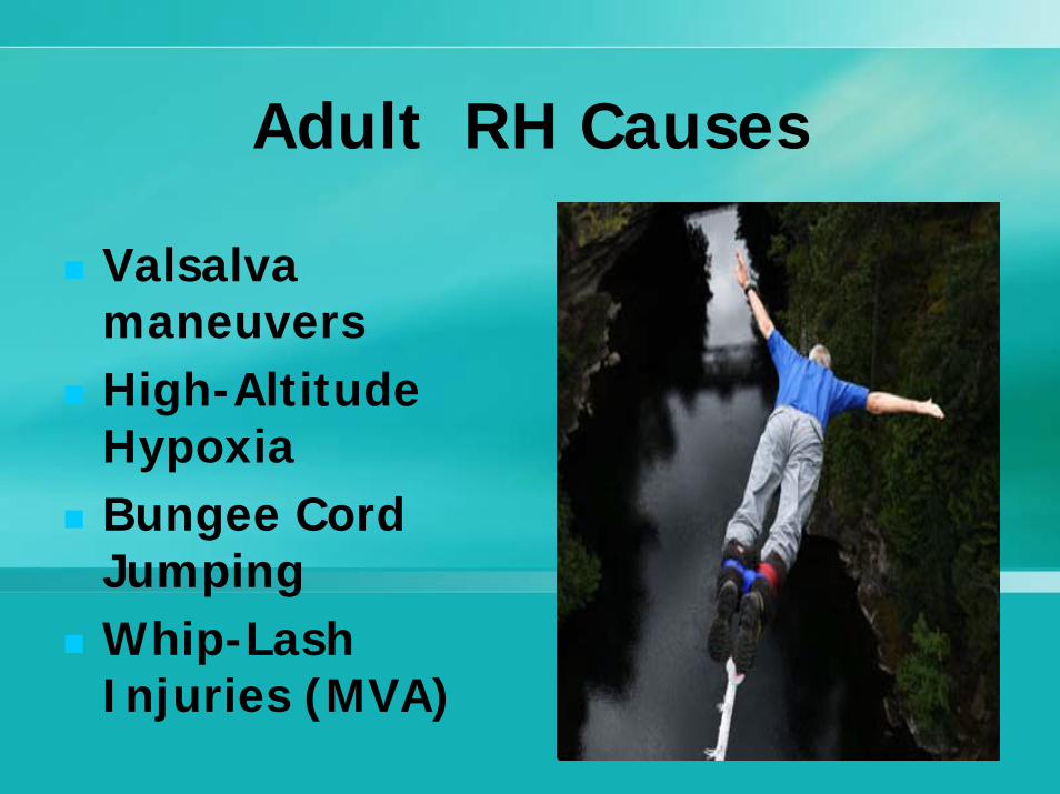
Adult RH Causes
ValsalvamaneuversHigh-Altitude HypoxiaBungee Cord JumpingWhip-Lash Injuries (MVA)

Retinal Hemorrhages
Normal Abnormal

Medical Management
Challenging and FrustratingSignificant brain injuries resulting in life-long physical and cognitive abnormalitiesDeathHistory is inaccurate which may delay treatmentEmotionally taxing to staff

Brain Injury
Injury caused by direct traumaHypoxic-ischemic injurySecondary injuries as a result of the trauma and hypoxic-ischemia

Treatment
ABCEndotracheal Intubation for Glascow < 8, apnea or cardiac arrestAvoid hypoxia and hypercarbiaHemodynamic monitoring: Arterial BPMaintain BP: give initial NS Bolus 20cc/kgKeep ICP <20 ( 1-15 mm Hg)Surgery for Trauma, Bleeds etc.

Treatment
If infant requires >40-60cc/kg fluid then inotropic/vasoactive support is neededDopamine, Epi, NE, Vasopressin * gtts etc.This is necessary to maintain adequate CPP (CPP=MAP-ICP)CPP: minimally accepted 60mm Hg (adults 70mm/Hg)Follow Neuro statusManage Seizures (fosphenytoin, benzodiazepines, phenobarbital etc)*note: hyponatremia described in Peds. Literature

DX Tests to Monitor
CBC with diff, coags, Fibrinogen, ABGs, LFTs, Lytes, Amylase, Lipase, BCX, UCX, CSF, CRPCXR, f/u CT, MRICT of AbdomenSkeletal SurveyEEGICP, MAP, CPPMany Systems Involved: Interdisciplinary Approach

Specific Goals
Maintain Cerebral Perfusion PressureMinimize ICP: Monitor CSF drainage, replace cc:ccsedation, decrease noxious stimuli, and osmolar therapyMaintain osmolarity: 300-310 mosm/l

Ventilation
Changes in Carbon Dioxide have a profound effect on cerebrovascularresponsesWith hyperventilation, cerebral blood flow decreases which decreases ICP…but at the risk of cerebral infarction with prolongationUntil recently (last 5 years), hyperventilation was a mainstay in therapyCurrently: Maintain Low-Normal PCo2

Ventilation
We used to provide temporary hyperventilation: (maintain PC02 low 20’s)Studies have shown that it worsens the outcomeCan cause ischemiaGoal: maintain PCO2 low normal range (35-45)Hypercapnia should also be avoided: may increase ICP

Treatment
Manage SeizuresObtain EEG: Barbiturate-induced coma reduces cerebral metabolism and can be achieved by seeing a “burst”suppression on the EEG

Temperature
Reducing body temperature reduces cerebral metabolic rate significantlyThere is also evidence for additional benefits to ischemic brain tissueHypothermia: Runs the risk with neutropenia, coags, and infection. The patient lacks the capacity to elicit a proper immune response. Underlying infection may be missed due to the inability to mount a fever.

L-carnitine Current TX
Numerous lines of evidence indicate that cerebral ischemic insults disrupt normal respiratory activity in mitochondria.Glutamate mediated intracellular calcium
accumulation and free radical generation are thought to be major mechanisms that contribute to cell death in hypoxic-ischemic brain injury. Study Demonstrated: l-carnitine protects against glutamate- and KA-induced neurotoxicity.
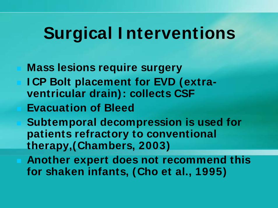
Surgical Interventions
Mass lesions require surgeryICP Bolt placement for EVD (extra-ventricular drain): collects CSFEvacuation of BleedSubtemporal decompression is used for patients refractory to conventional therapy,(Chambers, 2003)Another expert does not recommend this for shaken infants, (Cho et al., 1995)

End of Part IEnd of Part I

Trivia QuestionWhat Syndrome Do All of These People
Have?

Answer:“The Mick Jagger Syndrome”

Please“Curb Your Enthusiasm”

Part IICase Study
Problem-Based Learning

Run Forrest Run !!
It is 5PM one winter evening a few weeks before Christmas. You have not done your Peds rotation. You are getting ready to leave clinical and have plans for holiday “cheer” and Carols with friends. Suddenly you hear overhead: “Pediatric Trauma Level One: ETA 5 Minutes”. Your heart begins to pound. You look at your preceptor and via mental telepathy you both run to the trauma bay. No caroling for you this evening. The patient arrives via Life Lion in the trauma bay.
Case Study November, 2006
1 y/o male presented to Community Hospital with seizures, change in mental status, fever, and lethargy.
Was that a trauma page?

Patient Information
1 y/o male, 10 kg, ex 25 weeker, NICU X 3 months, Vent X2 months with diuretic therapy & 02. hx of BPD, RAD, GERD, mild developmental delay, HX of PDA: s/pClosure with Indomethacin at birth.
Vitals: HR: 70, RR: 16 and irregular, BP: 115/40, Sats: 88%, Temp: 40 ® Foley in place: no U/O yet, Home Meds: albuterolq4 ATC, reglan, zantac, Iron

Patient Information
Arrived Boarded and Collared, CT: shows: HUGE Sub-Dural Bleed and sub-arachnoidbleed, patient is on IVF @ 60cc/hr of D5 ¼NS + 20meqkcl/Liter, s/p 3 boluses of NS: 20c/kg in the ER for “shock” on Room Air, Pupils: Right Pupil dilated, you see conjunctival hemorrhages OUleft pupil 3mm sluggish, Anterior Fontanelle bulging, mottled, cap refill >6 seconds, poor peripheral pulses, circumoral cyanosis, decerebrateposturing

LabsPT: 16, INR: 2.0, PTT: 50Lfts: elevated, Albumin: 4.8CXR: hypoventilatorychangesABG: 7.27/50/70/18/BD -10, ICA++: 0.59BGM: 60(note: these labs were from 2 hours ago)
NA: 124, K: 6.0, Cl: 120, Co2: 10, Bun 25, Cr: 1.5, Glu: 58Ca: 7, wbc 22, hgb: 7.8, Hct: 24, Plt: 125Neut: 85, L 15, Bands 25EKG: shows mild peaked T waves approaching sinus bradyIV Access: 22 gauge PIV X1 in Right AC

QuestionWhat are some Causes of
Hyponatremia?
1. Dehydration2. Meningitis3. Subarachnoid Hemorrhage4. SIADH and Cerebral Salt Wasting5. All of the Above

Progression of CaseThe patient is intubated using STP and Rocuronium
The child is started on a fentanyl gtt @ 2 mcg/kg/hr, PRBC (10cc/kg) are put on a pump over 2 hours, FFP 10cc/kg, and treatment is initiated for hyperkalemia. The Ab CT shows a duodenal hematoma, the repeat CXR: confirms ETT placement w/atelectatic areas, the child receives a 4 Fr. Rt. Femoral CVL and placement is confirmed via Xray (you can use the line), an Arterial Line is placed

Progression of Case
There is a depressed skull fracture, the repeat head CT shows cerebral edema with a brain stem shift, OGT is placed and abdomen is decompressed: you get fresh blood. You repeat the trauma panel of labs + Blood Cultures. You also give Tylenol 15mg/kg/PR. CXR: Several Rib Fractures (new)

Peds Doses of Blood Products
1. Plt: 1 unit/10kg if <10kg: 1 Unit: note: 1 unit ~50cc
2. PRBC: 5-20cc/kg3. FFP: 10-15cc/kg4. Cryo: adult: 10units (15cc/unit)5. Peds Cryo: <1yr: 10cc/kg, >1 yr: 1
unit/5kg6. 5 % albumin: 5-15cc/kg

Questions
What implications does a depressed skull fracture have on a febrile patient and what would you do and why? What med and dose?
This child’s Temp is 40 ®: Is this the time for a lumbar puncture?

AnswerPatient At Risk For MeningitisTreatment:Decadron: 0.6mg/kg divided by 4 = 1.5mg IV X1Ceftriaxone: 100mg/kg X1 (1gm)Vancomycin: 15mg/kg X1 (150mg)Give in That OrderNote: no steroids if HSV is suspected or less than 6 weeksNO LP !!!!

QuestionsGroup Participation
1. What would you give immediately to treat the cerebral edema? What IVF would you run? And Why?
2. What is in LR that is bad for this patient? How much per Liter? (actually 2 things may be bad)
3. What does giving hypotonic fluid to this pt. contribute to?

AnswersMannitol To Treat Cerebral Edema: 0.25-0.5gm/kg over 20 minutes (use filter)K+ in LR: Bad for this Patient: Hyperkalemic Already, Lactate may be badHypotonic Fluids Contribute to DilutionalHyponatremiaFluids Containing Dextrose Contribute to Cerebral Edema in the Trauma Patient

Progression of Case
The child is transported to the OR. Neuro Surgery and General Surgery are Ready The neuro surgeon begins the emergency craniotomy to evacuate the bleed and place bolt with EVD.Vent Settings: 1.o Fi02, TV: 90, Peep: +3, Rate: 18 The child begins to seize. Urine is dilute and large volume now…

QuestionsGroup Participation
1. What is the best treatment for pediatric seizures?
2.What could be the cause of the seizures? List some neuroprotective measures in the OR?
2. What is the different between phenytoinand fosphenytoin? What is the loading dose? When can you check a level?

Answers
Seizures: Lorazepam 0.1mg/kg (dilute), Midazolam: 0.1mg/kgCauses: Fever, Meningitis, Hyponatremia, Bleed etc.Neuroprotective: temp, Iso, Isotonic Fluids, ?steroids, anticonvulsant load, EEG suppression (meds)

Pediatric Status Epilepticus
Lorazepam (0.05-0.1 mg/kg IV/IO slowly infused over 2-5 min) has rapid onset and long duration of anticonvulsant action. It is preferred over diazepam. Midazolam (0.1-0.2 mg/kg IM) is most effective when IV or IO access is not available. Midazolam is the only benzodiazepine that can be administered safely intramuscularly with equivalent rapid onset and moderate duration of action.

Seizures
Phenytoin (18-20 mg/kg IV/IO) or fosphenytoin (15-20 mg/kg IV/IO) loading doses: These long-acting anticonvulsants usually are infused if benzodiazepines do not stop the seizures. A full loading dose should be delivered unless the patient is known to have a current therapeutic level.

Intravenous Phenytoin vs Fosphenytoin
fosphenytoin is a water-soluble prodrugof phenytoin.It is rapidly converted into phenytoin in vivo by phosphatase enzymes.The half-life of this conversion is 8-15 min and is independent of the plasma concentrations of either fosphenytoin or phenytoin.

Intravenous Phenytoin vsFosphenytoin
phenytoin has very poor water solubility. It requires slow infusion in glucose free solutions to avoid precipitation.fosphenytoin is supplied in phenytoinequivalents (PE) to obviate the need for learning new dosage schedules or calculating equivalent dosages.In general, has less CV effects (arrhythmias, hypotension)

Characteristics Fosphenytoin Phenytoin
Routes of Administration
IV or IM IV
Time to Max Serum Level
20 Min 20 min
IV Solution Compatability
Dextrose or NS
NS Only
Max IV Infusion Rate
150mg PE/min
50mg/min

Side Effect Fosphenytoin phenytoin
Local pain or burning
1% 37%
Hypotension 2% 13%
itching / burning (transient, not serious)
9% 0%
Purple glove syndrome (see below)
none reported 3 to 7%
1000-mg IV loading dose
$90.00 $6.72

Case Progression
The patient now has large U/O has DILUTE Urine (that is a hint)…but think out of the box..Vitals: HR: 55, BP: now low 90/55, Sats: 98%, Temp: 38.9 ®, CVP: 3We gave Versed: 1mg IV X1, fosphenytoin 20mg PE/kg/ X1…Still Seizing? YES…

The labs come back:
ABG: 7.35/28/450/15/BD-10, ICA++: 0.4Wbc: 36K, hgb: 7, Hct: 22, plt: 60, N: 90, L 30, M 4, Bands 40CRP: 10 CVP: 2-3, Serum Osmolarity: 600mmol/L Pt: 14, INR: 1.8, PTT: 35Na: 118, K: 3.8, Cl: 130, C02 15, Bun 35, Cr: 2.8, Ca+: 5.6, Glucose: 30
QuestionsGroup Participation
What are the implications of these labs?
What is your plan?

Case Progression10cc/kg PRBC, 1 unit platelets, 3%
hypertonic Saline: 2cc/kg (slowly): seizures stop…IVF are continued @ NS 1 ½ MIVF (60cc/hr) to change to ½ NS in 4 hours. We also gave Decadron: 0.15mg/kg/dose X1…why?Decreased Fi02 to 0.6%Atropine: 0.2mg IV X1

Case Progression
D25%: 2cc/kg then recheck BGMThe child is given: Vanco: 15mg/kg and Ceftriaxone 1 gm The Crani is done. The general surgeon is evacuating the bleed and repairing the duodenum. (Now…here’s the rub)The patient has insensible losses from the open gut, bleeding etc. Despite this, the patient is putting out large amount of clear urine and has electrolyte disturbances.

3% Hypertonic Saline
2cc/kg until Seizures StopInfusion Rate: 1cc/minDo not exceed 12cc/kg1meq/2ccNote: check institution policy*Can cause central pontinedemyelination

QuestionWhat possible factor is most likely
not contributing to the higher urine output?
A. DIB. Fluid ResuscitationC. Improved Renal and Splanchic BFD. SIADH

New Lab Data
New Labs:Hgb: 11, Hct: 30, Coags: normalVitals: HR now: 150, BP: 88/46 and dropping, Sats: 99% PH 7.28, PCo2: 30, Po2: 300, BD: -12, HCo3: 8, K: 4.0, Ca: 9

What Differential Diagnosis is possible to explain the dilute
urine?
A. High Output Renal FailureB. DIC. Cerebral Salt Wasting SyndromeD. Fluid OverloadE. All of the Above

What is the Diagnosis R/T the hypovolemia, acidosis, and dilute
urine output?
A. Cerebral Salt Wasting SyndromeB. DIC. SIADHD. Too many hotdogs at Three Rivers Stadium

DiagnosisCerebral Salt Wasting Syndrome
We determined through concise examination of our data that this child has Cerebral Salt Wasting. Fluid Restriction is NOT Correct. Can cause cerebral infarctions. With SIADH (Faseret al.) fluid restriction of ¾ MIVF improves hyponatremia. In patients with SAH and undiagnosed CSW fluid restriction is detrimental. Therefore, look at all the parameters.

DiagnosisCerebral Salt Wasting Syndrome
With SIADH you have euvolemia, normal of increased CVP, normal HR, normal hct, increased EC volume, normal alb, decreased bun/cr, normal K+.With CSW: you have: decreased EC fluid volume, increased albumin, increased hct (we saw that after transfusions), increased Bun/Cr, normal or increased K+, decreased CVP and BP, and increased HR.

Case Progression
We proceeded with the latter part of the surgery, did not restrict fluids and the NA was improving. By 10PM: The NA was 130 (up from 124 from 3PM in the ER: initial labs) The patient was on NS @ 1 ½MIVF to change to ½ NS by 10PM in PICU. The BP and HR were approaching normal and we gave a 5% albumin bolus 10cc/kg. The child was stabilized and transported to the PICU intubated with a bolt and EVD.

Case Progression
No vasopressors were on board. (good for organ perfusion…the issue was volume) The child was extubated 3 days later (was started on the head injury protocol @ HMC). He then went to the rehab unit and has some minor deficits. (hard to judge because his baseline was delayed). Time will tell.

Cerebral Salt Wasting Syndrome
CSWS is defined as “true” hyponatremiawhich occurs when there is a primary loss of sodium into the urine without an increase in total systemic volume. It is related to acute or chronic damage of the central nervous system.The most important component of this caseis the TIMING of the INJURY. You don’t usually see CSW right away much like With NAT (non-accidental trauma)…The child usually doesn’t present to the ED right away, thus, delaying the diagnosis due to late clinical manifestations

When further patients were studied (eg. SAH, head injury), plasma volume was found to be reduced, ADH levels were appropriate for serum osmolality and patients did not respond to fluid restriction as expected for SIADHIf hyponatremia is due to CSW, fluid restriction may actually aggravate the clinical condition (esp in vasospasm of SAH) and lead to cerebral infarction - patients respond to salt and volume replacement (ie. opposite treatment!)

Causes of CSWS
Humeral mechanisms: increase in circulating atrial natriuretic peptide (ANP) and brain natriuretic peptide (BNP) are thought to be contributing factors to the development of CSWS. Brain insult from the following disease processes are thought to also increase the risk of developing CSWS:
Sub-Arachnoid Hemorrhage Intracerebral Hemorrhage/Stroke Cerebral Neoplasm/Intracranial Surgery Increased Intracranial Pressure Tuberculous Meningitis

Signs and Symptoms of CSWS:Physical signs of CSWS: associated with severe hyponatremia or intravascular depletion.
Hypovolemia (low CVP), shock SX Absence of Weight gain Orthostatic tachycardia/hypotension Increased capillary refill time/increased skin turgorDry mucous membranes Sunken anterior fontanel (in infants)Large Volume of Dilute Urine

Hallmark: Low EC Volume with CSWS
Differentiating the Diagnosis of CSWS and SIADH:Identical acute cerebral insults may cause either SIADH or CSW. The clinical manifestation of both conditions can be virtually identical. The only true discriminative feature is that extracellular volume is “expanded” in SIADH and is “low” in CSWS.

CSWS vs. SIADH
CSWSU/O high, diluteHigh Serum Osm.Urine NA: highHypovolemicSerum Na+: lowTX: NS (fluid)Replete Sodium
SIADHU/O low, conc.Low Serum Osm.Urine NA: highEuvolemicSerum Na+: lowTX: Fluid Restriction

Treatment of CSWS:
Making the distinction between CSW and SIADH is of particular importance with regard to therapy. The following treatment regiment is recommended for treatment of patients who are suffering from CSWS:
Treat the underlying neurological process Volume replacement (to maintain a positive salt balance) IV Hydration with 0.9% NaCl infusion IV Hydration with hypertonic 3% NaCl infusion Colloids may effective to absorb third-space fluid Blood products may be useful for volume expansion Urine replacement (cc for cc) and CSF: if EVD in placePositive sodium balance Fludocortisone (enhances sodium reabsorption, can cause hypokalemia)

Clinical Manifestations to Monitor CSWS SIADH
ECF volume ( the primary wayto differentiate SIADH and CSWS) Decreased IncreasedHematocrit Increased normal
Albumin concentration Increased normal
BUN/creatinine increased Decreased
Potassiumnormal or high normal
Uric acidnormal or low Decreased
Treatmentnormal saline
fluid restriction

Pennsylvania State Program“Pennsylvania Shaken Baby Syndrome Prevention and Awareness Program”Mark S. Dias, M.D., FAAP: Neurosurgeon: HMCDean J. Bonsall, M.D.: Pediatric OphthalmologistKelly Cappos, RN, BSN, CPUR, CLNC, HMCCarroll Rottmund, RN, BSN, CCRN, CLNC, HMC

In 2002, the 42 hospitals that provided maternity services in Central Pennsylvania were asked to partner together and participate in a shaken baby education, research, and child abuse prevention effort. The ultimate goal is to decrease the incidence of infant abusive head trauma statewide!

Dr. Dais and the shaken baby team were awarded a $2.8 million dollar grant from the Centers for Disease Control (CDC) in October 2007 to continue the hospital-based program
The ultimate goal is to decrease the incidence of infant abusive head trauma statewide!

Moral of the StoryDon’t Shake or Drop your Baby

One Hundred Years from nowIt will not matter
what kind of car I drove,What kind of house I lived in,
how much money was in my bank account
nor what my clothes looked like.But the world may be a better place
becauseI was important in the life of a child.
Forest E. Witcraft
Thank youMarie