shear bond strength of orthodontic brackets cemented with a zinc oxide-polyvinyl cement
TRANSCRIPT

Shear bond strength of orthodontic brackets cementedwith a zinc oxide-polyvinyl cement
Steve Martin, DDS,· and Franklin Garcia-Godoy, DDS, MSb
San Antonio, Texas
The purpose of this study was to compare the shear bond strengths and enamel surface structureafter debonding a conventional metal bracket and a polycrystalline ceramic bracket bonded witha bipolar zinc oxide-polyvinyl cement (F-21) or a light-cured resin cement (Transbond). Fortyextracted human premolars were used. The buccal enamel surfaces were used, and the teethrandomly divided into four groups of 10 teeth each: group 1: conventional metal bracket (Unitek)bonded with Transbond; group 2: metal bracket bonded with F-21; group 3: ceramic bracket(Transcend 2000) bonded with Transbond; and group 4: ceramic bracket bonded with F-21. Thebrackets were bonded to the etched enamel surfaces according to manufacturer's instructions. Allspecimens were stored in distilled water for 24 hours and then thermocycled for 300 cyclesbetween 5° C and 55° C. The specimens were mounted in dental stone and placed in the Instron ata crosshead speed of 0.5 mm/min with a knife-edged blade. Immediately after debonding, theenamel surface and bracket-enamel interface were evaluated visually and with a stereomicroscope.Representative samples were then examined with the scanning electron microscope. The analysisof variance and Student-Newman-Keuls tests were performed. The results in megapascals wereGroup 1: 19,6 (±9,6); group 2: 14,3 (±4,6); group 3: 28,8 (±12,6); and group 4: 18,5 (±7,5).Group 3 was statistically significantly different (P < 0.008) from all other groups. Groups 1, 2, and 4were not significantly different. Debonding occurred mainly at the bracket-resin interface in group1 (two enamel fractures), mixed (bracket-resin, resin-enamel interface) in group 2, at theenamel-resin interface (with four specimens fracturing the enamel) in group 3, and at thebracket-resin interface (with one specimen fracturing the enamel) in group 4. The scanning electronmicroscope evaluation revealed that after debonding, the ceramic bracket group with Transbondhad the roughest enamel surface. (AM J ORTHOD DENTOFAC ORTHOP 1994;106:615-20.)
Ceramic brackets have been introduced toprovide improved esthetics in orthodontic treatment. Several studies have reported on the bondstrength of ceramic brackets to enamel. The ceramic brackets currently available are mainly composed of monocrystalline or polycrystalline aluminum oxide and more recently of a polycarbonateflexible base. The physical properties of thesebrackets differ from metal brackets. Ceramicbrackets are brittle and even the smallest surfacecracks can dramatically reduce the load requiredfor fracture. 1,2
Ceramic brackets will not produce a chemicaladhesion to the adhesive. Therefore, to obtainretention of ceramic brackets to enamel, indenta-
From the Umversity of Texas Health Science Center at San Antomo.Presented by Dr. Martin In partial fulfillment of the requirements for theCertificate Program, Department of Pediatric Dentistry, University ofTexas Health Science Center at San Antonio.'Postdoctoral Resident."Professor, Departments of Pediatric Dentistry and Restorative Dentistry.Copynght © 1994 by the American Association of Orthodontists.0889-5406/94/$3.00 + 0 8/1/45731
tions or undercuts and a coating of silica and asilane coupling agent are applied to the base of thebracket. However, these brackets can producegreater enamel abrasiveness and enamel fractureduring debonding because of their increased bondstrength.r"
The purpose of this study was to compare theshear bond strengths and enamel surface structureafter debonding a conventional metal bracket and apolycrystalline ceramic bracket bonded with a bipolar zinc oxide-polyvinyl cement or a light-curedresin cement.
MATERIALS AND METHODShear bond strength test
Erupted intact noncarious extracted human premolars stored in distilled water for no longer than 3 monthswere cleaned with a scaler and then with a fine flour ofpumice and water slurry using a rubber prophylaxis cupon a slow-speed conventional handpiece. The teeth wereextracted because of orthodontic reasons. Only teeth notshowing a fractured and/or frosty enamel on the buccalsurface were chosen. Forty teeth were selected and
615
616 Martin and Garcia-Godoy
stored for 24 hours in deionized water at room temperature to prevent dehydration.
The buccal enamel surfaces were not physically altered (flattened or abraded) to leave the natural contours to simulate clinical conditions as closely as possible and to obtain better adaptation of the orthodonticbrackets.
The teeth were randomly divided into four groups of10 teeth each.Group 1: A 37% phosphoric acid gel (Vococid, Voco,Cuxhaven, Germany; batch # 1609) was applied to theenamel surface with a disposable foam pellet for 15seconds. The etch ant was then rinsed with a steadystream of deionized water for 20 seconds, and the teethdried with oil-free compressed air for 15 seconds. Transbond adhesive primer (Unitek, Monrovia, Calif.; batch119PCA) was applied with a brush, for 20 seconds,oil-free compressed air gently blown, and Transbondadhesive paste (Unitek, Monrovia, Calif.; batch 119PCA)applied to the bracket base (conventional premolarmetal bracket, 0.022 wire slot, Unitek, Monrovia, Calif.)and light-cured for three 20-second exposures (Command, Kerr, Romulus, Mich.). The light-curing tip wasplaced at the gingival, occlusal, and mesial aspects of thebrackets.Group 2: Similar to group 1, but etching for 60 secondsand with Polibond and F21, a zinc oxide-polyvinyl cement (Voco, Cuxhaven, Germany, batch # 1605, 1606).Polibond resin was mixed with one drop of Polibondliquid, applied to the enamel with disposable brushes,gently air-dried and a second layer applied. The F21cement was mixed according to the manufacturer's instructions: one scoop: two drops, and applied to thebracket base. The bracket was placed over the Polibondtreated enamel surface and held in place with cottonpliers for 1 minute.Group 3: Similar to group 1, but premolar ceramicbrackets (0.022 wire slot, Transcend Series 2000, Unitek,Monrovia, Calif.) were used. The brackets were lightcured for three 20-second exposures (Command, Kerr,Romulus, Mich.). The light was placed at the gingival,occlusal, and mesial aspects of the brackets.Group 4: Similar to group 1, but etching for 60 seconds.Transcend Series 2000 brackets and F21 cement wereused.
All 40 teeth were bonded and bracketed by oneoperator who used hand pressure and cotton pliers. In allspecimens, the manufacturer's instructions were followed. After bracket bonding, the teeth were left at roomtemperature for 20 minutes to permit complete polymerization of the adhesive and then stored at room temperature in distilled water for 24 hours. All teeth, withbrackets bonded, were thermocycled for 300 cycles? incold and hot deionized water (5° to 55° C) with dwelltimes of 30 seconds.
After thermocycling, the teeth were immediatelymounted in dental stone in a position that allowed theshearing force to be perpendicular to the bonded
Amencan Journal of Orthodontics and Dentofactal OrthopedicsDecember 1994
bracket. On completion of the mounting procedure andplaster setting, the plastic cups with the teeth and brackets were placed on the Instron at a crosshead speed of0.5 mm/min with a knife-edged blade placed as close aspossible to the bracket-tooth interface. Each specimenwas prevented from dehydration during the testing procedure by storage in a closed plastic vial with gauzeimpregnated with distilled water. The bracket surfacearea was calculated before placing the cements. Theresults were recorded in megapascals and an analysis ofvariance and the Student-Newman-Keuls procedureswere used to evaluate the statistical significance. Thefailure site or interface in which the bond failure occurred was recorded at this time and confirmed later withthe stereomicroscope. Selected samples were also evaluated with the scanning electron microscope (SEM).
Enamel morphology
Immediately after shearing, all specimens were independently evaluated visually and stereomicroscopicallyby the authors for any gross enamel surface changes. A98% agreement was noted. A concensus by the authorswas obtained for the cases where no agreement wasreached. Representative samples were mounted on aluminum stubs and sputter-coated with gold/palladium andexamined in a JEOL JSM-840A SEM (JEOL Ltd., Tokyo, Japan). The micromorphology of the enamel surfaceof each group was evaluated.
RESULTSShear bond strength
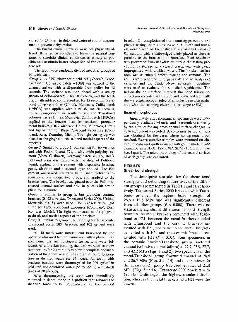
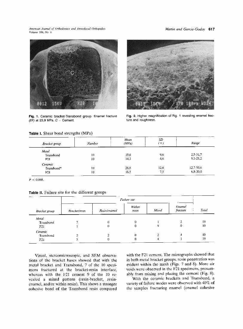
The descriptive statistics for the shear bondstrengths and debonding failure sites of the different groups are presented in Tables I and II, respectively. Transcend Series 2000 brackets with Transbond provided the highest bond strength of28,8 ± 12,6 MPa and was significantly differentfrom all other groups (P < 0.008). There was nostatistically significant difference in bond strengthbetween the metal brackets cemented with Transbond or F21, between the metal brackets bondedwith Transbond and the ceramic brackets cemented with F21, nor between the metal bracketscemented with F21 and the ceramic brackets cemented with F21 (P < 0.05). Four specimens inthe ceramic bracket-Transbond group fracturedenamel (cohesive enamel failure) at 17,7,23,9,22,7,and 42,2 MPa (Figs. 1 and 2); two specimens in themetal- Transbond group fractured enamel at 26,8and 26,7 MPa (Figs. 3 and 4) and one specimen inthe ceramic-F21 group fractured enamel at 21,8MPa (Figs. 5 and 6). Transcend 2000 brackets withTransbond displayed the highest standard deviation, whereas the metal brackets with F21 were thelowest.
Amencan Journal of Orthodontics and Dentofactal OrthopedicsVolume 106, No 6
'.......... ,
Martin and Garcia-Godoy 617
" 0012
c
..X20 f"".__._".~""
o~lr 151{"tj:
Fig. 1. Ceramic bracket-Transbond group. Enamel fracture(EF) at 23,9 MPa. C = Cement.
Table I. Shear bond strengths (MPa)
Fig. 2. Higher magnification of Fig. 1 revealing enamel fracture and roughness.
Mean SDBracket group Number (MPa) (±) Range
MetalTransbond 10 19,6 9,6 2,5-31,7F21 10 14,3 4,6 9,1-25,2
CeramicTransbond* 10 28,8 12,6 12,7-50,6F21 10 18,5 7,5 6,8-30,0
P < 0.008.
Table II. Failure site for the different groups
Fallure sue
I IWithm I )
EnamelBracket group Bracketlresin Resin/enamel resin Mixed fracture Total
MetalTransbond 7 0 0 1 2 10F21 1 0 0 9 0 10
CeramicTransbond 2 2 0 2 4 10F21 5 0 0 4 1 10
Visual, stereomicroscopic, and SEM observations of the bracket bases showed that with themetal bracket and Transbond, 7 of the 10 specimens fractured at the bracket-resin interface,whereas with the F21 cement 9 of the 10 revealed a mixed pattern (resin-bracket, resinenamel, and/or within resin). This shows a strongercohesive bond of the Transbond resin compared
with the F21 cement. The micrographs showed thatin both metal bracket groups, resin penetration wasevident within the mesh (Figs. 7 and 8). More airvoids were observed in the F21 specimens, presumably from mixing and placing the cement (Fig. 8).
With the ceramic brackets and Transbond, avariety of failure modes were observed with 40% ofthe samples fracturing enamel (enamel cohesive

618 Martin and Garcia-Godoy Ameru an Journal oj Orthodonucs and Dentofu; ut! Ortliotin!« \D..cembcr 1994
.,..
c
00139 1SKV
E
o
Fig. 3. Metal bracket-Transbond group. Enamel fracture (EF)at 26,7 MPa. C = Cement; E = enamel.
Fig. 5. Ceramic bracket-F21 group. Enamel fracture at 21,8MPa. C = Cement, E = enamel; 0 = dentin.
\.
o
WD29
BB
',.'
Fig. 4. Higher magnification of Fig. 2 revealing enamel fracture and structure.
Fig. 6. Fitting surface of bracket from Fig. 5. Note extensiveenamel fracture BB = Bracket base; 0 = dentin.
failure). With the F21 cement, 50% of the specimens failed at the bracket-resin interface and 40%displayed a mixed failure mode. Only one specimen(10%) fractured enamel.
Enamel morphology
In the metal bracket groups, very little enameldamage could be determined visually except for thespecimens that completely fractured the enamel(Fig. 3). However, with the SEM it was evident thatthe removal of the bracket produced microfractures on the enamel surface. The enamel surface
displayed fractured enamel to a depth of approximately 10 p.m. In the outside area of the bracket,the enamel showed a typical "coral-like" micromorphology as described by Garcia-Godoy andGwinnett."
In both the ceramic bracket groups. four specimens fractured enamel (Figs. I and 2). After debonding the ceramic brackets. more specimens revealed enamel damage than in the metal bracketgroups. The degree of enamel damage varied fromsmall enamel cracks to enamel fracture.
In both ceramic and metal bracket groups, the

Amencan Journal of Onhodontu:s and Dentofacial OrthopedicsVolume 106, No 6
Martin and Garcia-Godoy 619
" ) )~
C
~~);'~I~J 9 15KU X20 1rom WD29
)-" '-"'''-'''''-
BB
t•
.1/
c
Fig. 7. Transbond resin penetration within metal bracketmesh. BB = Bracket base; C = resin.
Fig. 8. Bracket fitting surface showmq F21 resin penetrationwithin metal bracket mesh. Air voids are evident. C = Cement.
specimens failing at the resin-enamel interface orshowing mixed failure patterns revealed minimalenamel damage.
DISCUSSION
The present study showed that Transcend 2000ceramic brackets produced higher bond strength toenamel than the metal bracket. In a previous study,Transcend 2000 also displayed the highest shearbond strength values compared with other types ofceramic brackets. 11 This could be explained because Transcend 2000 uses fused aluminum oxideparticles to provide more condensed micromechanical retention (increasing the bonding area)than the metal bracket that relies exclusively inmacromechanical retention. Undoubtedly, bracketbase surface characteristics greatly influence bondstrength. This study also supports others who showthat ceramic brackets provide higher bondstrengths than metal brackets do.":"
In the present study, a 60-second enamel etching time was used for the F21 cement according tothe manufacturer's instructions. Recent studieshave suggested that etching for 15 to 20 seconds tobond orthodontics brackets and resins is adequate because, although lower bond strength valueswere recorded, they were not statistically significantly different." Further studies should evaluatewhether enamel fracture incidence is reduced withboth ceramic and metal brackets when a reducedetching time is used.
In orthodontic bonding, the highest bond
strength obtained by a bracket may not be the mostdesirable characteristic because orthodontic treatment is a "temporary" situation and eventually thebrackets must be removed. Therefore, in orthodontics, a clinically acceptable bond strength to permitthe intraoral orthodontic forces would be the mostdesirable characteristic. Moreover, the high bondstrength obtained with some orthodontic bracketscould produce a clinical problem during and afterdebonding. Successful clinical bonding would beachieved by a shear bond strength of 6 to 8 MPa. 13
On the basis of these considerations, the bracketsand cements tested in this study provided bondstrength values above the acceptable range reported to withstand clinical orthodontic forces.
The protruding crystals of Transcend 2000 polycrystalline brackets did provide a strong bondstrength with most of the resin remaining on theenamel, bonded to the bracket or fracturing theenamel.
The increased film thickness of resin cementsmay result in a weak interface produced by agreater polymerization shrinkage and thermal expansion of the resin matrix." This study did notmeasure the film thickness of the adhesives used,but it could also account for the lower valuesobtained with the F21 cement. However, because inthis study only the ceramic bracket-Transbondgroup revealed a statistically significant differencefrom all other groups, it could be concluded thatthe ceramic bracket-resin combination does provide the highest bond strength regardless of film

620 Martin and Garcia-Godoy
thickness. Evaluation of brackets with several adhesives with different filler contents should beconducted to obtain the best combinations forclinical purposes.
In this study, Transbond revealed the strongestcohesive value. However, in orthodontics this wouldnot necessarily be a beneficial property. Perhaps aluting agent with a weaker cohesive value would bepreferable to increase failure or debonding of thebracket within the cement rather than at theenamel-cement interface. With this latter failuremode, the present study showed that visually noenamel damage was produced, but when examinedwith the SEM, most of the specimens had someevidence of enamel damage.
The increased evidence of successful orthodontic bonding with etch times of 15 seconds" suggestthat clinical evaluations of the F21 cement shouldbe performed to assess its potential as a bracketbonding material.
REFERENCES
1. Scott GE. Fracture toughness and surface cracks-the keyto understanding ceramic brackets. Angle Orthod 1988;58:5-8.
2. Winchester U. Bond strengths of five different ceramicbrackets: an in vitro study. Eur J Orthod 1991;13:293-305.
3. Jeiroudi MT. Enamel fracture caused by ceramic brackets.AM J ORTHOD DENTOFAC ORTHOP 1991;99:97-9.
4. Eliades T, Viazis AD, Eliades G. Bonding of ceramic brackets to enamel: morphologic and structural considerations.AM J ORTHOD DENTOFAC ORTHOP 1991;99:369-75.
Amencan Journal of Orthodontics and Dentofacial OnhopedirDecember 1994
5. Bramble LM. A paradigm of the marketplace. AM JORTHOD DENTOFAC ORTHOP 1988;94:354-5.
6. Viazis AD, DeLong R, Douglas WH, Bevis RR, SpeidelTM. Enamel abrasion from ceramic orthodontic brackets: aspecial case report. AM J ORTHOD DENTOFAC ORTHOP1990;98:103-9.
7. Redd TB, Shivapuja PK. Debonding ceramic brackets: effects on enamel. J Clin Orthod 1991;25:475-81.
8. Franklin S, Garcia-Godoy F. Shear bond strengths andeffects on enamel of two ceramic brackets. J Clin Orthod1993;27:83-8.
9. Crim GA, Garcia-Godoy F. Thermocycling: effect of duration and storage time. J Prosthet Dent 1987;25:475-81.
10. Garcia-Godoy F, Gwinnett AI. Effect of etching times andmechanical pretreatments on the enamel of primary teeth:an SEM study. Am J Dent 1991;4:115-9.
11. Ostertag AJ, Dhuru VB, Ferguson DJ, Meyer RA. Shear.torsional and tensile bond strengths of ceramic bracketsusing three adhesive filler concentrations. AM J ORTHODDENTOFAC ORTHOP 1991;100:251-8.
12. Kinch AP, Taylor H, Warltier R, et al. A clinical trialcomparing the failure rates of directly bonded bracketsusing etch times of 15 or 60 seconds. AM J ORTHODDENTOFAC ORTHOP 1988;94:476-83.
13. Reynolds IR. A review of direct orthodontic bonding. Br JOrthod 1975;2:171-8.
14. Evans LB, Powers JM. Factors affecting in vitro bondstrength of no-mix orthodontic cements. AM J ORTHOD1985;87:508-12.
Reprint requests to:Dr. Franklin Garcia-GodoyDepartment of Pediatric DentistryUniversity of Texas Health Science Center7703 Floyd Curl Dr.San Antonio, TX 78284-7888