short title: erks and jnks regulate egr-1 response to h2o2 · 1 short title: erks and jnks regulate...
TRANSCRIPT

1
Short title: ERKs and JNKs regulate Egr-1 response to H2O2
ERKs and JNKs mediate hydrogen peroxide-induced Egr-1 expression and nuclear
accumulation in H9c2 cells
Ioanna-Katerina S. Aggeli, Isidoros Beis, Catherine Gaitanaki*
Department of Animal and Human Physiology, School of Biology, Faculty of Sciences,
University of Athens, Panepistimioupolis Ilissia, 157 84 Athens, GREECE
*Corresponding author: C. Gaitanaki, PhD. Dept. of Animal and Human Physiology,
School of Biology, Faculty of Sciences, University of Athens, Panepistimioupolis Ilissia,
157 84 Athens, GREECE. Tel : +30 210 7274136; fax : +30 210 7274635 e-mail:

2
Summary
One of the most significant insults that jeopardize cardiomyocyte homeostasis is a surge
in reactive oxygen species (ROS) i.e. in the failing myocardium. Early growth response
factor-1 (Egr-1) has been found to act as a transcriptional regulator in multiple biological
processes known to exert deleterious effects on cardiomyocytes. We thus investigated the
signaling pathways involved in its regulation by H2O2. Egr-1 mRNA levels were found to
be maximally induced after 2h in H2O2-treated H9c2 cells. Egr-1 respective response at
the protein level, was found to be maximally induced after 2 h of treatment with 200 μM
H2O2, remaining elevated for 6 h, declining thereafter. H2O2-induced upregulation of Egr-
1 mRNA and protein levels was ablated in the presence of agents inhibiting ERKs
pathway (PD98059) and JNKs (SP600125, AS601245). Immunofluorescent experiments
revealed H2O2-induced Egr-1 nuclear sequestration to be also ERK- and JNK-dependent.
Overall, our results show for the first time ERKs and JNKs’ fundamental role in
regulating Egr-1 response to H2O2 treatment in cardiac cells at multiple levels: mRNA,
protein and subcellular distribution. Nevertheless, further studies are required so as to
elucidate the specific physiological role of Egr-1 regarding the modulation of gene
expression and determination of cell fate.
Key Words: early growth response factor-1 (Egr-1); ERKs; JNKs; H2O2; signaling

3
Abbreviations
Early growth response factor-1 (Egr-1); Extracellular signal-regulated kinases (ERKs);
cJun-N-terminal kinases (JNKs); Mitogen-activated protein kinases (MAPKs);
glyceraldehyde-3-phosphate dehydrogenase (GAPDH); Dimethyl-sulfoxide (DMSO);
Sodium dodecyl sulphate-polyacrylamide gel electrophoresis (SDS-PAGE); polymerase
chain reaction (PCR)
4
Introduction
Oxidative stress mediated by excessive reactive oxygen species (ROS) has been
shown to compromise heart function, having deleterious effects on cardiac myocytes
(Byrne et al. 2003, Ferrari et al. 2004). Indeed, apoptotic mechanisms have been found to
be triggered in a variety of cardiovascular pathologies including: atherosclerosis,
ischemic episodes, myocardial infarction as well as in the case of ischemia/reperfusion
injury (Balla et al. 1991, Feuerstein and Young 2000, Flotats and Carrio 2003). Ample
data from gene expression profiling experiments have led to the identification of a
genetic survival program activated in a plethora of ROS-related cardiac disorders. Among
the antiapoptotic genes mediating cell preservation under these adverse conditions the
early growth response factor-1 (Egr-1) is also included (Depre et al. 2001).
The transcription factor Egr-1 (also termed NGF1-A, Zif/268, Krox-24) was
originally identified as an immediate early response gene (Milbrandt 1987) bearing both
growth inhibitory (Huang et al. 1997, Levin et al. 1995) or growth promoting properties
(Eid et al. 1998). It contains a DNA binding domain that consists of three zinc fingers
(Lemaire et al. 1988, Lim et al. 1987) and binds to a GC-rich sequence in the promoter of
its target genes (Cao et al. 1993). Egr-1 is rapidly induced by differentiation signals
(Sukhatme et al. 1988) as well as by heat shock, UV light (Lim et al. 1987) and ionizing
radiation (Datta et al. 1992). In terms of its tissue distribution, Egr-1 expression is highest
in brain and heart (Lanoix et al. 1998).
In particular, Egr-1 has been found to mediate transcriptional regulation of a variety
of inflammatory and coagulant genes involved in atherosclerotic pathogenesis following
vascular injury i.e. transforming growth factor-β, intercellular adhesion molecule-1

5
(ICAM-1), plasminogen activator inhibitor-1 as well as platelet-derived growth factor A
and B (PDGF-A and B) (Khachigian et al. 1996, Yan et al. 2000). Egr-1 is also expressed
in atherosclerotic plaques (McCaffrey et al. 2000) and mechanically injured carotid
arteries (Santiago et al. 1999). Eliciting salutary changes and mediating cardiac
remodeling by altering the expression of genes like: atrial natriuretic factor (ANF) and α-
or β-myosin heavy chain (α- or β-MHC), Egr-1 has also been shown to promote
preservation of the heart contractile machinery (Bruneau et al. 1996, Saadane et al. 1999).
In addition to this, Fahmy and Khachigian (2002) have marked that reduction of Egr-1
levels resulted in suppression of smooth muscle cell proliferation limiting intimal
hyperplasia in balloon-injured carotid.
Numerous reports confirm that among the signal transduction pathways involved in
Egr-1 regulation, MAPK subfamilies are included, with compelling evidence reporting
the particularly crucial role of extracellular signal-regulated kinases (ERKs) especially in
cardiomyocytes (Chiu et al. 1999, deHager et al. 2001, Hodge et al. 1998). MAPKs
constitute a highly conserved family of serine/threonine protein kinases which are
activated via dual phosphorylation of a specific threonine and tyrosine residue (Goedert
et al. 1997). The three best-studied MAPKs subfamilies include: ERKs, cJun-N-terminal
kinases (JNKs) and p38-MAPK (Goedert et al. 1997, Kyriakis and Avruch 1996). Upon
activation, MAPKs can be found in both the cytoplasm and nucleus, where they interact
with their substrates, i.e. other protein kinases, cytoskeletal proteins as well as
transcription factors (Bogoyevitch 2000, Kyriakis and Avruch 1996).
Given the emerging importance of Egr-1 function in the myocardium under
oxidative stress conditions (Ross 1998), as well as the fact that the mechanism regulating

6
its expression remains elusive, this study was undertaken in an effort to decipher the
redox signal transduction pathways involved in H2O2-induced Egr-1 response in the
context of cardiac myocytes. Thus, we used H9c2 cardiomyoblasts as our experimental
setting. This clonal cell line derived from embryonic heart ventricle, retains properties of
signaling pathways of adult cardiomyocytes (Kimes and Brandt 1976) which accounts for
its extensive use in studies investigating signal transduction mechanisms in
cardiomyocytes (Han et al. 2004, Su et al. 1999, Tanaka et al. 2003, Turner et al. 1998).
Overall, in the present study, we demonstrate for the first time, the involvement of both
ERK and JNK signaling pathways in Egr-1 mRNA and protein levels upregulation along
with its nuclear sequestration in H2O2-treated cardiac cells.

7
Methods
Materials
Hydrogen peroxide was purchased from Merck (Darmstadt, Germany). DMSO,
leupeptin, trans-epoxy-succinyl-L-leucylamido-(4-guanidino) butane (E-64),
dithiothreitol (DTT) and phenylmethylsulphonyl fluoride (PMSF) were obtained from
Sigma-Aldrich (St Louis, Missouri, USA). SP600125, AS601245 and PD98059 were
purchased from Calbiochem-Novabiochem (La Jolla, CA, USA) while SB203580 was
from Alexis Biochemicals (Lausen, Switzerland). Nitrocellulose (0.45 μm) was obtained
from Schleicher & Schuell (Keene NH, USA). Prestained molecular mass markers were
from New England Biolabs (Beverly, MA, USA). Secondary antibodies were from
DakoCytomation (Glostrup, Denmark). Primers for the detection of Egr-1 and GAPDH
were synthesized by Invitrogen Life Technologies (California, USA). Super RX film was
purchased from Fuji photo film GmbH (Dusseldorf, Germany). General laboratory
reagents were purchased from Sigma-Aldrich or Merck.
Cell cultures, treatments and reagents
H9c2 cells (passage 18-25; American Type Culture Collection, Rockville, MD, USA)
were cultured in DMEM (PAA Laboratories GmbH, Pasching, Austria) supplemented
with 10% (v/v) heat inactivated fetal bovine serum (PAA Laboratories GmbH) and
antibiotics, under an atmosphere of 95% air / 5% CO2 at 37oC. Experiments were carried
out using mononucleated myoblasts after serum had been withdrawn for 24 h. Hydrogen
peroxide (200μΜ) was added to the medium for the times indicated. This concentration
of hydrogen peroxide is used routinely for gene expression studies in cardiomyocyte

8
experimental settings exposed to oxidative stress (Kemp et al. 2003). When
pharmacological inhibitors were used, they were dissolved in DMSO and added to the
medium 30min prior to treatment with 200μΜ H2O2 as follows: PD98059 (25 μΜ),
SP600125 (10 μΜ), AS601245 (1 μΜ), SB203580 (10 μΜ), cycloheximide (20 μM) and
actinomycin D (5 μg/ml). Cells were left untreated (control) or incubated with either
DMSO or the inhibitors alone or with the inhibitors followed by exposure to 200 μΜ
H2O2 for 1h (mRNA studies) or 2h (protein studies), respectively. Control experiments
with DMSO alone were performed for the respective duration (1,5h for Egr-1 mRNA
studies and 2,5h for Egr-1 protein studies).
Preparation of nuclear extracts
Nuclear extracts were prepared as previously described (Aggeli et al. 2006). Cells were
harvested into buffer A (10 mM Tris-HCl pH 7.9, 10 mM KCl, 1.5 mM MgCl2, 0.3 mM
Na3VO4, 200 M leupeptin, 10 M E-64, 5 mM DTT, 300 M PMSF). Samples were
centrifuged (10,000 g, 5 min, 4oC) in a BR4i Jouan centrifuge, and the supernatants
discarded. Pellets were re-suspended in buffer A containing 0.1% (v/v) Nonidet P40 (10
min, 4oC). After centrifugation (10,000 g, 5 min, 4oC), pellets were re-suspended in
buffer C [20 mM Hepes pH 7.9, 420 mM NaCl, 1.5 mM MgCl2, 0.2 mM EDTA, 25%
(v/v) glycerol, 0.3 mM Na3VO4, 200 M leupeptin, 10 M E-64, 5 mM DTT, 300 M
PMSF]. After centrifugation (12,000 g, 5 min, 4oC) supernatants (nuclear extract) were
boiled with 0.33 vol. of SDS-PAGE sample buffer [SB4X: 0.33 M Tris-HCl (pH 6.8),
10% (w/v) SDS, 13% (v/v) glycerol, 20% (v/v) 2-mercaptoethanol, 0.2% (w/v)
9
bromophenol blue]. Protein concentrations were determined using the BioRad Bradford
assay reagent (Bio-Rad, Hercules, California, USA).
Immunoblotting
Protein samples (30 μg) from nuclear fraction extracts were separated by SDS-PAGE on
8% (w/v) polyacrylamide gels and transferred electrophoretically onto nitrocellulose
membranes. Nonspecific binding sites were blocked with 5% (w/v) nonfat milk powder
in TBST [20 mM Tris-HCl pH 7.5, 137 mM NaCl, 0.1% (v/v) Tween 20] for 30 min at
room temperature. Subsequently, membranes were incubated overnight with the
appropriate primary antibody [1:1000 anti-Egr-1 (sc-110, Santa Cruz Biotechnology, Inc.
California, USA) or 1:2000 anti-actin (A2103, Sigma-Aldrich St Louis, Missouri, USA)]
at 4oC. After washing in TBST (3 x 5 min) blots were incubated with the respective
horseradish peroxidase-conjugated secondary antibody 1:5000 in TBST containing 1%
(w/v) nonfat milk powder (60 min). After washing in TBST (3 x 5 min), bands were
detected using enhanced chemiluminescense (ECL) (Amersham Biosciences, Uppsala,
Sweden) and quantified by scanning densitometry (Gel Analyzer v. 1.0).
RNA preparation, cDNA synthesis and ratiometric Reverse transcription PCR (RT-PCR)
The expression of endogenous Egr-1 was determined by ratiometric reverse transcription
of total RNA followed by PCR analysis. Total RNA was extracted from cells using Trizol
(Invitrogen Life Technologies), according to the manufacturer’s instructions. For cDNA
synthesis, 2 μg of total RNA was denatured in the presence of 5 p mole oligo-dT primer
in a reaction volume of 13.5 μl at 65oC for 5 min. Reverse transcription was performed

10
with M-MLV Reverse Transcriptase (Invitrogen Life Technologies), first strand buffer
(Promega, Madison, USA), dithiothreitol (Promega) and deoxy-nucleotide triphosphates
(dNTPs) (Promega). The first strand reaction was incubated at 37oC for 1 h. Termination
of the reaction was achieved by inactivation of the reverse transcriptase at 70oC for 5
min. PCR for Egr-1 was performed using 1.5 Units Taq (Bioron GmbH, Ludwigshafen,
Germany) with sense 5’-GTG CGA GTG GAG ATC GGA AT-3’ and antisense 5’- GTA
ACC GCA GCA TTC CAA CT-3’ primers, based on the sequence of rat Egr-1 [Genbank
accession no. NM012551]. These primers amplify a 205-base pair PCR product. After a 5
sec denaturation at 95oC, PCR was carried out for 25 cycles (95oC for 30 sec, 59oC for 30
sec and 72oC for 30 sec), and then a final extension was done at 72oC for 4 min. PCR (25
cycles) for GAPDH was performed using the following primers: sense 5'-ACC ACA
GTC CAT GCC ATC AC-3' and antisense 5'-TCC ACC ACC CTG TTG CTG TA-3'
[Genbank accession no. X02231]. cDNA samples derived from “control” and treated
cells were always amplified simultaneously. PCR products were separated on a 2% (w/v)
agarose gel supplemented with ethidium bromide (EtBr) at a final concentration of 100
g/l. Band intensities were determined using an appropriate image analysis programme
(Gel Analyzer v. 1.0). All values were normalized for the amount of GAPDH mRNA
and estimation of fragment band size (Egr-1 205 bp, GAPDH 452 bp) was performed by
comparison with GeneRuler 100bp DNA ladder (Fermentas Life Sciences Inc., Hanover,
USA).

11
Immunofluorescence staining
Cells were grown on appropriate chamber slides in plating medium and were treated after
serum had been withdrawn for 24 h. Subsequently, cells were fixed with 4% (v/v)
formaldehyde in phosphate buffer saline (PBS) pH 7.4 for 15 min at RT, washed in PBS
(x3) and incubated (5 min, RT) with 1% (w/v) BSA in PBS containing 0.3% (v/v) Triton
X-100. Incubation with the primary antibody against Egr-1 (1:100, 1 h, 37oC) was
followed by 0.5 h incubation at 37oC with an Alexa Fluor 488-conjugated anti-rabbit
secondary antibody (1:250) (green fluorescense). After washing, cell nuclei were stained
using TO-PRO-3 iodide (642/661) (1 μΜ in DMSO) (red fluorescence). Following
mounting, chamber slides were visualized under a laser scanning confocal Zeiss Axiovert
BioRad Radience 2100 microscope.
Statistical evaluations
All data are presented as means S.E.M. Comparisons between control and treatment
were performed using Student’s paired t-test. A value of at least P<0.05 was considered
to be statistically significant.

12
Results
H2O2 stimulates Egr-1 mRNA levels in H9c2 cells in a JNK- and ERK-dependent
manner.
There is emerging evidence revealing Egr-1 diverse biological effects under
stressful conditions. Given the importance of the triggered cellular responses by oxidative
insults in the myocardium, Egr-1 transcriptional response to hydrogen peroxide treatment
was examined in the present study by exposure of H9c2 cells to 200 μΜ H2O2. Thus,
Egr-1 mRNA was found to be induced from 1 h (3.04±0.52 fold relative to control),
maximized at 2 h (6.03±0.16 fold relative to control) (Figure 1A upper panel) decreasing
thereafter. GAPDH (glyceraldehydes-3-phosphate dehydrogenase) mRNA levels were
also assayed as a housekeeping gene (Figures 1A, C bottom panels). Data shown (Figures
1B, D) represents densitometric analysis of Egr-1 PCR product bands normalized for the
respective GAPDH values. Subsequently, to probe into the actual pathways transducing
this effect, we tried to determine the signaling cascades involved in stimulation of Egr-1
transcript levels by H2O2. To this end, various pharmacological inhibitors were used:
PD98059: that blocks the ERK1/2 pathway, SP600125 and AS601245: both selective
JNKs inhibitors and SB203580: a p38-MAPK inhibitor. The effect of cycloheximide
which is known to suppress de novo protein synthesis as well as actinomycin D which is
a widely used transcription inhibitor, were also examined. DMSO as well as the
inhibitors alone had no effect on Egr-1 mRNA levels (data not shown). As shown in
Figure 1C (upper panel) and Figure 1D, we observed that pre-treatment of H9c2 cells
with PD98059 and SP600125 as well as AS601245 almost abrogated H2O2-stimulated
Egr-1 response. These results also indicate that ERKs and JNKs participate in H2O2-

13
induced Egr-1 mRNA upregulation in H9c2 cells. In contrast, there is no apparent
intermediacy of p38-MAPK in the observed response. Furthermore, the latter was
abolished in the presence of actinomycin D, a result indicative of Egr-1 regulation at the
transcriptional level. Additionally, H2O2-induced Egr-1 levels were markedly enhanced
in the presence of cycloheximide, an effect that confirms Egr-1 to function as an
immediate early response gene.
ERKs and JNKs are involved in Egr-1 protein upregulation by H2O2 in H9c2 cells.
Subsequently, an effort was made to examine the time-dependent profile of Egr-1
response to H2O2 at the protein level. As shown in Figure 2A and 2C, a sustained
upregulation of Egr-1 protein levels was observed in samples from nuclear extracts at 1 h
after the onset of stimulation (2.17±0.17 fold relative to control) with maximal values
being attained at 2 h (3.71±0.15 fold relative to control) and being sustained for at least 6
h, declining thereafter. Additionally, using various inhibitors, the contribution of a
number of signaling pathways to Egr-1 protein upregulation was assessed. DMSO as well
as the inhibitors alone had no effect on Egr-1 protein levels (data not shown). Our results
suggest that the latter are upregulated via a mechanism involving ERKs and JNKs, since
the respective inhibitors almost ablated the observed response (Figure 2B, D). SB203580,
a p38-MAPK inhibitor, had no effect. Equal protein loading was verified by reprobing
the membranes with a specific anti-actin antibody (Figures 2A, B bottom panels).

14
Distribution pattern of Egr-1 in H2O2-treated H9c2 cells
Our aforementioned findings prompted us to look into Egr-1 distribution pattern as
well as into the mechanism modulating the latter. To our knowledge, this is the first
report describing regulation of Egr-1 subcellular localization in the context of cardiac
cells exposed to hydrogen peroxide. Effectively, monitoring the distribution profile of
this transcription factor, we observed that in untreated cells (Figure 3-control) the basal-
minimal immunofluorescent signal detected was located in both the cytoplasm and
nucleus. Interestingly, after exposure of H9c2 cells for 2 h to 200 μΜ H2O2, there was a
significant enhancement of Egr-1 staining which was accumulated exclusively in the
nucleus (Figure 3- H2O2). In the presence of PD98059 as well as SP600125, ERKs and
JNKs selective inhibitors respectively, there was a marked decrease in Egr-1
immunostaining almost reaching basal levels, which was once more distributed in the
nucleus as well as in the cytoplasm (Figure 3- PD/H2O2 and SP/H2O2, respectively). A
minimal immunofluorescent signal was detected when the inhibitors were used alone in
both the cytoplasmic and nuclear compartments (data not shown). Thus, it appears that
after treatment with H2O2, one observes the nuclear sequestration of enhanced Egr-1
protein levels, a process found to be both ERK- and JNK-dependent.
15
Discussion
Several cardiac pathological conditions have as their prime cause exposure to
increased levels of reactive oxygen species (ROS) which induce the apoptotic death of
cardiac myocytes (Byrne et al. 2003, Feuerstein and Young 2000). Hydrogen peroxide
constitutes one of the most widely investigated ROS that has been found to exert a dual
effect by either stimulating proliferation or triggering apoptosis (Kanno et al. 2000,
Sundaresan et al. 1995, Wei et al. 2000). Early growth response factor-1 (Egr-1) was
originally characterized as an immediate early gene (IEG) that is induced by stimuli
implicated in vascular pathology (i.e. growth factors, cytokines, hypoxia, hyperoxia,
hemorrhagic shock injury) (Gess et al. 1997, Silverman and Collins T 1999). Given the
fact that accumulating reports account for a key role of Egr-1 and its targets in
orchestrating cellular response following oxidative stress (Jin et al. 2000), it was of
interest to probe into its regulation by H2O2 in H9c2 cardiac cells.
Accordingly, in our hands, exposure of H9c2 cells to 200 μΜ H2O2 resulted in the
transient upregulation of Egr-1 mRNA (Figure 1A, B). In aortic smooth muscle cells, Jin
et al. (2000) have shown H2O2-induced Egr-1 mRNA to peak within 1 h and to be
regulated by a tyrosine kinase-dependent mechanism. In our study, induction of Egr-1
mRNA levels was mediated via ERKs and JNKs signaling cascades, as evidenced by
abrogation of the observed effect in the presence of their respective pharmacological
inhibitors: PD98059 and SP600125 or alternatively AS601245 (Figure 1C, D). On the
contrary, SB203580, a p38-MAPK inhibitor, did not block H2O2-induced Egr-1 mRNA
stimulation. In line with our findings, numerous reports have shown ERKs to be involved
in EGR-1 mRNA upregulation by diverse stimuli in a plethora of cell types and tissues.

16
In particular, Hasan and Schafer (2008) have marked ERK-dependent Egr-1 mRNA
stimulation by hemin in vascular smooth muscle cells. In addition to this, Egr-1
transcription has also been shown to be regulated through an ERK-related mechanism, in
astrocytes treated with endothelin-3 (Biesiada et al. 1996) as well as in primary neonatal
cardiomyocytes treated with estrogen (deJager et al. 2001) and RAW macrophages
exposed to hypoxia (Mishra et al. 2006). However, in NIH3T3 cells, Lim et al. (1998)
have shown various forms of stress (UV radiation, heat shock) to induce Egr-1 gene via a
mechanism independent of ERKs, involving p38-MAPK and JNKs.
Interestingly, only a few investigators have observed the participation of JNKs in
Egr-1 mRNA levels stimulation. In particular, Chung et al. (2007) have reported
amitriptyline (an antidepressant inhibiting neurotransmitter reuptake) to induce Egr-1
gene expression in rat C6 glial cells via ERKs and JNKs, using their respective selective
inhibitors. Similarly, Choi et al. (2008) have demonstrated Egr-1 induction by curcumin
in U-87MG human glioblastoma cells to involve ERKs and JNKs. Given the differential
mechanisms implicated in each setting, one can postulate that involvement of MAPKs in
the transcriptional regulation of Egr-1 is stimulus- and cell type-specific. The observed
abrogation of H2O2-induced Egr-1 mRNA by actinomycin D, a known inhibitor of gene
transcription (McConkey et al. 1989a, 1989b), confirmed this response to be regulated at
the transcriptional level, while the additive effect of cycloheximide on induction of Egr-1
mRNA by H2O2, substantiated that the latter constitutes an immediate-early response,
underscoring Egr-1 function as an IEG (Milbrandt 1987) (Figure 1C, D).
Subsequently, taking into account the fact that in skeletal muscle cells induction of
Egr-1 mRNA by various stimuli (including endothelin 1, angiotensin II and alpha

17
adrenergic agonists) was followed by a translational block (Maass et al. 1994), it
appeared of interest to elucidate the mechanism modulating expression of H2O2-induced
Egr-1 protein levels in our experimental model. Correlating with studies reporting Egr-1
coordinated upregulation of mRNA and protein levels, we found Egr-1 protein to be
maximally induced at 2 h of H2O2 treatment, returning to basal levels after 6 h (Figure
2A, C). Supporting our findings, Shamin et al. (1999) reported Egr-1 upregulation at both
mRNA and protein levels, in neonatal cardiomyocytes exposed to endothelin-1,
angiotensin II or norepinephrine, with Hasan and Schafer (2008) also reporting a similar
effect in vascular smooth muscle cells exposed to hemin. What is more, regarding our
observation of Egr-1 protein upregulation by H2O2 in an ERK- and JNK-dependent
manner, numerous reports have also pointed to the involvement of ERKs i.e. in estrogen-
treated neonatal cardiac myocytes (deJager et al. 2001) and prostaglandin-treated cardiac
myocytes (Xu et al. 2008). Additionally, in accordance with our findings, Ahn et al.
(2007) have observed that Egr-1 protein upregulation in phorbol myristate-treated human
glioma cells was ERK- and JNK-dependent. However, contradicting our results, Wang et
al. (2005) have found p38-MAPK rather than ERKs or JNKs, to mediate isoproterenol-
induced Egr-1 protein expression in H9c2 cells.
In terms of Egr-1 subcellular localization, we have shown H2O2 to cause its nuclear
sequestration (Figure 3 - H2O2 vs. control). To our knowledge, our study is the first to
report the involvement of both ERKs (Figure 3 – PD/H2O2) and JNKs (Figure 3 –
SP/H2O2) in the enhanced expression and spatial distribution i.e. nuclear accumulation of
Egr-1 protein in cardiac myocytes exposed to H2O2, potentially mediating this
transcription factor’s interaction with its substrates, allowing thereafter for any

18
modulation of gene expression. In accordance with our findings, Moon et al. (2007) have
observed that in human intestinal epithelial cells exposed to sulindac sulfide, a non-
steroidal anti-inflammatory drug, the promoted expression and nuclear translocation of
Egr-1 was blocked in the presence of an ERK cascade inhibitor.
Egr-1 has been found to play a significant role in preservation of cardiac function
and pathogenesis of vascular diseases, with Okada et al. (2002) having noted the
fundamental contribution of Egr-1 induction to the development of cardiac allograft
vasculopathy. What is more, involvement of Egr-1 in regulation of sodium-calcium
exchanger-1 (NCX1) as well as in fibroblast growth factor-2 (FGF-2) gene expression in
cardiac myocytes, further substantiates Egr-1 cardioprotective properties (Jimenez et al.
2004, Wang et al. 2005). However, recent evidence also denotes Egr-1 possible
implication in the pathogenesis of myocardial ischemia/reperfusion injury, with Egr-1
inhibition leading to amelioration of hemodynamics in vivo and to a relief of myocardial
injuries in morphology and structure as evidenced by an increase of cell viability (Zhang
et al. 2008). The controversy concerning Egr-1 physiological role is further enhanced by
the report of Kasneci et al. (2009) who demonstrated Egr-1 to act as a transcriptional
repressor of calsequestrin (CSQ) resulting in its downregulation, with negative effects on
cardiac function. This is due to the fact that CSQ constitutes the major calcium storage
protein that links excitation–contraction coupling in the cardiac sarcoendoplasmic
reticulum (Chopra et al. 2007).
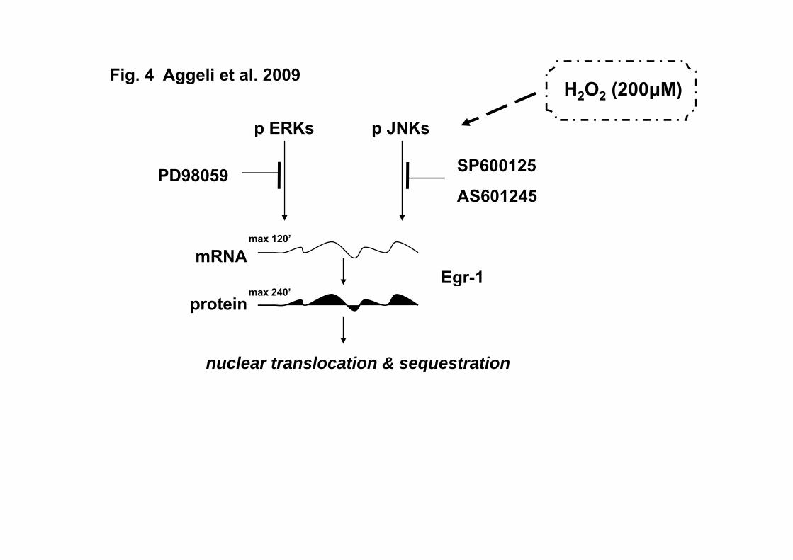
One can deduce from the above that elucidating the signal transduction pathways
mediating Egr-1 response to hydrogen peroxide appears compelling, particularly in
cardiac myocytes. Overall, our data disclose the role of ERKs and JNKs in the regulation

19
of Egr-1 temporal and spatial expression pattern in H2O2-treated cardiac cells. The
diagram in Figure 4 constitutes a schematic representation of our results. Further studies
are nevertheless required, so as to decipher and delineate the precise repertoire of effects
of this immediate responsive transcription factor in the complex context of the
myocardium, justifying Egr-1 characterization as a primary regulator of cell fate under
stressful conditions.
ACKNOWLEDGMENTS
This work was funded by Special Research Account of the University of Athens grants.

20
FIGURE LEGENDS
Fig. 1 Time course analysis of H2O2–induced Egr-1 mRNA upregulation in H9c2
cardiomyoblasts; a JNK- and ERK–mediated response. (A) H9c2 cells were exposed to
200 μΜ H2O2 for the times indicated. (C) H9c2 cells were left untreated (control) or pre-
incubated with 10 μM PD98059 (PD), 10 μM SP600125 (SP), 1 μΜ AS601245 (AS), 10
μΜ SB203580 (SB), 20 μM cycloheximide (CLX), and 5 mg/ml actinomycin D (ActD)
for 30 min, then exposed to 200 μΜ H2O2 for 1 h in the absence or presence of the
inhibitors. RNA was extracted and expression of Egr-1 (A and C upper panels) as well as
GAPDH (A and C lower panels) mRNA was analyzed by ratiometric RT-PCR. The
positions of the 500, 400, 300 and 200bp markers are indicated on the left of the panels.
After densitometric analysis of the PCR products, results were normalized for GAPDH
and the data is presented (B and D) as fold stimulation. Results are means ± SEM for at
least three independent experiments. * p<0.001 compared to control values; ** p<0.001
compared to identically treated cells in the absence of inhibitors.
Fig. 2 (A) Kinetics of Egr-1 protein expression levels in H2O2-treated H9c2
cardiomyoblasts. H9c2 cells were left untreated (control) or were exposed to 200 μΜ
H2O2 for the times indicated. (B) Effect of PD98059, SP600125, AS601245 and
SB203580 on Egr-1 response. H9c2 cells were left untreated or were pre-incubated with
10 μM PD98059 (PD), 10 μM SP600125 (SP), 1 μΜ AS601245 (AS) and 10 μΜ
SB203580 (SB) for 30 min, then exposed to 200 μΜ H2O2 for 2 h in the absence or
presence of the inhibitors. Nuclear cell extracts (30 μg) were subjected to SDS-PAGE
and immunoblotted with an antibody for total Egr-1 protein levels (A and B upper
21
panels). To verify equal loading, the membranes were then stripped and re-incubated with
a specific anti-actin antibody (A and B lower panels). Bands were quantified by laser
scanning densitometry (C and D). Blots and results shown are representative of at least
three independent experiments. Results are means ± SEM for at least three independent
experiments. * p<0.001 compared to control values; ** p<0.001 compared to identically
treated cells in the absence of inhibitors.
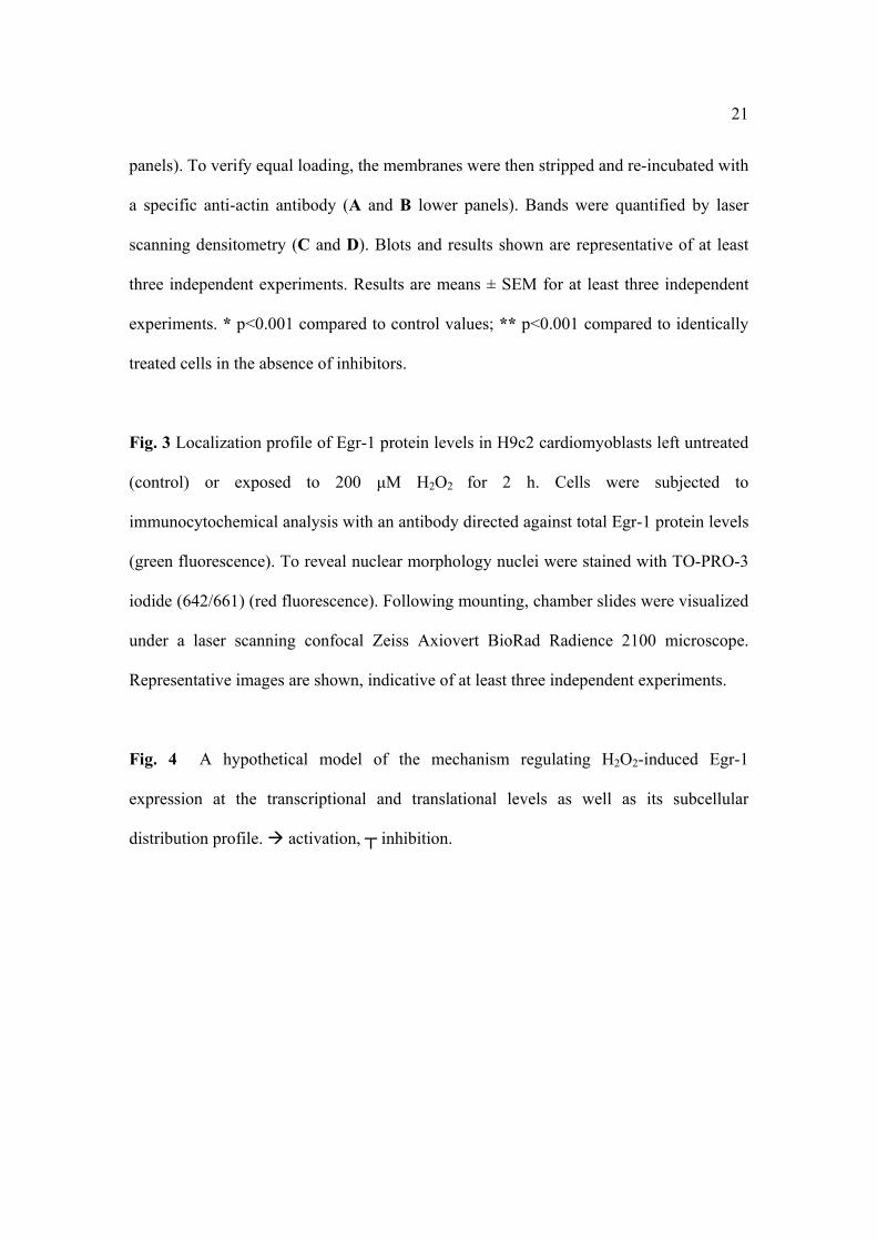
Fig. 3 Localization profile of Egr-1 protein levels in H9c2 cardiomyoblasts left untreated
(control) or exposed to 200 μΜ H2O2 for 2 h. Cells were subjected to
immunocytochemical analysis with an antibody directed against total Egr-1 protein levels
(green fluorescence). To reveal nuclear morphology nuclei were stained with TO-PRO-3
iodide (642/661) (red fluorescence). Following mounting, chamber slides were visualized
under a laser scanning confocal Zeiss Axiovert BioRad Radience 2100 microscope.
Representative images are shown, indicative of at least three independent experiments.
Fig. 4 A hypothetical model of the mechanism regulating H2O2-induced Egr-1
expression at the transcriptional and translational levels as well as its subcellular
distribution profile. activation, ┬ inhibition.

22
References
AGGELI IK, GAITANAKI C, BEIS I: Involvement of JNKs and p38-MAPK/MSK1
pathways in H2O2-induced upregulation of heme oxygenase-1 mRNA in H9c2 cells.
Cell Signal 18:1801-1812, 2006.
AHN BH, PARK MH, LEE YH, MIN DO S: Phorbol myristate acetate-induced Egr-
1 expression is suppressed by phospholipase D isozymes in human glioma cells.
FEBS Lett 581: 5940-5944, 2007.
BALLA G, JACOB HS, EATON JW, BELCHER JD, VERCELLOTTI GM: Hemin:
a possible physiological mediator of low density lipoprotein oxidation and endothelial
cell injury. Arterioscler Thromb 11: 1700-1711, 1991.
BIESIADA E, RAZANDI M, LEVIN ER: Egr-1 activates basic fibroblast growth
factor transcroption. J Biol Chem 271: 18576-18581, 1996.
BOGOYEVITCH MA: Signalling via stress-activated mitogen-activated protein
kinases in the cardiovascular system Cardiovasc Res 45: 826-842, 2000.
BRUNEAU BG, PIAZZA LA, deBOLD AJ: Alpha (1)-adrenergic stimulation of
isolated rat atria results in discoordinate increases in natriuretic peptide secretion and
gene expression and enhances Egr-1 and c-myc expression. Endocrinology 137: 137-
143, 1996.
BYRNE JA, GRIEVE DJ, CAVE AC, SHAH AM: Oxidative stress and heart failure.
Arch Mal Coeur Vaiss 96: 214-221, 2003.
CAO X, MAHENDRAN R, GUY GR, TAN YH: Detection and characterization of
cellular EGR-1 binding to its recognition site. J Biol Chem 268: 16949-16957, 1993.
23
CHIU JJ, WUNG BS, HSIEH HJ, LO LW, WANG DL: Nitric oxide regulates shear
stress-induced early growth response-1. Expression via the extracellular signal-
regulated kinase pathway in endothelial cells. Circ Res 85: 238-246, 1999.
CHOI BH, KIM CG, BAE YS, LIM Y, LEE YH, SHIN SY: p21 Waf1/Cip1
expression by curcumin in U-87MG human glioma cells: role of early growth
response-1 expression. Cancer Res 68: 1369-1377, 2008.
CHOPRA N, KANNANKERIL PJ, YANG T, HLAING T, HOLINSTAT I,
ETTENSOHN K, PFEOFER K, AKIN B, JONES LR, FRANZINI-ARMSTRONG
C, KNOLLMANN BC: Modest reduction of cardiac calsequestrin increase
sarcoplasmic reticulum Ca 2+ leak independent of luminal Ca 2+ and trigger
ventricular arrthymias in mice. Circ Res 101: 617–626, 2007.
CHUNG EY, SHIN SY, LEE YH: Amitriptyline induces early growth response-1
gene expression via ERK and JNK mitogen-activated protein kinase pathways in rat
C6 glial cells. Neurosc Lett 422: 43-48, 2007.
DATTA R, RUBIN E, SUKHATME V, QURESHI S, HALLAHAN D,
WEICHSELBAUM RR, KUFE DW: Ionizing radiation activates transcription of the
EGR1 gene via CArG elements. Proc Natl Acad Sci USA 89: 10149-10153, 1992.
DeJAGER T, PELZER T, MULLER-BOTZ S, IMAM A, MUCK J, NEYSES L:
Mechanisms of estrogen receptor action in the myocardium. J Biol Chem 276: 27873-
27880, 2001.

24
DEPRE C, TOMLINSON JE, KUDEJ RK, GAUSSIN V, THOMPSON E, KIM SJ,
VATNER DE, TOPPER JN, VATNER SF: Gene program for cardiac cell survival
induced by transient ishemia in conscious pigs. Proc Natl Acad Sci 98: 9336-9341,
2001.
EID MA, KUMAR MV, ICZKOWSKI KA, BOSTWICK DG, TINDALL DJ:
Expression of early growth response genes in human prostate cancer. Cancer Res 58:
2461-2468, 1998.
FAHMY RG, KHACHIGIAN LM: Antisense Egr-1 RNA driven by the CMV
promoter is an inhibitor of vascular smooth muscle cell proliferation and regrowth
after injury. J Cell Biochem 84: 575-582, 2002.
FERRARI R, GUARDIGLI G, MELE D, PERCOCO GF, CECONI C, CURELLO
S: Oxidative stress during myocardial ischaemia and heart faiklure. Curr Pharm Des
10: 1699-1711, 2004.
FEUERSTEIN GZ, YOUNG PR: Apoptosis in cardiac disease states: stress- and
mitogen-activated signaling pathways. Cardiovasc Res 45: 560-569, 2000.
FLOTATS A, CARRIO I: Non-invasive in vivo imaging of myocardial apoptosis and
necrosis. Eur J Nucl Med Mol Imaging 30: 615-630, 2003.
GESS B, WOLF K, KURTZ A: Lack of control by immediate early response genes
for the oxygn regulation of erythropietin gene expression. Pflugers Arch 433: 827-
831, 1997.
GOEDERT M, CUENDA A, CRAXTON M, JAKES R, COHEN P: Activation of the
novel stress-activated protein kinase SAPK4 by cytokines and cellular stresses is
25
mediated by SKK3 (MKK6); comparison of its substrate specificity with that of other
SAP kinases. EMBO J 16: 3563-3571, 1997.
HAN H, LONG H, WANG H, WANG J, ZHANG Y, WANG Z: Progressive
apoptotic cell death triggered by transient oxidative insult in H9c2 rat ventricular
cells: a novel pattern of apoptosis and the mechanisms. Am J Physiol Heart Circ
Physiol 286: H2169-2182, 2004.
HASAN RN, SCHAFER AI: Hemin upregulates Egr-1 expression in vascular smooth
muscle cells via ROS-ERK1/2-Elk-1 and NfκΒ. Circ Res 102: 42-50, 2008.
HODGE C, LIAO J, STOFEGA M, GUAN K, CARTER-SU C, SCHWARTZ J:
Growth hormone stimulates phosphorylation and activation of elk-1 and expression of
c-fos, egr-1, and junB through activation of extracellular signal-regulated kinases 1
and 2. J Biol Chem 273: 31327-31336, 1998.
HUANG RP, FAN Y, de BELLE I, NIEMEYER C, GOTTARDIS MM, MERCOLA
D, ADAMSON ED: Decreased Egr-1 expression in human, mouse and rat mammary
cells and tissues correlated with tumor formation. Int J Cancer 72: 102-109, 1997.
JIMENEZ SK, SHEIKH F, JIN Y, DETILLIEUX KA, DHALIWAL J, KARDAMI
E, CATTINI PA: Transcriptional regulation of FGF-2 gene expression in cardiac
myocytes. Cardiovasc Res 62: 548-557, 2004.
JIN N, HATTON ND, HARRINGTON MA, XIA X, LARSEN SH, RHOADES RA:
H2O2-induced Egr-1, Fra-1 and c-jun gene expression is mediated by tyrosine kinase
in aortic smooth muscle cells. Free Rad Biol Med 29: 736-746, 2000.

26
KANNO S, ISHIWAKA M, TAKAYANAGI M, TAKAYANAGI Y, SASAKI K:
Characterization of hydrogen peroxide-induced apoptosis in mouse primary cultured
hepatocytes. Biol Pharm Bull 23: 37-42, 2000.
KASNECI A, KEMENY-SUSS NM, KOMAROVA SV, CHALIFOUR LE: Egr-1
negatively regulates calsequestrin expression and calcium dynamics in ventricular
cells. Card Res 81: 695–702, 2009.
KEMP TJ, CAUSTON H, CLERK A: Changes in gene expression induced by
H(2)O(2) in cardiac myocytes. Biochem Biophys Res Commun 307: 416-421, 2003.
KHACHIGIAN LM, LINDNER V, WILLIAMS AJ, COLLINS T: Egr-1-induced
endothelial gene expression: a common theme in vascular injury. Science 271: 1427-
1431, 1996.
KIMES BW, BRANDT BL: Properties of a clonal muscle cell line from rat heart. Exp
Cell Res 98: 367-381, 1976.
KYRIAKIS JM, AVRUCH J: Sounding the alarm: protein kinase cascades activated
by stress and inflammation. J Biol Chem 271: 24313-24316, 1996.
LANOIX J, MULLICK A, HE Y, BRAVO R, SKUP D: Wild-type egr-1/Krox24
promotes and dominant-negative mutants inhibit, pluripotent differentiation of p19
embryonal carcinoma cells. Oncogene 17: 2495-2504, 1998.
LEMAIRE P, REVELANT O, BRAVO R, CHARNAY P: Two mouse genes
encoding potential transcription factors with identical DNA-binding domains are
activated by growth factors in cultured cells. Proc Natl Acad Sci USA 85: 4691-4695,
1988.

27
LEVIN WJ, PRESS MF, GAYNOR RB, SUKHATME VP, BOONE TC,
REISSMANN PT, FIGLIN RA, HOLMES EC, SOUZA LM, SLAMON DJ:
Expression patterns of immediate early transcription factors in human non-small cell
lung cancer. Oncogene 11: 1261-1269, 1995.
LIM CP, JAIN N, CAO X: Stress-induced immediate-early gene, egr-1, involves
activation of p38/JNK1. Oncogene 16: 2915-2926, 1998.
LIM RW, VARNUM BC, HERSCHMAN HR: Cloning of tetradecanoyl phorbol
ester-induced 'primary response' sequences and their expression in density-arrested
Swiss 3T3 cells and a TPA non-proliferative variant. Oncogene 1: 263-270, 1987.
MAASS A, GROHE C, SUKHATME VP, VETTER H, NEYSES L: Mitogenic
signals control translation of the Early growth response gene-1 in myogenic cells.
Biochem Biophys Res Commun 202: 1337-1345, 1994.
McCAFFREY TA, FU C, DU B, EKSINAR S, KENT KC, BUSH H Jr, KREIGER
K, ROSENGART T, CYBULSKY MI, SILVERMAN ES, COLLINS T: High-level
expression of Egr-1 and Egr-1-inducible genes in mouse and human atherosclerosis. J
Clin Invest 105: 653-662, 2000.
McCONKEY DJ, HARTZELL P, AMADOR-PEREZ JF, ORRENIUS S, JONDAL
M: Calcium-dependent killing of immature thymocytes by stimulation via the CD3/T
receptor comlpex. J Immunol 143: 1801-1806, 1989.
McCONKEY DJ, NICOTERA P, HARTZELL P, BELLOMO G, WYLLIE AH,
ORRENIUS S: Glucocorticoids activate a suicidal process in thymocytes through an
elevation of cytosolic Ca2+ concentration. Arch Biochem Biophys 269: 365-370, 1989.

28
MILBRANDT J: A nerve growth factor-induced gene encodes a possible
transcriptional regulatory factor. Science 238: 797-799, 1987.
MISHRA S, FUJITA T, LAMA VN, NAM D, LIAO H, OKADA M, MINAMOTO
K, YOSHIKAWA Y, HARADA H, PINSKY D: Carbon monoxide rescues ischemic
lungs by interrupting MAPK-driven expression of early growth response 1 gene and
its downstream target genes. Proc Natl Acad Sci USA 103: 5191-5196, 2006.
MOON Y, YANG H, KIM YB: Up-regulation of Egr-1 via ERK1/2 signals attenuates
sulindac sulfide-mediated cytotoxicity in the human intestinal epithelial cells. Toxicol
Appl Pharmacol 223: 155-163, 2007.
OKADA M, WANG CY, HWANG DW, SAKAGUCHI T, OLSON KE,
YOSHIKAWA Y, MINAMOTO K, MAZER SP, YAN SF, PINSKY DJ:
Transcriptional control of cardiac allograft vasculopathy by early growth response
gene-1 (Egr-1). Circ Res 91: 135-142, 2002.
ROSS R: The smooth muscle cell II. Growth of smooth musclen in culture and
formation of elastic fibers. J Cell Biol 50: 172-186, 1998.
SAADANE N, ALPERT L, CHALIFOUR LE: Expression of immediate early genes,
GATA-4, and Nkx2.5 in adrenergic-induced cardiac hypertrophy and during
regression in adult mice. Br J Pharmacol 127: 1165-1176, 1999.
SANTIAGO FS, LOWE HC, KAVURMA MM, CHESTERMAN CN, BAKER A,
ATKINS DG, KHACHIGIAN LM: New DNA enzyme targeting Egr-1 mRNA
inhibits vascular smooth muscle proliferation and regrowth after injury. Nat Med 5:
1264-1269, 1999.

29
SHAMIN A, PELZER T, GROHE C, NEYSES L: Induction of Egr-1 mRNA and
protein by endothelin 1, angiotensin II and norepinephrine in neonatal cardiac
myocytes. Mol Cell Biochem 195: 11-17, 1999.
SILVERMAN ES, COLLINS T: Pathways of Egr-1-mediated gene transcription in
vascular biology. Am J Pathol 154: 665-670, 1999.
SU C, CHONG K, EDELSTEIN K, LILLE S, KHARDPRI R, LAI CC: Constitutive
hsp70 attenuates hydrogen peroxide-induced membrane lipid peroxidation. Biochem
Biophys Res Commun 265: 279-284, 1999.
SUKHATME VP, CAO XM, CHANG LC, TSAI-MORRIS CH,
STAMENKOVITCH D, FERREIRA PC, COHEN DR, EDWARDS SA, SHOWS
TB, CURRAN T: A zinc finger-encoding gene coregulated with c-fos during growth
and differentiation, and after cellular depolarization. Cell 53: 37-43, 1988.
SUNDARESAN M, YU Z, FERRANS VJ, IRANI K, FINKEL T: Requirement for
generation of H2O2 for platelet-derived growth factor signal transduction. Science
270: 296-299, 1995.
TANAKA H, SAKURAI K, TAKAHASHI K, FUJIMOTO Y: Requirement of
intracellular free thiols for hydrogen peroxide-induced hypertrophy in
cardiomyocytes. J Cell Biochem 89: 944-955, 2003.
TURNER N, XIA F, AZHAR G, ZHANG X, LIU L, WEI JY: Oxidative stress
induces DNA fragmentation and caspase activation via the c-Jun NH2-terminal
kinase pathway in H9c2 cardiac muscle cells. J Mol Cell Cardiol 30: 1789-1801,
1998.

30
WANG C, DOSTANIC S, SERVANT N, CHALIFOUR LE: Egr-1 negatively
regulates expression of the sodium-calcium exchanger-1 in cardiomyocytes in vitro
and in vivo. Card Res 65: 187-194, 2005.
WEI T, NI Y, HOU J, CHEN C, ZHAO B, XIN W: Hydrogen peroxide-induced
oxidative damage and apoptosis in cerebellar granule cells: protection by gingo biloba
extract. Pharmacol Res 41: 427-433, 2000.
XU W, CHOU CL, SUN H, FUJINO H, CHEN QM, REGAN JW: FP prostanoid
receptor mediated induction of the expression of early growth response factor-1 by
activation of a Ras/Raf/MAPK signaling cascade. Mol Pharmacol 73: 111-118, 2008.
YAN SF, FUJITA T, LU J, OKADA K, SHAN S, ZOU Y, MACKMAN N,
PINSKY DJ, STERN DM: Egr-1 a master switch coordinating upregulation of
divergent gene families underlying ischemic stress. Nat Med 6: 1355-1361, 2000.
ZHANG Y, SHI G, ZHENG L, LV Y, GAO P, HUANG Z, GAO F, ZHOU Y: The
Protective Effect of Egr-1 Antisense Oligodeoxyribonucleotide on Myocardial Injury
Induced by Ischemia-reperfusion and Hypoxia reoxygenation. Cell Physiol Biochem
22: 645-652, 2008.

Fig. 1 Aggeli et al. 2009
A B
300bp200bp
Egr-1 (205bp)
Egr-1/GAPDH
6
A ion)
0 0.5 1 2 4 6 12 (h)H2O2 (200μΜ)
2
4
Egr-
1 m
RN
Aol
d st
imul
at
GAPDH (452bp)
500bp400bp
0 0.5 1 2 4 6 120
Time (h)
(fo
Time (h)

Fig. 1 Aggeli et al. 2009
DC300bp200bp Egr-1
(205bp)
Egr-1/GAPDH
6
A ion)
cont
rol
H2O
2
PD+
SP+
AS+
SB+
CLX
+A
ctD
+
2
4
Egr-
1 m
RN
Aol
d st
imul
ati
c C A
500bp400bp GAPDH
(452bp) trol 2
O 2
2O 2
2O 2
2O 2
2O 2
2O 2
2O 2
0
(fo
( p)
con H
PD/H
SP/H
AS/
H
SB/H
CLX
/H
Act
D/H
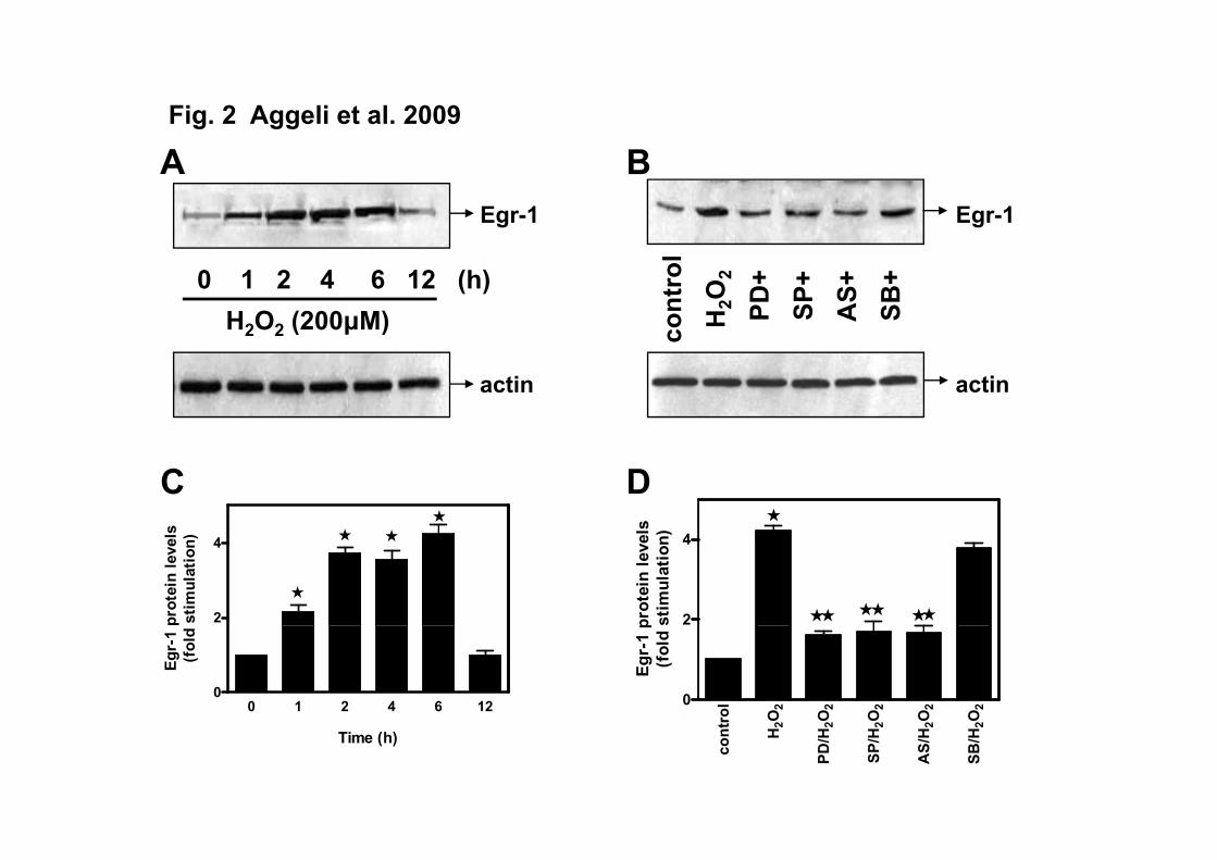
Fig. 2 Aggeli et al. 2009
AEgr-1
BEgr-1
cont
rol
H2O
2
PD+
SP+
AS+
SB+
H2O2 (200μΜ)0 1 2 4 6 12 (h)
actin
c
actin
2 2 ( μ )
s s
C D
2
4
prot
ein
leve
lsst
imul
atio
n)
2
4
prot
ein
leve
lsst
imul
atio
n)
0 1 2 4 6 120
Egr-
1 (f
old
rol 2
O 2
2O 2
2O 2
2O 2
2O 2
0
Egr-
1 p
(fol
d
Time (h)co
nt 2H
2PD
/H
2SP
/H
2A
S/H
2SB
/H

Fig. 3 Aggeli et al. 2009
Egr-1 nucleus merge
control
30μm 30μm 30μm
H2O2
20μm 20μm 20μm

Fig. 3 Aggeli et al. 2009
Egr-1 nucleus merge
PD/H2O2
30μm 30μm 30μm
SP/H2O2
30μm 30μm 30μm
H O (200μΜ)Fig. 4 Aggeli et al. 2009
p ERKs p JNKs
H2O2 (200μΜ)
SP600125
AS601245PD98059
Egr-1mRNA
max 120’
Egr 1protein
max 240’
nuclear translocation & sequestration