shoulder - aulakinesica.com.ar 9 cleland hombro… · measuring shoulder range of ... de las quejas...
TRANSCRIPT


Shoulder 9
CLINICAL SUMMARY AND RECOMMENDATIONS 378
Anatomy 379 Osteology 379 Arthrology 380 Scapulohumeral Rhythm 381 Ligaments 382 Muscles 384 Nerves 387
Patient History 389 Initial Hypotheses Based on Historical Findings 389 Diagnostic Utility of the Patient History for Identifying Labrum and Rotator Cuff Tears 389
Physical Examination Tests 390 Range of Motion 390 Assessing Strength and Proprioception 392 Muscle Length 393 Palpation 394 Assessing Alignment 395 Classifying Shoulder Disorders 397 Special Tests—Instability 398 Special Tests—Labral Tears 402 Special Tests—Subacromial Impingement 413 Special Tests—Rotator Cuff Tears 418 Special Tests—Brachial Plexus Palsy 425 Special Tests—Acromioclavicular Lesions 426 Combination of Tests 427
Outcome Measures 429 Appendix 430
Quality Assessment of Diagnostic Studies Using QUADAS 430 References 436

378 Netter’s Orthopaedic Clinical Examination: An Evidence-Based Approach
CLINICAL SUMMARY AND RECOMMENDATIONS
Patient History
Complaints Little is known about the utility of subjective complaints with shoulder pain. While a report of trauma does not seem clinically useful, a history of popping, clicking, or catching may be minimally helpful in diagnosing a labral tear (�LRs � 2.0).
Physical Examination
Range of Motion, Strength, and Muscle Length Assessment
Measuring shoulder range of motion (ROM) has consistently been shown to be highly reliable but is of unknown diagnostic utility. Visual assessments and func-tional tests of ROM are more variable and may be adequately reliable in some instances.
Assessing strength with manual muscle testing (MMT) appears to be reliable. Weak abduction and/or external rotation may be fairly useful in identifying subacromial impingement and/or full thickness rotator cuff tears. Weak internal rotation appears very helpful in identifying subscapularis tears (�LR � 7.5 to 20.0).
Assessments of shoulder muscle tightness are moderately reliable. However, the single study 1 done to test associated diagnostic utility found tight pectoralis minor muscles in all 90 participants regardless of whether they had shoulder problems or not (100% sensitivity, 0% specifi city).
Special Tests The apprehension test appears to be the most useful test in identifying shoulder in-stability, especially when defi ning a positive test by an “apprehensive response” (�LR � 7.1 to 20.2, �LR � .00 to .29) as opposed to “pain” (�LR � 1.1 to 3.1, �LR � .69 to .90). To a lesser extent, it may also be helpful in diagnosing labral tears.
Results of studies examining the diagnostic utility of tests to identify labral tears are highly variable. Even though most single tests do not appear very useful, one study found both the Kim test and the Jerk test to be very good at identifying labral tears (�LRs of 13.3 and 36.5, respectively). The same author also found the biceps load test I and II to be very effective at identifying superior labrum anterior posterior (SLAP) lesions (�LR � 30 for both).
A 2008 meta-analysis found both the Hawkins-Kennedy and Neer test to be minimally helpful for both ruling in and ruling out subacromial impingement. The presence of a “painful arc” during elevation may additionally be helpful in identifying the condition (�LR � .39, �LR � .32).
In addition to rotator cuff muscle weakness (above), the external and internal rota-tion lag signs appear to be very helpful at identifying infraspinatus and subscapu-laris tears respectively. Several other tests (bear-hug, belly-press, Napoleon) appear to be also very useful in diagnosing subscapularis tears.
Whereas several signs and symptoms are helpful in identifying brachial plexus nerve root avulsions, the shoulder protraction test appears to be the most useful (�LR � 4.8, �LR � .05).
Combinations of Findings
Even though combinations of tests are generally better than single tests, combi-nations of tests are only moderately helpful in identifying labral tears. The most effi cient pair seems to be the anterior apprehension and Jobe relocation tests (�LR � 5.4).
Another study 2 reported even better diagnostic utility when specifi c combina-tions of three tests were used. By selecting two highly sensitive tests (compres-sion rotation, anterior apprehension, and O’Brien tests) and one highly specifi c tests (Yergason, biceps load II, and Speed’s tests), users can be fairly confi dent in both ruling out and ruling in SLAP lesions.

9 SHOULDER 379
ANATOMY
Osteology
Right clavicle Superior
Inferior
Acromial end
Shaft body
Acromial facet
Posterior
Anterior
Sternal end
Trapezoid line
Conoid tubercle
Impression forcostoclavicular lig.
Sternal facetPosterior
Anterior
Subclavian groove (for subclavius m.)
Figure 9-2
Superior and inferior surface of clavicle.
AcromionCoracoid process
Acromial angle
Clavicle (cut)
Supraglenoidtubercle
Anatomical neck
Greater tubercle
Lesser tubercle
Surgical neck
Deltoid tuberosity
Intertubercularsulcus
Crest ofgreater tubercle
Crest oflesser tubercle
Medialsupracondylar ridge
Lateralsupracondylar ridge
CondylesMedialLateral
Radialfossa
Lateralepicondyle
Capitulum
Coronoid fossa
Trochlea
ScapulaHumerus
Head ofhumerus
Superior borderSuperior angle
Suprascapular notch
Neck
Medial border
Subscapular fossa
Infraglenoid tubercle
Lateral border
Inferior angle
Glenoidcavity ofscapula
Medial epicondyle
Figure 9-1
Anterior humerus and scapula.

380 Netter’s Orthopaedic Clinical Examination: An Evidence-Based Approach
Arthrology
Anterior sternoclavicular lig.Clavicle
Subclavius m.
Costoclavicular lig.
1st rib
Costal cartilages
2nd rib
Radiate sternocostal lig.
Interclavicular lig.
Costoclavicular lig.
Synchondrosis of 1st rib
Manubrium
Sternocostal (synovial) joint
Manubriosternal synchondrosis
Articular cavities of sternoclavicular joint
Articular disc of sternoclavicular joint
Suprascapular notchSuperior border
Superior angle
Supraspinous fossa
Spine
NeckInfraspinous fossa
Medial border
Lateral border
Inferior angle
Clavicle (cut)Coracoid process
Acromion
Acromial angleNotch connecting supraspinousand infraspinous fossae
Greater tubercleHead of humerus
Anatomical neckSurgical neckInfraglenoid tubercle
Deltoid tuberosity
Radial groove
Medial supracondylar ridgeLateral supracondylar ridge
Olecranon fossa
Lateral epicondyle
TrochleaGroove for ulnar n.
Medial epicondyle
Groove forcircumflexscapular vessels
HumerusScapula
Figure 9-3
Sternoclavicular joint.
Joint Type and Classifi cation Closed Packed Position Capsular Pattern
Glenohumeral Spheroidal Full abduction and external rotation
ER limited more than abduction, limited more than internal rotation and fl exion
Sternoclavicular Saddle Arm abducted to 90° Not reported
Acromioclavicular Plane synovial Arm abducted to 90°
Scapulothoracic Not a true articulation Not available Not available
9 SHOULDER 381
Scapulohumeral Rhythm
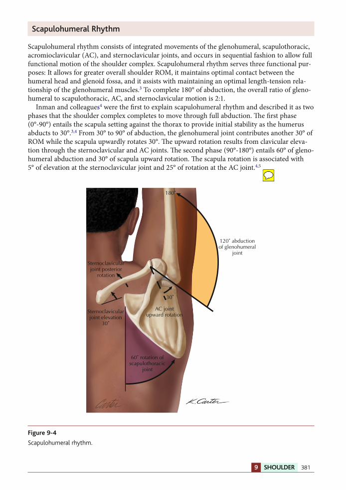
Scapulohumeral rhythm consists of integrated movements of the glenohumeral, scapulothoracic, acromioclavicular (AC), and sternoclavicular joints, and occurs in sequential fashion to allow full functional motion of the shoulder complex. Scapulohumeral rhythm serves three functional pur-poses: It allows for greater overall shoulder ROM, it maintains optimal contact between the humeral head and glenoid fossa, and it assists with maintaining an optimal length-tension rela-tionship of the glenohumeral muscles. 3 To complete 180° of abduction, the overall ratio of gleno-humeral to scapulothoracic, AC, and sternoclavicular motion is 2:1.
Inman and colleagues 4 were the fi rst to explain scapulohumeral rhythm and described it as two phases that the shoulder complex completes to move through full abduction. Th e fi rst phase (0°-90°) entails the scapula setting against the thorax to provide initial stability as the humerus abducts to 30°. 3 , 4 From 30° to 90° of abduction, the glenohumeral joint contributes another 30° of ROM while the scapula upwardly rotates 30°. Th e upward rotation results from clavicular eleva-tion through the sternoclavicular and AC joints. Th e second phase (90°-180°) entails 60° of gleno-humeral abduction and 30° of scapula upward rotation. Th e scapula rotation is associated with 5° of elevation at the sternoclavicular joint and 25° of rotation at the AC joint. 4 , 5
180˚
120˚ abductionof glenohumeral
joint
AC jointupward rotation
30˚
60˚ rotation of scapulothoracic
joint
Sternoclavicular joint elevation
30˚
Sternoclavicular joint posterior
rotation
Figure 9-4
Scapulohumeral rhythm.
382 Netter’s Orthopaedic Clinical Examination: An Evidence-Based Approach
Ligaments
Acromion
Coracoacromial lig.
Supraspinatus tendon (cut)
Coracohumeral lig.
Greater tubercle andlesser tubercle
of humerus
Transverse humeral lig.
Intertubercular tendon sheath(communicates with synovial cavity)
Acromioclavicular joint capsule (incorporating acromioclavicular lig.)
Subscapularis tendon (cut)
Biceps brachii tendon (long head)
Clavicle
Trapezoidlig. Coraco-
clavicularlig.Conoid
lig.
Superior transversescapular lig. andsuprascapular notch
Coracoid process
Communications ofsubtendinousbursa of subscapularis
Broken line indicatesposition of subtendinous bursa of subscapularis
Figure 9-5
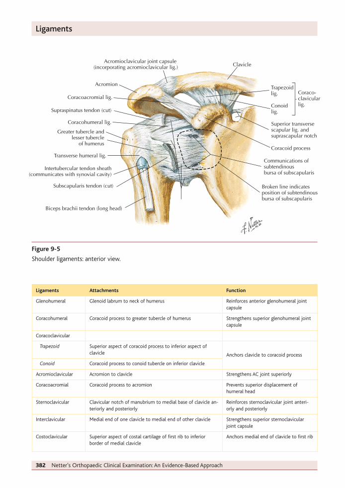
Shoulder ligaments: anterior view.
Ligaments Attachments Function
Glenohumeral Glenoid labrum to neck of humerus Reinforces anterior glenohumeral joint capsule
Coracohumeral Coracoid process to greater tubercle of humerus Strengthens superior glenohumeral joint capsule
Coracoclavicular
Trapezoid Superior aspect of coracoid process to inferior aspect of clavicle Anchors clavicle to coracoid process
Conoid Coracoid process to conoid tubercle on inferior clavicle
Acromioclavicular Acromion to clavicle Strengthens AC joint superiorly
Coracoacromial Coracoid process to acromion Prevents superior displacement of humeral head
Sternoclavicular Clavicular notch of manubrium to medial base of clavicle an-teriorly and posteriorly
Reinforces sternoclavicular joint anteri-orly and posteriorly
Interclavicular Medial end of one clavicle to medial end of other clavicle Strengthens superior sternoclavicular joint capsule
Costoclavicular Superior aspect of costal cartilage of fi rst rib to inferior border of medial clavicle
Anchors medial end of clavicle to fi rst rib

9 SHOULDER 383
Anteriorview
Deltoid m.(reflected)
Supraspinatus m.
Subdeltoid bursa fused withsubacromial bursa
Subscapularis m.
Capsular lig.
Subdeltoid bursa
Supraspinatus tendon
Capsular lig.Synovial membrane
AcromionAcromioclavicularjoint
Coracoacromial lig.Acromion
Supraspinatus tendon(fused to capsule)
Subdeltoid bursa
Infraspinatus tendon(fused to capsule)
Glenoid cavity(cartilage)
Teres minor tendon(fused to capsule)
Synovial membrane (cut edge)
Openings of subtendinousbursa of subscapularis
Joint opened: lateral view
Coracoid process
Coracohumeral lig.
Coronal section through joint
Biceps brachii tendon(long head)
Superiorglenohumeral lig.
Subscapularis tendon(fused to capsule)
Middleglenohumeral lig.
Inferiorglenohumeral lig.
Axillary recess
Glenoidcavity ofscapula
Deltoid m.
Glenoidlabrum
Figure 9-6
Shoulder (glenohumeral) joint.
Ligaments (continued)
384 Netter’s Orthopaedic Clinical Examination: An Evidence-Based Approach
Muscles
Posterior Muscles of Shoulder
Semispinalis capitis m.
Splenius capitis m.
Spinous process of C7 vertebra
Levator scapulae m.
Not connectedto upper limb
Rhomboid minor m.Rhomboid major m.
Acromion
Supraspinatus m.
Spine of scapula
Infraspinatus m.
Teres minor m.
Teres major m.Latissimus dorsi m.
Long head
Lateral head
Spinous process ofT12 vertebra
Tricepsbrachii m.
Trapezius m.
Deltoid m.
Infraspinatusfascia
Triangle ofauscultation
Figure 9-7
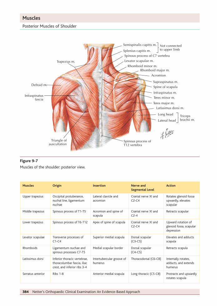
Muscles of the shoulder: posterior view.
Muscles Origin Insertion Nerve and Segmental Level
Action
Upper trapezius Occipital protuberance, nuchal line, ligamentum nuchae
Lateral clavicle and acromion
Cranial nerve XI and C2-C4
Rotates glenoid fossa upwardly, elevates scapular
Middle trapezius Spinous process of T1-T5 Acromion and spine of scapula
Cranial nerve XI and C2-4
Retracts scapular
Lower trapezius Spinous process of T6-T12 Apex of spine of scapula Cranial nerve XI and C2-C4
Upward rotation of glenoid fossa, scapular depression
Levator scapulae Transverse processes of C1-C4
Superior medial scapula Dorsal scapular (C3-C5)
Elevates and adducts scapula
Rhomboids Ligamentum nuchae and spinous processes C7-T5
Medial scapular border Dorsal scapular (C4-C5)
Retracts scapula
Latissimus dorsi Inferior thoracic vertebrae, thoracolumbar fascia, iliac crest, and inferior ribs 3-4
Intertubercular groove of humerus
Thoracodorsal (C6-C8) Internally rotates, adducts, and extends humerus
Serratus anterior Ribs 1-8 Anterior medial scapula Long thoracic (C5-C8) Protracts and upwardly rotates scapula
9 SHOULDER 385
Muscles
Anterior Muscles of Shoulder
Acromion
Deltopectoral triangle
Deltoid m.
Cephalic v.
Serratus anterior m.
External oblique m.
Bicepsbrachii m.
Long head
Short head
Anterior layer ofrectus sheath
6th costal cartilage
Sternum
Clavicle
ClavicularheadSternocostalheadAbdominalpart
Pectoralismajor m.
Trapezius m.
Sternocleidomastoid m.Deltoid branch ofthoracoacromial a.
Triceps brachii m.(lateral head)
Latissimus dorsi m.
Omohyoid m. andinvesting layer ofdeep cervical fascia
Figure 9-8
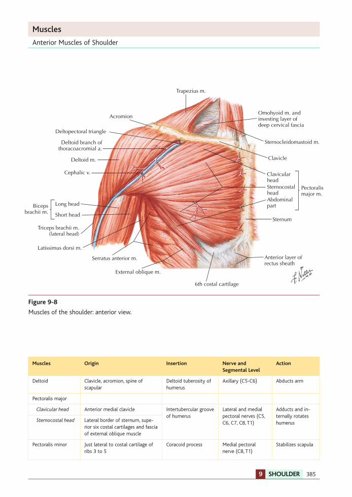
Muscles of the shoulder: anterior view.
Muscles Origin Insertion Nerve and Segmental Level
Action
Deltoid Clavicle, acromion, spine of scapular
Deltoid tuberosity of humerus
Axillary (C5-C6) Abducts arm
Pectoralis major
Clavicular head Anterior medial clavicle Intertubercular groove of humerus
Lateral and medial pectoral nerves (C5, C6, C7, C8, T1)
Adducts and in-ternally rotates humerus Sternocostal head Lateral border of sternum, supe-
rior six costal cartilages and fascia of external oblique muscle
Pectoralis minor Just lateral to costal cartilage of ribs 3 to 5
Coracoid process Medial pectoral nerve (C8, T1)
Stabilizes scapula
386 Netter’s Orthopaedic Clinical Examination: An Evidence-Based Approach
Muscles
Rotator Cuff Muscles
Acromion
Teres minor tendon
Infraspinatus tendon
Supraspinatus tendon
Acromioclavicular jointCoracoacromial lig.
Subscapularis tendonCoracoid process
Trapezoid lig.Conoid lig.
Coracoclavicular lig.Superior view
Infraspinatus m.
Spine of scapulaSupraspinatus m.
Clavicle
Superior borderof scapula
Subscapularis m.
Anterior view
Coracoacromial lig.
Superior transverse scapular lig. and suprascapular notch
Coracoid processAcromion
Supraspinatus tendon
Biceps brachii tendon (long head)
Subscapularis m.
Posterior view
Supraspinatus m. Spine of scapulaAcromion
Supraspinatus tendon
Infra-spinatus m.
Teres minor m.
Axillary n.
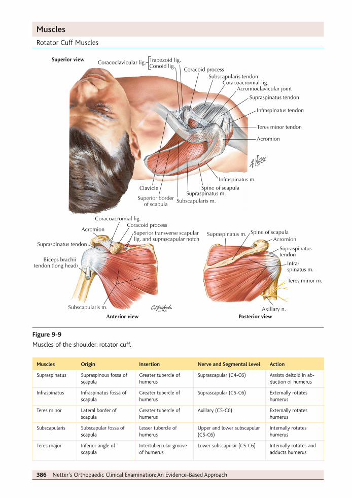
Figure 9-9
Muscles of the shoulder: rotator cuff.
Muscles Origin Insertion Nerve and Segmental Level Action
Supraspinatus Supraspinous fossa of scapula
Greater tubercle of humerus
Suprascapular (C4-C6) Assists deltoid in ab-duction of humerus
Infraspinatus Infraspinatus fossa of scapula
Greater tubercle of humerus
Suprascapular (C5-C6) Externally rotates humerus
Teres minor Lateral border of scapula
Greater tubercle of humerus
Axillary (C5-C6) Externally rotates humerus
Subscapularis Subscapular fossa of scapula
Lesser tubercle of humerus
Upper and lower subscapular (C5-C6)
Internally rotates humerus
Teres major Inferior angle of scapula
Intertubercular groove of humerus
Lower subscapular (C5-C6) Internally rotates and adducts humerus
9 SHOULDER 387
Nerves
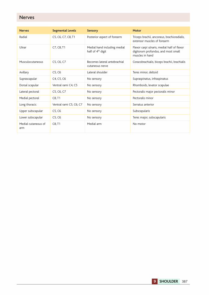
Nerves Segmental Levels Sensory Motor
Radial C5, C6, C7, C8, T1 Posterior aspect of forearm Triceps brachii, anconeus, brachioradialis, extensor muscles of forearm
Ulnar C7, C8, T1 Medial hand including medial half of 4 th digit
Flexor carpi ulnaris, medial half of fl exor digitorum profundus, and most small muscles in hand
Musculocutaneous C5, C6, C7 Becomes lateral antebrachial cutaneous nerve
Coracobrachialis, biceps brachii, brachialis
Axillary C5, C6 Lateral shoulder Teres minor, deltoid
Suprascapular C4, C5, C6 No sensory Supraspinatus, infraspinatus
Dorsal scapular Ventral rami C4, C5 No sensory Rhomboids, levator scapulae
Lateral pectoral C5, C6, C7 No sensory Pectoralis major pectoralis minor
Medial pectoral C8, T1 No sensory Pectoralis minor
Long thoracic Ventral rami C5, C6, C7 No sensory Serratus anterior
Upper subscapular C5, C6 No sensory Subscapularis
Lower subscapular C5, C6 No sensory Teres major, subscapularis
Medial cutaneous of arm
C8, T1 Medial arm No motor

388 Netter’s Orthopaedic Clinical Examination: An Evidence-Based Approach
Pectoralis minor tendon (cut)Coracoid process
AcromionCephalic v.
Musculocutaneous n.
Anterior circumflex humeral a.Axillary n. and posteriorcircumflex humeral a.
Pectoralis major m. (cut)
Coracobrachialis m.Deltoid m.
Biceps brachii m.
Musculocuta-neous n.
Brachialis m.
Thoracoacromial a.Acromial branch
Deltoid branchClavicular branch
Pectoral branchAxillary a.
Clavicle andsubclaviusm. (cut)
Suprascapular a. and n.
Dorsal scapular a. and n.Trapezius m.
Transverse cervical a.
Anterior scalene m.
Sternocleidomastoid m.
Phrenic n.
Omo-hyoid m.
Subclaviana. and v.
1st rib
Brachial plexus
Superior thoracic a.
Lateral pectoral n.
Medial pectoral n.
Deep a.of arm
Radial n.
Tricepsbrachii m.
Brachial vv.
Ulnar n.
Median n.
Brachial a.
Medial cutaneous n.of the forearm
Basilic v.
Ulnar n.
Medial cutaneousn. of arm
Intercostobrachial n.
Circumflexscapular a.
Lowersubscapular n.
Teres major m.
Subscapular a.
Latissimus dorsi m.
Thoracodorsal a. and n.Upper subscapular n.
Serratus anterior m.
Lateral thoracic a. and long thoracic n.
Pectoralis minor m. (cut)
Figure 9-10
Anterior axilla.
Nerves (continued)

9 SHOULDER 389
PATIENT HISTORY
Initial Hypotheses Based on Historical Findings
History Initial Hypothesis
Patient reports lateral/anterior shoulder pain with overhead ac-tivities or exhibits a painful arc
Possible subacromial impingement 6 , 7
Possible tendinitis 8 Possible bursitis 8
Patient reports of instability, apprehension, and pain with activ-ities, most often when shoulder is abducted and externally rotated
Shoulder instability 6 Possible labral tear if clicking is present 9 , 10
Decreased ROM and pain with resistance Possible rotator cuff or long head of the biceps tendinitis 11
Patient reports of pain and weakness with muscle loading, night pain. Age � 60
Possible rotator cuff tear 11
Patient reports poorly located shoulder pain with occasional ra-diation into elbow. Pain is usually aggravated by movement and relieved by rest. Age � 45. Females more often affected than males
Possible adhesive capsulitis 12
Patient reports fall on shoulder followed by pain over AC joint Possible AC sprain 6
Patient reports upper extremity heaviness or numbness with prolonged postures and when laying on involved side
Possible thoracic outlet syndrome 13 , 14
Possible cervical radiculopathy 15
Diagnostic Utility of the Patient History for Identifying Labrum and Rotator Cuff Tears
�LR Interpretation �LR
�10 Large �.1
5.0-10.0 Moderate .1-.2
2.0-5.0 Small .2-.5
1.0-2.0 Rarely important .5-1.0
Patient Report and Study Quality
Population Reference Standard Sens Spec �LR �LR
History of trauma 16
55 patients with shoulder pain scheduled for arthroscopy
Glenoid labral tear observed during arthroscopy
.50 (.35, .65)
.36 (.08, .65)
.79 (.46, 1.34)
1.38 (.6, 3.17)
History of pop, click, or catch 16
.55 (.4, .69)
.73 (.46, .99)
2.0 (.73, 5.45)
.63 (.38, 1.02)
History of trauma 11
448 patients with shoulder pain scheduled for arthroscopy
Rotator cuff tear observed during arthroscopy
.36 .73 1.33 .88
Reports of night pain 11
.88 .20 1.10 .60

390 Netter’s Orthopaedic Clinical Examination: An Evidence-Based Approach
PHYSICAL EXAMINATION TESTS
Range of Motion
Reliability of Range of Motion Measurements
ICC or � Interpretation
.81-1.0 Substantial agreement
.61-.80 Moderate agreement
.41-.60 Fair agreement
.11-.40 Slight agreement
.0-.10 No agreement
Measurment of internal rotationin 90° of abduction
Measurment of external rotationin 90° of abduction
Figure 9-11
Range of motion measurements.
Test Procedure Instrumentation Population Reliability
Passive fl exion 17
Universal goniometer 100 patients referred for physical therapy for shoul-der impairments
Intra-examiner: ICC � .98 Inter-examiner: ICC � .89
Passive extension 17 Intra-examiner: ICC � .94 Inter-examiner: ICC � .27
Passive abduction 17 Intra-examiner: ICC � .98 Inter-examiner: ICC � .87
Active elevation 18
Visual estimation of ROM
201 patients with shoulder pain
Affected side: ICC � .88 (.84, .91) * Unaffected side: ICC � .76 (.67, .82) *
Passive elevation 18 Affected side: ICC � .87 (.83, .90) * Unaffected side: ICC � .73 (.66, .79) *
Passive external rotation 18
Affected side: ICC � .73 (.22, .88) * Unaffected side: ICC � .34 (.00, .65) *
Passive horizontal adduction 18
Affected side: ICC � .36 (.22, .48) * Unaffected side: ICC � .18 (.04, .32) *
* Inter-examiner only. ICC, Intraclass correlation coeffi cient;

9 SHOULDER 391
Range of Motion
Reliability of Functional Range of Motion Tests
ICC or � Interpretation
.81-1.0 Substantial agreement
.61-.80 Moderate agreement
.41-.60 Fair agreement
.11-.40 Slight agreement
.0-.10 No agreement
Figure 9-12
Hand behind back (functional internal rotation of shoul-der test).
Test and Measure Test Procedure Population Inter-examiner Reliability
Hand to neck 19
Visual estimation of ROM graded on a scale of 0 to 3 or 4
46 patients with shoulder pain
Intra-examiner: ICC � .80 (.63, .93) Inter-examiner: ICC � .90 (.69, .96)
Hand to scapula 19 Intra-examiner: ICC � .90 (.72, .92) Inter-examiner: ICC � .90 (.69, .94)
Hand to opposite scapula 19 Intra-examiner: ICC � .86 (.65, .90) Inter-examiner: ICC � .83 (.75, .96)
Active abduction 20
ROM assessed visually to nearest 5°. Pain assessed as “no pain,” “little pain,” “much pain,” and “excruciating pain”
91 patients with shoulder pain
ROM: ICC � .96 Pain: � � .65
Passive abduction 20 ROM: ICC � .96 Pain: � � .69
Painful arc with active abduction 20
Presence of: � � .46 Starting ROM: ICC � .72 Ending ROM: ICC � .57
Painful arc with passive abduction 20
Presence of: � � .52 Starting ROM: ICC � .54 Ending ROM: ICC � .72
Passive external rotation 20 ROM: ICC � .70 Pain: � � .50
Hand behind back 20
As above except ROM graded on a scale of 0 to 7
ROM: � � .73 Pain: � � .35
Hand in neck 20 ROM: � � .52 Pain: � � .52
Springing test 1st rib 20 Examiner exerts force with the 2nd metacarpophalangeal joint on the 1st rib of the patient, assessing ROM (normal or restricted), pain (present or absent), and joint stiffness (present or absent)
ROM: � � .26 Stiffness: � � .09 Pain: � � .66

392 Netter’s Orthopaedic Clinical Examination: An Evidence-Based Approach
Assessing Strength and Proprioception
ICC or � Interpretation
.81-1.0 Substantial agreement
.61-.80 Moderate agreement
.41-.60 Fair agreement
.11-.40 Slight agreement
.0-.10 No agreement
Reliability of Assessing Strength
Test and Measure Test Procedure Population Test-Retest Reliability
Within-Day Between-Days
Serratus anterior strength 21
With subject supine with arm at 90° of shoulder fl exion and 105° of shoul-der horizontal adduction, subject presses toward ceiling while holding weighted apparatus
30 asymp-tomatic students
Inter-examiner ICC � .90-.93
ICC � .83-.89
Serratus anterior endurance 21
As above, with patient holding weight equal to 15% of body weight
Inter-examiner ICC � .71-.76
ICC � .44-.62
Lower trapezius 22 With patient prone and using a hand-held dynamometer on the spine of the scapula, resistance is applied to scapular adduction and depression
40 patients with shoulder pain
ICC � .93 (.89, .96) ICC � .89 (.68, .95)
Serratus anterior 22 With patient supine with shoulder and elbow at 90° and using handheld dynamometer on the elbow, resis-tance is applied to scapular protraction
ICC � .93 (.88, .96) ICC � .94 (.88, .97)
Middle trapezius 22 With patient prone and using a hand-held dynamometer on the spine of the scapula, resistance is applied to scapular retraction
ICC � .94 (.90, .97) ICC � .94 (.82, .97)
Upper trapezius 22 With patient sitting and using a hand-held dynamometer on the superior scapula, resistance is applied to scap-ular elevation
ICC � .95 (.92, .97) ICC � .96 (.91, .98)
Reliability of Assessing Proprioception
Test and Measure Test Procedure Population Test-Retest Reliability
Joint position sense 23 With patient standing, examiner measures full ex-ternal rotation (ER) and internal rotation (IR) of shoulder with inclinometer. Target angles are deter-mined as 90% of IR and 90% of ER. With patient blindfolded, examiner guides patient’s arm into target angle position and holds it for 3 sec. The pa-tient’s arm is returned to neutral. The patient is in-structed to return the arm to target angle. Examiner takes measurement with inclinometer
31 asymptomatic subjects
IR ICC � .98 ER ICC � .98
9 SHOULDER 393
Muscle Length
Reliability of Determining Length of Pectoralis Minor
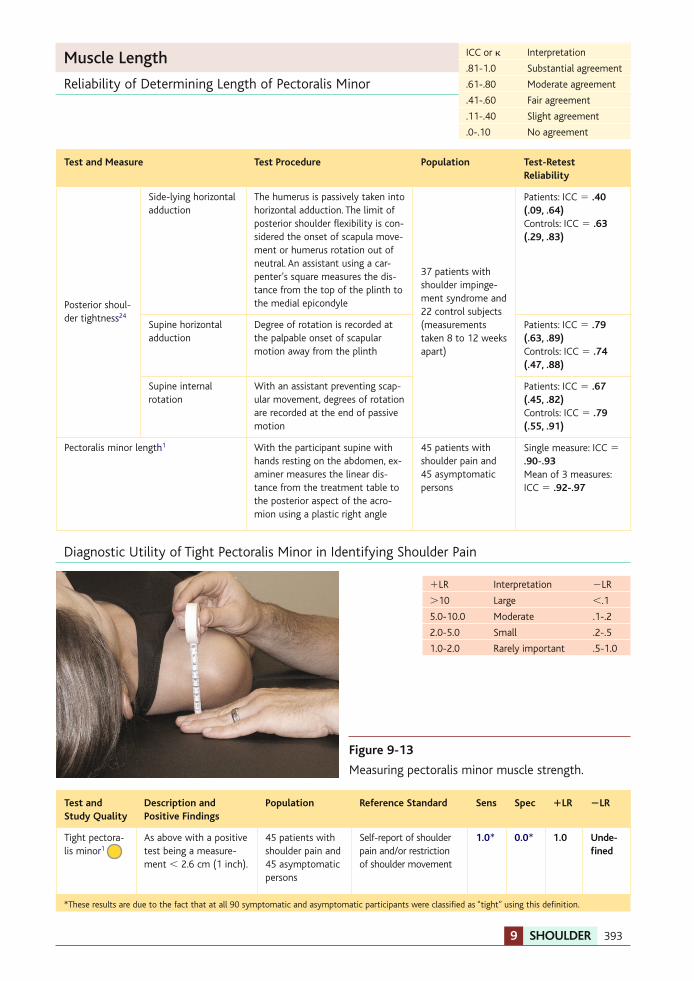
Figure 9-13
Measuring pectoralis minor muscle strength.
Test and Measure Test Procedure Population Test-Retest Reliability
Posterior shoul-der tightness 24
Side-lying horizontal adduction
The humerus is passively taken into horizontal adduction. The limit of posterior shoulder fl exibility is con-sidered the onset of scapula move-ment or humerus rotation out of neutral. An assistant using a car-penter’s square measures the dis-tance from the top of the plinth to the medial epicondyle
37 patients with shoulder impinge-ment syndrome and 22 control subjects (measurements taken 8 to 12 weeks apart)
Patients: ICC � .40 (.09, .64) Controls: ICC � .63 (.29, .83)
Supine horizontal adduction
Degree of rotation is recorded at the palpable onset of scapular motion away from the plinth
Patients: ICC � .79 (.63, .89) Controls: ICC � .74 (.47, .88)
Supine internal rotation
With an assistant preventing scap-ular movement, degrees of rotation are recorded at the end of passive motion
Patients: ICC � .67 (.45, .82) Controls: ICC � .79 (.55, .91)
Pectoralis minor length 1 With the participant supine with hands resting on the abdomen, ex-aminer measures the linear dis-tance from the treatment table to the posterior aspect of the acro-mion using a plastic right angle
45 patients with shoulder pain and 45 asymptomatic persons
Single measure: ICC � .90 - .93 Mean of 3 measures: ICC � .92-.97
Diagnostic Utility of Tight Pectoralis Minor in Identifying Shoulder Pain
�LR Interpretation �LR
�10 Large �.1
5.0-10.0 Moderate .1-.2
2.0-5.0 Small .2-.5
1.0-2.0 Rarely important .5-1.0
ICC or � Interpretation
.81-1.0 Substantial agreement
.61-.80 Moderate agreement
.41-.60 Fair agreement
.11-.40 Slight agreement
.0-.10 No agreement
Test and Study Quality
Description and Positive Findings
Population Reference Standard Sens Spec �LR �LR
Tight pectora-lis minor 1
As above with a positive test being a measure-ment � 2.6 cm (1 inch).
45 patients with shoulder pain and 45 asymptomatic persons
Self-report of shoulder pain and/or restriction of shoulder movement
1.0 * 0.0 * 1.0 Unde-fi ned
* These results are due to the fact that at all 90 symptomatic and asymptomatic participants were classifi ed as “tight” using this defi nition.
394 Netter’s Orthopaedic Clinical Examination: An Evidence-Based Approach
Palpation
Reliability of Palpating the Subacromial Space
ICC or � Interpretation
.81-1.0 Substantial agreement
.61-.80 Moderate agreement
.41-.60 Fair agreement
.11-.40 Slight agreement
.0-.10 No agreement
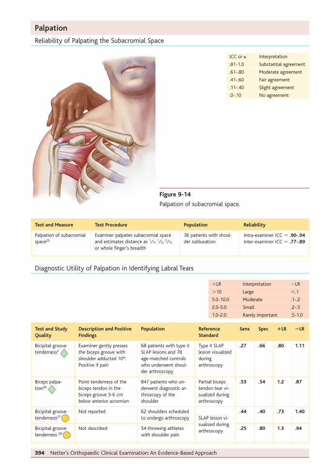
Figure 9-14
Palpation of subacromial space.
Test and Measure Test Procedure Population Reliability
Palpation of subacromial space 25
Examiner palpates subacromial space and estimates distance as 1⁄4, 1⁄2, 3⁄4, or whole fi nger’s breadth
36 patients with shoul-der subluxation
Intra-examiner ICC � .90-.94 Inter-examiner ICC � .77-.89
Diagnostic Utility of Palpation in Identifying Labral Tears
�LR Interpretation �LR
�10 Large �.1
5.0-10.0 Moderate .1-.2
2.0-5.0 Small .2-.5
1.0-2.0 Rarely important .5-1.0
Test and Study Quality
Description and Positive Findings
Population Reference Standard
Sens Spec � LR � LR
Bicipital groove tenderness 2
Examiner gently presses the biceps groove with shoulder adducted 10°. Positive if pain
68 patients with type II SLAP lesions and 78 age-matched controls who underwent shoul-der arthroscopy
Type II SLAP lesion visualized during arthroscopy
.27 .66 .80 1.11
Biceps palpa-tion 26
Point tenderness of the biceps tendon in the biceps groove 3-6 cm below anterior acromion
847 patients who un-derwent diagnostic ar-throscopy of the shoulder
Partial biceps tendon tear vi-sualized during arthroscopy
.53 .54 1.2 .87
Bicipital groove tenderness 27
Not reported 62 shoulders scheduled to undergo arthroscopy SLAP lesion vi-
sualized during arthroscopy
.44 .40 .73 1.40
Bicipital groove tenderness 28
Not described 54 throwing athletes with shoulder pain
.25 .80 1.3 .94
9 SHOULDER 395
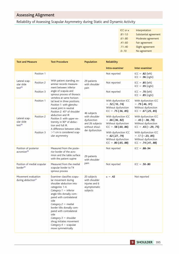
Assessing Alignment
Reliability of Assessing Scapular Asymmetry during Static and Dynamic Activity
ICC or � Interpretation
.81-1.0 Substantial agreement
.61-.80 Moderate agreement
.41-.60 Fair agreement
.11-.40 Slight agreement
.0-.10 No agreement
Test and Measure Test Procedure Population Reliability
Intra-examiner Inter-examiner
Lateral scap-ular slide test 29
Position 1
With patient standing, ex-aminer records measure-ment between inferior angle of scapula and spinous process of thoracic vertebra at same horizon-tal level in three positions. Position 1: with glenohu-meral joint in neutral Position 2: 45° of shoulder abduction and IR Position 3: with upper ex-tremity in 90° of abduc-tion and full IR. A difference between sides �1 cm is considered scap-ular asymmetry
29 patients with shoulder pain
Not reported ICC � .82 (left) ICC � .96 (right)
Position 2 Not reported ICC � .85 (left) ICC � .95 (right)
Position 3 Not reported ICC � .70 (left) ICC � .85 (right)
Lateral scap-ular slide test 30
Position 1
46 subjects with shoulder dysfunction and 26 subjects without shoul-der dysfunction
With dysfunction ICC � .52 (.10, .74) Without dysfunction ICC � .75 (.56, .85)
With dysfunction ICC � .79 (.46, .91) Without dysfunction ICC � .67 (.25, .85)
Position 2 With dysfunction ICC � .66 (.36, .82) Without dysfunction ICC � .58 (.60, .86)
With dysfunction ICC � .45 (�.38, .78) Without dysfunction ICC � .43 (�.29, .75)
Position 3 With dysfunction ICC � .62 (.27, .79) Without dysfunction ICC � .80 (.65, .88)
With dysfunction ICC � .57 (�.23, .85) Without dysfunction ICC � .74 (.41, .88)
Position of posterior acromion 29
Measured from the poste-rior border of the acro-mion and the table surface with the patient supine
29 patients with shoulder pain
Not reported ICC � .88-.94
Position of medial scapular border 29
Measured from the medial scapular border to T4 spinous process
Not reported ICC � .50-.80
Movement evaluation during abduction 31
Examiner classifi es scapu-lar movement during shoulder abduction into categories 1-4. Category 1 � inferior angle tilts dorsally com-pared with contralateral side Category 2 � medial border tilts dorsally com-pared with contralateral side Category 3 � shoulder shrug initiates movement Category 4 � scapulae move symmetrically
20 subjects with shoulder injuries and 6 asymptomatic subjects
� � .42 Not reported

396 Netter’s Orthopaedic Clinical Examination: An Evidence-Based Approach
Lateral slide test position 1
Lateral slide test position 3
Lateral slide test position 2
Figure 9-15
Detecting scapular asymmetry.
Assessing Alignment (continued)
Reliability of Assessing Scapular Asymmetry during Static and Dynamic Activity

9 SHOULDER 397
Classifying Shoulder Disorders
Reliability of Classifying Shoulder Disorders
ICC or � Interpretation
.81-1.0 Substantial agreement
.61-.80 Moderate agreement
.41-.60 Fair agreement
.11-.40 Slight agreement
.0-.10 No agreement
Posterior view revealsatrophy of scapular anddeltoid muscles. Brokenlines, indicating positionof spine of scapula andaxis of humerus on eachside, show little or nomotion in right shoulder
Markedly limited range ofmotion on right side comparedwith that on left side. Slight abduction capability largely due to elevation and rotation ofscapula. All joint motionsrestricted and painfulat extremes. Atrophy ofshoulder muscles
Coronal section of shouldershows adhesions betweencapsule and periphery ofhumeral head
Adhesions ofperipheral capsuleto distal articularcartilage
Adhesions obliteratingaxillary foldof capsule
Figure 9-16
Adhesive capsulitis of the shoulder.
Classifi cation Description of Procedure Population Inter-examiner Reliability
Bursitis 32 Examiners use patient history combined with “selective tissue tension” examination during active movements, passive movements, and isometric strength assessments
56 painful shoulders
� � .35-.58
Capsulitis 32 � � .63-.82
Rotator cuff lesion 32 � � .71-.79
Other diagnosis 32 � � .69-.78
Capsular syndrome 33
Examiner obtains patient history. Physical examination consists of active, passive, and resistive movements. Determi-nation of ROM, presence of painful arc or capsular pattern, and degree of muscle weakness are identifi ed
201 patients with shoulder pain
� � .63 (.50, .76)
Acute bursitis 33 � � .50 (�.10, 1.0)
AC syndrome 33 � � .24 (�.06, .53)
Subacromial syndrome 33 � � .56 (.45, .68)
Rest group (does not fi t any cate-gory above) 33
� � .39 (.24, .54)
Mixed group (patient presents with two or more above classifi cations) 33
� � .14 (�.03, .30)

398 Netter’s Orthopaedic Clinical Examination: An Evidence-Based Approach
Subcoracoid dislocation (most common)
Subglenoid dislocation
Subclavicular dislocation (uncommon). Very rarely, humeral head penetrates between ribs, producing intrathoracic dislocation
Special Tests—Instability
Reliability of Tests to Identify Shoulder Instability
ICC or � Interpretation
.81-1.0 Substantial agreement
.61-.80 Moderate agreement
.41-.60 Fair agreement
.11-.40 Slight agreement
.0-.10 No agreement
Figure 9-17
Shoulder instability.
Test and Measure Test Procedure Population Reliability
Sulcus sign 34 With patient supine, examiner applies inferior distraction to shoulder. Amount of laxity is graded on a 0-3� scale. 0 repre-sents no laxity. 3� represents maximum laxity
43 healthy college athletes
Inter-examiner � � .03-.06
Intra-examiner � � .01-.20
9 SHOULDER 399
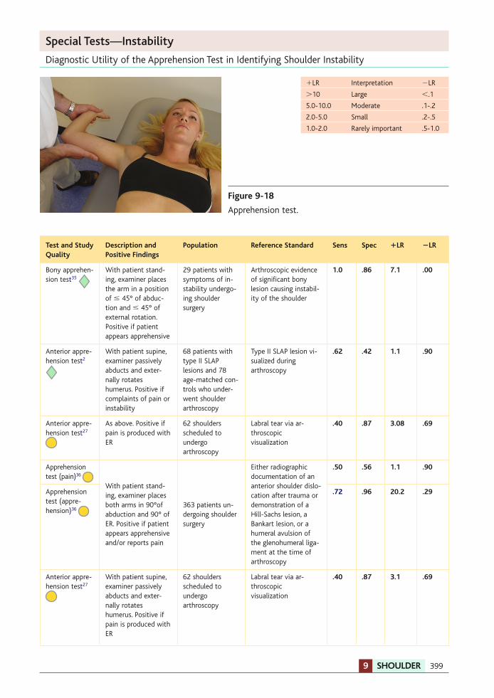
Special Tests—Instability
Diagnostic Utility of the Apprehension Test in Identifying Shoulder Instability
�LR Interpretation �LR
�10 Large �.1
5.0-10.0 Moderate .1-.2
2.0-5.0 Small .2-.5
1.0-2.0 Rarely important .5-1.0
Figure 9-18
Apprehension test.
Test and Study Quality
Description and Positive Findings
Population Reference Standard Sens Spec �LR �LR
Bony apprehen-sion test 35
With patient stand-ing, examiner places the arm in a position of � 45° of abduc-tion and � 45° of external rotation. Positive if patient appears apprehensive
29 patients with symptoms of in-stability undergo-ing shoulder surgery
Arthroscopic evidence of signifi cant bony lesion causing instabil-ity of the shoulder
1.0 .86 7.1 .00
Anterior appre-hension test 2
With patient supine, examiner passively abducts and exter-nally rotates humerus. Positive if complaints of pain or instability
68 patients with type II SLAP lesions and 78 age-matched con-trols who under-went shoulder arthroscopy
Type II SLAP lesion vi-sualized during arthroscopy
.62 .42 1.1 .90
Anterior appre-hension test 27
As above. Positive if pain is produced with ER
62 shoulders scheduled to undergo arthroscopy
Labral tear via ar-throscopic visualization
.40 .87 3.08 .69
Apprehension test (pain) 36
With patient stand-ing, examiner places both arms in 90°of abduction and 90° of ER. Positive if patient appears apprehensive and/or reports pain
363 patients un-dergoing shoulder surgery
Either radiographic documentation of an anterior shoulder dislo-cation after trauma or demonstration of a Hill-Sachs lesion, a Bankart lesion, or a humeral avulsion of the glenohumeral liga-ment at the time of arthroscopy
.50 .56 1.1 .90
Apprehension test (appre-hension) 36
.72 .96 20.2 .29
Anterior appre-hension test 27
With patient supine, examiner passively abducts and exter-nally rotates humerus. Positive if pain is produced with ER
62 shoulders scheduled to undergo arthroscopy
Labral tear via ar-throscopic visualization
.40 .87 3.1 .69
400 Netter’s Orthopaedic Clinical Examination: An Evidence-Based Approach
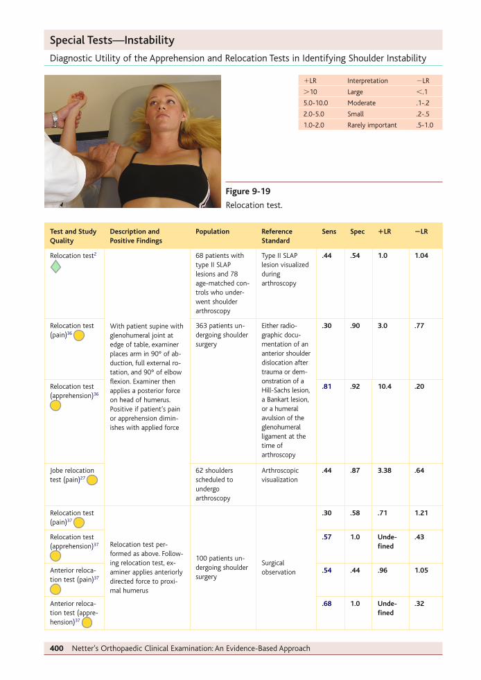
Special Tests—Instability
Diagnostic Utility of the Apprehension and Relocation Tests in Identifying Shoulder Instability
�LR Interpretation �LR
�10 Large �.1
5.0-10.0 Moderate .1-.2
2.0-5.0 Small .2-.5
1.0-2.0 Rarely important .5-1.0
Figure 9-19
Relocation test.
Test and Study Quality
Description and Positive Findings
Population Reference Standard
Sens Spec �LR �LR
Relocation test 2
With patient supine with glenohumeral joint at edge of table, examiner places arm in 90° of ab-duction, full external ro-tation, and 90° of elbow fl exion. Examiner then applies a posterior force on head of humerus. Positive if patient’s pain or apprehension dimin-ishes with applied force
68 patients with type II SLAP lesions and 78 age-matched con-trols who under-went shoulder arthroscopy
Type II SLAP lesion visualized during arthroscopy
.44 .54 1.0 1.04
Relocation test (pain) 36
363 patients un-dergoing shoulder surgery
Either radio-graphic docu-mentation of an anterior shoulder dislocation after trauma or dem-onstration of a Hill-Sachs lesion, a Bankart lesion, or a humeral avulsion of the glenohumeral ligament at the time of arthroscopy
.30 .90 3.0 .77
Relocation test (apprehension) 36
.81 .92 10.4 .20
Jobe relocation test (pain) 27
62 shoulders scheduled to undergo arthroscopy
Arthroscopic visualization
.44 .87 3.38 .64
Relocation test (pain) 37
Relocation test per-formed as above. Follow-ing relocation test, ex-aminer applies anteriorly directed force to proxi-mal humerus
100 patients un-dergoing shoulder surgery
Surgical observation
.30 .58 .71 1.21
Relocation test (apprehension) 37
.57 1.0 Unde-fi ned
.43
Anterior reloca-tion test (pain) 37
.54 .44 .96 1.05
Anterior reloca-tion test (appre-hension) 37
.68 1.0 Unde-fi ned
.32
9 SHOULDER 401
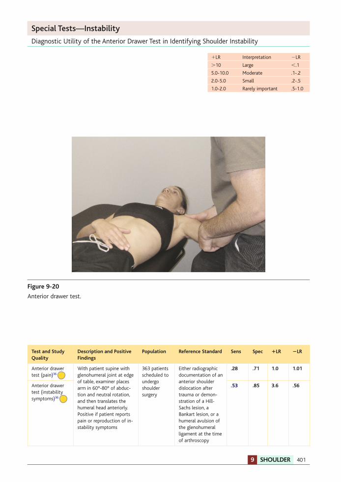
Special Tests—Instability
Diagnostic Utility of the Anterior Drawer Test in Identifying Shoulder Instability
�LR Interpretation �LR
�10 Large �.1
5.0-10.0 Moderate .1-.2
2.0-5.0 Small .2-.5
1.0-2.0 Rarely important .5-1.0
Figure 9-20
Anterior drawer test.
Test and Study Quality
Description and Positive Findings
Population Reference Standard Sens Spec �LR �LR
Anterior drawer test (pain) 36
With patient supine with glenohumeral joint at edge of table, examiner places arm in 60°-80° of abduc-tion and neutral rotation, and then translates the humeral head anteriorly. Positive if patient reports pain or reproduction of in-stability symptoms
363 patients scheduled to undergo shoulder surgery
Either radiographic documentation of an anterior shoulder dislocation after trauma or demon-stration of a Hill-Sachs lesion, a Bankart lesion, or a humeral avulsion of the glenohumeral ligament at the time of arthroscopy
.28 .71 1.0 1.01
Anterior drawer test (instability symptoms) 36
.53 .85 3.6 .56

402 Netter’s Orthopaedic Clinical Examination: An Evidence-Based Approach
Special Tests—Labral Tears
Reliability of the Crank Test
ICC or � Interpretation
.81-1.0 Substantial agreement
.61-.80 Moderate agreement
.41-.60 Fair agreement
.11-.40 Slight agreement
.0-.10 No agreement
Figure 9-21
Crank test.
Test Description Population Inter-examiner Reliability
Crank test 16 See page 403 55 patients with shoulder pain scheduled for arthroscopic surgery � � .20 (�.05, .46)
9 SHOULDER 403
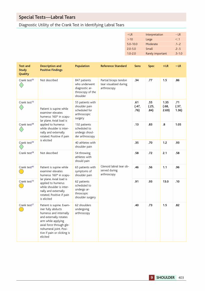
Special Tests—Labral Tears
Diagnostic Utility of the Crank Test in Identifying Labral Tears
�LR Interpretation �LR
�10 Large �.1
5.0-10.0 Moderate .1-.2
2.0-5.0 Small .2-.5
1.0-2.0 Rarely important .5-1.0
Test and Study Quality
Description and Positive Findings
Population Reference Standard Sens Spec �LR �LR
Crank test 26
Not described 847 patients who underwent diagnostic ar-throscopy of the shoulder
Partial biceps tendon tear visualized during arthroscopy
.34 .77 1.5 .86
Crank test 16
Patient is supine while examiner elevates humerus 160° in scapu-lar plane. Axial load is applied to humerus while shoulder is inter-nally and externally rotated. Positive if pain is elicited
55 patients with shoulder pain scheduled for arthroscopic surgery
Glenoid labral tear ob-served during arthroscopy
.61 (.47, .76)
.55 (.25, .84)
1.35 (.68, 2.69)
.71 (.37, 1.36)
Crank test 38
132 patients scheduled to undergo shoul-der arthroscopy
.13 .83 .8 1.05
Crank test 39
40 athletes with shoulder pain
.35 .70 1.2 .93
Crank test 28 Not described 54 throwing athletes with should pain
.58 .72 2.1 .58
Crank test 40
Patient is supine while examiner elevates humerus 160° in scapu-lar plane. Axial load is applied to humerus while shoulder is inter-nally and externally rotated. Positive if pain is elicited
65 patients with symptoms of shoulder pain
.46 .56 1.1 .96
Crank test 10
62 patients scheduled to undergo ar-throscopic shoulder surgery
.91 .93 13.0 .10
Crank test 27
Patient is supine. Exam-iner fully abducts humerus and internally and externally rotates arm while applying axial force through gle-nohumeral joint. Posi-tive if pain or clicking is elicited
62 shoulders undergoing arthroscopy
.40 .73 1.5 .82

404 Netter’s Orthopaedic Clinical Examination: An Evidence-Based Approach
Special Tests—Labral Tears
Diagnostic Utility of the Compression Rotation Test in Identifying Labral Tears
�LR Interpretation �LR
�10 Large �.1
5.0-10.0 Moderate .1-.2
2.0-5.0 Small .2-.5
1.0-2.0 Rarely important .5-1.0
Figure 9-22
Compression rotation test.
Test and Study Quality
Description and Positive Findings
Population Reference Standard
Sens Spec �LR �LR
Compression rotation test 2
With patient supine with arm abducted to 90° and elbow fl exed to 90°, exam-iner applies axial force to humerus. Humerus is cir-cumducted and rotated. Positive if pain or clicking is elicited
68 patients with type II SLAP lesions and 78 age-matched controls who under-went shoulder arthroscopy
Type II SLAP lesion visualized during arthroscopy
.61 .54 1.3 .72
Compression rotation test 41
426 patients who had undergone shoulder arthroscopy Labral tear visual-
ized during arthroscopy
.24 .76 1.0 1.0
Compression rotation test 28
Not described 54 throwing athletes with shoulder pain
.25 1.0 Unde-fi ned
.75
9 SHOULDER 405
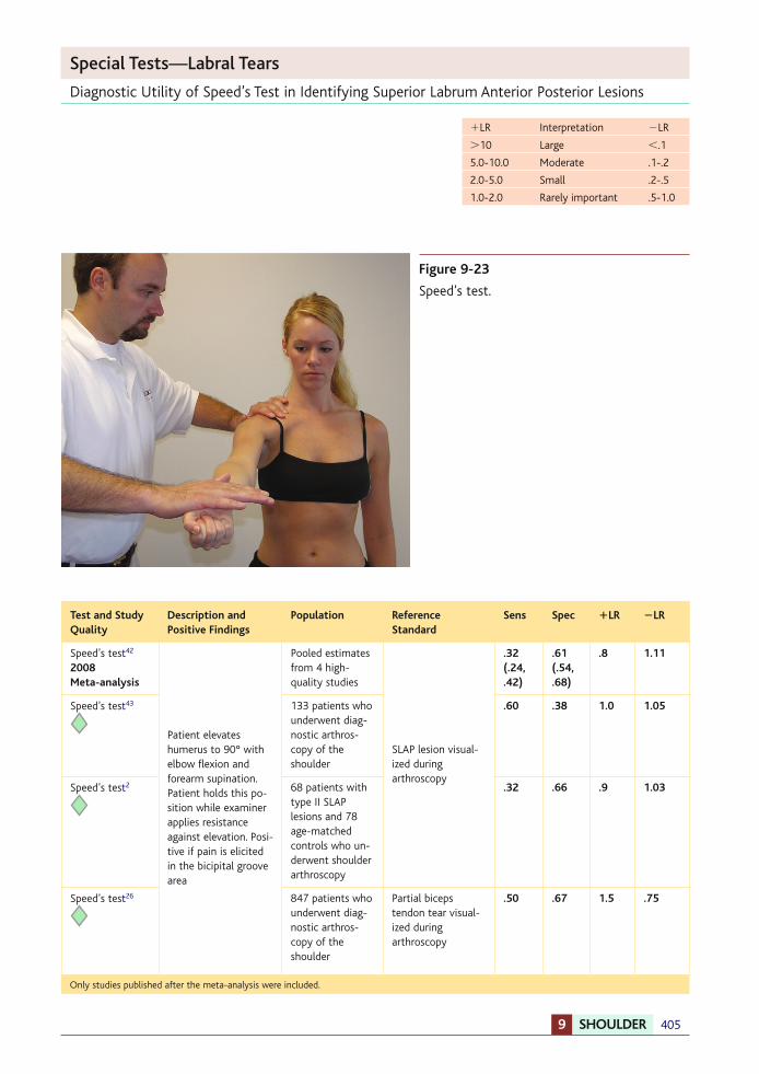
Special Tests—Labral Tears
Diagnostic Utility of Speed’s Test in Identifying Superior Labrum Anterior Posterior Lesions
�LR Interpretation �LR
�10 Large �.1
5.0-10.0 Moderate .1-.2
2.0-5.0 Small .2-.5
1.0-2.0 Rarely important .5-1.0
Figure 9-23
Speed’s test.
Test and Study Quality
Description and Positive Findings
Population Reference Standard
Sens Spec �LR �LR
Speed’s test 42 2008 Meta-analysis
Patient elevates humerus to 90° with elbow fl exion and forearm supination. Patient holds this po-sition while examiner applies resistance against elevation. Posi-tive if pain is elicited in the bicipital groove area
Pooled estimates from 4 high-quality studies
SLAP lesion visual-ized during arthroscopy
.32 (.24, .42)
.61 (.54, .68)
.8 1.11
Speed’s test 43
133 patients who underwent diag-nostic arthros-copy of the shoulder
.60 .38 1.0 1.05
Speed’s test 2
68 patients with type II SLAP lesions and 78 age-matched controls who un-derwent shoulder arthroscopy
.32 .66 .9 1.03
Speed’s test 26
847 patients who underwent diag-nostic arthros-copy of the shoulder
Partial biceps tendon tear visual-ized during arthroscopy
.50 .67 1.5 .75
Only studies published after the meta-analysis were included.
406 Netter’s Orthopaedic Clinical Examination: An Evidence-Based Approach
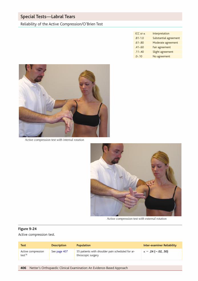
Special Tests—Labral Tears
Reliability of the Active Compression/O’Brien Test
ICC or � Interpretation
.81-1.0 Substantial agreement
.61-.80 Moderate agreement
.41-.60 Fair agreement
.11-.40 Slight agreement
.0-.10 No agreement
Active compression test with internal rotation
Active compression test with external rotation
Figure 9-24
Active compression test.
Test Description Population Inter-examiner Reliability
Active compression test 16
See page 407 55 patients with shoulder pain scheduled for ar-throscopic surgery
� � .24 (�.02, .50)

9 SHOULDER 407
Special Tests—Labral Tears
Diagnostic Utility of the Active Compression/O’Brien Test
�LR Interpretation �LR
�10 Large �.1
5.0-10.0 Moderate .1-.2
2.0-5.0 Small .2-.5
1.0-2.0 Rarely important .5-1.0
Test and Study Quality
Description and Positive Findings
Population Reference Standard
Sens Spec �LR �LR
Active com-pression test 16
Patient stands and fl exes arm to 90° with elbow in full extension. Patient then adducts arm 10° internally and rotates humerus. Exam-iner applies downward force to arm as patient resists. Patient then fully supinates arm and repeats procedure. Positive if pain is elicited with fi rst maneuver and reduced with second maneuver
55 patients with shoulder pain scheduled for arthroscopic surgery
Glenoid labral tear observed during arthroscopy
.55 (.4, .69)
.18 (�.05, .41)
.67 (.45, .98)
2.5 (.68, 9.13)
O’Brien test 43
133 patients who underwent diagnostic ar-throscopy of the shoulder
SLAP lesion visu-alized during arthroscopy
.94 .28 1.3 .21
O’Brien test 2
68 patients with SLAP lesions and 78 age-matched controls
.63 .53 1.3 .70
Active com-pression test 38
132 patients scheduled to undergo shoul-der arthroscopy
.63 .50 1.3 .74
Active com-pression test 39
40 athletes with shoulder pain
.78 .11 .1 2.00
Active com-pression test 41
426 patients who had under-gone shoulder arthroscopy
.47 .55 1.0 .96
Active com-pression test (palm down) 26
As above except positive if pain is elicited in tested position
847 patients who underwent diagnostic ar-throscopy of the shoulder
Partial biceps tendon tear vi-sualized during arthroscopy
.68 .46 1.3 .70
Active com-pression test (palm up) 26
.40 .57 .9 1.1
O’Brien test 40
As above except patient is seated
65 patients with symptoms of shoulder pain
.54 .31 .78 1.48
O’Brien test 27
62 shoulders undergoing arthroscopy
.63 .73 2.3 .51
O’Brien test 28
Not described 54 throwing athletes with shoulder pain
.54 .60 1.4 .77

408 Netter’s Orthopaedic Clinical Examination: An Evidence-Based Approach
Special Tests—Labral Tears
Diagnostic Utility of the Yergason Test in Identifying Labral Tears
�LR Interpretation �LR
�10 Large �.1
5.0-10.0 Moderate .1-.2
2.0-5.0 Small .2-.5
1.0-2.0 Rarely important .5-1.0
Figure 9-25
Yergason test.
Test and Study Quality
Description and Positive Findings
Population Reference Standard
Sens Spec �LR �LR
Yergason test 2
With patient standing with elbow at 90°, patient supinates forearm against examin-er’s resistance. During procedure, examiner pal-pates long head of biceps tendon. Positive if pain at biceps tendon
68 patients with type II SLAP lesions and 78 age-matched controls who underwent shoulder arthroscopy
SLAP lesion visual-ized during arthroscopy
.12 .87 .9 1.01
Yergason test 38
132 patients sched-uled to undergo shoulder arthroscopy
.13 .94 2.2 .93
Yergason test 27
62 shoulders sched-uled to undergo arthroscopy
.09 .93 1.29 .98
Yergason test 28
54 throwing ath-letes with shoulder pain
.13 1.0 Unde-fi ned
.87
Yergason test 44
152 subjects with shoulder pain scheduled to undergo surgery
Biceps tendon and/or labral tear visualized during arthroscopy
.43 .79 2.05 .72
9 SHOULDER 409
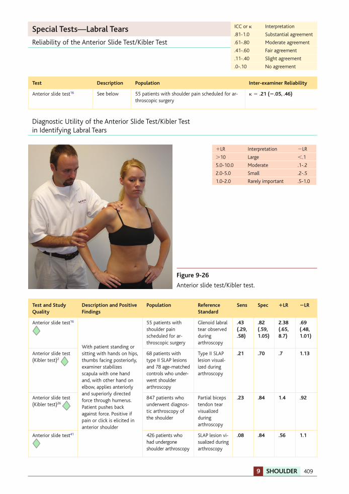
Special Tests—Labral Tears
Reliability of the Anterior Slide Test/Kibler Test
Figure 9-26
Anterior slide test/Kibler test.
ICC or � Interpretation
.81-1.0 Substantial agreement
.61-.80 Moderate agreement
.41-.60 Fair agreement
.11-.40 Slight agreement
.0-.10 No agreement
Test Description Population Inter-examiner Reliability
Anterior slide test 16 See below 55 patients with shoulder pain scheduled for ar-throscopic surgery
� � .21 (�.05, .46)
Diagnostic Utility of the Anterior Slide Test/Kibler Test in Identifying Labral Tears
�LR Interpretation �LR
�10 Large �.1
5.0-10.0 Moderate .1-.2
2.0-5.0 Small .2-.5
1.0-2.0 Rarely important .5-1.0
Test and Study Quality
Description and Positive Findings
Population Reference Standard
Sens Spec �LR �LR
Anterior slide test 16
With patient standing or sitting with hands on hips, thumbs facing posteriorly, examiner stabilizes scapula with one hand and, with other hand on elbow, applies anteriorly and superiorly directed force through humerus. Patient pushes back against force. Positive if pain or click is elicited in anterior shoulder
55 patients with shoulder pain scheduled for ar-throscopic surgery
Glenoid labral tear observed during arthroscopy
.43 (.29, .58)
.82 (.59, 1.05)
2.38 (.65, 8.7)
.69 (.48, 1.01)
Anterior slide test (Kibler test) 2
68 patients with type II SLAP lesions and 78 age-matched controls who under-went shoulder arthroscopy
Type II SLAP lesion visual-ized during arthroscopy
.21 .70 .7 1.13
Anterior slide test (Kibler test) 26
847 patients who underwent diagnos-tic arthroscopy of the shoulder
Partial biceps tendon tear visualized during arthroscopy
.23 .84 1.4 .92
Anterior slide test 41
426 patients who had undergone shoulder arthroscopy
SLAP lesion vi-sualized during arthroscopy
.08 .84 .56 1.1

410 Netter’s Orthopaedic Clinical Examination: An Evidence-Based Approach
Special Tests—Labral Tears
Reliability of Various Tests in Identifying Labral Tears
ICC or � Interpretation
.81-1.0 Substantial agreement
.61-.80 Moderate agreement
.41-.60 Fair agreement
.11-.40 Slight agreement
.0-.10 No agreement
Figure 9-27
Kim and jerk tests.
Test Description Population Reliability
Passive compression test 45 See page 411 61 patients undergoing arthroscopy for shoulder pain Inter-examiner � � .77
Kim test 46 See page 411 172 painful shoulders Inter-examiner � � .91

9 SHOULDER 411
Special Tests—Labral Tears
Diagnostic Utility of Various Tests in Identifying Labral Tears
�LR Interpretation �LR
�10 Large �.1
5.0-10.0 Moderate .1-.2
2.0-5.0 Small .2-.5
1.0-2.0 Rarely important .5-1.0
Test and Study Quality
Description and Positive Findings Population Reference Standard
Sens Spec �LR �LR
Passive com-pression test 45
With patient side lying with af-fected side up, examiner places one hand over the AC joint to stabilize the shoulder and the other hand on the elbow. Examiner then externally rotates the shoulder in 30° abduc-tion and gives axial compression while extending the arm. Positive if pain
61 patients un-dergoing arthros-copy for shoulder pain
SLAP lesion vi-sualized during arthroscopy
.82 .86 5.90 .21
Kim test 46
With patient sitting with arm ab-ducted 90°, examiner holds the elbow and lateral aspect of the proximal arm and applies a strong axial loading force. Examiner then elevates the arm to 135° and adds a posterior/inferior force. Positive if sudden onset of posterior shoulder pain 172 painful
shoulders
Labral tear visu-alized during arthroscopy
.80 .94 13.3 .21
Jerk test 46
With patient sitting, examiner holds scapula with one hand and inter-nally rotates and abducts the pa-tient’s arm to 90° with the other. Examiner then horizontally adducts the arm while applying an axial loading force. Sharp pain indicates a positive test
.73 .98 36.5 .28
Supine fl exion resis-tance test 43
With patient supine with arm resting in full fl exion and palm up, examiner grasps patient’s arm just distal to the elbow and asks the patient to lift the arm as if throw-ing. Positive if pain is felt deep inside the shoulder joint
133 patients who underwent diag-nostic arthros-copy of the shoulder
SLAP lesion vi-sualized during arthroscopy
.80 .69 2.6 .29
Resisted su-pination ex-ternal rota-tion test 39
With patient supine with arm ab-ducted 90° and elbow fl exed 70°, examiner supports the arm by the elbow. Examiner resists supination and gently maximally externally rotates the shoulder. Positive if shoulder pain, clicking, or catching is elicited
40 athletes with shoulder pain
.83 .82 4.6 .21
(Continued)

412 Netter’s Orthopaedic Clinical Examination: An Evidence-Based Approach
�LR Interpretation �LR
�10 Large �.1
5.0-10.0 Moderate .1-.2
2.0-5.0 Small .2-.5
1.0-2.0 Rarely important .5-1.0
Special Tests—Labral Tears (continued)
Diagnostic Utility of Various Tests in Identifying Labral Tears
Test and Study Quality
Description and Positive Findings Population Reference Standard
Sens Spec �LR �LR
Whipple test 2
The arm is fl exed 90° and adducted until the hand is opposite the other shoulder. The patient resists while examiner pushes downward on the arm. Positive if pain
68 patients with type II SLAP lesions and 78 age-matched controls who un-derwent shoulder arthroscopy
Type II SLAP lesion visualized during arthroscopy
.65 .42 1.1 .83
Biceps load test II 2
With patient supine, examiner grasps wrist and elbow. Arm is ele-vated 120° and fully externally rotated with elbow held in 90° of fl exion and forearm supinated. Ex-aminer then resists elbow fl exion by patient. Positive if resisted elbow fl exion causes pain
68 patients with type II SLAP lesions and 78 age-matched controls who un-derwent shoulder arthroscopy
.30 .78 1.4 .90
Biceps load test II 47
127 patients ex-periencing shoul-der pain sched-uled to undergo arthroscopy
.90 .97 30 .10
Posterior jerk test 28
Not described 54 throwing ath-letes with shoul-der pain
.25 .80 1.3 .72
Biceps load test 48
With patient supine, examiner grasps wrist and elbow. Arm is ab-ducted to 90° with elbow fl exed to 90° and forearm supinated. Exam-iner externally rotates arm until patient becomes apprehensive at which time ER is stopped. Patient fl exes elbow against examiner’s re-sistance. Positive if patient’s appre-hension remains or pain is produced
75 patients with unilateral recur-rent anterior shoulder dislocations
.90 .97 30 .10

9 SHOULDER 413
Special Tests—Subacromial Impingement
Reliability of Hawkins-Kennedy Test
ICC or � Interpretation
.81-1.0 Substantial agreement
.61-.80 Moderate agreement
.41-.60 Fair agreement
.11-.40 Slight agreement
.0-.10 No agreement
Figure 9-28
Hawkins-Kennedy test.
Test Description Population Reliability
Hawkins-Kennedy test 49 See below 33 patients with shoulder pain Test-retest � � 1.0 Inter-examiner � � . 91
Diagnostic Utility of Hawkins-Kennedy Test in Identifying Subacromial Impingement
�LR Interpretation �LR
�10 Large �.1
5.0-10.0 Moderate .1-.2
2.0-5.0 Small .2-.5
1.0-2.0 Rarely important .5-1.0
Test and Study Quality
Description and Positive Findings
Population Reference Standard Sens Spec �LR �LR
Hawkins-Kennedy test 4 2008 Meta-analysis
Patient is standing. The affected arm is forward-fl exed 90° and then forcibly medially rotated. Positive if the patient complains of pain.
Pooled estimates from 4 high-quality studies
Impingement syndrome diagnosed from sub-acromial injection or surgery
.79 (.75, .82)
.59 (.53, .64)
1.9 .36
Hawkins-Kennedy test 50
30 patients with new onset shoulder pain
Magnetic resonance imaging (MRI) con-fi rmed subacromial impingement
.74 .40 1.2 (.7, 2.3)
.65
MRI confi rmed subacro-mial bursitis
.80 .43 1.4 (.8, 2.4)
.47
Hawkins-Kennedy test 26
847 patients who underwent diag-nostic arthroscopy of the shoulder
Partial biceps tendon tear visualized during arthroscopy
.55 .38 .9 1.18
Only studies published after the meta-analysis were included.
414 Netter’s Orthopaedic Clinical Examination: An Evidence-Based Approach
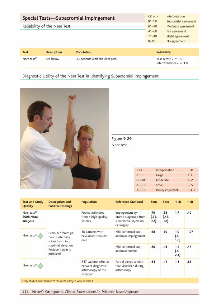
Special Tests—Subacromial Impingement
Reliability of the Neer Test
Figure 9-29
Neer test.
Test and Study Quality
Description and Positive Findings
Population Reference Standard Sens Spec �LR �LR
Neer test 42 2008 Meta-analysis
Examiner forces pa-tient’s internally rotated arm into maximal elevation. Positive if pain is produced
Pooled estimates from 4 high-quality studies
Impingement syn-drome diagnosed from subacromial injection or surgery
.79 (.75, .82)
.53 (.48, .58)
1.7 .40
Neer test 50
30 patients with new-onset shoulder pain
MRI confi rmed sub-acromial impingement
.68 .30 1.0 (.6, 1.6)
1.07
MRI confi rmed sub-acromial bursitis
.80 .43 1.4 (.8, 2.4)
.47
Neer test 26
847 patients who un-derwent diagnostic arthroscopy of the shoulder
Partial biceps tendon tear visualized during arthroscopy
.64 .41 1.1 .88
�LR Interpretation �LR
�10 Large �.1
5.0-10.0 Moderate .1-.2
2.0-5.0 Small .2-.5
1.0-2.0 Rarely important .5-1.0
Only studies published after the meta-analysis were included.
ICC or � Interpretation
.81-1.0 Substantial agreement
.61-.80 Moderate agreement
.41-.60 Fair agreement
.11-.40 Slight agreement
.0-.10 No agreement
Test Description Population Reliability
Neer test 49 See below 33 patients with shoulder pain Test-retest � � 1.0 Inter-examiner � � 1.0
Diagnostic Utility of the Neer Test in Identifying Subacromial Impingement

9 SHOULDER 415
Special Tests—Subacromial Impingement
Diagnostic Utility of Various Tests in Identifying Subacromial Impingement
�LR Interpretation �LR
�10 Large �.1
5.0-10.0 Moderate .1-.2
2.0-5.0 Small .2-.5
1.0-2.0 Rarely important .5-1.0
Test and Study Quality
Description and Positive Findings
Population Reference Standard Sens Spec �LR �LR
Painful arc sign 51
Patient actively ele-vates arm in scapular plane to full elevation. Positive if patient ex-periences pain between 60° and 120°
552 patients with shoulder pain
Arthroscopic visualization• All impingement• Bursitis• Partial thickness RCT• Full thickness RCT
.74 .71 .67 .76
.81 .47 .47 .72
3.9 1.3 1.3 2.7
.32 .62 .70 .33
Cross-body ad-duction test 51
Arm at 90° of fl exion. Examiner then adducts arm across the patient’s body. Positive if shoulder pain is produced
552 patients with shoulder pain
Arthroscopic visualization• All impingement• Bursitis• Partial thickness RCT• Full thickness RCT
.23 .25 .17 .23
.82 .80 .79 .81
1.3 1.3 .8 1.2
.94 .94 1.05 .95
Lift-off test (Gerber’s test) 50
Patient attempts to lift the affected arm off the back. Positive if unable to lift off back
30 patients with new-onset shoul-der pain
MRI confi rmed subacromial impingement
.68 .50 1.4 (.7, 2.7)
.64
MRI confi rmed subacromial bursitis
.93 .71 3.3 (1.4, 7.6)
.10
Lift-off test (Gerber’s test) 26
847 patients who underwent diag-nostic arthros-copy of the shoulder
Partial biceps tendon tear visualized during arthroscopy
.28 .89 2.5 .81
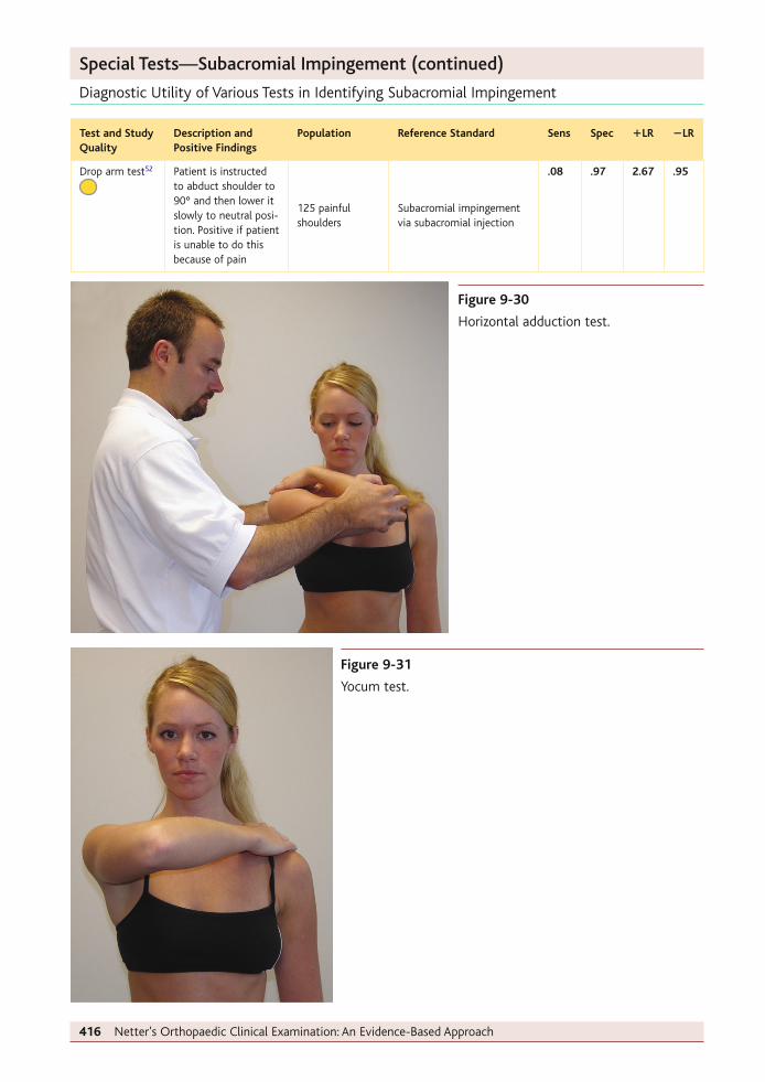
Yocum test 50
With patient seated or standing, patient places hand of in-volved shoulder on contralateral shoulder and raises elbow. Posi-tive if pain is elicited
30 patients with new-onset shoul-der pain
MRI confi rmed subacromial impingement
.79 .40 1.3 (.8, 2.3)
.53
MRI confi rmed subacromial bursitis
.80 .36 1.2 (.08, 2.0)
.56
Horizontal ad-duction test 52
Examiner forces pa-tient’s arm into hori-zontal adduction while elbow is fl exed. Posi-tive if pain is elicited
125 painful shoulders
Subacromial impingement via subacromial injection
.82 .28 1.14 .64
The painful arc test 52
Patient is instructed to perform straight plane abduction throughout full ROM. Positive if pain occurs between 60° and 100° of abduction
.33 .81 1.74 .83
(Continued)
416 Netter’s Orthopaedic Clinical Examination: An Evidence-Based Approach
Test and Study Quality
Description and Positive Findings
Population Reference Standard Sens Spec �LR �LR
Drop arm test 52
Patient is instructed to abduct shoulder to 90° and then lower it slowly to neutral posi-tion. Positive if patient is unable to do this because of pain
125 painful shoulders
Subacromial impingement via subacromial injection
.08 .97 2.67 .95
Figure 9-30
Horizontal adduction test.
Special Tests—Subacromial Impingement (continued)
Diagnostic Utility of Various Tests in Identifying Subacromial Impingement
Figure 9-31
Yocum test.

9 SHOULDER 417
Special Tests—Subacromial Impingement
Diagnostic Utility of Internal Rotation Resistance Strength Test in Differentiating Subacromial Impingement versus Intra-articular Pathology
Resistance against external rotation
Resistance against internal rotation
Figure 9-32
Internal rotation resistance strength test.
Zaslav 53 investigated the internal rotation resistance strength (IRRS) test’s ability to delineate intra-articular pathology from impingement syndrome in a group of 115 patients who underwent arthroscopic shoulder surgery. Th e IRRS test is performed with the patient standing. Th e exam-iner positions the patient’s arm in 90° abduction and 80° ER. Th e examiner applies resistance against ER and then IR in the same position. Th e test is considered positive for intra-articular pathology if the patient exhibits greater weakness in IR when compared with ER. If the patient demonstrated greater weakness with ER, they were considered positive for impingement syn-drome. Th e IRRS test demonstrated a sensitivity of .88, a specifi city of .96, a positive LR of 22.0, and a negative LR of .13.

418 Netter’s Orthopaedic Clinical Examination: An Evidence-Based Approach
Special Tests—Rotator Cuff Tears
Reliability of Special Tests for Identifying Supraspinatus and/or Infraspinatus Tears
Acute rupture (superior view). Often associatedwith splitting tear parallel to tendon fibers.Further retraction results in crescentic defect asshown on right
Retracted tear, commonly found atsurgery. Broken line indicates extent ofdebridement of degenerated tendonfor repair.
Subscapularis m.
Humerus
Biceps brachiitendon
Infraspinatus m.
Supraspinatus m.
Thickened, edematousbiceps brachii tendon
Figure 9-33
Superior rotator cuff tear.
Figure 9-34
Supraspinatus muscle test (empty can).
ICC or � Interpretation
.81-1.0 Substantial agreement
.61-.80 Moderate agreement
.41-.60 Fair agreement
.11-.40 Slight agreement
.0-.10 No agreement
Test Description Population Reliability
Supraspinatus muscle test (empty can) 49 Shoulder and elbow at 90° with arm in-
ternally rotated. Examiner then resists internal rotation force. Positive if patient gives way
33 patients with shoulder pain
Test-retest � � 1.0 Inter-examiner � � .94
Patte maneuver 49 Test-retest � � 1.0 Inter-examiner � � 1.0
9 SHOULDER 419
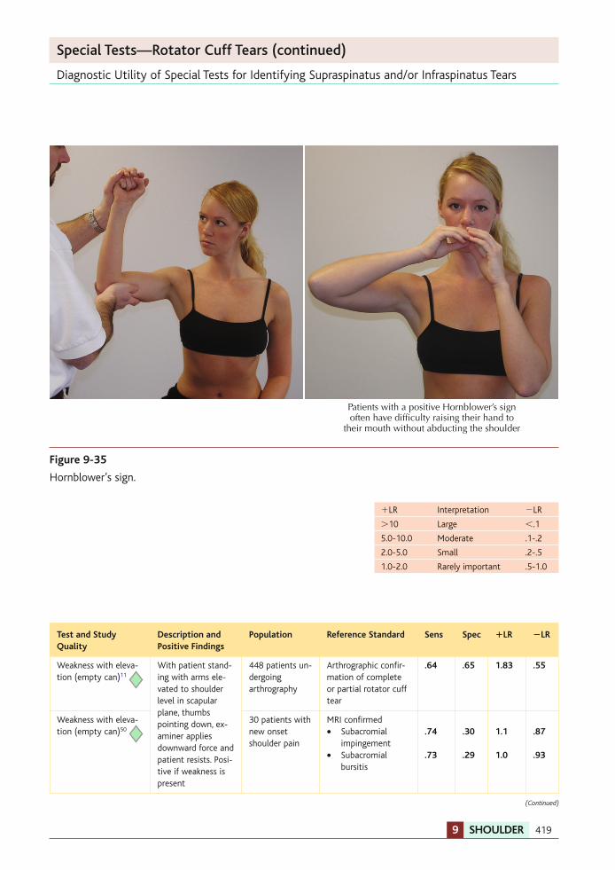
Diagnostic Utility of Special Tests for Identifying Supraspinatus and/or Infraspinatus Tears
Patients with a positive Hornblower’s signoften have difficulty raising their hand to
their mouth without abducting the shoulder
Figure 9-35
Hornblower’s sign.
Special Tests—Rotator Cuff Tears (continued)
�LR Interpretation �LR
�10 Large �.1
5.0-10.0 Moderate .1-.2
2.0-5.0 Small .2-.5
1.0-2.0 Rarely important .5-1.0
Test and Study Quality
Description and Positive Findings
Population Reference Standard Sens Spec �LR �LR
Weakness with eleva-tion (empty can) 11
With patient stand-ing with arms ele-vated to shoulder level in scapular plane, thumbs pointing down, ex-aminer applies downward force and patient resists. Posi-tive if weakness is present
448 patients un-dergoing arthrography
Arthrographic confi r-mation of complete or partial rotator cuff tear
.64 .65 1.83 .55
Weakness with eleva-tion (empty can) 50
30 patients with new onset shoulder pain
MRI confi rmed• Subacromial
impingement• Subacromial
bursitis
.74
.73
.30
.29
1.1
1.0
.87
.93
(Continued)

420 Netter’s Orthopaedic Clinical Examination: An Evidence-Based Approach
Test and Study Quality
Description and Positive Findings
Population Reference Standard Sens Spec �LR �LR
Supraspinatus muscle test 50
Examiner resists ab-duction of the arm at 90° with pa-tient’s arm neutral or internally rotated. Positive if patient gives way
30 patients with new onset shoulder pain
MRI confi rmed• Subacromial
impingement• Subacromial
bursitis
.58 .73
.20
.43
.7
1.3
2.10
.63
Supraspinatus muscle test 51
552 patients with shoulder pain
Arthroscopic visualization• All impingement• Bursitis• Partial thickness
RCT• Full thickness RCT
.44 .25 .32
.53
.90 .67 .68
.82
4.4 .80 1.0
2.9
.62 1.12 1.00
.57
Drop-arm test 51
Patient elevates fully and then slowly lowers arm. Positive if the arm suddenly drops or patient has severe pain
552 patients with shoulder pain
Arthroscopic visualization• All impingement• Bursitis• Partial thickness
RCT• Full thickness RCT
.27 .14 .14
.35
.88 .77 .78
.88
2.3 .60 .60
2.9
.83 1.12 1.10
.74
Infraspinatus muscle test (Patte test) 50
Elbow at 90° with arm neutrally rotated and ad-ducted to the trunk. Examiner then resists internal rota-tion force. Positive if patient gives way
30 patients with new-onset shoulder pain
MRI confi rmed• Subacromial
impingement• Subacromial
bursitis
.58
.73
.60
.71
1.5
2.5
.70
.38
Infraspinatus muscle test 51
552 patients with shoulder pain
Arthroscopic visualization• All impingement• Bursitis• Partial thickness
RCT• Full-thickness RCT
.42 .25 .19
.51
.90 .69 .69
.84
4.2 .80 .60
3.2
.64 1.09 1.17
.58
External rotation lag sign 54
With patient sitting, examiner holds the arm in 20° shoulder elevation (in the scapular plane), 5° from full external rotation, and 90° elbow extension. Patient maintains the position when examiner releases arm. Positive if unable to hold position
37 patients with shoulder pain
Supraspinatus or in-fraspinatus tear via ultrasound
.46 .94 7.2 (1.7, 31.0)
.60 (.40, .90)
Drop sign 54
With patient sitting, examiner holds the arm in 90° abduc-tion and full exter-nal rotation. Patient is asked to maintain the position when examiner releases arm. Positive if unable to hold position
.73 .77 3.2 (1.5, 6.7)
.30 (.20, .80)
(Continued)
9 SHOULDER 421
Test and Study Quality
Description and Positive Findings
Population Reference Standard Sens Spec �LR �LR
Passive elevation less than 170° 11
With patient supine, examiner maximally elevates shoulder
448 patients undergoing arthrography
Arthrographic confi r-mation of complete or partial rotator cuff tear
.30 .78 1.36 .90
Passive ER less than 70° 11
With patient supine with arm at side, examiner externally rotates arm
.19 .84 1.19 .96
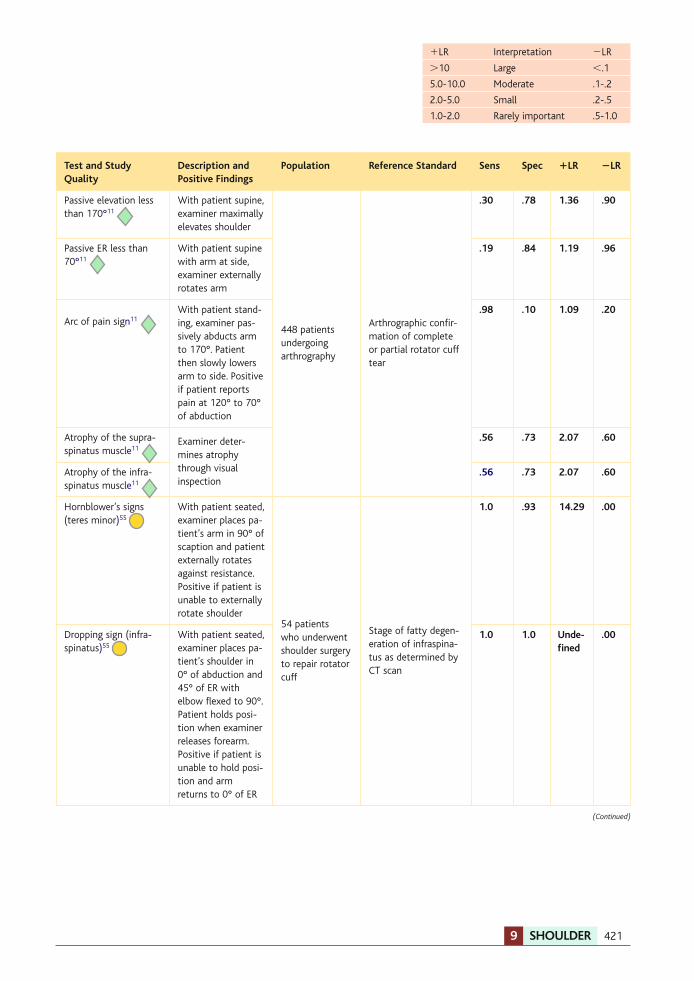
Arc of pain sign 11
With patient stand-ing, examiner pas-sively abducts arm to 170°. Patient then slowly lowers arm to side. Positive if patient reports pain at 120° to 70° of abduction
.98 .10 1.09 .20
Atrophy of the supra-spinatus muscle 11
Examiner deter-mines atrophy through visual inspection
.56 .73 2.07 .60
Atrophy of the infra-spinatus muscle 11
.56 .73 2.07 .60
Hornblower’s signs (teres minor) 55
With patient seated, examiner places pa-tient’s arm in 90° of scaption and patient externally rotates against resistance. Positive if patient is unable to externally rotate shoulder
54 patients who underwent shoulder surgery to repair rotator cuff
Stage of fatty degen-eration of infraspina-tus as determined by CT scan
1.0 .93 14.29 .00
Dropping sign (infra-spinatus) 55
With patient seated, examiner places pa-tient’s shoulder in 0° of abduction and 45° of ER with elbow fl exed to 90°. Patient holds posi-tion when examiner releases forearm. Positive if patient is unable to hold posi-tion and arm returns to 0° of ER
1.0 1.0 Unde-fi ned
.00
�LR Interpretation �LR
�10 Large �.1
5.0-10.0 Moderate .1-.2
2.0-5.0 Small .2-.5
1.0-2.0 Rarely important .5-1.0
(Continued)
422 Netter’s Orthopaedic Clinical Examination: An Evidence-Based Approach
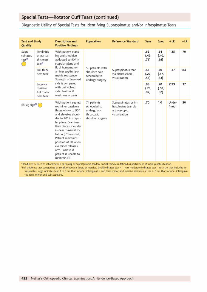
Test and Study Quality
Description and Positive Findings
Population Reference Standard Sens Spec �LR �LR
Supra-spinatus test 56
Tendinitis or partial thickness tear *
With patient stand-ing and shoulders abducted to 90° in scapular plane and IR of humerus, ex-aminer applies iso-metric resistance. Strength of involved side is compared with uninvolved side. Positive if weakness or pain
50 patients with shoulder pain scheduled to undergo surgery
Supraspinatus tear via arthroscopic visualization
.62 (.49, .75)
.54 (.40, .68)
1.35 .70
Full thick-ness tear †
.41 (.27, .55)
.70 (.57, .83)
1.37 .84
Large or massive full thick-ness tear †
.88 (.79, .97)
.70 (.58, .82)
2.93 .17
ER lag sign 57
With patient seated, examiner passively fl exes elbow to 90° and elevates shoul-der to 20° in scapu-lar plane. Examiner then places shoulder in near maximal ro-tation (5° from full). Patient maintains position of ER when examiner releases arm. Positive if patient is unable to maintain ER
74 patients scheduled to undergo ar-throscopic shoulder surgery
Supraspinatus or in-fraspinatus tear via arthroscopic visualization
.70 1.0 Unde-fi ned
.30
Special Tests—Rotator Cuff Tears (continued)
Diagnostic Utility of Special Tests for Identifying Supraspinatus and/or Infraspinatus Tears
* Tendinitis defi ned as infl ammation or fraying of supraspinatus tendon. Partial thickness defi ned as partial tear of supraspinatus tendon. † Full thickness tear categorized as small, moderate, large, or massive. Small indicates tear � 1 cm, moderate indicates tear 1 to 3 cm that includes in-
fraspinatus, large indicates tear 3 to 5 cm that includes infraspinatus and teres minor, and massive indicates a tear � 5 cm that includes infraspina-tus, teres minor, and subscapularis.
9 SHOULDER 423
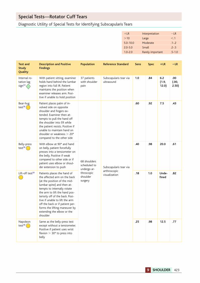
Special Tests—Rotator Cuff Tears
Diagnostic Utility of Special Tests for Identifying Subscapularis Tears
�LR Interpretation �LR
�10 Large �.1
5.0-10.0 Moderate .1-.2
2.0-5.0 Small .2-.5
1.0-2.0 Rarely important .5-1.0
Test and Study Quality
Description and Positive Findings
Population Reference Standard Sens Spec �LR �LR
Internal ro-tation lag sign 54
With patient sitting, examiner holds hand behind the lumbar region into full IR. Patient maintains the position when examiner releases arm. Posi-tive if unable to hold position
37 patients with shoulder pain
Subscapularis tear via ultrasound
1.0 .84 6.2 (1.9, 12.0)
.00 (.00, 2.50)
Bear-hug test 58
Patient places palm of in-volved side on opposite shoulder and fi ngers ex-tended. Examiner then at-tempts to pull the hand off the shoulder into ER while the patient resists. Positive if unable to maintain hand on shoulder or weakness � 20° compared to the other side
68 shoulders scheduled to undergo ar-throscopic shoulder surgery
Subscapularis tear via arthroscopic visualization
.60 .92 7.5 .43
Belly-press test 58
With elbow at 90° and hand on belly, patient forcefully presses into a tensiometer on the belly. Positive if weak compared to other side or if patient uses elbow or shoul-der extension to push
.40 .98 20.0 .61
Lift-off test 58
Patients places the hand of the affected arm on the back (at the position of the mid-lumbar spine) and then at-tempts to internally rotate the arm to lift the hand pos-teriorly off of the back. Posi-tive if unable to lift the arm off the back or if patient per-forms the lifting maneuver by extending the elbow or the shoulder
.18 1.0 Unde-fi ned
.82
Napoleon test 58
Same as the belly-press test except without a tensiometer. Positive if patient uses wrist fl exion � 30° to press into belly
.25 .98 12.5 .77

424 Netter’s Orthopaedic Clinical Examination: An Evidence-Based Approach
Negative test
Positive test
Figure 9-36
Lift-off test.
Special Tests—Rotator Cuff Tears (continued)
Diagnostic Utility of Special Tests for Identifying Subscapularis Tears
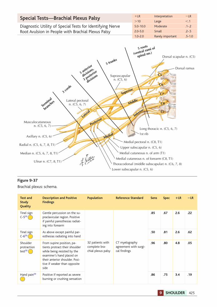
9 SHOULDER 425
Special Tests—Brachial Plexus Palsy
Diagnostic Utility of Special Tests for Identifying Nerve Root Avulsion in People with Brachial Plexus Palsy
Medial pectoral n. (C8, T1)
Medial cutaneous n. of arm (T1)
Medial cutaneous n. of forearm (C8, T1)
Upper subscapular n. (C5, 6)
Thoracodorsal (middle subscapular) n. (C6, 7, 8)
Lower subscapular n. (C5, 6)
Musculocutaneousn. (C5, 6, 7)
Axillary n. (C5, 6)
Radial n. (C5, 6, 7, 8, T1)
Median n. (C5, 6, 7, 8, T1)
Ulnar n. (C7, 8, T1)
Dorsal ramus
Long thoracic n. (C5, 6, 7)
Suprascapularn. (C5, 6)
Lateral pectoraln. (C5, 6, 7)Te
rminal
branch
es
3 cords
3 anter
ior
divisions,
3 posterio
r
divisions
3 trunks
5 roots
(ventral rami of
spinal nn.) Dorsal scapular n. (C5)
1st rib
Superior
Middle
Inferior
Later
al
Posterior
Medial
C5
C6
C7
C8
T1
Figure 9-37
Brachial plexus: schema.
�LR Interpretation �LR
�10 Large �.1
5.0-10.0 Moderate .1-.2
2.0-5.0 Small .2-.5
1.0-2.0 Rarely important .5-1.0
Test and Study Quality
Description and Positive Findings
Population Reference Standard Sens Spec �LR �LR
Tinel sign C-5 59
Gentle percussion on the su-praclavicular region. Positive if painful paresthesias radiat-ing into forearm
32 patients with complete bra-chial plexus palsy
CT myelography agreement with surgi-cal fi ndings
.85 .67 2.6 .22
Tinel sign C-6 59
As above except painful par-esthesias radiating into hand
.50 .81 2.6 .62
Shoulder protraction test 59
From supine position, pa-tients protract their shoulder while being resisted by the examiner’s hand placed on their anterior shoulder. Posi-tive if weaker than opposite side
.96 .80 4.8 .05
Hand pain 59
Positive if reported as severe burning or crushing sensation
.86 .75 3.4 .19

426 Netter’s Orthopaedic Clinical Examination: An Evidence-Based Approach
Injury to acromioclavicular joint.Usually caused by fall on tip ofshoulder, depressing acromion(shoulder separation)
Grade I. Acromioclavicularligaments stretched butnot torn; coracoclavicularligaments intact
Grade II. Acromioclavicularligaments ruptured and jointseparated; coracoclavicularligaments intact Grade III. Coracoclavicular and
acromioclavicular ligaments rupturewith wide separation of joint
Figure 9-38
Common mechanism of injury for acromioclavicular tears.
Special Tests—Acromioclavicular Lesions
Diagnostic Utility of Special Tests for Identifying Acromioclavicular Lesions
�LR Interpretation �LR
�10 Large �.1
5.0-10.0 Moderate .1-.2
2.0-5.0 Small .2-.5
1.0-2.0 Rarely important .5-1.0
Test and Study Quality
Description and Positive Findings
Population Reference Standard Sens Spec �LR �LR
O’Brien sign 60
Patient is standing. Examiner asks patient to fl ex arm to 90° with elbow in full extension. Patient then adducts arm 10° and internally rotates humerus. Examiner applies downward force to arm as patient resists. Patient fully supinates arm and repeats procedure. Positive if pain lo-calized to AC joint 1013 patients
with pain between mid clavicle and deltoid
AC joint infi ltration test: patients were in-jected with lidocaine in AC joint. Those who experienced at least a 50% reduction in symptoms within 10 minutes were consid-ered to have AC pathology
.16 .90 1.6 .93
Paxinos sign 60
Patient sits with arm by side. With one hand, examiner places thumb over posterolat-eral aspect of acromion and index fi nger superior to mid-portion of clavicle. Examiner then applies compressive force. Positive if pain is re-ported in area of AC joint
.79 .50 1.58 .42
Palpation of the AC joint 60
Not reported .96 .10 1.07 .40

9 SHOULDER 427
Combination of Tests
Diagnostic Utility of Combinations of Tests for Identifying Glenoid Labral Tears
�LR Interpretation �LR
�10 Large �.1
5.0-10.0 Moderate .1-.2
2.0-5.0 Small .2-.5
1.0-2.0 Rarely important .5-1.0
Test and Study Quality
Patient Population Reference Standard
Sens Spec � LR � LR
Pop �
crank 16
55 patients with shoulder pain sched-uled for arthroscopic surgery
Glenoid labral tear observed during arthroscopy
.27 (.14, .4)
.91 (.74, 1.08)
3.0 (.44, 20.67)
.8 (.62, 1.04)
Pop �
anterior slide 16
.16 (.05, .27)
1.0 (1.0, 1.0)
Undefi ned .84 (.74, .96)
Active compression �
Anterior slide 16
.25 (.12, .38)
.91 (.74, 1.08)
2.75 (.4, 19.09)
.83 (.64, 1.06)
Anterior slide �
Crank 16
.34 (.2, .48)
.91 (.74, 1.08)
3.75 (.55, 25.41)
.73 (.55, .96)
Crank test �
Apprehension test �
Relocation test �
Load and shift test �
Inferior sulcus sign 10
54 patients with shoulder pain
Arthroscopic visualization
.90 .85 6.0 .12
Jobe relocation �
O’Brien 27
62 shoulders sched-uled to undergo arthroscopy
As above
.41 .91 4.56 .65
Jobe relocation �
Anterior apprehension 27
.38 .93 5.43 .67
O’Brien �
Anterior apprehension 27
.38 .82 2.11 .76
Jobe �
O’Brien �
Apprehension 27
.34 .91 3.78 .73
See test descriptions under single tests.
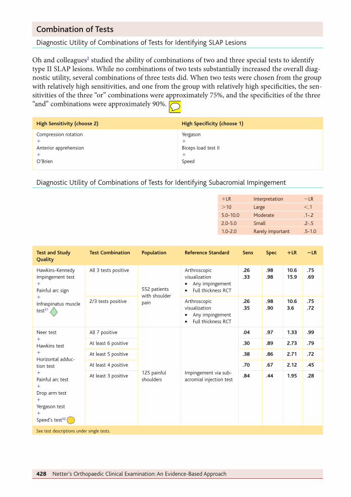
428 Netter’s Orthopaedic Clinical Examination: An Evidence-Based Approach
Combination of Tests
Diagnostic Utility of Combinations of Tests for Identifying SLAP Lesions
Oh and colleagues 2 studied the ability of combinations of two and three special tests to identify type II SLAP lesions. While no combinations of two tests substantially increased the overall diag-nostic utility, several combinations of three tests did. When two tests were chosen from the group with relatively high sensitivities, and one from the group with relatively high specifi cities, the sen-sitivities of the three “or” combinations were approximately 75%, and the specifi cities of the three “and” combinations were approximately 90%.
High Sensitivity (choose 2) High Specifi city (choose 1)
Compression rotation �
Anterior apprehension �
O’Brien
Yergason �
Biceps load test II �
Speed
Diagnostic Utility of Combinations of Tests for Identifying Subacromial Impingement
�LR Interpretation �LR
�10 Large �.1
5.0-10.0 Moderate .1-.2
2.0-5.0 Small .2-.5
1.0-2.0 Rarely important .5-1.0
Test and Study Quality
Test Combination Population Reference Standard Sens Spec �LR �LR
Hawkins-Kennedy impingement test �
Painful arc sign �
Infraspinatus muscle test 51
All 3 tests positive
552 patients with shoulder pain
Arthroscopic visualization• Any impingement• Full thickness RCT
.26 .33
.98 .98
10.6 15.9
.75 .69
2/3 tests positive Arthroscopic visualization• Any impingement• Full thickness RCT
.26 .35
.98 .90
10.6 3.6
.75 .72
Neer test �
Hawkins test �
Horizontal adduc-tion test �
Painful arc test �
Drop arm test �
Yergason test �
Speed’s test 52
All 7 positive
125 painful shoulders
Impingement via sub-acromial injection test
.04 .97 1.33 .99
At least 6 positive .30 .89 2.73 .79
At least 5 positive .38 .86 2.71 .72
At least 4 positive .70 .67 2.12 .45
At least 3 positive .84 .44 1.95 .28
See test descriptions under single tests.

9 SHOULDER 429
OUTCOME MEASURES
Outcome Measure Scoring and Interpretation Test-Retest Reliability
MCID
Upper Extremity Func-tional Index
Users are asked to rate the diffi culty of performing 20 functional tasks on a Likert-type scale ranging from 0 (extremely diffi cult or unable to perform activ-ity) to 4 (no diffi culty). A total score out of 80 is cal-culated by summing each score. The answers provide a score between 0 and 80, with lower scores repre-senting more disability
ICC � .95 61 Unknown (MDC � 9.1 ) 61
Disabilities of the Arm, Shoulder, and Hand (DASH)
Users are asked to rate the diffi culty of performing 30 functional tasks on a Likert-type scale. Twenty-one items relate to physical function, 5 items relate to pain symptoms, and 4 items related to emotional and social functioning. A total score out of 100 is calcu-lated with higher scores representing more disability
ICC � .90 62 10.2 62
Shortened Disabilities of the Arm, Shoulder, and Hand Question-naire (QuickDASH)
Users are asked to rate an 11-item questionnaire that addresses symptoms and physical function. A total score out of 100 is calculated with higher scores rep-resenting more disability
ICC � .90 63 8.0 63
Shoulder Pain and Dis-ability Index (SPADI)
Users are asked to rate their shoulder pain and disabil-ity on 13 items, each on a visual analog scale from 0 (no pain/diffi culty) to 100 (worst pain imaginable/so diffi cult requires help). Eight items relate to physi-cal function and 5 items relate to pain symptoms. A total score out of 100 is calculated with higher scores representing more disability
ICC � .89 62 13.1 62
American Shoulder and Elbow Surgeons (ASES) score
Users are asked to rate their shoulder pain on a 1-item and visual analog scale and functional ability on 10 items on a Likert-type scale ranging from 0 to 4. Pain and function are equally weighted to create a total score out of 100. Lower scores represent more pain and disability
ICC � .91 62 6.4 62
Numeric Pain Rating Scale (NPRS)
Users rate their level of pain on an 11-point scale ranging from 0 to 10, with high scores representing more pain. Often asked as “current pain” and “least,” “worst,” and “average” pain in the past 24 hours
ICC � .72 64 2 65 , 66
ICC, Intraclass correlation coeffi cient; MDC, minimal detectable change; MCID, minimum clinically important difference.
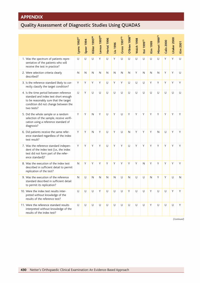
430 Netter’s Orthopaedic Clinical Examination: An Evidence-Based Approach
APPENDIX
Quality Assessment of Diagnostic Studies Using QUADAS Ly
ons
1992
67
Spee
r 19
94
Kib
ler
1995
68
Lero
ux 1
995 69
Her
tel 1
996
Liu
1996
Gro
ss 1
997 7
0
O’B
rien
199
8 71
Wal
ch 1
998
Ito
i 199
9 72
Kim
199
9
Mim
ori 1
999 73
Cal
is 2
000
Lita
ker
2000
Kim
200
1
1. Was the spectrum of patients repre-sentative of the patients who will receive the test in practice?
U U U Y U Y U U U U U U Y Y U
2. Were selection criteria clearly described?
N N N N N N N N Y N N N Y Y U
3. Is the reference standard likely to cor-rectly classify the target condition?
Y Y Y Y U Y Y U U U Y Y Y Y Y
4. Is the time period between reference standard and index test short enough to be reasonably sure that the target condition did not change between the two tests?
U Y U U U U U U U U U U U U U
5. Did the whole sample or a random selection of the sample, receive verifi -cation using a reference standard of diagnosis?
Y Y N Y U Y U Y Y Y Y Y Y Y Y
6. Did patients receive the same refer-ence standard regardless of the index test result?
Y Y N Y U Y U N Y Y Y N U Y Y
7. Was the reference standard indepen-dent of the index test (i.e., the index test did not form part of the refer-ence standard)?
Y Y Y Y U Y Y U Y Y Y Y Y Y Y
8. Was the execution of the index test described in suffi cient detail to permit replication of the test?
N Y Y Y Y Y Y Y Y N Y Y Y Y Y
9. Was the execution of the reference standard described in suffi cient detail to permit its replication?
N U N N N N U N U U N Y Y U N
10. Were the index test results inter-preted without knowledge of the results of the reference test?
U U U Y U U U Y U U Y U U Y Y
11. Were the reference standard results interpreted without knowledge of the results of the index test?
U U U U U U U U U U Y U U U Y
(Continued)

9 SHOULDER 431
Lyon
s 19
92 67
Spee
r 19
94
Kib
ler
1995
68
Lero
ux 1
995 69
Her
tel 1
996
Liu
1996
Gro
ss 1
997 7
0
O’B
rien
199
8 71
Wal
ch 1
998
Ito
i 199
9 72
Kim
199
9
Mim
ori 1
999 73
Cal
is 2
000
Lita
ker
2000
Kim
200
1
12. Were the same clinical data available when test results were interpreted as would be available when the test is used in practice?
U U U U U Y U U U U U U U Y N
13. Were uninterpretable/ intermediate test results reported?
U U U Y U U U U U U U U U Y U
14. Were withdrawals from the study explained?
U U U Y U U U U U U U U U Y U
Quality summary rating:
APPENDIX (continued)
Quality Assessment of Diagnostic Studies Using QUADAS
Y � yes, N � no, U � unclear. Good quality (Y - N � 10 to 14). Fair quality (Y - N � 5 to 9). Poor quality (Y - N � 4).
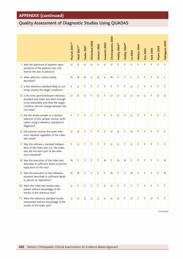
432 Netter’s Orthopaedic Clinical Examination: An Evidence-Based Approach
Mur
rell
2001
74
Wo
lf 2
001 7
5
Zas
lav
2001
McF
arla
nd 2
002
Stet
son
2002
Gua
nche
200
3
Chr
ono
poul
os
2004
Ho
ltby
200
4 44
Ho
ltby
200
4 56
Lo 2
004
Wal
ton
2004
Kim
200
5
Park
200
5
Mye
rs 2
005
Nak
agaw
a 20
05
1. Was the spectrum of patients repre-sentative of the patients who will receive the test in practice?
U U U U U U U Y Y Y Y Y Y Y Y
2. Were selection criteria clearly described?
N N N U N U N Y Y Y Y Y Y U Y
3. Is the reference standard likely to cor-rectly classify the target condition?
Y U Y Y Y Y Y Y Y U Y Y Y Y Y
4. Is the time period between reference standard and index test short enough to be reasonably sure that the target condition did not change between the two tests?
U U U Y U Y U U U U U U Y U U
5. Did the whole sample or a random selection of the sample, receive verifi -cation using a reference standard of diagnosis?
Y Y Y Y Y Y Y U Y U Y Y Y Y Y
6. Did patients receive the same refer-ence standard regardless of the index test result?
U U Y Y Y Y U U Y N Y Y Y Y Y
7. Was the reference standard indepen-dent of the index test (i.e., the index test did not form part of the refer-ence standard)?
Y U Y Y Y Y U U Y U Y Y Y Y Y
8. Was the execution of the index test described in suffi cient detail to permit replication of the test?
N Y Y Y Y N Y N N Y Y Y Y Y N
9. Was the execution of the reference standard described in suffi cient detail to permit its replication?
N N Y Y Y Y N Y Y N Y Y Y Y N
10. Were the index test results inter-preted without knowledge of the results of the reference test?
U Y Y Y Y U U Y Y Y Y Y Y Y Y
11. Were the reference standard results interpreted without knowledge of the results of the index test?
U U U U U U U U Y U Y Y U Y Y
APPENDIX (continued)
Quality Assessment of Diagnostic Studies Using QUADAS
(Continued)

9 SHOULDER 433
Mur
rell
2001
74
Wo
lf 2
001 7
5
Zas
lav
2001
McF
arla
nd 2
002
Stet
son
2002
Gua
nche
200
3
Chr
ono
poul
os
2004
Ho
ltby
200
4 44
Ho
ltby
200
4 56
Lo 2
004
Wal
ton
2004
Kim
200
5
Park
200
5
Mye
rs 2
005
Nak
agaw
a 20
05
12. Were the same clinical data available when test results were interpreted as would be available when the test is used in practice?
U U Y Y Y Y U Y U U Y U Y Y U
13. Were uninterpretable/ intermediate test results reported?
U U U U U U U U U U U U Y U U
14. Were withdrawals from the study explained?
U U U Y U Y U U U U Y U Y Y U
Quality summary rating:
APPENDIX (continued)
Quality Assessment of Diagnostic Studies Using QUADAS
Y � yes, N � no, U � unclear. Good quality (Y - N � 10 to 14). Fair quality (Y - N � 5 to 9). Poor quality (Y - N � 4).

434 Netter’s Orthopaedic Clinical Examination: An Evidence-Based Approach
Bart
h 20
06
Bert
elli
2006
Pare
ntis
200
6
Farb
er 2
006
Gill
200
7
Kim
200
7
Lew
is 2
007
Mill
er 2
008
Bush
nell
2008
Oh
2008
Silv
a 20
08
Ebin
ger
2008
Wal
swo
rth
2008
Kib
ler
2009
76
Levy
20
09
77
1. Was the spectrum of patients repre-sentative of the patients who will receive the test in practice?
Y U Y Y U Y Y Y Y Y Y Y Y U U
2. Were selection criteria clearly described?
U U Y N Y Y Y Y Y Y Y U U U U
3. Is the reference standard likely to cor-rectly classify the target condition?
Y Y Y Y Y Y N Y Y Y U Y Y Y Y
4. Is the time period between reference standard and index test short enough to be reasonably sure that the target condition did not change between the two tests?
U U U U U U Y Y U Y Y Y U U U
5. Did the whole sample or a random selection of the sample, receive verifi -cation using a reference standard of diagnosis?
Y Y Y Y Y Y Y Y Y Y Y Y Y N Y
6. Did patients receive the same refer-ence standard regardless of the index test result?
Y Y Y Y Y Y Y Y Y Y Y Y Y N Y
7. Was the reference standard indepen-dent of the index test (i.e., the index test did not form part of the refer-ence standard)?
Y Y Y Y Y Y Y Y Y Y Y Y Y Y Y
8. Was the execution of the index test described in suffi cient detail to permit replication of the test?
Y Y Y Y Y Y Y Y Y Y Y Y Y Y U
9. Was the execution of the reference standard described in suffi cient detail to permit its replication?
Y Y U Y Y Y U Y Y Y Y Y Y Y U
10. Were the index test results inter-preted without knowledge of the results of the reference test?
Y Y Y Y Y Y U Y Y Y Y Y Y Y Y
11. Were the reference standard results interpreted without knowledge of the results of the index test?
U U U U U U U Y U U Y U Y U U
APPENDIX (continued)
Quality Assessment of Diagnostic Studies Using QUADAS
(Continued)

9 SHOULDER 435
Bart
h 20
06
Bert
elli
2006
Pare
ntis
200
6
Farb
er 2
006
Gill
200
7
Kim
200
7
Lew
is 2
007
Mill
er 2
008
Bush
nell
2008
Oh
2008
Silv
a 20
08
Ebin
ger
2008
Wal
swo
rth
2008
Kib
ler
2009
76
Levy
20
09
77
12. Were the same clinical data available when test results were interpreted as would be available when the test is used in practice?
U U Y Y Y N U Y Y Y Y Y Y Y N
13. Were uninterpretable/ intermediate test results reported?
U U Y U Y Y U U U U U U Y U U
14. Were withdrawals from the study explained?
U U U U Y Y Y Y U Y Y Y Y U U
Quality summary rating:
APPENDIX (continued)
Quality Assessment of Diagnostic Studies Using QUADAS
Y � yes, N � no, U � unclear. Good quality (Y - N � 10 to 14). Fair quality (Y - N � 5 to 9). Poor quality (Y - N � 4).
436 Netter’s Orthopaedic Clinical Examination: An Evidence-Based Approach
1. Lewis JS, Valentine RE . Th e pectoralis minor length test: a study of the intra-rater reliability and diagnos-tic accuracy in subjects with and without shoulder symptoms . BMC Musculoskelet Disord . 2007 ; 8 : 64 .
2. Oh JH, Kim JY, Kim WS , et al . Th e evaluation of various physical examinations for the diagnosis of type II superior labrum anterior and posterior lesion . Am J Sports Med . 2008 ; 36 : 353 - 359 .
3. Norkin CC, Levangie PK . Th e shoulder complex . In: Joint Structure and Function: A Comprehensive Analy-sis . 2nd ed . Philadelphia : FA Davis ; 1992 . 240 - 261 .
4. Inman VT, Saunders SJB, Abbott LC . Observations on the function of the shoulder joint. 1944 . Clin Orthop . 1996 ; 330 : 3 - 12 .
5. Neumann DA . Shoulder complex . In: Kinesiology of Musculoskeletal System: Foundations for Physical Reha-bilitation . St. Louis : Mosby ; 2002 . 189 - 248 .
6. Brody LT . Shoulder. Current Concepts of Orthopaedic Physical Th erapy (11.2.6) . La Crosse, WI : Orthopaedic Section, American Physical Th erapy Association ; 2001 .
7. Michener LA, Walsworth MK, Burnet EN . Eff ective-ness of rehabilitation for patients with subacromial impingement syndrome: a systematic review . J Hand Th er . 2004 ; 17 : 152 - 164 .
8. Hartley A . Practical Joint Assessment . St Louis : Mosby ; 1995 .
9. Berg EE, Ciullo JV . A clinical test for superior glenoid labral or “SLAP” lesions . Clin J Sport Med . 1998 ; 8 : 121 - 123 .
10. Liu SH, Henry MH, Nuccion SL . A prospective evalu-ation of a new physical examination in predicting glenoid labral tears . Am J Sports Med . 1996 ; 24 : 721 - 725 .
11. Litaker D, Pioro M, El Bilbeisi H , et al . Returning to the bedside: using the history and physical examina-tion to identify rotator cuff tears . J Am Geriatr Soc . 2000 ; 48 : 1633 - 1637 .
12. Cleland J, Durall CJ . Physical therapy for adhesive capsulitis . Physiotherapy . 2002 ; 88 : 450 - 457 .
13. Rayan GM, Jensen C . Th oracic outlet syndrome: pro-vocative examination maneuvers in a typical popula-tion . J Shoulder Elbow Surg . 1995 ; 4 : 113 - 117 .
14. Winsor T, Brow R . Costoclavicular syndrome: its di-agnosis and treatment . JAMA . 2004 ; 196 : 109 - 111 .
15. Wainner RS, Gill H . Diagnosis and nonoperative management of cervical radiculopathy . J Orthop Sports Phys Th er . 2000 ; 30 : 728 - 744 .
16. Walsworth MK, Doukas WC, Murphy KP , et al . Reli-ability and diagnostic accuracy of history and physical examination for diagnosing glenoid labral tears . Am J Sports Med . 2008 ; 36 : 162 - 168 .
17. Riddle DL, Rothstein JM, Lamb RL . Goniometric reli-ability in a clinical setting. Shoulder measurements. Phys Th er . 1987 ; 67 : 668 - 673 .
18. Terwee CB, de Winter AF, Scholten RJ , et al . Interob-sever reproducibility of the visual estimation of range
of motion of the shoulder . Arch Phys Med Rehabil . 2005 ; 86 : 1356 - 1361 .
19. Yang JL, Lin JJ . Reliability of function-related tests in patients with shoulder pathologies . J Orthop Sports Phys Th er . 2006 ; 36 : 572 - 576 .
20. Nomden JG, Slagers AJ, Bergman GJ , et al . Interob-server reliability of physical examination of shoulder girdle . Man Th er . 2009 ; 14 : 152 - 159 .
21. Wang SS, Normile SO, Lawshe BT . Reliability and smallest detectable change determination for serratus anterior muscle strength and endurance tests . Physio-ther Th eory Pract . 2006 ; 22 : 33 - 42 .
22. Michener LA, Boardman ND, Pidcoe PE, Frith AM . Scapular muscle tests in subjects with shoulder pain and functional loss: reliability and construct validity . Phys Th er . 2005 ; 85 : 1128 - 1138 .
23. Dover G, Powers ME . Reliability of joint position sense and force-reproduction measures during inter-nal and external rotation of the shoulder . J Athl Train . 2003 ; 38 : 304 - 310 .
24. Borstad JD, Mathiowetz KM, Minday LE , et al . Clini-cal measurement of posterior shoulder fl exibility . Man Th er . 2007 ; 12 : 386 - 389 .
25. Boyd EA, Torrance GM . Clinical measures of shoul-der subluxation: their reliability . Can J Public Health . 1992 ; 83 ( Suppl 2 ): S24 - S28 .
26. Gill HS, El Rassi G, Bahk MS , et al . Physical examina-tion for partial tears of the biceps tendon . Am J Sports Med . 2007 ; 35 : 1334 - 1340 .
27. Guanche CA, Jones DC . Clinical testing for tears of the glenoid labrum . Arthroscopy . 2003 ; 19 : 517 - 523 .
28. Nakagawa S, Yoneda M, Hayashida K , et al . Forced shoulder abduction and elbow fl exion test: a new simple clinical test to detect superior labral injury in the throwing shoulder . Arthroscopy . 2005 ; 21 : 1290 - 1295A .
29. Nijs J, Roussel N, Vermeulen K , et al . Scapular posi-tioning in patients with shoulder pain: a study examining the reliability and clinical importance of 3 clinical tests . Arch Phys Med Rehabil . 2005 ; 86 : 1349 - 1355 .
30. Odom CJ, Taylor AB, Hurd CE , et al . Measurement of scapular asymmetry and assessment of shoulder dys-function using the Lateral Scapular Slide Test: a reli-ability and validity study . Phys Th er . 2001 ; 81 : 799 - 809 .
31. Kibler WB, Uhl TL, Maddux JW , et al . Qualitative clinical evaluation of scapular dysfunction: A reliabil-ity study . J Shoulder Elbow Surg . 2002 ; 11 : 550 - 556 .
32. Hanchard NC, Howe TE, Gilbert MM . Diagnosis of shoulder pain by history and selective tissue tension: agreement between assessors . J Orthop Sports Phys Th er . 2005 ; 35 : 147 - 153 .
33. de Winter AF, Jans MP, Scholten RJ , et al . Diagnostic classifi cation of shoulder disorders: interobserver agreement and determinants of disagreement . Ann Rheum Dis . 1999 ; 58 : 272 - 277 .
REFERENCES
9 SHOULDER 437
34. Levy AS, Lintner S, Kenter K , et al . Intra- and in-terobserver reproducibility of the shoulder laxity ex-amination . Am J Sports Med . 1999 ; 27 : 460 - 463 .
35. Bushnell BD, Creighton RA, Herring MM . Th e bony apprehension test for instability of the shoulder: a prospective pilot analysis . Arthroscopy . 2008 ; 24 : 974 - 982 .
36. Farber AJ, Castillo R, Clough M , et al . Clinical assessment of three common tests for traumatic anterior shoulder instability . J Bone Joint Surg Am . 2006 ; 88 : 1467 - 1474 .
37. Speer KP, Hannafi n JA, Altchek DW, Warren RF . An evaluation of the shoulder relocation test . Am J Sports Med . 1994 ; 22 : 177 - 183 .
38. Parentis MA, Glousman RE, Mohr KS , et al . An eval-uation of the provocative tests for superior labral an-terior posterior lesions . Am J Sports Med . 2006 ; 34 : 265 - 268 .
39. Myers TH, Zemanovic JR, Andrews JR . Th e resisted supination external rotation test: a new test for the di-agnosis of superior labral anterior posterior lesions . Am J Sports Med . 2005 ; 33 : 1315 - 1320 .
40. Stetson WB, Templin K . Th e crank test, the O’Brien test, and routine magnetic resonance imaging scans in the diagnosis of labral tears . Am J Sports Med . 2002 ; 30 : 806 - 809 .
41. McFarland EG, Kim TK, Savino RM . Clinical assess-ment of three common tests for superior labral anterior-posterior lesions . Am J Sports Med . 2002 ; 30 : 810 - 815 .
42. Hegedus EJ, Goode A, Campbell S , et al . Physical ex-amination tests of the shoulder: a systematic review with meta-analysis of individual tests . Br J Sports Med . 2008 ; 42 : 80 - 92 ; discussion 92 .
43. Ebinger N, Magosch P, Lichtenberg S, Habermeyer P . A new SLAP test: the supine fl exion resistance test . Arthroscopy . 2008 ; 24 : 500 - 505 .
44. Holtby R, Razmjou H . Accuracy of the Speed’s and Yergason’s tests in detecting biceps pathology and SLAP lesions: comparison with arthroscopic fi ndings . Arthroscopy . 2004 ; 20 : 231 - 236 .
45. Kim YS, Kim JM, Ha KY , et al . Th e passive compres-sion test: a new clinical test for superior labral tears of the shoulder . Am J Sports Med . 2007 ; 35 : 1489 - 1494 .
46. Kim SH, Park JS, Jeong WK , et al . Th e Kim test: a novel test for posteroinferior labral lesion of the shoulder—a comparison to the jerk test . Am J Sports Med . 2005 ; 33 : 1188 - 1192 .
47. Kim SH, Ha KI, Ahn JH , et al . Biceps load test II: a clinical test for SLAP lesions of the shoulder . Arthros-copy . 2001 ; 17 : 160 - 164 .
48. Kim SH, Ha KI, Han KY . Biceps load test: a clinical test for superior labrum anterior and posterior lesions in shoulders with recurrent anterior dislocations . Am J Sports Med . 1999 ; 27 : 300 - 303 .
49. Johansson K, Ivarson S . Intra- and interexaminer reliability of four manual shoulder maneuvers used to identify subacromial pain . Man Th er . 2009 ; 14 : 231 - 239 .
50. Silva L, Andreu JL, Munoz P et al . Accuracy of physi-cal examination in subacromial impingement syn-drome . Rheumatology (Oxford) . 2008 ; 47 : 679 - 683 .
51. Park HB, Yokota A, Gill HS , et al . Diagnostic accuracy of clinical tests for the diff erent degrees of subacro-mial impingement syndrome . J Bone Joint Surg Am . 2005 ; 87 : 1446 - 1455 .
52. Calis M, Akgun K, Birtane M , et al . Diagnostic values of clinical diagnostic tests in subacromial impinge-ment syndrome . Ann Rheum Dis . 2000 ; 59 : 44 - 47 .
53. Zaslav KR . Internal rotation resistance strength test: a new diagnostic test to diff erentiate intra-articular pa-thology fropm outlet (Neer) impingement syndrome in the shoulder . J Shoulder Elbow Surg . 2001 ; 10 : 23 - 27 .
54. Miller CA, Forrester GA, Lewis JS . Th e validity of the lag signs in diagnosing full-thickness tears of the rotator cuff : a preliminary investigation . Arch Phys Med Rehabil . 2008 ; 89 : 1162 - 1168 .
55. Walch G, Boulahia A, Calderone S , et al . Th e “drop-ping” and “hornblower’s” signs in evaluation of rotator-cuff tears . J Bone Joint Surg Br . 1998 ; 80 : 624 - 628 .
56. Holtby R, Razmjou H . Validity of the supraspinatus test as a single clinical test in diagnosing patients with rotator cuff pathology . J Orthop Sports Phys Th er . 2004 ; 34 : 194 - 200 .
57. Hertel R, Ballmer FT, Lombert SM, Gerber C . Lag signs in the diagnosis of rotator cuff rupture . J Shoul-der Elbow Surg . 1996 ; 5 : 307 - 313 .
58. Barth JR, Burkhart SS, De Beer JF . Th e bear-hug test: a new and sensitive test for diagnosing a subscapularis tear . Arthroscopy . 2006 ; 22 : 1076 - 1084 .
59. Bertelli JA, Ghizoni MF . Use of clinical signs and computed tomography myelography fi ndings in de-tecting and excluding nerve root avulsion in complete brachial plexus palsy . J Neurosurg . 2006 ; 105 : 835 - 842 .
60. Walton J, Mahajan S, Paxinos A , et al . Diagnostic values of tests for acromioclavicular joint pain . J Bone Joint Surg Am . 2004 ; 86-A : 807 - 812 .
61. Stratford PW, Binkley JM, Stratford DM . Develop-ment and initial validation of the upper extremity functional index . Physiotherapy Canada . 2001 ; 53 : 259 - 263 .
62. Roy JS, MacDermid JC, Woodhouse LJ . Measuring shoulder function: a systematic review of four ques-tionnaires . Arthritis Rheum . 2009 ; 61 : 623 - 632 .
63. Mintken PE, Glynn P, Cleland JA . Psychometric prop-erties of the shortened disabilities of the Arm, Shoul-der, and Hand Questionnaire (QuickDASH) and Numeric Pain Rating Scale in patients with shoulder pain . J Shoulder Elbow Surg . 2009
64. Li L, Liu X, Herr K . Postoperative pain intensity as-sessment: a comparison of four scales in Chinese adults . Pain Med . 2007 ; 8 : 223 - 234 .
65. Farrar JT, Berlin JA, Strom BL . Clinically important changes in acute pain outcome measures: a validation study . J Pain Symptom Manage . 2003 ; 25 : 406 - 411 .
66. Farrar JT, Portenoy RK, Berlin JA , et al . Defi ning the clinically important diff erence in pain outcome mea-sures . Pain . 2000 ; 88 : 287 - 294 .

438 Netter’s Orthopaedic Clinical Examination: An Evidence-Based Approach
67. Lyons AR, Tomlinson JE . Clinical diagnosis of tears of the rotator cuff . J Bone Joint Surg Br . 1992 ; 74 : 414 - 415 .
68. Kibler WB . Specifi city and sensitivity of the anterior slide test in throwing athletes with superior glenoid labral tears . Arthroscopy . 1995 ; 11 : 296 - 300 .
69. Leroux JL, Th omas E, Bonnel F, Blotman F . Diagnos-tic value of clinical tests for shoulder impingement syndrome . Rev Rhum Engl Ed . 1995 ; 62 : 423 - 428 .
70. Gross ML, Distefano MC . Anterior release test. A new test for occult shoulder instability . Clin Orthop Relat Res . 1997 ; 105 - 108 .
71. O’Brien SJ, Pagnani MJ, Fealy S , et al . Th e active com-pression test: a new and eff ective test for diagnosing labral tears and acromioclavicular joint abnormality . Am J Sports Med . 1998 ; 26 : 610 - 613 .
72. Itoi E, Kido T, Sano A , et al . Which is more useful, the “full can test” or the “empty can test” in detecting the torn supraspinatus tendon? Am J Sports Med . 1999 ; 27 : 65 - 68 .
73. Mimori K, Muneta T, Nakagawa T, Shinomiya K . A new pain provocation test for superior labral tears of the shoulder . Am J Sports Med . 1999 ; 27 : 137 - 142 .
74. Murrell GA, Walton JR . Diagnosis of rotator cuff tears . Lancet . 2001 ; 357 : 769 - 770 .
75. Wolf EM, Agrawal V . Transdeltoid palpation (the rent test) in the diagnosis of rotator cuff tears . J Shoulder Elbow Surg . 2001 ; 10 : 470 - 473 .
76. Kibler WB, Sciascia AD, Hester P , et al . Clinical utility of traditional and new tests in the diagnosis of biceps tendon injuries and superior labrum anterior and posterior lesions in the shoulder . Am J Sports Med . 2009 ; 37 : 1840 - 1847 .
77. Levy O, Relwani JG, Mullett H , et al . Th e active ele-vation lag sign and the triangle sign: new clinical signs of trapezius palsy . J Shoulder Elbow Surg . 2009 ; 18 : 573 - 576 .