shoulder arthroscopic technique for performing remplissage ... med... · arthroscopic technique for...
TRANSCRIPT

*smith&nephew
KNEE
HIP
SHOULDER
EXTREMITIES
SHOULDER TECHNIQUE GUIDE
Larry D. Field, MD
Arthroscopic Technique for Performing Remplissage using the HEALICOIL™ PK Suture Anchor
Arthroscopic Technique for Performing Remplissage using the HEALICOIL™ PK Suture Anchor
As Described by:Larry D. Field, MD
Director, Upper Extremity Service Mississippi Sports Medicine and Orthopaedic Center Jackson, MS
The following technique guide was prepared under the guidance of Larry Field, MD. Created under close collaboration with the surgeon, it contains a summary of medical techniques and opinions based upon his training and expertise in the field, along with his knowledge of Smith & Nephew’s products. Smith and Nephew does not provide medical advice and recommends that surgeons exercise their own professional judgement when determining a patient’s course of treatment. This guide is presented for educational purposes only. Prior to performing this technique, or utilizing any product referenced herein, please conduct a thorough review of each product’s indications, contraindications, warnings, precautions and instructions as detailed in the Instructions for Use provided with the individual components.
Table of ContentsIntroduction ...................................................................................... 3
Surgical Technique ........................................................................... 5
Step 1: Patient Set-up and Arthroscopic Assessment .................. 5
Step 2: Debridement ..................................................................... 6
Step 3: Anchor Placement..............................................................7
Step 4: Suture Passing ...................................................................7
Step 5: Bankhart Repair ................................................................ 8
Step 6: Remplissage Suture Tying ................................................. 8
Postoperative Management ............................................................ 9
References .......................................................................................10
3Technique Guide 06979 V1
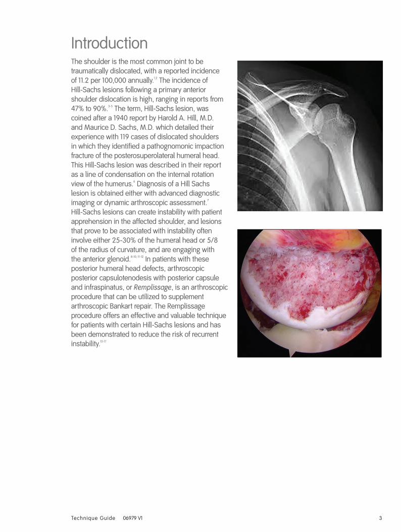
IntroductionThe shoulder is the most common joint to be traumatically dislocated, with a reported incidence of 11.2 per 100,000 annually.1,2 The incidence of Hill-Sachs lesions following a primary anterior shoulder dislocation is high, ranging in reports from 47% to 90%.3-5 The term, Hill-Sachs lesion, was coined after a 1940 report by Harold A. Hill, M.D. and Maurice D. Sachs, M.D. which detailed their experience with 119 cases of dislocated shoulders in which they identified a pathognomonic impaction fracture of the posterosuperolateral humeral head. This Hill-Sachs lesion was described in their report as a line of condensation on the internal rotation view of the humerus.6 Diagnosis of a Hill Sachs lesion is obtained either with advanced diagnostic imaging or dynamic arthroscopic assessment.7 Hill-Sachs lesions can create instability with patient apprehension in the affected shoulder, and lesions that prove to be associated with instability often involve either 25-30% of the humeral head or 5/8 of the radius of curvature, and are engaging with the anterior glenoid.8-10, 11-12 In patients with these posterior humeral head defects, arthroscopic posterior capsulotenodesis with posterior capsule and infraspinatus, or Remplissage, is an arthroscopic procedure that can be utilized to supplement arthroscopic Bankart repair. The Remplissage procedure offers an effective and valuable technique for patients with certain Hill-Sachs lesions and has been demonstrated to reduce the risk of recurrent instability.13-17

4 Technique Guide 06979 V1
Patient Positioning / Portal PlacementIt is the author’s preference to postion the patient in a lateral decubitus position when managing Hill-Sachs lesions, although the procedure can be performed in the beach chair position as well if preferred. The patient is tilted posteriorly 30° to the vertical plane rather than “straight” lateral decubitus. The arm is then prepped and draped in sterile fasion and suspended in approximately 10-15 pounds of balanced suspension during the procedure. Standard arthroscopic portals are used and are further discussed below in the surgical technique.

5Technique Guide 06979 V1
Figure 1a
Figure 1b
Figure 1c
Surgical TechniqueThe surgical technique is composed of 6 surgical steps, which includes a Bankart repair.
Step 1: Patient Set-up and Arthroscopic Assessment
1a. Set-up (Figure 1a)
The operative shoulder should be evaluated while supine on the operating room table for stability and range of motion while focusing on the position of the arm when the humeral head begins to engage or sub lux anteriorly to glenoid; this is compared with the contralateral extremity. The author prefers the lateral decubitus position for the patient, with the arm suspended in balanced traction and the shoulder tilted approximately 30 degrees posterior from the vertical plane thus improving access to the anterior shoulder during Bankart repair.
1b. Posterior Portal (Figure 1b)
Standard technique for posterior portal placement is used, placing the portal 2cm inferior and 2cm medial to the posterolateral corner of the acromion, in the soft spot.
1c. Diagnostic Arthroscopy (Figure 1c)
After portals are established, diagnostic arthroscopy is begun, paying close attention for the presence of a Hill-Sachs lesion while noting the size, location, and orientation.

6 Technique Guide 06979 V1
Figure 2
1d. Dynamic Arthroscopic Assessment (Figure 1d)
The arm is then removed from traction for the dynamic assessment of the shoulder which should include taking the shoulder from the neutral position into external rotation at 0°, 45° and 90° of abduction, and an anterior drawer test is performed while the glenohumeral joint is viewed from the posterior portal. (Anterior portal can be used for dynamic assessment, as well). Engaging Hill-Sachs lesions are an indication to proceed with remplissage in addition to Bankart procedure. After completion of the dynamic assessment, a standard anterior portal is established using standard technique while viewing through the posterior portal.
NOTE: A second more inferiorly located posterior portal may be required to adequately access the humeral head defect for anchor placement and suture passage later in procedure.
Step 2: Debridement (Figure 2)
Debridement of the Hill-Sachs lesion must be performed for complete evaluation of lesion. Debridement is carried out with an arthroscopic shaver making sure to remove any unnecessary tissue present in the defect. This is performed through the posterior portal while viewing from the anterior portal. Debridement is also performed to help stimulate the bony bed to assist in healing of the soft tissues.
Figure 1d

7Technique Guide 06979 V1
Figure 3
Figure 4
Step 3: Anchor Placement (Figure 3)
After debridement is performed, complete visualization of the Hill Sachs lesion is now possible and attention is taken to appropriate placement of anchors. One or two HEALICOIL™ PK 5.5mm double loaded suture anchors are typically used by author, depending on the size of lesion. Anchors are placed immediately adjacent to articular cartilage of the posterior humeral head defect
Step 4: Suture Passing (Figure 4)
With the use of a retrograde suture passer, all suture limbs are then passed sequentially form inferior to superior through the capsule and infraspinatus along the axis of Hill-Sachs Lesion. All sutures used for the remplissage procedure are left untied until completion of the Bankart repair.
NOTE: It is very important to complete Remplissage anchor placement and suture passage through to posterior capsule and infraspinatus prior to repair of the Bankart lesion since re-tensioning of the anterior ligamentous structures that occurs with Bankart repair results in significant posterior humeral head translation. This obligate posterior humeral head shift dramatically reduces the "working space" available to perform the Remplissage procedure within the posterior glenohumeral joint space adjacent to the Hill-Sachs lesion.
HEALICOIL™ PK Suture Anchor Ref. #72203379

8 Technique Guide 06979 V1
Figure 5
Step 5: Bankhart Repair (Figure 5)
After completion of posterior anchor placement and suture passing, 2 additional portals are made for the Bankart repair, the first including a mid-anterior portal, placed immediately superior to subscapularis tendon with an appropriate angle for placing anchors into the glenoid rim. The second portal is an anterior superior portal, which is used for viewing while a soft tissue elevator is inserted through the mid anterior portal and used to elevate and remove any scarred capsular or labral tissues that may inhibit adequate reattachment of the labrum. Typically, 3 or 4 SUTUREFIX Ultra double loaded suture anchors are used, depending on size of lesion, and spaced between the 5:30 and 1 o’clock positions and the 6:30 to 11 o’clock positions, for right and left shoulders respectively. Anchor placement and suture tying is started inferiorly and moved superiorly along glenoid rim. While passing suture limbs, they are placed in a more inferior position to corresponding anchor to help restore adequate tissue tension on the labrum. Often with the labral repair, the author includes anteroinferior capsular tissue when passing sutures providing a capsular imbrication as well as superior shift.
Step 6: Remplissage Suture Tying (Figure 6)
After Completion of the Bankart repair, the Remplissage sutures are now tied while viewing form the anterior portal. This is performed by retrieving the sutures through a cannula in the posterior portal. After sutures are retrieved, they are blindly tied through the cannula but only after the cannula is advanced down to the infraspinatus fascia. Alternatively, direct visualization of suture tying can be performed by accessing the subacromial space and visualizing the process directly. The author prefers a sliding knot with 3 alternating half hitches for knot tying, and this is repeated with all suture limbs. After all sutures are tied, arthroscopic evaluation of glenohumeral joint stability is then performed to confirm appropriate glenohumeral joint balance and centering.
SUTUREFIX ULTRA Suture Anchor Ref. # 72203854
Figure 6
9Technique Guide 06979 V1
Postoperative ManagementPatient is placed into sling with abduction pillow prior to leaving operative room. Rehab begins with passive pendulum exercises for first two weeks while maintaining active motion distally to shoulder, instructions given to patient to perform at home. At 2-6 weeks, formal physical therapy under supervision is begun and passive elevation to 90° with passive external rotation to 30° is added to the pendulum exercises already being performed. Strengthening at 2 weeks begins with scapular engagement exercises with assistance and education at therapy. Typically at 6 weeks, active motion is progressed in all patients with progression from isometric strengthening to more dynamic exercise including periscapular and rotator cuff strengthening. By 12 weeks, patients should have obtained full ROM and now begin functional strengthening in multiplanar positions.18 The author maintains and recommends a close working relationship with your physical therapist so patient protocols can be adjusted as needed.
The views and opinions expressed for postoperative care are solely those of the surgeon(s) and do not reflect the views of Smith & Nephew, Inc. In no event shall Smith & Nephew, Inc., be liable for any damages whatsoever (including, without limitation, damages for loss of business profits, business interruption, loss of business information, or other pecuniary loss) arising out of the use of or inability to use the expressed views.
10 Technique Guide 06979 V1
References1. Field, LD, Stiefel, EC: Remplissage: an arthroscopic technique for management of the engaging Hill-Sachs
lesion, Oper Tech Sports Med 21:232-237, 2013.
2. Simonet WT, Melton LJ, Cofield RH, et al: Incidence of anterior shoulder dislocation in Olmstead County, Minnesota. Clin Orthop Relat Res 186: 186-191, 1984
3. Taylor DC, Arciero RA: Pathologic changes associated with shoulder dislocations. Arthroscopic and physical examination findings in first-time, traumatic anterior dislocations. Am J Sports Med 25: 306-311, 1997.
4. Widjaja AB, Tran A, Bailey M, Proper S: Correlation between Bankart and Hill-sachs lesions in anterior shoulder dislocation. ANZ J Surg 76:436-438, 2006.
5. Calandra JJ, Baker CL, Uribe J: The incidence of Hill-sachs lesions in initial anterior shoulder dislocations. Arthroscopy 5: 254-257, 1989.
6. Hill HA, Sachs MD: The grooved defect of the humeral head: a frequently unrecognized complication of dislocations of the shoulder joint. Radiology 35: 690-700, 1940.
7. Kodali P, Jones MH, Polster J, et al: Accuracy of measurement of Hill-sachs lesions with computed tomography. 20: 1328-1334, 2011.
8. Hovelius L: Anterior dislocation of the shoulder in teenager and young adults.Fiver year prognosis. J Bone Joint Surg 69: 393-399, 1987.
9. Hovelius L, Augustini BG, Fredin H, et al: Primary anterior dislocation of the shoulder in younger patients. J Bone Joint Surg 78: 1677-1684, 1996.
10. Hovelius L, Eriksson K, Fredin H, et al: Recurrences after initial dislocation of the shoulder. J Bone Joint Surg 65: 343-349, 1983.
11. Burkhart SS, Danaceau SM: Articular arc length mismath as a cause of failed bankart repair. Arthroscopy 16: 740-744, 2000.
12. Burkhart SS, DeBeer JF: Traumatic glenohumeral bone defects and their relationship to failure of arthroscopic Bankart repairs: significance of the inverted-pear glenoid and the humeral engaging Hill-sachs lesion. Arthroscopy 16: 677-694, 2000.
13. Kirkley A, Werstine R, Ratjek A, Griffin A: Prospective randomized clinical trial comparing the effectiveness of immediiatearthrosocpic stabilization versus immobilization and rehabilitation in first traumatic anterior dislocations of the shoulder: long term evaluation. Arthroscopy 21:55-63, 2005.
14. Franceschi F, Papalia R, Rizzello G, et al: Remplissage repair-new frontiers in the prevention of recurrent shoulder instability: a 2 years follow up comparative study. Am J Sports Med 40: 2462-2469, 2012.
15. Boileau P, O'Shea K, Vargas P, et al: Anatomical and functional results after arthroscopic HIll-sachsremplissage. J Bone Join Surg 94: 618-626, 2012.
16. Zhu YM, Lu Y, Zhang J, et al: Arthroscopic bankart repair combines with remplissage technique for the treatment of anterior shoulder instability with engaging Hill-sachs lesion: a report of 49 cases with a minimum 2 year follow-up. Am J Sports Med 39: 1640-1647, 2011.
17. Nourissat G, Kilinc As, Werther JR, et al: A prospective, comparative, radiological, and clinical study of the influence of the "remplissage" procedure on shoulder range of motion after stabilization by arthroscopic Bankart repair. Am J Sports Med 39: 2147-2152, 2011.
18. Tokish JM, Abrams JS: Arthroscopic Managementof Anterior Instability in Patients with Moderate Humeral Bone Loss: The Remplissage Technique. AAOS Advanced Reconstruction: Shoulder 2 Chapter 10; 97-109, 2016.
11Technique Guide 06979 V1 Technique Guide 06311 V1
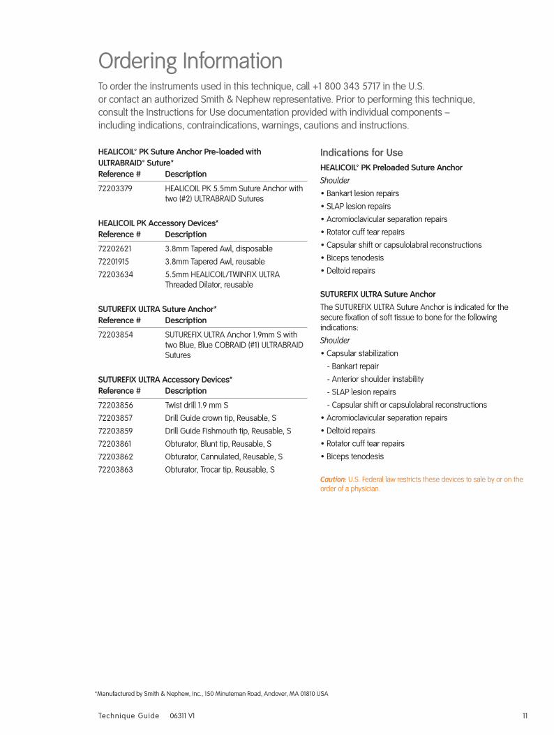
HEALICOIL™ PK Suture Anchor Pre-loaded with ULTRABRAID™ Suture* Reference # Description
72203379 HEALICOIL PK 5.5mm Suture Anchor with two (#2) ULTRABRAID Sutures
HEALICOIL PK Accessory Devices* Reference # Description
72202621 3.8mm Tapered Awl, disposable
72201915 3.8mm Tapered Awl, reusable
72203634 5.5mm HEALICOIL/TWINFIX ULTRA Threaded Dilator, reusable
SUTUREFIX ULTRA Suture Anchor* Reference # Description
72203854 SUTUREFIX ULTRA Anchor 1.9mm S with two Blue, Blue COBRAID (#1) ULTRABRAID Sutures
SUTUREFIX ULTRA Accessory Devices* Reference # Description
72203856 Twist drill 1.9 mm S
72203857 Drill Guide crown tip, Reusable, S
72203859 Drill Guide Fishmouth tip, Reusable, S
72203861 Obturator, Blunt tip, Reusable, S
72203862 Obturator, Cannulated, Reusable, S
72203863 Obturator, Trocar tip, Reusable, S
Indications for UseHEALICOIL™ PK Preloaded Suture Anchor Shoulder
• Bankart lesion repairs
• SLAP lesion repairs
• Acromioclavicular separation repairs
• Rotator cuff tear repairs
• Capsular shift or capsulolabral reconstructions
• Biceps tenodesis
• Deltoid repairs
SUTUREFIX ULTRA Suture Anchor The SUTUREFIX ULTRA Suture Anchor is indicated for the secure fixation of soft tissue to bone for the following indications:
Shoulder
• Capsular stabilization
- Bankart repair
- Anterior shoulder instability
- SLAP lesion repairs
- Capsular shift or capsulolabral reconstructions
• Acromioclavicular separation repairs
• Deltoid repairs
• Rotator cuff tear repairs
• Biceps tenodesis
Caution: U.S. Federal law restricts these devices to sale by or on the order of a physician.
Ordering InformationTo order the instruments used in this technique, call +1 800 343 5717 in the U.S. or contact an authorized Smith & Nephew representative. Prior to performing this technique, consult the Instructions for Use documentation provided with individual components – including indications, contraindications, warnings, cautions and instructions.
*Manufactured by Smith & Nephew, Inc., 150 Minuteman Road, Andover, MA 01810 USA

Smith & Nephew, Inc.150 Minuteman RoadAndover, MA 01810 USA
www.smith-nephew.comT +1 978 749 1000 US Customer Service: +1 800 343 5717
™ Trademark of Smith & Nephew. ©2017 Smith & Nephew. All rights reserved. Printed in USA. 06979 V1 4/17
Supporting healthcare professionals for over 150 years