signa explorer - imv-imaging.co.uk · peak amplitude 33 mt/m per axis peak slew rate 120 t/m/s per...
TRANSCRIPT
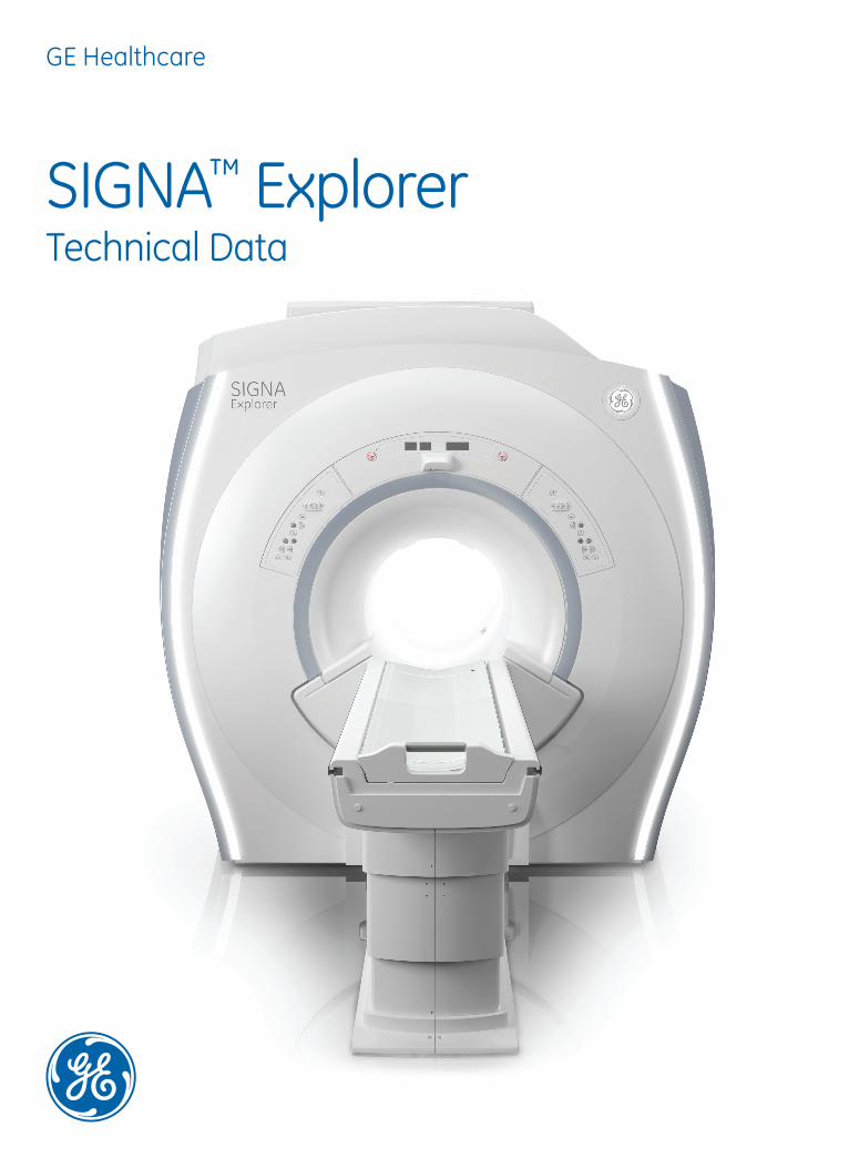
GE Healthcare
SIGNA™ Explorer Technical Data

ContentsMagnet ................................................................................................................................................................ 3
Patient Table ...................................................................................................................................................... 4
Gradient System ............................................................................................................................................... 5
Host computer and Recon engine ................................................................................................................. 6
RF Transmit and Receive ................................................................................................................................. 7
RF Coils and arrays ........................................................................................................................................... 8
Imaging and display parameters .................................................................................................................. 9
Workflow .......................................................................................................................................................... 11
Parallel Imaging† ............................................................................................................................................. 13
Post processing ............................................................................................................................................... 14
ScanTools ........................................................................................................................................................ 15
Neuro Imaging† ............................................................................................................................................... 18
The Silent Neuro Exam Package .................................................................................................................. 19
Body and Breast Imaging† ............................................................................................................................. 20
Orthopedic Imaging† ...................................................................................................................................... 22
Cardiac and Vascular Imaging† ..................................................................................................................... 23
Pediatric† Imaging‡ ......................................................................................................................................... 25
Site Planning and power consumption ....................................................................................................... 26

MagnetThe cornerstone of MRIThe SIGNA Explorer system features the stable, short-bore, highly homogeneous superconducting CXK4 magnet.
High HomogeneityThe homogeneity of the magnet contributes to consistently achieving high image quality in applications such as:
• Large FOV imaging, up to 50 cm x 50 cm x 50 cm
• Off-center imaging such as Liver, Shoulders and dual-breasts.
• Robust and dependable Fat saturation and
• Other advanced applications such as tumor staging-screening, Diffusion EPI, Tensor Imaging and Spectroscopy
Operating Field 1.5 Tesla
Shim coils 18 superconducting coils
Magnet shielding Active Shield with 3 Linear shim channels
EMI shielding 99% shielding factor
Size (L x W x H) with enclosures in cm
195 x 251 x 238
Magnet cooling Liquid Helium only
Temporal field stability < 0.1 ppm/hr
Long term magnet stability < 0.1 ppm
Manufacturer GE Healthcare
Cryogen Tank 2,000 Lit
Boil off rate Zero boil off Under normal operating conditions
Cryogen refill period Typically 4 years
Patient comfort features:The SIGNA Explorer magnet enclosures are designed for patient comfort.
• Dual-flared patient bore with 105 cm flare in the front
• 45.7 cm spacious vertical space between the table-top and the gantry inside the bore
• In-bore lighting and ventilation
• Two-way In-bore Intercom system
• Feet-first positioning
• Laser alignments for axial and sagittal reference planes
• Dual sided controls
‡ Volume Root–Mean–Square (V – RMS method with 24 measurements in each of 13 planes
Large Volume Root–Mean–Square LV – RMS is the most rigerous method with over 173,000 measurement collected over spherical volume. LV RMS measurements make use of the entire imaging chain.
Magnet Homogeneity‡
V-RMS Homogeneity Specifications
Diameter of Spherical Volume -DSV
Specified Minimum ppm
Typical ppm
10 cm < 0.02 < 0.004
20 cm < 0.06 < 0.02
30 cm < 0.14 < 0.06
40 cm < 0.35 < 0.27
45 cm < 0.97 < 0.81
48 cm < 2.00 < 1.65
LV-RMS HomogencityDSV (Diametrical Spherical Volume)
Specified Minimum ppm
Typical ppm
10 cm < 0.05 < 0.02520 cm < 0.25 < 0.0530 cm < 0.50 < 0.2540 cm < 1.00 < 0.5045 cm < 1.23 < 0.6348 cm < 2.00 < 0.95
3
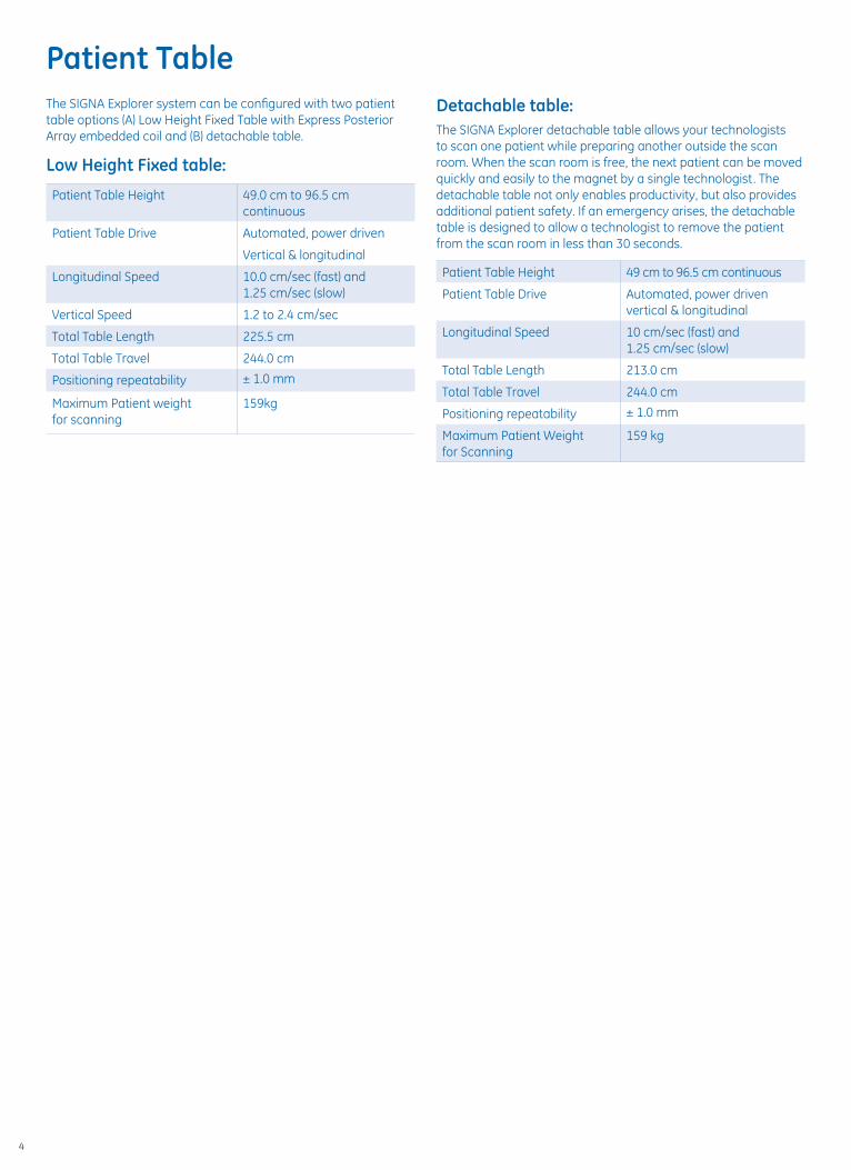
Detachable table:The SIGNA Explorer detachable table allows your technologists to scan one patient while preparing another outside the scan room. When the scan room is free, the next patient can be moved quickly and easily to the magnet by a single technologist. The detachable table not only enables productivity, but also provides additional patient safety. If an emergency arises, the detachable table is designed to allow a technologist to remove the patient from the scan room in less than 30 seconds.
Patient Table Height 49 cm to 96.5 cm continuous
Patient Table Drive Automated, power driven vertical & longitudinal
Longitudinal Speed 10 cm/sec (fast) and 1.25 cm/sec (slow)
Total Table Length 213.0 cm
Total Table Travel 244.0 cm
Positioning repeatability ± 1.0 mm
Maximum Patient Weight for Scanning
159 kg
Patient TableThe SIGNA Explorer system can be configured with two patient table options (A) Low Height Fixed Table with Express Posterior Array embedded coil and (B) detachable table.
Low Height Fixed table:
Patient Table Height 49.0 cm to 96.5 cm continuous
Patient Table Drive Automated, power driven
Vertical & longitudinal
Longitudinal Speed 10.0 cm/sec (fast) and 1.25 cm/sec (slow)
Vertical Speed 1.2 to 2.4 cm/sec
Total Table Length 225.5 cm
Total Table Travel 244.0 cm
Positioning repeatability ± 1.0 mm
Maximum Patient weight for scanning
159kg
4
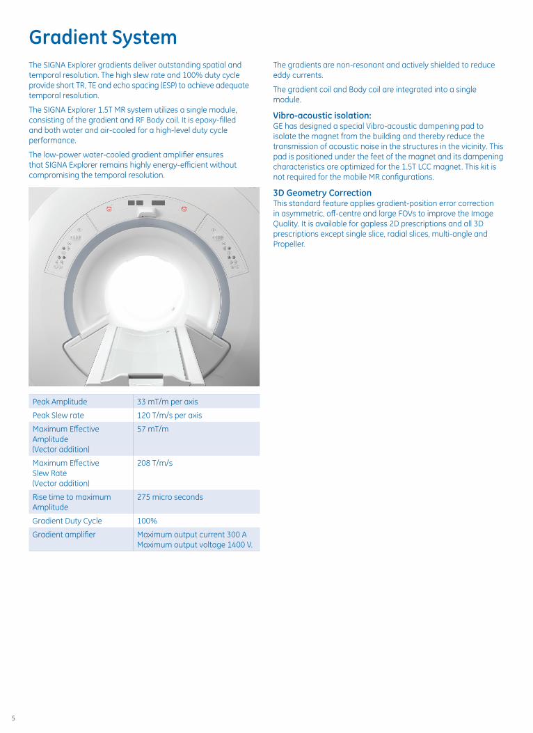
The SIGNA Explorer gradients deliver outstanding spatial and temporal resolution. The high slew rate and 100% duty cycle provide short TR, TE and echo spacing (ESP) to achieve adequate temporal resolution.
The SIGNA Explorer 1.5T MR system utilizes a single module, consisting of the gradient and RF Body coil. It is epoxy-filled and both water and air-cooled for a high-level duty cycle performance.
The low-power water-cooled gradient amplifier ensures that SIGNA Explorer remains highly energy-efficient without compromising the temporal resolution.
Peak Amplitude 33 mT/m per axis
Peak Slew rate 120 T/m/s per axis
Maximum Effective Amplitude (Vector addition)
57 mT/m
Maximum Effective Slew Rate (Vector addition)
208 T/m/s
Rise time to maximum Amplitude
275 micro seconds
Gradient Duty Cycle 100%
Gradient amplifier Maximum output current 300 AMaximum output voltage 1400 V.
Gradient SystemThe gradients are non-resonant and actively shielded to reduce eddy currents.
The gradient coil and Body coil are integrated into a single module.
Vibro-acoustic isolation:GE has designed a special Vibro-acoustic dampening pad to isolate the magnet from the building and thereby reduce the transmission of acoustic noise in the structures in the vicinity. This pad is positioned under the feet of the magnet and its dampening characteristics are optimized for the 1.5T LCC magnet. This kit is not required for the mobile MR configurations.
3D Geometry CorrectionThis standard feature applies gradient-position error correction in asymmetric, off-centre and large FOVs to improve the Image Quality. It is available for gapless 2D prescriptions and all 3D prescriptions except single slice, radial slices, multi-angle and Propeller.
5
SIGNA Explorer provides adequate processing speed and image storage capacity to handle even the most demanding applications.Host ComputerMain CPU Intel Xeon Quadcore Processor
Clock rate 3.6 GHz
Main Memory 32 GB
Cabinets Convertible minitower
Hard disk 3 x 300 GB Hard-disk
Image Storage 400,000 uncompressed 256 x 256 image storage
DVD Image exchange and short-term storage is possible with DVD writer
The DVD capacity is 35,000 images per 4.7 GB DVD
Recon EngineReconstruction performance today is challenged by explosive growth in data, and increased computational complexity. The amount of data to be stored and processed continues to increase with the advances in MR system technology. The SIGNA Explorer is designed to meet that challenge with 8 GB-memory and fast recon speed.
Main Processor Quad-core Intel processor
Clock rate 2.4 GHz
Memory 48 GB ECC DDR3 1333
Hard disk storage 300 GB and 1 100 GB
Ethernet Transfer speed 1 GB/s
Reconstruction speed 14,000 FFT/second at 256 x 256 matrix for full FOV
Operator console:Scan control keyboard assembly with intercom speaker, microphone and volume controls, and emergency stop switch.
An enhanced multi-tasking, simultaneous processing architecture manages all processes to help prevent loss of work and disruption when desktops are switched.
Display Monitor:• 24.1” LCD landscape Color Monitor
• Flicker free and High 1920 x 1200 dot resolution
• Automatic backlight technology
• Wide Viewing Angle Technology with 1000:1 contrast ratio
• Digital DVI interface
Host computer and Recon engine
6

The SIGNA Explorer is based on 8/16 high bandwidth receivers. Signals from independent receive coils within an array, are directly connected to independent receive channels for excellent SNR in all applications.
Optical RFConventional design of the MR scanner places the RF receivers in the electronics room. In such design strategies, the MR signal is susceptible to random electrical noise prior to being digitized by the ADCs.
The OpTix RF receivers are located inside the magnet enclosures in the shielded scan room, isolated from external noise sources. The MR signal is digitized within the scan room and transmitted via fiber optic cables to the reconstruction engine in the electronics room. The close proximity of the receivers to the patient reduces noise and improves image quality.
Optical RF technology increases SNR for all volume acquisitions, independent of which surface coil is being used.
RF Transmit
RF amplifier Water-cooled Compact, solid-state
Maximum output power 10 kw for body and 2 kw for head
Maximum RF field >24 µT
RF Exciter Frequency 63.86 ± 0.65 MHz
Amplitude control 16 bit
Frequency resolution >0.596Hz/step
Phase resolution <0.0055 deg/step
Amplitude stability <0.09 dB/minute
Phase stability <1.5 deg/minute
Frequency stability 14 parts per billion
RF pulse control Digital
RF Receive
Receive channels 16 selectable
Analogue to Digital converters 16 selectable
Sampling rate 80 MHz @ 16 bits per channel
ADC sampling resolution 16 bit
Receive signal filtering/decimation
Digital, non-recursive, linear FIR
Quadrature demodulation Digital
Receive dynamic range >145 dB/Hz
Receive signal resolution Up to 32 bits
Pre-amplifier noise figure <0.5 dB for body
RF Transmit and Receive
7

SIGNA Explorer offers an assortment of quadrature and multi-channel array coils to ensure outstanding image quality and coverage.
All the configurations include following two Transmit/Receive coils:
Transmit / Receive head coil
• 16-rung quadrature birdcage coil with look-out mirror
• Patient friendly with split-top design
• 28 cm diameter and 38 cm length
• Applications include brain, extremities and other miscellaneous applications when other specialty coils are not available
Transmit Receive integrated body coil
• 16-rung quadrature birdcage coil
• 60 cm inner diameter for up to 50 cm FOV
The Express coil suite, compatible with the fixed table configuration, is designed to provide outstanding coverage, and convenient workflow.
Express Head-Neck array
• 16-element
• Optimized for Brain, C-spine and Neuro-Vascular MRA
• Removable top with Open-face design
• Lookout mirror
Express Posterior Array coil embedded in the cradle
• 12-element coil with superb SI coverage of thorax, abdomen, pelvis, and TL spine
• High-sensitivity elements ensure excellent contrast and resolution
Express Anterior array AA coil 9-element for the 16-channel system
• 12-ch imaging of the thorax, abdomen and pelvis in conjuction with Express Posterior Array
• Offers improved SNR and coverage up to 56 cm in SI direction
• Light weight with openings for EKG leads
Optional Surface coils
8-channel lnvivo Brain array
8-channel Breast array
3-channel shoulder array
8-channel shoulder array
Quadrature Knee/Foot birdcage coil with chimney design
8-channel Foot/Ankle array
8-channel Transmit/Receive Invivo Knee array
8ch Body array
16ch Flex Array Large 23 x 70 cm
16ch Flex Array Medium 23 x 48 cm
Endo-rectal coil with Interface
8-channel Wrist array
4ch Flex Coil Large 53.5 x 24.0 cm
4ch Flex Coil Small 37.5 x 24.0 cm
3” Round coil for orbits. ankle. IAC, wrist and other small structures
5” Round coil for orbits. ankle. lAC, wrist and other small structures
Dual Array adapter for simultaneous use of 2 x 3” round coils for the applications such as high-resolution dual TMJ
Optional Surface coils for detachable patient tables only
16ch 29-element Head-Neck-spine array
12-channel Body Array
8-channel lnvivo NV array
8-channel CTL spine array
Some of the coils need 16-channel configuration. Please contact GE representative for more details or refer to the techno-commercial offer for SIGNA Explorer
RF Coils and arrays
8

Imaging planes Direct axial, sagittal, coronal, oblique and double/triple oblique plane imaging
3 Plane Localizer pulse sequence
Slice thickness • 0.5 mm to 100 mm in 0.1 mm increments in 2D mode
• 0.1mm to 10 mm in 0.1 mm increments in 3D mode
• Operator variable inter-slice spacing in increments as small as 0.1 mm. Interleave multi-slice imaging
• 4 – 512 contiguous slice volume imaging in increments of 2 in 3D mode
• 2D Standard Multi-Slice Imaging with as many as 2048 slices
FOV • Body Coil: 1 to 50 cm in 1 cm increments in all planes
• Ability to scan off-center FOV and Asymmetric FOV
Imaging Matrix • Phase encoding from 32 to 1024 in steps of 32. Frquency ecoding of 64 to 1024
• Respiratory Triggering concurrent with ECG gating
• ZIP (Zero Filled Interpolation Processing) methods:
• Through-plane ZIP (slice) reconstructs images interpolated between acquired slices for high-resolution 3D reformations
• In-plane ZIP (matrix) reconstructs data at a higher resolution matrix than selected, for optimized balance between SNR and spatial resolution. Options include 512 ZIP and 1024 ZIP
Gating • Peripheral Gating
• Respiratory gating
• VCG Fating
• Cardiac adaptive digital gating
Image Display • 256 Image buffer (256 x 256) at 35 fps
Imaging and display parametersImage Display Features
• Zoom/Roam/Flip/Rotate/Scroll
• Explicit Magnify & Magnifying Glass
• Image Measurement Tools Grid On/Off
• Cross Reference/User Annotation
• Exam/Series Page
• Hide Graphics/Erase Annotation/Screen Save
• Accelerator Command Bar
• Compare Mode/Reference Image/ Image Enhance
• ClariView Image Filtering
• 5 image filters to achieve image sharpening and image smoothing
• Minified Reference Scoutview
• Cine Paging (up to 4 windows and 128 images/window)
• Add/Subtract /Edit Patient Data
Image Annotation Features
• Two Graphic/Text planes overlay the entire screen
• Grid placement with anatomical reference on an image
• Drawing and annotation may be added to and removed from images
Imaging Parameters
• Respiratory Triggered FSE
• Flow Compensation
• Respiratory Compensation
• Peripheral Pulse Gating
• Graphic Prescription
• Explicit Saturation (SAT outside and inside the FOV and concatenated SAT)
• Graphic Saturation (oblique and cursor placement SAT inside the FOV)
• No Phase Wrap
• Fat/Water (Spectral Chem-Sat) Saturation
• Extended Dynamic Range
• Phase and Frequency Offset
• Asymmetric FOV
• Square Pixel
9

Networking Protocols supported
• DICOM 3.0 Basic Grayscale Print Service Class
• DICOM 3.0 send, receive and query/retrieve
• InSite point-to-point
• TCP/IP (for system administration)
DICOM Conformance Standards
• DICOM 3.0 Modality Work List Service Class supported with optional Connect Pro software
• DICOM 3.0 Storage Service Class
• Service Class User (SCU) for image send
• Service Class Provider (SCP) for image receive
• DICOM 3.0 Query/Retrieve Service Class
• DICOM 3.0 Storage Commitment Service Class
• DICOM 3.0 Basic Grayscale Print Service Class
TR, TE, and Echo Spacing
128x128 256x256
2D Spin Echo
Shortest TR in ms 6.0 8.0
Shortest TE in ms 2.2 2.2
Min. Slice Thickness 0.6 mm 0.6 mm
Minimum FOV 10 mm 10 mm
2D Fast Spin Echo
Shortest TR in ms 10.0 11.0
Shortest TE in ms 2.2 2.2
Min. Slice Thickness 0.5 mm 0.5 mm
Min. Echo Spacing in ms 2.2 2.2
Max ETL in SSFSE 264 264
2D Fast Grad. Echo
Shortest TR in ms 2.3 2.8
Shortest TE in ms 0.8 1.0
Min. Slice Thickness 0.7 mm 0.7 mm
Minimum FOV 10 mm 10 mm
3D Fast Grad. Echo
Shortest TR in ms 0.88 1.2
Shortest TE in ms 0.22 0.30
Min. Slice Thickness 0.1 mm 0.1 mm
EPI
Shortest TR in ms 5.0 5.0
Shortest TE in ms 1.2 1.6
Min. Slice Thickness 0.6 mm 0.6 mm
Minimum FOV 40 mm 40 mm
Min. Echo Spacing at 25 cm FOV in ms
0.70 1.064
Min. Echo Spacing at 48 cm FOV in ms
0.504 0.716
Minimum shots 1 1
Maximum b value s/mm2 10,000 10,000
10

expected from that protocol is preserved. This feature removes the guess work and random changes that novice operators tend to do with TR, bandwidth, spacing.
For the advanced imaging user, of course, the advanced mode and expert mode are always available.
Modality WorklistThe modality worklist (MWL) provides an automated method of obtaining exam and protocol information for a patient directly from a DICOM Worklist server. The protocol may be selected well in advance of the patient’s arrival at the MR suite, thereby simplifying exam preparation and reducing necessary work by the technologist during the time-critical procedure. The ConnectPro software enables the DICOM worklist server class for the SIGNA Explorer Operator’s Console.
Protocol libraries and propertiesThe SIGNA Explorer system protocols are organized into two main libraries, GE Optimized and Site Authored. For quick search and selection, each protocol may be archived with independent properties based on patient demographics, anatomy, type of acquisition, or identification number.
ProtoCopyThis feature enables a complete exam protocol to be shared with the click of a mouse. The exam protocol can originate from either a library or previously acquired exam. This enables routine archive of protocols for emergency backup and simple management of libraries across multiple systems.
Workflow ManagerOnce a protocol has been selected for an exam, it is automatically loaded into the Workflow Manager. The Workflow Manager controls image prescription, acquisition, processing, visualization, and networking and may fully automate these steps if requested.
Inline viewingInline viewing allows the user to conveniently view, compare, and analyze images without having to switch to the Browser. Simply select the series to view from the Workflow Manager and the images are displayed along with standard image display tools. By selecting multiple series at a time, a user can perform image comparisons. The integrated viewer allows the user to seamlessly move between scanning and image viewing.
Inline processingThe SIGNA Explorer workflow automates many of theroutine tasks that previously required user interaction. Thisdramatically reduces the workload for the user and helpsensure that consistent and repeatable images are presentedfor review. Processing steps are automatically completedimmediately after the data has been reconstructed and theimages saved into the database. These automated processing steps can be saved in the Protocol Library to ensure consistent exam workflow for each type of patient.
For certain tasks, such as vascular segmentation, the usermust accept the results, or complete additional steps prior tosaving the images to the database. In these cases the data isautomatically loaded into the appropriate tool, then the system will await further instruction by the user. Examples of fully automated and partially automated inline processing include:
Ready Interface SystemThe SIGNA Explorer Ready Interface incorporates many features designed to lighten the workload of the technologists. The SIGNA Explorer includes an automated protocol-driven workflow and user interface designed for consistency in generating high-quality imaging for all patients and from all technologists. Simultaneous scanning, reconstructions, filming, archiving, networking, and post-processing can help achieve productivity, efficiency, and streamlined data management.
Ready Brain ApplicationAn MRI examination of the brain consists of a number of connected steps. Ready Brain provides the flexibility to automate these connected steps from acquiring a localizer image, prescribing acquisition planes, scanning relevant series, performing post-processing up to transferring the final image data to a reading station. By standardizing the steps of an exam and the location of the scan planes, such automation could result in greater consistency, especially in the longitudinal follow-up.
Ready Brain features an automatic localizer, automatic calculation of the mid-sagittal plane for 2D/3D prescription, determination of the AC-PC line, and correction for extreme (>45 degree) rotation.
Express coil productivityThe Express coil suite, compatible with the fixed table configuration, is designed to provide convenient workflow when imaging the central nervous system, thorax, abdomen and pelvis.
AutoStartIf AutoStart is selected, once the landmark position has been set and the technologist exits the scan room, the Workflow Manager will automatically start the acquisition.
Slider BarThe SIGNA Explorer scanning has been simplified with
• Basic mode – designed for routine imaging
• Advanced mode – essential parameters are displayed such as FOV, slice thickness, TR, TE, Spacing
• Export Mode – where all the parameters are displayed and controlled
The Slider Bar is designed to help you select optimal scan-time for the most routine exam protocols by selecting within the recommended 5 levels of scantime-resolution combinations. The parameters behind each level are optimized, such that even with the recommended shortest scan-time, the fundamental contrast
Workflow
11

Inline processing capabilitiesDiffusion Weighted Images ADC/eADC Maps
Automatic compute and save
Diffusion Tensor Images FA/ADC Maps Automatic compute and save
Image Filtering: A-E, SCIC, PURE Automatic compute and save
Maximum/MinimumIntensity Projection Automatic compute and save
Reformat to orthogonal planes Automatic compute and save
T2 Map for cartilage evaluation Automatic compute and save
FiberTrak Automatic load
Spectroscopy – Single voxel brain and breast metabolite
Automatic compute and save
3D Volume Viewer Automatic load
Spectroscopy – 2D/3D Chemical Shift Imaging
Automatic load
BrainStat (Functool) Automatic load
Image Fusion Automatic load
IVI (Volume Viewer) Automatic load
Pasting Automatic load
SER (Functool) Automatic load
eDWI Automatic compute and save
3D ASL Automatic compute and save
LinkingLinking automates the prescription of images for each series in an exam. Once the targeted anatomical region has been located the Linking feature combines information from a prescribed imaging series to all subsequent series in the Workflow Manager. All series that have been linked may automatically be prescribed (Rx) and no further interaction will be needed by the technologist to initiate the scan. Series can have common fields of view, obliquity, slice thickness, anatomical coverage, saturation bands, or shim volumes.
Protocol Icon ShortcutsThis unique and operator-friendly feature shows anatomical-pictorial shortcuts (icons) in the ‘Ready Shortcuts’ tab on the exam screen. These shortcuts icons are fixed. However the protocols assigned to each icon can be customized in the ‘custom shortcuts’ of the exam screen.
The pictorial icons are Brain, Pituitary, Vascular Brain, C-spine, T-spine, L-spine, Shoulder joint, Wrist joint, Hip joint, Knee joint, Liver-Pancreas, Kidneys, Prostate and Uterus.
12
ARC – Auto Calibrating Reconstruction ARC helps to eliminate breath-hold mismatch errors by imbedding the calibration data within the scan data. In addition, this reconstruction permits small FOV imaging by reducing focal parallel imaging artifacts from the exam. Supporting both 1D and 2D acceleration, net acceleration factors of up to 3.4 can be achieved.
ASSET - Array Spatial Sensitivity Encoding TechniqueASSET imaging option is a 1D image-based parallel imaging technique used to speed up the data acquisition. For temporally sensitive acquisitions, ASSET is designed to reduce image blurring and motion and enable greater anatomical coverage.
Parallel imaging acceleration factors ranging from 1 to 3.0 are supported depending on the coil.
With the SIGNA Explorer system, the following applications are parallel imaging enabled.
• 2D Spin Echo
• 2D T2 FLAIR
• 2D FLAIR EPI
• 3D FIESTA-C
• Inhance non-contrast MRA
• 3D SWAN
• 2D FSE
• 2D FRFSE
• 2D FSE-IR
• 2D T1FLAIR
• 2D FSE Double IR
• 2D FSE Triple IR
• 2D T2MAP
• 2D FSE-XL IDEAL
• 2D FRFSE-XL IDEAL
• 2D SSFSE
• 2D SSFSE-IR
• 2D SSFSE MRCP
• 2D SSFSE 3-plane
• 3D FRFSE
• 3D CUBE
• 2D FGRE
• 2D FSPGR
• 2D FIESTA
• 2D FIESTA Fat Sat
• 2D FIESTA Fast CINE
• 2D MDE
• 2D MFGRE
• 3D TOF GRE
• 3D TOF SPGR
• 3D FGRE
• 3D FSPGR
• 3D FGRE IDEAL
• 3D FSPGR IDEAL
• 3D Quick STEP
• 3D Fast TOF GRE
• 3D Fast TOF SPGR
• 3D FIESTA
• 3D MDE
• 3D TRICKS
• 3D LAVA
• 3D Dual Echo
• 3D VIBRANT
• 2D GRE-EPI
• 2D SE-EPI
• 2D DW-EPI
• 2D DT-EPI
• BRAVO
• LAVA-Flex
• Propeller 3.0
† Some product features may not be available in all countries
Parallel Imaging†
13

Multi-Projection Volume Reconstruction (MPVR)
Quick way to generate volumetric images for MR or CT Angiography without threshold data or removing unwanted anatomy.
An entire volume is used to generate images in any plane, creating real time frames of reference at the same time.
Multi-Planar Reformation (MPR)
Provides real time assessment of anatomy in off-axis planes. Sagittal, coronal, oblique, and curved planar reformations.
Other standard analysis features
Curved reformations
Batch reformations
Interactive Vascular Imaging (IVI)
Comparison Mode
Multi-image ROI
SCIC & PURE Surface coil intensity correction (SCIC) and phase array uniformity correction (PURE)
MR Pasting Combines a series of MR images acquired in multiple stations of the body into a single image. Pasting 1.1 pro-vides the convenience of viewing a single image rather than several images. The applications include pasting of multiple images of sagittal spine imaging and carotid to abdominal aorta and peripheral vascular imaging.
FuncTool PerformanceFuncTool Performance is an advanced MR-specific post-processing package with multiple algorithms to help you analyze and visualize the spatial and time-course based MR data. The package runs on the main console and optionally on the Advantage* Workstation.
Post processingResults are displayed in various formats including time-intensity curves, parametric color overlays and metabolite ratio maps.
These include:
• ADC and eADC maps
• Correlation coefficients for mapping of motor strip and visual/auditory stimuli
• Automatic assessment of Blood Volume, Blood Flow, Mean Transit Time and Time to Peak
• Metabolite maps (needs 2D CSI package)
• FuncTool Fusion for overlay of multiple images acquired from separate acquisitions
• Spectroscopy baseline correction
• BrainStat GVF
• Archive or network generated report
• Generate single click DICOM SR report
• Generate report in database
• Compute and annotate
FuncTool also plays critical role in display and visualization of the data acquired with optional advanced applications such as
• 3D Arterial Spin Labeling
• Starmap R2*
• Diffusion Tensor Imaging
• DWI
• BOLD data acquired with GRE-EPI
• FiberTrack
• MR Touch - MR Elastograms
14

GE ScanTools have evolved in to a comprehensive suite of pulse sequences and applications.
Spin Echo Pulse sequencesSpin Echo and Fast Spin Echo sequences have been the mainstay in MR imaging due to their robust signal and well understood contrast behavior. The XL version of the FSE is designed to enable an extended train of echoes to reduce the T2 blurring and the fast recovery FSE helps in reducing TR without compromising the T2 weighting.
• 2D Spin Echo
• 2D FSE-XL
• 2D SSFSE (single shot)
• 2D SSFSE IR
• 2D FRFSE-XL (Fast Recovery version)
Single-Shot Fast-Spin EchoAn ultra-fast technique that permits complete image acquisition following a single RF excitation. It can acquire slices in less than one second, making it an excellent complement to T2-weighted brain, abdominal imaging and MRCP studies.
3D FRFSECoupled with respiratory gating, this 3D FSE sequence uses a novel “recovery” pulse at the end of each echo train to recapture signal for the next repetition. These features result in high-resolution three-dimensional images for MR cholangiopancreatography (MRCP) studies.
Gradient Echo Pulse sequencesThe main advantage of GRE sequences is its shorter TR enabled by lower flip angles and gradient inversions instead of 180° RF pulses. Consequently, the GRE sequences are better suited to a variety of 3D volumetric applications including ce-MRA techniques.
SIGNA Explorer system’s innovative imaging chain and high fidelity gradients offer short TRs and TEs.
• 2D GRE/SPGR
• 2D FGRE/FSPGR
• 3D GRE/SPGR
• 3D FGRE/FSPGR
• ECG gated FGRE
• ECG gated Fast SPGR
• IR-prepared 3D-GRE with ARC parallel imaging for Brain Volume imaging BRAVO
3D Dual EchoWith improvements in parallel imaging and RF coil arrays, volumetric imaging in body is becoming a standard of care. The 2D/3D Dual Echo sequence produces in-phase and out-of-phase images in a single breath-hold. As a result, the high-resolution images are in perfect alignment helping to simplify the diagnostic process. In addition, the excellent SNR of the 3D acquisition permits the thinner slices than are traditionally available using 2D techniques.
LAVALAVA delivers excellent spatial and temporal resolution with large volume slice coverage in significantly shorter total scan times than those possible with conventional 3D gradient echo techniques. LAVA provides outstanding Liver breath-held imaging without compromising in-plane spatial resolution.
LAVA-XVLAVA-XV utilizes auto-calibrating ARC parallel imaging technique that needs no coil sensitivity maps. It is less sensitive to motion artifacts compared to the conventional LAVA based on ASSET.
2D and 3D MERGEMultiple Echo Recombined Gradient Echo (MERGE) uses multiple echoes to generate high-resolution images of the C-spine with excellent gray-white matter differentiation. By combining early echoes with high SNR and late echoes with improved contrast, the result is improved cord contrast within the spinal column.
2D/3D TOF2D Gated TOF Imaging, 3D TOF Imaging and enhanced 3D TOF Imaging are based on conventional gradient echo imaging techniques. TOF imaging techniques rely primarily on flow-related enhancements to distinguish the moving spins from the stationary spins.
2D/3D Phase contrastThese techniques demonstrate flow velocities and directional properties in vessels and other moving fluids such as cerebral spinal fluid and aortic flow.
Inversion Recovery Pulse sequencesThis is an SE sequence with preparatory 180° pulse to flip the net magnetization vector by 180° to null the signal from a particular entity, very often water. The variants of the IR available with our ScanTools are:
• 2D T1 FLAIR (Fluid Attenuated IR)
• 2D T2 FLAIR (Fluid Attenuated IR)
• STIR – Short TI Inversion Recovery
• Double IR and Triple IR for black blood imaging in assessment of carotid plaques cardiac imaging
Echo Planar sequencesWith echo-planar imaging, a single echo train is employed to collect data from all lines of k-space during one TR. In the single shot version, all the k-space lines are collected in a single TR and in case of the multi-shot EPI it is collected over multiple TRs. EPI is an excellent choice for diffusion weighted imaging.
SIGNA Explorer offers comprehensive EPI techniques with short echo spacing:
• EPI SE
• EPI GRE
• EPI Diffusion Weighted
• EPI FLAIR (single shot version helps in DWI)
ScanTools
15

SPECIAL - Spectral Inversion at LipidsSpectral Inversion at Lipids (SPECIAL) uses an inversion pulse transmitted at the frequency of fat and timed to the null point of fat. This results in higher signal produced from protons bound in water and a decreased signal from nuclei precessing at the frequency of fat. The flip angle of the inversion pulse is optimized based on the prescribed TI, TR, and flip angle. SPECIAL is available with 3D FRGE and 3D Fast TOF sequences.
SmartPrepSmartPrep uses a special tracking pulse sequence to monitor the MR signal through a user-prescribed volume to detect the arrival of an injected contrast bolus and to trigger the acquisition, for optimum contrast enhancement. It does so by integrating the following:
• Efficient multi-station scouting capabilities
• Ultra flexible multi station Graphic Rx for precise multi-station RF tuning with pre-scan ahead
• Automated triggering, table motion and coil switching Application of robust fat-suppression with SPECIAL (Spectral Inversion at Lipid)
• The result is acquisition techniques that deliver consistently high-resolution results by making routine use of elliptic-centric encoding and ZIP reconstruction to create images as large as 1024x1024 pixels
SmartStepThe SmartStep adds programmable stepping capability to SmartPrep to plan and optimize your contrast enhancements.
Fluoro-Triggered MRAFluoro-Triggered MRA images the selected anatomy continuously, enabling the user to manually trigger each acquisition as soon as the MR-operator is satisfied with the level of vessel enhancement. This switch-over occurs almost instantly, in less than one second.
TRICKSTime Resolved Imaging of Contrast KineticS (TRICKS) technology is designed to use intricate temporal sampling with complex data recombination to accelerate the temporal resolution of 3D dynamic imaging – without compromising spatial resolution. Easy to set up, TRICKS rapidly generates time-resolved 3D images of blood vessels to help meet the challenge of capturing peak arterial phases. With TRICKS, the different vascular phases can be extracted quickly after image acquisition.
3D FIESTA-CThis phase-cycled FIESTA reduces sensitivity to susceptibilities that may be encountered when imaging in the posterior fossa. It provides exquisite contrast that is equated for visualization of the internal auditory canal. It is also well-suited for T2 imaging through the cervical spine.
3D FIESTA3D FIESTA (Fast Imaging Employing Steady-state Acquisition is a technique that uses an extremely short repetition time (TR) between RF pulses such that high-resolution 3D volume images can be acquired rapidly. The 3D FIESTA technique is especially useful for the rapid acquisition of high-spatial-resolution images of static structures such as cochlea, internal auditory canal, or joints.
ART Acoustic Reduction TechnologyThis feature Reduces modifies pulse sequences without compromising the image quality to reduce the acoustic noise to improve the patient comfort.
Other ScanTools applicationsScanTools also includes many advanced applications such as real-time imaging, BOLD acquisitions and multistation vascular imaging.
3D Cube3D Cube has potential to replace several slice-by-slice, plane-after-plane 2D FSE acquisitions with a single 3D volume scan, providing you with T1, T2, T2 FLAIR or PD sequences. You can easily reformat sub-millimeter isotropic volume data from a single acquisition into any plane without gaps, and with the same resolution as the original plane. Our self-calibrating ARC parallel imaging helps eliminate artifacts while accelerating the image acquisition.
BOLD fMRI analysis on FuncToolCorrelation Coefficient algorithm is used to analyze an image set acquired with the EPI. Neuronal activity of either motor or cognitive functions can be mapped by fMRI through changes in signal intensity arising from bulk magnetic susceptibility-induced relaxation changes resulting from variations in blood flow and oxygenation. A BOLD image acquisition is typically a single shot, multiphase, GRE-EPI with high number of phases. Within each series, you can acquire multiple phases and multiple slices.
The pre-selected images from the series can be loaded in to FuncTool to automatically compute parametric images. Magnification factor, WW/WL, and threshold functions are available for post processing.
BrainSTAT GVFThe BrainSTAT post processing application automatically generates parametric maps for neuro Blood Flow, Blood Volume. Mean Transit Time, and Time to Peak signal intensity. A Gamma Variate fitting algorithm is used to automatically estimate the arterial input function, then calculate the values for the four parametric maps. The maps may be saved in DICOM format and fused with high resolution anatomic datasets for improved visualization of tissue and anatomy.
Diffusion Weighted EPISingle Shot FLAIR EPI and Single Shot, diffusion-weighted EPI with b-values up to 7,000 s/mm2 is standard with SIGNA Explorer that is based on
• Automatic isotropic diffusion-weighted image generation
• Multi-NEX capability
• Online image processing
• ADC maps
Flow Analysis: Allows Flow Quantification in vasculature such as carotid artery.
i-Drive Pro real-time imagingi-drive Pro brings real-time interactive imaging to the MR system for imaging of organs such as heart, GI tract and spine. It allows the user to change scan parameters easily, including during scanning, to evaluate the results immediately.
16

3D COSMICCoherent Oscillatory State Acquisition for the manipulation of imaging contrast is a 3D sequence for imaging of axial c-spine. COSMIC uses modified fast GRE pulse sequence with steady-state free precession segmented multi-shot centric k-space acquisition. This improves the CNR and SNR of c-spine tissue including the spinal cord, vertebral disks, nerve root canal and contrast between CSF and nerve roots.
2D FatSat FIESTAFast Imaging Employing STeady-state Acquisition (FIESTA) is designed to produce high SNR images extremely rapidly and with excellent contrast between tissues. The contrast relies on a steady state for the transverse magnetization, which builds as a series of radio frequency pulses and special gradient pulses are repeated after an extremely short repetition time, TR. FIESTA accentuates the signal from tissues that have a long T2 and short T1. FIESTA has the capability to suppress the signal from fat, especially to create more contrast between the vasculature and surrounding tissues.
2D FIESTA CINEFast Imaging Employing STeady state Acquisitions is a fully balanced steady-state coherent imaging pulse sequence that has been designed to produce high SNR images at very short TR. The pulse sequence uses fully balanced gradients to re-phase the transverse magnetization at the end of each TR interval. This sequence accentuates the contrast of anatomy with high T2/T1 ratios (such as the cardiac blood pool), while suppressing the signal from tissues with low T2/T1 ratios (such as muscle and myocardium). This enhances the contrast between the myocardium and the blood pool.
VCG Gating Allows more robust trigger detection.
IVIIVI is an interactive user interface that allows operators to remove background from MR angiography images for angiographic and maximum intensity (MIP) projections in multiple scan planes.
Connect ProConnectPro software enables the DICOM worklist server class for the operator-console, making it possible for the console to query your HIS/RIS by name, modality, or scheduled date, and to download patient demographics directly to the scanner. This may require separate gateway hardware to connect non DICOM-compatible HIS/RIS systems to the MR system.
Performed Procedure StepPerformed Procedure Step (PPS) is an important automated connectivity capability and a key step towards a film-less and paperless environment. In conjunction with the GE PACS broker, it automatically notifies the HIS/RIS and PACS systems of procedure status, which then closes the loop on the information gathered from patient arrival through billing. This results in improved patient care and enhanced productivity.
17

The following NEURO applications are standard with the SIGNA Explorer ScanTools.
• Ready Brain
• Diffusion Weighted EPI
• EPI 2D GRE for BOLD acquisitions
• EPI FLAIR
• 2D STIR
• 3D Cube
• 3D FIESTA
• 3D FIESTA C
• 3D COSMIC
• 2D/3D MERGE
• MR Pasting
• 2D/3D TOF
• 2D/3D Phase Contrast Angio
• Magnetization Transfer Contrast
• T1-SE MEMP
• T1 FLAIR
• T2 FLAIR
• T1/T2 FSE
Optional Advanced Neuro applications PROPELLER 3.0PROPELLER 3.0 is based on fast spin echo (FSE) and its name is derived from its unique k-space acquisition, in which data are acquired in radial “blades” that rotate similar to the propeller on an airplane. Since each blade passes through the center of k-space, PROPELLER has low sensitivity to motion artifacts and high contrast-to-noise properties. This makes it well-suited for producing high-resolution image quality even under challenging circumstances. Available in all-imaging planes, PROPELLER offers motion-insensitive T2, T2 FLAIR, T1 FLAIR, and PD contrasts in all anatomies. It is available in all imaging planes and all anatomies - with special relevance in brain, abdomen and MSK.
Diffusion-weighted PROPELLER is an alternative to the conventional echoplanar imaging, to produce high quality images in the skull base even in the presence of dental work, craniotomies or other abnormalities that cause a magnetic field disturbance.
SWANSWAN lets you visualize and delineate small vessels microbleeds, large vascular structures, and iron or calcium deposits in the brain. SWAN captures a broad spectrum of contrast characteristics specific to a wide range of tissue components using a multi-TE acquisition technique. The multi-TE approach is inherently less affected by chemical shift, leading to clear images.
SWAN 2.0SWAN phase imaging based on complex coil combine reconstruction
IDEALThis sequence and reconstruction package acquires multiple echoes at different echo times with a fast spin echo readout to create water-only, fat-only, in-phase and out-of-phase images. IDEAL is designed for imaging those difficult regions such as the neck and spine where inhomogeneous magnetic fields yield failures with traditional fat saturation techniques.
Diffusion Tensor Imaging with Fiber TrackingThis package expands the EPI capability to include diffusion tensor imaging, a technique that acquires diffusion information in up to 150 different diffusion directions. It generates image contrast based on the degree of diffusion anisotropy in cerebral tissues such as white matter. FuncTool capabilities on the console (included with ScanTools) create Fractional Anisotropy (FA), Apparent Diffusion Weighted (ADC) and T2-Weighted TRACE maps.
The FiberTrak post-processing utility generates eigen-vector information from the diffusion tensor acquisition and processing. Using a robust and efficient seeding process, three-dimensional renderings of the diffusion along white matter tracts are generated.
PROBE PRESS single voxel spectroscopyPROBE PRESS single-voxel spectroscopy allows you to non-invasively evaluate the relative concentrations of in-vivo metabolites and lets you acquire and display volume-localized, water-suppressed H1 spectra in single voxel mode. The package includes automated recon, cquisition set-up and graphic prescription of spectroscopic volumes. The standard sequence consists of three slice-selective RF pulses with crusher gradients. The PRESS sequence makes use of reduced flip angles to decrease TE time of the sequence. The key advantage of PRESS (over STEAM) is that it provides up to twice the SNR and hence decreased exam time or voxel size. It is the sequence of choice for all Hydrogen single voxel spectroscopy data acquisitions with TE values ≥ 35 ms.
PROBE – 2D CSI / 3D CSIThis extends the PROBE-PRESS capabilities with simultaneous multi-voxel in-plane acquisitions. Post-processing, including the generation of metabolite maps is automatically generated with FuncTool Performance package.
Express Spine AnnotationEnables semi-automatic annotation of vertebral bodies on saggital T2W spine images.
† Some product features may not be available in all countries
Neuro Imaging†
18

3D ASL (Arterial Spin labeling)3D ASL utilizes water in arterial blood as an endogenous contrast medium to help visualize tissue perfusion and provide quantitative assessment of cerebral blood flow (CBF) in ml/100 g/min. 3D ASL deploys stocked spiral FSE readout with modulated fiip angle to acquire 3D volumetric data with increased SNR and less image distortion compared to conventional 2D EPI-bosed ASL techniques. A pulsed continuous labeling is applied to Iobel arteriat blood close to the imaging volume thus improving conspicuity of flowing blood. Selective. interwoven pulses ore then used to saturate ond invert the imaging volume, in order to achieve better background suppression, and reduce sensitivity to motion. The 3D volume con be reformatted to axial sagittal. coronal or oblique planes. The quantitative color CBF mops can be generated and stored in DICOM format. 3D ASL helps generate robust, reproducible images and perfusion maps with high SNR. reduced motion artifacts ond less distortion in high magnetic susceptibility regions.
eDWIThe enhanced Diffusion Weighted Imaging technique has been designed to provide high signal-to-noise-ratio diffusion images of the liver and brain with short acquisition time Its multi-B feature is designed to provide measurement of apparent diffusion coefficient (ADC) map with reduced effect of perfusion. In addition. “3 in 1” technique applies diffusion weighting to all three grodients simultaneously, helping Improve sensitivity. Built in tetrahedral feature applies four different diffusion weighing combinations of x. y, and z gradients simultaneously to acquire isotropic diffusion weighted images with high signal to noise ratio and shorter TE. Its smart NEX feature significantly reduces the acquisition time. An inversion recovery is deployed to provide robust fat suppression. Enhanced DWl package includes the acquisition sequence and post-processing tools.
3D PROMO3D PROMO provides a real time 3D navigator based motion correction algorithm correcting for the 6 rigid body terms where re-acquisition of severely corrupted data provides robust high quality motion reduced 3D outcomes. 3D PROMO is compatible with both T2 and T2 FLAIR CUBE acquisitions.
The Silent Neuro Exam PackageThe Silent Neuro Exam Package includes a set of sequences designed to generate high-resolution T1, T2, Flair and PD weighted images. The Silenz imaging sequence delivers 3D isotropic images with T1 and/ or PD contrast with sound levels that are within 3dB(A) of the ambient conditions.
Newly enhanced gradient waveforms are employed to minimize the acoustic noise of FSE, 3D Cube and Propeller-based acquisitions to generate T2 and T2 Flair weighted images. In addition, the localizer and pre-scan calibration sequences are optimized to deliver a complete neuro exam at near silent levels.
19

Following BODY applications are standard with the SIGNA Explorer ScanTools.
• LAVA and LAVA XV
• 3D FRFSE-XL for high res MRCP
• 2D and 3D Dual Echo
• 2D SSFSE
• 2D FSE-XL
• 2D STIR/ FIRM
• T2 Fatsat FRFSE-XL
• 2D FatSat FIESTA
• LAVA XV
• 3D Cube
• 2D FSPGR with Fatsat
• 3D FSPGR
• Multi-phase variable delays
Optional Advanced Body and Breast applicationsStarMapStarMap is a technique that acquires multiple echoes at different TE times at each location resulting in images that represent different T2 and T2* weighting. Post-processing of the images is employed to generate gray scale and color maps of the T2 or T2* signal decay across the echoes, which can be useful in the assessment of the presence of iron.
VIBRANTVIBRANT* (Volume Imaging for Breast AssessmeNT) permits high definition bilateral imaging of both breasts in the time that it normally takes to image a single breast. VIBRANT integrates ASSET parallel imaging, bilateral shimming, and a patented fat-suppression technique developed specifically for breast imaging. VIBRANT allows the slices to be acquired in either the sagittal or axial orientation.
BREASEBREASE is a TE averaged PRESS spectroscopy acquisition help characterize breast anatomy.
LAVA FlexLAVA Flex is a 3D FSPGR imaging technique that generates fat/water in-phase and out-of-phase echoes in a single acquisition. Up to 4 types of image contrasts may be achieved within oneacquisition: in phase, out of phase, water only, fat only. The water only contrast differs from a conventional fat suppressed image in that an inversion prep pulse is not applied for fat suppression. In fact, the fat information is removed leaving a water only image that may potentially be used in place of a LAVA type image. LAVA Flex uses ARC. (Auto Calibrating Reconstruction for Cartesian Sampling), a 2D self-calibratedparallel imaging technique that allows for acceleration in both phase and slice directions for supported coils.
† Some product features may not be available in all countries
eDWIThe enhanced Diffusion Weighted Imaging technique hasbeen designed to provide high signal-to-noise-ratio diffusionimages of the liver and brain with short-acquisition time. Itsmultiple b-value feature is designed to provide measurement of apparent diffusion coefficient (ADC) map with reduced effect of perfusion. In addition, “3 in 1” technique applies diffusion weighting to all three gradients simultaneously, helping improve sensitivity. Built in tetrahedral feature applies four different diffusion weighing combinations of x, y, and z gradients simultaneously to acquire isotropic diffusion weighted images with high signal to noise ratio and shorter TE. Its smart NEX feature significantly reduces the acquisition time. Inversion recovery has been deployed to provide robust fat suppression. Enhanced DWI package includes the acquisition sequence and post-processing tools.
Propeller 3.0As extensively described in the Neuro section, Propeller 3.0 offers excellent applications in motion-sensitive abdominal imaging.
MR TouchMR-TouchTM is a non-invasive method to measure relative tissue stiffness with MR. MR Touch™ combines hardware, acquisition and reconstruction algorithms to produce color-coded anatomical images, called Elastograms, showing varying degrees of elasticity or stiffness. The image contrast is related to relative stiffness of soft tissue and is generated from the real-time data acquisition during tissue palpation with low amplitude and low frequency sound waves. The hardware component is comprised of an active sound wave generator and a passive transducer that produces small vibrations in the area of the patient to be scanned.
The acquisition software also triggers the sound wave generator to produce synchronized vibrations on the surface of the patient during the data acquisition. The reconstruction algorithms generate images that show the propagation of waves through the tissue (phase images) and also the corresponding strain wave and relative stiffness images. Parallel imaging is used to accelerate image acquisition.
Body NavigatorsBody Navigators are designed to deliver real time and robust free breathing respiratory motion-compensation to improve body applications.
IDEAL IQIDEAL IQ is a GE exclusive technique that builds upon the original IDEAL (Iterative Decomposition of water and fat with Echo Asymmetry and Least-squares estimation) technique.
IDEAL IQ acquires multiple images of the anatomy at separate echo times to calculate the phase differences and determine triglyceride fat and water content per pixel. It exploits the resonance frequency differences between triglyceride-fat and water, measured as phase differences in multiple echoes, to resolve the triglyceride-fat and water. It provides reliable and uniform water-fat separation in the presence of B0 field inhomogeneity and improves the accuracy of water-fat separation by estimating and correcting for T2* decay between echoes and by more accurately modeling triglyceride fat’s spectral profile as multiple peaks rather than a single peak. The result is a triglyceride fat-fraction map image that reflects the spatial distribution of relative concentration of triglyceride fat within a voxel.
Body and Breast Imaging†
20

PROSEPROSE (PROstate Spectroscopy and imaging Examination), is a noninvasive imaging technique to evaluate prostate lesions.
VIBRANT FlexVIBRANT Flex utilizes dual echo acquisition with 2D ARC parallel imaging to produce water-only, fat-only, in-phase, and out-of-phase images of the breast in a single scan. The Flex processing eliminates fat saturation failures in inhomogeneous regions to provide a clear depiction of the underlying breast anatomy.
FOCUSFOCUS delivers a highly efficient method for increasing the resolution in Single Shot DW EPI sequences. The outcome delivers robust high resolution imaging while removing artifacts typically induced from motion, image back-folding or unsuppressed tissue. In addition, the reduced field of view imaging leads to a reduction in blurring that translates into an overall improvement to the image quality result. The sequence utilizes 2D selective excitation pulses in DW-EPI acquisitions to limit the prescribed phase encoded field of view.
21
Following ortho applications are standard with the SIGNA Explorer ScanTools.
• 2D STIR
• 2D FSE-XL
• 2D GRE
• 3D FIESTA
• 3D Cube
• 3D FSPGR/FGRE
• FATSAT
Optional Advanced Ortho applicationsPropeller 3.0As extensively described in the Neuro section, Propeller 3.0 offers excellent applications in motion-sensitive imaging of joints.
IDEALAreas such as the foot/ankle, shoulder, and off-isocenter wrist make fat saturation a challenge. With IDEAL, water, fat, in-phase, and out-of-phase images can be generated even in the presence of large static field variations. This sequence can produce consistent and reliable images in challenging anatomical areas.
CartiGram T2 MapCartigram is a non-invasive technique for the visualization of subtle changes in the cartilage ultra-structure. It employes special multi-echo T2 weighted technique to generate multiple images per location. The acquired data is processed in FuncTool to produce the T2 color maps that better demonstrate subtle changes compared to the conventional gray scale visualization.
MAVRIC SLMAVRIC SL is a novel pulse sequence based on 3D FSE with a unique reduction-algorithm designed to image in the presence of MR conditional implants. MAVRIC SL helps reduce artifacts caused by metal in both in-plane and through-plane dimensions. This helps in imaging of the soft and bone tissue adjacent to metal instrumentation.
† Some product features may not be available in all countries
Orthopedic Imaging†
22
The other technologies used in this application are – ASSET parallel imaging, SPECIAL spectral fat saturation and respiratory gating.
Inhance 3D DeltaFlow non-contrast MRAInhance 3D DeltaFlow, based on 3D FSE, has been specially designed for peripheral arterial imaging.
The cardiac-gated 3D FSE acquires two echoes - one in diastole and the other in systole. The slow arterial flow during diastole results in bright arteries in the diastolic-images while faster arterial flow during systole results in darker arteries in the systolic- images. Subtraction of the two provides arterial-only images with excellent suppression of venous-flow and the background signal.
Interleaved acquisition and ASSET parallel imaging with optimized k-space trajectory helps respectively reduce motion miss-registration and improve vessel visualization. In addition, the use of partial-Fourier and coronal plane acquisition allows for considerably reduced scan time.
iDrive Pro PlusiDRIVE Pro Plus expands the capabilities of standard iDrive Pro with:
• Geometric changes to image plane location, obliquity, rotation, center FOV and FOV size
• Contrast parameters such as spatial pre-saturation on/off, special sat pulses, flow comp and RF spoiling
• Application of a non-selective IR pulse
• Swapping phase and frequency
It starts with an intuitive point-and-click user interface and live, on-image navigation icons. It continues with click-of-the-mouse image book-marking and a suite of localization and drawing tools, and includes capabilities from 10-level undo/redo, built-in time, autoNEX and click-of-the-mouse display/review/save, all to streamline even the most complex exams and manipulations.
QuickSTEPQuickSTEP automatically prescribes, acquires, and combines images from multiple stations for fast acquisition and exam completion. To complete the entire exam in as little as 6 minutes, the system will automatically acquire mask datasets from multiple stations without any user intervention. Secondary images are then acquired at the same independent table positions. The system will automatically subtract the mask images from the secondary dataset and combine the resulting images from the multiple stations into one series. The user needs only to complete a quick review of the data prior to insertion of images into the database.
StarMapStarMap is a technique that acquires multiple echoes at different TE times at each location resulting in images that represent different T2 and T2* weighting. Post-processing of the images is employed to generate gray scale and color maps of the T2 or T2* signal decay across the echoes, which can be useful in the assessment of the presence of iron.
The following Cardiac applications are standard with the SIGNA Explorer ScanTools.
• VCG Gating
• VCG Gated GRE and Fast SPGR
• Double and Triple IR
• Fast Cine
• iDRIVE Pro realtime imaging
• Fluro-Trigerred MRA
• TRICKS
• 2D FIESTA Cine
• SmartPrep for vascular runoffs
• SmartStep for vascular runoffs
• MR Pasting
• 2D/3D Phase contrast Angio
• 2D/3D TOF
• 3D Fast TOF with GRE/SPGR for ce-MRA
Optional Advanced Cardio-vascular applicationsInhance 3D Velocity non-contrast MRAThis technique, based on 3D Phase Contrast, is specially designed for neuro and renal arteries. The role of phase contrast MRA is well established in visualization of complex, multi-directional flow in brain as well as renal arteries. The Inhance 3D Velocity takes this technique few steps ahead by
• 3D Volumetric acquisition with ASSET parallel imaging
• Improved background suppression and Respiratory trigger to enable abdominal applications
• Respiratory trigger to enable abdominal applications
Inhance 2D Inflow non-contrast MRAThis technique is based on 2D TOF, which is known for its speed and simplicity while imaging the unidirectional blood flow such as in carotids, popliteal and femoral.
The Inhance 2D Inflow technique further improves the effectiveness of 2D TOF by employing:
• Peripheral Gating
• Centric k-space acquisition to match peak systolic signal
• Dummy RF for saturating diastolic signal
• More views/segment and
• ASSET parallel imaging
Inhance Inflow IR non-contrast MRAThe high-SNR, bright-blood behavior of the steady-state sequences, such as FIESTA is well known. The Inhance IFIR further enhances this phenomenon by applying a selective inversion pulse over the region of interest to invert arterial, venous and static tissue, and then, at the null point of the venous blood, applying an excitation pulse to generate strong arterial signal.
† Some product features may not be available in all countries
Cardiac and Vascular Imaging†
23

Cardiac TaggingThis application is used for the visualization of contractile function. It combines cardiac-gated FastCINE gradient-recalled echo with spatial SAT pulses applied throughout the FOV. Using the operator’s choice of diagonal stripes or a grid pattern, tagging is applied once per R-R interval, immediately following the R-wave ECG trigger, just before the start of data acquisition.
FGRE Time CourseThe FGRE Time Course PSD is a fast gradient-echo sequence
optimized for time-course studies. FGRE TC utilizes single-echo acquisition to help reduce sensitivity to echo mis-alignment or system calibrations variations. ASSET parallel imaging and shortened RF pulse design are incorporated to help improve temporal resolution and reduce motion related artifacts. In addition to selective notch pulse, it also supports non-selective saturation pulse for excellent background suppression and multi-plane imaging capability.
PS-MDEPhase Sensitive Inversion Recovery reduces the sensitivity of inversion delay times in the suppression of myocardial signal for MDE results by utilization of a phase-sensitive reconstruction of the resultant image. The use of the phase image provides a more robust outcome and image appearance.
Black Blood Single Shot Fast Spin EchoBlack Blood SSFSE is available for dual or triple inversion pre-pulse single shot FSE based acquisition for morphological imaging of the heart and vessels. The inversion pre-pulses allows for nulling of the blood pool for improved visualization of vessels and heart structures. Utilization of single shot acquisitions enables single breath-hold multi-slice coverage which leads to larger volume coverage in fewer breath-holds for patient tolerance as well as reduction of overall exam times.
Cine IR (Cine Inversion Recovery)Cine IR is very useful for approximating the myocardial null point for a subsequent myocardial viability assessment with delayed enhancement (MDE) techniques. Cine IR is a conventional ECG-gated, gradient-recalled echo FASTCARD or FASTCINE acquisition with a multi-phase readout and an inversion recovery (IR) preparation.
A single adiabatic inversion pulse is generated upon detection of the cardiac R-wave to trigger the multi-phase readout. Multi-phase images are generated within the cardiac cycle, each at a progressively longer TI time.
2D Delayed EnhancementThis technique uses an IR prepared, cardiac-gated fast gradient echo sequence to acquire images whose appearance depends on the tissue’s T1 relaxation time. The IR-preparation step allows various tissues to be suppressed or enhanced. The IR prep pulse in this sequence is non-selective; i.e. it excites the entire volume inside the body coil, rather than a specific slice.
3D FatSat FIESTA3D FatSat FIESTA is software designed for imaging of the coronary arteries. The software acquires 3D images using FIESTA (Fast Imaging Employing STeady-state Acquisition). Fat suppression is applied to accentuate the coronary arteries. The use of VAST (Variable Sampling in Time) technology greatly shortens breath-holding requirements or allows for higher spatial resolution. 3D Fiesta is mandatory for the 3D Heart application.
3D Heart3D Heart has been designed for comprehensive whole heart imaging with
• 3D Fast GRE/SPGR for high resolution myocardial evaluation
• 3D Delayed Enhancement
• Cine IR
• Navigator
The whole heart volume is acquired in several slabs, using a multi-slab localizer that allows easy whole-heart prescription, compared to prescribing specific anatomical views in 2D acquisitions. A T2 preparation is deployed to improve the contrast to noise ratio between myocardium and the coronary for 3D FatSat Fiesta. A navigator echo pulse that detects motion of the diaphragm is utilized to enable free breathing acquisition. The navigator has been optimized to improve robustness, and includes a slab-tracking feature that automatically shifts slab positions based on the detected diaphragm location to improve motion suppression and increase scan efficiency. The multi-slab acquisition minimizes the effect of respiratory drift and heart rate variability on image quality. Furthermore, the SNR is improved with mulit-slab due to less blood saturation effect. An optimized phase ordering and steady state preparation has also been used to improve CNR and SNR. 3D Fatsat FIESTA is mandatory for the 3D Heart application.
Together with the 3D Fatsat FIESTA, the 3D Heart offers a strong foundation for needle free non-contrast imaging of the Coronary vasculature.
24
† MR Scanning has not been established as safe for imaging fetuses or infants. Carefully compare the benefits of MR versus alternative procedures before scanning to control risk to the patient. A physician needs to decide to scan pregnant or infant patients
Pediatric† Imaging‡
There have been many advances in MRI in the recent past that have led to significant increase in the referrals for pediatric imaging. The most notable design aspects of the SIGNA Explorer in that context are:
• High homogeneity magnet for no-compromise large FOV exams
• Well lit and well-ventilated short inner bore with flared openings
• ART – Acoustic Noise Reduction Technology
• Low RF amplifier power only 10 kW ensures that the SAR limits are not exceeded
• Wide selection of surface coils such as Quad T/R coil with large inner bore, 4ch and 16ch Flex coils and 5” and 3” Circular coils
• ASSET and ARC parallel imaging
• Half NEX and Half Fourier
• Variety of 3D techniques
Moreover, following optional clinical applications, described extensively in previous sections, also go a long way in ensuring productivity and diagnostic confidence.
• Propeller 3.0 for motion-insensitive applications
• 3D ASL – Arterial Spin Labeling for quantitative perfusion using endogenous contrast medium – water
• IDEAL technique for achieving 4-contrasts in a single acquisition
• IDEAL IQ for Fat-quantification
• Diffusion Tensor and Fiber Tracking for applications in epilepsy and other congenital conditions
• 2D/3D Neuro spectroscopy
• 3D Cube for faster volumetric imaging
• Inhance suite of applications for non-contrast vascular studies
• SWAN for better sensitivity towards micro-bleeds.
• Starmap for assessment of Iron. This is useful in differential diagnosis and follow-up of Thalassemia
• Cartigram for non-invasive T2 mapping of the cartilage and other tissues.
‡ Some product features may not be available in all countries
25

Accessory PackageThe scanner comes with the Unified coil phantom set, customer diagnostic software, operator manuals, and patient log books.
InSite* Remote DiagnosticsRemote service and applications support including magnet monitoring, is readily available. This capability also allows downloading of applications software including the trial software available to users through our eFlex program.
WarrantyThe published Company warranty in effect on the date of shipment shall apply. The Company reserves the right to make changes.
Regulatory Compliance:The SIGNA Explorer 1.5T MR System is a CE-complaint device that satisfies Electro-Magnetic Compatibility (EMC) and Electro-Magnetic Interface (EMI) regulations, pursuant to IEC-60601. Laser alignment devices contained within this product are appropriately labeled according to the requirements of the Center for Devices and Radiological Health.
Fringe Field
Axial Radial
0.5 mT 4.00 m 2.5 m
0.1 mT 5.70 m 3.4 m
Weight of the key system components
Magnet with enclosures and cryogen 5532 kg
Electronics Cabinet 900 kg
Patient Table (Fixed) 136 kg
Patient table detachable Signa Lite 150 kg
Electrical Power Supply
Recommended Configuration
3-phase Grounded WYE with Neutral and Ground (5 wire system).
Alternate Configuration 3 phase DELTA with Ground (4 wire). Recommend corner Grounded Delta configuration.
Frequency 50 ± 3.0Hz or 60 ± 3.0 Hz (Local Voltage adoption may be required)
Voltage 480/415/400/380/208/200 Vrms
Temperature and Humidity
Temperature in degrees Celsius
Humidity in percent
Magnet Room 15° – 21° C 30 to 60
Control Room 15° – 32° C 30 to 75
Equipment Room 15° – 28° C 30 to 75
For more details, please contact GE representative or refer to the GE pre-install manual.
Mobile MR installationsTrailer-compatible configurations are available. Please contact your nearest GE representative for the list of qualified trailer companies and other details.
Site Planning and power consumption
26

DICOM ComplianceImages generated by the SIGNA Explorer scanner adhere to the 2004 version of the DICOM conformance standard.
Objects created by the system include:
• MR images
• Secondary-capture images, both grayscale and color
• Grayscale Softcopy Presentation State (GSPS)
• Structured reports
• In addition to supporting MOD, CD-R and DVD-R for
• DICOM interchange, the system also supports:
• CT images
• PET images
• RT structure set
• GEMS PET raw information
Transactions supported as a Storage Class User (SCU) or Store Class Provider (SCP) include:
• DICOM store with storage commit (SCU)
• DICOM store (SCU/SCP)
• DICOM modality worklist (SCU)
• DICOM performed procedure step (SCU)
• DICOM query retrieve (SCU/SCP)
• DICOM print – grayscale and color – (SCU)
• Basic application level confidentiality profile as a de-identifier
• Finally, this system supports the following IHE Technical Profiles. Scheduled workflow with the following options:
• Patient-based worklist query
• Broad worklist query
• Assisted-acquisition protocol setting
• Patient information reconciliation
• Simple image and numeric report
• Consistent presentation of images
Power management technologyDue to multiple power supplies and amplifiers employed in a modern day MRI, power surges may occur. SIGNA Explorer offers robust power management technology to reduce the peak instantaneous power demand.
Power consumption
50 Hz 60 Hz
Average Power 13.3 kW 14.0 kW
Standby (no scan) 4.6 kW 5.0 kW
Overnight sleep mode 2.8 kW 2.8 kW
Peak Instantaneous power < 5s
24 kW 27 kW
Continuous sustained power > 5s
17.2 kW 17.4 kW
Cryocooler power consumption
50 Hz 60 Hz
Cryo-cooler (Shield cooler)
6.5 kW 7.5 kW
Minimum room dimensions
With Equip Room No Equip Room
Magnet Room 3.54m x 5.7m 3.54m x 5.7m
Control Console Room 1.52m x 2.13m 3.4m x 1.9m
Electronics Equipment Room
2.7m x 2.5m
Total Area needed ex-cluding chillers
31m2 27m2
Note: SIGNA Explorer is the official product name.
27

DOC1595929
©2014 General Electric Company – All rights reserved.
General Electric Company reserves the right to make changes in specifications and features shown herein, or discontinue the product described at any time without no-tice or obligation. Contact your GE Representative for the most current information.
GE and GE Monogram are trademarks of General Electric Company.
*Trademarks of the General Electric Company.
GE Healthcare, a division of General Electric Company.
www.gehealthcare.com
About GE Healthcare GE Healthcare provides transformational medical technologies and services to meet the demand for increased access, enhanced quality and more affordable healthcare around the world. GE (NYSE: GE) works on things that matter - great people and technologies taking on tough challenges. From medical imaging, software & IT, patient monitoring and diagnostics to drug discovery, biopharmaceutical manufacturing technologies and performance improvement solutions, GE Healthcare helps medical professionals deliver great healthcare to their patients.
GE Healthcare3000 North Grandview Blvd.Waukesha, WI 53188U.S.A.