significant incidental cardiac disease on thoracic ct
TRANSCRIPT

PICTORIAL REVIEW Open Access
Significant incidental cardiac disease onthoracic CT: what the general radiologistneeds to knowMaren Krueger2, Paul Cronin1, Mohamed Sayyouh1 and Aine Marie Kelly1*
Abstract
Objective: Incidental cardiac findings are often found on chest CT studies, some of which may be clinicallysignificant. The objective of this pictorial review is to illustrate and describe the appearances and management ofthe most frequently encountered significant cardiac findings on non-electrocardiographically gated thoracic CT.Most radiologists will interpret multidetector chest CT and should be aware of the imaging appearances,significance, and the appropriate next management steps, when incidental significant cardiac disease isencountered on thoracic CT.
Conclusion: This article reviews significant incidental cardiac findings which may be encountered on chest CTstudies. After completing this review, the reader should not only be familiar with recognizing clinically significantcardiac findings seen on thoracic CT examinations but also have the confidence to direct their furthermanagement.
Keywords: Cardiac disease, Incidental finding, Significant, Chest computed tomography (CT), Management
Teaching points
� With the continued rise in advanced imaging,particularly multidetector row computed tomography(CT), radiologists who interpret trauma or inpatientbody CT will diagnose incidental cardiac disease onchest CT, which may require swift action or furthermanagement.
� In sick patients and inpatients, thoracic and cardiaccomorbidities frequently occur and thereforeradiologists should be aware of the appearances ofsignificant cardiac findings on chest CT so that theycan direct further appropriate management.
IntroductionChest (thoracic and cardiovascular) computed tomography(CT) is commonly performed for a variety of acute androutine clinical indications and is the third most commonCT procedure performed after abdominal pelvic and brain
CT [1]. The numbers of CT’s performed are also rising,with an average increase of 7.8% annually between 1996and 2010 [2]. The incidence and prevalence of chest symp-toms and disorders in the general adult population is com-mon, and it may be higher in ill in-patients, with manyindications generating chest CT examinations. In addition,patients undergoing CT are often middle aged and olderand may have existing heart and lung comorbidities, someof which will manifest on thoracic CT. Given that thoracicand cardiac disease etiological factors and disease pro-cesses overlap, radiologists who interpret body CT will fre-quently encounter cardiac findings on chest CT [3].Most hospitals and outpatient medical centers now
utilize multi-detector row CT (MDCT) which allowsbetter spatial resolution and for more efficient through-put and increased numbers of patients scanned [4].With this increase in CT imaging, body or chest radiol-ogists are encountering more and more incidental find-ings and the challenge is to determine which findingsare significant and will require further action or man-agement. Previous studies of patients undergoing CT(including CT to evaluate for pulmonary embolism)have reported cardiac abnormalities, in 61 to 78% of
* Correspondence: [email protected] of Radiology, Division of Cardiothoracic Radiology, University ofMichigan, 1500 East Medical Center Drive, Ann Arbor, MI 48109, USAFull list of author information is available at the end of the article
Insights into Imaging
© The Author(s). 2019 Open Access This article is distributed under the terms of the Creative Commons Attribution 4.0International License (http://creativecommons.org/licenses/by/4.0/), which permits unrestricted use, distribution, andreproduction in any medium, provided you give appropriate credit to the original author(s) and the source, provide a link tothe Creative Commons license, and indicate if changes were made.
Krueger et al. Insights into Imaging (2019) 10:10 https://doi.org/10.1186/s13244-019-0693-y
cases [5, 6]. Similarly, many radiologists will frequentlyinterpret non-cardiac or non-electrocardiographically(ECG) gated thoracic CT and will likely come acrosscardiac disease and findings, some of which will requirefurther management or specific action to be taken [7–10]. The high volume of thoracic CT examinationswithout ECG gating (including pulmonary embolismCT) represents an opportunity for radiologists to com-prehensively evaluate and comment on the presence orabsence of cardiac disease, which may influence futureclinical decisions [11].Some of these cardiac entities will be significant,
requiring further management, which may include re-ferral for other imaging, referral to another specialty,intervention, or imaging follow up. Therefore, radiolo-gists should not only be familiar with the appearancesof the more commonly occurring significant cardiacfindings encountered on thoracic CT but also be ableto confidently direct their further management.Incidental significant cardiac findings that might be
encountered on thoracic CT will include shunts (bothintracardiac and extracardiac), valvular anomalies anddiseases, coronary anomalies and disease, chamber andwall masses, and myocardial and pericardial disease.The purpose of this pictorial review is to demonstrate
the more common and significant cardiac findings seenon non-cardiac, non-ECG gated chest CT (thoracic CT)examinations; discuss their incidence and prevalencewhen known; describe and illustrate their CT imaging fea-tures; generate an appropriate differential diagnosis; andfinally, to outline the appropriate next management steps.
Cardiac shuntsAtrial septal defectAbout 60% of atrial septal defects (ASDs) are secun-dum defects, found in the region of the fossa ovalis,and over two-thirds of patients with secundum de-fects are female [12, 13]. Although, isolated small pa-tent foramen ovale or ostium secundum ASDs havean estimated prevalence of up to 24% in MDCT andcoronary CT patients and up to 25–35% in autopsystudies, most are not visualized [14, 15]. Associationsinclude mitral valve prolapse.Less common ASDs include the ostium primum
ASD in the lowermost atrial septum (about 35% ofASDs), associated with Downs syndrome and endo-cardial cushion defects and presenting early in life[12, 13]. More rare ASDs include the superior sinusvenosus defect in the superior atrial septum, usuallyassociated with partial anomalous pulmonary venousreturn and the inferior sinus venosus defect in the in-ferior portion of the right atrium at the junction ofthe inferior vena cava with the right atrium and the
coronary sinus ASD, a deficiency of the wall betweenthe coronary sinus and the left atrium [16].Tiny shunts < 0.5 cm in diameter are usually not of
hemodynamic significance with larger shunts > 2 cmbeing associated with hemodynamic disturbance [17].Some ASDs will not be diagnosed until the fourthand fifth decades of life and most frequently presentwith progressive dyspnea on exertion. Atrial septal defectsget progressively larger with age due to hypertrophy of theleft ventricle with decreased compliance.On imaging, larger atrial septal defects may be seen on
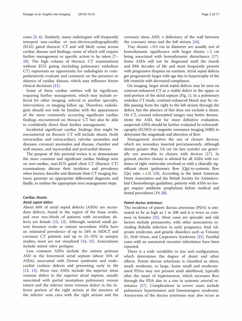
contrast-enhanced CT as a visible defect in the upper ormid-portion of the atrial septum (Fig. 1). In a pulmonaryembolus CT study, contrast-enhanced blood may be vis-ible passing from the right to the left atrium through thedefect, but the absence of this does not exclude a shunt.On CT, coronal reformatted images may better demon-strate the ASD, but for more definitive evaluation,suspected ASDs should be further evaluated by echocardi-ography (ECHO) or magnetic resonance imaging (MRI) todetermine the magnitude and direction of flow.Management involves the use of closure devices,
which are nowadays inserted percutaneously, althoughshunts greater than 3.8 cm (in late systole) are gener-ally not amenable to closure with devices [17]. Ingeneral, elective closure is advised for all ASDs with evi-dence of right ventricular overload or with a clinically sig-nificant shunt (pulmonary flow [Qp]-to-systemic flow[Qs] ratio > 1.5) [18]. According to the latest AmericanHeart Association and the British Society for Antimicro-bial Chemotherapy guidelines, patients with ASDs no lon-ger require antibiotic prophylaxis before medical anddental procedures [19, 20].
Patent ductus arteriosusThe incidence of patent ductus arteriosus (PDA) is esti-mated to be as high as 1 in 500 and it is twice as com-mon in females [21]. Most cases are sporadic and riskfactors include prematurity, with other associations in-cluding Rubella infection in early pregnancy, fetal val-proate syndrome, and genetic disorders such as Trisomy21, Holt Oram, and Carpenters Syndrome [21]. Familialcases with an autosomal recessive inheritance have beenreported.There is a wide variability in size and configuration,
which determines the degree of shunt and othereffects. Patent ductus arteriosus is classified as silent,small, moderate, or large. Some small and moderate-sized PDAs may not present until adulthood, typicallyafter the onset of hypertension, which increases flowthrough the PDA due to a rise in systemic arterial re-sistance [17]. Complications in severe cases includepulmonary hypertension and Eisenmengers syndrome.Aneurysms of the ductus arteriosus may also occur as
Krueger et al. Insights into Imaging (2019) 10:10 Page 2 of 17
a complication. Associations include ASD and ven-tricular septal defects (VSD).On contrast-enhanced CT imaging, a PDA may be seen
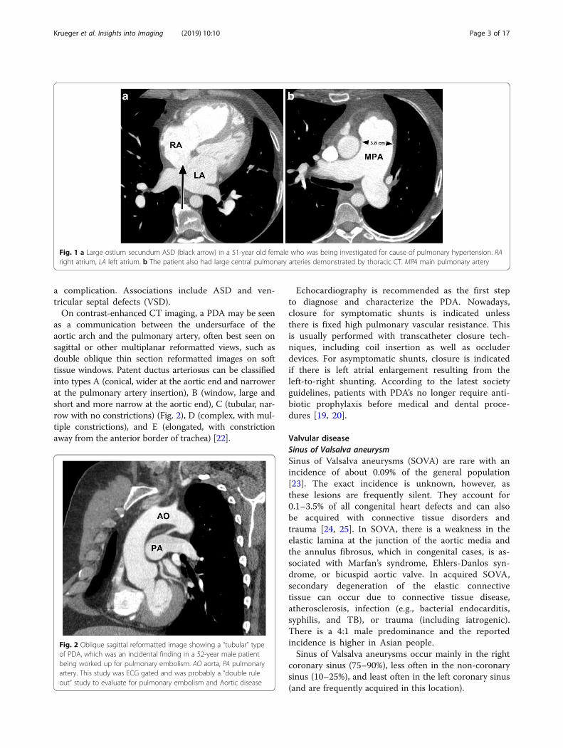
as a communication between the undersurface of theaortic arch and the pulmonary artery, often best seen onsagittal or other multiplanar reformatted views, such asdouble oblique thin section reformatted images on softtissue windows. Patent ductus arteriosus can be classifiedinto types A (conical, wider at the aortic end and narrowerat the pulmonary artery insertion), B (window, large andshort and more narrow at the aortic end), C (tubular, nar-row with no constrictions) (Fig. 2), D (complex, with mul-tiple constrictions), and E (elongated, with constrictionaway from the anterior border of trachea) [22].
Echocardiography is recommended as the first stepto diagnose and characterize the PDA. Nowadays,closure for symptomatic shunts is indicated unlessthere is fixed high pulmonary vascular resistance. Thisis usually performed with transcatheter closure tech-niques, including coil insertion as well as occluderdevices. For asymptomatic shunts, closure is indicatedif there is left atrial enlargement resulting from theleft-to-right shunting. According to the latest societyguidelines, patients with PDA’s no longer require anti-biotic prophylaxis before medical and dental proce-dures [19, 20].
Valvular diseaseSinus of Valsalva aneurysmSinus of Valsalva aneurysms (SOVA) are rare with anincidence of about 0.09% of the general population[23]. The exact incidence is unknown, however, asthese lesions are frequently silent. They account for0.1–3.5% of all congenital heart defects and can alsobe acquired with connective tissue disorders andtrauma [24, 25]. In SOVA, there is a weakness in theelastic lamina at the junction of the aortic media andthe annulus fibrosus, which in congenital cases, is as-sociated with Marfan’s syndrome, Ehlers-Danlos syn-drome, or bicuspid aortic valve. In acquired SOVA,secondary degeneration of the elastic connectivetissue can occur due to connective tissue disease,atherosclerosis, infection (e.g., bacterial endocarditis,syphilis, and TB), or trauma (including iatrogenic).There is a 4:1 male predominance and the reportedincidence is higher in Asian people.Sinus of Valsalva aneurysms occur mainly in the right
coronary sinus (75–90%), less often in the non-coronarysinus (10–25%), and least often in the left coronary sinus(and are frequently acquired in this location).
Fig. 1 a Large ostium secundum ASD (black arrow) in a 51-year old female who was being investigated for cause of pulmonary hypertension. RAright atrium, LA left atrium. b The patient also had large central pulmonary arteries demonstrated by thoracic CT. MPA main pulmonary artery
Fig. 2 Oblique sagittal reformatted image showing a “tubular” typeof PDA, which was an incidental finding in a 52-year male patientbeing worked up for pulmonary embolism. AO aorta, PA pulmonaryartery. This study was ECG gated and was probably a “double ruleout” study to evaluate for pulmonary embolism and Aortic disease
Krueger et al. Insights into Imaging (2019) 10:10 Page 3 of 17
The upper limits of normal for sinus diameter in menis 4 cm and in women, 3.5 cm, with slight variations,when adjusted for body surface area [23]. Depending onthe location of the aneurysm, there may be compressionof other structures by the aneurysm. Aortic regurgitationis a complication. Surgery is indicated for rupturedcases, and for cases with associated aortic regurgitation.Management of un-ruptured SOVA is controversial, butelective intervention is recommended for large lesions,to prevent rupture later. Intervention is also indicatedwhere there is compression of the right ventricular out-flow tract, or if the lesion is complicated by arrhythmiaor infection.On imaging, the distance from the outer edge of the
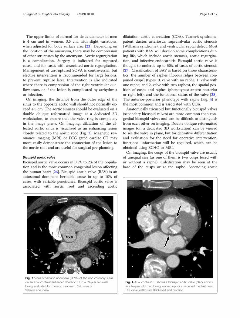
sinus to the opposite aortic wall should not normally ex-ceed 4.5 cm. The aortic sinuses should be evaluated on adouble oblique reformatted image at a dedicated 3Dworkstation, to ensure that the valve ring is completelyin the image plane. On imaging, dilatation of the af-fected aortic sinus is visualized as an enhancing lesionclosely related to the aortic root (Fig. 3). Magnetic res-onance imaging (MRI) or ECG gated cardiac CT maymore easily demonstrate the connection of the lesion tothe aortic root and are useful for surgical pre-planning.
Bicuspid aortic valveBicuspid aortic valve occurs in 0.5% to 2% of the popula-tion and is the most common congenital lesion affectingthe human heart [26]. Bicuspid aortic valve (BAV) is anautosomal dominant heritable cause in up to 10% ofcases, with variable penetrance. Bicuspid aortic valve isassociated with aortic root and ascending aortic
dilatation, aortic coarctation (COA), Turner’s syndrome,patent ductus arteriosus, supravalvular aortic stenosis(Williams syndrome), and ventricular septal defect. Mostpatients with BAV will develop some complications dur-ing life, which include aortic stenosis, aortic regurgita-tion, and infective endocarditis. Bicuspid aortic valve isthought to underlie up to 50% of cases of aortic stenosis[27]. Classification of BAV is based on three characteris-tics: the number of raphes [fibrous ridges between con-joined cusps] (types: 0, valve with no raphe; 1, valve withone raphe; and 2, valve with two raphes), the spatial pos-ition of cusps and raphes (phenotypes: antero-posterioror right-left), and the functional status of the valve [28].The anterior-posterior phenotype with raphe (Fig. 4) isthe most common and is associated with COA.Anatomically tricuspid but functionally bicuspid valves
(secondary bicuspid valves) are more common than con-genital bicuspid valves and can be difficult to distinguishfrom each other on imaging. Double oblique reformattedimages (on a dedicated 3D workstation) can be viewedto see the valve in plane, but for definitive differentiationand evaluation for the need for operative intervention,functional information will be required, which can beobtained using ECHO or MRI.On imaging, the cusps of the bicuspid valve are usually
of unequal size (as one of them is two cusps fused withor without a raphe). Calcification may be seen at thebase of the cusps or at the raphe. Ascending aortic
Fig. 3 Sinus of Valsalva aneurysm (SOVA) of the non-coronary sinuson an axial contrast enhanced thoracic CT in a 59-year old malebeing evaluated for thoracic neoplasm. SVA sinus ofValsalva aneurysm
Fig. 4 Axial contrast CT shows a bicuspid aortic valve (black arrows)in a 62-year old man being worked up for a widened mediastinum.The valve leaflets are thickened and calcified
Krueger et al. Insights into Imaging (2019) 10:10 Page 4 of 17

dilatation occurs mostly due to cystic medial necrosis(bicuspid aortopathy) rather than secondary to valvestenosis (post stenotic dilatation) (Fig. 4). Patients withBAV and aortic root or ascending aortic diametersgreater than 4 cm should undergo serial evaluation withMRI or CT, and yearly evaluation when the aortic diam-eter exceeds 4.5 cm [29]. Operative intervention is rec-ommended when the aortic root or ascending aorticdiameter is greater than 5.5 cm or if the rate of increaseis more than 0.5 cm per year [29, 30].
Coronary arterial disordersCoronary artery diseaseCoronary arterial calcification (CAC) is a marker ofthe burden of coronary artery atherosclerosis but itsrelationship to plaque instability is less predictable.Coronary arterial calcification can be an early indica-tor of coronary artery disease in asymptomatic indi-viduals being worked up for surgery or othersuspected chest pathology. Coronary artery diseasewas the most frequent cardiac finding on chest CT inprior studies with the left anterior descending coron-ary artery being the most frequently involved [6]. Thesame authors note that the presence of coronary ar-terial calcification was often not mentioned in chestCT reports [6]. Coronary arterial calcification can beeasily seen even on non-ECG gated unenhanced CT.Radiologists interpreting chest CT should not onlyevaluate the anatomic origin of the coronary arteriesbut also evaluate for the extent and severity of coron-ary arterial disease. Patients with coronary arterial cal-cification have increased risk for cardiac events,including ischemia, arrhythmia, hypotension, myocar-dial infarction, coronary arterial intervention or sur-gery, and death. For each standard deviation increasein CAC scores on non-ECG gated CT, the odds ratiofor death increased by 50%, when adjusted for trad-itional cardiovascular disease risk factors [31]. Detec-tion of coronary arterial calcification might enable thepatient to undergo primary preventative measuressuch as dietary changes, exercise, aspirin, or statintreatment. Coronary arterial calcification has a highspecificity and negative predictive value [6]. In pa-tients who are undergoing CT prior to surgery, it isimportant for the surgeon and anesthetist to knowabout the coronary arterial calcification burden, asthe patient may need functional imaging or cardiologyconsultation first or a different surgical approach oranesthetic plan.
Coronary artery ectasia and aneurysmCoronary artery aneurysms are commonly defined as a50% or greater localized increase in diameter of theaffected vessel compared with the adjacent arterial
segment [32]. Giant coronary artery aneurysms canreach several cm in diameter [33]. Coronary artery ecta-sia is defined as diffuse coronary artery dilation with lessthan 50% increase in diameter [34]. Coronary artery an-eurysms are most commonly caused by atherosclerosisin adults or by Kawasaki disease in children [35]. Otheretiologies include trauma (including iatrogenic), arteritis(granulomatosis with polyangitis, polyarteritis nodosa,systemic lupus arteriosus), infection (syphilis, staphylo-coccus), dissection, connective tissue disorders (Mar-fan’s, Ehlers-Danlos), and cocaine abuse [35].Patients may be asymptomatic or present with heart
failure or angina due to embolic or thrombotic phenom-ena. Hypertension is a risk factor. Aneurysms are mostcommonly found in the right coronary artery (RCA,40%), followed by the left anterior descending (LAD,32%) and circumflex (LCX, 23%) coronary artery [36].Aneurysms have traditionally been diagnosed by echo-cardiography (ECHO) or catheter angiography, but thelatter may underestimate the size of the aneurysm ifthere is considerable intraluminal thrombus [37]. Now-adays, they are increasingly found on CT and MR im-aging [35]. On imaging, coronary artery aneurysms areseen as fusiform dilatations of a portion of the coronaryartery (Fig. 5). Some have classified them according tothe extent of involvement (diffuse or discrete ectasia andnumber of vessels affected). Differential diagnosis in-cludes coronary artery fistula and aberrant origin of thecoronary artery from the pulmonary artery (Bland-White-Garland syndrome) [34].Further evaluation can be carried out non-invasively
with electrocardiographically (ECG) gated cardiac CT orcardiac magnetic resonance imaging (CMR). Manage-ment is unclear with some advocating surgical or endo-vascular repair (usually to treat any areas of stenosis incases of atherosclerosis) and others taking a more con-servative approach, including anti platelet agents andanticoagulation [38].
Coronary artery fistulaA coronary artery fistula (CAF) is defined as a single ormultiple direct precapillary connections between abranch of a coronary artery and the lumen of a cardiacchamber (coronary cameral fistula), or an arterial/venousstructure including the coronary sinus, superior venacava, pulmonary artery, pulmonary vein, or bronchialvein (coronary arteriovenous fistula) [39, 40]. Coronaryartery fistulas are rare, representing 0.2 to 0.4% of allcongenital heart disease (CHD), 14% of all coronaryanomalies, and often incidentally found on angiographicimaging [41, 42]. The majority of CAF are congenital,but some are acquired secondary to trauma, infection,neoplasms, or iatrogenic injury. They can also occuras a result of intracardiac congenital heart operations,
Krueger et al. Insights into Imaging (2019) 10:10 Page 5 of 17

transcutaneous techniques used for myocardial biopsyor coronary angioplasty, or as a complication ofKawasaki disease [41, 43]. As more cardiac trans-plants are performed and patients undergo myocardialbiopsies, the incidence of acquired CAF in thispatient population is increasing.Fistulas most commonly arise from the RCA (50 to
60% of cases), the left anterior descending coronaryartery (25 to 42%), the circumflex (18%), or both cor-onary arteries (5%) [33, 45]. Single fistulas are morecommon (74 to 90%) than multiple (10 to 16%) [43].The most common (60 to 90%) site of drainage is theright side of the heart [44–46]. The remaining 10%may be connected to the pulmonary artery, coronarysinus, superior vena cava (SVC), pulmonary vein, leftatrium (LA), left ventricle (LV), or have multiple con-nections [47]. Termination into a cardiac chamber orvascular structure with lower pressure may lead toenlargement and tortuosity of the artery [46]. Theclinical presentation of CAFs depends on the severityof the left-to-right shunt, with the majority detectedin adult patients being usually asymptomatic. Coron-ary angiography can reliably demonstrate the proximalpart of the CAF and allow evaluation of the size andnumber of fistulas present. However, coronary fistulasdraining into low-pressure chambers of the heart maynot be well-visualized by conventional angiographybecause of significant dilution of the contrastmedium. Electrocardiographically gated MDCT is use-ful to image CAF and provide a road map for treat-ment planning [39].Symptomatic CAF (most commonly dyspnea due to
pulmonary hypertension or heart failure as a result ofleft to right shunting, chest pain due to chronic
angina secondary to a coronary “steal” phenomenon)were usually treated with surgery, though nowadayssome can be treated percutaneously [40, 47]. Manage-ment of asymptomatic CAF is more controversial,with some advocating surgical management for largerfistulae (fistula luminal diameter is ≥ 2, the luminaldiameter of the reference vessel) [40, 45]. On CT im-aging, single or multiple connections will be seenpassing from the coronary artery to a cardiac cham-ber, pulmonary or systemic artery (Fig. 6). Referral toa cardiologist for further evaluation (which may in-clude ECG gated cardiac CT or MRI for surgicalplanning with unclear etiology) is recommended.
Anomalous coronary artery originCoronary arteries can have anomalous origins from acontralateral sinus of Valsalva, the pulmonary artery, oranother coronary artery or its branches [48–50]. Themost serious anomaly occurs when a left or right coron-ary artery arises from the contralateral sinus of Valsalvaand courses between the aorta and pulmonary trunk(intra-arterial or “malignant” course). Other high-riskanatomic features include a slit-like orifice, an intra-mural course (through the aortic wall), and an acute takeoff angle [51]. Death is thought to be caused by ischemiaresulting from ostial occlusion due to angulation orkinking at the coronary orifice or compression withinthe aortic wall or between the aorta and the pulmonaryartery during diastole [46].In contrast, anomalous coronary arteries that course
anterior to the pulmonary artery (pre-pulmonic), pos-terior to the aorta (retro-aortic), or more caudal (tothe pulmonary valve) across the ventricular septum(trans-septal) do not usually have hemodynamic
Fig. 5 a Axial contrast enhanced CT image demonstrating a coronary artery aneurysm (a) of the left anterior descending coronary artery (blackarrow) in a 56-year-old female who was being evaluated for aortic disease. b Multiplanar reformatted images in the sagittal oblique plane alsodemonstrate the aneurysm (a) of the left anterior descending coronary artery (black arrow). A aneurysm, AO aorta, PA pulmonary artery, LA leftatrium, LSPV left superior pulmonary vein. Please note that this examination was an ECG gated aortic CTA, but images were included as it wassuch a good example of coronary artery aneurysm
Krueger et al. Insights into Imaging (2019) 10:10 Page 6 of 17

consequences and are regarded as “benign.” An originof the left coronary artery from the pulmonary artery(Bland-White-Garland syndrome) or less commonlyorigin of the right coronary artery from the pulmon-ary artery is also considered as malignant as it is fre-quently associated with myocardial ischemia anddeath in early childhood [46]. Electrocardiographicallygated MDCT coronary angiography has emerged asthe standard of reference for evaluation of coronaryartery anomalies. On imaging, the abnormal originand course will be visible as a contrast-enhanced ves-sel passing anterior to the pulmonary artery, posteriorto the aorta, or coursing between the two vessels(Fig. 7). Sagittal reformatted images are useful forevaluating whether the course is intra-arterial ortrans-septal, which has implications for treatment[52]. Treatment of “malignant” anomalous coronaryarteries is usually surgical, but there have been a fewreports of successful endovascular repair in cases withfunctional obstruction [53, 54].
Abnormalities of the cardiac wallsLeft ventricular aneurysm, pseudoaneurysm, anddiverticulumIt can be difficult to distinguish true left ventricularaneurysms from pseudoaneurysms or congenital diver-ticula, but correct differentiation is crucial since pseu-doaneurysms have a high risk of rupture and surgicalrepair is recommended, while true aneurysms anddiverticula can often be managed medically. Two fea-tures which may help distinguish these entities in-clude location and ostial (neck) diameter.
Left ventricular (LV) true aneurysms occur after myo-cardial infarction with the majority at the apex, or an-terolateral wall (Fig. 8) [55]. More posterior infarcts arethought to be lethal due to involvement of the papillarymuscles and associated severe mitral valve regurgitation,with less reported. Left ventricular true aneurysms arethinned areas of scarred myocardium which can be
Fig. 6 a, b A 62-year-old woman being evaluated for weight loss. a Axial thick slab maximum intensity projection (MIP) CT image shows thecoronary artery fistula (white arrow) arising from the left anterior descending coronary artery (black arrow). b Axial CT image shows the coronaryartery fistula emptying into the main pulmonary artery (white arrow). Please note that this examination was an ECG gated aortic CTA, but imageswere included as it was such a good example of coronary artery fistula
Fig. 7 Axial contrast enhanced CT demonstrates the RCA (blackarrow) arising from the left coronary cusp in a 43-year-old femalebeing evaluated for hoarseness and vocal cord paralysis. Ao aorta, PApulmonary artery
Krueger et al. Insights into Imaging (2019) 10:10 Page 7 of 17

dyskenitic or akinetic, and complicated by heart failure,arrhythmia, stasis of blood flow, thrombus, and emboli.Pseudoaneurysms are usually located posteroinferiorly
and may be caused by ischemia, infarction, or trauma(Fig. 8) [55]. A pseudoaneurysm occurs when rupture ofthe free LV wall in myocardial infarction is contained byoverlying adherent pericardium or not contained. Leftventricular pseudoanerysms usually rupture, resulting inimmediate death, and as a result are not commonly seenby imaging. If the patient survives, pseudoaneurysmsmay cause congestive heart failure with embolic eventsas the cavity is noncontractile or dyskinetic, due to slowflow of blood and thrombosis [55]. Left ventricular aneu-rysms tend to have wide, more evident openings in con-trast to the narrow ostium of pseudoaneurysms ordiverticula which can be difficult to visualize [55, 56].Diverticula are rare congenital anomalies and typically
include endocardium, full thickness myocardium andpericardium, arising near the apex, except for the iso-lated fibrous variety, found at the heart base or in thesubvalvular area (Fig. 8) [56, 57]. Muscular diverticulaare often associated with midline defects involving theabdominal wall (omphalocoele), sternum, diaphragm,and heart (ASD) (Cantrell syndrome) [58]. Left heartcatheterization (ventriculography) was the gold standardto visualize muscular diverticula contracting in systole,in contrast to pseudoaneurysms which do not contract.Nowadays, dynamic imaging with ECG gated MDCT orfunctional cine MRI can demonstrate the form andfunction less invasively. The presence of myocardiumsurrounding the aneurysmal cavity suggests a trueaneurysm (thinned myocardium) or diverticulum (fullthickness myocardium) and myocardial discontinuitysuggests pseudoaneurysm.Finally, true aneurysms in any location within the left
ventricle are much more common than pseudoaneurysms
or diverticula; therefore, location is not an adequate criter-ion for clinical decision making. The more posterior theoutpouching, the more difficult it is to detect [59].
Myocardial diseaseDilated cardiomyopathyIn dilated cardiomyopathy (DCM), there is impairedcontraction of the left or both ventricles in the absenceof ischemic heart disease [60]. Its etiology is unknown inabout half of cases (idiopathic DCM) but may be gen-etic, viral, metabolic, or toxic. Patients present clinicallywith progressive heart failure. Complications includearrhythmia, thromboembolism, or sudden death [61].Ejection fraction is reduced and end systolic and enddiastolic left ventricular volumes are increased. Diagno-sis may be made on ECHO but results are variable, andnowadays MRI is the modality of choice for diagnosisand follow up [62]. On axial non-ECG gated CT images,if a dilated left ventricular chamber is seen (greater than5.5 cm) with a uniform left ventricular wall thicknessless than 0.7 cm, further evaluation with MRI and orECHO should be suggested (Fig. 9). This is because theleft ventricular wall will be thinner (and the chambermore dilated) in end diastole.
Left ventricular hypertrophyLeft ventricular hypertrophy (LVH) is assessed using es-timates of left ventricular mass, which usually includesmeasurements of end diastolic left ventricular mid cavitydiameter, left ventricular posterior wall, and septal wallthicknesses. On non-gated CT, left ventricular wallthickness is likely to be overestimated since imaging maynot represent end diastole. However, some suggest LVHwhen the free or septal ventricular wall exceeds 2.0 to2.5 cm in thickness on axial CT [3]. If LVH is suspectedon CT, a search should be made for etiologies including
Fig. 8 a Axial contrast-enhanced CT demonstrates a left ventricular true aneurysm (white arrowheads) in a 71-year-old male who was beingevaluated for empyema. He had a history of myocardial infarction. b Axial CT image through the chest demonstrates a left ventricularpseudoaneurysm with a narrow neck (black arrow) in a young male being evaluated for fever and chest pain. LA left atrium, RA right atrium, LVleft ventricle, PsA pseudoaneurysm. c Axial CT image through the lower chest and upper abdomen demonstrates a small apical left ventriculardiverticulum (asterisk) in a 55-year-old male being evaluated for thoracic neoplasm. LLL left liver lobe, RV right ventricle
Krueger et al. Insights into Imaging (2019) 10:10 Page 8 of 17

aortic or mitral valve disease, coarctation, or a left toright shunt (Fig. 10).LVH is usually diagnosed by electrocardiogram or on
echocardiography (ECHO) and if suspected on non-ECGgated CT, further evaluation should be recommendedusing these modalities and by cardiac MRI [63].
Hypertrophic cardiomyopathyHypertrophic cardiomyopathy (HCM) is a genetic dis-order, with an incidence of 1 in 500, typically with auto-somal dominant inheritance, variable penetrance, andexpression [60]. Myocardial hypertrophy occurs in the ab-sence of any hypertrophic stimulus and HCM is the mostcommon cardiac cause of sudden death in apparentlyhealthy young adults and athletes. Diagnosis is usuallymade by ECHO and other testing including ECG and con-firmed by cardiac catheterization or MRI imaging which
will demonstrate a hypertrophied non-dilated left ven-tricle. In HCM, myocardial hypertrophy is typicallyheterogenous, with several forms including asymmetrichypertrophy (the most common, with anteroseptal or midventricular thickening) (Fig. 11), symmetric hypertrophy,apical hypertrophy, and mass like hypertrophy [64, 65].Asymmetric septal wall hypertrophy causes obstruc-
tion of the left ventricular outflow tract (LVOT) in up toa third of cases. The mitral valve may be affected sec-ondary to left ventricular outflow tract obstruction withsystolic anterior wall motion (SAM), or because of a pri-mary abnormality in the valve itself. Anatomic narrow-ing of the LVOT during systole and SAM contribute tocause dynamic subaortic obstruction [64, 65]. On axialCT images, narrowing may be seen at the level of theleft ventricular outflow tract, in association with thicken-ing of the mid ventricular septum, and non-invasive as-sessment of the anatomic and functional changes withMRI can be recommended [60, 64, 65]. In older patients,a sigmoid shape to the interventricular septum may beseen but is not usually associated with hemodynamicchanges [66].
Myocardial calcificationMyocardial calcification may be dystrophic, metastatic,or idiopathic [67, 68]. Dystrophic calcifications are morecommon and most often seen following myocardial in-farction, with calcification reported in 8% of cases 6 yearsafter infarction, and the most common site is in the an-terior wall of the left ventricle (LV) [67]. The myocardialcalcification results from necrosis, hemorrhage, or fibro-sis and usually underestimates the size of the infarct. OnCT imaging, dystrophic myocardial calcification is thinand curvilinear and usually found within the peripheryof the infarct, at the interventricular septum and apex,distributed away from the aortic root (Fig. 12). Apicalleft ventricular aneurysms commonly calcify. Other
Fig. 9 Axial contrast-enhanced CT demonstrates dilatedcardiomyopathy (DCM) on an axial contrast enhanced image in a65-year-old man being evaluated for PE
Fig. 10 a Axial contrast-enhanced CT demonstrates left ventricular hypertrophy (LVH) on an axial contrast enhanced image in a 70-year-old manbeing evaluated for back pain. b Sagittal oblique multiplanar reformatted MPR images (short axis) demonstrate concentric hypertrophy of the leftventricular wall
Krueger et al. Insights into Imaging (2019) 10:10 Page 9 of 17
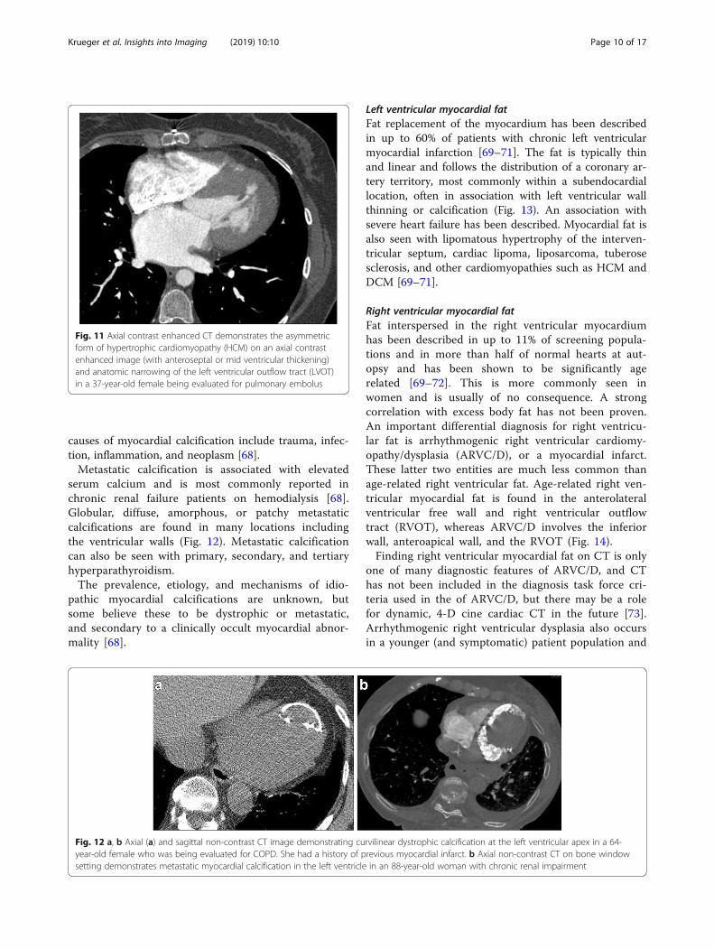
causes of myocardial calcification include trauma, infec-tion, inflammation, and neoplasm [68].Metastatic calcification is associated with elevated
serum calcium and is most commonly reported inchronic renal failure patients on hemodialysis [68].Globular, diffuse, amorphous, or patchy metastaticcalcifications are found in many locations includingthe ventricular walls (Fig. 12). Metastatic calcificationcan also be seen with primary, secondary, and tertiaryhyperparathyroidism.The prevalence, etiology, and mechanisms of idio-
pathic myocardial calcifications are unknown, butsome believe these to be dystrophic or metastatic,and secondary to a clinically occult myocardial abnor-mality [68].
Left ventricular myocardial fatFat replacement of the myocardium has been describedin up to 60% of patients with chronic left ventricularmyocardial infarction [69–71]. The fat is typically thinand linear and follows the distribution of a coronary ar-tery territory, most commonly within a subendocardiallocation, often in association with left ventricular wallthinning or calcification (Fig. 13). An association withsevere heart failure has been described. Myocardial fat isalso seen with lipomatous hypertrophy of the interven-tricular septum, cardiac lipoma, liposarcoma, tuberosesclerosis, and other cardiomyopathies such as HCM andDCM [69–71].
Right ventricular myocardial fatFat interspersed in the right ventricular myocardiumhas been described in up to 11% of screening popula-tions and in more than half of normal hearts at aut-opsy and has been shown to be significantly agerelated [69–72]. This is more commonly seen inwomen and is usually of no consequence. A strongcorrelation with excess body fat has not been proven.An important differential diagnosis for right ventricu-lar fat is arrhythmogenic right ventricular cardiomy-opathy/dysplasia (ARVC/D), or a myocardial infarct.These latter two entities are much less common thanage-related right ventricular fat. Age-related right ven-tricular myocardial fat is found in the anterolateralventricular free wall and right ventricular outflowtract (RVOT), whereas ARVC/D involves the inferiorwall, anteroapical wall, and the RVOT (Fig. 14).Finding right ventricular myocardial fat on CT is only
one of many diagnostic features of ARVC/D, and CThas not been included in the diagnosis task force cri-teria used in the of ARVC/D, but there may be a rolefor dynamic, 4-D cine cardiac CT in the future [73].Arrhythmogenic right ventricular dysplasia also occursin a younger (and symptomatic) patient population and
Fig. 11 Axial contrast enhanced CT demonstrates the asymmetricform of hypertrophic cardiomyopathy (HCM) on an axial contrastenhanced image (with anteroseptal or mid ventricular thickening)and anatomic narrowing of the left ventricular outflow tract (LVOT)in a 37-year-old female being evaluated for pulmonary embolus
Fig. 12 a, b Axial (a) and sagittal non-contrast CT image demonstrating curvilinear dystrophic calcification at the left ventricular apex in a 64-year-old female who was being evaluated for COPD. She had a history of previous myocardial infarct. b Axial non-contrast CT on bone windowsetting demonstrates metastatic myocardial calcification in the left ventricle in an 88-year-old woman with chronic renal impairment
Krueger et al. Insights into Imaging (2019) 10:10 Page 10 of 17

there is often associated right ventricular wall dilatation[74]. The diagnosis of ARVC/D is challenging, due tothe absence of unique diagnostic criteria, incompletepenetrance, and variable disease expression. In addition,diagnostic tests are not sensitive enough, findings arenon-specific, and the diagnostic criteria used are sub-jective [73]. Therefore, if ARVC/D is suspected, referralto a cardiologist for complete assessment of diagnostictask force criteria for ARVC/D (which include featureson cardiac MRI) is recommended.
Chamber enlargementLeft atrial enlargementLeft atrial enlargement can be caused by mitral valvulardisease (stenosis and regurgitation), atrial fibrillation, leftto right shunts, left ventricular hypertrophy (LVH), leftventricular failure, dilated cardiomyopathy, and ischemicheart disease. A search should be made for thrombus inthe left atrium or its appendage. In addition, evaluationof the mitral valve for calcification or thickening can bemade, as well as checking the other chamber sizes andwall thicknesses, and looking for coronary arterial calci-fication. Left atrial anteroposterior measurements shouldideally be made on a three-chamber view (aortic outflowtract, left atrium, and left ventricle) at the end of leftventricular systole [75]. Measurements of the largest an-teroposterior left atrial diameter (made at the level ofthe aortic valve), greater than 4.5 cm on axial non-ECGgated CT, had 94% specificity (53% sensitivity) for leftatrial dilatation [76]. Normalized reference range volumemeasurements for the left atrium and other cardiacchambers have been established using dynamic magneticresonance imaging by Maceira and colleagues [77].
Right atrial enlargementRight atrial enlargement can be caused by pulmonaryarterial hypertension, tricuspid valvular disease (stenosisand regurgitation), atrial fibrillation, left to right shunts,left ventricular hypertrophy (LVH), and ischemic heartdisease. There are no accepted standardized measure-ments for right atrial sizes. Normalized reference rangevolume measurements for the right atrium and othercardiac chambers have been established using dynamicmagnetic resonance imaging [77].
Left ventricular enlargementLeft ventricular enlargement (LVE) can be indicativeof many pathological entities, including cardiomyopa-thies, ischemic heart disease and valvular dysfunction.Non-ECG gated CT images likely will not be ob-
tained in end-diastole and so the ventricular cavitysize may be underestimated. A recent study suggestedthat left ventricular chamber measurements greaterthan 5.6 cm (made half way between the apex andthe valve, from inner wall to inner wall) on axialnon-ECG gated CT had a 100% specificity (78% sensi-tivity) and negative predictive value of 93% (positivepredictive value of 80%) [78]. Therefore, dimensionalthresholds are best used to provide specific detection(rule in) and to suggest for chamber enlargement,rather than being able to exclude it. Normalized refer-ence range volume measurements for the left ven-tricle and other cardiac chambers are well establishedby dynamic magnetic resonance imaging [79].
Fig. 13 Axial image demonstrates fat replacement in an apical leftventricular infarct on a patient being evaluated for chronic dyspneaby high resolution computed tomography (HRCT)
Fig. 14 Axial non-contrast CT (which was performed to follow up alung nodule) demonstrates fat replacement in the free wall of theright ventricle (white arrowheads) in a 50-year-old male
Krueger et al. Insights into Imaging (2019) 10:10 Page 11 of 17

Right ventricular enlargementRight ventricular enlargement is difficult to assess, dueto its complex shape and variability and lack of specificlandmarks that can be used as reference points. Rightventricular enlargement is found with pulmonary hyper-tension, pulmonary valve stenosis or regurgitation, leftto right shunts (ventricular septal defect), and cardiomy-opathy. Normalized reference range volume measure-ments for the right ventricle and other cardiac chambershave been established using dynamic magnetic reson-ance imaging [80].
Pericardial diseaseConstrictive pericarditisConstrictive pericarditis results from infections, tubercu-losis, connective tissue disorder, neoplasm, or trauma[81]. Although pericardial calcification is found in up to28% of cases of constrictive pericarditis, the finding doesnot necessarily cause constrictive physiology [64]. Peri-cardial constriction from pericardial fluid or from pro-gressive pericardial fibrosis (with a stiff or less compliantpericardium) causes impaired ventricular filling duringdiastole and diastolic heart failure. This leads to dilata-tion of the inferior vena cava, hepatic veins, and rightatrium with a normal or reduced volume right ventricle(RV). An elongated cone-shaped RV and asigmoid-shaped interventricular septum are sometimesseen (Fig. 15). Constrictive pericarditis can be difficult todistinguish from restrictive cardiomyopathy clinically and
radiologically. However, differentiation is important asconstrictive pericarditis can sometimes be treated surgi-cally by pericardiectomy [82]. The two entities can be dis-tinguished on imaging based on whether the pericardiumis of normal thickness (restrictive cardiomyopathy) or ofabnormally increased thickness (constrictive pericarditis).MR imaging with flow sensitive sequences can help distin-guish these two entities [82]. If pericardial constriction issuspected, further imaging with ECHO and cardiac MRIand referral to cardiology is recommended.
HemopericardiumPericardial effusion results when more than 60 mL offluid accumulates in the pericardial sac. Hemorrhagicpericardial effusion is better characterized on unen-hanced CT and measures between 40 and 60 HU(Hounsfield units), compared to less than 10 HU forsimple fluid/transudates and between 10 to 20 HU forexudates (Fig. 16). Hemopericardium can occur follow-ing left ventricular free wall rupture in myocardial in-farction, with ventricular or coronary aneurysmalrupture, in postsurgical patients, in the setting oftrauma, and with malignancy [83].
Pericardial calcificationPericardial calcification commonly results from trauma,infection, hemorrhage, or therapeutic radiation [67].Traumatic etiologies include surgical exploration duringpericardiotomy for cardiac shunt surgery. Tuberculosisand histoplasmosis as infectious etiologies are now lesscommonly seen in the developed world. Calcificationresulting from radiation will confirm to the radiationport used. The calcification may be a focal plaque or athin curvilinear focus following the cardiac contour.
Fig. 15 Axial contrast-enhanced CT shows thickening of the visceraland parietal pericardium (black arrows) with fluid in between thepericardial layers in a 77-year-old male patient being investigated forloculated pleural effusion. This pericardial thickening and fluid areresulting in constrictive physiology (constrictive pericarditis),evidenced by a decreased volume, cone-shaped right ventricle, anda sigmoid-shaped interventricular septum (black asterisks)
Fig. 16 Non-contrast axial image from a dedicated aortic CTthrough the lower chest, showing hemopericardium in a 43-year-oldmale who being evaluated for congestive heart failure and lowejection fraction, following aortic valve surgery
Krueger et al. Insights into Imaging (2019) 10:10 Page 12 of 17

Lesions are usually 1 to 2 mm thick but can increase to1 to 2 cm in thickness in chronic cases. Pericardial calci-fication is most commonly found in the atrioventricular(AV) groove and in the lower and diaphragmatic por-tions of the pericardium (Fig. 17). It is most commonly aglobal process, involving both sides of the heart. Myo-cardial calcification tends to localize to the left heart(where the myocardium is thickest) and is thinner thanpericardial calcification [67]. Up to 28% of patients withconstrictive pericarditis have pericardial calcification, butthe converse is not true [64]. Pericardial calcificationsmay be present without any physiological compromiseto the heart. If pericardial calcification is seen on a chestCT, evaluation of the images for signs of constriction isrecommended, with consideration for ECHO and or car-diac MRI to further evaluate [84, 85].
Filling defects in the cardiac chambersThrombiThrombus is the most common filling defect found incardiac chambers with a predilection for the left atrialappendage and left ventricular apex (Fig. 18) [85]. Insinus rhythm, left atrial appendage thrombi are usuallyassociated with mitral valve disease or left atrial chamberand appendage dysfunction. The incidence of left atrialthrombi is significantly higher in patients with atrial fib-rillation and mitral valve disease [86]. Right atrialthrombus is more likely to occur in the presence of cen-tral venous catheters. Patients with wall motion abnor-malities, such as left ventricular aneurysm, are atincreased risk for left ventricular thrombi. Patients withprosthetic cardiac valves and pacemakers are also at in-creased risk. Intracardiac thrombi are complicated by
arterial embolism in up to 20% of cases, and thereforeearly identification with subsequent therapy is important[86]. On CT imaging, thrombi typically do not enhancebut chronic thrombi can appear heterogeneous with per-ipheral fibrous capsule formation or they may calcify[85]. Cardiac MRI can help distinguish thrombi frommasses, such as tumor thrombus or myxomas.
MyxomasCardiac myxomas are the most common primary neo-plasm and account for about half of all primary benigncardiac tumors [87, 88]. The majority (75%) arise withinthe left atrium, typically at the interatrial septum nearthe limbus of the fossa ovalis. Most are sporadic, but oc-casionally there is a familial predisposition or an associ-ated clinical complex (e.g., Carney complex). Myxomasmay be asymptomatic or present with cardiac obstruct-ive symptoms, embolic phenomena, and constitutionalsymptoms. On contrast-enhanced CT imaging, myx-omas are usually lower in attenuation (occasionally thesame attenuation as myocardium) within the contrastfilled cardiac chamber (Fig. 19). They can arise from anyendocardial surface and their attachment can be difficultto determine by CT. Myxomas may be heterogenous inattenuation due to calcification (particularly in the rightheart), areas of hemorrhage, thrombus, or hemosiderindeposition. Origin and size can sometimes be used tohelp differentiate myxomas from thrombus with myx-omas being larger, and often arising from the fossa ovalisas opposed to the left atrial appendage [89].
Malignant cardiac massesMetastasesMetastases to the heart and pericardium are 20 to 40times more common than primary cardiac malignan-cies and are generally associated with a poor progno-sis [90]. Cardiac metastases are more frequentlycarcinomas than sarcomas, with prevalences of be-tween 1.5 and 25% reported for patients with malig-nant tumors [91]. Due to their proximity to the heart,many metastases originate in the lung or breast, butall types of malignant disease can involve the heartwith an increased propensity reported for melanoma[91]. Tumors involve the pericardium and heart byone of four pathways (direct contiguous spread,hematogenous dissemination, transvenous extension,or retrograde lymphatic invasion). Metastases to theheart and pericardium can manifest as a pulmonaryor mediastinal mass with direct invasion, as myocar-dial masses (due to hematogenous spread) as a cen-tral mass extending into the left atrium with venousextension), or as a pericardial effusion or nodularity(due to lymphatic extension) [92]. Direct extensioncan occur with bronchial, breast, esophageal, and
Fig. 17 Axial CT image demonstrates chunky pericardial calcificationon a non-contrast image, in the AV grooves and bilaterally in a 36-year-old male being investigated for chronic pain
Krueger et al. Insights into Imaging (2019) 10:10 Page 13 of 17

thymic neoplasms due to proximity of these tumors.With hematogenous metastases in the heart and peri-cardium, spread can occur via the coronary arteries,and there is usually evidence of hematogenous metas-tases in other organs (Fig. 20). Transvenous extensionof tumor thrombus can occur in the right atriumthrough the superior and inferior vena cava, or intothe left atrium via the pulmonary veins.
Primary cardiac tumorsPrimary cardiac tumors are rare with a reported inci-dence of less than 0.02 to 0.056% with pericardial tu-mors being even rarer [93]. Primary malignant cardiactumors account for 25% of primary cardiac tumors [91].Primary cardiac neoplasms are frequently asymptomaticuntil they are large and produce non-specific symptoms.Before the advent of cross-sectional imaging, primary
cardiac tumors were rarely diagnosed before death.Now, they are found in the living and characterizationhas become important in directing their management.The most common primary tumor in adults is angiosar-coma [94]. Angiosarcoma predominantly presents inmiddle-aged male patients, with a slight preponderancereported in males. Angiosarcomas usually involve theright atrium with right heart failure. On CT, these tu-mors manifest as a discrete mass protruding into a car-diac chamber or as a diffuse infiltrating mass with areasof necrosis (Fig. 21) [91]. These highly vascular tumorsexhibit areas of necrosis and pericardial invasion leadsto hemorrhagic pericardial effusion and or cardiac tam-ponade. The prevalence of metastatic disease is reportedat 66 to 89% and the prognosis is poor.
Fig. 18 a Axial CT image demonstrating thrombus (black arrow) in the right atrium in a 73-year-old patient who had CT to evaluate forpulmonary embolus. b Axial CT image demonstrates thrombus within the appendage of the left atrium (white arrows) of another patient whohad atrial fibrillation who was being evaluated for pleural effusions
Fig. 19 Axial contrast-enhanced CT shows a pedunculated myxomain the right atrium, which extends through the tricuspid valve intothe right ventricle. RA right atrium, M myxoma, RV right ventricle, LVleft ventricle
Fig. 20 Axial CT with contrast demonstrates nodules and masses(black arrows) in the interventricular septum and the left ventricularfree myocardial wall, in a 78-year-old male patient with metastaticmalignant melanoma. There is also a large necrotic lymph node inthe left mediastinum adjacent to the cardiac implants
Krueger et al. Insights into Imaging (2019) 10:10 Page 14 of 17

Rhabdomyosarcoma is the most common primary car-diac tumor in infants and children, with a slight malepredominance. Rhabdomyosarcomas can be multiple,are more likely to occur on the valves, and can occur inall chambers, with variable presentation. They tend toinvolve the myocardium and can invade the pericardiumwith nodular masses, rather than diffuse sheet-like thick-ening. The other sarcomas (undifferentiated, leiomyosar-coma, fibrosacroma, and osteosarcoma) typically involvethe left heart chambers and cause left heart failure [94].Low attenuation components in a mass arising typicallyfrom the posterior wall of the left atrium may be seenwith leiomyosarcomas, while chondroid or osteoid ele-ments may be seen with osteosarcomas. Unfortunately,delayed diagnosis leads to a poor prognosis with an aver-age survival of 1 year. Cardiac lymphoma is defined asone that is mostly confined to the heart or pericardium(to distinguish it from invasive non-Hodgkin’s lymph-oma) and almost all primary cardiac lymphomas are ag-gressive B cell lymphomas. Unlike other primary cardiacmalignancies, primary cardiac lymphoma has a favorableresponse to chemotherapy. They are more frequent inthe right atrium and pericardial effusion is a commonfeature, and sometimes the only finding visible on im-aging. Findings of primary cardiac lymphoma on CT arenon-specific.CT provides good soft tissue detail and can depict cal-
cification, ossification or fat, or make a tissue diagnosisin the case of lipomas. However, cardiac MR imaginghas the added advantage of providing functional
information and utilizes multiple planes which can helppre-therapy planning [95, 96].
ConclusionCardiac disease and pathology are commonly seen onthoracic CT performed for other indications. Relativelycommon and significant entities include filling defectsdue to thrombus or myxoma. Other not uncommonlyencountered entities include small left to right shunts,the sequelae of prior MI (such as ventricular fat or cal-cium deposition), or pericardial constriction. Rarer butsignificant entities described include coronary artery fis-tula or aneurysm, anomalous coronary artery course, leftventricular aneurysm, or pseudoaneurysm and malignantcardiac tumors (metastases). Radiologists should beaware of the imaging features of these cardiac diseasesencountered on thoracic CT, as some will require spe-cific further referral or diagnostic testing. After complet-ing this article, the reader should be familiar withclinically significant cardiac findings seen on chest CTexaminations and be confident to direct the next man-agement steps.
AbbreviationsARVC/D: Arrhythmogenic right ventricular cardiomyopathy/dysplasia;ASD: Atrial septal defects; AV: Atrioventricular; BAV: Bicuspid aortic valve;CAF: Coronary artery fistula; CHD: Congenital heart disease; COA: Aorticcoarctation; CT: Computed tomography; DCM: Dilated cardiomyopathy; ECGgated: Electrocardiographically gated; ECHO: Echocardiography;HCM: Hypertrophic cardiomyopathy; LA: Left atrium; LAD: Left anteriordescending; LCX: Circumflex; LV: Left ventricle; LVE: Left ventricularenlargement; LVH: Left ventricular hypertrophy; LVOT: Left ventricular outflowtract; MDCT: Multi-detector row CT; MRI: Magnetic resonance imaging;PDA: Patent ductus arteriosus; RV: Right ventricle; RVOT: Right ventricularoutflow tract; SAM: Systolic anterior wall motion; SOVA: Sinus of Valsalvaaneurysm; SVC: Superior vena cava; VSD: Ventricular septal defects
Authors’ contributionsMK and AK carried out the initial manuscript drafting and imaging collection.PC provided some images later. MK and AK participated in the design of thestudy and performed the literature reviews. AK and MK conceived of thestudy and participated in its design and coordination. All authors read andapproved the final manuscript.
Competing interestsThe authors declare that they have no competing interests.
Publisher’s NoteSpringer Nature remains neutral with regard to jurisdictional claims inpublished maps and institutional affiliations.
Author details1Department of Radiology, Division of Cardiothoracic Radiology, University ofMichigan, 1500 East Medical Center Drive, Ann Arbor, MI 48109, USA.2Fulford Radiology, Base Hospital, Private Bag 2016, New Plymouth, Taranaki4342, New Zealand.
Received: 12 July 2018 Accepted: 10 January 2019
References1. National Council on Radiation Protection and Measurements. Report No.
160, Ionizing Radiation Exposure of the Population of the United States.
Fig. 21 Axial contrast-enhanced CT demonstrates a mass (blackarrows) involving the right atrium and ventricle, (later confirmed tobe angiosarcoma by histology) on axial contrast enhanced CT andreformatted images in a 27-year-old female being investigated forpossible pulmonary embolism (shortness of breath and dizziness)
Krueger et al. Insights into Imaging (2019) 10:10 Page 15 of 17

Available at: http://ncrponline.org/publications/reports/ncrp-report-160/.Accessed on 23 Jan 2019
2. Smith-Bindman R, Miglioretti DL, Johnson E et al (2012) Use of diagnosticimaging studies and associated radiation exposure for patients enrolled inlarge integrated health care systems, 1996-2010. JAMA 307:2400–2409
3. Lee SH, Seo JB, Kang JW, Chae EJ, Park SH, Lim TH (2008) Incidental cardiacand pericardial abnormalities on chest CT. J Thorac Imaging 23:216–226
4. Berland LL (2011) The American College of Radiology strategy for managingincidental findings on abdominal computed tomography. Radiol Clin NorthAm 49:237–243
5. Foley PW, Hamaad A, El-Gendi H, Leyva F (2010) Incidental cardiac findingson computed tomography imaging of the thorax. BMC Res Notes 3:326
6. Choy G, Kröpil P, Scherer A et al (2013) Pertinent reportable incidentalcardiac findings on chest CT without electrocardiography gating: review of268 consecutive cases. Acta Radiol 54:396–400
7. Bruzzi JF, Rémy-Jardin M, Delhaye D, Teisseire A, Khalil C, Rémy J (2006)When, why, and how to examine the heart during thoracic CT: part 1, basicprinciples. AJR Am J Roentgenol 186:324–332
8. Guthaner DF, Wexler L, Harell G (1979) CT demonstration of cardiacstructures. AJR Am J Roentgenol 133:75–81
9. Scherer A, Choy G, Kröpil P, Lanzman RS, Mödder U, Abbara S (2009)Cardiac pathologies incidentally detected with non-gated chest CT. Rofo181:1127–1134
10. Lim KC, Chai P, Teo LS (2011) Incidental cardiac abnormalities on non-electrocardiogram-gated multi-detector computed tomography imaging ofthe thorax and abdomen. Singapore Med J 52:906–912 quiz 913
11. McKie SJ, Hardwick DJ, Reid JH, Murchison JT (2005) Features of cardiacdisease demonstrated on CT pulmonary angiography. Clin Radiol 60:31–38
12. Wu JC, Child JS (2004) Common congenital heart disorders in adults. CurrProbl Cardiol 29:641–700
13. Webb G, Gatzoulis MA (2006) Atrial septal defects in the adult: recentprogress and overview. Circulation 114:1645–1653
14. Kosehan D, Akin K, Koktener A, Cakir B, Aktas A, Teksam M (2011)Interatrial shunt: diagnosis of patent foramen ovale and atrial septaldefect with 64-row coronary computed tomography angiography. Jpn JRadiol 29:576–582
15. Saremi F, Channual S, Raney A et al (2008) Imaging of patent foramen ovalewith 64-section multidetector CT. Radiology 249:483–492
16. Kafka H, Mohiaddin RH (2009) Cardiac MRI and pulmonary MR angiographyof sinus venosus defect and partial anomalous pulmonary venousconnection in cause of right undiagnosed ventricular enlargement. AJR AmJ Roentgenol 192:259–266
17. Berko NS, Haramati LB (2012) Simple cardiac shunts in adults. SeminRoentgenol 473:277–288
18. Akagi T (2015) Current concept of transcatheter closure of atrial septaldefect in adults. J Cardiol Jan 65:17–25
19. Wilson W, Taubert KA, Gewitz M et al (2007) Prevention of infectiveendocarditis: guidelines from the American Heart Association: a guidelinefrom the American Heart Association rheumatic fever, endocarditis, andKawasaki disease committee, council on cardiovascular disease in theYoung, and the council on clinical cardiology, council on cardiovascularsurgery and anesthesia, and the quality of care and outcomes researchinterdisciplinary working group. Circulation 116:1736–1754
20. Gould FK, Elliott TS, Foweraker J et al (2006) Guidelines for the preventionof endocarditis: report of the working party of the British Society forantimicrobial chemotherapy. J Antimicrob Chemother 57:1035–1042
21. Schneider DJ (2012) The patent ductus arteriosus in term infants, children,and adults. Semin Perinatol 36:146–153
22. Krichenko A, Benson LN, Burrows P, Möes CA, McLaughlin P, Freedom RM(1989) Angiographic classification of the isolated, persistently patent ductusarteriosus and implications for percutaneous catheter occlusion. Am JCardiol 63:877–880
23. Weinreich M, Yu PJ, Trost B (2015) Sinus of valsalva aneurysms: review ofthe literature and an update on management. Clin Cardiol 38:185–189
24. Feldman DN, Roman MJ (2006) Aneurysms of the sinuses of Valsalva.Cardiology 106:73–81
25. White CS, Plotnick GD (2001) Case 33: sinus of valsalva aneurysm. Radiology219:82–85
26. Ko SM, Song MG, Hwang HK (2012) Bicuspid aortic valve: spectrum ofimaging findings at cardiac MDCT and cardiovascular MRI. AJR Am JRoentgenol 198:89–97
27. Michelena HI, Prakash SK, Della Corte A et al (2014) Bicuspid aortic valve:identifying knowledge gaps and rising to the challenge from theInternational Bicuspid Aortic Valve Consortium (BAVCon). Circulation 129:2691–2704
28. Braverman AC, Güven H, Beardslee MA, Makan M, Kates AM, Moon MR(2005) The bicuspid aortic valve. Curr Probl Cardiol 30:470–522
29. Nishimura RA, Otto CM, Bonow RO et al (2014) 2014 AHA/ACC Guideline forthe Management of Patients with Valvular Heart Disease: executivesummary: a report of the American College of Cardiology/American HeartAssociation Task Force on Practice Guidelines. Circulation 129:2440–2492Erratum in: Circulation 129:e650
30. Vahanian A, Alfieri O, Andreotti F et al (2012) Guidelines on themanagement of valvular heart disease (version 2012): the Joint Task Forceon the Management of Valvular Heart Disease of the European Society ofCardiology (ESC) and the European Association for Cardio-Thoracic Surgery(EACTS). Eur J Cardiothorac Surg 42:S1–S44
31. Hughes-Austin JM, Dominguez A 3rd, Allison MA et al (2016) Relationship ofcoronary calcium on standard chest CT scans with mortality. JACCCardiovasc Imaging 9:152–159
32. Devabhaktuni S, Mercedes A, Diep J, Ahsan C (2016) Coronary arteryectasia-a review of current literature. Curr Cardiol Rev 12:318–323
33. Crawley PD, Mahlow WJ, Huntsinger DR, Afiniwala S, Wortham DC (2014)Giant coronary artery aneurysms: review and update. Tex Heart Inst J 41:603–608
34. Restrepo CS, Lane MJ, Murillo H (2012) Cardiac aneurysms,pseudoaneurysms, and diverticula. Semin Roentgenol 47:262–276
35. Tan KT, Challenor V, McGann G (2007) CT diagnosis of coronary arteryaneurysms. Int J Cardiol 118:273–274
36. Gowda RM, Dogan OM, Tejani FH, Khan IA (2005) Left main coronary arteryaneurysm. Int J Cardiol 105:115–116
37. Hoey ET, Nagra I, Ganeshan A (2011) Cardiac aneurysms and diverticula:magnetic resonance and computed tomography appearances. Curr ProblDiagn Radiol 40:72–84
38. Shambrook JS, Chowdhury R, Brown IW, Peebles CR, Harden SP (2010)Cross-sectional imaging appearances of cardiac aneurysms. Clin Radiol 65:349–357
39. Zenooz NA, Habibi R, Mammen L, Finn JP, Gilkeson RC (2009) Coronaryartery fistulas: CT findings. Radiographics 29:781–789
40. Reddy G, Davies JE, Holmes DR, Schaff HV, Singh SP, Alli OO (2015)Coronary artery fistulae. Circ Cardiovasc Interv 8:e003062
41. Luo L, Kebede S, Wu S, Stouffer GA (2006) Coronary artery fistulae. Am JMed Sci 332:79–84
42. Buccheri D, Luparelli M, Chirco PR, Piraino D, Andolina G, Assennato P(2016) A call to action for an underestimated entity: our algorithm fordiagnosis and management of coronary artery fistula. Int J Cardiol 221:1081–1083
43. Mangukia CV (2012) Coronary artery fistula. Ann Thorac Surg 93:2084–209244. Gowda RM, Vasavada BC, Khan IA (2006) Coronary artery fistulas: clinical and
therapeutic considerations. Int J Cardiol 107:7–1045. Abdelmoneim SS, Mookadam F, Moustafa S et al (2007) Coronary artery fistula:
single-center experience spanning 17 years. J Interv Cardiol 20:265–27446. Shriki JE, Shinbane JS, Rashid MA et al (2012) Identifying, characterizing, and
classifying congenital anomalies of the coronary arteries. Radiographics 32:453–468
47. Latson LA (2007) Coronary artery fistulas: how to manage them. CatheterCardiovasc Interv 70:110–116
48. Komatsu S, Sato Y, Ichikawa M et al (2008) Anomalous coronary arteries inadults detected by multislice computed tomography: presentation of casesfrom multicenter registry and review of the literature. Heart Vessels 23:26–34
49. Kim SY, Seo JB, Do KH et al (2006) Coronary artery anomalies: classificationand ECG-gated multi-detector row CT findings with angiographiccorrelation. Radiographics 26:317–333 discussion 333-314
50. Zeina AR, Blinder J, Sharif D, Rosenschein U, Barmeir E (2009) Congenitalcoronary artery anomalies in adults: non-invasive assessment withmultidetector CT. Br J Radiol 82:254–261
51. Krupiński M, Urbańczyk-Zawadzka M, Laskowicz B et al (2014) Anomalousorigin of the coronary artery from the wrong coronary sinus evaluated withcomputed tomography: "high-risk" anatomy and its clinical relevance. EurRadiol 24:2353–2359
52. Brothers JA, Whitehead KK, Keller MS et al (2015) Cardiac MRI and CT:differentiation of normal ostium and intraseptal course from slitlike ostium
Krueger et al. Insights into Imaging (2019) 10:10 Page 16 of 17

and interarterial course in anomalous left coronary artery in children. AJRAm J Roentgenol 204:W104–W109
53. Vadivelu R, Bagga S (2013) Is endovascular therapy the right choicefor treatment of functional compression of anomalous right coronaryartery arising from left coronary sinus with interarterial course? BMJCase Rep https://casereports.bmj.com/content/casereports/2013/bcr-2012-007856.full.pdf. Accessed on 23 Jan 2019
54. Shah N, Cheng VE, Cox N, Soon K (2015) Percutaneous coronaryintervention of an anomalous left Main coronary artery arising from theright sinus of Valsalva. Heart Lung Circ 24:e123–e126
55. Brown SL, Gropler RJ, Harris KM (1997) Distinguishing left ventricular aneurysmfrom pseudoaneurysm. A review of the literature. Chest 111:1403–1409
56. Cianciulli TF, Del Carmen Gonzalez Colaso P, Saccheri MC et al (2009) Leftventricular diverticulum, a rare echocardiographic finding: two adultpatients and review of the literature. Cardiol J 16:76–81
57. Romagnoli A, Ricci A, Morosetti D, Fusco A, Citraro D, Simonetti G (2015)Congenital left ventricular diverticulum: multimodality imaging evaluationand literature review. J Saudi Heart Assoc 27:61–67
58. Makkuni P, Kotler MN, Figueredo VM (2010) Diverticular and aneurysmalstructures of the left ventricle in adults: report of a case within the contextof a literature review. Tex Heart Inst J 37:699–705
59. Zoffoli G, Mangino D, Venturini A et al (2009) Diagnosing left ventricularaneurysm from pseudo-aneurysm: a case report and a review in literature. JCardiothorac Surg 4:11
60. Soler R, Rodríguez E, Remuiñán C, Bello MJ, Díaz A (2003) Magneticresonance imaging of primary cardiomyopathies. J Comput Assist Tomogr27:724–734
61. Mohan SB, Parker M, Wehbi M, Douglass P (2002) Idiopathic dilatedcardiomyopathy: a common but mystifying cause of heart failure. Cleve ClinJ Med 69:481–487
62. Shehata ML, Turkbey EB, Vogel-Claussen J, Bluemke DA (2008) Role ofcardiac magnetic resonance imaging in assessment of nonischemiccardiomyopathies. Top Magn Reson Imaging 19:43–57
63. Armstrong AC, Gidding S, Gjesdal O, Wu C, Bluemke DA, Lima JA (2012) LVmass assessed by echocardiography and CMR, cardiovascular outcomes,and medical practice. JACC Cardiovasc Imaging 5:837–848
64. Bogaert J, Olivotto I (2014) MR imaging in hypertrophic cardiomyopathy:from magnet to bedside. Radiology 273:329–348
65. Maron MS (2012) Clinical utility of cardiovascular magnetic resonance inhypertrophic cardiomyopathy. J Cardiovasc Magn Reson 14:13
66. Funabashi N, Umazume T, Takaoka H et al (2013) Sigmoid shapedinterventricular septum exhibit normal myocardial characteristics and has arelationship with aging, ascending aortic sclerosis and its tilt to leftventricle. Int J Cardiol 168:4484–4488
67. Gowda RM, Boxt LM (2004) Calcifications of the heart. Radiol Clin North Am42:603–617 vi-vii
68. Nance JW Jr, Crane GM, Halushka MK, Fishman EK, Zimmerman SL (2015)Myocardial calcifications: pathophysiology, etiologies, differential diagnoses,and imaging findings. J Cardiovasc Comput Tomogr 9:58–67
69. Kimura F, Matsuo Y, Nakajima T et al (2010) Myocardial fat at cardiacimaging: how can we differentiate pathologic from physiologic fattyinfiltration? Radiographics 30:1587–1602
70. Jacobi AH, Gohari A, Zalta B, Stein MW, Haramati LB (2007) Ventricularmyocardial fat: CT findings and clinical correlates. J Thorac Imaging 22:130–135
71. Zafar HM, Litt HI, Torigian DA (2008) CT imaging features and frequency ofleft ventricular myocardial fat in patients with CT findings of chronic leftventricular myocardial infarction. Clin Radiol 63:256–262
72. Kirsch J, Johansen CK, Araoz PA, Brady PA, Williamson EE, Glockner JF (2010)Prevalence of fat deposition within the right ventricular myocardium inasymptomatic young patients without ventricular arrhythmias. J ThoracImaging 25:173–178
73. Gandjbakhch E, Redheuil A, Pousset F, Charron P, Frank R (2018) Clinicaldiagnosis, imaging, and genetics of arrhythmogenic right ventricularcardiomyopathy/dysplasia: JACC state-of-the-art review. J Am Coll Cardiol72:784–804
74. Rastegar N, Burt JR, Corona-Villalobos CP et al (2014) Cardiac MR findingsand potential diagnostic pitfalls in patients evaluated for arrhythmogenicright ventricular cardiomyopathy. Radiographics 34:1553–1570
75. Kuchynka P, Podzimkova J, Masek M et al (2015) The role of magneticresonance imaging and cardiac computed tomography in the assessmentof left atrial anatomy, size, and function. Biomed Res Int 2015:247865
76. Huckleberry J, Haltom S, Issac T, Gabaldon J, Ketai L (2012) Accuracy of non-ECG-gated computed tomography angiography of the chest in assessmentof left-sided cardiac chamber enlargement. J Thorac Imaging 27:354–358
77. Maceira AM, Cosin-Sales J, Prasad SK, Pennell DJ (2016) Characterization ofleft and right atrial function in healthy volunteers by cardiovascularmagnetic resonance. J Cardiovasc Magn Reson 18:64
78. Kathiria NN, Devcic Z, Chen JS et al (2015) Assessment of left ventricularenlargement at multidetector computed tomography. J Comput AssistTomogr 39:794–796
79. Maceira AM, Prasad SK, Khan M, Pennell DJ (2006) Normalized leftventricular systolic and diastolic function by steady state free precessioncardiovascular magnetic resonance. J Cardiovasc Magn Reson 8:417–426
80. Maceira AM, Prasad SK, Khan M, Pennell DJ (2006) Reference rightventricular systolic and diastolic function normalized to age, gender andbody surface area from steady-state free precession cardiovascular magneticresonance. Eur Heart J 27:2879–2888
81. Welch TD, Oh JK (2015) Constrictive pericarditis: old disease, newapproaches. Curr Cardiol Rep 17:20
82. Young PM, Glockner JF, Williamson EE et al (2012) MR imaging findings in76 consecutive surgically proven cases of pericardial disease with CT andpathologic correlation. Int J Cardiovasc Imaging 28:1099–1109
83. Chaturvedi A, Vargas D, Ocazionez D (2018) CT for evaluation of acutepericardial emergencies in the ED. Emerg Radiol 25:321–328
84. Cremer PC, Kwon DH (2015) Multimodality imaging of pericardial disease.Curr Cardiol Rep 17:24
85. Bogaert J, Francone M (2013) Pericardial disease: value of CT and MRimaging. Radiology 267:340–356
86. Mortensen KH, Gopalan D, Balan A (2013) Atrial masses on multidetectorcomputed tomography. Clin Radiol 68:e164–e175
87. Egolum UO, Stover DG, Anthony R, Wasserman AM, Lenihan D, Damp JB(2013) Intracardiac thrombus: diagnosis, complications and management.Am J Med Sci 345:391–395
88. Díaz Angulo C, Méndez Díaz C, Rodríguez García E, Soler Fernández R, RoisSiso A, Marini Díaz M (2015) Imaging findings in cardiac masses (part I):study protocol and benign tumors. Radiologia 57:480–488
89. Grebenc ML, Rosado-de-Christenson ML, Green CE, Burke AP, Galvin JR (2002)Cardiac myxoma: imaging features in 83 patients. Radiographics 22:673–689
90. Scheffel H, Baumueller S, Stolzmann P et al (2009) Atrial myxomas andthrombi: comparison of imaging features on CT. AJR Am J Roentgenol 192:639–645
91. Díaz Angulo C, Méndez Díaz C, Rodríguez García E, Soler Fernández R, RoisSiso A, Marini Díaz M (2016) Imaging findings in cardiac masses. Part II:malignant tumors and pseudotumors. Radiologia 58:26–37
92. Anavekar NS, Bonnichsen CR, Foley TA et al (2010) Computed tomographyof cardiac pseudotumors and neoplasms. Radiol Clin North Am 48:799–816
93. Chiles C, Woodard PK, Gutierrez FR, Link KM (2001) Metastatic involvement ofthe heart and pericardium: CT and MR imaging. Radiographics 21:439–449
94. Restrepo CS, Vargas D, Ocazionez D, Martínez-Jiménez S, Betancourt Cuellar SL,Gutierrez FR (2013) Primary pericardial tumors. Radiographics 33:1613–1630
95. Restrepo CS, Largoza A, Lemos DF et al (2005) CT and MR imaging findingsof malignant cardiac tumors. Curr Probl Diagn Radiol 34:1–11
96. Motwani M, Kidambi A, Herzog BA, Uddin A, Greenwood JP, Plein S (2013)MR imaging of cardiac tumors and masses: a review of methods andclinical applications. Radiology 268:26–43
Krueger et al. Insights into Imaging (2019) 10:10 Page 17 of 17