silane endodontic irrigant - fitebac dental...a b s t r a c t objective. to analyze effect of...
TRANSCRIPT

d e n t a l m a t e r i a l s 3 5 ( 2 0 1 9 ) 1264–1278
Available online at www.sciencedirect.com
ScienceDirect
jo ur nal ho me pag e: www.int l .e lsev ierhea l th .com/ journa ls /dema
An in vitro study of a novel quaternary ammoniumsilane endodontic irrigant
U. Daooda,∗,1, A. Paroliaa,1, A. Elkezzaa, C.K. Yiub, P. Abbott c,J.P. Matinlinnad, A.S. Fawzyc
a Clinical Dentistry, Restorative Division, School of Dentistry, International Medical University Kuala Lumpur, 126,Jalan Jalil Perkasa 19, 57000 Bukit Jalil, Wilayah Persekutuan Kuala Lumpur, Malaysiab Paediatric Dentistry and Orthodontics, Faculty of Dentistry, The University of Hong Kong, Prince Philip DentalHospital, 34 Hospital Road, Sai Ying Pun, Hong Kong Special Administrative Regionc UWA Dental School, University of Western Australia, 17 Monash Avenue, Nedlands, WA 6009, Australiad Dental Materials Science, Applied Oral Sciences, Faculty of Dentistry, The University of Hong Kong, 34 HospitalRoad, Sai Ying Pun, Hong Kong Special Administrative Region
a r t i c l e i n f o
Article history:
Received 2 March 2019
Received in revised form
19 May 2019
Accepted 22 May 2019
Keywords:
Root dentine
Quaternary ammonium silane
Condensation
E. faecalis
Antimicrobial
Raman spectroscopy
Confocal microscopy
a b s t r a c t
Objective. To analyze effect of NaOCl + 2% quaternary ammonium silane (QAS)-containing
novel irrigant against bacteria impregnated inside the root canal system, and to evaluate its
antimicrobial and mechanical potential of dentine substrate.
Methods. Root canal was prepared using stainless steel K-filesTM and ProTaperTM and sub-
jected to manual and ultrasonic irrigation using 6% NaOCl + 2% CHX, 6% NaOCl + 2% QAS and
saline as control. For confocal-microscopy, Raman spectroscopy and SEM analysis before and
after treatment, Enterococcus faecalis cultured for 7 days. Raman spectroscopy analysis was
done across cut section of gutta percha/sealer-dentine to detect resin infiltration. Indenta-
tion of mechanical properties was evaluated using a Berkovich indenter. The contact angle
of irrigants and surface free energy were evaluated. Mineralization nodules were detected
through Alazarin red after 14 days.
Results. Control biofilms showed dense green colonies. Majority of E. faecalis bacteria were
present in biofilm fluoresced red in NaOCl + 2% QAS group. There was reduction of 484 cm−1
Raman band and its intensity reached lowest with NaOCl + 2% QAS. There was an increase in
1350–1420 cm−1 intensity in the NaOCl + 2% CHX groups. Gradual decrease in 1639 cm−1 and
1609 cm−1 Raman signal ratios were seen in the resin-depth region of 17 �m>, 14.1 �m> and
13.2 �m for NaOCl + 2% QAS, NaOCl + 2% CHX and control groups respectively. All obturated
groups showed an intact sealer/dentine interface with a few notable differences. 0.771 and
83.5% creep indentation distance for NaOCl + 2% QAS ultrasonic groups were observed. High-
est proportion of polar component was significantly found in the NaOCl + 2% QAS groups
which was significantly higher as compared to other groups. Mineralized nodules were
increased in NaOCl + 2% QAS.
∗ Corresponding author at: Clinical Dentistry, Restorative Division, Faculty of Dentistry, International Medical University Kuala Lumpur,126, Jalan Jalil Perkasa 19, 57000 Bukit Jalil, Wilayah Persekutuan Kuala Lumpur, Malaysia.
E-mail address: [email protected] (U. Daood).https://doi.org/10.1016/j.dental.2019.05.0200109-5641/© 2019 The Academy of Dental Materials. Published by Elsevier Inc. All rights reserved.

d e n t a l m a t e r i a l s 3 5 ( 2 0 1 9 ) 1264–1278 1265
Significance. Favorable antimicrobial and endodontic profile of the NaOCl + 2% QAS solution
might suggest clinical use for it for more predictable reduction of intracanal bacteria.
© 2019 The Academy of Dental Materials. Published by Elsevier Inc. All rights reserved.
1
MtaetpccfvtsdmddMu
cilrellpuitdo[eiCtada
bttsra
. Introduction
icroorganisms breach dental hard tissues via acidic dissolu-ion [1] through cracks in tooth or exposed dentinal tubulesnd reach pulp space to cause infection [2]. Given that,ndodontic failure may occur because of bacterial penetra-ion or persistence within the root canal system as result ofoor disinfection procedure [3]. Plentiful rinsing is required tolean and disinfect the root canal system for preparation andompletion of endodontic procedure [4]. Current guidelinesor root canal treatment [5] advocate the idea for the use ofoluminous irrigating solution avoiding its extrusion throughhe apical foramen. Irrigation is usually carried out using ayringe and a needle, which may not be able to clean andisinfect remote areas of the root canal system [6]. During nor-al clinical practice many techniques have been adapted by
ental practioners. Therefore, many elaborate methods wereeveloped in the past to improve root canal disinfection [7].ost widely used adjunct method for root canal irrigation isltrasonic agitation [8].
Most of the attention has been given to the use ofhlorhexidine (CHX) when dealing with antimicrobials. CHXs bactericidal in high concentrations and bacteriostatic inow concentrations, primarily against gram-positive bacte-ia [9]. Chlorhexidine is water-soluble and has reversiblelectrostatic bonding between protonated amine groups [10]eading to eventual leaching out from bonded interfaces, withoss of antimicrobial and protease inhibitory activities in theresence of calcium due to chelation [11]. When NaOCl issed in conjunction with CHX, the former is left behind
nside the canal system, and CHX forms a brown precipi-ate [12], containing para-chloroaniline (PCA) with subsequentiscoloration and a possible hindrance in the sealing of thebturating material along with blockage of the lateral canals
13]. Therefore, an intermediate rinse is recommended afterach irrigant solution. Some studies suggest that the precip-tate contains PCA due to the chemical interaction betweenHX and NaOCl [14]. Even so, other studies have suggested
hat this precipitate did not contain PCA [15]. However, theim of the present laboratory study was not to investigate theeposits formed. Moreover, there is a clinical need to look forlternative irrigation material and regimen.
Nevertheless, some issues like incomplete elimination ofacteria, and weakening of antibacterial actions have ledo exploration of alternative options. It is, therefore, vitalo develop a novel irrigant or disinfectant with predictable
resent an effective class of antimicrobial agents [16]. Thesol–gel process has been used for the design of silica (SiO2)-based materials in the context of silica gel formation. Thehydrolysis process produces silanol groups ( Si OH) and thereaction proceeds and is followed by condensation of Si O Sibonds with inorganic polymerization [17]. The reactions indi-cate a typical sequence of condensation products that includemonomer, dimers and higher order molecular ring products[18]. The silicon atom center has a direct interaction with thesolvents as the condensation occurs near the terminal silanolgroups, forming a chain-like structure inside the sol and net-work like gels [19].
The most common compounds that participate in thesilica sol-gel chemical reaction are tetramethoxysilane(TMOS) or tetraethoxysilane (TEOS). Tetraethoxysilane actsas an anchoring unit in the formation of the 3D siloxanenetwork. As hydrolysis and condensation proceed further,the OH and O Si-units, attached to a specific siliconatom, are increased [20]. The experimental versions ofnovel quaternary ammonium silane (SiQAS) disinfectant(K21 molecule) examined in the present study were syn-thesised by sol–gel reaction. Because methanol is producedduring the hydrolysis and condensation reactions of SiQACthe molecule is potentially toxic for intraoral use [21].Hence, SiQAC has been substituted with 3-(triethoxysilyl)-propyldimethyloctadecyl ammonium chloride (i.e. the ethoxysubstituted SiQAC, abbreviated as Et-SiQAC) for coupling withtetraethoxysilane (TEOS) via sol–gel synthesis. This resultedin the generation of ethanol- or acetone-soluble, fully-hydrolysed, partially-condensed QAS (1-octadecanaminium,N,N’-[[3,3-bis[[[3-(dimethyloctadecylammonio)propyl] dihydroxysilyl]oxy]-1,1,5,5,-tetrahydroxyl-1,5-trisiloxanediyl]di-3,1-propanediyl]-bis[N,N-dimethyl] chloride(1:4), with the CAS number 1566577-36-3 (codenamed K21).The yellow, partially condensed solid was converted into gran-ular powder in absence of ethanol by pressurized reactionmixture under low pressure. The compound was imme-diately dissolved in absolute ethanol to produce solutionscontaining 2 wt%, QAS to be used as the experimental irrigantin the present study. However, it is not known whether aQAS-containing disinfectant has the same inhibitory effectsagainst bacteria when impregnated in radicular dentine andcanals.
Accordingly, the objectives of present laboratory study wereto investigate the effect of a NaOCl + 2% QAS-containing disin-fectant/irrigant system against bacteria impregnated into theroot canal system, and to evaluate its antimicrobial potential.
trong antibacterial effects. That said, such a novel mate-ial should exhibit no adverse effect on the subsequentlypplied resin sealers. Quaternary ammonium compounds rep-
1 Equal contribution authors.
Considering the complex endodontic system and the difficultyof penetrating dentinal tubules, with concerns of possible PCA
and deposits being formed after use of CHX and NaOCl, thereis a potential to exploit the QAS disinfectant as an irrigantfor a feasible therapeutic approach against biofilm infection
s 3 5
1266 d e n t a l m a t e r i a lwithin the root canal system. The null hypotheses tested werethat NaOCl + 2% QAS cavity irrigants had (i) no antimicrobialeffect on Enterococcus faecalis species biofilm architecture, (ii)no effect on E. faecalis species adhesion on dentine structure,and (iii) no effect on mechanical properties of dentine sub-strate.
2. Materials and methods
2.1. Manufacture of quaternary ammonium silanepowder
The 2% QAS irrigant examined was synthesized by sol-gel reaction between 1 mol of TEOS (Mw 208 g mol−1) and4 mol of Et-SiQAC (Mw 538 g mol−1) as reported previouslyDaood et al. [35]. Briefly, 1 mol of TEOS (Mw 208 g mol−1) and4 mol of 3-(triethoxysilyl)-propyldimethyloctadecyl ammo-nium chloride (i.e. the ethoxy substituted SiQAC, abbreviatedas Et-SiQAC; Mw 538 g mol−1). In a typical synthesis, 2.08 gof TEOS was blended with 29.89 g of 3-(triethoxysilyl)-propyldimethyloctadecyl ammonium chloride (72% of Et-SiQAC dissolved in ethanol) and 5 mL of ethanol (to renderthe blend more homogeneous). Hydrolysis was initiated bythe addition of 10.08 g of 0.02 M HCl-acidified deionized water.The pH was set 1.66, representing 3.5 times the stoichiomet-ric molar concentration of water required, to ensure completehydrolysis. Completion of the hydrolysis reaction took approx-imately 3 h and it was indicated by the disappearance ofabsorbance peaks of the Si O C peak at 1078 cm−1 and the
OC2H5 peak at 1172 cm−1 as it was monitored by FTIR. TheIR spectrum of the fully hydrolysed reaction was characterisedby the presence of silanol groups, water and ethanol. Therewas presence of Si O Si cyclic or open chained species iden-tified as impurities (Fig. 1A). Since ethanol is a by-product ofthe sol–gel reaction and there is elimination of methanol, QASwas successfully used without further purification [22] in allour studies [23,54,62].
2.2. Canal irrigation
Eighty extracted non-carious human single rooted anteriorteeth were obtained from patients (21–29 years old) with theirinformed consent under a protocol reviewed and approvedby Institutional Review Board of International Medical Uni-versity, JC Committee (IMU 425–2018). All the experimentswere carried out in accordance with approved guidelinesand regulations. Teeth were stored in 0.02% sodium azideat 4 ◦C to prevent bacterial growth and were used in lessthan 1 month after extraction. Teeth were cleaned using peri-odontal curettes to remove periodontal tissues and storedin saline. A low-speed diamond edge-coated disc (Bredent
®,
Senden, Germany) mounted on a milling machine (K9 MillingApparatus-990, Kavo, München, Germany) under water cool-ing was used to decoronate the tooth 1 mm below the
cemento-enamel junction to obtain the root segment. Rootcanal preparation was performed by a single trained endodon-tist. Root canals were shaped using stainless steel K-filesTM(Dentsply Sirona, Tulsa Dental, USA) using watch wind motion.
( 2 0 1 9 ) 1264–1278
For ProTaperTM shaping, rotary instruments (both DentsplyMaillefer, Ballagues, Switzerland) were used up to size X4and irrigated with 2 mL of 6% NaOCl (Calasept, UpplandsVäsby, Sweden) after each use. Smear layer of root canals wereremoved using 6% NaOCl and 17% EDTA (Pulpdent Corpora-tion, Warwick, UK) for 2 min and canals thoroughly rinsedwith sterile saline after each irrigation. These teeth were fur-ther subjected to sterilization by autoclave for 20 min at 121 ◦C.Thereafter, these teeth were randomly assigned in six studygroups (n = 5) and subjected to one of the following irrigationprotocols:
Group A: 6% NaOCl + 2% CHX (Manual irrigation using 30-gauge side vented needle, Endo-EZE, Ultradent).
Group B: 6% NaOCl + 2% CHX (UltraSonic agitation, VDWUltra, VDW GmbH, Munich, Germany).
Group C: 6% NaOCl + 2% QAS (Manual irrigation using 30-gauge side vented needle, Endo-EZE, Ultradent).
Group D: 6% NaOCl + 2% QAS (UltraSonic agitation, VDWUltra, VDW GmbH, Munich, Germany).
Group E: Saline (Manual irrigation using 30-gauge sidevented needle, Endo-EZE, Ultradent).
Group F: Saline (UltraSonic agitation, VDW GmbH, Munich,Germany).
2.3. Teeth preparation
For confocal microscopy, Raman spectroscopy and SEM anal-ysis before and after treatment, the teeth (n = 5 per group)were prepared to a size of 50 K-file which was 1 mm shortof the apical foramen using crown down technique. Crownswere sectioned using low-speed diamond edge-coated disc(Bredent
®, Senden, Germany) mounted on a milling machine
and length standardized to 16 mm from coronal to apex ofthe root. EDTA was added for 3 min for removal of the smearlayer and the teeth were autoclaved at 120 ◦C for 30 min. E.faecalis (ATCC 29212) was cultured in 10 mL BHI broth supple-mented with 8% sucrose (pH 7.4) and a minimal amount ofxylitol (0–2%) was added at 37 ◦C for 48 h and later incubatedat 37 ◦C for 24 h. After 4000 rpm centrifugation for 15 min,each of the cell pellets was washed three times with sterilephosphate buffered solution (PBS, 0.01 M, pH 7.2), later re-suspended (O.D reading of 0.11 at 660 nm) in 100 mL of therespective growth medium and adjusted to a concentration ofMcFarland 3 (109 cells/mL) before use.
Five milliliters of BHI broth was mixed with equal weight ofbacterial inoculum using sterilized syringes of sufficient vol-ume to fill the root canal during a 7-day period. After 7 days,each tooth, under aseptic conditions, were dried with sterilepaper points. After subjecting the specimens to different irri-gation protocols (no irrigation protocol for control specimens),two parallel grooves were prepared on external surfaces inthe mesio-distal direction of each tooth to facilitate split frac-ture. Final separation was made using a chisel and a hammerand teeth were then taken for SEM, confocal microscopy andRaman analysis.
2.4. Raman spectroscopy for resin sealer penetration
After irrigation with designated protocols for each studygroup, the root canals were dried with paper points and speci-

d e n t a l m a t e r i a l s 3 5 ( 2 0 1 9 ) 1264–1278 1267
Fig. 1 – (A) Comparison between the partially and fully condensed K21 QAS solution with ethanol as the reference; (B)Representative line map (scans) across sealer interface of different control and after different irrigation protocols specimens.The spectral contribution is recorded at 1639 cm−1 and 1609 cm−1 obtained ratio intensities representing the penetration ofdifferent groups. (C) Raman spectra of E. faecalis biofilms on root dentine specimens treated with different irrigationprotocols. Spectral differences of control and treated specimens can be seen in the 484 cm−1 region after normalization.Biofilm colonies after inoculation and irrigation protocol exposure providing initial comparison with standard signatureRaman spectra and change in intensities. (D) Raman spectra of root dentine with treated biofilms cultivation. Yellow regionindicates the variation of peaks after treatment at Amide I region with peak based discrimination. (For interpretation of ther d to
mbaiuwofusFRaatRilt
eferences to color in this figure legend, the reader is referre
ens (n = 3) were then randomly assigned to each study group,ased on the irrigation protocol. The materials (RealSeal SEnd gutta percha) for obturation were manipulated accord-ng to the manufacturer’s instructions and canals sealedsing single cone obturation technique. Each root sectionas cut horizontally into approximately 1 mm thick slices tobtain the middle third sections and each section subjectedor Raman spectroscopy analysis. The spectra were obtainedsing a Raman spectrometer equipped with a Leica micro-cope and lenses (JY LabRam HR 800, Horiba Jobin Yvon,rance) with curve-fitting Raman software (Labspec 5). Theaman spectra was obtained using the 785 nm laser sourcet 50× magnification and a spectral resolution of 1 cm−1 and
laser power of 4.94 mW. The laser beam was focused acrosshe cut section of the gutta percha/sealer-dentine interface.
eadings were taken from the central regions of the specimennterface starting in the resin region of the specimen (on theeft), across the interface and ending onto the dentin side ofhe specimen (on the right) in 1 �m steps using the x–y–z stage
the web version of this article.)
to detect resin infiltration and background-corrected for darkcounts with intensity normalization. Origin 8 (Origin Lab Corp,Northampton, MA) was used to calculate sealer penetration.The peak intensity at 1609 cm−1 was considered as an inter-nal standard for aromatic C C and 1639 cm−1 which is thealiphatic C C, is indicative of cured seal.
2.5. Biofilm preparation and Raman spectroscopy dataacquisition
For Raman spectroscopy measurements, specimens wereremoved after 7 days from culture plates, and allowed todry for 15 min at 35 ◦C. The specimens were transferred tomicroscopic slides (low fluorescent quartz). Areas of 5 �m cho-sen randomly across all dried biofilm disc specimens. Raman
spectra were recorded using a Raman spectrometer (Nanocat,University of Malaya) equipped with a Leica microscope andlenses (JY LabRam HR 800, Horiba Jobin Yvon, France) withcurve-fitting Raman spectroscopy software (Labspec 5). The
s 3 5
1268 d e n t a l m a t e r i a lspectra were collected at 785 nm wavelength with argon ion632.8 nm laser at a <500 �W power and a 100× objective (asuperior signal/noise ratio). The exposure time was 60 s as thesample (including the biofilm specimens) was translated at0.5 �m intervals in X and Y directions with 1 s exposure timein the information-rich region between 200 and 3200 cm−1. Allspectra were recorded at room temperature and in dark toavoid any peaks (background noise) originating due to ambi-ent light. Raman peaks were centered at 434 cm−1 (assigned tostretching vibration of V2PO4) [24], 960 cm−1 (hydroxyapatitePO4) [25] for inorganic species, and, 484 cm−1 (polysaccharidesor carbohydrates) [26], 1655 cm−1 (amide I {C O}) [27], and1454 cm−1 (pyrrolidine rings of proline and hydroxyprolineinside collagen) [28] for organic to the peak at 1070 cm−1.
2.6. Confocal analysis
Non-destructive identification of QAS within the root denti-nal surface was performed using 0.1 wt% aqueous solution ofsodium fluorescein (46960 Bioreagent, Millipore Sigma, Darm-stadt, Germany).
After irrigation with NaOCl + 2% QAS the specimens (n = 3)were rinsed in 0.1 wt% fluorescein for 24 h. The specimensrinsed with deionised water were examined using confocallaser scanning microscope (CLSM, Leica Fluoview FV 1000,Olympus, Tokyo, Japan), equipped with a 60×/1.4 NA oilimmersion lens using 488 nm argon/helium laser beam and633 nm krypton ion laser illumination, both in the reflectionand fluorescence modes. Reflected and fluorescence signalswere detected with a photomultiplier tube to a depth of 20 �mand then converted to single-projection images for better visu-alization and qualitative analysis. Stacks of fluorescent imagesobtained of the biofilm were examined using bioimageL soft-ware (v.2.0. Malmö, Sweden), which provides information onthe structure of the biofilm, including green and red-stainedbacteria volume on a two dimensional x–y section based oncolor segmentation algorithms written in MATLAB.
2.7. SEM analysis
For SEM analysis of biofilms, the specimens (preparationdescribed above in Section 2.2) were placed onto the SEMsample holder and imaging was performed at 3500× magni-fications at a working distance of 5–8 mm to aid in locatingthe same area after specimen placement, to be used as mark-ers for accurate co-registration. Next, images were cropped tothe same size to enable batch processing in the segmentationstep.
As expected, neutralization of QAS irrigant would takeplace by the buffering action of dentine leading to a con-densation of 3D network within and around the dentinetubules. Dentine canals in specimens (n = 3) were subjected toNaOCl + 2% CHX or NaOCl + 2% QAS irrigation protocol accord-ing to the groups. Two parallel grooves were made onto theexternal surfaces, in the mesio-distal direction to facilitate asplit fracture. Final separation was made using a chisel and a
hammer. All specimens were dehydrated in ascending gradesof ethanol (33%, 66%, 85%, 95%, 2 × 100%, for 20 min in each)and immediately transferred to the pressure chamber of thecritical point drying machine (CPD 30, Leica). The specimens( 2 0 1 9 ) 1264–1278
were mounted on aluminium stubs (specimen holders) withdouble-sided conductive tape, sputter-coated with 30 nm thicklayer Au/Pd (120 s) and examined using a SEM (Philips/FEI XL30FEG SEM) at an accelerated voltage of 10 kV at different mag-nifications and images evaluated by two examiners.
The interface morphology between root dentine and resin-based sealer were investigated using SEM. From each apicalthird, two 1 mm slices were cut, polished sequentially to 1200grit size by silicon carbide papers (Carbimet, Buehler, LakeBluff, IL, USA) and slices were prepared for resin infiltra-tion into dentine and resin tag formation. The slices wereacid etched using 35% phosphoric acid (15 s) and then rinsedusing distilled water for 15 s. The specimens were immersedin 5.25% NaOCl solution for 15 min and rinsed under run-ning water for 5 min. Next, all specimens were post fixedusing osmium tetroxide (OsO4), rinsed using PBS solution,dehydrated in ascending ethanol for 20 min. and dried in acritical-point dryer (CPD30 Baltec, Leica, Guyancourt, France).After air-drying, the specimens were mounted on stubs andgold sputtered for 120 s under vacuum and imaged using aPhilips/FEI XL30 FEG at an accelerating voltage of 10 kV.
2.8. Nano-indentation testing
A low-speed diamond edge-coated disc (Bredent®
, Senden,Germany) mounted on a milling machine (K9 MillingApparatus-990, Kavo, Germany) under water cooling was usedto decoronate the tooth 1 mm below the cemento-enameljunction to obtain the root segment. After following the irriga-tion protocols, indentation mechanical properties of radicularroot canal dentine (n = 5) were evaluated using NanoscopeIII (Digital Instruments, Santa Barbara, CA) with a Berkovichindenter (Hysitron, Minneapolis, MN, USA). An average of tenmeasurements were taken 1.5 mm apart at 6 N force, 2 Hzspeed and 10 indentations per cycle at the root canal lumen.The indentations were available at the following parameters:the first indentation cyclic (the distance between first andlast cycles), the creep indentation distance (a progressive riseduring application of force from the first indentation) andincreased indentation (the distance between the first and lastindentation ‘distances’). In addition to this, specimens werecarbon coated and a Philips/FEI XL30 FEG unit at an accelerat-ing voltage of 10 kV was used to confirm the appearance andlocation of indentations.
2.9. Contact angles and surface free energymeasurements
After selecting the designated irrigation regimens for root den-tine specimens, the teeth were horizontally sectioned 1 mmbelow the cemento-enamel juncion to obtain 5 mm dentineblocks (DB) for each group. Each dentine block was split into 2semi-cylindrical halves. Deionized water was used as the ref-erence liquid for contact angle measurements. Each irrigant
reference liquid was applied in the form of a drop (2 �L) andprofile recorded using contact angle analyzer (Dental Simula-tion Lab, IMU Laboratory). After analysis, Image J software wasused for calculating the contact angle fitting within the con-
5 ( 2
tf
�
wewLt
2
Msastr0gtA(hcditittio
hidwisaTrmcb1iti
2
Aw2tOm
d e n t a l m a t e r i a l s 3
our of the droplet, placed on the dentine surface. The surfaceree energy was calculated using the equation [29]:
L(1 – cos�) = 2[(�LD�S
D)0.5 + (�L+�S
−)0.5 + (�L+�S
−)0.5] (1)
here, �, � are the contact angle and surface tension of refer-nce liquid used. L and S subscripts represent different phases,hereas superscripts D, −, and + represent dispersive and the
ewis bases. All images captured were taken in 5 min afterheir placement on the dentine surface.
.10. Alazarin red staining
ineralization nodules were detected through Alazarin redtaining after performing 14 days of induction followingpplication of root canal irrigation systems on root dentineurface (n = 5). A 0.5 mm thick dentine disc was prepared fromhe middle-root dentine of each tooth giving square-shapedoot dentine discs. The final disc thickness of approximately.4 mm. It was achieved by grinding the outer side with wet 320rit silicon carbide papers. The NIH 3T3 MF cell line was cul-ured in Dulbecco’s Modified Eagle’s Medium (DMEM, Sigmaldrich, St. Louis, MO, USA) with 1% penicillin/streptomycin
10,000 U/100 �g/mL) and 10% fetal bovine serum (FBS) in aumidified incubator with 5% CO2 incubator at 37 ◦C. Theells were cultured to seventh passages and expanded to 4×ilution as the cells reached 85% confluence. After 3 days of
ncubation, the cells (3 × 10⁴) were seeded on the pulpal side ofhe root dentine discs (0.28 cm2) in 24-well plates (n = 5) in anncubator with 5% CO2 and 95% air at 37 ◦C. Next the discs wereransferred back to the same wells to receive the irrigationreatments. The irrigation formulations were applied by plac-ng discs into respective petri dishes containing a 2 mm depthf irrigation formulations for 2 min, followed by blot-drying.
All procedures were performed in a vertical laminar flowood. After the treatment, the root dentine discs were placed
n the CO2 incubator for additional 24 h. The root dentineiscs and cell culture were rinsed twice with PBS, and theyere fixed for 1 h using 70% ethanol. After rinsing the spec-
mens thrice using de-ionized water, the specimens weretained using 40 mmol/l Alizarin Red solution (Sigma-Aldrich),djusted to a pH of 4.0 with ammonium hydroxide for 20 min.hen, the cells were rinsed again with de-ionized water foremoval of excess dye. Photographs were taken using a light
icroscopy (LW 0.52 Nikon Japan, Eclipsee). The cells wereounted (0.28 cm2) in 24-well plates. The cells were then incu-ated with 10% cetylpyridinium chloride (Sigma-Aldrich) for5 min to solubilize the nodules. The absorbance of the result-ng solution formed for each group was calculated based onhe mean value of the control group at 14 days as 100% stain-ng.
.11. Statistical analysis
ll data are presented as mean ± standard deviation and theyere analyzed using a statistical package (SigmaStat Version
0, SPSS, Chicago, IL, USA software) assessed for a normal dis-ribution using the Shapiro–Wilk test for normality (p > 0.05).ne-way analysis of variance followed by the Tukey post hocultiple comparison test, was used to evaluate the antimi-
0 1 9 ) 1264–1278 1269
crobial effect on the percentages of live and dead bacteriaand micro-indentation analysis. All statistical analysis useda 95% confidence limit, so that the p values ≥ 0.05 were notconsidered statistically significant. The surface free energywas calculated using one-way analysis of variance (p < 0.05).One-way ANOVA was also conducted to determine significantdifferences amongst Alazarin red staining groups (p-value of0.05).
3. Results
Raman spectroscopy at 1 �m across the interface in all exper-imental groups is shown in Fig. 1B. A gradual decrease in the1639 cm−1 and 1609 cm−1 peak ratios are seen in the region of17 �m>, 14.1 �m> and 13.2 �m for NaOCl + 2% QAS, NaOCl + 2%CHX and control groups respectively, ending in a plateauassigned by the 1639 cm−1/1609 cm−1 ratios. The results indi-cate a comprehensive penetration of the sealer in NaOCl +2%QAS groups, confirming also a complex interaction betweenthe sealer and specimens. The abrupt decrease in ratio inten-sities of 1639 cm−1/1609 cm−1 in all groups indicate resistanceto sealer penetration (Fig. 1B).
From the spectra it can be observed that the bands in thisregion respond mostly with treatment on the biofilm and typ-ify changes within the absorbance spectrum with intensitychanges with different irrigation specimen groups (Fig. 1C).There were obvious variations in spectra with the biofilmchanges at 484 cm−1 according to the compounds used. Theweak bands refer to the glycosidic link or the ring waggingof possible polysaccharides within the biofilm or spectro-scopic signature due to linkages of polysaccharides. There wasa gradual reduction and striking difference of the 484 cm−1
band as the intensity reaches the lowest with NaOCl + 2% QASgroups. This suggests a decrease in the carbohydrate contentwithin the biofilm, again a factor that is more pronounced withthe QAS groups. These carbohydrate moieties are more likelycomplex C O and C C stretches coupled with C H deforma-tions.
The changes within the groups affecting the differentbiofilms, although highly reproducible, are far more pro-nounced in Fig. 1D, as tests were performed in triplicates.There is an increase in the underlying intensity in the1350–1420 cm−1 region in the biofilm in the NaOCl + 2% CHXgroups suggesting an increase in the carbohydrate content(p < 0.05), a factor more pronounced for biofilm backbone. Thenew band at 993 cm−1 supports the argument that we areobserving an increase in carbohydrate content in the biofilm(p < 0.05). The resolution at 1100 cm−1 to 1150 cm−1 indicatesthe peptidoglycan and polysaccharide in this region of thebiofilm, which are seen typically high in the NaOCl + 2% CHXgroups (Fig. 3).
Representative images of biofilm formed on the specimensare presented in Fig. 2. In the control samples (Fig. 2A), the E.faecalis biofilm appears to be in higher quantity covering thedentine surface. The biofilms show densely clustered green
colonies (98.71 ± 0.71 live bacteria) with minimal areas of deadbacterial cells. Results of one-way ANOVA verified significantlylower biofilm volume and thickness after the use of irrigationprotocols, especially in the case of NaOCl + 2% QAS groups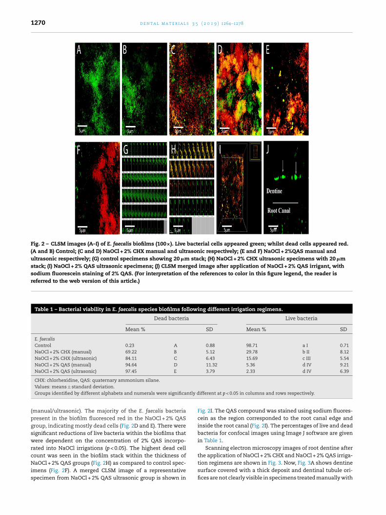
1270 d e n t a l m a t e r i a l s 3 5 ( 2 0 1 9 ) 1264–1278
Fig. 2 – CLSM images (A–I) of E. faecalis biofilms (100×). Live bacterial cells appeared green; whilst dead cells appeared red.(A and B) Control; (C and D) NaOCl + 2% CHX manual and ultrasonic respectively; (E and F) NaOCl + 2%QAS manual andultrasonic respectively; (G) control specimens showing 20 �m stack; (H) NaOCl + 2% CHX ultrasonic specimens with 20 �mstack; (I) NaOCl + 2% QAS ultrasonic specimens; (J) CLSM merged image after application of NaOCl + 2% QAS irrigant, withsodium fluorescein staining of 2% QAS. (For interpretation of the references to color in this figure legend, the reader isreferred to the web version of this article.)
Table 1 – Bacterial viability in E. faecalis species biofilms following different irrigation regimens.
Dead bacteria Live bacteria
Mean % SD Mean % SD
E. faecalisControl 0.23 A 0.88 98.71 a I 0.71NaOCl + 2% CHX (manual) 69.22 B 5.12 29.78 b II 8.12NaOCl + 2% CHX (ultrasonic) 84.11 C 6.43 15.69 c III 5.54NaOCl + 2% QAS (manual) 94.64 D 11.32 5.36 d IV 9.21NaOCl + 2% QAS (ultrasonic) 97.45 E 3.79 2.33 d IV 6.39
CHX: chlorhexidine, QAS: quaternary ammonium silane.
ntly d
Values: means ± standard deviation.Groups identified by different alphabets and numerals were significa(manual/ultrasonic). The majority of the E. faecalis bacteriapresent in the biofilm fluoresced red in the NaOCl + 2% QASgroup, indicating mostly dead cells (Fig. 2D and E). There weresignificant reductions of live bacteria within the biofilms thatwere dependent on the concentration of 2% QAS incorpo-rated into NaOCl irrigations (p < 0.05). The highest dead cellcount was seen in the biofilm stack within the thickness of
NaOCl + 2% QAS groups (Fig. 2H) as compared to control spec-imens (Fig. 2F). A merged CLSM image of a representativespecimen from NaOCl + 2% QAS ultrasonic group is shown inifferent at p < 0.05 in columns and rows respectively.
Fig. 2I. The QAS compound was stained using sodium fluores-cein as the region corresponded to the root canal edge andinside the root canal (Fig. 2I). The percentages of live and deadbacteria for confocal images using Image J software are givenin Table 1.
Scanning electron microscopy images of root dentine afterthe application of NaOCl + 2% CHX and NaOCl + 2% QAS irriga-
tion regimens are shown in Fig. 3. Now, Fig. 3A shows dentinesurface covered with a thick deposit and dentinal tubule ori-fices are not clearly visible in specimens treated manually with
d e n t a l m a t e r i a l s 3 5 ( 2 0 1 9 ) 1264–1278 1271
Fig. 3 – Representative SEM images of root dentine following application of (A–C) NaOCl + 2% CHX manual and ultrasonicirrigation protocols. The NaOCl + 2% CHX did not infiltrate well into dentine, but formed huge deposits on the surface. (D)After application of NaOCl + 2% QAS irrigation protocol, the QAS layer was formed on top in the form of a thin crust, in bothmanual (D) and ultrasonic (E and F) specimens. (G) QAS was seen to be phase separated in the presence of water fromdentinal tubules producing spherical structures after ultrasonication. (H) Dense clusters of E. faecalis bacterial cells wereobserved as micro-colonies on the dentine in untreated specimens; (I–J) recolonization of E. faecalis on root canal walls isseen in specimens after irrigation with NaOCl + 2% CHX manual (I) and ultrasonic (J) specimens; almost completee QAS
NdvssdgfesFTtrIbtacs(sa
radication of bacterial colonies seen after using NaOCl + 2%
aOCl + 2% CHX. The groups display multiple and singulareposits covering the sample. Scratches on dentine surface areisible, created by the mechanical preparation, however thesecratches show less deposits in the NaOCl + 2% CHX treatedpecimens with ultrasonic irrigation protocol (Fig. 3B). Theegree of dentinal orifice exposure was the lowest for theseroups as most of the orifices remained obliterated. The sur-ace is relatively coarse and the deposits are predominantlyvenly spread. Fig. 3 shows a crust formation on the dentineurface, in both manual (Fig. 3D) and ultrasonically (Fig. 3E and) treated specimens with NaOCl + 2% QAS irrigation protocols.here is seen condensation of 2% QAS as spherical bodies on
op of the dentinal tubules which is indicative of a phase sepa-ation inside the tubules due to the presence of water (Fig. 3G).n the absence of any treatment E. faecalis formed monolayeriofilms and micro-colonies are visible and remain the samehroughout the specimen with cells chaining and clumpingnd thereby forming complex biofilms. The most dramatichanges in the biofilm were observed in the NaOCl + 2% QAS
pecimens following both manual (Fig. 3K) and ultrasonicFig. 3L) irrigation protocols, as compared to NaOCl + 2% CHXpecimens (Fig. 3I and J). The SEM micrographs demonstratebsence of bacterial colonies in the NaOCl + 2% QAS groups,protocols in both manual (K) and ultrasonic (L) specimens.
suggesting that there was increased cell lysis and removal.There were small colony formations in NaOCl + 2% CHX groupsdue to restructuring of biofilms from monolayers into micro-colonies. While observing specimens from the QAS groups inboth manual and ultrasonic irrigation protocols, there wasmaximum detachment of E. faecalis biofilms.
Representative SEM images of the resin sealer/dentineinterface of the specimens are shown in Fig. 4. All obturatedspecimen groups show an intact sealer/dentine interface witha few notable differences between the control, NaOCl + 2%CHX and NaOCl + 2% QAS groups. Well-formed and exten-sive, long resin tags can be seen forming a continuous intactinterface, and also possibly a hybrid layer with the overlyingresin sealer, heavily concentrated at the sealer/dentine inter-face of the NaOCl + 2% QAS manual and ultrasonic specimengroups. The resin tags are penetrating deep into the dentinaltubules appearing as smooth tubular rods (Fig. 6E and F) ascompared to the control (Fig. 4A and B). As the irrigation reg-imen changed, there appears to be a corresponding change
in the sealer penetration. However, the NaOCl + 2% CHX spec-imens (Fig. 4C and D) show an interfacial gap between theresin tags and the sealer, especially in the manual irrigationgroup (Fig. 4C). No pattern is seen in the manual and ultra-
1272 d e n t a l m a t e r i a l s 3 5 ( 2 0 1 9 ) 1264–1278
Fig. 4 – Scanning electron microscope images of sealer-dentine interface in (III A and B) manual and ultrasonic control, (III Cand D) manual and ultrasonic NaOCl + 2% CHX specimens, and (III E and F) manual and ultrasonic NaOCl + 2% QAS
s.
specimens. Note the formation of sealer tags in all specimensonic control specimens, suggesting a correlation betweenthe irrigation and sealer penetration. In the NaOCl + 2% CHXspecimens (Fig. 4C and D), the sealers appeared mixed, butnon-homogenous, suggesting not all tubules were filled withsealer.
The depth-dependent loading in nano-indentation testsis a good discriminator between the groups and showedthat the elastic moduli were constant within a range of50 nm–150 nm contact depth. The total indentation increaseat 95% confidence interval was of 0.771 and 83.5% (95% CI)for creep indentation distance for the NaOCl + 2% QAS ultra-sonic groups (Fig. 5). Within this target measurement range,the average elastic modulus for the NaOCl + 2% QAS ultra-sonic group (>29.1 ± 3.39 GPa) was significantly higher thancompared to the control (<25.3 ± 4.4 GPa) and the NaOCl + 2%CHX (<27.2 ± 2.9) groups (p < 0.05) after ultrasonication. Thedifferences between the control and other groups are shown inTable 2. As expected the differences were observed in nano-indentation and elastic moduli indicating measurements ofcreep indentation and the indentation increase are muchhigher in the NaOCl + 2% QAS groups, both in the manualand ultrasonic irrigation protocols when compared to all othergroups (Fig. 5).
The surface free energy of root canal dentine afterapplication of intracanal irrigant is showed in Fig. 6.Results of one-way ANOVA analysis showed a significantdifference among the groups (p < 0.01). As compared to
control (±109 m/J/m2 manual; ±140 m/J/m2 manual), the val-ues for NaOCl + 2% CHX (both groups) (±99 m/J/m2 manual;±103 m/J/m2 manual) were reduced due to a significant differ-ence in the polar components. The highest proportion of polarcomponent was significantly found in the NaOCl + 2% QASgroups which was significantly higher as compared to all othergroups (±144 m/J/m2 manual; ±157 m/J/m2 manual). For theNaOCl + 2% QAS groups, the estimated �S
D values remaineddifferent as the �S values increased when compared to control.
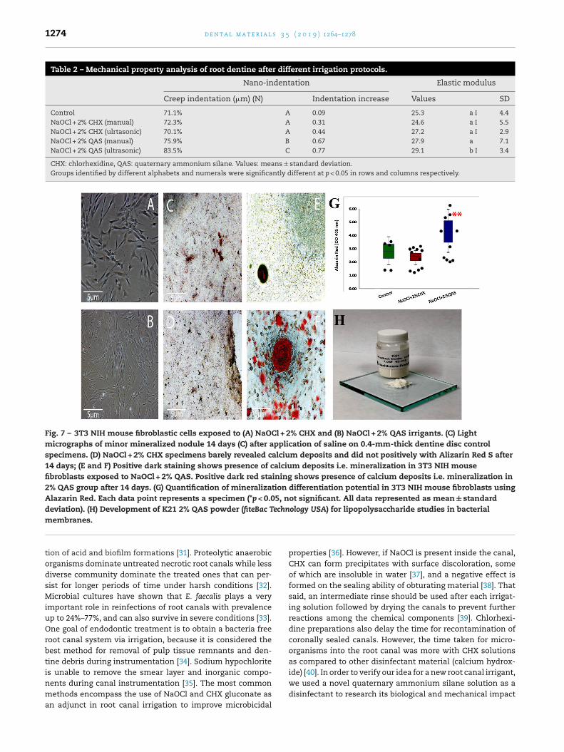
Deposition of mineralized nodules 14 days after applicationof irrigation formulations are shown in Fig. 7. Fig. 7A and B isa depiction of 3T3 NIH mouse fibroblastic cells after exposureto the NaOCl + 2% CHX (Fig. 7D) and NaOCl + 2% QAS groups(Fig. 7E) respectively. Mineralized nodules were significantlysmaller in the NaOCl + 2% CHX (±2.8) specimens when com-pared to the NaOCl + 2% QAS (±5.1) specimens and the control(±3.3). In the control specimens, the NIH 3T3 mouse fibrob-lasts cells revealed positively stained calcium deposits whenAlazarin red was used. Lesser staining than in the controlgroups were seen in the NaOCl + 2% CHX groups. Massive pos-itive dark stainings were seen representing calcium deposits(mineralization) in the NaOCl + 2% QAS groups (Fig. 7E). Theuse of NaOCl + 2% QAS significantly increased nodule forma-tion after 14 days) (p < 0.05).
4. Discussion
The bacterial influence of a more acidogenic environment and
aciduric microbiota is a direct consequence of frequent andprolonged acidification, changing the ecological balance inthe tooth substrate [30]. In the approach to reduce bacterialacidification mentioned, most attention is paid to reduc-
d e n t a l m a t e r i a l s 3 5 ( 2 0 1 9 ) 1264–1278 1273
Fig. 5 – Micro indentation distance increase, first indentation cycle and creep indentation parameters measured in the firstcycle for (A) manual and (B) ultrasonic NaOCl + 2% QAS irrigation specimens. (C) Indentation load function after depthprofiling of elastic modulus (extension stretch identified by arrow) for ultrasonic groups showing statistical similarity forcontrol and NaOCl + 2% CHX groups (*). (D) A representative photograph of deposits/precipitates formed after consequent useof NaOCl and chlorhexidine on culture plates. Note the colour change and deposits in the mixed liquids ranging from darkreddish brown to light orange as well as formation of precipitate in varying amounts. (For interpretation of the references tocolor in this figure legend, the reader is referred to the web version of this article.)
Fig. 6 – Contact angle and (B) surface free energy of root canal dentine with different irrigation regimens. Surface free energyis represented as dispersive and shows significantly higher values for NaOCl + 2% QAS, both manual and ultrasonicspecimens. Inset SEM surfaces of root dentine treated with manual NaOCl + 2% CHX and NaOCl + 2%QAS. Both images showpresence of deposits and siloxane layer on root dentine surfaces respectively.
1274 d e n t a l m a t e r i a l s 3 5 ( 2 0 1 9 ) 1264–1278
Table 2 – Mechanical property analysis of root dentine after different irrigation protocols.
Nano-indentation Elastic modulus
Creep indentation (�m) (N) Indentation increase Values SD
Control 71.1% A 0.09 25.3 a I 4.4NaOCl + 2% CHX (manual) 72.3% A 0.31 24.6 a I 5.5NaOCl + 2% CHX (ulrtasonic) 70.1% A 0.44 27.2 a I 2.9NaOCl + 2% QAS (manual) 75.9% B 0.67 27.9 a 7.1NaOCl + 2% QAS (ultrasonic) 83.5% C 0.77 29.1 b I 3.4
CHX: chlorhexidine, QAS: quaternary ammonium silane. Values: means ± standard deviation.Groups identified by different alphabets and numerals were significantly different at p < 0.05 in rows and columns respectively.
Fig. 7 – 3T3 NIH mouse fibroblastic cells exposed to (A) NaOCl + 2% CHX and (B) NaOCl + 2% QAS irrigants. (C) Lightmicrographs of minor mineralized nodule 14 days (C) after application of saline on 0.4-mm-thick dentine disc controlspecimens. (D) NaOCl + 2% CHX specimens barely revealed calcium deposits and did not positively with Alizarin Red S after14 days; (E and F) Positive dark staining shows presence of calcium deposits i.e. mineralization in 3T3 NIH mousefibroblasts exposed to NaOCl + 2% QAS. Positive dark red staining shows presence of calcium deposits i.e. mineralization in2% QAS group after 14 days. (G) Quantification of mineralization differentiation potential in 3T3 NIH mouse fibroblasts usingAlazarin Red. Each data point represents a specimen (*p < 0.05, not significant. All data represented as mean ± standarddeviation). (H) Development of K21 2% QAS powder (fiteBac Technology USA) for lipopolysaccharide studies in bacterial
membranes.tion of acid and biofilm formations [31]. Proteolytic anaerobicorganisms dominate untreated necrotic root canals while lessdiverse community dominate the treated ones that can per-sist for longer periods of time under harsh conditions [32].Microbial cultures have shown that E. faecalis plays a veryimportant role in reinfections of root canals with prevalenceup to 24%–77%, and can also survive in severe conditions [33].One goal of endodontic treatment is to obtain a bacteria freeroot canal system via irrigation, because it is considered thebest method for removal of pulp tissue remnants and den-tine debris during instrumentation [34]. Sodium hypochlorite
is unable to remove the smear layer and inorganic compo-nents during canal instrumentation [35]. The most commonmethods encompass the use of NaOCl and CHX gluconate asan adjunct in root canal irrigation to improve microbicidalproperties [36]. However, if NaOCl is present inside the canal,CHX can form precipitates with surface discoloration, someof which are insoluble in water [37], and a negative effect isformed on the sealing ability of obturating material [38]. Thatsaid, an intermediate rinse should be used after each irrigat-ing solution followed by drying the canals to prevent furtherreactions among the chemical components [39]. Chlorhexi-dine preparations also delay the time for recontamination ofcoronally sealed canals. However, the time taken for micro-organisms into the root canal was more with CHX solutionsas compared to other disinfectant material (calcium hydrox-
ide) [40]. In order to verify our idea for a new root canal irrigant,we used a novel quaternary ammonium silane solution as adisinfectant to research its biological and mechanical impact
5 ( 2
om
nabfacciftdmaiivQ
ttcettoaathabTCtiiTrtabbr
iafiacpssttpl
d e n t a l m a t e r i a l s 3
n the root dentine surface, because the precipitates formedaybe more cytotoxic than NaOCl and CHX alone [41].Indeed, organosilane quaternary ammonium salts are a
ew class of antimicrobials, with reactive silanol groups as result of generation of hydrolysis OF QAS that are covalentlyonded to substrates via Si O linkages engaging microbiocidalunctions [42]. 3-(Trimethoxysilyl) propyldimethyloctadecylmmonium chloride (SiQAC) has been widely used for antimi-robial coatings of medical devices. It penetrates bacterialell membranes causing cell death of bacteria as they comen contact. SiQAC salts do not end with polymerizable endunctional groups [43]. In general, the positively charged qua-ernary amine charged (N+) compounds cause bacterial lysisue to their interaction with the negatively charged bacterialembranes. With the use of SiQAC along an anchoring tetra-
lkoxysilane and other organofunctional trialkoxysilanes [44],t can produce different antibacterial activities, which remainndependent of the loss of surface layer [45]. By altering the pHalue of the system, hydrolysis and condensation reactions ofAS can be differentially controlled [46].
To evaluate the antibacterial effects of QAS irrigation pro-ocol, confocal microscopy analysis was done which showshat the QAS irrigation protocol has a significant antimi-robial effect against E. faecalis biofilms. Most importantly,ndodontic disease is considered as a biofilm-induced infec-ion, as bacteria survive in an adaptive mechanism persistinghe endodontic infection [47]. Given this, the incorporationf 2% QAS in the irrigation protocol has potential to renderntibacterial properties without a discernable decrease after
prolonged period of time. The QAS compound kills bac-eria on contact [48] using TEOS as an anchoring unit andaving organic functional groups which are covalently boundnd detectable in specimens that showed completely deadacterial cells (94.64 ± 11.32 and 97.45 ± 3.79 for ultrasonic).his is why the first null hypothesis can be rejected. TheLSM analysis revealed green and red fluorescence intensi-
ies within the different biofilms. Most of the bacteria presentn the biofilm fluoresced green colour in the control group,ndicating that the bacteria were mostly alive in the control.he majority of the bacteria present in the biofilms fluoresceded colour in the QAS groups. This refers to immobilization ofhe SiQAC molecules that are providing greater antimicrobialctivity. Because of the silanol groups, QAS can be covalentlyonded via O Si O linkages to exert non-migrating micro-iocidal functions. The protective antimicrobial film formedesults in circumventing or slowing down biofilm formation.
Raman spectroscopy is a widely used powerful tool fordentification of bacterial components within the biofilm masst molecular level [49]. Now, another interesting observationor biofilm in Fig. 1C is the apparent shift of 484 cm−1 whichs usually assigned to polysaccharides or carbohydrates [50]nd has multi-dimensional information on special cellularomponents and their presence. Even so, the use of differentrotocols marked a difference in intensities and deviations intructural elements as the control specimens showed inten-ive signals at 484 cm−1. Moreover, the Raman signals assigned
o these polysaccharides were also detected in other samplesreated with different irrigation protocols. Based on this com-atibility, NaOCl + 2% QAS (manual and ultrasonic) showed theeast amount of intensity (Fig. 1C), followed by NaOCl+2%CHX
0 1 9 ) 1264–1278 1275
ultrasonic and manual, respectively. These Raman signaturesof carbohydrates appeared at lower intensities in QAS spec-imens hypothesizing that more bacterial colonies are beingaffected, and endorsing the rejection of null hypothesis. Therewas also a substantial difference in intensity ratio of Ramanbands at 484 cm−1 and 1130 cm−1 with some shifts (data notshown). The set of bands showed to be highly sensitive andspecific for biochemical changes.
There was appearance of new bands near at 993 cm−1 and1335 cm−1 in the control specimens (Fig. 1D) strengthening theobservation for the increase in carbohydrate content as part ofthe biofilm matrix [51]. However, the intense bands were dulland of lower intensity in the experimental groups, giving aprecise description consistent with the absence of additional,albeit unidentified, carbohydrates. Another interesting obser-vation is the apparent change in the amide I (C O stretch)band, the �-sheet secondary structure that involves amideand carbonyl functional groups, producing spectral changes.It was not possible to avoid such conformational changes dueto the native structure within the biofilm, where the intensityof amide I is low in QAS groups, a frequency distinguishablefrom the substrate. The lowest amide I values are usually mea-sured at the interior of cell nucleus where more nucleic acidsare found [52]. In addition, from the results of the study, theNaOCl + 2% QAS groups showed higher amounts of sealer pen-etration (Fig. 1B) compared to other groups (p < 0.05). Hence,including 2% QAS within the irrigation protocol may providea long-term antibacterial solution. The unique addition of 2%QAS with a NaOCl irrigant could be considered pivotal and anadvisable approach for the attainment of maximum antibacte-rial and avoid reinfections. This laboratory study also indicatesthe valid use of Raman spectroscopy, allowing detailed spec-tral analysis for resin penetration of the sealer.
Analysis of the dentine surface and biofilm formed on den-tine substrate had been performed using SEM. The contrastingresults in SEM between the CHX and QAS groups might beattributed to the nature of two different solutions used forthe irrigation protocol (Fig. 3). The thick deposits with com-plete coverage of the dentinal tubule orifices are clearly visiblein specimens treated both manually and ultrasonically withNaOCl + 2% CHX specimens (Fig. 3B and C). Chlorhexidine canreact with remaining NaOCl inside the root canal formingsmall precipitate clusters, which could be para-chloroanilinedeposits (Fig. 3A–C). However, the purpose of the present lab-oratory study was not to study the nature of the deposits perse. The impact of an endodontic irrigating solution producingsuch deposits was clearly visible in the SEM analysis of resintags formed. The current investigation compared the quality,density and length of resin tags formed in each group (Fig. 4).The type of irrigation protocol performed and the morphologyof resin tags formed were directly related. The length of resintags increased in the QAS groups (Fig. 4E and F) despite the 2%QAS groups forming a crust type deposition on the surfaceof dentine. The presence of bridged organosiloxane groupsin the molecular backbone of QAS, which has affinity for theresinous adhesive [53], might have resulted in better penetra-
tion. Without the use of resin sealer, QAS did not infiltrate wellinside the dentine (Fig. 4C and D). Moreover, the said crust pro-duced by 2% QAS was completely dissolved and subsequentlycould no longer be identified inside the resin matrix. Conden-
s 3 5
1276 d e n t a l m a t e r i a lsation of QAS was observed on the dentine surface indicatinga phase separation in the presence of water coming from thedentinal tubules (Fig. 3G). With QAS groups, there might havebeen an increase in surface hydrophobicity by changing thesurface free energy of the dentine substrate [54], resulting inbetter wettability and enhanced infiltration of resin sealer.These organofunctional silanes also act as water scavengersreacting with wholesome water molecules hydrolysing thealkoxy groups present within the silanes converting them intoalcohol (in this case: ethanol) molecules [55]. In addition, theformation of hydrophobic layer due to the presence of QASleads to more durable hybrid layer formation with reduction ofwater leading to facilitation of infiltration of adhesives (Ramanspectroscopy data not shown).
SEM results suggested that NaOCl with 2% QAS was themost effective method for removing E. faecalis biofilm in theroot canal system, in both manual and ultrasonic way, whencompared to other irrigation techniques. This said methoddebrided and decontaminated the root surfaces both mechan-ically and chemically. This could be attributed to the knownbactericidal effects of NaOCl and synergistic effects of 2% QAS(Fig. 3K and L). That said, the second null hypothesis canbe rejected. The possible reasons for the differences in theefficacy of different irrigation protocols could be the resultof advanced synergistic effects of QAS. Previous study hasreported important information on the antimicrobial effects of2%QAS on biofilms indeed [56]. One of the major etiologies ofendodontic failure is the persistence of bacterial biofilm evenafter endodontic therapy [57]. Many methods have been exam-ined including machine-assisted irrigation [58] as E. faecalisis a well-studied micro-organism in the endodontic litera-ture because of its virulence [59]. The use of a tetrafunctionalorganosilane as the anchoring unit for the antimicrobial tri-alkoxysilane molecules enables a three-dimensional networkto be formed once condensation is brought to completionwithin the dentinal substrate [60].
Our surface free energy results (Fig. 6) did not contrastthe existing SEM and confocal results. Even so, there was anincreased surface energy in the NaOCl + 2% QAS groups, whichmay be due to the increased surface roughness due to the pres-ence of QAS surface crust. The results for the flow of irrigantsare in concordance with other current data demonstratingthat NaOCl + 2% QAS based irrigants exhibited higher flow val-ues than the other study groups. The explanation might beprimarily due to the complex interactions between the non-polar and polar components of the root canal and the irrigantsdispensed on it. As for the model for calculating surface freeenergy, the droplet volume and substrate type were equal, andthis is why we believe that the difference in the irrigation con-tent may have caused these said discrepancies. The ethanolinclusion the 2% QAS may have increased the surface freeenergy is suggesting that this irrigation protocol may be anattractive method for endodontic therapy. Once there is theformation of a smear layer on the dentine surface, its charac-teristics can also determine the level of surface free energy asit is increased with the increase in surface roughness due to
h
changes in the �S values, reflecting increase surface densityon dentine surface [61].From a practicing endodontist’s point of view, the currentresults strongly support the use and introduction of this irri-
( 2 0 1 9 ) 1264–1278
gation protocol as an alternative. In addition, the obtainedcontact angles along with surface free energy studies confirmhigher wettability to the long-term success of root canal treat-ment. On the other hand, the use of EDTA is known to softenthe dentine and affect the mechanical properties. The use ofquaternary ammonium silanes, despite exhibiting temporalhydrolytic stability after condensation, bring some stabilitywith their molecular siloxane bridges (Table 2). Now, this isrejecting the third null hypothesis that QAS has no effect onmechanical properties of dentine substrate. Ongoing work isbeing performed to study in depth effects on biofilm at theexopolysaccharide backbone.
Even so, the exudation of dentinal fluid along withcytoplasmic elongations within dentinal tubules may havereduced the inward transdentinal diffusion of QAS agentstowards the dental pulp. However, this assumption shouldbe verified in vivo before a final conclusion can be drawn.No detrimental effect on mineralized nodule productions wasobserved in the NaOCl + 2% QAS groups after 14 days (Fig. 5).The normal deposition of mineralized nodules in NaOCl + 2%QAS formulations indicates that the dentine pulp repara-tive processes, which are essential tertiary dentine formation,were minimally affected following NaOCl + 2% QAS applica-tion, rendering it a safe irrigant for use.
The 2% QAS irrigation formulation did not show cyto-toxic effects on 3T3 NIH mouse fibroblast cells (Fig. 7). Thepredominance of the anti-inflammatory phenotype after itsapplication may stimulate healing and tissue repair [62]. Itis noteworthy that the newly developed antibacterial quater-nary ammonium silane has also been proven to increase theresistance of dentine collagen to degradation and is promisingoption for use as a protease inhibitor [33] to improve durabilityof resin-dentin bonds. Moreover, the favorable antimicro-bial and endodontic profile of the NaOCl + 2% QAS could beascribed to its use as a potential irrigation concept for morepredictable reduction of intracanal bacteria and enhancing theversatility of the material and its potential use in dentistry.Future work is underway in the development of a 2% QASpowder formulation to investigate the detailed effects of QASon undifferentiated stem cells and bacterial lipopolysaccha-ride membranes looking at its important role in regenerativeendodontics.
5. Conclusion
Within the limits of the present study, it may that the newlydeveloped quaternary ammonium silane (2% QAS) increasesits bacterial efficacy when used in conjunction with NaOCl asan irrigant impregnated inside a root canal. Considering thecomplex endodontic system and the difficulty of penetratingdentinal tubules, there is a potential to exploit the QAS dis-infectant as an irrigant for a feasible therapeutic approachagainst biofilm infection within the root canal system.
Acknowledgements
This work was supported in part by the grant IMU 425/2018,School of Dentistry, International Medical University. Theauthors thank the labs at Nanocat University of Malaya, School

5 ( 2
oLf
r
6/.
d e n t a l m a t e r i a l s 3
f Dentistry of the International Medical University Kualaumpur, the Faculty of Dentistry of at University of Hong Kongor the research experiments and analysis.
e f e r e n c e s
[1] Hojo S, Takahashi N, Yamada T. Acid profile in cariousdentin. J Dent Res 1991;70:182–6.
[2] Nagaoka S, Miyazaki Y, Liu HJ, Iwamoto Y, Kitano M,Kawagoe M. Bacterial invasion into dentinal tubules ofhuman vital and non-vital teeth. J Endod 1995;21:70–3.
[3] Siqueira Jr JF. Aetiology of root canal treatment failure: whywell-treated teeth can fail. Int Endod J 2001;34:1–10.
[4] Willershausen I, Wolf TG, Schmidtmann I. Survey of rootcanal irrigating solutions used in dental practices withinGermany. Int Endod J 2015;48:654–60.
[5] American Association of Endodontics. Guide to clinicalendodontics. 6th ed; 2013. Available at:www.nxtbook.com/nxtbooks/aae/guidetoclinicalendodontics[Accessed 10 October 2017].
[6] Boutsioukis C, van der Sluis LW. Syringe irrigation: blendingendodontics and fluid dynamics. In: Basrani B, editor.Endodontic irrigation: chemical disinfection of the rootcanal system. New York: Springer; 2015. p. 45–64.
[7] Haapasalo M, Shen Y, Qian W, Gao Y. Irrigation inendodontics. Dent Clin North Am 2010;54:299–303.
[8] Dutner J, Mines P, Anderson A. Irrigation trends amongAmerican Association of Endodontists members: aweb-based survey. J Endod 2012;38:37–40.
[9] Rosenthal S, Spangberg L, Safavi K. Chlorhexidinesubstantivity in root canal dentin. Oral Surg Oral Med OralPathol Oral Radiol Endod 2004;98:488–92.
[10] Breschi L, Mazzoni A, Nato F. Chlorhexidine stabilizes theadhesive interface: a 2-year in vitro study. Dent Mater2010;26:320–5.
[11] Gendron R, Grenier D, Sorsa T, Mayrand D. Inhibition of theactivities of matrix metalloproteinases 2, 8, and 9 bychlorhexidine. Clin Diagn Lab Immunol 1999;6:437–9.
[12] Zehnder M. Root canal irrigants. J Endod 2006;32:389–98.[13] Gupta H, Kandaswamy D, Manchanda SK, Shourie S.
Evaluation of the sealing ability of two sealers after usingchlorhexidine as a final irrigant: an in vitro study. J ConservDent 2013;16:75–8.
[14] Basrani BR, Manek S, Mathers D. Determination of4-chloroaniline and its derivatives formed in the interactionof sodium hypochlorite and chlorhexidine by using gaschromatography. J Endod 2010;36:312–4.
[15] Thomas JE, Sem DS. An in vitro spectroscopic analysis todetermine whether parachloroaniline is produced frommixing sodium hypochlorite and chlorhexidine. J Endod2010;36:315–7.
[16] Makvandi P, Jamaledin R, Jabbari M, Nikfarjam N,Borzacchiello. Antibacterial quaternary ammoniumcompounds in dental materials: a systematic review. DentMater 2018;6:851–67.
[17] Turova NY, Turevskaya EP, Kessler VG, Yanovskaya MI. Thechemistry of metal alkoxides. Norwell: Kluwer AcademicPress; 2002.
[18] Danks AE, Hall SR, Schnepp Z. The evolution of ‘sol–gel’chemistry as a technique for materials synthesis. MaterHoriz 2016;3:91–5.
[19] Levy David, Zayat Marcos, editors. The sol–gel handbook:synthesis, characterization, and applications. 1st ed.Wiley-VCH Verlag GmbH & Co. KGaA; 2015. Published 2015by Wiley-VCH Verlag GmbH & Co. KGaA.
0 1 9 ) 1264–1278 1277
[20] Hoffmann F, Fröba M. Vitalising porous inorganic silicanetworks with organic functions—PMOs and related hybridmaterials. Chem Soc Rev 2011;40:608–20.
[21] Bindler F, Voges R, Laugel P. The problem of methanolconcentration admissible in distilled fruit spirits. Food AdditContam 1988;5:343–51.
[22] Daood U, Yiu CKY, Burrow MF, Niu LN, Tay FR. Effect of anovel quaternary ammonium silane on dentin proteaseactivities. J Dent 2017;58:19–27.
[23] Daood U, Yiu CKY, Burrow MF. Effect of a novel quaternaryammonium silane cavity disinfectant on cariogenic biofilmformation. Clin Oral Invest 2019,http://dx.doi.org/10.1007/s00784-019-02928-7.
[24] Soares LE, do Espirito Santo AM, Junior AB, Zanin FA, daSilva Carvalho C, de Oliveira R, et al. Effects of Er:YAG laserirradiation and manipulation treatments on dentincomponents, part 1: Fourier transform-Raman study. JBiomed Opt 2009;14:1–7.
[25] Alebrahim MA, Krafft C, Popp J. Raman imaging to studystructural and chemical features of the dentin enameljunction. Mater Sci Eng 2015;92:1–9.
[26] Schuster KC, Reese I, Urlaub E, Gapes JR, Lendl B.Multidimensional information on the chemical compositionof single bacterial cells by confocal Ramanmicrospectroscopy. Anal Chem 2000;72:5529–34.
[27] Ramakrishnaiah R, Rehman G, Basavarajappa S, Al KkhuraifAA, Durgesh BH, Khan AS, et al. Applications of Ramanspectroscopy in dentistry: analysis of tooth structure. ApplSpectrosc Rev 2015;50:332–50.
[28] Silva Júnior ZS, Botta SB, Ana PA, Franca CM, Fernandes KP,Ferrari RA, et al. Effect of papain-based gel on type I collagen— spectroscopy applied for microstructural analysis. Sci Rep2015;5:11448.
[29] Koehne T, Marshall RP, Jeschke A, Kahl-Nieke B, Schinke T,Amling M. Osteopetrosis, osteopetrorickets andhypophosphatemic rickets differentially affect dentin andenamel mineralization. Bone 2013;53:25–33.
[30] Nobuhiro T, Bente N. Ecological hypothesis of dentin androot caries. Caries Res 2016;50:422–31.
[31] Li Y, Tanner A. Effect of antimicrobial interventions on theoral microbiota associated with early childhood caries.Pediatr Dent 2015;37:226–44.
[32] Figdor D, Sundqvist G. A big role for the very small —understanding the endodontic microbial flora. Aust Dent J2007;52(1Suppl):S38–51.
[33] Stuart CH, Schwartz SA, Beeson TJ, Owatz CB. Enterococcusfaecalis: its role in root canal treatment failure and currentconcepts in retreatment. J Endod 2006;2:93–8.
[34] Haapasalo M, Shen Y, Wang Z, Gao Y. Irrigation inendodontics. Braz Dent J 2014;21:299–303.
[35] Teixeira CS, Felippe MCS, Felippe WT. The effect ofapplication time of EDTA and NaOCl on intracanal smearlayer removal: an SEM analysis. Int Endod J 2005;38:285–90.
[36] Marco S, Rullo R, Albino A. The thioredoxin system in thedental caries pathogen Streptococcus mutans and thefood-industry bacterium Streptococcus thermophilus.Biochimie 2013;95:2145–56.
[37] Vivacqua-Gomes N, Ferraz CC, Gomes BP, Zaia AA, TeixeiraFB, Souza-Filho FJ. Influence of irrigants on the coronalmicroleakage of laterally condensed gutta-percha rootfillings. Int Endod J 2002;35:791–5.
[38] Zehnder M. Root canal irrigants. J Endod 2006;32:389–98.[39] Vouzara T, Koulaouzidou E, Ziouti F, Economides N.
Combined and independent cytotoxicity of sodium
hypochlorite, ethylenediaminetetraacetic acid andchlorhexidine. Int Endod J 2016;49:764–73.
s 3 5
1278 d e n t a l m a t e r i a l[40] Gomes BP, Sato E, Ferraz CC, Teixeira FB, Zaia AA, Souza-Filho FJ. Evaluation of time required for recontamination ofcoronally sealed canals medicated with calcium hydroxideand chlorhexidine. Int Endod J 2003;36:604–9.
[41] Cintra LT, Watanabe S, Samuel RO, et al. The use of NaOCl incombination with CHX produces cytotoxic product. Clin OralInvestig 2014;18:935–40.
[42] Zhang W, Luo XJ, Niu LN, Liu SY, Zhu WC, Epasinghe J, et al.One-pot synthesis of antibacterial monomers with dualbiocidal modes. J Dent 2014;42:1078–95.
[43] Ahlström B, Thompson RA, Edebo L. The effect ofhydrocarbon chain length, pH, and temperature on thebinding and bactericidal effect of amphiphilic betaine esterson Salmonella typhimurium. APMIS 1999;107:318–24.
[44] Matinlinna JP, Lung CYK, Tsoi JKH. Silane adhesionmechanism in dental applications and surface treatments: areview. Dent Mater 2018;34:13–28.
[45] Gong SQ, Niu LN, Kemp LK, Yiu CK, Ryou H, Qi YP, et al.Quaternary ammonium silane-functionalized, methacrylateresin composition with antimicrobial activities andself-repair potential. Acta Biomater 2012;8:3270–82.
[46] Salon MCB, Bayle PA, Abdelmouleh M, Boufi S, Belgacem MN.Kinetics of hydrolysis and self-condensation reactions ofsilanes by NMR spectroscopy. Colloid Surf A 2008;312:83–91.
[47] Al-Ahmad A, Ameen H, Pelz K, et al. Antibiotic resistanceand capacity for biofilm formation of different bacteriaisolated from endodontic infections associated withroot-filled teeth. J Endod 2014;40:223–30.
[48] Tiller JC, Liao CJ, Lewis K, et al. Designing surfaces that killbacteria on contact. Proc Natl Acad Sci U S A 2001;98:5981–5.
[49] Efrima S, Zeiri L. Understanding SERS of bacteria. J RamanSpectrosc 2008;40:277–88.
[50] Schuster KC, Reese I, Urlaub E, Gapes JR, Lendl B.Multidimensional information on the chemical compositionof single bacterial cells by confocalRaman microspectroscopy. Anal Chem 2000;72:5529–34.
[51] Carey Paul R, Gibson Blake R, Gibson Jordan F, GreenbergMichael E, Heidari-Torkabadi Hossein, Pusztai-CareyMarianne, et al. Defining molecular details of the chemistryof biofilm formation by Raman microspectroscopy.
( 2 0 1 9 ) 1264–1278
Bichemistry 2017;56(17):2247–50,http://dx.doi.org/10.1021/acs.biochem.7b00116.
[52] Baldassarre L, Giliberti V, Rosa A, Ortolani M, Bonamore A,Baiocco P, et al. Mapping the amide I absorption in singlebacteria and mammalian cells with resonant infrarednanospectroscopy. Nanotechnology 2016;27:075101.
[53] Lung CYK, Matinlinna JP. Resin bonding to silicatizedzirconia with two and cross-linking silane, part I:experimental. Silicon 2010;2:153–61.
[54] Daood D, Yiu CKY, Burrow MF, Niu LN, Tay FR. Effect of anovel quaternary ammonium silane cavity disinfectant ondurability of resin-dentine bond. J Dent 2017;60:77–86.
[55] Mancheno-Posso P, Dittler RF, Lewis D, Juang P, XY JI, Xu XH,et al. Review of status, trends, and challenges in workingwith silane and functional silanes. In: Moriguchi K, UtagawaS, editors. Silane, chemistry, applications and performance.New York: Nova Science Publishers; 2013. p. 66–87.
[56] Ya-ping G, Ji-yao L, Mohamed MM, Christopher WC, HockinHKX, Tay FR, et al. Quaternary ammonium silane-basedantibacterial and anti-proteolytic cavity cleanser. DentMater 2018;12:1814–27.
[57] Baumgartner JC. Microbiologic aspects of endodonticinfections. J Calif Dent Assoc 2004;32:459–68.
[58] Stuart CH, Schwartz SA, Beeson TJ, Owatz CB. Enterococcusfaecalis: its role in root canal treatment failure and currentconcepts in retreatment. J Endod 2006;32:93–8.
[59] Dumani A, Yoldas O, Yilmaz S, Koksal F, Kayar B, Akcimen B,et al. Polymerase chain reaction of Enterococcus faecalis andcandida albicans in apical periodontitis from Turkishpatients. J Clin Exp Dent 2012;4:e34.
[60] Liu Y, Xu D, Wu Y, Sun H, Gao H. Comparative study on thehydrolysis kinetics of substituted ethoxysilanes byliquid-state 29Si NMR. J Non Cryst Solids 2004;343:61–70.
[61] Inoue N, Tsujimoto A, Takimoto M, Ootsuka E, Endo H,Takamizawa T, et al. Surface free-energy measurements asindicators of the bonding characteristics of single step
self-etching adhesives. Eur J Oral Sci 2010;118:525–30.[62] Daood U, Yiu CKY. Transdentinal cytotoxicity andmacrophage phenotype of a novel quaternary ammoniumsilane cavity disinfectant. Dent Mater 2019;2:206–16.