simultaneous detection of circulating immunological ... detection of... · m. de amici3 & p....
TRANSCRIPT

Simultaneous detection of circulating immunological parametersand tumor biomarkers in early stage breast cancer patientsduring adjuvant chemotherapy
B. Rovati1 & S. Mariucci1 & S. Delfanti1 & D. Grasso1 & C. Tinelli2 & C. Torre3 &
M. De Amici3 & P. Pedrazzoli1
Accepted: 22 December 2015# International Society for Cellular Oncology 2016
AbstractBackground Chemotherapy-induced immune suppressionhas mainly been studied in patients with advanced cancer,but the influence of chemotherapy on the immune system inearly stage cancer patients has so far not been studied system-atically. The aim of the present study was to monitor the im-mune system during anthracycline- and taxane-based adjuvantchemotherapy in early stage breast cancer patients, to assessthe impact of circulating tumor cells on selected immune pa-rameters and to reveal putative angiogenic effects of circulat-ing endothelial cells.Methods Peripheral blood samples from 20 early stage breastcancer patients were analyzed using a flow cytometric multi-color of antibodies to enumerate lymphocyte and dendritic cellsubsets, as well as endothelial and tumor cells. An enzyme-linked immunosorbent assay (ELISA) was used to measurethe levels of various serological factors.Results During chemotherapy, all immunological parametersand angiogenesis surrogate biomarkers showed significant de-creases. The numbers of circulating tumor cells showed sig-nificant inverse correlations with the numbers of T helper
cells, a lymphocyte subset directly related to effective anti-tumor responses. Reduced T helper cell numbers may contrib-ute to systemic immunosuppression and, as such, the activa-tion of dormant tumor cells.Conclusions From our results we conclude that adjuvant che-motherapy suppresses immune function in early stage breastcancer patients. In addition, we conclude that the presence ofcirculating tumor cells, defined as pan-cytokeratin+, CD326+,CD45− cells, may serve as an important indicator of a patient’simmune status. Further investigations are needed to firmlydefine circulating tumor cells as a predictor for the successof breast cancer adjuvant chemotherapy.
Keywords Adjuvant breast cancer chemotherapy .Cytokine .
Lymphocyte subsets . Dendritic cells . Endothelial cells .
Tumor cells
1 Introduction
Tumors and chemotherapy (CT) are known to significantlyalter immune responses [1, 2], but surprisingly few studieshave been reported on how cancer stages may affect immuneresponses during CT. Recent studies on therapy responses inbreast cancer (BC) have focused primarily on the relevance ofmolecular or genetic biomarkers, while little attention hasbeen paid to the significance of cellular immune responsesto clinical outcome [3], despite the fact that BC patients havea lower baseline immune response than healthy individuals[4–6]. There is also evidence indicating that low lymphocytecounts predict a greater likelihood of disease recurrence inpatients with early stage BC [7], as also poorer disease-freeand overall survival rates in metastatic BC patients [8, 9].
In cancer patients subjected to CT, the clinical immunode-ficiency is primarily related to Tcell depletion, associatedwith
Electronic supplementary material The online version of this article(doi:10.1007/s13402-015-0264-2) contains supplementary material,which is available to authorized users.
* B. [email protected]
1 SC Oncologia e Laboratorio di Citofluorimetria, e Terapie Cellulari,Fondazione IRCCS Policlinico San Matteo, Pavia, Italy
2 Servizio di Biometria e Statistica Medica, Fondazione IRCCSPoliclinico San Matteo, Pavia, Italy
3 SC Pediatria, Laboratorio di Immuno Allergologia, FondazioneIRCCS Policlinico San Matteo, Pavia, Italy
Cell Oncol.DOI 10.1007/s13402-015-0264-2

inhibition of the ability of dendritic cells (DCs) to induce bothprimary and secondary T and B cell responses [10]. Naturalkiller (NK) cells constitute an important component of innateimmunity, able to limit viremia and to mediate spontaneouskilling of various tumor cells, even before the adaptive im-mune system is activated [11]. Complete restoration of immu-nocompetence following anti-neoplastic therapy implies theprogressive recovery of various cell subpopulations, whichis a complex process that is dependent on the type, the dose,the scheduling and the interactions of the drugs used [10].
In recently reported studies evidence has been obtained forimmunological suppression of tumor cells in a dormant state[12]. The mechanisms underlying the activation of dormanttumor cells and metastatic disease are, however, not fully un-derstood. Besides genetic and immunological factors [13, 14],angiogenesis may also trigger the activation of dormant tumorcells [15–18]. Angiogenesis is an essential step in tumorgrowth and metastasis and may be measured via circulatingendothelial cells (CECs) and bone marrow-derived circulatingendothelial progenitor cells (CEPs) [19]. It has amply beenshown that angiogenesis is regulated by chemokines, a groupof cytokines involved in the migration of leukocytes that dis-play pleiotropic immunologic effects in addition to promotingthe proliferation of tumor cells and mediating organ-specificmetastasis. In particular, it has been found that chemokinesand their receptors play important roles in BC development.As yet, however, few reports have focused on changes inchemokine concentrations during BC chemotherapy, especial-ly adjuvant chemotherapy [20, 21].
The aims of the present study were (1) to monitor in earlystage BC patients the influence of anthracycline- and taxane-based adjuvant chemotherapy both on the immune system, byevaluating the distribution of lymphocyte subsets, and on cir-culating DCs including their subsets and some serologicalfactors selected for their relevance to BC, (2) to analyze therelationship between circulating tumor cells (CTCs) and thequantitative distribution of lymphocyte subsets, in order togain further insight into possible tumor-induced immune sup-pression and (3) to evaluate angiogenesis by changes in sur-rogate biomarkers of CECs and CEPs before and duringchemotherapy.
2 Materials and methods
2.1 Study population
In total 20 early stage breast cancer (BC) patients aged 34–71 years (median age 53.5 years), referred to the Medical On-cology Unit of the IRCCS Foundation Policlinico San Matteo,were consecutively enrolled in this study from March 2012 toSeptember 2013. Inclusion criteria were: (a) women 18 yearsold or older with stage I to III BC and receiving adjuvant
chemotherapy, based on clinical-pathological characteristics,(b) absence of relevant comorbidities and (c) ability to signinformed consent prior to entering the study. Exclusion criteriawere: (a) pregnancy or breastfeeding and (b) known HIV pos-itivity. Eighteen women completed the study and two womenwithdrew from the study for family and personal reasons. Eachpatient provided written informed consent before entering thestudy and the study was conducted following approval by theEthical Committee and Study Protocols Review Board of theIRCCS Foundation Policlinico San Matteo. All proceduresused in this study are in agreement with the 1975 HelsinkiDeclaration [22]. A summary of the characteristics of the studypopulation is listed in Table 1.
Table 1 Clinical-pathological characteristics of study population
Characteristics Numbers Percentage
Total patients 20 100
Age (years)
Median 53.5 –
Range 34–71 –
Menopausal status
Premenopausal 8 40
Postmenopausal 12 60
Tumor size (mm)
pT1≤ 20 8 40
pT2> 20≤ 50 11 55
pT3> 50 1 5
Nodal status
Node-negative 7 35
Node-positive 13 65
Histological type
Ductal 15 75
Lobular 1 5
Mixed 4 20
Grading
II 6 30
III 14 70
Estrogen Receptor (ER) status
Negative 12 60
Positive 8 40
Progesterone receptor (PR) status
Negative 13 65
Positive 7 35
Human epidermal growth factor 2 (HER2) expression
Negative 14 70
Positive 6 30
Proliferative index Ki67
Negative (<15 %) 2 10
Positive (>15 %) 18 90
B. Rovat et al.

2.2 Treatment schedule
The chemotherapy regimen applied consisted of four cy-cles of AC (adriamycin 60 mg/m2 plus cyclophosphamide600 mg/m2) administered on day 1 every 3 weeks, follow-ed by 12 weekly administrations of paclitaxel (80 mg/m2).Antiemetic, corticosteroids and histamine receptor blockerswere administered according to the institutional guidelines.Prophylactic use of pegfilgrastim, a PEGylated form of therecombinant human granulocyte colony-stimulating factor(GCSF), was adopted in six patients during the secondcycle of adriamycin and cyclophosphamide (AC)-basedtherapy, after evidence of a G4 neutropenia episode atthe nadir of the first cycle. No GCSF was used in thepaclitaxel-based treatment. This schedule was adopted inour department at the time of the study in patients with arecent diagnosis of breast cancer that, based on current inter-national guidelines, required anthracycline- and taxane-basedchemotherapy. Human epidermal growth factor receptor 2(HER2) positive patients received trastuzumab (Herceptin®)with weekly administrations in conjunction with paclitaxel.This treatment was continued every 3 weeks at the end ofthe chemotherapy cycle, thereby completing 1 year of chemo-therapy. According to their clinical-pathological characteris-tics, patients received hormone therapy (based on menopausalstatus) and/or complementary radiotherapy at the end of thechemotherapy program, as required by internationalguidelines.
2.3 Study design
All cellular populations were evaluated by flow cytometry(FCM, see below) at the start of the study (baseline) toestablish correlations with the tumor stage, histologicalgrade, lymph node status, hormone receptor status, HER2status, menopausal status, age and tumor size. Further-more, each cellular parameter was analyzed during admin-istration of the adjuvant therapy. Finally, the followingserological factors were tested using an enzyme-linked im-munosorbent analysis (ELISA) assay, at baseline and be-fore the administration of the 1st and 12th cycle oftaxane-based therapy: human vascular endothelial growthfactor (VEGF), human stromal cell-derived factor 1 alpha(CXCL12/SDF-1α) and human epidermal growth factorreceptor 2 (sp185/HER2). Immunophenotype profiles oflymphocyte and DC subsets from 31 to 19 healthy fe-males (HFs), respectively, selected in order to excludecomorbidities that could modulate the immune system[23, 24], were used as references. The percentage andabsolute number of CECs with their apoptotic fraction(CEC-APO) and CEPs in a group of 11 HFs, selected aspreviously reported [25], were used as references. All con-trol groups consisted of age-matched HFs.
2.4 Flow cytometry (FCM)
Peripheral blood samples were collected in 4 ml Vacutainer(Becton Dickinson, Basel, Switzerland) tubes, containing liq-uid tripotassium ethylene diamine tetra-acetic acid (K3EDTA)as an anticoagulant, and processed within 2 h after collection.As is routine in our laboratory [23–25], flow cytometric anal-yses were carried out on whole blood without any enrichmentprocedure to avoid enrichment artefacts. A panel of monoclo-nal antibodies (MoAbs), including anti-CD45 to identify he-matopoietic cells, in conjunction with appropriate analysisgates, was used to enumerate the different cellular popula-tions. Blood erythrocytes were removed by adding 1 ml lysissolution (VersaLyse, Immunotech Beckman Coulter, Mar-seille, France) to 100 μl whole blood samples and incubationfor 20min at room temperature (RT) in the dark. Evaluation ofnucleated cells from the whole blood samples was performedusing a Navios flow cytometer (Beckman Coulter, USA).FCM data were analysed using the Kaluza flow cytometryanalysis v1.1 software package. The precision, accuracy andstability of the cell counts were verified using internationalquality controls purchased from the United KingdomNationalExternal Quality Assessment Scheme (UK NEQAS LI, Shef-field, UK) [26].
2.5 Multi-color staining and FCM of lymphocyte and DCsubsets
The following mouse anti-human conjugated MoAbs(Beckman Coulter, USA) were used for lymphocyte subsetdetection: fluorescein isothiocyanate (FITC)-labelled anti-CD4 (clone 13B8.2) and anti-CD45RO (clone UCHL1), phy-coerythrin (PE)-labelled anti-CD25 (clone B1.49.9) and anti-CD45RA (clone 2H4LDH11LDB9), phycoerythrin-texas red(ECD)-labelled anti-CD16 (clone 3G8) and anti-CD20 (cloneB9E9), phycoerythrin cyanin-5.5 (clone PC5.5)-labelled anti-CD19 (clone J3.119) and anti-CD56 (clone N901), phycoery-thrin cyanin-7(PC7)-labelled anti-HLA-DR (clone Immu357)and anti-CD38 (clone LSI98-4-3), allophycocyanin (APC)-labelled anti-CD3 (clone UCHT1) and anti-CD4 (clone13B8.2), APC alexa fluor 700-labelled anti-CD8 (cloneB9.11) and anti-CD23 (clone 9P25) and APC alexa fluor750-labelled anti-CD45 (clone J.33). Mouse anti-human con-jugated MoAbs (Beckman Coulter, USA) were used for DCsubset identification: FITC-labelled anti-CD3 (cloneUCHT1), anti-CD16 (clone 3G8), anti-CD19(clone 89B),anti-CD20 (clone H299), anti-CD14 (clone RM052), anti-CD34 (clone S81), anti-CD56 (clone N901) and anti-CD11b(clone 94), APC-labelled anti-HLA-DR (clone Immu357),PE-labelled anti-CD11c (clone BU15) and anti-CD123 (cloneSSDCLY107D2) and APC alexa fluor 750-labelled anti-CD45 (clone J.33). As gating strategy, we applied a gate inthe CD45 versus side scatter (SSC) dot plot on CD45-positive
Simultaneous detection of circulating immunological parameters and tumor
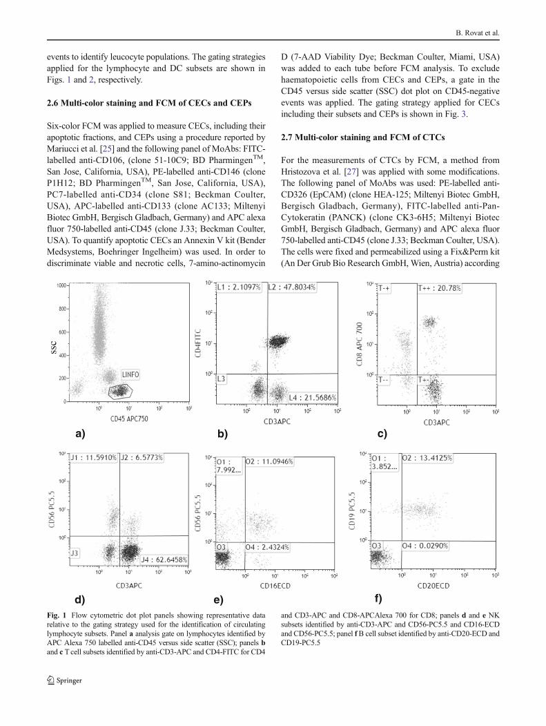
events to identify leucocyte populations. The gating strategiesapplied for the lymphocyte and DC subsets are shown inFigs. 1 and 2, respectively.
2.6 Multi-color staining and FCM of CECs and CEPs
Six-color FCM was applied to measure CECs, including theirapoptotic fractions, and CEPs using a procedure reported byMariucci et al. [25] and the following panel of MoAbs: FITC-labelled anti-CD106, (clone 51-10C9; BD PharmingenTM,San Jose, California, USA), PE-labelled anti-CD146 (cloneP1H12; BD PharmingenTM, San Jose, California, USA),PC7-labelled anti-CD34 (clone S81; Beckman Coulter,USA), APC-labelled anti-CD133 (clone AC133; MiltenyiBiotec GmbH, Bergisch Gladbach, Germany) and APC alexafluor 750-labelled anti-CD45 (clone J.33; Beckman Coulter,USA). To quantify apoptotic CECs an Annexin V kit (BenderMedsystems, Boehringer Ingelheim) was used. In order todiscriminate viable and necrotic cells, 7-amino-actinomycin
D (7-AAD Viability Dye; Beckman Coulter, Miami, USA)was added to each tube before FCM analysis. To excludehaematopoietic cells from CECs and CEPs, a gate in theCD45 versus side scatter (SSC) dot plot on CD45-negativeevents was applied. The gating strategy applied for CECsincluding their subsets and CEPs is shown in Fig. 3.
2.7 Multi-color staining and FCM of CTCs
For the measurements of CTCs by FCM, a method fromHristozova et al. [27] was applied with some modifications.The following panel of MoAbs was used: PE-labelled anti-CD326 (EpCAM) (clone HEA-125; Miltenyi Biotec GmbH,Bergisch Gladbach, Germany), FITC-labelled anti-Pan-Cytokeratin (PANCK) (clone CK3-6H5; Miltenyi BiotecGmbH, Bergisch Gladbach, Germany) and APC alexa fluor750-labelled anti-CD45 (clone J.33; Beckman Coulter, USA).The cells were fixed and permeabilized using a Fix&Perm kit(An Der Grub Bio Research GmbH, Wien, Austria) according
a) b) c)
d) e) f)
ssc
Fig. 1 Flow cytometric dot plot panels showing representative datarelative to the gating strategy used for the identification of circulatinglymphocyte subsets. Panel a analysis gate on lymphocytes identified byAPC Alexa 750 labelled anti-CD45 versus side scatter (SSC); panels band c Tcell subsets identified by anti-CD3-APC and CD4-FITC for CD4
and CD3-APC and CD8-APCAlexa 700 for CD8; panels d and e NKsubsets identified by anti-CD3-APC and CD56-PC5.5 and CD16-ECDand CD56-PC5.5; panel f B cell subset identified by anti-CD20-ECD andCD19-PC5.5
B. Rovat et al.

to the manufacture’s instructions. During the permeabilizationstep, an anti-PANCK antibody was added to the perme-abilization buffer in a final volume of 100 μl. In each sample5×105 to 1×10⁶ cellular events were counted. Circulatingtumor cells (CTC) were defined as EpCAM+, PANCK+,CD45−. A blood sample was considered CTC-positive whenat least one EpCAM+, PANCK+, CD45− cell was detected.Viable and necrotic cells were differentiated through 7-AADstaining. The gating strategy applied for CTC detection isshown in Fig. 4.
The efficiency of the method was tested using 15 patientswith metastatic BC as clinical Bpositive controls^ based onresults from previous studies [28]. In addition, 18 peripheralblood samples from healthy volunteers to which differentnumbers (5 to 1,000) of SKBR3 or MCF7 breast cancer-derived cells were added, were used in parallel as a secondpositive control group. As negative controls, fresh blood sam-ples were collected from 20 randomly-selected, age-matchedhealthy females.
2.8 Serological factor analyses
All serological samples were collected, assayed and de-termined in the same session to avoid inter-assay varia-tion and to detect changes over time. Human VEGF,
CXCL12/SDF-1α and sp185/HER2 immunoassays wereperformed according to the manufacturer’s instructions(R&D Systems, Minneapolis, MN, for VEGF andCXCL12/SDF-1α; Cusabio Biotech, Wuhan, China, forsp185/HER2) employing a quantitative sandwich en-zyme procedure. Human VEGF and CXCL12/SDF-1αare expressed in pg/ml and human sp185/HER2 in ng/ml. The minimum detectable dose (MDD) of VEGF,using the Calibrator Diluent RD6U, is typically lessthan 9.0 pg/ml and the range of sample values in serumis 62–707 pg/ml. For SDF-1α 41 assays were evaluatedand its minimum detectable dose was estimated to rangefrom 1.0 to 47 pg/ml, while the mean MDD was 18 pg/ml and the range of sample values in EDTA plasma was1,360–2,900 pg/ml. In both cases the MDD was deter-mined by adding two standard deviations to the meanoptical density value of twenty zero-standard replicatesand calculating the corresponding concentrations. Theminimum detectable dose of human sp185/HER2 is typ-ically less than 0.04 ng/ml and the detection range is0.156–10 ng/ml. The sensitivity of this assay, or lowerlimit detection (LLD), was defined as the lowest proteinconcentration that could be differentiated from zero. TheLLD was determined from the mean of 20 zero-standardreplicates plus three standard deviations.
a)
c)
b)
d)
SS
C
CO
CK
FIT
C
CD45 APC750 HLA-DRAPC
HLA-DR APC HLA-DR APC
CD
11c
PE
CD
123
PE
Fig. 2 Flow cytometric dot plotpanels showing representativedata relative to the gating strategyfor the identification of circulatingdendritic cells (DCs) and theirsubsets, with the lineage-negativeanalytical procedure. Panel aleukocyte analysis region appliedto APC 750 labelled anti-CD45versus side scatter (SSC) forexclusion of debris. Panel b DCcell population after labellingwith a cocktail of FITC-conjugated MoAbs recognizinglineage-negative associatedantigens (see text, paragraph 2.5)and APC labelled anti-HLA-DRMoAb. Panels c and dDC subsetsidentified by anti-HLA-DR-APCand CD11c-PE for DC1 andHLA-DR-APC and CD123-PEfor DC2
Simultaneous detection of circulating immunological parameters and tumor

2.9 Statistical analyses
The cellular populations were normally distributed (Shapiro-Wilk test) and the results were, therefore, expressed as meanvalues ±SD. Linear regression models for repeated measureswere used to compare quantitative variables among the base-line, AC-therapy and taxane-therapy groups. T-test for inde-pendent data and χ2 statistics or Fisher’s exact test, as appro-priate, were applied to compare differences between healthyfemales and patient groups. P <0.05 was considered statisti-cally significant. All tests were two-sided. Data analyses wereperformed using the STATA statistical package (version 13;Stata Corporation, College Station, 2013, Texas, USA).
3 Results
At baseline, few significant correlations were observed be-tween tumor stage, histological grade, lymph node status, hor-mone receptor status, HER2 status, menopausal status, ageand tumor size (data not shown). Next, cellular populationswere analyzed after the administration of the therapy, i.e., afteradriamycin and cyclophosphamide (AC)-based therapy and atthe end of taxane-based therapy. Finally, serological factors
were tested at baseline and before the administration of the1st and 12th cycle of taxane-based therapy. Eighteen women(90 %) completed the study and two women (10 %) withdrewfrom the study for family and personal reasons. In six patients(33 %) with a neutropenia episode, pegfilgrastim was admin-istered as a prophylactic. Currently, the disease has progressedin two women (10 %). Baseline data were compared withthose from a group of age-matched healthy females (HF).The clinical-pathological characteristics of the study popula-tion, at the time of the initial investigation, are listed in detailin Table 1.
3.1 Comparison between baseline patients and healthyfemales
Baselinemeans, percentages and absolute values for lymphocytesubsets and for DCs and CECs, including their subsets, werecompared with those in a HF population. At baseline, the totallymphocyte numbers in BC patients were similar to those of thehealthy controls, although statistically significant increases inpercentages and absolute numbers of NK CD16+ (p=0.032;0.048), NK CD3+-CD56+ (p=0.0004, p=0.0008) and doublepositive CD4+-CD8+ T cells (p=0.0004, p=0.0014) were ob-served. Also the CD16+-CD56+ NK subset showed an increase,
a) b) c)
d) e) f) g)
SS
C
SS
C
SS
C
Fig. 3 Flow cytometric dot plot panels showing representative picturesrelative to the gating strategy utilized for identification of circulatingendothelial cells (CECs) both resting and activated (rCECs and aCECs)with their apoptotic fractions (CEC-APO) and circulating progenitorendothelial cells (CEPs). Panel a gate analysis used to exclude
hematopoietic cells expressing the CD45 antigen versus side scatter(SSC). Panel b gate to distinguishing live from dead cells by 7AADstaining. Panel c, d, e, f and g sequential steps determining CECs (withrCEC and aCEC subsets), CEC-APO and CEPs
B. Rovat et al.

but this increase was not significant (p=0.2261). The activated Tand B lymphocyte subsets (CD4+-CD25+ and CD19+-CD23+)also showed statistically significant increases, in both percent-ages and absolute numbers (p <0.0001, p <0.0001 andp=0.0001, p=0.0001). Early B lymphocyte subset (CD19+-CD38+) cells were significantly decreased, in both percentageand absolute number (p=0.0292, p=0.0290). As for changes inDC subsets and DC1:DC2 ratios, significant increases in per-centages and absolute numbers were evident, whereas the DC2subset was significantly decreased in both percentage and abso-lute number. Comparisons of mean values, with p-values, ofimmunological cell subsets in patients at baseline versus HF(percentages and absolute numbers) are summarized in Fig. 5and Table 2, respectively. Finally, we found that circulating en-dothelial subsets (CECs), including their apoptotic fraction, andprogenitor endothelial cells (CEPs) did not show any statisticallysignificant changes in BC patients at baseline (data not shown).
3.2 Modification of cellular parameters during adjuvanttreatment
Mean values with p-values of T and B lymphocytes and NKand DC cell subsets after AC-based therapy and at the end oftaxane-based therapy, versus baseline (percentages and
absolute numbers), are summarized in Fig. 6 and Table 3,respectively.
3.2.1 T lymphocyte subsets
After AC-based therapy, we observed significant decreases inthe numbers of leucocytes (p <0.0001) and total lymphocytes(p <0.0001) and in the absolute numbers of mature (CD3+) Tcells (p <0.0001), double-positive (CD4+-CD8+) cells(p=0.038) and in the percentages and absolute numbers ofT helper (CD3+-CD4+) cells (p=0.011, p <0.0001). In con-trast, we found that the percentage of cytotoxic (CD3+-CD8+)T cells significantly increased (p=0.038). Among the CD4+
subsets, the percentage and absolute number of T naïve (CD4+-CD45RA+) cells were found to be decreased (p= 0.04, p<0.0001). In contrast, the percentage of T memory (CD4+-CD45RO+) cells was found to be increased (p=0.04), whichin turn led to a significant increase in the CD45RO+:CD45RA+
ratio (p=0.041), while their absolute number exhibited a sig-nificant decrease (p=0.005). Finally, the activated (CD4+-CD25+) T cell subset showed a significant decrease in absolutenumber (p = 0.011). At the end of taxane-based therapyleucocytes, total lymphocytes and each T lymphocyte subset,except the percentage of T memory cells which recovered to
CTC
a) b)
c)
SS
C
SS
C
Fig. 4 Flow cytometric dot plotpanels showing representativepictures relative to the gatingstrategy used for the identificationof circulating tumor cells (CTCs).Panel a gate analysis used toexclude hematopoietic cellsexpressing the CD45 antigenversus side scatter (SSC). Panel bgate to distinguish live from deadcells by 7AAD staining. Panel cCTCs identified by anti-EpCAM-PE and anti- Pan-Cytokeratin(PANCK) FITC
Simultaneous detection of circulating immunological parameters and tumor

baseline values, exhibited the same trend as seen in the AC-based therapy, in both percentages and absolute numbers.
3.2.2 B lymphocyte subsets
After AC-based therapy, significant decreases in absolutenumbers and percentages of the CD19+ and CD20+ B cellsubsets (p <0.0001, p <0.0001 and p <0.0001, p <0.0001),the activated CD19+-CD23+ B cell subset (p <0.0001, p<0.0001) and the early CD19+-CD38+ B cell subset
(p=0.002, p=0.001), were observed. The same shifts, in bothpercentages and absolute numbers, were observed at the endof the taxane-based therapy for each B cell subset.
3.2.3 NK cells subsets
Despite the lymphopenia, which followed AC-based therapy,the NK cell numbers did not significantly decrease and, as aconsequence, significant increases in the percentages of NKsubsets, i.e., CD16+ (p=0.043), CD16+-CD56+ (p=0.048),
0 20 40 60 80
CD3+ ( Mature T cell)
CD3+-CD4+ ( Helper T(Th) cell)
CD3+-CD8+ ( Cytotoxic/supp (Tc) T cell)
CD4+-CD8+ ( Th-Tc double positive cell)
CD4+: CD8+ ( Th cell: Tc cell)
CD4+-CD25+ ( Activated T subset cell)
CD3+-HLA-DR+ ( Activated T subset cell)
CD4+-CD45RA+ ( Naïve T cell)
CD4+-CD45RO+ ( Memory T cell)
D45 RO+-CD45RA+ ( Intermediate cell subset)
D45RO+: CD45RA+ ( Memory cell: naïve cell )
%
T lymphocyte
PatientsHF
p -value
0 5 10 15 20
CD3--CD56+ - NK cell
CD16+ - NK cell subset
CD16+-CD56+ - NK cell subset
CD3+-CD56+ - NKT cell subset
CD19+ - B cell
CD20+ - B cell
CD19+-CD23+ - Activated B cell subset
CD19+-CD38+ - Early B cell subset
%
NK Cell
B Lymphocyte
0 1 2 3
DCs+ - DC total
DC1+ - DC myeloid cell subset
DC2+ - DC plasmacitoid cell subset
DC1+: DC2+ - Myeloid :plasmacitoid DC
%
Dendritic cell
0.21
0.1739
0.5373
0.0004
0.1763
<0.0001
0.1422
0.049
0.0838
0.3361
0.0526
0.6687
0.0328
0.1616
0.0004
0.7759
0.9503
0.0001
0.0292
0.8894
0.026
<0.0001
0.0006
Fig. 5 Mean percentage levels ofbiomarker lymphocyte and DCsubsets among baseline patientsversus healthy females (HF)
B. Rovat et al.

CD3−-CD56+ (p=0.036) and CD3+-CD56+ (p=0.026) cells,were observed. At the end of the 12th cycle of taxane-basedtherapy, the percentages of the respective NK cell subsets hadincreased further.
3.2.4 DCs and their subsets
After AC-based therapy, a significant decrease was observedin the total dendritic cell (DC) number (p=0.016) and thepercentages and absolute numbers of DC subsets, i.e., DC1(p=0.012, p=0.02) and DC2 (p=0.02, p=0.017) and of theDC1:DC2 ratio (p=0.045). After taxane-based therapy, thetotal DCs (p <0.0001, p <0.0001) and their subsets (DC1and DC2) showed significant decreases in percentages andabsolute numbers (p=0.001, p <0.0001 and p <0.0001, p<0.0001).
3.2.5 CECs and their subsets
After AC-based treatment, the total CECs showed a non-significant decrease in both percentage and absolute number,
while significant decreases in the rCEC (p=0.048) and CEC-APO subsets were observed in both percentages and absolutenumbers (p=0.032, p=0.004). At the end of 12th cycle oftaxane-based therapy the total CECs, including resting andapoptotic subsets, showed a significant decrease in the abso-lute number (p=0.036, p=0.047, p=0.003). After AC- andtaxane-based therapy, the CEP numbers did not show anysignificant changes from the baseline values. Mean values,with p-values, of CECs and their subsets and CEPs, afterAC-based therapy and at the end of taxane-based therapy,versus baseline both in percentages and absolute numbers,are listed in Tables 4 and 5, respectively.
3.3 Modification of serological parametersduring adjuvant treatment
VEGF, SDF-1α and HER2 serum levels were assessed byELISA, both at baseline and during adjuvant treatment. Atbaseline, the VEGF and SDF-1α mean values were withinthe normal reference ranges, while the HER2 mean valuesshowed an increase compared to the reference range. After
Table 2 Comparison of cellular immunological profiles among baseline patients and healthy females
Parameter Description Healthy females (n°31) Patients (n°20) p-ValueMean (sd) cells/μl Mean (sd) cells/μl
Leucocyte White cells 6485(1284) 6877(1537) 0.3295
Lymphocyte White cells subset 2241(339) 2190(489) 0.6616
T lymphocyte
CD3+ Mature T cell 1713(305) 1585(507) 0.2649
CD3+-CD4+ Helper T(Th) cell 942(331) 1061(257) 0.1792
CD3+-CD8+ Cytotoxic/supp (Tc) T cell 558(152) 601(223) 0.4162
CD4+-CD8+ Th-Tc double positive cell 20(12) 73(86) 0.0014
CD4+-CD25+ Activated T subset cell 48(26) 184(61) <0.0001
CD3+-HLA-DR+ Activated T subset cell 127(212) 198(151) 0.2003
CD4+-CD45RA+ Naïve T cell 457(216) 527(149) 0.2115
CD4+-CD45RO+ Memory T cell 598(198) 512(206) 0.1424
CD45 RO+-CD45RA+ Intermediate cell subset 102(102) 65(42) 0.1310
NK Cell
CD3−-CD56+ NK cell 238(137) 266(168) 0.5176
CD16+ NK cell subset 248(102) 326(173) 0.0480
CD16+-CD56+ NK cell subset 217(86) 260(164) 0.2261
CD3+-CD56+ NKT cell subset 134(97) 266(168) 0.0008
B Lymphocyte
CD19+ B cell 222(85) 225(81) 0.9008
CD20+ B cell 220(83) 216(74) 0.8617
CD19-CD23+ Activated B cell subset 79(40) 151(78) 0.0001
CD19-CD38+ Early B cell subset 92(52) 61(41) 0.0290
Dendritic cell (DC)
DCs+ DC total 31(12) 30(23) 0.8395
DC1+ DC myeloid cell subset 14(7) 21(18) 0.0561
DC2+ DC plasmacitoid cell subset 17(8) 5(3) <0.0001
Simultaneous detection of circulating immunological parameters and tumor

AC-based therapy the VEGF level showed a significant in-crease (p = 0.011), while the SDF-1α and HER2 levelsshowed significant decreases (p=0.001 and p=0.004), re-spectively, compared to baseline values. At the end of the12th cycle of taxane-based therapy, the VEGF level againexhibited a significant increase (p=0.037), while the SDF-1α and HER2 levels were found to decrease again, although
this decrease was significant only for SDF-1α (p=0.003). Theserum cytokine data, with their mean values (± SD) and p-values, after AC-based and at the end of taxane-based therapyversus baseline, are summarized in Table 6. Additionally, weanalyzed relationships between VEGF levels and the absolutenumbers of CECs and their subsets and CEPs, but no directcorrelations were detected.
Dendritic cell
p<0.001p=0.003
p=0.011
p=0.046
p<0.001p<0.001
p=0.046
p=0.004
p=0.011
p<0.001
p=0.004
p=0.003
p=0.053p=0.042
p=0.040
p=0.020p=0.036
NK Cell
p=0.0003p=0.043p=0.014
p=0.043p=0.043 p<0.001
p<0.001p=0.043
p<0.001p<0.001
p<0.001p<0.001
p<0.001p<0.001
p<0.001p=0.002
p<0.001
p=0.012
p=0.002
p<0.001
p<0.001
p=0.044
p=0.006
CD3+ ( Mature T cell)
CD3+-CD4+ ( Helper T(Th) cell)
CD3+-CD8+ ( Cytotoxic/supp (Tc) T cell)
CD4+-CD8+ ( Th-Tc double positive cell)
CD4+: CD8+ ( Th cell: Tc cell)
CD4+-CD25+ ( Activated T subset cell)
CD3+-HLA-DR+ ( Activated T subset cell)
CD4+-CD45RA+ ( Naïve T cell)
CD4+-CD45RO+ ( Memory T cell)
CD45 RO+-CD45RA+ ( Intermediate cell subset)
CD45RO+: CD45RA+ ( Memory cell: naïve cell )
CD3--CD56+ - NK cell
CD16+ - NK cell subset
CD16+-CD56+ - NK cell subset
CD3+-CD56+ - NKT cell subset
CD19+ - B cell
CD20+ - B cell
CD19+-CD23+ - Activated B cell subset
CD19+-CD38+ - Early B cell subset
B Lymphocyte
DCs+ - DC total
DC1+ - DC myeloid cell subset
DC2+ - DC plasmacitoid cell subset
DC1+: DC2+ - Myeloid :plasmacitoid DC
0 20 40 60 80
Baseline (20)
AC-Therapy (19)
Taxane-Therapy (18)
0 10 20 30 %
0 1 2 3%
%
T lymphocyte
Fig. 6 Mean percentage levels ofbiomarker lymphocyte and DCsubsets among baseline patientsversus anthracycline-(AC) andtaxane-based treatment
B. Rovat et al.

3.4 Relationship between CTCs and immune parameters
We found that during adjuvant chemotherapy the circulatingtumor cells (CTCs) showed no significant correlations withany of the peripheral immunological cell populations, exceptfor an inverse correlation with the absolute numbers of Thelper (CD3+-CD4+) cells (p=0.03), the percentages and ab-solute numbers of the CD45RO+-CD45RA+ T cell intermedi-ate subset (p=0.01 and p=0.02) and the DC1: DC2 ratio(p = 0.03). Comparisons of CTCs in the AC-based andtaxane-based treatment groups versus baseline groupsare listed in Table 7.
4 Discussion
The measurement of immunoregulatory cell levels in periph-eral blood is important for the clinical management of cancerand for therapeutic approaches that may affect the immuno-logical status of the patient. Numerous reports have indicatedthat both the presence of a tumor and the application of
chemotherapy may impair immune responses [1, 2]. As ofyet, most reports have focused primarily on immune changesin advanced stage cancer patients [29–33], while the immunestatus of early stage cancer patients has been less well studied.Here, we have monitored early breast cancer (BC) patientsbefore and during adjuvant CT administration, using differentfactors selected for their relevance to BC: circulating lympho-cyte and DC subsets, serological factors (VEGF, SDF-1α andHER2), circulating tumor cells (CTCs), circulating endotheli-al cells (CECs) and their subsets and bone marrow-derivedcirculating endothelial progenitor cells (CEPs). In addition,all cellular populations and serological factors were evaluatedat the start of the study (baseline) to establish correlations withclinical-pathological parameters. However, few significantcorrelations were observed. It is important to note here thatthe majority of the patients included in this study was estrogenreceptor (ER) negative. The patients enrolled in this studywere diagnosed with early stage BC, and thus candidates foradjuvant chemotherapy. Among hormone-responsive BC car-rying patients with positive prognostic factors, except ER-positive cases with high Ki67 and/or HER2 positivity
Table 3 Comparison of cellular immunological profiles among anthracycline- (AC) and taxane-based therapy versus baseline
Parameter Description Baseline (n.20) AC-Therapy (n.19) p-Value Taxane Therapy (n.18) p-ValueMean (sd) cells/μl Mean (sd) cells/μl Mean (sd) cells/μl
Leucocyte White cells 6877(1537) 5862(2596) <0.0001 4820(1191) <0.0001
Lymphocyte White cells subset 2190(489) 1353(444) <0.0001 1230(318) <0.0001
T lymphocyte
CD3+ Mature T cell 1585(507) 1067(395) <0.0001 913(340) <0.0001
CD3+-CD4+ Helper T(Th) cell 1061(257) 589(222) <0.0001 444(205) <0.0001
CD3+-CD8+ Cytotoxic/supp (Tc) T 601(223) 509(231) 0.054 473(190) 0.002
CD4+-CD8+ cell Th-Tc double positive cell 73(86) 38(44) 0.038 30(18) 0.014
CD4+-CD25+ Activated T subset cell 184(61) 135(60) 0.011 102(47) <0.0001
CD3+-HLA-DR+ Activated T subset cell 198(151) 198(216) 0.990 164(149) 0.407
CD4+-CD45RA+ Naïve T cell 527(149) 226(177) <0.0001 182(84) <0.0001
CD4+-CD45RO+ Memory T cell 512(206) 397(172) 0.005 275(171) 0.005
CD45RO+-CD45RA+ Intermediate cell subset 65(42) 120(198) 0.265 72(58) 0.706
NK Cell
CD3−-CD56+ NK cell 266(168) 198(117) 0.043 231(151) 0.179
CD16+ NK cell subset 326(173) 225(121) 0.003 278(154) 0.075
CD16+-CD56+ NK cell subset 260(164) 186(117) 0.011 226(153) 0.161
CD3+-CD56+ NKT cell subset 266(168) 155(134) 0.696 145(89) 0.504
B Lymphocyte
CD19+ B cell 225(81) 23(27) <0.0001 16(20) <0.0001
CD20+ B cell 216(74) 10(12) <0.0001 13(20) <0.0001
CD19+-CD23+ Activated B cell subset 151(78) 13(11) <0.0001 12(19) <0.0001
CD19+-CD38+ Early B cell subset 61(41) 14(23) 0.001 10(14) <0.0001
Dendritic cell (DC)
DCs+ DC total 30(23) 13(11) 0.016 8(6) <0.0001
DC1+ DC myeloid cell subset 21(18) 8(8) 0.02 5(5) 0.001
DC2+ DC plasmacitoid cell subset 5(3) 5(3) 0.017 3(2) <0.0001
Simultaneous detection of circulating immunological parameters and tumor

(Luminal B), the majority is currently not subjected to adju-vant chemotherapy according to international guidelines (12thSt Gallen International Breast Cancer Conference, 2011). Ourresults may, therefore, be particularly reflective of the ER-negative subgroup of BC patients.
4.1 Lymphocyte subsets in early stage breast cancerbefore and after adjuvant chemotherapy
Patients with advanced tumors usually present with compleximmune system dysfunctions, but the mechanisms underlyingthis phenomenon are as yet not completely understood [34].Lymphopenia is frequently observed in patients with ad-vanced cancers, before and during the administration of che-motherapy [35–38]. However, it remains unclear whetherlymphopenia within specific lymphocyte subsets influenceslong term outcome, in particular progression-free survival(PFS) and overall survival (OS), in patients with solid tumorsor lymphomas [39]. Compared to healthy controls, our earlystage BC patients did not exhibit lymphopenia or significantchanges in the percentages and absolute numbers of T and Bcell subsets, except for a significant increase in CD4+CD8+
double positive T cells, activated T and B cells and NK cellsubsets. Therefore, at presentation our BC population, in con-trast to previous reports [4–6, 30, 40], does not show a deficitin the immune parameters studied, but rather an activation ofT and B cell subsets and an increase in NK cells, an important
component of innate immunity, able to limit viremia and tomediate spontaneous killing of various tumor cells, even be-fore the adaptive immune system is activated [10].
Chemotherapy (CT) is considered to be a major cause ofimmune deficiency in cancer patients, but the impact of cyto-toxic CT on the immune system has as yet not been fullyelucidated. Also, little information is available on the actualimpact of antineoplastic drugs such anthracyclines on the im-mune system [41]. In a preliminary study Mackall et al. [42]reported immunosuppressive effects of cytotoxic CT, such asdecreases in T total and T helper cell subsets, following stan-dard adjuvant CTand radiotherapy in BC patients. Significantreductions in the absolute numbers of T helper cells were alsoreported by Sewell et al. [43] in BC patients treated withstandard CT, and by Sara et al. [44] in patients with solidtumors treated with CT. Schroeder et al. [45] reported specificalterations in T cell populations in patients with BC, such as areduction in the absolute T cell number, but not in theCD4+:CD8+ ratio. Melichar et al. [46] observed some changesin lymphocyte subsets in a cohort of BC patients that weresuggestive of T cell activation. Our group previously notedlittle impact of a topotecan-based CT on lymphocyte subsetsin either naïve or pre-treated ovarian cancer patients [47].Similarly, Collovà et al. [41] found no significant impairmentof the immune system in BC patients during intensive CTprograms with PEG support, with a rapid restoration of mostimmune competent cell populations.
Table 4 Comparison of circulating endothelial cells (CECs) with their subsets (resting (CECr), activated (CECa) and apoptotic cells (CEC-APO) andcirculating progenitors cells (CEPs) at baseline patients versus AC-based therapy
Parameter Description Percentage count p-Value Absolute count p-Value
Baseline (n.20)Mean (sd)
AC-Therapy (n.19)Mean (sd)
Baseline (n.20)Mean (sd)
AC-Therapy (n.19)Mean (sd)
CECs CD146+CD34+CD133−CD45− 0.0002± 0.0002 0.0002± 0.0001 0.257 18± 14 11 ± 13 0.166
CECr CD146+CD34+CD133−CD106−CD45− 0.0002± 0.0002 0.0001± 0.0001 0.207 16± 15 7 ± 7 0.048
CECa CD146+CD34+CD133CD106+CD45− 0.0002± 0.0005 0.00003 ± 0.0001 0.625 1.4 ± 3.6 3 ± 13 0.464
CEPs CD146+CD34+CD133+CD45− 0.0004± 0.0001 0.00004 ± 0.0001 0.861 1.9 ± 5 2 ± 5 0.815
CEC-APO CD146+CD34+CD133−AnnV+CD45- 0.0001± 0.0001 0.00009 ± 0.0001 0.032 11 ± 10 4 ± 6 0.004
Table 5 Comparison of circulating endothelial cells (CECs) with their subsets (resting (CECr), activated (CECa) and apoptotic cells (CEC-APO) andcirculating progenitors cells (CEPs) at baseline patients versus taxane-based therapy
Parameter Description Percentage count p-Value Absolute count p-Value
Baseline (n.20)Mean (sd)
Taxane-therapy(n.18) Mean (sd)
Baseline (n.20)Mean (sd)
Taxane-therapy(n.18) Mean (sd)
CECs CD146+CD34+CD133−CD45− 0.0002± 0.0002 0.0002 ± 0.0002 0.459 18 ± 14 8 ± 9 0.036
CECr CD146+CD34+CD133−CD106−CD45− 0.0002± 0.0002 0.0001 ± 0.0002 0.318 16 ± 15 7 ± 9 0.047
CECa CD146+CD34+CD133CD106+CD45− 0.0002± 0.0005 0.00003 ± 0.0001 0.591 1.4 ± 3.6 1.05 ± 2.99 0.743
CEPs CD146+CD34+CD133+CD45− 0.0004± 0.0001 0.00005 ± 0.0001 0.782 1.9 ± 5 2.88± 9.67 0.737
CEC-APO CD146+CD34+CD133−AnnV+CD45- 0.0001± 0.0001 0.0001 ± 0.0002 0.384 11 ± 10 2.66± 5.12 0.003
B. Rovat et al.

In the present study we found, after AC-based therapy ofearly stage BC patients, significant decreases in the absolutenumbers of leucocytes and total lymphocytes compared tobaseline levels. Consequently, the absolute numbers of all Tand B cell subset showed significant decreases, although theabsolute numbers of NK subsets did not significantly de-crease. In agreement with literature data [10, 40–47], wefound that the percentages of T cells, T helper cells includingtheir T naïve subset, and B lymphocyte subsets exhibited sig-nificant decreases after AC-based therapy. In contrast, the NKsubsets, and the T cytotoxic and T memory subsets showedsignificant increases, suggesting a partial preservation of theimmune competence. At the end of taxane-based therapy, thesame trend was observed for the lymphocyte subsets, both inpercentages and absolute numbers. Taken together, we con-clude that in these patients anthracycline-based CTmay be themajor cause of the observed immune deficiency, whichpersisted through to the end of the therapeutic program. It isimportant to emphasize here that the immunodeficiency ob-served was partial and only concerned the adaptive immunity,while the innate immune system was not affected. Taxane haspreviously been reported to exert immunostimulatory effectsthat are supposed to be implicated in antitumor activity [48].These findings may have important clinical implications forthe design of immunotherapeutic approaches.
4.2 DC subsets in early stage breast cancerbefore and after adjuvant chemotherapy
Dendritic cells (DCs), the most powerful antigen-presentingcells, play a fundamental role in the induction of antitumorimmune responses [49]. Peripheral blood DCs can be dividedin two distinct subsets: myeloid and plasmacytoid DCs, based
on their origins, phenotypic features and functions. MyeloidDCs are effective T cell stimulators that can induce tumor-specific immune responses, whereas the function ofplasmacytoid DCs is as yet uncertain [50]. Adequate numbersand activities of these cells are crucial for eliciting effectiveantitumor effects and for the efficacy of cancer immunother-apies. Previous studies have reported that DCs isolated fromcancer patients may exhibit both quantitative and functionaldeficiencies [49–53]. Gabrilovich et al. [51] observed e.g. re-duced DC numbers in peripheral blood samples from patientswith various malignancies. Della Bella et al. [52] reported thatwithin a group of invasive breast cancer patients, no correla-tion could be found between DC count and cancer stage. Inour baseline population we observed, compared to healthycontrols, no statistically significant changes in total DCs, butwe found a significant increase in the percentages and theabsolute numbers of the DC1 subsets and the DC1: DC2 ra-tios, with the DC2 subset showing a significant decrease inpercentage and absolute number. An increase in the myeloidDC subset may potentiate the tumor-specific T cells response,as reflected here by increases in the activated T and B cellsubsets. Indeed, suppression of DC differentiation representsanother important immune escape mechanism that has beenencountered in some cancer patients [53]. Furthermore, DCsare now known to be potent stimulators of NK cell activationthrough the action of several cytokines. This may explain thepersistence of the NK cells seen here [10]. At the end of theAC-based therapy, a significant decrease was observed in thepercentages and absolute numbers of DC1 subsets andDC1:DC2 ratios, while the total DCs decreased only in abso-lute numbers. Finally, at the end of the 12th cycle of taxane-based therapy, DCs and their subsets showed significant de-creases in percentages and absolute numbers. As previouslyobserved in lymphocyte subsets and DCs, our results suggest
Table 6 Comparison of serum cytokine profiles among anthracycline- (AC) and taxane-based therapy versus baseline
Parameter Description BaselineMean(sd) (n.20)
AC-TherapyMean(sd) (n.19)
p-Value Taxane-TherapyMean(sd) (n.18)
p-Value
Human VEGF human Vascular EndothelialGrowth Factor
404 ± 199 745 ± 512 0.011 771 ± 684 0.037
Human CXCL12/SDF-1α human Stromal cell-DerivedFactor 1 alpha
2245± 364 1841± 337 0.001 1891 ± 300 0.003
sp185/HER2 human Epidermal GrowthFactor Receptor 2
11.5 ± 2.24 9.43± 1.86 0.004 10.1 ± 2.69 0.116
Table 7 Comparison of circulating tumor cells (CTC) among anthracycline- (AC) and taxane-based therapy versus baseline
Parameter Description Baseline (n.20)Mean (sd)
AC-Therapy (n.19)Mean (sd)
p-Value Taxane Therapy (n.18)Mean (sd)
p-Value
nCTC* CD326+PANCK+CD45− 0.70(0.92) 0.73(0.80) 0.915 0.77(1.06) 0.825
*n.CTC absolute number/3.75 ml, PANCK Pan-Cytokeratin
Simultaneous detection of circulating immunological parameters and tumor

that in early stage BC patients anthracycline-based CTmay bethe major cause of immune deficiency, whereas taxane-basedtherapy may have played a relatively minor role.
4.3 Serological factors before and after adjuvantchemotherapy
In addition to the cellular immunological profiles, also theserological profiles of VEGF, SDF-1α and HER2 were eval-uated before and during AC-based and taxane-based chemo-therapy. VEGF is predominantly responsible for angiogenicsignalling in endothelial cells [54, 55]. In a previous study ithas been reported that the plasma level of VEGFmay correlatewith the CEC numbers [56]. Furstemberger et al. [57] reportedthat elevated CEC levels decreased significantly afteranthracycline- and taxane-based therapy, but that these levelsdid not reach the level of the healthy controls. In our studypopulation, the mean serum VEGF levels observed at baselinewere within reference ranges, while during adjuvant-basedtherapy the VEGF levels were found to be significantly in-creased, although we did not detect a direct correlation be-tween VEGF levels and CEC counts. The latter may be dueto the fact that CEC numbers in tumor patients may be influ-enced by various additional factors, such as the localization ofthe tumor, the extent of tumor vascularisation and the prolif-eration status of the CECs. Additionally, preclinical data sug-gest a role of VEGF and SDF-1α in the homing andneoangiogenesis of metastases, including a synergistichypoxia-dependent pro-angiogenic effect of VEGF andSDF-1α [58]. SDF-1α, also known as CXCL12, is the onlyknown ligand for the CXC chemokine receptor 4 (CXCR4).This receptor plays a role in cellular motility, adhesion, che-motaxis, angiogenesis and metastasis. Though involved inmany biological processes, the SDF-1-CXCR4 signalling axishas been shown to play an important role in BC [59, 60].CXCR4 has been found to be over-expressed in both primaryinvasive and in situ ductal carcinomas, suggesting an impor-tant role for the SDF-1-CXCR4 axis at several stages of thedisease. The exact impact of CXCR4 signalling on primaryBC development, however, remains to be defined [61, 62]. Inour study population, the serum SDF-1αmean values at base-line were within the normal range, whereas during adjuvant-based therapy a significant decrease, compared to baselinevalues, was observed.
Furthermore, we measured serum levels of the human epi-dermal growth factor receptor-2 (HER2), a receptor that hasbeen found to play an important role in BC [63, 64]. Ampleclinical studies have confirmed its prognostic significance inmetastatic BC [65–70]. In patients with locally-advanced dis-ease, the serum HER2 level has prognostic significance, andits increase has been associated with increased sensitivities toanthracycline-based CT and endocrine therapies. However,insufficient data are currently available on the value of serum
HER2 testing in patients with early BC in order to allow ameaningful comparison with established BC prognostic fac-tors, or to evaluate its potential for a possible early diagnosticand prognostic screen in these patients [71]. In our study pop-ulation, the baseline mean serum HER2 levels were found tobe increased compared to the normal reference ranges. How-ever, we did not observe a direct correlation between serumHER2 levels and tissue HER2 status, nor between serumHER2 levels and clinical-pathological parameters. We didfind, however, that AC-treatment, but not taxane treatment,resulted in decreased serum HER2 levels.
4.4 Circulating tumor cells during adjuvantchemotherapy
The systemic immunosuppression observed during adjuvantchemotherapy may contribute to activation of dormant tumorcells [12, 72]. The occurrence of circulating tumor cells(CTCs) in peripheral blood of patients with metastatic BCmay be associated with a poor prognosis [8, 9], but there areas yet few data on the importance of these cells in patients withnon-metastatic disease. Here, we failed to observe any signif-icant correlation between CTC positivity and other establishedprognostic markers such as tumor stage, lymph node status,hormone receptor status, HER2 status, menopausal status andtumor size. According to previous reports, however, the de-tection of one or more CTCs as a potential independent prog-nostic factor in non-metastatic BC patients, might be useful inidentifying early-stage patients at risk of disease progression[73, 74]. The CTC numbers did not correlate significantlywith the numbers of peripheral blood lymphocyte subsets,except for inverse correlations observed for the DC1:DC2ratio and the intermediate cell subset, in both percentagesand absolute numbers, and the T helper cell subset in absolutenumber. The latter subset is directly related to an effectiveanti-tumor response, since these cells not only support theactivation and expansion of cytotoxic T lymphocytes,but they are also essential for Tcell priming in order to generatean effective CD8+ T cell memory. The T helper cell deficitobserved may contribute to systemic immune suppression andactivation of dormant tumor cells. Further studies should clarifythe exact effect of CTCs on the adaptive immune system.
4.5 Circulating endothelial and progenitor cellsbefore and after adjuvant chemotherapy
In addition to genetic factors [13, 14] and immunological sta-tus, angiogenesis [15, 16] is an essential step in tumor growthand metastasis [17, 18]. Angiogenesis may be quantifiedthrough the identification of bone marrow-derived circulatingendothelial progenitor cells (CEPs) and mature circulating en-dothelial cells (CECs). Previously, elevated CEC levels werereported as an adverse prognostic indicator for survival in
B. Rovat et al.

different types of cancer, suggesting that CEC quantificationcould be useful in identifying patients who might benefit fromanti-angiogenesis therapy [75, 76]. Since ample studies haveindicated that CEC and CEP levels may correlate with tumorsize and grading [77–82], we hypothesised that tumor-bearingpatients might have higher CEC and CEP levels than healthycontrols. However, no significant differences were found. Per-haps CEC and CEP levels depend not only on the presence ofa tumor, but also on other factors such as vascular and lym-phatic invasion. Although most patients who received adju-vant CT had no gross residual tumor, vascular or lymphaticinvasion is possible and may explain the CEC and CEP kinet-ics. Previous reports have been aimed at investigating howCECs and CEPs may be affected by different chemotherapeu-tic agents. Certain agents, such as taxanes and fluorouracil,have been found to rapidly induce CEP mobilization and sub-sequent tumor homing, while other agents, such asgemcitabine, cisplatin and doxorubicin, fail to do so [83,84]. Furstenberger et al. [56] analyzed the levels of CECsand CEPs before and after neoadjuvant chemotherapy in BCpatients and found that, after two cycles of CT, the CEC levelsdecreased and the CEP levels increased. This phenomenonmay be explained by CEPs behaving as progenitor cells thatcould be mobilized from the bonemarrow by a regular dose ofCT. Although CEPs will gradually differentiate into CECs, thenext course of CT may destroy the cells that are in transitionbefore they can fully differentiate into CECs. This may ex-plain why the CEC levels decreased and the CEP levels in-creased during CT administration. Finally, many preclinicalstudies have indicated that CEPs may contribute to tumorgrowth [78, 79, 85], which raises the concern that CT maymobilize CEPs and trigger additional tumor growth. In ourstudy population, we found that total CECs, including theirresting and apoptotic subset levels, decreased during adjuvantCT, whereas the CEP levels showed no significant changescompared to baseline values. Therefore, our results indicatethat CEC and CEP counts change during CT treatment andthat these changes should be borne in mind in planning anyfuture study using CECs and CEPs as surrogate markers forangiogenesis status during CT treatment.
5 Conclusions
From our data we conclude that adjuvant CT, especially AC-based therapy, may have an immunosuppressive effect on pa-tients with early stage BC and that this effect may persist tillthe end of the CT program. The immunodeficit observed is,however, partial and concerns only the adaptive immunity,while the innate immunity is not affected. Moreover, thetaxane program is known to exert immunostimulatory effects,implicated in antitumor activity. Although the CTCs showedfew significant correlations with peripheral blood lymphocyte
subsets, their assessment may yield important information onthe immune status of patients with non-metastatic BC, a pa-tient subset in which the role of CTCs is not yet clarified.Further studies are required to verify the influence of CTCson the adaptive immune system. Finally, the changes in CECand CEP levels observed during therapy provide interestinginformation on the role of angiogenesis in early stage BC. Alimitation of our study is the relatively small sample size.Therefore, future investigations should be carried out on largerpatient cohorts in order to firmly establish that the adaptiveimmune system of these patients can be suppressed by che-motherapy, and to elucidate its clinical significance and thefactors influencing immune recovery in order to develop po-tential rational therapeutic interventions.
Acknowledgments Wewish to thank all the patients and volunteers fortaking part in the study, as well as the nursing and the medical staff of theOncologic Unit at the Fondazione IRCCS Policlinico San Matteo, fortheir support in the acquisition of clinical samples. The authors thankGiorgia Testa and Sabrina Nigrisoli (SC Pediatria, Laboratorio diImmuno Allergologia, Fondazione IRCCS Policlinico S. Matteo, Pavia)for technical support and Paul Baines (Cardiff, UK) for English languagesupport.
Compliance with ethical standards All procedures involving humanparticipants were in accordance with the ethical standards of the institu-tional and/or national research committee and with the 1964 Helsinkideclaration and its later amendments or comparable ethical standards.
Conflict of interest The authors declare that they have no conflict ofinterest.
Informed consent Informed consent was obtained from all individualparticipants included in the study.
Funding This work was supported by the Fondazione IRCCSPoliclinico San Matteo, Pavia (Hospital Research grant n° 08067611 toP. Pedrazzoli) and Bquota 5×1000 dell’imposta su reddito delle personefisiche^ designed for health research.
References
1. R.V. Gardner, Long term hematopoietic damage after chemothera-py and cytokine. Front. Biosci. 4, e47–e57 (1999)
2. R.G. Van der Most, A. Currie, B.W. Robinson, R.A. Lake,Cranking the immunologic engine with chemotherapy: using con-text to drive tumor antigen cross-presentation towards useful anti-tumor immunity. Cancer Res. 66, 601–604 (2006)
3. D.H. Kang, M.T. Weaver, N.J. Park, B. Smith, T. McArdle, J.Carpenter, Significant impairment in immune recovery after cancertreatment. Nurs. Res. 58, 105–114 (2009)
4. M.J. Campbell, J. Scott, H.T. Maecker, J.W. Park, L.J. Esserman,Immune dysfunction and micrometastases in women with breastcancer. Breast Cancer Res. Treat. 91, 163–171 (2005)
5. I. Caras, A. Grigorescu, C. Stavaru, D.L. Radu, I. Mogos, G. Szegli,A. Salageanu, Evidence for immune defects in breast and lung
Simultaneous detection of circulating immunological parameters and tumor

cancer patients. Cancer Immunol. Immunother. 53, 1146–1152(2004)
6. G. Konievic, I. Spuzic, Stage dependence of NK cell activity and itsmodulation by interleukin 2 in patients with breast cancer.Neoplasma 40, 81–855 (1993)
7. C. Wiltschke, M. Krainer, A.C. Budinsky, A. Berger, C. Muller, R.Zeillinger, P. Speiser, E. Kubista, M. Eibl, C.C. Zeilinski, Reducedmitogenic stimulation of peripheral blood mononuclear cells as aprognostic parameter for the course of breast cancer: a prospectivelongitudinal study. Br. J. Cancer 71, 1292–1296 (1995)
8. Y. Nieto, E.J. Shpall, I.K. McNiece, S. Nawaz, J. Beaudet, S.Rosinski, J. Pellom, V. Slat-Vasquez, P.A. McSweeney, S.I.Bearman, J. Murphy, R.B. Jones, Prognostic analysis of earlylymphopcyte recovery in patients with advanced breast cancer re-ceiving high-dose chemotherapy with an autologous hematopoieticprogenitor cell transplant. Clin. Cancer Res. 10, 5076–5086 (2004)
9. I.F. Porrata, J.N. Ingle, M.R. Litzow, S. Geyer, S.N. Markovic,Prolonged survival associated wit early lymphocyte recovery afterautologous hematopoietic stem cell transplantation for patients withmetastatic breast cancer. Bone Marrow Transplant. 28, 865–871(2001)
10. S. Mariucci, B. Rovati, M.Manzoni, M.G. Della Porta, G. Comolli,S. Delfanti, M. Danova, Lymphocyte subpopulation and dendriticcell phenotyping during antineoplastic therapy in human solid tu-mors. Clin. Exp. Med. 11, 199–210 (2011)
11. G. Ferlazzo, M. Pack, D. Thomas, C. Paludan, D. Schmid, T.Strowing, G. Bougras, V.A. Muller, L. Moretta, C. Munz, DistinctCT roles of IL-12 and IL-15 in human natural killer cell activationby dendritic cells from secondary lymphoid organs. Proc. Natl.Acad. Sci. U. S. A. 101, 16606–16611 (2004)
12. S. Zhao, Y. Liu, Q. Zhang, H. Li, M. Zhang, W. Ma, W. Zhao, J.Wang, M. Yang, The prognostic role of circulating tumor cells(CTCs) detected by RT-PCR in breast cancer. A meta-analysis ofpublished literature. Breast Cancer Res. Treat. 130, 809–816 (2011)
13. L.D. Wood, D.W. Parsons, S. Jones, J. Lin, T. Sjoblom, R.J. Leary,D. Shen, S.M. Boca, T. Barber, J. Ptak, N. Silliman, S. Szabo, Z.Dezso, V. Ustyanksky, T. Nikolskaya, Y. Nikolsky, R. Karchin, P.A.Wilson, J.S. Kaminker, Z. Zhang, R. Croshaw, J. Willis, D.Dawson, M. Shipitsin, J.K. Willson, S. Sukumar, K. Polyak, B.H.Park, C.L. Pethiyagoda, P.V. Pant, D.G. Ballinger, A.B. Sparks, J.Hartigan, D.R. Smith, E. Suh, N. Papadopoulos, P. Buckhaults,S.D. Markowitz, G. Parmigiani, K.W. Kinzler, V.E. Velculescu, B.Vogelstein, The genomic landscapes of human breast and colorectalcancers. Science 318, 1108–1113 (2007)
14. C.A. Klein, D. Holzel, Systemic cancer progression and tumor dor-mancy: mathematical models meet single cell genomics. Cell Cycle5, 1788–1798 (2006)
15. S. North, M. Moenner, A. Bikfalvi, Recent developments in theregulation of the angiogenic switch by cellular stress factors intumors. Cancer Lett. 218, 1–14 (2005)
16. M. Relf, S. LeJeune, P.A. Scott, S. Fox, K. Smith, R. Leek, A.Moghaddam, R. Whitehouse, R. Bicknell, A.L. Harris,Expression of the angiogenic factors vascular endothelial cellsgrowth factors, tumor growth factor beta-1, platelet-derived endo-thelial cell growth factor, placenta growth factor and pleiotrophin inhuman primary breast cancer and its relation to angiogenesis.Cancer Res. 57, 963–969 (1997)
17. M. Campoli, S. Ferrone, A.H. Zea, P.C. Rodriguez, A.C. Ochoa,Mechanisms of tumor evasion. Cancer Treat. Res. 23, 61–88 (2005)
18. H.R. Salih, V. Nüssler, Immune escape versus tumor tolerance: howdo tumor evade immune surveillance? Eur. J. Med. Res. 6, 323–632(2001)
19. C.C. Lin, C.Y. Liu, M.J. Chen, T.E. Wang, C.H. Chu, H.Y. Wang,S.C. Shit, M.L. Hsu, T.C. Hsu, Y.J. Chen, Profiles of circulatingendothelial cells and serum cytokines during adjuvant
chemoradiation in rectal cancer patients. Clin. Transl. Oncol. 15,855–860 (2013)
20. E. Razis, K.T. Kalogeras, V. Kotoula, A.G. Eleftheraki, N. Nikitas,R. Kronenwett, E. Timotheadou, C. Christodoulou, D. Pectasides,H. Gogas, R.M. Wirtz, T. Makatsoris, D. Bafaloukos, G.Aravantinos, D. Televantou, N. Pavlidis, G. Fountzilas, Improvedoutcome of high-risk early HER2 positive breast cancer with highCXCL13-CXCR5messenger RNA expression. Clin. Breast Cancer12, 183–193 (2012)
21. J. Wang, Q. He, Y.G. Shao, M. Ji, Chemokine fluctuate in theprogression of primary breast cancer. Eur. Rev. Med. Farmacol.Sci. 17, 596–608 (2013)
22. World Medical Association, Declaration of Helsinki: ethical princi-ples for medical research involving human subjects. J. Postgrad.Med. 48, 206–208 (2002)
23. B. Rovati, S. Mariucci, R. Poma, C. Tinelli, S. Delfanti, P.Pedrazzoli, An eight-colour flow cytometric method for the detec-tion of reference values of lymphocyte subsets in selected healthydonors. Clin. Exp. Med. 14, 249–259 (2014)
24. B. Rovati, S. Mariucci, M. Manzoni, K. Bencardino, M. Danova,Flow cytometric detection of circulating dendritics cells in healthysubjects. EJH 52, 45–52 (2008)
25. S. Mariucci, B. Rovati, S. Chatzileontiadou, K. Bencardino, M.Manzoni, S. Delfanti, M. Danova, A six-colour flow cytometricmethod for simultaneous detection of cell phenotype and apoptosisof circulating endothelial cells. Scand. J. Clin. Lab. Invest. 69, 433–438 (2009)
26. L. Whitby, W. Granger, I. Storie, K. Goodfellow, A. Sawle, J.T.Reilly, D. Barnett, Quality control of CD4+ T-lymphocyte enumer-ation : results from 9 years of the United Kingdom national externalquality assessment scheme for immune monitoring (1993–2001).Cytometry 50, 102–110 (2002)
27. T. Hristozova, R. Konschak, C. Stromberger, A. Fusi, Z. Liu, W.Weichert, A. Stenzinger, V. Budach, U. Keilholz, I. Tinhofer, Thepresence of circulating tumor cells (CTCs) correlates with lymphnode metastasis in nonresectable squamous cell carcinoma of thehead and neck region (SCCHN). Ann. Oncol. 22, 1878–1885(2011)
28. T.J. Molloy, A.J. Bosma, L.O. Baumbusch, M. Synnestvedt, E.Borgen, H.G. Russnes, E. Schlichting, L.J. van’t Veer, B. Naume,The prognostic significance of tumor cell detection in the peripheralblood versus the bone marrow in 733 early-stage breast cancerpatients. Breast Cancer Res. 3, R61 (2011)
29. C. Jochems, J. Schlom, Tumor- infiltrating immune cells and prog-nosis: the potential link between conventional cancer therapy andimmunity. Exp. Biol. Med. 236, 567–279 (2011)
30. I. Poschke, D. Mougiakakos, R. Kiessling, Camouflage and sabo-tage: tumor escape from the immune system. Cancer Immunol.Immunother. 60, 1161–1171 (2011)
31. J. de Boniface, I. Poschke, Y. Mao, R. Kiessling, Tumor-dependentdown regulation of the Zeta chain in T cell is detectable in earlybreast cancer and correlates with immune cell function. Int. J.Cancer 131, 129–139 (2012)
32. G.P. Dunn, L.J. Old, R.D. Schreiber, The immunobiology of cancerimmunosurveillance and immunoediting. Immunity 21, 137–148(2004)
33. R. Kim, M. Emi, K. Tanabe, K. Arihiro, Tumor driven evolution ofimmunosuppressive networks during malignant progression.Cancer Res. 66, 527–536 (2006)
34. S. Ferrari, F. Malugani, B. Rovati, C. Porta, A. Riccardi, M.Danova, Flow cytometric analysis of circulating dendritic cells sub-sets and intracellular cytokine production in advanced breast cancerpatients. Oncol. Rep. 14, 113–120 (2005)
35. M. Siddiqui, K. Ristow, S.N. Markovic, T.E. Witzig, T.M.Habermann, J.P. Colgan, D.J. Inwards, W.L. White, S.M. Ansell,I.N. Micallef, P.B. Johnston, T.G. Call, L.F. Porrata, Absolute
B. Rovat et al.

lymphocyte count predicts overall survival in follicular lympho-mas. Br. J. Haematol. 134, 596–601 (2006)
36. C. Borg, I. Ray-Coquard, I. Philip, G. Clapisson, N. Bendriss-Vermare, C. Menetrier-Caux, C. Sebban, P. Biron, J.Y. Blay, CD4lymphopenia as a risk factor for febrile neutropenia and early deathafter cytotoxic chemotherapy in adult patients with cancer. Cancer101, 2675–2680 (2004)
37. I. Ray-Coquard, H. Ghesquiere, T. Bachelot, C. Borg, P. Biron, C.Sebban, A. LeCesne, F. Chauvin, J.Y. Blay, ELYPSE Study Group,Identification of patients at risk for early death after conventionalchemotherapy in solid tumors and lymphomas. Br. J. Cancer 85,816–822 (2001)
38. Y. Oki, K. Yamamoto, H. Kato, Y. Kuwatsuka, H. Taji, Y. Kagami,Y. Morishima, Low absolute lymphocyte count is a poor prognosticmarker in patients with diffuse large B-cell lymphoma and suggestspatients’ survival benefit from rituximab. Eur. J. Haematol. 81,1209–1215 (2008)
39. J. Péron, C. Cropet, O. Tredan, T. Bachelot, I. Ray-Coquard, G.Clapisson, S. Chabaud, I. Philip, C. Borg, P. Cassier, I. LabidiGaly, C. Sebban, D. Perol, P. Biron, C. Caux, C. Menetrier-Caux,J.Y. Blay, CD4 lymphopenia to identify and-of-life metastatic can-cer patient. Eur. J. Cancer 49, 1080–1089 (2013)
40. I. Poschke, J. De Boniface, Y. Mao, R. Kiessling, Tumor-inducedchanges in the phenotype of blood-derived and tumor-associated Tcells of early stage breast cancer patients. Int. J. Cancer 131, 1611–1620 (2012)
41. E. Collovà, B. Rovati, D. Grasso, K. Bencardino, M. Manzoni, M.Danova, Effect of peg-Filgrastim-supported dose-dense adjuvantchemotherapy on the peripheral blood leukocyte phenotype inbreast cancer patients. Mol. Med. Rep. 2, 85–88 (2009)
42. C.L. Mackall, T-cell immunodeficiency following cytotoxic anti-neoplastic therapy: a review. Stem Cells 18, 10–18 (2000)
43. H.F. Sewell, C.F. Halbert, R.A. Robins, A. Galvin, S. Chan, R.W.Blamey, Chemotherapy induced differential changes in lymphocytesubsets and natural killer cell function in patients with advancedbreast cancer disease. Int. J. Cancer 55, 735–738 (1993)
44. E. Sara, A. Kotsakis, J. Souklekos, C. Kourousis, S. Kakolyris, E.Mavromanolakis, J. Vlachonicolis, V. Georgoulias, Post-chemotherapy lymphopoiesis in patients with solid tumors is char-acterized by CD4+ cell proliferation. Anticancer Res. 19, 471–476(1999)
45. W. Schroeder, A. Vering, M. Stegmuller, R. Strohmeier,Lymphocyte subsets in patients with ovarian and breast cancer.Eur. J. Gynaecol. 18, 474–479 (1997)
46. B. Melichar, M. Touskova, J. Dvorak, P. Jandik, O. Kopecky, Theperipheral blood leukocyte phenotype in patients with breast can-cer: effect of doxorubicin/paclitaxel combination chemotherapy.Immunopharmacol. Immunotoxicol. 23, 163–173 (2001)
47. S. Ferrari, B. Rovati, L. Cucca, C. Scarabelli, M. Presti, C. Beccaria,E. Collovà, C. Porta, M. Danova, Impact of the topotecan-basedchemotherapy on the immune system of advanced ovarian cancerpatients: an immunophenotypic study. Oncol. Rep. 9, 1107–1113(2002)
48. O.T. Chan, K.X. Yang, The immunological effects of taxanes.Cancer Immunol. Immunother. 49, 181–185 (2000)
49. A. Pinzon-Charry, C.S.K. Ho, R. Laherty, T. Maxwell, D. Walker,R.A. Gardiner, L. O’Connor, C. Pyke, C. Schmidt, C. Furnival, J.A.López, A population of HLA-DR+ immature cells accumulates inthe blood dendritic cells compartment of patients with differenttypes of cancer. Neoplasia 7, 1112–1122 (2005)
50. S.I. Labidi-Galy, V. Sisirak, P. Meeus, M. Gobert, I. Treilleux, A.Bajard, J.D. Combes, J. Faget, F. Mithieux, A. Cassignol, O.Tredan, I. Durand, C. Ménétrier-Caux, C. Caux, J.Y. Blay, I. Ray-Coquard, N. Bendriss-Vermare, Quantitative and functional alter-ations of plasmacytoid dendritic cells contribute to immune toler-ance in ovarian cancer. Cancer Res. 71, 5423–5434 (2011)
51. D.I. Gabrilovich, I.F. Ciernik, D. Carbone, Dendritic cells in anti-tumor immune responses. I defective antigen presentation in tumor-bearing hosts. Cell. Immunol. 170, 101–110 (1996)
52. S. Della Bella, M. Gennaro, M. Vaccari, C. Ferraris, S. Nicola, A.Riva, M. Clerici, M. Greco, M.L. Villa, Altered maturation of pe-ripheral blood dendritic cells in patients with breast cancer. Br. J.Cancer 89, 1463–1472 (2003)
53. B. Almand, J.R. Resser, B. Lindman, S. Nadaf, J.I. Clark, E.D.Kwon, D.P. Carbone, D.I. Gabrilovich, Clinical significance of de-fective dendritic cell differentiation in cancer. Clin. Cancer Res. 6,1755–1766 (2000)
54. G. Absenger, J. Szkandera, M. Stotz, M. Pichler, T. Winder, T.Langsenlehner, U. Langsenlehner, H. Samonigg, W. Renner, A.Gerger, A common and functional gene variant in the vascularendothelial growth factor a predicts clinical outcome in early-stage breast cancer. Mol. Carcinog. 52, E96–E102 (2013)
55. P. Mancuso, A. Burini, G. Pruneri, A. Goldhirsch, G. Martinelli, F.Bertolini, Resting and activated endothelial cells are increased inthe peripheral blood of cancer patients. Blood 97, 3658–3661(2001)
56. L.V. Beerpoot, N. Mehra, J.S. Vermaat, B.A. Zonnenberg, M.F.Gebbink, E.E. Voest, Increased levels of viable circulating endothe-lial cells are an indicator of progressive disease in cancer patients.Ann. Oncol. 15, 139–145 (2004)
57. G. Furstenberger, R. von Moos, R. Lucas, B. Thürlimann, H.J.Senn, J. Hamacher, E.M. Boneberg, Circulating endothelial cellsand angiogenic serum factor during neoadjuvant chemotherapy ofprimary breast cancer. Br. J. Cancer 94, 524–531 (2006)
58. M.D. Groves, K.R. Hess, V.K. Puduvalli, H. Colman, C.A. Conrad,M.R. Gilbert, J. Weinberg, M. Cristofanilli, W.K. Yung, T.J. Liu,Biomarkers of disease: cerebrospinal fluid vascular endothelialgrowth factor (VEGF) and stromal cell derived factor (SDF)-1levels in patients with neoplastic meningitis (NM) due to breastcancer, lung cancer and melanoma. J. Neurooncol. 94, 229–234(2009)
59. G. Lazennec, A. Richmond, Chemokines and chemokine receptors:new insights into cancer-related inflammation. Trends Mol. Med.16, 133–144 (2000)
60. L.V. Rodhes, M.R. Bratton, Y. Zhu, S.L. Tilghman, S.E. Muir, V.A.Salvo, C.R. Tate, S. Elliott, K.P. Nephew, B.M. Collins-Burow,M.E. Burow, Effects of SDF-1-CXCR4 signalling on microRNAexpression and tumorigenesis in estrogen receptor-alpha (ER-α)-positive breast cancer cells. Exp. Cell Res. 317, 2573–2581 (2011)
61. A. Muller, B. Homey, H. Soto, N. Ge, D. Catron, M.E. Buchanan,T.McClanahan, E. Murphy,W. Yuan, S.N.Wagner, J.L. Barrera, A.Mohar, E. Verástegui, A. Zlotnik, Involvement of chemokine recep-tors in breast cancer metastasis. Nature 410, 50–56 (2001)
62. M.C. Smith, K.E. Luker, J.R. Garbow, J.L. Prior, E. Jackson, D.Piwnica-Worms, G.D. Luker, CXCR4 regulates growth of bothprimary and metastatic breast cancer. Cancer Res. 64, 8604–8612(2004)
63. V. Ludovini, S. Gori, M. Colozza, L. Pistola, E. Rulli, I. Floriani, E.Pacifico, F.R. Tofanetti, A. Sidoni, C. Basurto, A. Rulli, L. Crinò,Evaluation of serum HER2 extracellular domain in early breastcancer patients: correlation with clinicopathological parametersand survival. Ann. Oncol. 19, 883–890 (2008)
64. V. Rossi, I. Sarotto, F. Maggiorotto, P. Berchialla, F. Kubatzki, N.Tomasi, S. Redana, R. Martinello, G. Valabrega, M. Aglietta, R.Ponzone, F. Montemurro, Moderate immunohistochemical expres-sion of HER-2 (2+) without HER- 2 gene amplification is a nega-tive prognostic factor in early breast cancer. Oncologist 17, 1418–1425 (2012)
65. R. Colomer, S. Montero, A. Lluch, B. Ojeda, A. Barnadas, A.Casado, B. Massutí, H. Cortés-Funes, B. Lloveras, CirculatingHER2 extracellular domain and resistance to chemotherapy in ad-vanced breast cancer. Clin. Cancer Res. 6, 2356–2362 (2000)
Simultaneous detection of circulating immunological parameters and tumor

66. D. Lüftner, P. Henschke, B. Flath, C. Akrivakis, S. Schnabel, B.Prinz, R. Geppert, K.D.Wernecke, K. Possinger, SerumHER2 /neuas a prediction and monitoring parameter in a phase II study withweekly paclitaxel in metastatic breast cancer. Anticancer Res. 24,895–906 (2004)
67. L.N. Harris, V. Liotcheva, G. Broadwater, M.J. Ramirez, P.Maimonis, S. Anderson, T. Everett, D. Harpole, M.B. Moore,D.A. Berry, D. Rizzeri, J.J. Vredenburgh, R.C. Bentley,Comparison of methods of measuring HER-2 in metastatic breastcancer patients treated with high-dose chemotherapy. J. Clin.Oncol. 19, 1698–1706 (2001)
68. A. Lipton, S.M. Ali, K. Leitzel, L. Demers, V. Chinchilli, L. Engle,H.A. Harvey, C. Brady, C.M. Nalin, M. Dugan, W. Carney, J.Allard, Elevated serumHer-2/neu level predicts decreased responseto hormone therapy in metastatic breast cancer. J. Clin. Oncol. 20,1467–1472 (2002)
69. G.B. Cook, I.E. Neaman, J.L. Goldblatt, D.R. Cambetas, M.Hussain, D. Lüftner, K.K. Yeung, D.W. Chan, M.K. Schwartz,W.J. Allard, Clinical utility of serum HER-2/neu testing on thebayer immuno 1 automated system in breast cancer. AnticancerRes. 21, 1465–1470 (2001)
70. F.J. Esteva, V. Valero, D. Booser, L.T. Guerra, J.L. Murray, L.Pusztai, M. Cristofanilli, B. Arun, B. Esmaeli, H.A. Fritsche, N.Sneige, T.L. Smith, G.N. Hortobagyi, Phase II study of weeklydocetaxel and trastuzumab for patients with HER-2-overexpressing metastatic breast cancer. J. Clin. Oncol. 20, 1800–1808 (2002)
71. R.R. Mehta, J.H. McDermott, T.J. Hieken, K.C.Marler, M.K. Patel,L.D.Wild, T.K. Das Gupta, Plasma c-erbB-2 levels in breast cancerpatients: prognostic significance in predicting response to chemo-therapy. J. Clin. Oncol. 16, 2409–2416 (1998)
72. I. Gruber, N. Landenbergen, A. Staebler, M. Hahn, D. Wallwiener,T. Fehm, Relationship between circulating tumor cells and periph-eral T cells in patients with primary breast cancer. Anticancer Res.33, 2233–2338 (2013)
73. A. Lucci, C.S. Hall, A.K. Lodhi, A. Bhattacharyya, A.E. Anderson,L. Xiao, I. Bedrosian, H.M. Kuerer, S. Krishnamurthy, Circulatingtumor cells in non-metastatic breast cancer: a prospective study.Lancet Oncol. 13, 688–695 (2012)
74. V. Mikulova, M. Cabinakova, I. Janatkova, O. Mestek, T. Zima, P.Tesarova, Detection of circulating tumor cells during follow-up ofpatients with early breast cancer: clinical utility for monitoring oftherapy efficacy. Scand. J. Clin. Lab. Invest. 19, 1–11 (2013)
75. T. Fleitas, V. Martinez-Sales, V. Vila, E. Reganon, D. Mesado, M.Martín, J. Gómez-Codina, J. Montalar, G. Reynés, Circulating
endothelial cells and microparticle as prognostic markers in ad-vanced non-small cell lung cancer. PLoS ONE 7, e47365 (2012)
76. P.Mancuso, F. Bertolini, Circulating endothelial cells as biomarkersin clinical oncology. Microvasc. Res. 79, 224–228 (2010)
77. B. Dome, J. Timar, J. Dobos, L. Meszaros, E. Raso, S. Paku, I.Kenessey, G. Ostoros, M. Magyar, A. Ladanyi, K. Bogos, J.Tovari, Identification and clinical significance of circulating endo-thelial progenitors cells in human non-small cell lung cancer.Cancer Res. 66, 7341–7347 (2006)
78. R.P. Naik, D. Jin, E. Chuang, E.G. Gold, E.A. Tousimis, A.L.Moore, P.J. Christos, T. de Dalmas, D. Donovan, S. Rafii, L.T.Vahdat, Circulating endothelial progenitors cells correlate to stagein patients with invasive breast cancer. Breast Cancer Res. Treat.107, 133–138 (2008)
79. P.K. Goon, G.Y. Lip, P.S. Stonelake, A.D. Blann, Circulating endo-thelial cells and circulating progenitors cells in breast cancer: rela-tionship to endothelial damage/dysfunction/apoptosis, clinicopath-ologic factors, and the Nottingham Prognostic Index. Neoplasia 11,71–779 (2009)
80. D. Goodale, C. Phay, W. Brown, L. Gray-Statchuk, P. Furlong, M.Lock, I. Chin-Yee, M. Keeney, A.L. Allan, Flow cytometric assess-ment of monocyte activation markers and circulating endothelialcells in patients with localized or metastatic breast cancer.Cytometry B Clin. Cytom. 76, 107–117 (2009)
81. H.K. Kim, K.S. Song, H.O. Kim, J.H. Chung, K.R. Lee, D.H. Lee,E.S. Lee, H.K. Kim, K.W. Ryu, J.M. Bae, Circulating numbers ofendothelial progenitors cells in patients with gastric and breast can-cer. Cancer Lett. 198, 83–88 (2003)
82. C. Richter-Ehrenstaein, J. Rentzsch, S. Runkel, A. Schneider, G.Schonfelder, Endothelial progenitors cells in breast cancer patients.Breast Cancer Res. Treat. 106, 343–349 (2007)
83. Y.H. Kuo, C.H. Lin,W.Y. Shau, T.J. Chen, S.H. Yang, S.M. Huang,C. Hsu, Y.S. Lu, A.L. Cheng, Dynamics of endothelial cells andprogenitors cells in breast cancer patients receiving cytotoxic che-motherapy. BMC Cancer 12, 620 (2012)
84. Y. Shaked, E. Henke, J.M. Roodhart, P. Mancuso, M.H.Langenberg, M. Colleoni, L.G. Daenen, S. Man, P. Xu, U.Emmenegger, T. Tang, Z. Zhu, L. Witte, R.M. Strieter, F.Bertolini, E.E. Voest, R. Benezra, R.S. Kerbel, Rapidchemotherapy-induced acute endothelial progenitor cell mobiliza-tion: implications for antiangiogenic drugs as chemosensitizingagents. Cancer Cell 14, 263–267 (2008)
85. J.W. Ho, R.W. Pang, C. Lau, C.K. Sun, W.C. Yu, S.T. Fan, R.T.Poon, Significance of circulating endothelial progenitor cells inhepatocellular carcinoma. J. Hepatol. 44, 836–843 (2006)
B. Rovat et al.