simultaneous multi-slice (slice accelerated) diffusion epi...
TRANSCRIPT

Simultaneous Multi-Slice (Slice Accelerated) Diffusion EPI: Early Experience for Brain Ischemia and Cervical LymphadenopathyVal M. Runge, M.D.1; Johannes K. Richter, M.D.2; Markus Klarhöfer, Ph.D.3; Thomas Beck, Ph.D.4; Johannes T. Heverhagen, M.D.1
1 Department of Diagnostic, Interventional and Pediatric Radiology, University Hospital of Bern, Inselspital, Bern, Switzerland 2 Clinics for Neuroradiology, University Hospital Zurich, Switzerland 3 Siemens Healthcare, Zurich, Switzerland 4 Siemens Healthcare, Erlangen, Germany
IntroductionIn single shot EPI, the entirety of k-space is traversed after one shot (excitation). Readout-segmentation acquires k-space in multiple shots for reduced TE and encoding time. Real-time reacquisition of unusable shots is also supported. The result is markedly improved image quality, with reduced susceptibility artifact and image blur. The challenge with this approach, termed RESOLVE, is the longer scan time, which scales with the number of readout segments. Here slice acceleration (simultaneous multi-slice) can play a very important role, to reduce scan time, when applied in combination with RESOLVE.
Simultaneous multi-slice (SMS) accelerated diffusion-weighted echo planar imaging employs an innovative acquisition and reconstruction scheme that allows multiple slices to be acquired simultaneously [1-4]. The approach offers a substantial
decrease in image acquisition time, or alternatively improved spatial / diffusion resolution. The advent of this technique is analogous to that years ago of 2D multi-slice, and as such may represent one of the major innovations in this decade for MRI with widespread clinical utility. This short article covers briefly the theory behind the approach, advantages and limitations, and applications in the brain and the soft tissues of the neck using clinical cases.
MethodThe breadth of capabilities and cur-rent limitations with SMS diffusion EPI in brain imaging are illustrated at 3T. In this approach (provided as a works-in-progress software package1), multi-ple slices are acquired simultaneously using blipped-CAIPIRINHA technique with the individual slices then recon-structed using a slice-GRAPPA method. Slice acceleration with axial imaging,
Case 1Slice acceleration with RESOLVE for decreased scan time. The patient presented with both Broca’s (motor) and Wernicke’s (sensory) aphasia, together with transient weakness of the right hand. A large early subacute infarct is seen in the left middle cere-bral artery and watershed territories. With an acceleration factor of 2, the scan time is reduced by 1 minute (from 3:08 to 2:06 min:sec), with no loss in image quality. Indeed, the resultant image has less blur.
as applied, requires a phased array coil with sufficient elements in the z-direction, which in this instance was accomplished by use of a 32-channel head coil.
For slice acceleration, the RF excitation is modified, to excite multiple slices simultaneously, and during readout, phase-blips are applied to shift / alias simultaneously excited slices. Aliased slices are separated during reconstruc-tion using a slice-GRAPPA approach, with a high-quality slice separation requiring an appropriate multi-element coil. SMS acceleration allows more slices per TR or TR to be reduced with the same slice coverage. Potentially there is no SNR loss due to under- sampling, and the g-factor penalty is reduced by employing gradient-based CAIPIRINHA.
non-accelerated SMS2
2:06
1 The product is still under development and not commercially available yet. Its future availability cannot be ensured.
3:08
Clinical Simultaneous Multi-Slice RESOLVE
92 MAGNETOM Flash | (63) 3/2015 | www.siemens.com/magnetom-world
For 2D diffusion-weighted imaging (DWI) of the brain, TR is typically ≈ 6000 msec. However a reduction to 3500 does not impact substantially image quality or signal-to-noise ratio (SNR). Making use of the possibility to shorten TR expands the potential of SMS accelerated imaging, whether slice thickness is maintained or reduced. Specifically, SMS acceleration can be used in clinical brain DWI in this manner to either shorten scan time or to allow thinner sections covering the entire brain within a reasonable acquisition time.
Depending upon level of the brain, likely coil dependent, SNR results vary. Near the vertex, SNR was essentially equivalent for all scans. At the level of the lateral ventricles, mild decreases in SNR were seen that could be attribut-able not only to the decrease in TR and slice thickness, but also to the number of simultaneously excited slices (g-factor of the coil). When comparing the standard 4 mm single shot scan, to the SMS acceleration 3, 2 mm, short TR scan, the decrease in SNR was 27%, likely primarily due to the thinner slice.
The combination of the readout seg-mented approach (RESOLVE) with slice acceleration (SMS RESOLVE) provides images with markedly reduced bulk
susceptibility effect as well as image blur, with 2 mm slices through the entire brain, in a relatively short scan time. Alternatively, if the slice thick-ness is kept at 4 mm – the standard for clinical imaging of the brain at 3T, scan time is reduced by a third, in the approach implemented. Not evalu-ated, but extremely simple and of substantial clinical value, would be the use of slice acceleration to acquire a higher number of b-values in the same scan time.
Similarly, SMS RESOLVE can be applied in the neck, provided a sufficient number of coil elements are present in the slice direction. Here, with a 3 mm slice thickness, the primary application would be for a reduction in scan time.
ConclusionThe utility of SMS in combination with RESOLVE is demonstrated in cerebral ischemia, by allowing – with equiva-lent image quality – scan acquisition time to be shortened or slice thickness to be reduced [5]. A reduction in scan time was also demonstrated for imag-ing of the soft tissues of the neck. SMS RESOLVE with slice acceleration 2 led to a scan time reduction from 3:08 (min:sec) to 2:06, with the refer-
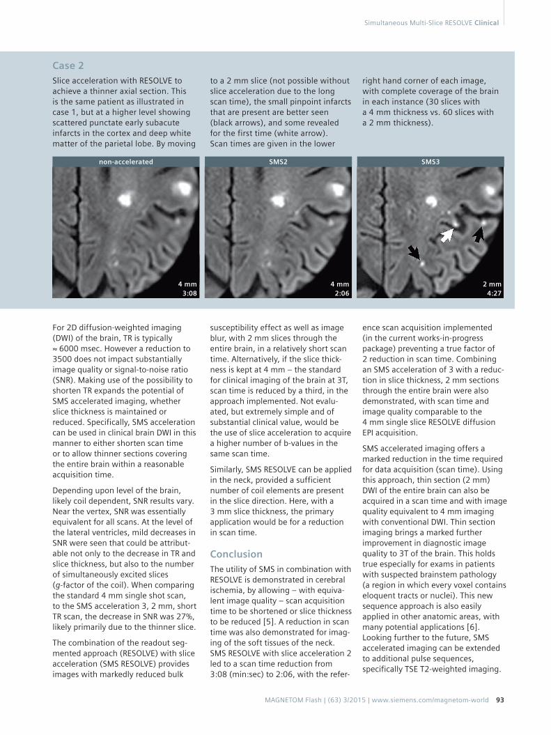
Case 2Slice acceleration with RESOLVE to achieve a thinner axial section. This is the same patient as illustrated in case 1, but at a higher level showing scattered punctate early subacute infarcts in the cortex and deep white matter of the parietal lobe. By moving
to a 2 mm slice (not possible without slice acceleration due to the long scan time), the small pinpoint infarcts that are present are better seen (black arrows), and some revealed for the first time (white arrow). Scan times are given in the lower
right hand corner of each image, with complete coverage of the brain in each instance (30 slices with a 4 mm thickness vs. 60 slices with a 2 mm thickness).
ence scan acquisition implemented (in the current works-in-progress package) preventing a true factor of 2 reduction in scan time. Combining an SMS acceleration of 3 with a reduc-tion in slice thickness, 2 mm sections through the entire brain were also demonstrated, with scan time and image quality comparable to the 4 mm single slice RESOLVE diffusion EPI acquisition.
SMS accelerated imaging offers a marked reduction in the time required for data acquisition (scan time). Using this approach, thin section (2 mm) DWI of the entire brain can also be acquired in a scan time and with image quality equivalent to 4 mm imaging with conventional DWI. Thin section imaging brings a marked further improvement in diagnostic image quality to 3T of the brain. This holds true especially for exams in patients with suspected brainstem pathology (a region in which every voxel contains eloquent tracts or nuclei). This new sequence approach is also easily applied in other anatomic areas, with many potential applications [6]. Looking further to the future, SMS accelerated imaging can be extended to additional pulse sequences, specifically TSE T2-weighted imaging.
non-accelerated SMS2 SMS3
4 mm 3:08
4 mm 2:06
2 mm 4:27
Simultaneous Multi-Slice RESOLVE Clinical
MAGNETOM Flash | (63) 3/2015 | www.siemens.com/magnetom-world 93

Case 3Slice acceleration with RESOLVE – the advantages of a thinner section. Bulk susceptibility artifacts on DWI are further reduced, and small pin-
Case 5In this example, the patient (with mul-tiple punctate, acute, left middle cere-bral artery distribution infarcts) was combative and moved throughout the exam despite sedation, degrading image quality. Motion artifact is great-est on the readout segmented DWI exam, due both to the long scan dura-tion (3:26 min:sec) and the acquisition scheme, and least on the 2 mm slice accelerated scan. This 67-year-old patient presented one day prior to the MR with global aphasia, a right facial palsy and – on other imaging studies – a distal M1 segment occlusion.
point lesions with restricted diffusion are better seen. And, as shown, in certain instances small pinpoint lesions can be visualized only on
the thinner sections, such as this small cortical infarct (arrow) in the left middle frontal gyrus.
Case 4Do we have good depiction of this lateral medullary infarct, with single shot imaging (upper row)? The con-ventional answer would be that the infarct is well delineated, with 4 mm slices acquired at 3T. But no, this is just a misconception, due to limited experience with thinner sections! This small infarct is not nearly as well seen as with 2 mm sections – acquired using slice acceleration (lower row), where the infarct is more sharply defined on each section and we have an additional slice (in between). The patient, an 87-year-old woman, presented one day prior to the MR with dizziness, nausea and vomiting, and left facial paralysis.
non-accelerated
single shot
accelerated
SMS2 SMS3
2 mm 4:27
4 mm 2:06
4 mm
2 mm
4 mm 3:08
readout segmented single shot accelerated
Clinical Simultaneous Multi-Slice RESOLVE
94 MAGNETOM Flash | (63) 3/2015 | www.siemens.com/magnetom-world

Case 6Axial (part 1, 6A-C) and sagittal (part 2, 6D, E) soft tissue neck images from a normal volunteer are presented, without and with slice acceleration. In part 1, a single-shot (ss) DWI exam is compared to RESOLVE acquired without and with slice acceleration. Note the artifactual foreshortening in the AP dimension on the ss exam, which leads to a lymph node (arrow) that is anterior to the submandibular gland on the left being projected over the gland. On the RESOLVE images, there is no anatomic distortion, with the effective spatial resolution also improved due to the absence of the artifactual blurring present in the ss exam (and inherent to this technique). The use of slice acceleration allowed the RESOLVE sequence to be obtained
in a very similar scan time as with the ss DWI, 2:07 vs 1:50 min:sec. In part 2, off-midline sagittal RESOLVE diffusion-weighted images are presented. Of intermediate signal intensity is a very small part of the submandibular gland with a high signal intensity small lymph node immediately anteriorly (in the middle of image), with a portion of the parotid gland seen in the more supe-rior portion of the image. Depiction of the multiple scattered, high signal intensity, normal lymph nodes and SNR are equivalent for the two scans, with slice acceleration reducing scan time by nearly a factor of 2 (from 3:44 to 2:07 min:sec). Images were acquired with the Head/Neck 64-channel coil.
Contact
Val M. Runge, M.D. Editor-in-Chief, Investigative Radiology Department of Diagnostic, Interventional and Pediatric Radiology University Hospital of Bern, Inselspital Bern, Switzerland [email protected]
single shot
readout segmented
readout segmented, SMS2
non-accelerated
SMS2
Acknowledgement All images were acquired at 3T on a MAGNETOM Skyra MR system.
References
1 Larkman DJ, Hajnal JV, Herlihy AH, et al. Use of multicoil arrays for separation of signal from multiple slices simultaneously excited. J Magn Reson Imaging. 2001;13(2):313-7.
2 Setsompop K, Gagoski BA, Polimeni JR, et al. Blipped-controlled aliasing in parallel imaging for simultaneous multislice echo planar imaging with reduced g-factor penalty. Magn Reson Med. 2012;67(5):1210-24.
3 Frost R, Jezzard P, Douaud G, et al. Scan time reduction for readout-segmented EPI using simultaneous multislice acceleration: Diffusion-weighted imaging at 3 and 7 Tesla. Magn Reson Med. 2015;74:136-49.
4 Poser BA, Anderson RJ, Guerin B, et al. Simultaneous multislice excitation by parallel transmission. Magn Reson Med. 2014;71(4):1416-27.
5 Runge VM, Aoki S, Bradley WG, Jr., et al. Magnetic Resonance Imaging and Computed Tomography of the Brain-50 Years of Innovation, With a Focus on the Future. Invest Radiol. 2015;50(9):551-6.
6 Filli L, Piccirelli M, Kenkel D, et al. Simul-taneous Multislice Echo Planar Imaging With Blipped Controlled Aliasing in Parallel Imaging Results in Higher Acceleration: A Promising Technique for Accelerated Diffusion Tensor Imaging of Skeletal Muscle. Invest Radiol. 2015;50(7):456-63.
6A
6B
6C
6D
6E
Simultaneous Multi-Slice RESOLVE Clinical
MAGNETOM Flash | (63) 3/2015 | www.siemens.com/magnetom-world 95