single molecule detection of parp1 and parp2 interaction with
TRANSCRIPT
Published online 15 December 2015 Nucleic Acids Research, 2016, Vol. 44, No. 6 e60doi: 10.1093/nar/gkv1476
Single molecule detection of PARP1 and PARP2interaction with DNA strand breaks and theirpoly(ADP-ribosyl)ation using high-resolution AFMimagingMaria V. Sukhanova1,2,†, Sanae Abrakhi2,†, Vandana Joshi2, David Pastre2, MikhailM. Kutuzov1, Rashid O. Anarbaev1,3, Patrick A. Curmi2, Loic Hamon2,* and Olga I. Lavrik1,3,*
1Institute of Chemical Biology and Fundamental Medicine, 630090, Novosibirsk, Russian Federation, 2INSERM,U1204, Laboratoire Structure––Activite des Biomolecules Normales et Pathologiques, Universited’Evry-Val-d’Essonne, F-91025 Evry, France and 3Novosibirsk State University, 630090, Novosibirsk, RussianFederation
Received February 18, 2015; Revised November 30, 2015; Accepted December 5, 2015
ABSTRACT
PARP1 and PARP2 are implicated in the synthesis ofpoly(ADP-ribose) (PAR) after detection of DNA dam-age. The specificity of PARP1 and PARP2 interactionwith long DNA fragments containing single- and/ordouble-strand breaks (SSBs and DSBs) have beenstudied using atomic force microscopy (AFM) imag-ing in combination with biochemical approaches.Our data show that PARP1 localizes mainly on DNAbreaks and exhibits a slight preference for nicks overDSBs, although the protein has a moderately highaffinity for undamaged DNA. In contrast to PARP1,PARP2 is mainly detected at a single DNA nick site,exhibiting a low level of binding to undamaged DNAand DSBs. The enhancement of binding affinity ofPARP2 for DNA containing a single nick was alsoobserved using fluorescence titration. AFM studiesreveal that activation of both PARPs leads to thesynthesis of highly branched PAR whose size de-pends strongly on the presence of SSBs and DSBsfor PARP1 and of SSBs for PARP2. The initial affinitybetween the PARP1, PARP2 and the DNA damagedsite appears to influence both the size of the PARsynthesized and the time of residence of PARylatedPARP1 and PARP2 on DNA damages.
INTRODUCTION
Poly(ADP-ribose) polymerase-1 (PARP1) and poly(ADP-ribose) polymerase-2 (PARP2) are nuclear proteins respon-
sible for the synthesis of polymers of ADP-ribose using �-NAD+ as substrate (1,2). These enzymes are implicated inresponse to cell DNA damage, recognizing damaged DNAgenerated under genotoxic stress or DNA breaks as a resultof the activity of DNA repair enzymes (3–7). Upon bind-ing to damaged DNA, PARP1(2) catalyse PARylation of it-self and/or of a number of nuclear proteins including DNArepair/replication factors (1,3,8). Although the catalytic do-mains of PARP1 and PARP2 have a high degree of homol-ogy, the PARP2 DNA binding domain is distinct from thatof PARP1 (4,9,10). This could reflect differences betweenthese proteins in their preferences for binding to damagedDNA, because their activity is regulated by their DNA-binding domains (5,10,11). PARP1(2) activity and DNAsubstrate specificity have been studied by an ensemble ofbiochemical approaches to estimate their preference for thedamaged DNA (11–15). Previously, analysis of PARP1(2)binding to damaged DNA was carried out mainly usingshort DNA duplexes; however in this case it is difficult toanalyse the interaction with a single DNA damage. The in-teraction of PARP1(2) with nicks and blunt ends on shortDNA duplexes can overlap. Therefore long DNA duplexeswith several DNA damages located at a distance from oneanother are better models to study the influence of bluntDNA ends, undamaged DNA sequences and a single dam-aged site on the specificity of protein–DNA interaction.
Here, we studied the interaction of PARP1(2) with longDNA fragments containing DNA breaks using atomic forcemicroscopy (AFM) imaging and fluorescence assays to es-timate the DNA binding characteristics of these proteins.We established new optimal conditions for adsorption ofDNA–PARP1(2) complexes on mica surface mediated by
*To whom correspondence should be addressed. Tel: +7 383 363 51 95; Fax: +7 383 363 51 53; Email: [email protected] may also be addressed to Loic Hamon. Tel: +33 169470179; Fax: +33 169470219; Email: [email protected]†These authors contributed equally to the paper as first authors.
C© The Author(s) 2015. Published by Oxford University Press on behalf of Nucleic Acids Research.This is an Open Access article distributed under the terms of the Creative Commons Attribution License (http://creativecommons.org/licenses/by-nc/4.0/), whichpermits non-commercial re-use, distribution, and reproduction in any medium, provided the original work is properly cited. For commercial re-use, please [email protected]
Downloaded from https://academic.oup.com/nar/article-abstract/44/6/e60/2499441by gueston 10 February 2018

e60 Nucleic Acids Research, 2016, Vol. 44, No. 6 PAGE 2 OF 12
putrescine (Pu2+). The conditions were exploited to detectthe interaction of PARP1 and PARP2 with long DNA frag-ments (1200-bp) containing only DSB ends or DSBs to-gether with a unique single strand break (SSB). Statisti-cal analysis of the AFM data in view of localizing proteinsbound to DNAs shows that PARP1 specifically binds toboth SSB and DSB with a preference for nicks. At the sametime, this protein binds to undamaged DNA, but to a lowerextent than to breaks. In contrast to PARP1, PARP2 bindsweakly to undamaged DNA and to DSB ends, and local-izes mainly to SSBs. These results correlate with fluores-cence data revealing that the binding affinity of PARP2 to1200-bp DNA is 5-fold weaker in comparison to the sameDNA fragment containing a nick. At the same time, the ap-pearance of a SSB in DNA leads only to a 2-fold increase inthe binding affinity of PARP1 to the DNA fragment. AFMdata show that PARylated PARP1(2) are still able to inter-act with DNA. Thus, PARylated PARP2 was detected nearnicks, while modified PARP1 was near blunt ends. The dataare in the agreement with previous biochemical findings in-dicating that the PARylated PARP1 can interact with dam-aged DNA (16,17). The length of the PAR polymer formedby PARP1(2) appears to be influenced by the initial bind-ing affinity of the proteins for the damaged site. Our obser-vations by a single-molecule study implicate PARP2 in therecognition of nicks and suggest its role in SSB repair.
MATERIALS AND METHODS
Chemicals, reagents and proteins
Chemical compounds (MgCl2, putrescine and spermi-dine) were purchased from Sigma-Aldrich. ‘Vivaspin’ ul-trafiltration spin columns were from ‘Sartorius Stedim.Biotech. GmbH’. Nb.BsmI, nicking endonuclease and plas-mid pBR322 were purchased from ‘New England Bio-Labs’. Taq DNA polymerase was purchased from ‘ThermoScientific’. Murine PARP2 was expressed in insect cellsand purified according to (18). Human PARP1 was ex-pressed in Escherichia coli and purified according to (19).The 1200-bp DNA fragment containing the Nb.Bsm1target sequence in the middle of the chain was pre-pared by the polymerase chain reaction (PCR). Plas-mid pBR322 was used as a template with the followingprimers: 5′-CGCCGCACTTATGACTGTCTTC-3′- for-ward primer and 5′-GCGTTAATGTCTGGCTTCTGA-3′-reverse primer. Although Taq DNA polymerase has a ten-dency to add an adenine nucleotide to the 3′-ends of PCRproducts, 1200-bp DNA was considered as DNA fragmentwith blunt ends. The PCR products were run on a 1%agarose gel and purified using a gel extraction kit (‘Fer-mentas’). Nicked DNAs were obtained by incubating the1200-bp DNA or pBR322 with Nb.Bsm1 nicking endonu-clease according to the recommended protocol. Formationof the nicked circular form of pBR322 was analysed by 0.8%agarose gel electrophoresis with EtBr staining. The 1400-bpDNA carrying ends with short overhangs (4 nt) was pre-pared using pBR322. The plasmid was cleaved with Pst1(ACGTC)/Sal1(CAGCT) and the fragment was purified ona 1% agarose gel. DNA concentrations were determined bymeasuring the absorbance at 260 nm.
Sample preparation for AFM imaging
PARP1 or PARP2 (1.75–14 nM) and DNAs (1.5 nM for1200-bp DNAs or 0.35 nM for nicked pBR322) were incu-bated in AFM deposition buffer (12.5 mM Hepes, pH 8.0,12.5 mM KCl, 1 mM DTT) on ice for 1–5 min. Just beforesample deposition, to adsorb the PARP1(2), DNA, DNA–PARP1 or DNA–PARP2 complexes, multivalent cationswere added to the solution to final concentrations of 10 mMMg2+, as well as 5 mM putrescine (Pu2+) or 50 �M sper-midine (Spd3+), after which a 10-�l droplet was depositedon the surface of freshly cleaved mica at room tempera-ture for 30 s. The mica surface was then rinsed with 0.02%uranyl acetate solution to stabilize the DNA–protein com-plexes in their conformations for AFM imaging in air (20).The sample was rapidly rinsed with pure water (Millipore)and air-dried before imaging. For experiments with auto-PARylation of PARP2, 3.5 nM PARP2 was incubated with1.5 nM nicked 1200-bp DNA in the AFM deposition bufferin the presence of 100 �M NAD+ on ice for 5–120 min.For experiments with auto-PARylation of PARP1, 35 nMPARP1 was incubated with 3.5 nM nicked pBR, 3.5 nM su-percoiled pBR or 13.7 nM 1200-pb DNA in the AFM depo-sition buffer in the presence of 100 �M NAD+ and 10 mMMgCl2 at 37◦C for 15–120 min. After incubation the sam-ples were diluted 10× in AFM deposition buffer and imme-diately deposited on mica. For AFM imaging, the sampleswere processed as described above.
AFM and image analysis
Sample imaging was performed in air at room temperaturein the tapping ModeTM with a MultimodeTM AFM (Veeco,Santa Barbara, CA, USA) operating with a NanoscopeIIIaTM controller. Olympus (Hamburg, Germany) siliconcantilevers AC160TS with resonance frequencies of about300 kHz and nominal spring constants of 10–100 N/mwere used. The scan frequency was typically 1.5 Hz perline and the modulation amplitude was a few nanometres.Data were acquired at a set point chosen to minimize tip–sample interaction force and a first or second order polyno-mial function served to remove the background. The ‘sec-tion’ tool in the Nanoscope Analysis software (version 1.50)was used to determine the molecular dimensions of theprotein particles. Cross-sections of the individual particlesimaged by AFM were made and the height and diameterat half-maximal height of each single particle were mea-sured. The DNA length and the position of PARP1(2) onDNA were analysed using ‘ImageJ’ software. The lengthof the DNA contour was manually traced on AFM im-ages. In the case of DNA–protein complexes, the contourlength was manually traced as the shortest possible DNApath through the bound protein. The size of PARylatedproteins were calculated using the following equation: S =�R2, where the R is the minimum radius of the circle inwhich PARylated proteins could be enclosed. The radiuswas measured from AFM images using the ‘section’ toolin the Nanoscope Analysis software. The percent of DNAbound to PARP1(2) was estimated as the ratio between thenumber of DNA fragments interacting with one or moreprotein molecules over the total DNA fragments adsorbedon mica. The binding specificity of PARP1(2) with DSB and
Downloaded from https://academic.oup.com/nar/article-abstract/44/6/e60/2499441by gueston 10 February 2018
PAGE 3 OF 12 Nucleic Acids Research, 2016, Vol. 44, No. 6 e60
SSB was quantified by estimating the percent of PARP1(2)complexes bound to the DNA lesion site (21). The protein–DNA complexes were classified according to whether theywere located at the ends, at the nick site or at the non-damaged sites of the model DNA fragments (Supplemen-tary Figure S1A–C), taking into account that the proteininteracts with the DSB ends when it is located within thelast 40 nm of each DNA end, and with SSB when the pro-tein is located between 194 and 234 nm from one DNA ex-tremity (Supplementary Figure S1D). The interval of 40 nmwas selected on the basis of the mean ± SD contour lengthmeasured for PARP1(2)–DNA complexes (SupplementaryFigure S1 E and F).
DNA binding constants and specificities
Binding specificities (S) and constants (Kd) were calculatedas described previously (21). Briefly, the method is basedon the determination of the average DNA fractional oc-cupancy of PARP1(2) at non-specific (undamaged DNA)sites, specific (nick) sites on the DNA and the DNA ends.The counting of protein–DNA complexes and DNA frag-ments was used to calculate the fractional occupancy, thatis the total number of DNA fragments (nFragment), the to-tal number of PARP1(2)–DNA complexes at undamagedDNA contours (nComplex,Und), the total number of DNA ter-mini (nComplex,Ends) and nicks (nComplex,Nick) bound by theseproteins (Supplementary Table S1). Taking into accountthe size of PARP1(2) in AFM images (Supplementary Fig-ure S1 D and F), the 1200-bp DNA fragment contains twoDNA ends and 976 non-specific sites; nicked 1200-bp DNAcontains one specific site, two DNA ends and 864 non-specific sites. The specificities and AFM-site specific bind-ing affinities (Kd) of PARP1(2) to nicked, to non-specificsites and to DNA ends were calculated directly estimatingthe number of specific and non-specific complexes and frac-tional occupancies of PARP1(2) bound to different DNAsites (Supplementary Table S1). Composite macroscopicbinding constants (Kd, AFM macro) of PARP1 and PARP2 to1200-bp DNA fragments were calculated using values of Kdfor specific and non-specific sites and DNA ends (21).
Fluorescent labelling of PARP1 and PARP2 and fluorescencetitration assays
For protein labelling, 5(6)-carboxyfluorescein N-hydroxysuccinimide ester (FSE) was used, in which thecarboxyl groups were activated with N-hydroxysuccinimide(NHS). The NHS ester is easily displaced by nucleophilicattack from primary amino groups of the N-terminalgroup of the protein (primary target at pH 8.0) and lysineside chains at the water-accessible surface of the protein,thus forming an amide bond with the original carboxylgroup of fluorescein at physiological pH. Purified PARP1or PARP-2 (1.8 pmol) were incubated with FSE (8 pmol)at 4◦C overnight in buffer containing 50 mM Hepes, pH7.5 and 100 mM NaCl. Unreacted FSE was removed bydialysis, followed by concentration of the labelled proteinson ultrafiltration spin columns. The concentration ofPARP1(2)-fluorescein conjugates and labelling efficiencywere determined using the extinction coefficients: �280 =
120 000 M−1cm−1 for PARP1, �280 = 70 415 M−1cm−1
for PARP2 and �280 = 23 400 cm−1 M−1 and �495 = 60000 M−1 cm−1 for fluorescein. The degree of labellingwas estimated as 25% for PARP1 and 28% for PARP2.Fluorescence titration experiments were performed byadding increasing amount of DNAs to a fixed concen-tration of protein (20 nM for PARP1-fluorescein and 90nM for PARP2-fluorescein) in buffer containing 12.5 mMHepes-KOH (pH 8.0), 12.5 mM KCl and 1 mM DTT.The excitation wavelength was set at 485 nm, and emissionwavelength at 520 nm. All experiments were performed at28◦C on a POLARstar Optima multidetection microplatereader (BMG Labtech, Offenburg, Germany) in a 96-wellassay ‘V’ bottom black plate (Axigen), and thus, one wellcontained one titration point. These experiments werecarried out in the same buffer as used for AFM samplepreparations. The volume of the reaction mixture was 50–100 �l. All reactions were mixed at room temperature. Theplate was incubated at 28◦C in the reader. Each experimentwas repeated at least three times.
The degree of binding (Db) was estimated using the fol-lowing equation:
Db = F − F0
Fmax − F0,
where F indicates the fluorescence intensity of the PARP1-fluorescein (or PARP2-fluorescein) conjugates at differentDNA concentrations, and F0 and Fmax are the fluorescenceintensities in the absence and at saturating levels of theDNA, respectively.
The dissociation constants (Kd) were calculated using thefollowing equation:
Db = 1
1 + Kd[C]
where [C] is the concentration of the DNA.
RESULTS
Imaging of PARP1(2)–DNA complexes in the presence ofpolyamines
AFM experiments make it possible to observe the inter-action between single molecules whereas traditional bio-chemical techniques indicate only average values from alarge number of interacting molecules (21–24). However,AFM imaging of DNA–protein complexes is not straight-forward since the complexes can dissociate during their ad-sorption and immobilization on the surface (23,25). The ab-sorption of DNA and of protein–DNA complexes on micarequires the presence of multivalent cations, such as mag-nesium (Mg2+), putrescine (Pu2+) or spermidine (Spd3+),in the deposition buffer or using fixing agents such as glu-taraldehyde (25–28). However, it has been shown that thecations can affect protein stability during the absorptionprocess (25,29–31). Therefore, at first, we determined theappropriate conditions for the deposition of free PARPson mica in the presence of different cations, namely Mg2+,Pu2+ or Spd3+. Representative AFM images of free PARP1and PARP2 are shown in Figure 1A. The average molecularheights and distribution of volumes for PARP1 and PARP2
Downloaded from https://academic.oup.com/nar/article-abstract/44/6/e60/2499441by gueston 10 February 2018

e60 Nucleic Acids Research, 2016, Vol. 44, No. 6 PAGE 4 OF 12
Figure 1. AFM images of PARP1 or PARP2 molecules deposited on micasurface in the presence of different counterions. (A) Images of PARP1 (2nM) or PARP2 (2 nM) in the presence of 10 mM Mg2+, 5 mM Pu2+ or50 �M Spd3+. Scale bar 500 nm; Z scale: 7 nm. (B) The average heightof PARP1 or PARP2 measured in images shown in (A) (n, number ofmolecules analysed). Results are mean ± SD of three independent samples.P-values were obtained by comparing the results by t-test, *, P < 0.05; **,P < 0.01; ***, P < 0.005; ns, not significant.
molecules in the presence of different cations were estimatedfrom these images (Figure 1B and Supplementary FigureS2). In contrast to Mg2+ or Pu2+, slight oligomerization ofboth proteins was observed in the presence of Spd3+ (Fig-ure 1B and Supplementary Figure S2). Thus, using Spd3+ inthe deposition process can induce protein aggregation thatcan complicate the analysis of DNA–PARP1(2) complexesby AFM.
PARP1 acts as a molecular sensor of DNA breaks and isinvolved in both SSB and DSB repair (1,7,32,33). PARP2participates in SSB repair but there is no strong evidencethat this protein is involved in DSB repair (8,34). In vitro,the interaction of PARP1(2) with DNA containing SSBsand DSBs has been studied by different techniques (10–15,35,36). However, AFM has never been employed in in-vestigating binding of PARPs with DNA containing thesetypes of lesions. Here we used a 1200-bp double-strandedDNA fragment (1200-bp DNA), and the same fragmentwith a nick in the middle of the chain (nicked 1200-bpDNA) to characterize PARPs and DNA complexes withAFM (Supplementary Figure S1D). The use of long DNAsubstrates can lead to a better discrimination of PARP1(2)binding to DSBs and nicks in the context of linear DNAduplexes.
Figure 2A and B show AFM images of 1200-bp DNAalone or after incubation with varying concentrations ofPARP1(2) proteins. Sample depositions on mica were per-formed in the presence of Pu2+ or Mg2+. Under these con-
Figure 2. Analysis of PARP1 and PARP2 complex formation with DNAcontaining DSB ends. (A) Binding of PARP1 or PARP2 to 1200-bp DNAanalysed by AFM. A total of 1200-bp DNA was incubated with 1.75–14 nM PARP1 or PARP2 and imaged by AFM in air. All images wereobtained using 5 mM Pu2+ for complex adsorption. White arrows indi-cate PARP1(2)–DNA complexes. Scale bar 1 �m; Z scale: 7 nm. (B) Im-age of 1200-bp DNA deposited on mica surface in the absence of PARP1or PARP2. Scale bar 500 nm; Z scale: 7 nm. (C) Quantitative analysis ofPARP1(2)–DNA complex formation depending on protein concentration,the number of DNA molecules analysed: 164 for 1.75 nM PARP1, 400 for1.75 nM PARP2, 138 for 3.5 nM PARP1, 326 for 3.5 nM PARP2, 117 for7 nM PARP1. At 7nM of PARP2, the detection of a specific interactionbetween the DNA fragment and the protein was complicated due to thehigh density of free PARP2 adsorbed on the surface. The results representmean ± SD of three independent samples. (D) Fluorescence measurementsof PARP1 and PARP2 binding to DNA. The reaction mixtures contain-ing the fluorescein-labelled protein PARP1 or PARP2 were titrated withincreasing amounts of 1200-bp DNA. Bars indicate the standard error ofthree independent experiments.
ditions, PARP1(2)–DNA complexes were not detected byAFM using a large range of Mg2+ concentrations from2 to 20 mM (data not shown). In contrast to Mg2+, thecomplexes were visualized in the presence of Pu2+ (Fig-ure 2A). Consequently, Pu2+ was selected as counterion forPARP1(2)–DNA complex adsorption. Under high PARP1concentrations, the formation of large aggregates and a de-crease of the amount of isolated molecules adsorbed onthe surface were observed (Figure 2A). PARP1 aggrega-tion on mica has already been demonstrated by AFM us-ing the chromatin system (37). In the same range of pro-tein concentrations, PARP2 showed a completely differentbehaviour since the density of DNA molecules remainedconstant while the number of proteins adsorbed on the sur-
Downloaded from https://academic.oup.com/nar/article-abstract/44/6/e60/2499441by gueston 10 February 2018

PAGE 5 OF 12 Nucleic Acids Research, 2016, Vol. 44, No. 6 e60
face significantly increased (Figure 2A). Thus, a concentra-tion of around 3.5 nM PARPs is more suitable for the com-parison of the efficiencies of DNA–protein complex forma-tion using AFM experiments for the following reasons; (i)aggregate formation for DNA–PARP1 complexes on micasurfaces is limited under these conditions, (ii) the numberof PARP2 molecules adsorbed on the surface remains suf-ficiently low to limit the formation of non-specific DNA–PARP2 complexes and (iii) the ratio of DNA–protein com-plexes over the total adsorbed DNA is sufficiently highto perform statistical analyses of PARP interaction withDNA. Under the conditions developed here, a low levelof complex formation between PARP2 and DNA was ob-served. Only 18% of DNA was involved in interaction withthis protein. In contrast to PARP2, PARP1 is more effec-tive in binding to dsDNA and the yield of complex wasabout 65% at 3.5 nM protein concentration (Figure 2C).The AFM results are in agreement with the results of flu-orescence titration experiments showing that the affinity ofPARP1 for 1200-bp DNA is about 5-fold higher than thatof PARP2 (Figure 2D).
To analyse the interaction of PARP1(2) with the DSBends, we estimated the position distributions of the proteinsbound to 1200-bp DNA fragments using 3.5 nM PARP1 orPARP2 (Figure 3). Representative AFM images of 1200-bpDNA in the presence of PARP1 or PARP2 are shown inFigure 3A and B. Analysis of the position distributions ofPARP1(2) molecules on DNA fragments shows that about40% of PARP1(2) molecules are located at DNA ends while60% interact with the internal region of the DNA molecules(Figure 3C). For detailed description of this DNA sub-strate and details of how the position of proteins along theDNA were defined and measured, see Supplementary Fig-ure S1. Although high levels of PARP1–DNA complex for-mation and low levels in the case of PARP2 were observed(Figure 2C), both proteins bind to DNA ends with similarspecificity (Table 1). At the same time, comparative anal-ysis of the site specific Kd values of the proteins indicatesthat PARP1 has about five time more affinity to DNA endsand non-specific DNA sites than that of PARP2 (Table 1).Interestingly, the composite Kd values of PARP1(2)–DNAcomplexes calculated from AFM-site specific constants areconsistent with the ones determined from fluorescence titra-tion experiments (Table 1).
Collectively, these data indicate that PARP1 interactsmore efficiently with both termini and undamaged parts oflong linear DNA than PARP2.
Specificity of PARP1 and PARP2 interaction with SSBs andDSBs
Both PARP1 and PARP2 have been shown to be in-volved in BER/SSB repair interacting with repair pro-teins and DNA intermediates, including DNA with anapurinic/apyrimidinic site, nick, short gap or flap lesions(5,7,8,13,16,38–41). Although PARP2 recognizes and bindsto gap- or flap-containing DNA structures (3,8,10,13), re-cently DNA with a 5′-phosphorylated SSB was identified asa preferential substrate for PARP2 activation (14). To testwhether a single DNA nick influences PARP1 and PARP2binding to long DNA molecules by AFM, nicked circular
Figure 3. AFM analysis of the interaction of PARP1 and PARP2 with un-damaged DNA and DNA ends. Large scale AFM images (left) of PARP1(A) and PARP2 (B) binding to 1200-bp DNA and zoomed images (right)of PARP1(2)–DNA complexes. A total of 1200-bp DNA was incubatedwith 3.5 nM PARP1 or PARP2 and imaged by AFM in air. Arrows indi-cate binding of PARP1(2) to DNA ends or undamaged DNA. Scale bar1 �m; Z scale: 7 nm. (C) Position distributions of PARP1 and PARP2 on1200-bp DNA; number of complexes analysed: 90 for PARP1 and 62 forPARP2. The results represent mean ± SD of three independent samples.
pBR or nicked 1200-bp DNA fragments were used. Thecircular shape of the pBR plasmid formed by endonucle-ase treatment was demonstrated by agarose gel and AFMimaging (Supplementary Figure S3). Both for PARP1 andPARP2, the presence of one protein molecule interactingwith the nicked plasmid was detected (Supplementary Fig-ure S4A). However, the compaction or crossover formationof the DNA on the AFM surface limits the detection of spe-cific DNA–protein interactions and complicates the statis-tical analysis of PARP complex formation (SupplementaryFigure S4A). Although nicked pBR does not appear to be agood model for AFM studies of interactions of PARP1 andPARP2 with the DNA nick site, we compared the affinity ofthese proteins for the substrate using fluorescence titration(Supplementary Figure S4B). Both proteins demonstratesimilar Kd values with nicked pBR, although PARP1 andPARP2 show clear differences in binding affinity to 1200-bpDNA (Figure 2D and Supplementary Figure S4B). In con-trast to nicked pBR, using linear 1200-bp DNA fragmentscarrying a single nick allowed to quantify the complexes of
Downloaded from https://academic.oup.com/nar/article-abstract/44/6/e60/2499441by gueston 10 February 2018

e60 Nucleic Acids Research, 2016, Vol. 44, No. 6 PAGE 6 OF 12
Table 1. DNA binding constants and specificities of PARP1 and PARP2 for the 1200-bp DNA fragment
PARP1 PARP2
Kd (nM) Specificity Kd (nM) Specificity
AFM-site specific constants:DNA ends 17.5 ± 1.0 360 ± 36 90.9 ± 6.2 323 ± 29.3Non-specific 6329 ± 518 29411 ± 1764Binding constants (AFM macro)a 3.30 ± 0.23 16.10 ± 1.01Binding constants (Flu)b 1.66 ± 0.02 7.65 ± 0.66
aComposite macroscopic binding constants of PARP1 and PARP2 to 1200-bp DNA fragments.bMacroscopic binding constant of PARP1 and PARP2 to a 1200-bp DNA measured by fluorescence titration in this study (Figure 2D).
PARP1(2) with DNA and to determine the specificity of in-teractions of these proteins with individual lesions (Supple-mentary Figure S1D). Using nicked 1200-bp DNA resultsin a slight increase in the amount of PARP1–DNA com-plexes formed in comparison to the same DNA fragmentwithout SSB (Figures 2C and 4C). About 65% of PARP1molecules are detected on SSB/DSB sites and only 35% arebound to undamaged DNA. Interestingly, the presence ofa single nick in the DNA significantly increases the amountof PARP2–DNA complexes detected by AFM, and the pro-tein is clearly localized at the nick site (Figure 4 C and B).The resulting distributions of PARP2 on nicked DNA re-veals its high preference for SSBs, since more than 70% ofall complexes are formed by PARP2 molecules interactingwith SSBs (Figure 4D). Thanks to high resolution imaging,it was also possible to determine the fractional occupanciesof the proteins bound to different DNA sites and to cal-culate their binding affinity and the specificity for nickedsites (Supplementary Tables S1 and S2). The data show thatPARP2 and PARP1 have a similar affinity for nicks; how-ever PARP2 reveals a higher specificity for this type of DNAbreaks (Table 2). Thus, PARP2 exhibits a significant prefer-ence for binding to nicked sites over DNA ends and undam-aged DNA. Notably, binding of the protein to nicks influ-ences the calculated composite macroscopic binding con-stants, and the affinity of PARP2 for nicked DNA frag-ments comes very close to that of PARP1 (Table 2). Theseresults are consistent with the ones of the determination ofthe Kd values of the PARP1(2) complex with a nicked 1200-bp DNA using fluorescence titration (Figure 4E). Both pro-teins show a higher affinity for DNA carrying a single nickas compared to DNAs with only blunt ends (Figures 2D and4E). When compared to PARP1, PARP2 has a weaker affin-ity for DNA containing only DSB ends, but a similar affinityfor nicked DNA (Figures 2D and 4E, Tables 1 and 2). Thus,when 1200-bp DNA fragments were used, the affinity ofPARP1 was higher than that of PARP2 due to the contribu-tion of PARP1 binding to both blunt ends and undamagedDNA (Table 1). However, our data demonstrate that bothproteins have a similar affinity for nick-containing DNA.Added to this, the high level of discrimination of nick byPARP2 detected by AFM indicates that binding of PARP2to DNA is significantly facilitated by SSB formation.
AFM imaging of PARP1 and PARP2 activation in the pres-ence of DSBs and SSBs
PARP1(2) catalyse the synthesis of PAR using NAD+ assubstrate (1,2,4–6). PARP1(2) activation results in the syn-
thesis of PAR, the branched negatively charged polymer,that is covalently attached to the PARPs themselves or toother acceptor proteins (1,5). PARylation of PARP1 leadsto suppression of its DNA binding activity and seems to bea factor regulating the interaction of the protein with DNA(1,5). As for PARP1, PAR synthesis catalysed by PARP2 isalso stimulated in the presence of DNA damage (3,8,9). Toaddress PARylation of PARP1(2) using AFM, imaging ofthese proteins after incubation with 1200-bp DNA, nicked1200-bp DNA or nicked or supercoiled pBR in the presenceof NAD+ was undertaken (Figures 5 and 6). When DNAsubstrates were incubated with PARP1 in the presence ofNAD+, the formation of branched polymers was detectedby AFM (Figure 5A and B). It should be noted that incu-bation of PARP1(2) with NAD+ without DNA did not leadto the appearance of PARylated proteins under the assayconditions used here (Supplementary Figure S5). The timecourse of PARP1 activation detected at the molecular levelallowed us to measure the average size of the PARylatedproteins in the presence of 1200-bp DNA, nicked 1200-bpDNA, and nicked or supercoiled pBR (Figure 5C). The sizewas estimated by determining the area of the circle with theradius, which encloses the auto-PARylated protein (Figure5D). Three major points could be underlined. First, the sizeof the PAR increases mainly within the first 15 min, but thepolymerization reaction proceeds for all the incubation pe-riod (Figure 5D). Second, PARylation of PARP1 was ob-served for all DNA substrates including supercoiled pBR,but the size of PAR and the amount of PARylated proteinsincreased significantly in the presence of DNA substrateswith breaks (Figure 5D). Third, in the case of DNA sub-strates containing both DSB and SSB, PARylated PARP1can still bind to DNA and has been mainly detected nearDSB ends (Figures 5A and 7).
When PARP2 was incubated with DNA in the presenceof NAD+ at 37◦C, PARylation of this protein was not ob-served by AFM, although it was detected by SDS gel elec-trophoresis (Supplementary Figure S6). The visualizationof PARP2 auto-PARylation by AFM was achieved by incu-bating the reaction mixtures on ice before sample loading onmica (Figure 6A and B). In the case of 1200-bp DNA, thePARylation catalysed by PARP2 was detected, but at a lowlevel (Figure 6C) while PARP2 activation was clearly ob-served after 15 min of incubation in the presence of nicked1200-bp DNA (Figure 6A and B). Thus, the presence ofa nick in the DNA fragment led to a significant increasein both the amount of PARylated protein and the size ofthe PAR polymer observed under the assay conditions used
Downloaded from https://academic.oup.com/nar/article-abstract/44/6/e60/2499441by gueston 10 February 2018

PAGE 7 OF 12 Nucleic Acids Research, 2016, Vol. 44, No. 6 e60
Figure 4. Comparative analysis of PARP1 and PARP2 binding to DNA containing a single nick. Large scale AFM images (left) of PARP1 (A) andPARP2 (B) binding to nicked 1200-bp DNA and zoomed images (right) of PARP1(2)–DNA complexes. Nicked 1200-bp DNA was incubated with 3.5nM PARP1 or PARP2 and imaged by AFM in air. Arrows indicate binding of PARP1(2) to DNA ends, nick or undamaged DNA. Scale bar 200 or 500nm; Z scale: 7 nm. (C) AFM analysis of PARP1(2)–DNA complex formation, number of DNA molecules analysed: 210 for PARP1 and 189 for PARP2.The results represent mean ± SD of three to six independent samples. (D) Analysis of the position distributions of PARP1 and PARP2 on nicked 1200-bpDNA, number of complexes analysed: 145 for PARP1 and 104 for PARP2. The results represent mean ± SD of three to six independent samples. (E)Fluorescence measurements of PARP1 or PARP2 binding to DNA. The reaction mixtures containing the fluorescein-labelled protein PARP1 or PARP2were titrated with increasing amounts of nicked 1200-bp DNA. Bars indicate the standard error of three independent experiments.
Downloaded from https://academic.oup.com/nar/article-abstract/44/6/e60/2499441by gueston 10 February 2018
e60 Nucleic Acids Research, 2016, Vol. 44, No. 6 PAGE 8 OF 12
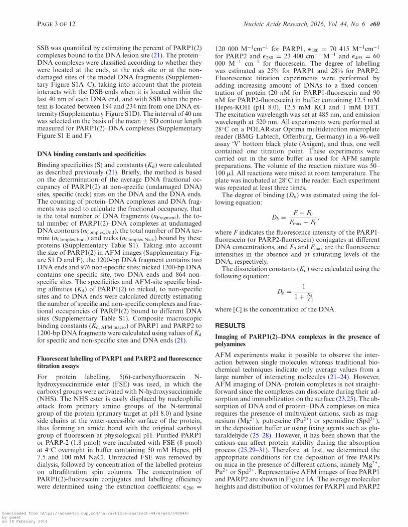
Table 2. DNA binding constants and specificities of PARP1 and PARP2 for nicked 1200-bp DNA fragment
PARP1 PARP2 2
Kd (nM) Specificity Kd (nM) Specificity
AFM-site specific constants:DNA nick 5.88 ± 0.51 1349 ± 134 3.7 ± 0.40 7297 ± 872Non-specific 7936 ± 476 27027 ± 1270Binding constants (AFM macro)a 2.3 ± 0.15 3.0 ± 0.29Binding constants (Flu)b 0.84 ± 0.09 1.6 ± 0.12
aComposite macroscopic binding constants of PARP1 and PARP2 to nicked 1200-bp DNA fragments.bMacroscopic binding constant of PARP1 and PARP2 to a 1200-bp DNA measured by fluorescence titration in this study (Figure 4E).
Figure 5. AFM analysis of PARP1 activation in the presence of the dif-ferent DNA substrates. AFM images show auto-PARylation of PARP1in the presence of nicked 1200-bp DNA (A), and 1200-bp DNA, circularnicked pBR or supercoiled pBR (B). Arrows indicate PARylated proteinsbound to DNA molecules. Scale bar: 500 nm; Z scale: 7 nm. (C) Compar-ative analysis of the size of PARylated PARP1 in the presence of differentDNA substrates. Number of PARylated molecules analysed: 145 for nicked1200-bp DNA, 202 for 1200-bp DNA, 114 for nicked pBR and 107 for su-percoiled pBR. Each data point represents the mean area ± SD with Rof PARylated molecules measured from images. All measurements of ra-dius (R) were done from the three to ten images obtained from three inde-pendent samples for each DNA substrate. The size of PARylated PARP1smaller than 1200 nm2 (R < 20 nm) were not taken into account. (D)Zoomed image of PARylated PARP1 bound to SSB. The minimum radiusof the circle (R) enclosing the PARylated protein was used to estimate thearea of PARylated proteins. Scale bar: 100 nm; Z scale: 7 nm.
here (Figure 6C). As for PARP1, the increase in size of thePAR polymer is mainly observed during the 15 min of thereaction; although the sizes of the PAR structures are in-creased at least 2-fold after incubation for 2 h on ice. Itshould be noted that interaction of PARylated PARP2, asalso PARP1, with DNA was detected by AFM images, but
Figure 6. AFM analysis of PARP2 activation in the presence of SSBs. (A)AFM images show time course of auto-PARylation of PARP2 in the pres-ence of nicked 1200-bp DNA. White arrows indicate PARylated proteinsbound to DNA molecules. Scale bar: 500 nm; Z scale: 7 nm. (B) Largescale AFM images of PARylated PARP2 in the presence of nicked 1200-bp DNA. Insert: zoomed image of a large PARylated PARP2 formed un-der these conditions. Scale bar: 500 nm; Z scale: 7 nm. (C) Comparativeanalysis of the size of PARylated PARP2 in the presence of 1200-bp DNAwith or without nick after 120 min of reaction. Number or PARylatedmolecules analysed: 354 for nicked 1200-bp DNA and 145 for 1200-bpDNA. Each data point represents the mean area ± SD with radius ofPARylated molecules measured from images. All measurements of the ra-diuses were done from the five images obtained from three independentsamples for each DNA substrate. The size of PARylated PARP2 smallerthan 1200 nm2 (R < 20 nm) were not taken into account.
mainly at the nick site (Figures 6A and 7). According tothe literature, poly(ADP-ribosyl)ation of PARPs leads toa decrease of its DNA binding activity and facilitates itsdissociation from DNA breaks (1–6). Although automodi-fied PARPs can still bind to the DNA, the amount of com-plexes of PARylated proteins with DNA is strongly reduced,such that the percentage of nicked 1200-bp DNA boundto PARylated proteins is about 5.5 and 3.7% for PARP1and PARP2 respectively. It is nearly one order of magnitudelower than the percentage of binding of unPARylated pro-teins to DNA (which correlates to a ≈50-fold effect on thebinding affinity of the proteins to nicks) (Table 2 and Sup-
Downloaded from https://academic.oup.com/nar/article-abstract/44/6/e60/2499441by gueston 10 February 2018

PAGE 9 OF 12 Nucleic Acids Research, 2016, Vol. 44, No. 6 e60
Figure 7. AFM analysis of the position distributions of PARylatedPARP1(2) on nicked 1200-bp DNA. PARP1 (3.5 nM) or PARP2 (3.5 nM)was incubated with 1200-bp nicked DNA (1.5 nM) at 37◦C for PARP1 andon ice for PARP2 for 60 min in the presence of 100 �M NAD+. Numberof complexes analysed: 89 for PARP1 and 55 for PARP2.
plementary Table S2). The interaction of PARylated pro-teins with DNA raises the question of their preferential lo-calization on the DNA. We examined the position distri-butions of PARylated PARP1(2) on nicked 1200-bp DNAby analysing a DNA molecules complexed with the modi-fied proteins (Figure 7). Our results show that PARylatedPAPR1 is mainly located at DNA ends and nick, whilePARylated PARP2 is preferentially located on the nick site(Figure 7). This is in agreement with the mode of binding ofunmodified PARP2 or PARP1 to individual sites observedby AFM for nicked 1200-bp DNA, when about 65% ofPARP1 or 85% of PARP2 complexes were bound to DNAbreaks (Figure 4D). The analysis of PARylation catalysedby the proteins as measured by AFM and gel electrophore-sis shows the increase in size of the PAR polymer producedin the presence of DSBs and SSB for PARP1 and SSB forPARP2 (Figures 5C and 6C, Supplementary Figure S7).This suggests that the extent of PARylation can be influ-enced by initial binding affinity of PARPs for DNA sites(Tables 1 and 2). To determine by AFM the effect of differ-ent DNA binding sites on the extent of PARP1(2) PARyla-tion, we measured the time course of changes in the sizefor the proteins activated by DSB, SSB or non-damagedDNA (Figure 8A). Figure 8B and D show the average sizeof PARylated PARP1 or PARP2 after incubation of the pro-teins with nicked 1200-bp DNA for 30, 60 and 120 min inthe presence of NAD+. These data demonstrate that af-ter 2 h of incubation the size of PARylated PARP1 de-tected at SSB and DSB was 25–40% greater than the size ofmodified PARP1 molecules located on undamaged DNA(Figure 8B). In the case of PARP2, its activation was de-tected mainly at DNA breaks (Figure 8D). Size of PARy-lated PARP2 detected at SSBs increases of 37% in compari-son with the modified form of proteins detected near DSBs.Thus, types of DNA damage have an influence on the sizeof the PAR polymers produced by PARPs.
Figure 8. AFM-based size determination of PARylated PARP1 andPARP2 according to position of the proteins on DNA. AFM images ofPARylated PARP1 (A) and PARP2 (C) located at different sites of nicked1200-bp DNA (from top to bottom: DNA end, nick and undamaged DNAfor PARP1, DNA end and nick for PARP2). Scale bar: 100 nm; Z scale: 7nm. (B) Time course of changes in the size of PARylated PARP1 during itsactivation at nick, DNA ends or undamaged DNA sites. (D) Time courseof changes in the size of PARylated PARP2 during its activation at nick orDNA ends. Each data point represents the mean area ± SD. P-values wereobtained by comparing the results by t-test, *, P < 0.05; **, P < 0.01; ***,P < 0.005; ns, not significant. All measurements of radiuses were donefrom more than 10 images obtained from three independent samples foreach DNA substrate.
DISCUSSION
PARP1 and PARP2 have been proposed to play a criticalrole in the detection and repair of DNA breaks (1,3,5,6,41).Here, we analysed the interaction of PARP1(2) with longDNA molecules to estimate the specificity of PARP1 andPARP2 interaction with SSBs and DSBs using a combina-tion of AFM imaging and biochemical approaches. Previ-ously, the AFM technique was only applied to study theinteraction of PARP1 with chromatin, plasmid and spe-cific DNA structures such as loops, hairpins and cruciform(42–45). Due to its ability to image single molecules, AFMprovides unique information on DNA–protein complexessuch as specificity of the interaction and cooperative as-pects of binding (21–25). However, the main problem ofthis approach remains the potential dissociation of DNA–protein complexes on mica surface during the adsorptionstep. As mica is a highly negatively charged surface as areDNA molecules, the adsorption and immobilization of nu-
Downloaded from https://academic.oup.com/nar/article-abstract/44/6/e60/2499441by gueston 10 February 2018

e60 Nucleic Acids Research, 2016, Vol. 44, No. 6 PAGE 10 OF 12
cleic acids alone or associated with proteins require the pres-ence of multivalent cations in the deposition buffer (25,26).Magnesium and polyamines such as putrescine or spermi-dine can be used as cations in the buffer (26,28). Spermi-dine at sub-millimolar concentrations induces strong DNAor DNA–protein complex adsorption allowing the observa-tion of protein–ssDNA complexes under various conditionsof ionic strength (26,27). It was shown that the concentra-tion of putrescine has to be in the millimolar range for ad-sorption of proteins with dsDNA on mica (26,28). We devel-oped a procedure for absorption of PARP–DNA complexeson mica surface mediated by putrescine. In contrast to Mg2+
and Spd3+, using of Pu2+ as counterions allowed to obtainrepresentative large-scale images of DNA–PARP1(2) com-plexes and compare them not only to specific interactionsof these proteins with DNA breaks but also their activa-tion in the presence of NAD+. Indeed, PAR synthesized byPARP1(2) is a negatively charged polymer and can be ad-sorbed on mica surface such as DNA molecules, allowingto estimate the size and structure of the poly(ADP-ribose)chains.
PARP1 is considered as a molecular sensor of both DNASSBs and DSBs (1,5,32,33,41). Our AFM data show thatabout 60% of PARP1 complexes were detected on DNAbreaks and 40% on undamaged DNA (Figure 4D). How-ever, considering the fact that undamaged sites are ∼1000-fold in excess over breaks, PARP1 binds predominantly toSSB/DSB rather than to undamaged DNA (Supplemen-tary Table S1). At the same time, distribution of PARP1between DSBs and SSBs is relatively similar, although thisprotein has higher affinity and specificity to nick sites (Ta-bles 1 and 2). Fluorescence titration assay confirmed thatPARP1 binds to 1200-bp DNA containing both a SSB andDSB with Kd value two times lower that the one for 1200-bpDNA without nick (Figures 2D and 4E).
PARP2 interaction with damaged DNA is less well doc-umented than PARP1 (10,13,14). The higher level of stimu-lation of PARP2 activity towards SSBs compared to DSBswas detected using short DNA duplexes representing DNAintermediates of the different DNA repair pathways (13,14).In our case, AFM detection at the single molecule level havedemonstrated a strong influence of a single nick on PARP2binding to DNA (Figures 2C and 4C), PARP2 moleculesare observed at SSB on nicked 1200-bp DNA in 75% of thetotal quantity of complexes (Figure 4D). Simultaneously,AFM and fluorescence titration experiments prove that thepresence of nick in the 1200-bp DNA fragment induces a∼5-fold increase in the affinity of PARP2 for the DNA sub-strate (Tables 1 and 2). Thus, in contrast to PARP1, PARP2has a low affinity for undamaged DNA, being more special-ized in recognizing SSB (Table 2). This specificity of PARP2for SSBs suggests the possibility that PARP2 contributes tothe SSB repair mechanism. Thus, AFM imaging providesdirect evidence that PARP1 and PARP2 indeed preferen-tially bind to DNA breaks in the context of an extendedDNA structure.
Another interesting finding of this study is the visual-ization of the PARylated proteins by AFM. Detection ofPARylated PARPs by AFM at high resolution allowed usto compare the size and structure of PAR synthesized bythe proteins (Figures 5–8). AFM images of PAR structures
formed under activation of PARP1 or PARP2 does notshow differences since PARs adopt a highly branched ‘star’shape for both PARPs used (Figures 5C and 6A). Like-wise, PARylated PARP1 and PARP2 interacting with SSBsor DSBs do not show differences in the PAR structure de-tected by AFM. In addition, measuring the size of PARy-lated proteins shows that modified PARP1(2) with R-valuelarger than 40 nm still interacts with DNA (Figure 8B andD). It is possible that ADP-ribose chain lengths synthesizedby PARP1(2) depend mainly on the initial affinity of theproteins for the site of DNA damage (Tables 1 and 2), i.e.high affinity binding leads to a longer residence time of theprotein at the specific site and thus offers the possibility ofsynthesizing a larger polymer. The size and the location ofPARylated PARP2 along DNA molecules in the presence ofboth SSBs and DSBs are in agreement with this hypothesis(Figures 7 and 8). The model of PARP1 ‘shuttling’ proposesthe regulation of PARP1 interaction with DNA throughauto-PARylation (1,5,46). This model implies that auto-poly(ADP-ribosyl)ation of PARP1 initiates dissociation ofthe enzyme–DNA complexes due to an electrostatic repul-sion between anionic PAR and DNA (5,46). Associationof PARylated PARP1(2) of varied size with different DNAsites observed by AFM may point to the weaker influenceof the electrostatic force on the stability of PARP1(2)–DNAcomplexes during the PARylation reaction. These AFM re-sults are in line with biochemical data wherein it was shownlowered affinity of PARylated PARP1 to DNA nick ratherthan an inability of the automodified protein to bind DNAlesions (16,17,39).
In conclusion, using analyses at the single molecule leveland long DNA fragments, we detected efficient interactionof PARP2 with SSB (Table 2). These results were confirmedby biochemical data that validate our experimental condi-tions used in AFM experiments. We then took advantageof our approach to study PARP1(2) activation and it ap-pears that the nature of strand interruption influences theefficiency of PARylation, while the polymer remains highlybranched in the case of reactions catalysed by PARP1 orPARP2. Our data permit us to reach the important con-clusion that PARylated PARP1 and PARP2 retain their in-teraction with DNA at their specific DNA sites (SSB orDSB) which initiated the reaction of PARP poly(ADP-ribosyl)ation. These data open perspectives on PAR struc-tural studies depending on the nature of the DNA damageand may bring important information on the function ofthe two proteins in the nucleus.
SUPPLEMENTARY DATA
Supplementary Data are available at NAR Online.
ACKNOWLEDGEMENTS
We are thankful to Dr Valerie Schreiber (Universite deStrasbourg, IREBS, Illkirch, France) for providing the re-combinant plasmid coding PARP2 and Dr Masahiko S.Satoh (Laval University Medical Centre (CHUQ), LavalUniversity, Quebec, Canada) for providing the recombinantplasmid coding PARP1. We would like to thank Dr Anne-Lise Haenni (Jacques Monod Institute, Paris, France) forcareful reading of the manuscript and useful comments.
Downloaded from https://academic.oup.com/nar/article-abstract/44/6/e60/2499441by gueston 10 February 2018
PAGE 11 OF 12 Nucleic Acids Research, 2016, Vol. 44, No. 6 e60
FUNDING
Russian Scientific Fund [14-24-00038 to O.I.L.]; InstitutNational de la Sante et de la Recherche Medicale; GenopoleEvry. Funding for open access charge: Russian ScientificFund [14-24-00038].Conflict of interest statement. None declared.
REFERENCES1. D’Amours,D., Desnoyers,S., D’Silva,I. and Poirier,G.G. (1999)
Poly(ADP-ribosyl)ation reactions in the regulation of nuclearfunctions. Biochem. J., 342, 249–268.
2. Ame,J.C., Spenlehauer,C. and de Murcia,G. (2004) The PARPsuperfamily. Bioessays, 26, 882–893.
3. Schreiber,V., Dantzer,F., Ame,J.C. and de Murcia,G. (2006)Poly(ADP-ribose): novel functions for an old molecule. Nat. Rev.Mol. Cell Biol., 7, 517–528.
4. Yelamos,J., Schreiber,V. and Dantzer,F. (2008) Toward specificfunctions of poly(ADP-ribose) polymerase-2. Trends Mol. Med., 14,169–178.
5. Lindahl,T., Satoh,M.S., Poirier,G.G. and Klungland,A. (1995)Post-translational modification of poly(ADP-ribose) polymeraseinduced by DNA strand breaks. Trends Biochem. Sci., 20, 405–411.
6. Satoh,M.S., Poirier,G.G. and Lindahl,T. (1993) NAD(+)-dependentrepair of damaged DNA by human cell extracts. J. Biol. Chem., 268,5480–5487.
7. Niedergang,C., Trucco,C., Flatter,E., De La Rubia,G., Oliver,J.,Rolli,V., Menissier de Murcia,J. and de Murcia,G. (1999) Involvementof poly(ADP-ribose) polymerase in base excision repair. Biochimie,81, 69–75.
8. Schreiber,V., Ame,J.C., Dolle,P., Schultz,I., Rinaldi,B., Fraulob,V.,Menissier de Murciam,J. and de Murcia,G. (2002) Poly(ADP-ribose)polymerase-2 (PARP-2) is required for efficient base excision DNArepair in association with PARP-1 and XRCC1. J. Biol. Chem., 21,23028–23036.
9. Ame,J.C., Rolli,V., Schreiber,V., Niedergang,C., Apiou,F., Decker,P.,Muller,S., Hoger,T., de Murcia,J.M. and de Murcia,G. (1999)PARP-2, a novel mammalian DNA damage dependentpoly(ADP-ribose) polymerase. J. Biol. Chem., 274, 17860–17868.
10. Schreiber,V., Ricoul,M., Ame,J.C., Dantzer,F., Meder,V.,Spenlehauer,C., Stiegler,P., Niedergang,C., Sabatier,L., Favaudon,V.et al. (2006) PARP-2, structure–function relationship. In: Burkle,A(ed). Poly(ADP-ribosyl)ation. pp. 13–31.
11. Pion,E., Ullmann,G.M., Ame,J.C., Gerard,D., de Murcia,G. andBombarda,E. (2005) DNA-induced dimerization ofpoly(ADP-ribose)polymerase-1 triggers its activation. Biochemistry,44, 14670–14681.
12. D’Silva,I., Pelletier,J.D., Lagueux,J., D’Amours,D., Chaudhry,M.A.,Weinfeld,M., Lees-Miller,S.P. and Poirier,G.G. (1999) Relativeaffinities of poly(ADP-ribose) polymerase and DNA-dependentprotein kinase for DNA strand interruptions. Biochim. Biophys. Acta,1430, 119–126.
13. Kutuzov,M.M., Khodyreva,S.N., Ame,J.C., Ilina,E.S.,Sukhanova,M.V., Schreiber,V. and Lavrik,O.I. (2013) Interaction ofPARP-2 with DNA structures mimicking DNA repair intermediatesand consequences on activity of base excision repair proteins.Biochimie, 95, 1208–1215.
14. Langelier,M.F., Riccio,A.A. and Pascal,J.M. (2014) PARP-2 andPARP-3 are selectively activated by 5′ phosphorylated DNA breaksthrough an allosteric regulatory mechanism shared with PARP-1.Nucleic Acids Res., 42, 7762–7775.
15. Lonskaya,I., Potaman,V.N., Shlyakhtenko,L.S., Oussatcheva,E.A.,Lyubchenko,Y.L. and Soldatenkov,V.A. (2005) Regulation ofpoly(ADP-ribose) polymerase-1 by DNA structure-specific binding.J. Biol. Chem., 280, 17076–17083.
16. Lavrik,O.I., Prasad,R., Sobol,R.W., Horton,J.K., Ackerman,E.J. andWilson,S.H. (2001) Photoaffinity labeling of mouse fibroblastenzymes by a base excision repair intermediate. J. Biol. Chem., 276,25541–25548.
17. Sukhanova,M., Khodyreva,S. and Lavrik,O. (2010) Poly(ADP-ribose)polymerase 1 regulates activity of DNA polymerase in long patchbase excision repair. Mutat. Res., 685, 80–89.
18. Ame,J.C., Kalisch,T., Dantzer,F. and Schreiber,V. (2011) Purificationof recombinant poly(ADP-ribose) polymerases. Methods Mol. Biol.,780, 135–152.
19. Sukhanova,M.V., Khodyreva,S.N. and Lavrik,O.I. (2004)Poly(ADP-ribose) polymerase-1 inhibits strand-displacementsynthesis of DNA catalyzed by DNA polymerase beta. Biochemistry(Mosc.), 69, 558–568.
20. Revet,B. and Fourcade,A. (1998) Short unligated sticky ends enablethe observation of circularised DNA by atomic force and electronmicroscopies. Nucleic Acids Res., 26, 2092–2097.
21. Yang,Y., Sass,L.E., Du,C., Hsieh,P. and Erie,D.A. (2005)Determination of protein-DNA binding constants and specificitiesfrom statistical analyses of single molecules: MutS-DNA interactions.Nucleic Acids Res., 33, 4322–4334.
22. Engel,A. and Muller,D.J. (2000) Observing single biomolecules atwork with the atomic force microscope. Nat. Struct. Biol., 7, 715–718.
23. Lyubchenko,Y.L., Gall,A.A. and Shlyakhtenko,L.S. (2014)Visualization of DNA and protein-DNA complexes with atomic forcemicroscopy. Methods Mol. Biol., 1117, 367–384.
24. Bustamante,C. and Rivetti,C. (1996) Visualizing protein-nucleic acidinteractions on a large scale with the scanning force microscope.Annu. Rev. Biophys. Biomol. Struct., 25, 395–429.
25. Pastre,D., Hamon,L., Sorel,I., Le Cam,E., Curmi,P.A. andPietrement,O. (2010) Specific DNA-protein interactions on micainvestigated by atomic force microscopy. Langmuir, 26, 2618–2623.
26. Pastre,D., Hamon,L., Landousy,F., Sorel,I., David,M.O., Zozime,A.,Le Cam,E. and Pietrement,O. (2006) Anionic polyelectrolyteadsorption on mica mediated by multivalent cations: a solution toDNA imaging by atomic force microscopy under high ionic strengths.Langmuir, 22, 6651–6660.
27. Hamon,L., Pastre,D., Dupaigne,P., Le Breton,C., Le Cam,E. andPietrement,O. (2007) High-resolution AFM imaging ofsingle-stranded DNA-binding (SSB) protein–DNA complexes.Nucleic Acids Res., 35, e58.
28. Hansma,H.G. and Laney,D.E. (1996) DNA binding to micacorrelates with cationic radius: assay by atomic force microscopy.Biophys. J., 70, 1933–1939.
29. Shi,W.X. and Larson,R.G. (2005) Atomic force microscopic study ofaggregation of RecA-DNA nucleoprotein filaments into left-handedsupercoiled bundles. Nano Lett., 5, 2476–2481.
30. Ristic,D., Modesti,M., van der Heijden,T., van Noort,J., Dekker,C.,Kanaar,R. and Wyman,C. (2005) Human Rad51 filaments on double-and single-stranded DNA: correlating regular and irregular formswith recombination function. Nucleic Acids Res., 33, 3292–3302.
31. Hoyer,W., Cherny,D., Subramaniam,V. and Jovin,T.M. (2004) Rapidself-assembly of alpha-synuclein observed by in situ atomic forcemicroscopy. J. Mol. Biol., 340, 127–139.
32. Wang,M., Wu,W., Rosidi,B., Zhang,L., Wang,H. and Iliakis,G. (2006)PARP-1 and Ku compete for repair of DNA double strand breaks bydistinct NHEJ pathways. Nucleic Acids Res., 34, 6170–6182.
33. Mansour,W.Y., Rhein,T. and Dahm-Daphi,J. (2010) The alternativeend-joining pathway for repair of DNA double-strand breaksrequires PARP1 but is not dependent upon microhomologies. NucleicAcids Res., 38, 6065–6077.
34. Beck,C., Robert,I., Reina-San-Martin,B., Schreiber,V. and Dantzer,F.(2014) Poly(ADP-ribose) polymerases in double-strand break repair:focus on PARP1, PARP2 and PARP3. Exp. Cell Res., 329, 18–25.
35. Benjamin,R.C. and Gill,D.M. (1980) Poly(ADP-ribose) synthesis invitro programmed by damaged DNA. A comparison of DNAmolecules containing different types of strand breaks. J. Biol. Chem.,255, 10502–10508.
36. Hengartner,C., Lagueux,J. and Poirier,G.G. (1991) Analysis of theactivation of poly(ADP-ribose) polymerase by various types of DNA.Biochem. Cell Biol., 69, 577–580.
37. Wacker,D.A., Ruhl,D.D., Balagamwala,E.H., Hope,K.M., Zhang,T.and Kraus,W.L. (2007) The DNA binding and catalytic domains ofpoly(ADP-ribose) polymerase 1 cooperate in the regulation ofchromatin structure and transcription. Mol. Cell Biol., 27, 7475–7485.
38. de Murcia,J.M., Niedergang,C., Trucco,C., Ricoul,M., Dutrillaux,B.,Mark,M., Oliver,F.J., Masson,M., Dierich,A., LeMeur,M. et al.(1997) Requirement of poly(ADP-ribose) polymerase in recoveryfrom DNA damage in mice and in cells. Proc. Natl. Acad. Sci. U.S.A.,94, 7303–7307.
Downloaded from https://academic.oup.com/nar/article-abstract/44/6/e60/2499441by gueston 10 February 2018

e60 Nucleic Acids Research, 2016, Vol. 44, No. 6 PAGE 12 OF 12
39. Khodyreva,S.N., Prasad,R., Ilina,E.S., Sukhanova,M.V.,Kutuzov,M.M., Liu,Y., Hou,E.W., Wilson,S.H. and Lavrik,O.I.(2010) Apurinic/apyrimidinic (AP) site recognition by the5′-dRP/AP lyase in poly(ADP-ribose) polymerase-1 (PARP-1). Proc.Natl. Acad. Sci. U.S.A., 107, 22090–22095.
40. Parsons,J.L. and Dianov,G.L., (2004) Monitoring base excisionrepair proteins on damaged DNA using human cell extracts.Biochem. Soc. Trans., 32, 962–963.
41. Parsons,J.L., Dianova,I.I., Allinson,S.L. and Dianov,G.L. (2005)Poly(ADP-ribose) polymerase-1 protects excessive DNA strandbreaks from deterioration during repair in human cell extracts. FEBSJ., 272, 2012–2021.
42. Smulson,M.E., Pang,D., Jung,M., Dimtchev,A., Chasovskikh,S.,Spoonde,A., Simbulan-Rosenthal,C., Rosenthal,D., Yakovlev,A. andDritschilo,A. (1998) Irreversible binding of poly(ADP)ribosepolymerase cleavage product to DNA ends revealed by atomic forcemicroscopy: possible role in apoptosis. Cancer Res., 58, 3495–3498.
43. Chasovskikh,S., Dimtchev,A., Smulson,M. and Dritschilo,A. (2005)DNA transitions induced by binding of PARP-1 to cruciformstructures in supercoiled plasmids. Cytometry, 68, 21–27.
44. Soldatenkov,V.A., Chasovskikh,S., Potaman,V.N., Trofimova,I.,Smulson,M. E. and Dritschilo,A. (2002) Transcriptional repressionby binding of poly(ADP-ribose) polymerase to promoter sequences.J. Biol. Chem., 277, 665–670.
45. Potaman,V.N., Shlyakhtenko,L.S., Oussatcheva,E.A.,Lyubchenko,Y.L. and Soldatenkov,V.A. (2005) Specific binding ofpoly(ADP-ribose) polymerase-1 to cruciform hairpins. J. Mol. Biol.,348, 609–615.
46. Tulin,A. and Spradling,A. (2003) Chromatin loosening bypoly(ADP)-ribose polymerase (PARP) at Drosophila puff loci.Science, 299, 560–562.
Downloaded from https://academic.oup.com/nar/article-abstract/44/6/e60/2499441by gueston 10 February 2018