six sigma dmaic improvement story - miami-dade
TRANSCRIPT

Slide 1
Bioterrorism Awareness:Protection of Human and Animal HealthFood animal veterinarians
Slide 2
Center for Food Security and Public Health Iowa State University - 2004
Why Are We Here?Why Are We Here?
• September 11, 2001 changed many things−Worst terrorist act in U.S.
history−More than 3,000 presumed
dead−Occurred on American soil− Increased sense of
vulnerability
The horrible events of September 11, 2001 changed our lives forever. The terrorist acts on that day cost more than 3,000 people their lives. They were the worst terrorist attacks on domestic soil in United States history. We are now experiencing a great sense of vulnerability and are constantly questioning our safety and that of our families. Top picture: Plumes of smoke poured over New York City as the World Trade Center collapsed to the ground. Bottom picture: Three unidentified rescue workers walked away from the crash site at the Pentagon. The Daily Progress photo by Dan Lopez via Associated Press.
Slide 3
Center for Food Security and Public Health Iowa State University - 2004
Biological AttackBiological Attack
• Bioterrorism attacks of 2001• Anthrax in postal system
• 22 cases• 5 deaths
• U.S. public health realm changed forever
The October, 2001 anthrax attacks were conducted via four envelopes mailed from Trenton, New Jersey containing Bacillus anthracis spores that were sent through the U.S. postal system. A fifth envelope was likely responsible for the Florida cases but was never recovered. Twenty-two cases of anthrax resulted; eleven inhalational and eleven cutaneous cases. In all, five people died from inhalational anthrax. The person/group responsible has not been identified.
Slide 4
Center for Food Security and Public Health Iowa State University - 2004
Preparedness Responsibilities: Veterinarians
Preparedness Responsibilities: Veterinarians
• Anticipate outbreaks on the local level• Collect and label samples• Know the agents • Know the typical signs of diseases
− Animals and human
• Know how to report suspected cases• Disseminate knowledge
Historically, infectious disease outbreaks are first recognized at the local level. As veterinarians, we should prepare ourselves by knowing: what agents could be used in an attack, what samples to collect and how to label and store them, what agents can infect animals (and what is enzootic in your area), how it’s transmitted, what signs to look for, and who to call if an intentional or accidental release of a biological agent is suspected. We should also be prepared to provide education to those most affected by the outbreak.
Slide 5
Center for Food Security and Public Health Iowa State University - 2004
OverviewOverview
• Bioterrorism• Zoonoses and bioterrorism• Disease control and biosecurity • U.S. Government agencies involved• Bioterrorism agents/diseases• Your role and responsibility
Today we will talk about bioterrorism and how we all have an important role in protecting our communities and our country. We will cover several topics including: generalities about bioterrorism, zoonotic disease and bioterrorism, disease control and biosecurity, the U.S. government agencies involved in preparing and protecting our nation, and a brief overview of potential bioterrorism agents. Finally, we will discuss the veterinarian’s responsibility: what to do if bioterrorism is suspected.

Slide 6
Center for Food Security and Public Health Iowa State University - 2004
Biological agents targeting humans, animals, or plants
Biological, chemical, or radiological agents targeting agriculture or its components
•Livestock•Food supply•Crops•Industry•Workers
TerrorismTerrorism
BioterrorismAgroterrorism Other
Conventional, radiological, nuclear, chemical,cyber
•Typically direct human targeting
“Biological warfare is defined as the use of microorganisms or toxins derived from living organisms, to cause death or disease in humans, animals, or plants in civilian settings. The definition would apply to the lone perpetrator acting independently, to state-supported terrorism, and to undeclared wars, as well as to declared armed conflict. Of the 3 targets (humans, animals, and plants) in the United States, the greatest threat would appear to be to human beings and animals.” (Huxsoll, D, Patrick W, Parrott C. Veterinary services in biological disasters. JAVMA 1987;190:714-722.) This definition will be used for bioterrorism for our purposes. The motivations of terrorists and terrorist groups to launch a bioterrorist attack are many, and may include the need for attention, exacting revenge, religious beliefs, desire to mimic God, apocalyptic fulfillment, the desire to create chaos within a society, the need to copy a previous incident (copycat actions), the desire to impress with new technology, or to inflict severe economic harm.
Slide 7
Center for Food Security and Public Health Iowa State University - 2004
Characteristics of a Biological Attack
Characteristics of a Biological Attack
• Difficult to detect release• Dissemination may cover large area• Possible secondary spread • Recognition of agent may be delayed
days to weeks• Difficulties in catching perpetrator
It can be difficult to detect when biological agents are released. Dissemination often covers a large geographic area and clinical cases may take days to weeks to recognize. There is also the possibility of secondary spread if the agent is contagious person-to-person or through a vector. These factors, especially the delayed recognition, allow the perpetrator plenty of time to leave the area.
Slide 8
Center for Food Security and Public Health Iowa State University - 2004
Time (Days)
No.
Affe
cted
Exposure
Symptoms
Seek Care
Infectious Disease OutbreakInfectious Disease Outbreak
This graph depicts the onset of an infectious disease outbreak. Note the time from exposure to the onset of symptoms. This demonstrates how cases may be delayed in their recognition, and by the time patients seek care, the perpetrator is long gone.
Slide 9
Center for Food Security and Public Health Iowa State University - 2004
Clues Suggesting Biological Agent Release
Clues Suggesting Biological Agent Release
• Clustering of morbidity or mortality−Temporally or geographically−Large numbers of animals and/or people−Atypical symptoms
• Normally healthy people affected • Unusual symptoms for area• Unusual age distribution• Disease occurring outside typical
season
Although there are many difficulties in detecting a bioterrorism attack, there are several clues that suggest a biological agent may have been released. Health-care providers should be alert to illness patterns and diagnostic clues that might indicate an unusual infectious disease outbreak associated with intentional release of a biologic agent. Indications of intentional release of a biologic agent include: 1) an unusual clustering of illness or mortality in a given geographic region or limited time frame for a large number of people or animals. This may also include abnormal or atypical unexplained symptoms; 2) normally healthy individuals suddenly becoming ill; 3) symptoms occurring in patients from an area that does not usually have clinical signs of that particular disease; 4) an unusual age distribution for common diseases (e.g. an increase in chickenpox-like illness among adult patients may be smallpox); or 5) the disease is occurring outside its “typical” season (e.g. flu-like symptoms in humans in June in the northern hemisphere).

Slide 10
Center for Food Security and Public Health Iowa State University - 2004
Many Agents are ZoonoticMany Agents are Zoonotic
• Disease may be seen in animals before humans
• Animals are sentinels− Pets, livestock,
wildlife
Many of potential bioterrorism agents are zoonotic. In some diseases, clinical signs may manifest in animals prior to humans. Pets in particular can act as important sentinels because they are present in large numbers and often live in close contact with humans. It is estimated that pets are present in 59% of U.S. households. Livestock are also present in high numbers especially in certain areas of the country. Many areas depend on livestock for their livelihood and this puts them at risk for bioterrorism or agroterrorism. Wildlife also play an important role in our communities because they could be important sources of infection for humans and animals, and could potentially contaminate large areas.
Slide 11
Center for Food Security and Public Health Iowa State University - 2004
Factors That Promote Transmission of Zoonoses
Factors That Promote Transmission of Zoonoses
• Frequent contact with domestic or wild animals
• Overlap with wildlife habitat• Intensive livestock production• Poor animal sanitation• Poor personal hygiene• Poor animal health
Some factors that promote zoonotic disease transmission include frequent contact with domestic or wild animals, and people visiting or living in areas that overlap with wildlife habitats or intensive livestock production. Other factors such as poor animal sanitation, poor animal health, and poor personal hygiene can also promote transmission.
Slide 12
Center for Food Security and Public Health Iowa State University - 2004
Disease Control: Client EducationDisease Control: Client Education
• Disinfect/clean up areas contaminated with animal waste−Livestock, pets, wildlife, rodents
• Basic hygiene−Wash hands−Child supervision
There are many things the public can do to protect themselves from exposure to zoonotic agents. Keep areas that have been contaminated with animal waste clean and disinfected. Follow proper hygiene, especially hand washing.
Slide 13
Center for Food Security and Public Health Iowa State University - 2004
Zoonoses Control:Client Education
Zoonoses Control:Client Education
• Proper pet selection• Use caution at petting zoos• Cook food properly• Control strays• Communication with physician and
veterinarian• Follow guidelines for
immunocompromised people
Encourage your clients to select the pet that is right for them, especially if they are immunocompromised. You should also educate your clients about petting zoos and that those animals may carry diseases that affect humans. Clients should be aware of proper guidelines for preparing and cooking food to decrease risk of disease. Control stray animal populations and encourage clients to call appropriate authorities if they observe a stray animal. Finally, it is important that your clients communicate with their physician and you on a regular basis, especially if they are immunocompromised, so that all parties are aware of what animals they may be living with or exposed to because of their occupation.
Slide 14
Center for Food Security and Public Health Iowa State University - 2004
Biosecurity Educationfor the Producer
Biosecurity Educationfor the Producer
• Develop and implement a biosecurity plan
• Train employees to help maintain the plan
• Post signs restricting access to areas of the farm and control traffic flow
Biosecurity begins at the farm entrance. Veterinarians should work with their clients to develop and implement a biosecurity plan that addresses the concern of the producer’s facility. Veterinarians are the local experts in infectious disease and their relationship with their clients makes them the perfect candidate for writing biosecurity plans. Veterinarians understand the risks involved and their clients should view biosecurity plans as an important service and added value to their operation. It is important to train all employees in the biosecurity plan and make sure they understand their responsibilities to the operation. Producers should also post signs to regulate access to certain areas of the farm, to help prevent unauthorized entry and control traffic flow. Photo courtesy of D. Bickett-Weddle, DVM, ISU.
Slide 15
Center for Food Security and Public Health Iowa State University - 2004
Biosecurity Educationfor the Producer
Biosecurity Educationfor the Producer
• Regulate visitors• Keep visitors sanitary−Clean clothing, boots−Disposable plastic
shoe/boot covers
• Implement insect, bird, and animal control
• Secure water, feed, and nutrient sources
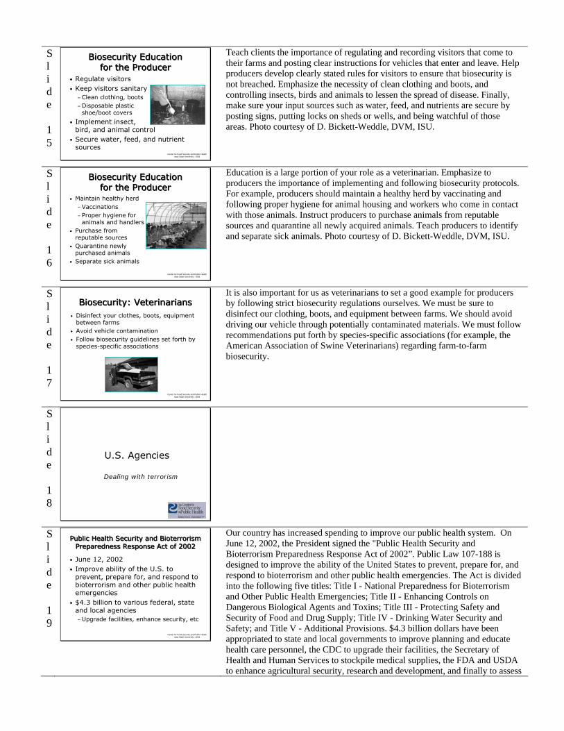
Teach clients the importance of regulating and recording visitors that come to their farms and posting clear instructions for vehicles that enter and leave. Help producers develop clearly stated rules for visitors to ensure that biosecurity is not breached. Emphasize the necessity of clean clothing and boots, and controlling insects, birds and animals to lessen the spread of disease. Finally, make sure your input sources such as water, feed, and nutrients are secure by posting signs, putting locks on sheds or wells, and being watchful of those areas. Photo courtesy of D. Bickett-Weddle, DVM, ISU.
Slide 16
Center for Food Security and Public Health Iowa State University - 2004
Biosecurity Educationfor the Producer
Biosecurity Educationfor the Producer
• Maintain healthy herd−Vaccinations−Proper hygiene for
animals and handlers• Purchase from
reputable sources• Quarantine newly
purchased animals• Separate sick animals
Education is a large portion of your role as a veterinarian. Emphasize to producers the importance of implementing and following biosecurity protocols. For example, producers should maintain a healthy herd by vaccinating and following proper hygiene for animal housing and workers who come in contact with those animals. Instruct producers to purchase animals from reputable sources and quarantine all newly acquired animals. Teach producers to identify and separate sick animals. Photo courtesy of D. Bickett-Weddle, DVM, ISU.
Slide 17
Center for Food Security and Public Health Iowa State University - 2004
Biosecurity: VeterinariansBiosecurity: Veterinarians• Disinfect your clothes, boots, equipment
between farms• Avoid vehicle contamination• Follow biosecurity guidelines set forth by
species-specific associations
It is also important for us as veterinarians to set a good example for producers by following strict biosecurity regulations ourselves. We must be sure to disinfect our clothing, boots, and equipment between farms. We should avoid driving our vehicle through potentially contaminated materials. We must follow recommendations put forth by species-specific associations (for example, the American Association of Swine Veterinarians) regarding farm-to-farm biosecurity.
Slide 18
U.S. Agencies
Dealing with terrorism
Slide 19
Center for Food Security and Public Health Iowa State University - 2004
Public Health Security and Bioterrorism Preparedness Response Act of 2002
Public Health Security and Bioterrorism Preparedness Response Act of 2002
• June 12, 2002• Improve ability of the U.S. to
prevent, prepare for, and respond to bioterrorism and other public health emergencies
• $4.3 billion to various federal, state and local agencies−Upgrade facilities, enhance security, etc
Our country has increased spending to improve our public health system. On June 12, 2002, the President signed the "Public Health Security and Bioterrorism Preparedness Response Act of 2002”. Public Law 107-188 is designed to improve the ability of the United States to prevent, prepare for, and respond to bioterrorism and other public health emergencies. The Act is divided into the following five titles: Title I - National Preparedness for Bioterrorism and Other Public Health Emergencies; Title II - Enhancing Controls on Dangerous Biological Agents and Toxins; Title III - Protecting Safety and Security of Food and Drug Supply; Title IV - Drinking Water Security and Safety; and Title V - Additional Provisions. $4.3 billion dollars have been appropriated to state and local governments to improve planning and educate health care personnel, the CDC to upgrade their facilities, the Secretary of Health and Human Services to stockpile medical supplies, the FDA and USDA to enhance agricultural security, research and development, and finally to assess

vulnerability and develop response plans.
Slide 20
Center for Food Security and Public Health Iowa State University - 2004
Department of Homeland Security (DHS)
Department of Homeland Security (DHS)
• Established January, 2003• Mission −Prevent, protect, and respond to acts of
terrorism on U.S. soil• Established four policy directorates −Responsibilities for coordinating HHS
and USDA−Guard borders and airports, coordinate
the response for future emergencies, analyze threats and intelligence, protect our critical infrastructure
On November 25, 2002, President Bush signed the "Homeland Security Act of 2002" into law. On January 24, 2003 the Department of Homeland Security (DHS) was established. Twenty-two federal agencies were brought together to streamline and centralize efforts to protect our nation’s homeland. This was the most significant transformation of the U.S. Government since 1947 when the branches of the U.S. Armed Forces were merged into the Department of Defense. The DHS provides one point of contact for state and local groups and the private sector. The mission of the DHS is to prevent terrorist attacks within the US, protect against terrorist attacks by decreasing our vulnerability, and minimizing damage from potential attacks and natural disasters. The DHS is organized into: Four Policy directorates (bureau or department): Border and Transportation Security (guard borders and airports), Emergency Preparedness and Response (coordinate the response for future emergencies), Information Analysis and Infrastructure Protections (analyze threats and intelligence), and Science and Technology (protect our critical infrastructure); a Management Directorate; the US Coast Guard; and the US Secret Service. Within the four policy directorates are multiple responsibilities for coordinating the efforts of the Department of Health and Human Services (HHS) and the United States Department of Agriculture (USDA).
Slide 21
Center for Food Security and Public Health Iowa State University - 2004
Centers for Disease Control and Prevention
Centers for Disease Control and Prevention
• CDC's Mission−Promote health and quality
of life by preventing and controlling disease, injury, and disability
• Preparing for bioterrorism since 1998• One of the first agencies to respond to
anthrax incidents of 2001
The Centers for Disease Control and Prevention (CDC) is recognized as the lead federal agency for protecting the health and safety of people at home and abroad. They provide credible information to enhance health decisions and promote health through strong partnerships. The CDC serves as the national focus for developing and applying disease prevention and control, environmental health, and health promotion and education activities designed to improve the health of the people of the United States. The CDC is headquartered in Atlanta, Georgia, and is an agency of the Department of Health and Human Services. Dr. Julie L. Gerberding is the Director. CDC has been responding to public health emergencies for decades and has been preparing for bioterrorism in particular since 1998. CDC's bioterrorism plans were put into action in Fall 2001 as one of the first agencies to respond during the anthrax outbreaks. One of the primary elements learned from these attacks was the importance of rapid identification. CDC works hard to help local and state health departments increase their capabilities for early detection.
Slide 22
Center for Food Security and Public Health Iowa State University - 2004
Strategic National StockpileStrategic National Stockpile
• 12-hour Push Package− Complete package of
medical materials
• Vendor Managed Inventory− Tailored to suspected agents
The mission of the Strategic National Stockpile (SNS), previously called the National Pharmaceutical Stockpile, is to ensure the availability of life-saving pharmaceuticals, antidotes and other medical supplies and equipment necessary to counter the effects of nerve agents, biological pathogens and chemical agents. The SNS Program stands ready for immediate deployment to any U.S. location in the event of a terrorist attack using a biological, toxin or chemical agent directed against a civilian population. These response packages are stored in strategic locations around the U.S. to ensure rapid delivery anywhere in the country. Following the federal decision to deploy, the SNS will typically arrive by air or ground in two phases. The first phase shipment is called a 12-hour Push Package. “12” because it will arrive in 12 hours or less, “push” because a state need only ask for help—not for specific items, and “package” because the

Program will ship a complete package of medical material to respond to a broad range of threats. Also available are inventory supplies known as Vendor Managed Inventory, or VMI packages. VMI packages can be tailored to provide pharmaceuticals, vaccines, medical supplies and/or medical products specific to the suspected or confirmed agent or combination of agents. A CDC team of five or six technical advisors will also be deployed at the same time as the first shipment. Known as a Technical Advisory Response Unit (TARU), this team is comprised of pharmacists, emergency responders, and logistics experts that will advise local authorities on receiving, distributing, dispensing, replenishing, and recovering SNS material. The SNS was tested in a real-life terrorist attack in response to the tragic events of September 11th and all facets of the New York operation performed exactly as intended.
Slide 23
Center for Food Security and Public Health Iowa State University - 2004
Insert Your State’s Info HereInsert Your State’s Info Here
The Iowa slides have been included as an example. Delete them and put in the information appropriate for your state.
Slide 24
Center for Food Security and Public Health Iowa State University - 2004
Preparing IowaPreparing Iowa
• Iowa’s Homeland Security −Administered by
Iowa Emergency Management Division
−Works with public and private partners
www.iowahomelandsecurity.org
Iowa’s Homeland Security Office works under the direction of the Federal Homeland Security Office. The mission of the Iowa Homeland Security office is to develop and coordinate the implementation of a comprehensive state strategy to secure the State of Iowa from terrorist threats or attacks and to coordinate the State of Iowa’s efforts to detect, prepare for, prevent, protect against, respond to and recover from terrorist attacks within Iowa. Iowa’s Homeland Security Office assesses current capabilities and assets of state government, identifies critical assets important to Iowa citizens, and ensures that they are protected, works with local emergency management agencies in each of Iowa’s 99 counties, and facilitates communication between many state departments and agencies, including the Iowa Department of Public Health and the Iowa Department of Agriculture and Land Stewardship.
Slide 25
Center for Food Security and Public Health Iowa State University - 2004
Preparing IowaPreparing Iowa
• Iowa Department of Public Health www.idph.state.ia.us/odedp
• Iowa Department of Agriculture and Land Stewardship− Highly infectious animal disease program− IRVIN: Iowa Rapid Veterinary Information
Network
• CFSPH training veterinarians to educate others
The Iowa Department of Public Health established the Office of Disease Epidemiology and Disaster Preparedness (ODEDP) in October 2001. The ODEDP encompasses two centers, the Center for Acute Disease Epidemiology (CADE) and the Center for Disaster Operations and Response (CDOR). The ODEDP leads development and implementation of an integrated system of health and public health services in preparedness for and response to disaster/terrorism incidents, outbreaks of infectious disease, and other public health threats and emergencies. The Iowa Department of Agriculture and Land Stewardship (IDALS) has been working on response plans for outbreaks of highly infectious animal diseases including the chain of communication between state agencies. The Iowa Rapid Veterinary Information Network (IRVIN) is a network of 860 licensed Iowa veterinarians who receive “burst” emails notifying them of the current concern. IRVIN was put to the test last summer when West Nile Virus was spreading throughout our state and has been used to inform practitioners of the latest pseudorabies status of Iowa. The Center for Food Security and Public Health at Iowa State University trained veterinarians on diseases the CDC has categorized as priority agents for bioterrorism preparedness (Category A, B, C), and these veterinarians will in turn educate other veterinarians, food producers, and the general public to increase awareness.

Slide 26
Category ABC Agent Overview
In this section, we first discuss how the CDC Category ABC disease/agent list was established and then overview the diseases.
Slide 27
Center for Food Security and Public Health Iowa State University - 2004
ClassificationClassification
• Prepared by the CDC’s Bioterrorism Preparedness and Response Office
• Category A: highest priority• Category B: second highest priority• Category C: third highest priority
In 1999 Congress requested that the national public health capabilities for response to acts of biological terrorism be upgraded. The CDC was designated as the lead agency for overall public health planning. In order to focus their preparedness efforts, the CDC needed to select and prioritize biological agents based on the threat they posed to public health. A group of national experts including infectious disease specialists, Department of Health and Human Services personnel, civilian and military intelligence experts, and law enforcement officials gathered to establish the list. The general criteria used for selection and prioritization were: 1) the public health impact based on illness and death; 2) the delivery potential to large populations based on stability and ability to mass produce and distribute a virulent agent; 3) potential for person to person transmission; 4) the public perception as related to public fear and potential civil disruption; 5) the special public health preparedness needs, stockpiles required, surveillance and diagnostic needs. Special attention was given to those agents that had previously been used or researched as a bioweapon. Based on these criteria, agents were scored and divided into A, B and C Categories. This is not a federally legislated list and is subject to change based on review of agents. Using this standardized system allows the CDC to add or remove agents. As veterinarians it is important to be aware of these agents and review these diseases as they relate to veterinary patients.
Slide 28
Center for Food Security and Public Health Iowa State University - 2004
“Weaponization” of Agents“Weaponization” of Agents
• Alter characteristics of a pathogen to make it a more effective weapon−Enhance transmission− Increase virulence−Resistant to antibiotics−Evade vaccine protection −Alter clinical signs
For each Category ABC disease we discuss, we will briefly review the agent, highlighting transmission and clinical signs in humans and animals, discuss the agent as a bioweapon, and then how we can prevent and control disease. However, before discussing the diseases, it is important to understand weaponization of an agent. If an agent has been weaponized, characteristics of the pathogen may have been altered to make it a more effective weapon. For example, the transmission of a pathogen may be enhanced or the virulence increased; the organism may have been altered to make it resistant to antibiotics it would otherwise be susceptible to; weaponization of an organism may allow it to evade the normal protective immunity induced by vaccine, or it may even alter the clinical signs. It is difficult to know. However, reviewing the agents and what we currently know about them is still important for our enhanced awareness of these agents.
Slide 29
Center for Food Security and Public Health Iowa State University - 2004
Note to presenterNote to presenter
• As time allows select diseases you would like to review.
• If you have limited time you should focus on the Category A agents.
• The disease coverage is brief. If you would like more information on a disease, refer to the fact sheet or to the disease specific presentation.
All diseases important for food animal veterinarians are included in this presentation.

Slide 30
Center for Food Security and Public Health Iowa State University - 2004
Category A : Agents/DiseasesCategory A : Agents/Diseases
• Anthrax• Botulism• Plague• Smallpox• Tularemia• Viral hemorrhagic fevers
The agents/diseases in Category A are Anthrax, Botulism, Plague, Smallpox, Tularemia, and Viral hemorrhagic fevers (four bacteria and two viruses).
Slide 31
Center for Food Security and Public Health Iowa State University - 2004
Anthrax: The AgentAnthrax: The Agent
• Bacteria: Bacillus anthracis• Forms spores• Human disease −Skin− Intestinal−Pulmonary
• Animal disease−Septicemia and rapid death
Anthrax results from infection by Bacillus anthracis, a spore forming, Gram positive aerobic rod. Anthrax can be found as a spore in the soil worldwide; it is particularly common in parts of Africa, Asia and the Middle East. In the United States, foci of infection occur in South Dakota, Nebraska, Mississippi, Arkansas, Texas, Louisiana and California, with smaller areas in other states. Spores can remain viable for decades in the soil or animal products such as dried or processed hides and wool. Spores can also survive for 2 years in water, 10 years in milk, and up to 71 years on silk threads. However, the vegetative organisms are thought to be destroyed within a few days during the decomposition of unopened carcasses (exposure to oxygen induces spore formation). There are three forms of the disease in humans. 1) Cutaneous anthrax which develops after skin infections. This form is characterized by a papular skin lesion, which becomes surrounded by a ring of fluid-filled vesicles (as shown in picture). Most lesions (malignant carbuncle) are non-painful and resolve spontaneously, but disseminated, fatal infections occur in approximately 20% of cases. 2) Intestinal anthrax develops after eating contaminated meat. The initial symptoms may be mild malaise and gastrointestinal symptoms. Severe symptoms can develop and rapidly progress to shock, coma and death. 3) Pulmonary anthrax occurs after inhaling spores in contaminated dust. Natural infections are mainly seen among workers who handle infected hides, wool, and furs (wool sorter’s disease). Symptoms may include fever, tiredness, and malaise; a nonproductive cough and mild chest pain may be present. Then follows an acute onset of severe respiratory distress with fatal septicemia and shock within one to two days. Fatalities may be prevented if treated early, however, when symptoms are flu-like and non-specific, early treatment is not sought. In animals, sheep, cattle, and horses are very susceptible, while dogs, rats, and chickens are resistant to disease. In ruminants sudden death may be the only sign. However, the disease may manifest as flu-like symptoms; chronic infections often have edema. Photo: 7 mo. old child who visited a network news office in Oct. 2001 with his parent. Child developed cutaneous anthrax
Slide 32
Center for Food Security and Public Health Iowa State University - 2004
Anthrax: The BioweaponAnthrax: The Bioweapon
• History• Available & easily
produced• Spores infective• Aerosolization• Low lethal dose• High mortality• Person-to-person transmission rare
In the 1950’s and 1960’s, B. anthracis was part of the U.S. bioweapons research program. In 1979, there was an accidental release of aerosol anthrax from a military compound in the Soviet Union. The neighboring residents experienced high fevers, difficulty breathing, and a large number died. Fatality estimates ranged from 200-1,000. In 1992, Russian President Boris Yeltsin finally acknowledged that the release occurred from a large scale military research facility. In 1991, Iraq admitted it had done research on B. anthracis as a bioweapon. There are several characteristics of B. anthracis make it attractive as a bioweapon. It is widely available and relatively easy to produce. The spores are infective, resistant, and remain infective when aerosolized. The lethal dose for inhalation of spores is low and mortality is high; the case-fatality rate for inhalational anthrax could approach 100%. Untreated pulmonary and intestinal infections are almost always fatal, especially if recognized too late for effective treatment. Person-to-person transmission of anthrax is very rare and has been reported only in cases of cutaneous anthrax. Photo courtesy of D. Bickett-Weddle, DVM, ISU.

Slide 33
Center for Food Security and Public Health Iowa State University - 2004
Anthrax: The ResponseAnthrax: The Response
• Vaccine−Humans−Animals
• Antibiotics−Treatment−Prophylaxis
• Disinfection −Sporicidal agents, sterilization
Vaccines are available for humans who have a high risk of infection. Efficacy of the vaccine against inhalation of B. anthracis is unknown, and reactogenicity of the vaccine is mild to moderate. Vaccine is available for livestock. Natural strains of B. anthracis are usually susceptible to a variety of antibiotics, but effective treatment depends on early recognition of the symptoms. Treatment for cutaneous anthrax is usually effective, but pulmonary and intestinal forms are difficult to recognize and mortality rates are much higher. Prophylactic antibiotics are appropriate for all exposed humans. Anthrax spores are resistant to heat, sunlight, drying, and many disinfectants, but are susceptible to sporicidal agents or sterilization.
Slide 34
Center for Food Security and Public Health Iowa State University - 2004
Botulism: The AgentBotulism: The Agent
• Clostridium botulinum – Gram pos, spore-forming bacteria
• 7 different neurotoxins−Types A-G
• Clinical signs−Flaccid paralysis−Pigs, dogs, and cats fairly resistant
Botulism, or “limber neck” in waterfowl, is caused by toxins produced by Clostridium botulinum. It is a Gram positive, spore-forming, toxin-producing obligate anaerobic bacillus. The spores are ubiquitous in soil. Botulism was first discovered by a German physician, Justinius Kerner in 1793. He called the substance “wurstgift” and found it in spoiled sausages. During this period of time, sausage was made by filling a pig’s stomach with meat and blood, boiling it in water then storing it at room temperature, which were ideal conditions for clostridial spores to survive. Botulism gets it name from “botulus” which is Latin for sausage. United States federal regulations for food preservation resulted following several outbreaks of botulism in the U.S. Botulism spores germinate and release 7 different antigenic types of neurotoxins, classified as A through G. Different neurotoxin types affect different species. Only a few nanograms of the toxin can cause severe illness and all cause flaccid paralysis. Neurologic clinical signs, including generalized weakness, dizziness, dysphagia, and flaccid paralysis, are similar in all species affected. In humans, gastrointestinal symptoms may proceed the neurologic symptoms because the preformed toxin is ingested. In animals, many species of mammals and birds can be affected. Clinical disease is most often in wildfowl, poultry, mink, cattle, sheep and horses. Ruminants and horses will often drool while humans experience dry mouth. Paralysis of the respiratory muscles leading to death may occur in 24 hours in severe cases. Waterfowl are especially sensitive and pigs, dogs, and cats are fairly resistant. Canadian cooperative wildlife health centre http://wildlife.usask.ca/bookhtml/botulism/botulismc.htm
Slide 35
Center for Food Security and Public Health Iowa State University - 2004
Botulism: The BioweaponBotulism: The Bioweapon
• Used by Aum Shinrikyo cult in Japan
• Aerosolized• Easy to produce and
transport• Potent and lethal• Most poisonous substance
known
Botulinum toxins are known to have been weaponized by several countries and terrorist groups in the past. It was part of the U.S. bioweapons program, Iraq has produced large volumes of this toxin, and the Aum Shinrikyo cult in Japan tried to use it unsuccessfully in 1990. The botulinum toxins are relatively easy to produce and transport. Botulinum toxin is extremely potent and lethal and is the single most poisonous substance known. Signs of a deliberate release of the toxin, either via aerosol, food, or water, is expected to cause clinical illness similar to foodborne illness. Additionally, uncommon toxin types, such as C, D, F, or G, may be the culprits and thus raise suspicion of an intentional release. The photo depicts a young child who had contracted botulism through a natural source. Note the limp appearance of the neck and arms. 75% of natural botulism cases occur in children under 1 year of age. California Department of Health Services http://www.dhs.cahwnet.gov/dcdc/InfantBot/toxfig2.htm

Slide 36
Center for Food Security and Public Health Iowa State University - 2004
Botulism: The ResponseBotulism: The Response
• Toxoids for high risk people• Antitoxin available−Case-by-case basis
• Botulinum toxins are easily inactivated with many disinfectants and heat
In endemic areas, toxoids are typically used in horses, cattle, sheep, and goats, and investigational toxoids for high risk laboratory workers are available. However, these toxoids are not effective for post-exposure prophylaxis. Botulinum antitoxin (trivalent) is sometimes used in animals but response depends on the type of toxin causing the disease and the species of animal. In humans, if given early, the antitoxin may decrease the severity of disease and shorten the duration of symptoms. It has severe side effects and is only used on a case-by-case basis. The U.S. Army has an investigational heptavalent antitoxin. Antibiotics may be warranted if a wound is involved but immediate intensive care may be the only treatment. Botulinum toxins can be inactivated by sunlight in 1 to 3 hours as well as bleach, sodium hydroxide, or chlorinated water. The spores are very resistant in the environment but moist heat (120°C for at least 15 min) will destroy them. Israel Veterinary Medical Association http://www.isrvma.org/article/56_3_4.htm
Slide 37
Center for Food Security and Public Health Iowa State University - 2004
Plague: The AgentPlague: The Agent
• Yersinia pestis−Gram neg, transmitted by
fleabites, aerosol, direct contact• Symptoms: Humans−Bubonic, septicemic, pneumonic
• Symptoms: Animals−Cat: similar to human−Dogs, livestock: Somewhat
resistant
Plague is caused by Yersinia pestis, a Gram negative coccobacillus. Transmission can occur via three main routes. Of these, transmission from a flea bite is most common. People (hunters especially) can be directly infected by handling the tissues of infected rodents (reservoir host) or their fleas. Human cases of plague typically occur in April through November, when fleas and their hosts are most active. Plague is a continuum of illness, progressing from one form to the next if left untreated. In humans and cats, there are three forms of disease: 1.) Bubonic is the most common and accounts for roughly 80% of cases, and includes flu-like symptoms and very swollen, painful lymph nodes (called “bubo”, shown in the bottom photo, an enlarged axillary lymph node.) Without treatment 50-60% of bubonic cases are fatal. 2.) Septicemic plague, is manifested as septic shock, disseminated intravascular coagulation, and necrosis of extremities can be seen (this is often seen in the finger tips, tip of the nose, and toes and is the result of microthrombi blocking capillaries and the circulation to these areas). Without treatment 100% of septicemic cases are fatal. 3.) Pneumonic is the least common form but the most fatal. Respiratory distress and hemoptysis are seen with pneumonic plague. This is the only form of plague that can be transmitted person-to-person or animal-to-person because the agent is aerosolized with a cough. Human cases have developed from domestic cat exposure. The outdoor domestic cat can be infected by eating infected rodents or acquiring infected rodent fleas. They then expose their owners to the infected flea or respiratory aerosol when coughing. Rare cases of bite or scratch transmission of plague from cats to people have been documented. The most likely route of transmission is via respiratory aerosol, infecting people with primary pneumonic plague. In rodents, the reservoir host, there may be epidemics of plague or they can maintain the virus in natural cycles and be asymptomatic. Wild carnivores, canines, and farm animals appear to be very resistant (seroconvert without clinical disease), although dogs have been infected experimentally. Image at top of slide: Male Xenopsylla cheopis (oriental rat flea) engorged with blood. This flea is the primary vector of plague in most large plague epidemics in Asia, Africa, and South America. Both male and female fleas can transmit the infection.
Slide 38
Center for Food Security and Public Health Iowa State University - 2004
Plague: The BioweaponPlague: The Bioweapon
• WHO estimate−50kg agent: City population 5 million−150,000 cases pneumonic plague−Potential mortality: 100,000
• Available• Person-to-person transmission • Pneumonic form ~ 100% fatal if
untreated
Plague has been a part of the bioweapons research programs in several countries, including the U.S. and Soviet Union. In WWII the Japanese reportedly released plague-carrying fleas over Chinese cities killing at least 109 people. Other methods of aerosolizing plague have been studied and would be more damaging. In 1970, the World Health Organization assessment estimated that, in a worst case scenario, a dissemination of 50 kg of Y. pestis in an aerosol cloud over a city of 5 million could result in 150,000 cases of pneumonic plague, 80,000-100,000 of which would require hospitalization and 36,000 of which would be expected to die. This does not include secondary cases and their resulting deaths, which could bring total mortality to 100,000. Plague occurs in many areas of the world, making it readily available. Pneumonic plague can be

highly contagious. A spotted ground squirrel, Spermophilus spilosoma, as pictured, can be a carrier of Y. pestis infected fleas and involved in plague epizootics.
Slide 39
Center for Food Security and Public Health Iowa State University - 2004
Plague: The ResponsePlague: The Response
• Antibiotics generally effective if given early
• Killed vaccine available • Isolation of sick individuals• Susceptible to a number of common
disinfectants
Antibiotics are effective in the early stages of bubonic or pneumonic plague; in pneumonic plague, their efficacy is often limited after 24 hours. Prophylactic antibiotics may also be given when a person has close contact with a person or animal (less than 2 meters away) with suspected pneumonic plague. Killed bacteria have been used in plague vaccines since 1896. However, only one vaccine—a formalin-inactivated preparation—is currently licensed for use in the United States. The efficacy of the inactivated plague vaccine in humans has not been measured in controlled studies. Researchers have not determined whether vaccination protects against inhalational exposure. The U.S. Public Health Service requires that all cases of suspected plague be reported immediately to local and state health departments and that the diagnosis be confirmed by CDC. As required by the International Health Regulations, CDC reports all U.S. plague cases to the World Health Organization. Y. pestis is susceptible to a number of disinfectants.
Slide 40
Center for Food Security and Public Health Iowa State University - 2004
Smallpox: The AgentSmallpox: The Agent
• Variola virus, Orthopoxvirus• Eradicated from the world in 1977• Narrow host range: Humans only• Transmission: Person-to-person,
fomites, aerosols• Clinical signs−Flu-like, progressive skin
eruptions
Smallpox results from infection by variola virus (genus Orthopoxvirus, family Poxviridae). The last naturally acquired case of smallpox occurred in 1977 and the last two laboratory-acquired infections were in 1978. In 1980, the World Health Organization (WHO) declared that endemic smallpox had been eradicated. Currently, the only known stocks of virus are stored at the Centers for Disease Control and Prevention (CDC) in Atlanta and the Institute for Viral Preparations in Moscow. Humans are the only mammals that are naturally susceptible to infection. The smallpox virus is transmitted from human-to-human and patients are known to be infectious from the time the rash appears until the time the scabs have separated (approximately 7 to 10 days). Virus is spread by direct contact or inhalation of aerosols. Transmission on fomites, such as contaminated clothing or bedclothes, is possible for short periods of time; however, variola does not remain viable for more than 2 days outside a human host. Smallpox has an acute onset; the initial clinical signs may include fever, malaise, rigors, vomiting, headache, backache and occasionally delirium. The characteristic skin lesions usually appear 2 to 3 days later; the first signs are macules, which develop into papules and eventually pustular vesicles. These lesions are most common on the face and extremities and develop in synchronous “crops.” Two forms of smallpox may be seen, variola minor and variola major. Variola minor is a mild disease and variola major is a more severe disease, which in a small percentage of people develops into either hemorrhagic or malignant forms. The malignant form has a mortality rate of 95%.

Slide 41
Center for Food Security and Public Health Iowa State University - 2004
Smallpox: The BioweaponSmallpox: The Bioweapon
• Used historically• Disease signals a bioterrorism event−Susceptible population
• Easy to produce large scale• Aerosolization• Secondary spread−Person-to-person −Fomites
• Mortality approximately 30%
Smallpox has a history as a bioweapon. During the French and Indian War, soldiers were reported to have distributed blankets infected with smallpox to the Native American Indian population. Japan considered its use during WWII, and the Soviet Union was reported to have produced massive quantities of the virus in the 1980s. Since the disease has been eradicated from the world, any new case of smallpox would signal a bioterrorism event. The virus is a threat as a bioweapon because it is relatively easy to produce it in large amounts. Also, aerosolized virus would be expected to infect large numbers of individuals, younger people have no immunity against this disease, and the resistance of those vaccinated more than 10 years ago is unknown. Secondary spread would be a concern as the virus is transmitted person-to-person and on fomites. The overall mortality rate for variola major is 3% in vaccinated individuals and 30% in unvaccinated. This disease would greatly stress our medical facilities and require extensive care for controlling the public’s reaction.
Slide 42
Center for Food Security and Public Health Iowa State University - 2004
Smallpox: The ResponseSmallpox: The Response
• No specific treatment• Vaccinia virus vaccination • Vaccinia Immune Globulin• Isolation of infected individuals• Ring vaccination program• Disinfection of environment, clothing
with various chemicals, boiling or autoclaving
No effective treatment other than supportive therapy is known; cidofovir and other antiviral agents are under investigation. The vaccine is composed of vaccinia virus and is administered through scarification with a bifurcated needle. Vaccination was an effective and essential component of the eradication of this disease from the world. The vaccine is effective in preventing or reducing disease if given within a few days of exposure. Vaccinia Immune Globulin (VIG) may also be helpful in post-exposure prophylaxis. Currently the vaccine is available to military personnel and first responder health care workers throughout the United States. Other control measures include quarantine of infected individuals and all contacts and initiation of a ring vaccination program. Disinfection of variola virus can be done with various agents and can also be destroyed by autoclaving or boiling for 10 minutes.
Slide 43
Center for Food Security and Public Health Iowa State University - 2004
Tularemia: The AgentTularemia: The Agent
• Francisella tularensis• Transmitted by ingestion,
inhalation, vectors, direct contact through skin
• Six clinical forms in humans
UlceroglandularGlandular
Tularemia, or “rabbit fever”, is caused by Francisella tularensis, a Gram negative bacteria. The disease can be transmitted by ingestion of infected, undercooked meat (rabbit); bites from infected ticks, and less commonly deerflies; through direct contact with blood or tissues of infected animals (especially rabbits); and inhalation of contaminated dust. Initial symptoms are flu-like and they include fever, chills, headache, and myalgia. In humans there are six clinical forms of tularemia – glandular and ulceroglandular are the most common presentation of this disease. An ulcer may or may not be present at site of infection and local lymph nodes are enlarged. Oculoglandular occurs when conjunctiva become infected by rubbing eyes with contaminated fingers or by splashing contaminated materials in the eyes. The oropharyngeal presentation is caused by ingestion of organism in contaminated food (undercooked meat) or water. Typhoidal and pneumonic forms usually occur following inhalation, or hematogenous spread of the organism. Both of these forms tend to present as atypical pneumonia and most fatalities occur with these forms and can be as high as 30-60% if untreated. This photo is of the Dermacentor variabilis (American dog tick) which is an effective transmitter of tularemia. Image from: Iowa State University-Entomology Dept Image Gallery http://www.ent.iastate.edu/imagegal/ticks/aamer/aamerfanddvarf.html; Girl with ulcerating lymphadenitis due to tularemia, Kosovo, April 2000 Image from CDC website: http://www.cdc.gov/ncidod/eid/vol8no1/01-0131.htm; Ulcer caused by tularemia. Image from: CDC Photo Image Library (http://phil.cdc.gov/Phil/results.asp?page=1)

Slide 44
Center for Food Security and Public Health Iowa State University - 2004
Tularemia: The AgentTularemia: The Agent
• Sheep, young pigs, horses, dogs, cats• Sudden fever, lethargy, stiffness, prostration, and death
• Wildlife• Usually find dead• Rabbits behave strangely
• Cattle, older pigs resistant
In animals the full spectrum of clinical signs is not known. Sheep, young pigs, horses, dogs, and cats are susceptible to tularemia. Signs of septicemia such as fever, lethargy, anorexia, and coughing are most commonly seen. In wildlife, clinical disease is not often seen and animals are found dead or moribund. However, when infected hares and cottontails are observed, they behave strangely in that they are easily captured because they run slowly, rub their noses and feet on the ground, experience muscle twitches, are anorectic, have diarrhea, and are dyspnic. These lagomorphs are an important reservoir for human infection. Older swine and bovine seem to be resistant to disease and are asymptomatic.
Slide 45
Center for Food Security and Public Health Iowa State University - 2004
Tularemia: The BioweaponTularemia: The Bioweapon
• Stable • Aerosolized• Low infective dose via inhalation• Case fatality: 30-60% (untreated )• WHO estimation: 1970−50 kg agent: City population 5 million
250,000 ill19,000 deaths
In the 1950-60’s, the United States military developed weapons which aerosolized F. tularensis, and it is suspected that other countries may have included this organism in their bioweapons research program as well. There are many characteristics that make F. tularensis a good agent for bioterrorism. It is stable, survives in mud, water, and dead animals for long periods of time, and has previously been stabilized as a bioweapon. Only a low dose is needed to cause inhalational disease. Case fatality rates of the typhoidal and pneumonic forms are reported to be 30-60% if untreated. In 1970, the World Health Organization (WHO) estimated that if 50kg of virulent F. tularensis particles were aerosolized over a city with 5 million people, the result would be 250,000 illnesses and 19,000 deaths. Recently, the CDC estimated the economic losses associated with an outbreak of tularemia to be $5.4 billion for every 100,000 people exposed.
Slide 46
Center for Food Security and Public Health Iowa State University - 2004
Tularemia: The ResponseTularemia: The Response
• Person-to-person transmission not documented
• Antibiotics effective if early or prophylactic
• Vaccine−For high risk individuals−Unknown efficacy
against inhalationaltularemia
Person-to-person transmission has not been documented with a tularemia infection, so secondary spread is of little concern. However, infectious organisms can be found in blood and other tissues so care must be taken when handling infected material. Antibiotics are generally effective if given early in the infectious process and as a prophylaxis. There is a live attenuated vaccine, given intradermally by scarification, that is available to individuals at high risk for exposure to the bacteria. The vaccines efficacy against high dose respiratory challenge is unknown. Disinfection of the bacteria is easily accomplished with many common disinfectants. However, the bacteria is stable at freezing temperatures for months to years. Image from: CDC PHIL: (http://phil.cdc.gov/phil/detail.asp?id=979)
Slide 47
Center for Food Security and Public Health Iowa State University - 2004
Viral Hemorrhagic Fevers:The Agents
Viral Hemorrhagic Fevers:The Agents
−Early: Fever, fatigue−Severe: Bleed from internal
organs, body orifices−Progression to shock &
seizures
• Animals: Only non-human primates susceptible
• Ebola, Marburg, Lassa, Machupo• Human clinical presentation
Vincent Massey
“Viral hemorrhagic fevers” refer to a group of illnesses that are caused by several distinct families of RNA viruses and today we will discuss Ebola, Marburg, Lassa and Machupo. Thanks to the book “The Hot Zone” and the movie “Outbreak”, the general public is aware and fearful of these diseases. These viruses cause a multisystemic syndrome which we refer to as viral hemorrhagic fever (VHF). Generally the overall vascular system is damaged and the body’s ability to regulate itself is impaired. Many types of hemorrhagic fever viruses cause mild disease while others cause a more life threatening condition. Specific signs and symptoms vary by the type of VHF, but initially they include marked fever, fatigue, dizziness, muscle aches, loss of strength, and exhaustion. Patients with severe cases of VHF have bleeding under the skin, in internal organs, or from body orifices like the mouth, eyes, or ears. The disease continues to progress to shock, nervous system malfunction, coma, delirium, and seizures. Some types of VHF are associated with renal failure and multiorgan dysfunction. Fluids and skin of Ebola infected individuals contain infectious virus. Most animals seem to be resistant except non-human primates. Photo Vincent Massey Collegiate, Winnipeg http://ens-news.com/ens/sep2000/2000-09-21-10.asp

Slide 48
Center for Food Security and Public Health Iowa State University - 2004
VHF: The BioweaponsVHF: The Bioweapons
• Aerosolized • Not readily available, require
specialized production• Person-to-person and nosocomial
transmission occur• Untreated fatality rate variable−Humans: 25-90%−Non-human primates: 50-100%
Information on the development of VHF for weaponization or research in bioweapons programs is limited. However, they are included in the lists of agents of concern in bioterrorism because of their potential to be weaponized (perhaps aerosolized) and because many are highly lethal. Person-to-person and nosocomial transmission occurs. The mortality rates vary with the type of virus, with the highest being Ebola (50-90%) and Marburg (23-25% humans, 80-100% in experimentally infected monkeys). Mortality rates for Machupo is lower (5-30% humans, 80-100% in non-human primates) and for Lassa-virus infection (30-50% humans, 53-60% in experimentally infected rhesus monkeys). Mortality rates in natural infections could be lower. Due to the communicability of these viruses and the visual picture the public has of these diseases, it is suspected that public panic and social disruption would be high if these diseases were present in the U.S.
Slide 49
Center for Food Security and Public Health Iowa State University - 2004
VHF: The ResponseVHF: The Response
• Intensive supportive care• Ribavirin has shown some efficacy • Susceptible to bleach solutions,
phenolic disinfectants, and UV light
Patients receive supportive therapy, but generally speaking, there is no other treatment or established cure for VHFs. Ribavirin, an anti-viral drug, has been effective in treating some individuals with Lassa fever but is not approved by the US Food and Drug Administration (FDA) for treatment of VHFs. There are no licensed vaccines to prevent or treat most of these diseases. These viruses are susceptible to bleach solutions, phenolic disinfectants and ultraviolet light. The photo is of a human arm with signs of bleeding underneath the skin. FLO-LIM Information Systems http://www.flo-lim.com/flo-lim_biot_viralhemorrhagicfever_ecchymosis-arm.htm
Slide 50
Center for Food Security and Public Health Iowa State University - 2004
Category B: Agents/DiseasesCategory B: Agents/Diseases
• Brucellosis• Glanders• Melioidosis • Psittacosis • Q Fever
• Typhus fever• Viral encephalitis• Toxins• Food Safety Threats • Water Safety Threats
The diseases we just reviewed were the Category A agents, i.e. those that are given highest priority by the CDC. This next group is the Category B agents/diseases and have been given second priority by the CDC. The first four in this group are bacteria, then two rickettsial organisms (Q Fever and Typhus fever), then one group of viruses (the viral encephalitides focusing on VEE), followed by some select toxins, and organisms that pose a threat to food and water.
Slide 51
Center for Food Security and Public Health Iowa State University - 2004
Brucellosis: The AgentBrucellosis: The Agent
• Gram-negative bacteria• Ingestion, inhalation, or
direct contact• Clinical signs−Humans: Cyclic fever and
flu-like symptoms−Animals: Reproductive signs
Brucellosis, or undulant fever, is caused by various species of Brucella, a Gram-negative, facultative intracellular rod. The organism can persist in the environment and indefinitely if frozen in aborted fetuses or placentas. Transmission occurs via ingestion of infected food or consuming infected unpasturized milk or dairy products, via inhalation of infectious aerosols (a means of infection in abattoirs), or through contact with infected tissues through a break in the skin or mucous membranes. Brucellosis can involve any organ or organ system and have a very insidious onset with varying clinical signs. The one common sign in all patients is an intermittent/irregular fever with variable duration, thus the term undulant fever. There are 3 forms of the disease in humans. In the acute form (<8 weeks from illness onset), symptomatic, nonspecific, and flu-like symptoms occur. The undulant form (< 1 yr. from illness onset and symptoms) include undulant fevers, and arthritis. In chronic form (>1 yr. from onset), symptoms may include chronic fatigue-like syndrome, and depressive episodes. Illness in people can be very protracted and painful and can result in an inability to work and loss of income. In animals, the clinical signs are mainly reproductive in nature, such as abortions, epididymitis, orchitis, and also fistulous withers in horses. Photo courtesy of D. Bickett-Weddle, DVM, ISU.

Slide 52
Center for Food Security and Public Health Iowa State University - 2004
Brucellosis: The AgentBrucellosis: The Agent
NoSheepB. ovis
YesDogs, other canidsB. canis
YesSwine, hares, reindeer, caribou, rodentsB. suis
YesGoats, sheep, cattleB.melitensis
YesCattle, bison, elk, horsesB. abortus
Human Pathogen
Natural HostSpecies
This table illustrates the many species of Brucella and their distinct natural hosts. However, many are also human pathogens with B. melitensis being the most pathogenic.
Slide 53
Center for Food Security and Public Health Iowa State University - 2004
Brucellosis: The BioweaponBrucellosis: The Bioweapon
• History• Highly infectious• Easily aerosolized• Stable • Prolonged incubation period−May make diagnosis difficult
• Person-to-person unlikely
In the 1950’s when the U.S. bioweapons research program was active, Brucella suis was the first agent weaponized. The World Health Organization prepared a bioterrorism scenario looking at aerosolized B. melitensis (which has more serious consequences for humans than B. suis) spread along a line with the prevailing winds with optimal meteorologic conditions. It was assumed that the infectious dose to infect 50 (ID50) percent of the population would require inhalation of 1,000 vegetative cells. The case fatality rate was estimated to be 0.5% with 50% of the people being hospitalized and staying an average of seven days. It is highly infective and fairly stable in this form. Incubation period in humans is one week up to several months, which often complicates the diagnosis due to the latency of clinical signs. Person-to-person transmission is very rare.
Slide 54
Center for Food Security and Public Health Iowa State University - 2004
Brucellosis: The ResponseBrucellosis: The Response
• Long term antibiotics generally effective
• Vaccinate calves, no human vaccine• Eliminate reservoir• Standard precaution
to avoid exposure• Thorough disinfection
Prolonged antibiotics are necessary to penetrate these facultative intracellular pathogens. Combination therapy has shown the best efficacy for treatment in humans. Vaccinating calves has helped eliminate infection in these animals, thus decreasing possible exposure to humans. Strict adherence to federal laws of identifying, segregating and/or culling infected animals is essential to success. Properly protect yourself to prevent exposure to tissues and body secretions of infected animals by wearing gloves, masks, goggles, and coveralls. Pasteurization or boiling milk and avoid eating unpasteurized dairy products will help decrease human exposure to brucellosis. The organism is susceptible to many disinfectants. Photo courtesy of D. Bickett- Weddle, DVM, ISU.
Slide 55
Center for Food Security and Public Health Iowa State University - 2004
Glanders: The AgentGlanders: The Agent• Burkholderia mallei: Gram-negative • Transmission by
ingestion, inhalation, or direct contact−Animal-to-human
transmission is inefficient
• Clinical signs−Humans & horses: Cutaneous &
pulmonary lesions, rapidly fatal illness
There are several common names associated with glanders and they include Equinia, Farcy and Malleus. Glanders is caused by a Gram negative bacteria, Burkholderia mallei (formerly Pseudomonas mallei). It is closely related to the next bacteria we will overview – Burkholderia pseudomallei that causes Melioidiosis (which we will review next). B. mallei is transmitted by ingestion or inhalation of infected tissues or fluids, and also through contact with broken skin or mucous membranes. Horses, mules and donkeys are the major host of this organism. Cats can be infected and may be particularly susceptible. Dogs, goats and camels can also be infected, but ruminants appear to be resistant. The clinical disease in horses and humans is similar. Transmission from animal to human appears to be inefficient. Infection by contact leads to ulceration of the skin, mucous membranes and soft tissues, as pictured on the slide. Infection by inhalation leads to acute glanders that results in pulmonary abscesses and nasal ulcers. Chronic glanders affects the joints and muscles forming ulcerated and purulent lesions. The photo is of a donkey with a ulcerative lesion on his lip. www.vet.uga.edu/vpp/gray_book/ Images/056.htm.
Slide 56
Center for Food Security and Public Health Iowa State University - 2004
Glanders: The BioweaponGlanders: The Bioweapon
• History−WWI Russian horses−WWII Chinese civilians, horses, POW’s
• Easy to produce• Aerosolized, highly infectious• Mortality high in chronic form−50-70%
• Person to person transmission: Rare
During World War I, glanders was believed to have been spread deliberately to infect large numbers of Russian horses and mules on the Eastern Front. This had an effect on troop and supply convoys, as well as on artillery movement, which were dependent on horses and mules. Human cases in Russia increased with these infections during and after WWI. During World War II the Japanese deliberately infected horses, civilians, and prisoners of war with B. mallei at the Pinfang (China) Institute. In 1943-44 the U.S. studied this agent as a possible biological weapon but did not weaponize it. After World War II the former Soviet Union is believed to have evaluated B. mallei as a potential bioweapon agent. In a single year in the 1980s, the Soviet Union produced more than 2,000 tons of dry agent for glanders. B. mallei can be aerosolized and infection via this route is almost always fatal if untreated. Even with treatment, the chronic form of the disease can develop and kill 50-70% of those infected despite hospitalization. Cases of human-to-human transmission have been reported, but are rare.
Slide 57
Center for Food Security and Public Health Iowa State University - 2004
Glanders: The ResponseGlanders: The Response
• No vaccine • Antibiotic therapy likely effective• Destroyed by various chemicals
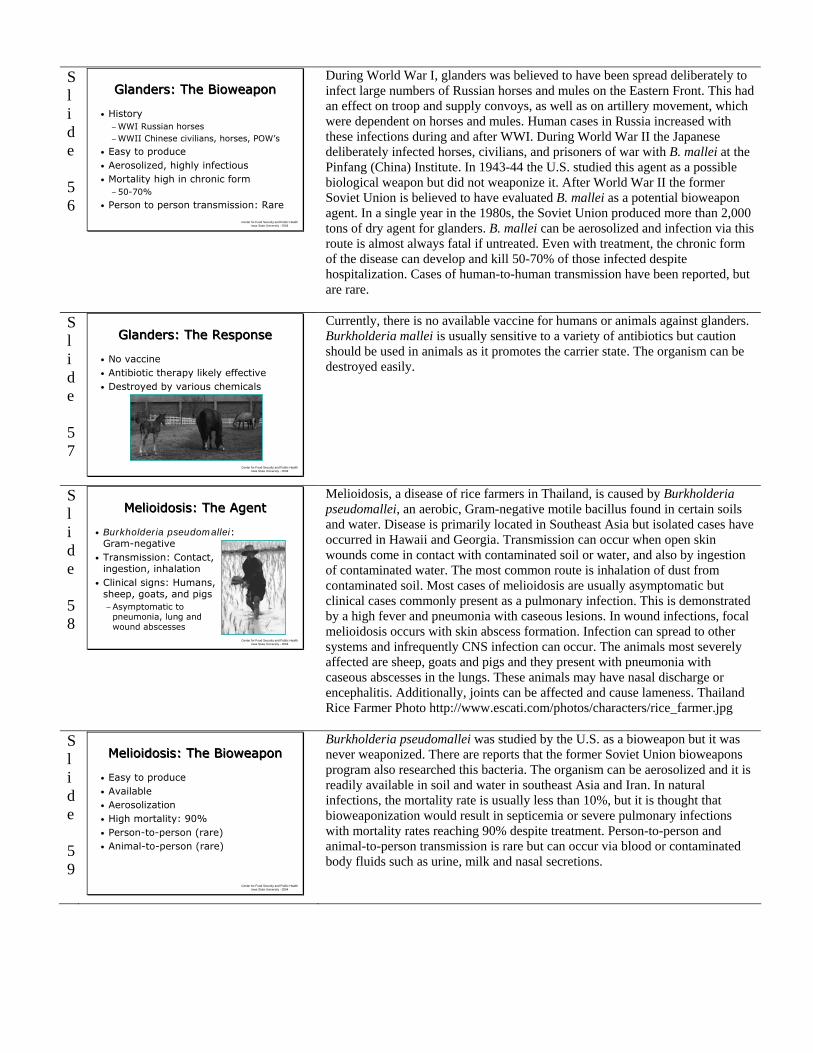
Currently, there is no available vaccine for humans or animals against glanders. Burkholderia mallei is usually sensitive to a variety of antibiotics but caution should be used in animals as it promotes the carrier state. The organism can be destroyed easily.
Slide 58
Center for Food Security and Public Health Iowa State University - 2004
Melioidosis: The AgentMelioidosis: The Agent
• Burkholderia pseudomallei: Gram-negative
• Transmission: Contact, ingestion, inhalation
• Clinical signs: Humans, sheep, goats, and pigs−Asymptomatic to
pneumonia, lung and wound abscesses
Melioidosis, a disease of rice farmers in Thailand, is caused by Burkholderia pseudomallei, an aerobic, Gram-negative motile bacillus found in certain soils and water. Disease is primarily located in Southeast Asia but isolated cases have occurred in Hawaii and Georgia. Transmission can occur when open skin wounds come in contact with contaminated soil or water, and also by ingestion of contaminated water. The most common route is inhalation of dust from contaminated soil. Most cases of melioidosis are usually asymptomatic but clinical cases commonly present as a pulmonary infection. This is demonstrated by a high fever and pneumonia with caseous lesions. In wound infections, focal melioidosis occurs with skin abscess formation. Infection can spread to other systems and infrequently CNS infection can occur. The animals most severely affected are sheep, goats and pigs and they present with pneumonia with caseous abscesses in the lungs. These animals may have nasal discharge or encephalitis. Additionally, joints can be affected and cause lameness. Thailand Rice Farmer Photo http://www.escati.com/photos/characters/rice_farmer.jpg
Slide 59
Center for Food Security and Public Health Iowa State University - 2004
Melioidosis: The Bioweapon Melioidosis: The Bioweapon
• Easy to produce • Available• Aerosolization• High mortality: 90%• Person-to-person (rare)• Animal-to-person (rare)
Burkholderia pseudomallei was studied by the U.S. as a bioweapon but it was never weaponized. There are reports that the former Soviet Union bioweapons program also researched this bacteria. The organism can be aerosolized and it is readily available in soil and water in southeast Asia and Iran. In natural infections, the mortality rate is usually less than 10%, but it is thought that bioweaponization would result in septicemia or severe pulmonary infections with mortality rates reaching 90% despite treatment. Person-to-person and animal-to-person transmission is rare but can occur via blood or contaminated body fluids such as urine, milk and nasal secretions.

Slide 60
Center for Food Security and Public Health Iowa State University - 2004
Melioidosis: The ResponseMelioidosis: The Response
• Long-term, multiple antibiotics effective
• Vaccines available: Not in U.S.
• Easily destroyed by disinfectants
B. pseudomallei is susceptible to various antibiotics, but relapses can occur once treatment is stopped. Long-term treatment may be necessary and multiple drugs may be needed. Vaccines are available in some countries, but not the U.S., and they are not effective against large challenge doses. In endemic areas, avoid contact with soil and water during the wet season. The organism can destroyed by numerous disinfectants.
Slide 61
Center for Food Security and Public Health Iowa State University - 2004
Psittacosis: The AgentPsittacosis: The Agent
• Chlamydophila psittaci−Gram-negative
• Occurs worldwide• Reportable in U.S.• Clinical disease in humans and birds−Asymptomatic −Systemic illness with severe pneumonia
The fourth bacterium in the Category B list is Chlamydophila psittaci. It is a Gram- negative, obligate intracellular bacteria. Psittacosis or avian chlamydiosis naturally occurs worldwide and is usually a sporadic disease. In the U.S., psittacosis is reportable in humans, but true incidence is unknown due to poor reporting compliance and frequent misdiagnosis. There are 50-100 confirmed cases per year in the U.S. (1-2 deaths per year), but some estimate as many as 100-200 cases actually occur annually. Pet store employees, owners of pet birds, and poultry processing plant workers account for the majority of the reported cases. The organism is transmitted by inhaling contaminated dust from feathers or bird droppings. The elementary body form of C. psittaci is the infectious form and is very resistant to drying. Infectivity of C. psittaci has been documented in straw or on hard surfaces for 2-3 weeks, canary feed for 2 months, in poultry litter for up to 8 months, and in diseased turkey carcasses for less than 1 year. In humans, clinical signs range from asymptomatic to systemic illness with severe pneumonia. Pneumonia occurs most commonly in adults 30-60 years old. Other signs include abrupt onset of fever, chills, headache, nonproductive cough, and breathing difficulty. In birds, clinical symptoms of avian chlamydiosis includes depression, ruffled feathers, inappetence, nasal discharge, respiratory distress, yellowish-green or green diarrhea, and conjunctivitis. Egg production may decrease. Pigeons and ducks may have neurologic signs.
Slide 62
Center for Food Security and Public Health Iowa State University - 2004
Psittacosis: The BioweaponPsittacosis: The Bioweapon
• Easily obtained• Aerosolized• Stable in the
environment• Person-to-person
transmission rare• Low mortality
This organism has previously been part of bioweapons research programs. Some characteristics that may make psittacosis a potential bioweapon include its worldwide occurrence, making it easy to obtain. The agent is easily aerosolized, and very stable in the environment. Person-to-person transmission is rare, although it occasionally spreads during paroxysmal coughing. Treated cases are rarely fatal but in severe untreated infections, mortality rates range from 10-40%. These chicks are eating at a centralized automatic feeder.
Slide 63
Center for Food Security and Public Health Iowa State University - 2004
Psittacosis: The ResponsePsittacosis: The Response
• Antibiotics generally effective
• Decontamination possible with most disinfectants
While antibiotics are generally effective, relapses can occur. Most disinfectants are effective against C. psittaci. Avoid exposure to infective organism when handling birds and their excreta.

Slide 64
Center for Food Security and Public Health Iowa State University - 2004
Q Fever: The AgentQ Fever: The Agent
• Coxiella burnetii • Transmission: Inhalation,
direct contact, ingestion, ticks• Disease symptoms−Humans:
Acute: Flu-like + pneumonia & hepatitisChronic: Endocarditis, osteomyelitis
−Animals: Most asymptomaticSheep, cattle and goats: Abortions
Q fever (“query” or “puzzling” fever) is caused by Coxiella burnetii, an obligate intracellular parasite, which is currently considered a rickettsial agent (new studies may change its family). The disease has been found worldwide, except in New Zealand. Transmission occurs by inhalation or direct contact of infectious organism; it also occurs following ingestion of the of the organism, and ticks spread the infection among ruminants and sometimes people. The organism is shed in high numbers in placental tissue and body fluids, and is highly infectious (one organism can cause disease). There was a report of a case where a cat infected with Q fever had kittens in the same room where a child’s birthday party was being held. Several of the children developed Q fever. Coxiella burnetii forms an unusual spore-like structure and can survive 7-10 days on wool at room temperature, 1 month on fresh meat in cold storage, and more than 40 months in skim milk. However, it is killed by pasteurization. Two clinical forms of disease occur in humans, acute (less than 6 months duration) and chronic (greater than 6 months). Symptoms of acute disease vary in severity and duration and usually manifest as self-limited febrile or flu-like illness, but pneumonia or hepatitis may also occur. Chronic disease occurs in 1-5% of those infected and the most common complication is heart related (endocarditis). Farm animals, including sheep, cattle, and goats, are the most important reservoirs of disease and are usually asymptomatic. Abortions, stillbirths, mastitis in dairy cattle, and complicated deliveries have been reported in these animals. Dogs, cats, rabbits, horses and many other animals can harbor the organism, but is usually asymptomatic.
Slide 65
Center for Food Security and Public Health Iowa State University - 2004
Q Fever: The BioweaponQ Fever: The Bioweapon
• History• Easily accessible• Environmentally
resistant• Highly infectious• Aerosolization−Travel ½ mile by wind
• Low mortality- chronic morbidity
This agent was part of the U.S. bioweapons research in the 1950’s and 1960’s. Some reports suggest that a portion of the information about the human infectivity of this organism (i.e. one organism can cause disease) was gained during experiments at the bioweapon research facility. This agent could be used as a bioweapon because it is easily accessible, very resistant, highly infectious, and is stable when aerosolized. Coxiella burnetii organisms can be carried up to ½ mile or more by the wind. Mortality is low with this disease. The picture is of a crop duster, and contrary to popular belief, experts believe that wide dissemination could be done with any type of plane, not something that requires intensive training to operate. Image: USDA website.
Slide 66
Center for Food Security and Public Health Iowa State University - 2004
Q Fever: The ResponseQ Fever: The Response
• Often self-limiting disease• Antibiotic therapy may limit
complications• Vaccine developed, not available in
U.S.• Variable susceptibility to
disinfectants
Although the disease is often self-limiting, antibiotics are generally effective at shortening the course of acute illness and reducing the risk of complications. Treatment of chronic cases is more difficult and may require long-term antibiotic therapy. A vaccine for Q fever has been developed and has successfully protected humans in occupational settings in Australia, but is not commercially available in the United States. A vaccine for use in animals has also been developed, but is not available in the United States. C. burnetii is highly resistant to physical and chemical agents. Variable susceptibility has been reported for disinfectants.
Slide 67
Center for Food Security and Public Health Iowa State University - 2004
Typhus Fever: The AgentTyphus Fever: The Agent
• Rickettsia prowazekii: Rickettsial organism• Endemic in Eastern Europe, Middle East,
and parts of Africa• Transmitted in feces of
human body louse • Clinical signs: Humans
− Fever, headache, maculareruptions, and petechial rash
• Not seen in domestic animals
J. Kalisch
The second rickettsial organism of the Category B agents is Rickettsia prowazekii. This disease, also called epidemic typhus, killed about 10% of the English population in 1557. It is transmitted by arthropod vectors, primarily the human body louse (not the human head louse). It is an obligate intracellular parasite in both humans and lice. Lice infected with R. prowazekii die about two weeks after ingesting the infected bloodmeal. The organism is not transmitted to the louse eggs (no transovarial transmission). As a result, the mammalian host is essential in long term propagation of R. prowazekii. Patients are infective for lice during the febrile illness and possibly for 2-3 days after their temperature returns to normal. Infected body louse pass rickettsia in their feces within 2-6 days after the blood meal. The rickettsia may remain viable in dead lice for weeks and in the feces for 2-3 days. The disease is characterized by the sudden appearance of headaches and high fever for two weeks. Once infected,

simultaneous symptoms may include chills, prostration, coughing, severe muscular pain, bronchial disturbances, and mental confusion. A macular eruption (dark spot on the skin) appears on the fifth to sixth day, initially on the upper trunk, which then spreads to the entire body except the face, palms, and soles of the feet. Mortality rate increases with age and may reach 60% in untreated persons older than 50 years. In epidemic conditions mortality can approach 100%. Domestic animals are not susceptible to R. prowazekii. Human body louse J. Kalisch, University of Nebraska.
Slide 68
Center for Food Security and Public Health Iowa State University - 2004
Typhus Fever: The BioweaponTyphus Fever: The Bioweapon
• WHO estimation: 1970−50 kg agent; 5 million people in city−125,000 ill−8,000 deaths
• Available• Can be aerosolized
in lice fecesU.S. Typhus Commission
In 1970 R. prowazekii was one of the agents selected by the WHO to estimate casualties if the agent was aerosolized as a bioweapon. The estimate for a city of 5 million was that 300,000 would be exposed, 125,000 would become ill, and 8,000 would die. The reason this agent might be considered in biowarfare is that it is readily available, remains infective in lice feces for several weeks, and can be aerosolized in the feces. Note: this photo shows the macular eruptions on the skin of a human with Typhus.
Slide 69
Center for Food Security and Public Health Iowa State University - 2004
Typhus Fever: The ResponseTyphus Fever: The Response
• Antibiotics are generally effective• Vaccine, not commercially available
Antibiotics early are generally effective at curing this disease and will decrease the number of relapses. No vaccine is commercially available, but experimental vaccines are produced by military sources in the U.S. and may be available for high-risk situations. The organism is susceptible to common disinfectants. Photo depicts macular eruptions on the skin of a typhus patient.
Slide 70
Center for Food Security and Public Health Iowa State University - 2004
Viral Encephalitis: The AgentViral Encephalitis: The Agent
• The Alphaviruses: EEE, WEE, and VEE• Transmitted via mosquito• Clinical signs−Humans, horses, donkeys,
mules: Often asymptomatic to flu-like
−Encephalitis in small proportions
• Birds are asymptomatic carriers, act as sentinels
This is the only viral group in the list of Category B agents. This group of equine encephalitis viruses are RNA viruses in the Alphavirus genus. Eastern, Western, and Venezuelan Equine Encephalitis viruses are transmitted by mosquitoes. The female mosquito takes a bloodmeal from a viremic host, generally birds for EEE and WEE, and birds and horses for VEE. The virus replicates in the salivary glands of the mosquito and is transmitted back to birds or to dead end hosts, such as humans and horses, where overt disease occurs. In humans, infections can be asymptomatic or cause flu-like illness. In a small proportion of cases viral encephalitis can occur and lead to permanent neurological damage or death. Horses, donkeys and mules have similar clinical signs as humans. The disease in these animals often precede human cases by several weeks. EEE and VEE have mortality rates of 40-90%; WEE has a lower mortality rate ranging from 20-30%. Birds are asymptomatic carriers. The detection of viremia in sentinel birds is detected via ELISA.

Slide 71
Center for Food Security and Public Health Iowa State University - 2004
Viral Encephalitis:The Bioweapon
Viral Encephalitis:The Bioweapon
• Easy to produce• Aerosolization• High rate of infection• Person-to-person transmission
possible
VEE was tested in the U.S. bioweapons program in the 1950s and 1960s. It is thought that other countries have also weaponized VEE. All U.S. stocks of VEE were destroyed, along with the other agents that were part of the program. VEE can be produced in large amounts by unsophisticated and inexpensive systems. The virus can be aerosolized or spread by releasing infected mosquitoes. Humans are highly susceptible and approximately 90-100% of exposed individuals could become infected and have clinical signs, although most are mild. Equids would also be susceptible and disease would occur simultaneously with human disease. There is a low overall human case-fatality rate.
Slide 72
Center for Food Security and Public Health Iowa State University - 2004
Viral Encephalitis:The Response
Viral Encephalitis:The Response
• Supportive care• Vaccine −Equine−Human: High risk
• Virus unstable in environment
Antibiotics are not effective for treatment and there are no effective antiviral drugs available. Treatment involves supportive care. There is a trivalent formalin inactivated vaccine available for horses for WEE, EEE, VEE in the United States, but the human vaccines are limited to those who are researchers and at a high risk of exposure. All of the virus types are unstable in the environment. Photo courtesy of D. Bickett-Weddle, DVM, ISU.
Slide 73
Center for Food Security and Public Health Iowa State University - 2004
Toxins: The AgentsToxins: The Agents
• Staphylococcal enterotoxin B (SEB)
• Ricin toxin from castor plant
• Clostridium perfringens epsilon toxin
Next we will discuss three toxins that satisfy the criteria for Category B agents. They are Staphylococcal enterotoxin B, ricin toxin from the castor plant, and Clostridium perfringens epsilon toxin. Toxins are biological agents produced by living organism such as bacteria, plants, or animals. Photo of soldiers eating- USARIEM http://www.usariem.army.mil/nbd/nutri.htm
Slide 74
Center for Food Security and Public Health Iowa State University - 2004
SEB: The AgentSEB: The Agent
• Staphylococcal enterotoxin B (SEB)• A common cause of food poisoning • Clinical signs: Humans−Fever, chills, headache, myalgia−Non-productive cough if inhaled−GI signs if swallowed
• Animals: Likely similar to human
Bacterial toxins are more difficult to produce than plant toxins but are much more toxic. SEB produced by Staphylococcus aureus is an example of a bacterial toxin. SEB is one of the toxins responsible for staphylococcal food poisoning in humans. It is a superantigen and acts by stimulating cytokine release and an overwhelming immune response. Routes of exposure could include inhalation or ingestion. In humans, the clinical signs resulting from SEB exposure via ingestion would occur in approximately 4-10 hours. Following inhalation exposure, clinical signs would occur in approximately 3-12 hours. The general symptoms include sudden onset of high fever persisting for several days, chills, and myalgia. Following inhalation, a non-productive cough, chest pain, and dyspnea can occur. Following ingestion, nausea, vomiting, and diarrhea predominate. In animals, although S. aureus is associated with many diseases, there is little information about clinical signs resulting from SEB. Experimental exposure of mice and monkeys to aerosolized SEB resulted in diarrhea and vomiting, followed by depression, dyspnea, and shock; some animals died.

Slide 75
Center for Food Security and Public Health Iowa State University - 2004
Ricin: The AgentRicin: The Agent
• Ricin toxin from bean of castor plant• Available worldwide• Clinical signs−Acute onset of fever,
chest tightness, cough,dyspnea, nausea
Plant toxins are easy to produce at a low cost because raw materials are available worldwide such as the ricin toxin from the bean of the castor plant. Annually, of one million tons of castor beans are processed worldwide in the production of castor oil. The waste product is five percent ricin by weight. The toxin is stable and extremely toxic by several routes of exposure, including respiratory. Many species are affected and have an acute onset of fever, chest tightness, cough, dyspnea, and nausea. Castor plant- Cooperative extension, Purdue University http://www.vet.purdue.edu/depts/addl/toxic/plant11.htm
Slide 76
Center for Food Security and Public Health Iowa State University - 2004
Epsilon Toxin: The AgentEpsilon Toxin: The Agent
• Clostridium perfringens type B and D• Increases intestinal and vascular
permeability, liver and neurological damage
• Clinical signs−Calves: Diarrhea, abdominal pain,
listlessness, neurologic−Sheep, goats: Watery to bloody
diarrhea, neurologic−Humans: Little information
C. perfringens types B and D produce epsilon toxin. C. perfringens type B infection causes severe enteritis in young calves, foals, piglets, and lambs (lamb dysentery) and C. perfringens type D causes enterotoxemia (overeating disease, pulpy kidney disease) in sheep and goats, and rarely cattle. The clinical signs of enterotoxemia are primarily a result of epsilon toxin, which causes increased intestinal and vascular permeability, and liver damage. In calves, clinical signs include diarrhea, abdominal pain and listlessness. Sheep and goats have watery to bloody diarrhea. Neurologic signs can be common in all. There is little or no information about the effects of epsilon toxin on humans. Human disease is usually caused by C. perfringens type A and C.
Slide 77
Center for Food Security and Public Health Iowa State University - 2004
Toxins: The BioweaponToxins: The Bioweapon
• History• Aerosolized: SEB, ricin• Available worldwide• Easy to produce, stable• Many species affected• No person-to-person transmission
Toxins have been researched and used as bioweapons. For example, the U.S. stockpiled and researched SEB during its bioweapons program before it was terminated in 1969. Ricin is thought to have been used in the assassination of Bulgarian defector Georgi Markov in London in 1978. It is said that a ricin pellet was injected into Markov by a specially engineered weapon disguised as an umbrella. Markov died three days after the incident. In Minnesota during 1991, four members of the Patriots Council, an antigovernment extremist group, were arrested for plotting to kill a US Marshall with ricin. The group planned to mix the homemade ricin with DMSO and then smear it on the door handles of the Marshall's car. The plan was discovered and all four men were arrested. There are reports of ricin use in the Iran-Iraq war in the 1980’s and also reports that it was found in Al Qaeda caves in Afghanistan in 2002. In 2003 equipment for producing ricin along with traces of the toxin were found in a London apartment. Although aerosolized SEB would not likely produce significant mortality, it could render 80% or more of exposed people clinically ill and unable to perform their duties for 1-2 weeks. The demand on the medical and logistical systems could be overwhelming. If inhaled, ricin toxin may cause pathologic changes within 8 hours and severe respiratory symptoms and failure in 36-72 hours. All of these toxins are available worldwide, relatively easy to produce, stable, and affect many species. There is no concern about transmission of the toxin from an affected individual.
Slide 78
Center for Food Security and Public Health Iowa State University - 2004
Toxins: The ResponseToxins: The Response
• Supportive care• No vaccines currently available for
SEB or ricin• Vaccines for animals for clostridial
disease• Toxins are inactivated with common
disinfectants
Management includes supportive care. Currently there are no effective antitoxins or human vaccines available. Development, safety, and immunogenicity testing for a human vaccine is anticipated for the near future. Preliminary animal studies for a SEB vaccine have been encouraging. There is promising research in animals with vaccines for ricin and antiserum production. Currently there are animal vaccines available for clostridial disease, but none have been developed for humans. These toxins can be inactivated with common disinfectants and if exposure is suspected, decontamination of the area or exposed skin should be done with soap and water or with bleach.

Slide 79
Center for Food Security and Public Health Iowa State University - 2004
Food Safety ThreatsFood Safety Threats• Campylobacter species• Salmonella species• E. coli 0157:H7 • Viruses, parasites,
chemicals, toxins• Ingestion of
contaminated food• Gastrointestinal upset
There are an estimated 250 pathogens that can cause foodborne related illnesses. The most commonly recognized foodborne infections are caused by Campylobacter, Salmonella and E. coli O157:H7. Viruses, parasites, natural or manufactured chemicals and organism toxins can also cause foodborne disease. Most agents cause gastrointestinal upset (nausea, diarrhea, abdominal cramps) which are self-limiting in 4-5 days. However, some agents have severe complications if left untreated. Campylobacter is considered the leading bacterial cause of foodborne related diarrhea, with an estimated 2.4 million people affected each year. Common sources include raw or undercooked poultry, raw milk or items contaminated with infected animal or human feces. Animal sources include poultry, cattle, puppies, kittens and pet birds. Many serotypes of Salmonella can also cause foodborne related illness. S. enteritidis and S. typhimurium are the most common isolates, accounting for about 41% of U.S. human cases. Common sources for Salmonella food-borne illness include raw poultry or eggs, beef, unwashed fruit and alfalfa sprouts. Pet reptiles can also be a common source. E. coli O157:H7 is another major pathogen of foodborne related illnesses. Common sources include undercooked or raw hamburger, salami, lettuce and alfalfa sprouts. It has also been associated with unpasteurized milk, apple juice or cider as well as contaminated well water. Cattle are the most common animal source although other mammals can serve as a source. Severe sequela can occur with E.coli infection.
Slide 80
Center for Food Security and Public Health Iowa State University - 2004
Food Safety Threats: The Bioweapon
Food Safety Threats: The Bioweapon
• 1984, The Dalles, Oregon−Bagwan Shree Rajneesh cult−Contaminated salad bars
Salmonella typhimurium
−Goal: Incapacitate voters−751 people ill
Restaurants have served as a conduit for terrorism before. In 1984, the Bagwan Shree Rajneesh cult in The Dalles, Oregon deliberately contaminated salad bars with Salmonella typhimurium in an attempt to incapacitate voters for an upcoming local election. At least 10 restaurants were affected and possibly one supermarket. Additionally, coffee creamers in some restaurants were also contaminated. The incident resulted in 751 people becoming ill. It wasn’t until one of the cultists broke a vow of silence and came forth one year later that the details of the “attack” became known. Plans were being made to contaminate the water also. This incident was meant to be a trial run before a full scale biological crime.
Slide 81
Center for Food Security and Public Health Iowa State University - 2004
Food Safety Threats: The Response
Food Safety Threats: The Response
• Constant vigilance to improve food safety
• Food irradiation at processing plants• Wash hands and utensils frequently• Proper cooking temperature and
storage
There must be constant vigilance for improving food safety and processing, both in the home and commercial settings. Food irradiation is the latest technology used at processing plants to help eliminate the risk of bacterial contamination. However, people should always follow good hygiene when working with food. Always wash your hands with soap and warm water before and after handling food. Wash raw fruits and vegetables before eating. After contact with raw meat or poultry, wash hands, utensils and kitchen surfaces with hot soapy water. Cook beef and beef products to an internal temperature of 160 oF. Cook poultry and eggs to an internal temperature of 170-180 oF. Eat cooked foods promptly and refrigerate any leftovers within 2 hours after cooking.
Slide 82
Center for Food Security and Public Health Iowa State University - 2004
Water Safety ThreatsWater Safety Threats
• 53% of US drinking water is from ground water
• Cryptosporidium parvum- protozoa
• Vibrio cholerae-bacteria
Fifty-three percent of the U.S. drinking water comes from ground (well, aquifer) water. Surface waters such as lakes, reservoirs, and streams, are used by cities but require filtration and/or chemical treatment. Two organisms are listed in the Category B list as examples of water safety threats. They are Cryptosporidium parvum and Vibrio cholerae.
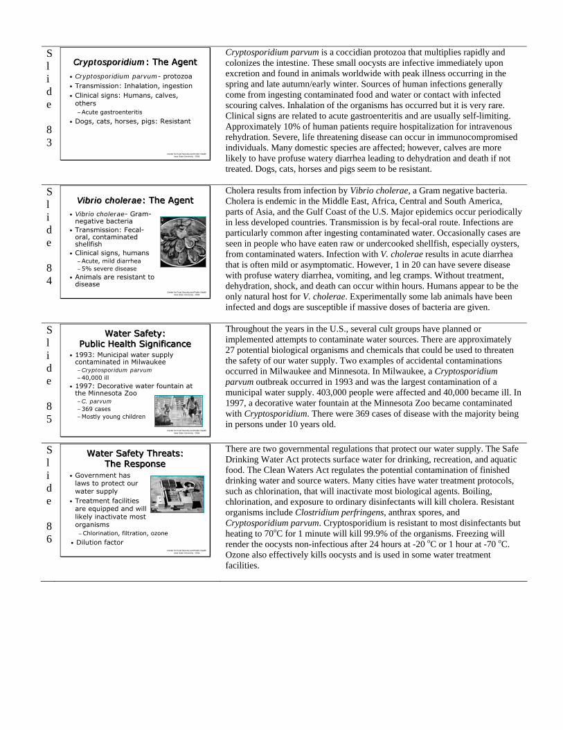
Slide 83
Center for Food Security and Public Health Iowa State University - 2004
Cryptosporidium: The AgentCryptosporidium: The Agent
• Cryptosporidium parvum- protozoa• Transmission: Inhalation, ingestion• Clinical signs: Humans, calves,
others−Acute gastroenteritis
• Dogs, cats, horses, pigs: Resistant
Cryptosporidium parvum is a coccidian protozoa that multiplies rapidly and colonizes the intestine. These small oocysts are infective immediately upon excretion and found in animals worldwide with peak illness occurring in the spring and late autumn/early winter. Sources of human infections generally come from ingesting contaminated food and water or contact with infected scouring calves. Inhalation of the organisms has occurred but it is very rare. Clinical signs are related to acute gastroenteritis and are usually self-limiting. Approximately 10% of human patients require hospitalization for intravenous rehydration. Severe, life threatening disease can occur in immunocompromised individuals. Many domestic species are affected; however, calves are more likely to have profuse watery diarrhea leading to dehydration and death if not treated. Dogs, cats, horses and pigs seem to be resistant.
Slide 84
Center for Food Security and Public Health Iowa State University - 2004
Vibrio cholerae: The AgentVibrio cholerae: The Agent
• Vibrio cholerae- Gram-negative bacteria
• Transmission: Fecal-oral, contaminated shellfish
• Clinical signs, humans−Acute, mild diarrhea−5% severe disease
• Animals are resistant to disease
Cholera results from infection by Vibrio cholerae, a Gram negative bacteria. Cholera is endemic in the Middle East, Africa, Central and South America, parts of Asia, and the Gulf Coast of the U.S. Major epidemics occur periodically in less developed countries. Transmission is by fecal-oral route. Infections are particularly common after ingesting contaminated water. Occasionally cases are seen in people who have eaten raw or undercooked shellfish, especially oysters, from contaminated waters. Infection with V. cholerae results in acute diarrhea that is often mild or asymptomatic. However, 1 in 20 can have severe disease with profuse watery diarrhea, vomiting, and leg cramps. Without treatment, dehydration, shock, and death can occur within hours. Humans appear to be the only natural host for V. cholerae. Experimentally some lab animals have been infected and dogs are susceptible if massive doses of bacteria are given.
Slide 85
Center for Food Security and Public Health Iowa State University - 2004
Water Safety: Public Health Significance
Water Safety: Public Health Significance
• 1993: Municipal water supply contaminated in Milwaukee−Cryptosporidum parvum−40,000 ill
• 1997: Decorative water fountain at the Minnesota Zoo−C. parvum−369 cases −Mostly young children
Throughout the years in the U.S., several cult groups have planned or implemented attempts to contaminate water sources. There are approximately 27 potential biological organisms and chemicals that could be used to threaten the safety of our water supply. Two examples of accidental contaminations occurred in Milwaukee and Minnesota. In Milwaukee, a Cryptosporidium parvum outbreak occurred in 1993 and was the largest contamination of a municipal water supply. 403,000 people were affected and 40,000 became ill. In 1997, a decorative water fountain at the Minnesota Zoo became contaminated with Cryptosporidium. There were 369 cases of disease with the majority being in persons under 10 years old.
Slide 86
Center for Food Security and Public Health Iowa State University - 2004
Water Safety Threats: The Response
Water Safety Threats: The Response
• Government has laws to protect our water supply
• Treatment facilities are equipped and will likely inactivate most organisms−Chlorination, filtration, ozone
• Dilution factor
There are two governmental regulations that protect our water supply. The Safe Drinking Water Act protects surface water for drinking, recreation, and aquatic food. The Clean Waters Act regulates the potential contamination of finished drinking water and source waters. Many cities have water treatment protocols, such as chlorination, that will inactivate most biological agents. Boiling, chlorination, and exposure to ordinary disinfectants will kill cholera. Resistant organisms include Clostridium perfringens, anthrax spores, and Cryptosporidium parvum. Cryptosporidium is resistant to most disinfectants but heating to 70oC for 1 minute will kill 99.9% of the organisms. Freezing will render the oocysts non-infectious after 24 hours at -20 oC or 1 hour at -70 oC. Ozone also effectively kills oocysts and is used in some water treatment facilities.

Slide 87
Center for Food Security and Public Health Iowa State University - 2004
Category CCategory C
• Nipah virus• Hantavirus
We will now discuss the diseases of third highest priority on the CDC Category Agent List, both of which are viruses.
Slide 88
Center for Food Security and Public Health Iowa State University - 2004
Nipah Virus: The AgentNipah Virus: The Agent
• Paramyxovirus• Fruit bat reservoir• Clinical signs−Humans: Encephalitis−Pigs: Respiratory, neurological−Dogs and cats: “Distemper”
Nipah virus was discovered as a Paramyxovirus in Malaysia in 1999, and causes a severe respiratory disease in pigs and severe encephalitis in humans. The reservoir for the virus is thought to be fruit bats, which are called flying foxes. Suspected transmission of the virus occurs from bats roosting in fruit trees close to pig confinements. The virus then spreads rapidly through the swine herd by direct contact or aerosolization (usually coughing). It can then be passed to humans, dogs, cats and other species. Transmission can also occur from direct contact with infected body fluids. To date, no person-to-person or bat-to-person transmission has been reported. In humans, the incubation period is 3-14 days. Initial symptoms include fever, headache, dizziness, drowsiness, disorientation and vomiting. Some cases show signs of respiratory illness. In severe cases, a rapidly progressive encephalitis can occur with a mortality rate of 40%. In swine, Nipah virus is highly contagious and easily spread. Many pigs are asymptomatic. Clinical signs include acute fever (>104 oF), tachypnea and dyspnea with open mouth breathing, and a loud, explosive barking cough may also be noted. Occasionally, neurological signs can occur. Clinical signs in pigs were noted 1-2 weeks before illness in humans making swine a sentinel for human disease. Disease in other animal species is poorly documented. Other species demonstrate respiratory and neurological signs. Photo of a Malayan flying fox.
Slide 89
Center for Food Security and Public Health Iowa State University - 2004
Nipah Virus: The BioweaponNipah Virus: The Bioweapon
• Aerosolization potential
• Wide host range• No person-to-person
transmission expected
• High morbidity and mortality
Nipah virus is described as an emerging pathogen with potentially high morbidity and mortality as well as a major health impact. Currently transmission of the disease involves close contact with pigs but aerosolization may be a possible bioterrorist method of dispersal. The potential for this virus to infect a wide range of hosts and produce significant mortality in humans makes this virus a public health concern. Photo from Dr. James Roth-ISU of hog confinement barns that were affected during the Nipah virus outbreak in Malaysia, 1999.
Slide 90
Center for Food Security and Public Health Iowa State University - 2004
Nipah Virus: The ResponseNipah Virus: The Response
• Avoid contact with all infected animals and fluids
• Vaccine being researched• Call authorities
immediately
Nipah virus is a very dangerous pathogen and is classified as a Biolevel 4 agent. If you suspect an outbreak, contact your state veterinarian and state public health veterinarian IMMEDIATELY! Avoid all contact with potentially infected species (pigs, dogs, cats) until the proper authorities are consulted. Nipah virus can be readily inactivated by detergents. Routine cleaning and disinfection with sodium hypochlorite or several commercially available detergents is expected to be effective.

Slide 91
Center for Food Security and Public Health Iowa State University - 2004
Hantavirus: The AgentHantavirus: The Agent• Bunyaviridae family• Asymptomatic reservoir: Rodents• Transmission: Inhalation,
ingestion, direct contact• Human clinical signs−Fever, myalgia, headache−Hantavirus Pulmonary Syndrome −Hemorrhagic Fever with Renal Syndrome
• Not seen in domestic animals
Hantavirus is an RNA virus in the Bunyaviridae family. It is recognized as causing hantavirus pulmonary syndrome (HPS) and hemorrhagic fever with renal syndrome (HFRS) in humans. Rodents are the asymptomatic reservoir host and the deer mouse (Peromyscus maniculatus) is the primary carrier in all areas of the United States, except the southeast, where the cotton rat (Sigmodon hispidus) and the rice rat (Oryzomys palustris) are involved. It is important to remember that the house mouse is not a carrier. Transmission to humans most commonly occurs when they disturb the microenvironment of rodents and breathe aerosolized infectious particles from rodent excrement. Direct contact with rodent excreta on human mucous membranes or through skin abrasions may also result in transmission. The virus particles can contaminate food consumed by humans and cause infection, and in very rare cases a bite from an infected rodent can precipitate the disease. Clinical signs in humans initially include fatigue, fever, myalgia, and headache. The disease can progress a severe respiratory syndrome, HPS in the U.S., or hemorrhagic fever with renal syndrome (HFRS) in Asia and Europe. Approximately 40% of patients die within the first 48 hours due to uncorrected hypoxia and shock. The disease is not seen in domestic animals.
Slide 92
Center for Food Security and Public Health Iowa State University - 2004
Hantavirus: The BioweaponHantavirus: The Bioweapon
• Aerosolized• Hospitalization• Unexpected disease
in the U.S.−HFRS limited to
Asia/Europe to date
Hantavirus is a newly emerging disease and information regarding its use as a bioweapon is limited. It is listed here because it satisfies the criteria for Category C agents, as it is emerging and could be mass disseminated. Hantavirus disease progresses rapidly, requires hospitalization, and mortality rate can reach 40%. If hemorrhagic fever with renal syndrome (HFRS) was introduced to the U.S., diagnosis may be delayed because that is not the form of the disease that is usually observed.
Slide 93
Center for Food Security and Public Health Iowa State University - 2004
Hantavirus: The ResponseHantavirus: The Response
• Supportive care• Limit exposure to
rodent excrement−Wear gloves, face
mask
• Virus is deactivated with bleach
Treatment of patients with HPS requires early and aggressive intensive care. Antiviral drugs, such as ribavirin, have questionable efficacy, possibly due to the late introduction after disease onset. Avoid exposure to rodents and their excrement. Wear latex rubber gloves, and possibly a face mask, when cleaning up areas when cleaning potentially contaminated areas. Hantavirus is susceptible to common disinfectants. Disinfect gloves before removal and wash hands thoroughly.
Slide 94
Center for Food Security and Public Health Iowa State University - 2004
Other Important Diseases
Other Important Diseases
• Transmissible Spongiform Encephalopathy (TSE)
• Rift Valley Fever• Hendra Virus• West Nile Virus• Foot and Mouth Disease• Monkeypox
The next six diseases that will be covered include Transmissible Spongiform Encephalopathy (TSE), Rift Valley Fever, Hendra, West Nile Virus, Foot and Mouth Disease, and Monkeypox. These agents/diseases are not part of the CDC Category ABC list, but we at the Center for Food Security and Public Health decided to include them because they are important zoonotic diseases.

Slide 95
Center for Food Security and Public Health Iowa State University - 2004
Transmissible Spongiform Encephalopathy: The AgentTransmissible Spongiform
Encephalopathy: The Agent• Prions−Proteinaceous infectious particles−Mutated proteins
• Very long incubation period• Neurological signs in all species• No treatment available
Transmissible spongiform encephalopathy (TSE) describes a group of diseases that are thought to be caused by prions (short for proteinaceous infectious particles). These abnormal proteins cause a variety of diseases in various species. In humans, variant Creutzfeldt-Jakob disease (vCJD), Kuru, Gerstmann-Straussler-Scheinker syndrome (GSS), and fatal familial insomnia (FFI) can occur. In animals, bovine spongiform encephalopathy (BSE), or “mad cow disease”, scrapie in sheep, chronic wasting disease (CWD) in deer and elk, mink spongiform encephalopathy (TME), and feline spongiform encephalopathy (FSE) can occur. The incubation period for most of these diseases is many years which complicates diagnosis. In humans with vCJD, clinical signs include depression and schizophrenia leading to ataxia and involuntary muscle movement. This eventually progresses to complete immobility and muteness. In animals, initial clinical signs can be subtle, but usually involve behavioral changes, such as excitability, nervousness, aggressiveness, and increased sensitivity to noise. Sheep with scrapie typically exhibit intense pruritus. The terminal state of TSEs in cattle, deer and elk, can result in extreme wasting despite a good appetite. Additionally, tremors and muscle fasciculations, especially in the neck and face, can occur. There is no known treatment at this time.
Slide 96
Center for Food Security and Public Health Iowa State University - 2004
Bovine Spongiform Encephalopathy
Bovine Spongiform Encephalopathy
• Mad cow disease• Incubation: 2 to 8 years• 1995, United Kingdom −vCJD−People exposed to BSE
Before bovine offal ban in 1989
• Active U.S. surveillance since 1990
Bovine spongiform encephalopathy (BSE) in cattle is thought to have occurred from feeding meat or bone meal from scrapie-infected sheep to cattle, or from spontaneous genetic mutation in a cow then fed to other cows. The first cases of BSE appeared in the U.K. in 1986. The incubation period ranges from 2 to 8 years and is always fatal. BSE is the only disease that has been shown to be transmissible to humans. BSE presents itself as variant Creutzfeldt Jakob disease in humans. In 1995, ten human cases similar to Creutzfeldt-Jakob disease (CJD) were reported in the U.K. However, the disease was affecting people at a younger age, eliciting behavioral changes not seen with classic CJD and demonstrated different brain lesions. Currently, it is thought that consumption of BSE contaminated beef products (prior to the U.K.’s specified bovine offal ban in 1989) may be responsible for the disease. The mortality rate is 100% for cattle and humans. The United States began active surveillance for BSE in 1990. In May 2003 BSE was diagnosed in an 8 year old angus beef cow in Alberta, Canada. All herd mates tested negative. The US reported its first case of BSE in a 6½ year old dairy cow in Washington state; this cow had been imported from Canada.
Slide 97
Center for Food Security and Public Health Iowa State University - 2004
TSE: The ResponseTSE: The Response
• Very resistant−Heat, sterilization and disinfectants
• Early identification not possible −Lack of host immune response−Long incubation period
• No effective treatment or vaccine • Surveillance program• Import restrictions
Destruction of prions is extremely difficult since they are very resistant to heat, normal sterilization processes, and disinfectants. Early identification of the prion is also difficult because it does not evoke a detectable immune or inflammatory response in the host. Additionally, there is an extremely long incubation period. Currently no effective treatment is available, although experimental drugs are under investigation. In response to the threat of BSE and other TSEs, the CDC has activated a surveillance program in the U.S. Additionally, the Red Cross has restricted blood donors from the U.K. or persons who have lived for more than 6 months in an European country known to have BSE. To prevent BSE from entering the U.S., severe import restrictions were placed on live ruminants and certain ruminant products from countries with known BSE occurrence. These restrictions were later extended to include importation of ruminants and certain ruminant products from all European countries. Additionally, in August 1997, the FDA instituted regulations to prohibit the use of mammalian protein, with a few exceptions, in ruminant animal feeds.

Slide 98
Center for Food Security and Public Health Iowa State University - 2004
Rift Valley Fever: The AgentRift Valley Fever: The Agent
• Phlebovirus in family Bunyaviridae• Transmission: Mosquito, inhalation,
contact with infected body fluids• Clinical signs−Humans: Flu-like, fever,
headacheSevere disease: Retinitis, hemorrhagic fever
−Animals: Abortions, death in neonates
Rift Valley Fever (RVF) is an RNA virus caused by a Phlebovirus in the family Bunyaviridae. Rift Valley fever is a disease that is endemic throughout most of Africa. It can be transmitted by mosquitoes, inhalation of virus, or direct contact with the virus in infected body fluids and aborted fetuses. Mosquito eggs can be infected transovarially and lay dormant for many years in the dry soil of grassland areas. Following heavy rainfalls, the eggs hatch and these newly infected mosquitoes seek a feed source (human or animal). Once a ruminant or human is infected, they serve as an amplifying host with a viremia that infects other mosquitoes. Typically humans are asymptomatic or have self-limiting flu-like symptoms. In less than 1% of humans infected, severe disease can occur resulting in retinitis, hemorrhagic fever or encephalitis. Progression to shock, coma, and death occurs in about 50% of these patients. In sheep, cattle and goats, RVF causes a very high rate of abortion and death in neonates. Clinical signs most commonly seen include fever, mucopurulent nasal discharge and possibly vomiting. Mortality in adult animals, especially those that have aborted, can be 20-30%. Photo depicts a newborn lamb and a ewe with a retained placenta.
Slide 99
Center for Food Security and Public Health Iowa State University - 2004
Rift Valley Fever: The Bioweapon
Rift Valley Fever: The Bioweapon
• WHO estimate: 1970−50 kg of virus aerosolized−35,000 incapacitated−400 deaths (1% mortality)
• Stable at most temperatures• Inactivated by various chemicals
The WHO prepared an estimate of casualties if RVF virus was aerosolized. The estimate suggests that if 50 kg of the agent were disseminated from an airplane, it would have a 1 km downwind reach with 35,000 humans incapacitated and 400 deaths (1% mortality). The virus is very stable and inactivated by various chemicals.
Slide 100 Center for Food Security and Public Health
Iowa State University - 2004
Rift Valley Fever: The Response
Rift Valley Fever: The Response
• Vaccinate ruminants in endemic areas
• Control mosquitoes• Avoid contact with
infected tissues & blood−Wear protective clothing
• No person-to-person transmission
Immunization of sheep, goats and cattle in endemic areas is the most effective method of controlling the disease. The current vaccine can be abortigenic and teratogenic but is usually less harmful than the effect of the disease. Current research is being conducted to develop a safer vaccine. Vaccines for humans are not commercially available. Avoid and control mosquito vectors and wear personal protective clothing when handling infected tissues. If RVF is suspected, the state or federal veterinarian should be contacted immediately and movement of animals should be restricted. To date, no person-to-person transmission has been documented. Photo depicts protective gloves and mask.
Slide 101 Center for Food Security and Public Health
Iowa State University - 2004
Hendra Virus: The AgentHendra Virus: The Agent
• Newly discovered− Australia
• Fruit bats• Transmission: Urine, body fluids• Incubation: 6-18 days• Humans
− Flu-like illness, respiratory failure
• Horses, cats− Acute respiratory signs, nasal discharge,
fever, encephalitis, sudden death
Hendra virus is one of three new Paramyxoviruses (Australian bat lyssavirus, Hendra virus and Nipah virus) recently discovered. It was first identified in Australia in 1994; twenty-one horses were affected with severe respiratory illness, of which 14 died or were euthanized. Three humans were also affected, two of which died. The reservoir for the virus has been found to be fruit bats (flying foxes). To date, natural infections have only been documented in horses and humans. Experimental infections have been reported in cats, horses and guinea pigs. Hendra virus does not appear to be highly contagious, but can be spread during close contact. Infected cats can transmit the infection to horses through their urine. Additionally, horses can be infected by eating feed contaminated with the virus. Infected animals can spread the virus to humans, but the method of transmission is unknown. It is thought to be through contact with body fluids (urine, blood, oral cavity) of the infected animal. Aerosol transmission appears to be inefficient. No person-to-person transmission has been reported to date. The incubation period is 6-18 days and initial symptoms in humans resemble viral flu-like signs. This rapidly progresses to respiratory failure or encephalitis, followed by death. In horses and experimentally infected cats, signs include acute respiratory dyspnea, nasal discharge (clear to serosanguinous), anorexia, depression and fever (up to 105.8 oF). Most horses

become ataxic and head pressing may be occasionally seen. This is followed by sudden death 1-3 days after the onset of clinical signs.
Slide 102 Center for Food Security and Public Health
Iowa State University - 2004
Hendra Virus: The ResponseHendra Virus: The Response
• Little is known about disease• Highest level of security to work with
the agent• Potentially serious consequences−High mortality rate−Lack of treatment
Currently, little is known about Hendra virus. Hendra virus is considered a biolevel 4 agent (highest–level security). Since there were 2 human deaths out of 3 human cases, mortality may be high in the event of an outbreak or attack. Currently there is no known treatment, although ribavirin may be useful.
Slide 103 Center for Food Security and Public Health
Iowa State University - 2004
West Nile Virus: The AgentWest Nile Virus: The Agent
• Flavivirus• Transmission
− Mosquitoes: Culex species− Blood transfusion, organ
donation, breast feeding• Animals: Horses, birds,
mammals, and reptiles• Humans
− Duration: 3-6 days− 80% have no signs− 20% develop “West Nile Fever”
West Nile virus (WNV) is a Flavivirus that can cause severe encephalitis in humans, horses, birds and other animal species. Transmission typically occurs from a mosquito vector. Culex species are the most important maintenance vectors in the eastern U.S., although WNV has been detected in 29 species of mosquitoes. It has been isolated from ticks, but their role in transmission is still unclear. A few cases of WNV have been transmitted through blood transfusions, organ donation, and breast feeding of infants. Bird species are the primary reservoir of WNV. Mosquitoes pick up the virus from birds in a bloodmeal and then transmit it to mammals via bites. It is thought that viremia in humans and horses is NOT high enough to serve as a reservoir source. Horses are the most commonly affected domestic animals and many are asymptomatic. Of those that do become ill, about 40% result in death. Clinical signs for horses include a wide variety of neurological signs, ranging from facial paralysis and head tilt, to recumbency and seizures. In humans, the incubation period for WNV is approximately 3-14 days. Eighty-percent of persons infected will be asymptomatic and approximately 20% will develop a mild illness, termed “West Nile Fever.” Signs begin with acute fever (usually >39 oC), headache, and myalgia, and gastrointestinal symptoms. Illness usually lasts less than a week, but prolonged fatigue is common. Approximately 1 in 150 WNV infections will result in severe neurological disease called “West Nile encephalitis,” “West Nile meningitis” or “West Nile meningoencephalitis”. Symptoms of severe infection include headache, high fever, muscle weakness, and paralysis. This can occur in patients of all ages. The case-fatality rate ranges from 3-15% and is highest among the elderly. Year-round transmission of WNV is possible in warmer climates.
Slide 104 Center for Food Security and Public Health
Iowa State University - 2004
West Nile Virus: Public Health Significance
West Nile Virus: Public Health Significance
• Human illness in U.S. in 2003−9,100 cases, 222 deaths
• Horses illness in U.S. in 2003−4,554 cases−40% of ill result in death
• Method of introduction to U.S. unknown
*data current as of 1/30/04
In 2003, 9,100 human cases of WNV were reported in the U.S., with 222 deaths. Among non-fatal cases, the median age was 47 years old; 53% were male. Reported equine cases totaled 4,554. Forty percent resulted in death. (Data current as of Jan 30, 2004). There is much speculation concerning the introduction of WNV into the United States. The source has never been determined. Image: Male Culex pipiens from Iowa State University Dept. of Entomology. http://www.ent.iastate.edu/imagegal/diptera/culicidae/cxpipm.html

Slide 105 Center for Food Security and Public Health
Iowa State University - 2004
Spread of WNV in the U.S.: 1999-2002
Spread of WNV in the U.S.: 1999-2002
This map depicts the spread of West Nile Virus since its appearance in the United States in 1999. This illustrates WNV activity in birds, horses, mosquitoes, other animals, and humans. Image taken from: www.cdc.gov/ncidod/dvbid/westnile/surv&control.htm#map1
Slide 106 Center for Food Security and Public Health
Iowa State University - 2004
West Nile Virus:The Response
West Nile Virus:The Response
• Treatment: Supportive care• Vaccine available for horses, not humans• Source elimination
− Mosquito larval habitats
• Personal protection− Reduce time outdoors− Wear long pants and sleeves− Use mosquito repellent
Treatment for WNV involves supportive care. Ribavirin and interferon may be helpful but, currently no specific antiviral has been approved by the FDA. A WNV vaccine for horses has been available since 2001 and became fully licensed by the USDA for use by licensed veterinarians in November 2002. Development of a human vaccine is underway. Acambis (a vaccine manufacturing company) has developed a live attenuated vaccine and hopes to begin clinical trials by 2003. Prevention remains the best defense against WNV by reducing the source and eliminating larval habitats. Additionally, WNV infection can be prevented by reducing your contact with mosquitoes. Personal protection measures include reducing time outdoors, particularly in early evening hours, wearing long pants and long sleeved shirts, and applying mosquito repellent containing DEET to exposed skin areas. Do not use DEET products on your pets. The concentration of DEET in mosquito products is too high to be safe for cats and dogs and they may develop severe neurological problems. Only use approved mosquito repellant products on pets, but note that not all products labeled for use on dogs can be used on cats.
Slide 107 Center for Food Security and Public Health
Iowa State University - 2004
Foot and Mouth Disease: FMDFoot and Mouth Disease: FMD
• Picornavirus• Transmission: Direct contact,
aerosol, fomites• Species: Cloven-hooved
animals (not horses)• Signs: Fever, vesicles,
salivation, lameness• Extremely rare, mild
symptoms in people
Foot and mouth disease (FMD) is caused by a highly contagious Picornavirus. FMD is transmitted by direct contact, aerosol, and fomites. Direct contact with large infective droplets from the breath of an infected animal, or contact with infective body fluids like saliva, feces or urine are potential modes of FMD transmission. The milk and semen of infected animals may be contagious for up to four days prior to the commencement of clinical signs. Humans and animals that come in contact with an FMD infected animal may serve as a source for transmission of the virus to susceptible animals through large respiratory droplets (direct transmission) and small size aerosol particles (aerosol transmission). The wind is able to carry the virus for long distances under the correct conditions. Fomite (indirect transmission) occurs when an inanimate object comes in contact with an animal that is infected and is then introduced to a susceptible animal. Examples of fomites include contaminated clothing, footwear, contaminated vehicles, feed or water sources, and meat and meat by-products in which the pH has remained above 6.0. The incubation period for FMD is 2 to 12 days with an average of 3 to 8 days. The virus is shed before clinical signs develop in infected animals. Initial clinical signs in cattle are fever, excessive salivation, depression, and anorexia caused by painful vesicles of the lips, tongue, gums, nostrils, and teats. Lameness is caused by hoof lesions in the area of the coronary band and interdigital space. The vesicles rupture, leaving large painful sores which may become secondarily infected. Cattle are the indicator host, and they are generally the first species to show signs. Their lesions are more severe and progress more rapidly than in other species. In pigs, sheep, and goats the clinical signs are similar to cattle but milder. Lameness tends to be the predominant sign. Pigs are considered the amplifying hosts because virus particle concentration in aerosols is very high. Sheep and goats are maintenance hosts because they have very mild clinical signs and diagnosis can be delayed.

Slide 108 Center for Food Security and Public Health
Iowa State University - 2004
FMD: Agroterrorism ThreatFMD: Agroterrorism Threat
• Most important livestock disease in the world
• U.S. agriculture as a target−One sixth of the U.S. domestic
product is tied to agriculture− Immunologically naive population
• Vulnerabilities− Increased travel, poor biosecurity
FMD is considered by many to be the most economically devastating livestock disease virus in the world. This is largely due to the fact that it is highly transmissible, results in economic losses in animal production, and depopulation is the most effective means of control. FMD is an agroterrorism threat to the U.S. because agriculture is a large part of the U.S. economy and our livestock are susceptible to the virus. One sixth of the US domestic product is tied to agriculture and one eighth of all U.S. jobs are directly or indirectly tied to agriculture. The U.S. share of the global market for agricultural goods averages just under 20 percent. Since U.S. farms produce far beyond the domestic demand for many crops, maintaining a competitive agricultural system in the global market is critical to ensuring the economic viability of U.S. agriculture. An attack on the agricultural industry will have devastating direct effects, but indirectly the export system and reliant industries such as equipment manufacturers, restaurants, and tourism would also be influenced. Agricultural exports in 2003 were valued at $56 billion. Factors that make the U.S. agriculture system at risk for FMD introduction include travelers to and from countries with FMD, meat and food products of animal origin not approved for importation, other items or things that could be contaminated with FMDV including live animals, semen and embryos, cell cultures, vaccines, hormones, and specimens for research. Our vulnerabilities for introduction of FMD by agroterrorists include noncompliance or poor biosecurity, centralized feed supplies that could be contaminated, livestock auction barns, rapid transportation industry, a narrow geographic distribution in high density livestock production, and lack of a compulsory federal animal identification system for rapid tracing of animal movements.
Slide 109 Center for Food Security and Public Health
Iowa State University - 2004
FMD: The ResponseFMD: The Response
• USDA upgrading safeguarding measures
• Strict biosecurity• Notify authorities immediately• Response and recovery plans−Quarantine−Depopulation−Disinfection
• Vaccination – complex decision
The USDA has upgraded the safeguarding measures in place to prevent introduction of FMD into the U.S. Following strict and complete biosecurity protocols is the best means of prevention. If there is a breach in these protocols and FMD is introduced, a response and recovery plan is initiated. This includes immediately contacting state and federal veterinarians, a confirmatory diagnosis, quarantine, depopulation, and disinfection. Depopulation protocols include plans for the infected premises, contact exposed premises, and contiguous premises. Proper destruction of all exposed cadavers, litter and animal products are required. Proper disinfection of all contact premises and infected materials is also required. Use of vaccination in an outbreak is complex. A decision to vaccinate during an outbreak would be made by collaboration of USDA, state, and local officials. The vaccine available is serotype specific and does not prevent infection. The vaccine, an inactivated virus containing an adjuvant, is recommended to be given in two initial vaccinations, 1-month apart depending on the antigenic relationship between vaccine and outbreak strains to susceptible animals. The use of vaccine as a prevention strategy is not done in the U.S. because it would be costly and probably ineffective due to the many subtypes circulating worldwide. In addition, a vaccination program would affect exportation and could potentially cost livestock producers billions of dollars. Under current policy, depopulation of vaccinated animals would still be required before our export markets could be reopened. This concludes the overview of the diseases. The disease list is long and may seem overwhelming. However, this brief overview is meant to sharpen and refresh skills and information you have learned before. We hope this review has increased your awareness of these diseases.

Slide 110 Center for Food Security and Public Health
Iowa State University - 2004
Monkeypox: The AgentMonkeypox: The Agent
• Orthopoxvirus, related to smallpox
• Transmission− Reservoir may be African squirrel− Bites, aerosol, direct contact− Zoonotic, animal-to-animal,
person-to-person
• Animals: Fever, rash, pustules conjunctivitis
• Humans: Flu-like, rash, pustules, lymphadenopathy
Monkeypox virus is a naturally occurring relative of variola (smallpox) virus and is endemic in central and western Africa. Monkeypox disease is clinically indistinguishable from smallpox, with the exception that monkeypox is less severe and there is often notable enlargement of cervical and inguinal lymph nodes. The virus was first identified and named when it was isolated from laboratory monkeys in 1958. The first isolation of the monkeypox virus from humans in Africa was in 1970. The reservoir for monkeypox virus may be an African squirrel. Many different rodents, rabbits, and primates are susceptible to infection. The virus is transmitted through bites, aerosols, or direct contact with lesions or body fluids from infected animals or humans. Fomite transmission is also possible. Transmission can be from animal to person, animal to animal or person to person. Epidemiological evidence in Africa indicates a rate of person-to-person transmission of 3.3 to 30%. The incubation period is approximately 12 days for humans and 6-7 days for animals. In rodents symptoms include fever, conjunctivitis, cough, lethargy and a blister-like rash. The disease in non-human primates is usually fever followed by a self-limiting rash. In humans, flu-like symptoms occur in the first 10 days, followed by the development of the rash (macular, papular, vesicular or pustular) and enlarged lymph nodes. An infected animal or person is contagious one day before clinical symptoms and for 21 days after symptoms or until scabs heal. Case-fatality rates reported from a rural African outbreak ranged from 1-10% with higher death rates among children. Images courtesy of CDC and USDA APHIS.
Slide 111 Center for Food Security and Public Health
Iowa State University - 2004
Monkeypox: Public Health Significance
Monkeypox: Public Health Significance
• 2003 U.S. Outbreak− Zoonotic disease− 6 Midwestern states
• Animal illness− Suspect cases: 93− Confirmed cases: 10
• Human illness− Suspect cases: 72− Confirmed cases: 37
All had contact with infected prairie dogs
• Potential bioweapon
In June 2003, monkeypox was diagnosed for the first time in humans in the United States. Trace back investigations identified that the virus was introduced into the U.S. in a shipment of 800 small mammals which arrived in Texas on April 9 from Ghana, Africa. It appears that infected rodents (dormice, Gambian giant pouched rats, rope squirrels) from this shipment were placed in contact with prairie dogs at an animal distribution facility in Illinois. The prairie dogs were then sold and went to 6 states [Illinois, Indiana, Kansas, Missouri, Ohio, and Wisconsin]. There have been 93 prairie dogs suspected of being infected with monkeypox virus. The CDC confirmed 4 of those cases. The CDC has also confirmed monkeypox in 1 Gambian rat, 3 dormice, and 2 rope squirrels from the original shipment. In 2003, 72 human suspect cases of monkeypox were reported to CDC. Thirty seven were confirmed and 35 were investigated. The majority of human cases reported some contact with an infected prairie dog. To date there are no confirmed cases of transmission between humans. In addition, there are no reports of human cases due to contact with animals other than prairie dogs or cases of transmission to other animal species in contact prairie dogs or African rodents. Monkeypox has not been reported in dogs or cats, and their susceptibility is unknown. Weaponization of monkeypox has raised concern as to whether or not it would constitute a threat similar to that posed by variola virus (smallpox). “Nevertheless, (a) the pathogenicity of monkeypox for humans, (b) the potential morbidity of an aerosolized monkeypox virus attack, and (c) the theoretical potential that genetic recombination could produce a modified animal poxvirus with enhanced virulence for humans have raised the specter that another poxvirus besides variola might constitute either a serious biowarfare threat or a reemergent public health problem.” From: Textbook of Military Medicine: Medical aspects of chemical and biological warfare, 1997. The map shows the distribution and number of human cases confirmed in the US as of July 11, 2003.

Slide 112 Center for Food Security and Public Health
Iowa State University - 2004
Monkeypox: The ResponseMonkeypox: The Response
• Treatment: supportive care• Smallpox vaccination
− Moderately protective (85% of cases)− 30 individuals in 2003, no adverse events
• Infection Control− EPA registered detergent disinfectant− 0.5% sodium hypochlorite (bleach)
• Embargo• Euthanasia of animals• Quarantine for 6 weeks
Treatment for monkeypox is primarily supportive care and the illness typically lasts between 2 to 4 weeks. Vaccination with the vaccinia virus (smallpox vaccine) affords approximately 85% protection against monkeypox. It is recommended that individuals exposed to monkeypox be vaccinated with the smallpox vaccine, up to 14 days post-exposure. In the 2003 outbreak in the U.S., 30 people were vaccinated (23 post-exposure) with vaccinia and there were no adverse events. Biosafety precautions should be adhered to when dealing with suspected or confirmed cases, including hand washing and personal protective equipment (i.e., mask, gown, gloves, and eye protection). Recommended guidelines for the decontamination and handling of environment and soiled bedding should be followed. [see Monkeypox Infections In Animals: Updated Interim Guidance for Veterinarians at http://www.cdc.gov/ncidod/monkeypox/animalguidance.htm]. Any EPA-registered hospital detergent-disinfectant or 0.5% sodium hypochlorite (bleach) will be effective against the virus. A joint order issued by the FDA and CDC in July 2003 restricted the importation of any rodents from Africa and banned transportation of prairie dogs and 6 species of African rodents within the US. The only allowable transportation of these animals is to veterinarians or animal control officers or as directed by state, local, or federal authorities. The CDC recommended euthanasia of all African rodents from the original April 9, 2003 shipment. Also, any prairie dog in contact with any of the animals from that shipment or any prairie dog housed at an infected premise should be euthanized. Only people who were recently vaccinated for smallpox should perform necropsies on animals infected with monkeypox. Other animal species in contact with infected rodents or prairie dogs should be quarantined for 6 weeks.
Slide 113
The Veterinarian’s Responsibility
The final section of the presentation addresses our responsibilities/opportunities as veterinarians.
Slide 114 Center for Food Security and Public Health
Iowa State University - 2004
Opportunities for the Veterinary ProfessionOpportunities for the Veterinary Profession
• Integrate into the public health system−Be aware, contribute, assist in
development of surveillance programs−Report trends in disease and clinical
signs−Be involved with emergency response
plans at all levels
Most of the Category ABC agents are zoonotic and 75% of all emerging diseases are zoonotic. Our veterinary profession must become more active and an integral component in the public health system. To do this, the veterinary profession should: 1) Be aware, contribute, and assist in development of local and state surveillance programs. Practitioners should participate in sample collection and recognize the early signs of an outbreak or new disease. 2) Report trends in clinical signs and diseases known to be zoonotic. This can potentially allow for early warning and interpretation before human cases occur. 3) Integrate into emergency response plans at all levels. Veterinarians deal daily with infectious diseases, many of which are zoonotic, and have a good understanding of biosecurity issues. For additional information see the following article: Donald L. Noah. Biological terrorism against animals and humans: a brief review and primer for action. JAVMA 2002;221:40-43.

Slide 115 Center for Food Security and Public Health
Iowa State University - 2004
The Veterinarian’s Responsibility
The Veterinarian’s Responsibility
• Guardian of animal and public health• Sharpen awareness of potential
bioterrorism• Alert officials early• You are the expert−Provide leadership and input to clients
and community
As veterinarians, it is your responsibility to be a guardian of animal and public health, as stated in the oath we took upon graduation. It is important to sharpen your awareness of potential bioterrorism threats and alert officials if you see signs in animals or people that suggest either accidental or intentional introduction of a biological agent. Provide leadership and sound scientific input to your clients and the community because you are an expert in this area.
Slide 116 Center for Food Security and Public Health
Iowa State University - 2004
What to do if bioterrorism is suspected
What to do if bioterrorism is suspected
• Stay informed and remain calm• Response is event specific• Response is everyone's responsibility• Follow the advice of public health
officials • Follow federal and state guidelines• Movement restrictions may be
necessary
In conclusion, if a bioterrorism event is suspected it is important to stay informed of the situation and remain calm. Each event is very specific and reactions to them can be quite different. It is everyone’s responsibility to follow the advice of public health officials and abide by federal and state guidelines. Movement restrictions may be necessary to prevent spread of disease. In a society that is free to come and go as we please, this may be difficult to accept at first. Remember that it is for the betterment of society and the particular situation to follow the advice of educated and trained infectious disease and bioterrorism specialists.
Slide 117 Center for Food Security and Public Health
Iowa State University - 2004
ContactsContacts
• Phone numbers to know−State Veterinarian−State Public Health
Veterinarian−APHIS- Area Veterinarian
in Charge−Public Health Officials
A handout with phone numbers of state veterinarians, state public health veterinarians, and APHIS area veterinarian in charge is provided. It is important to keep this list close at hand. If in doubt it is better to call and let the officials decide if your situation needs further investigation. The faster an outbreak can be identified, the faster it can be contained and controlled.
Slide 118 Center for Food Security and Public Health
Iowa State University - 2004
SummarySummary
• Bioterrorism is a real threat • Public health infrastructure is being
strengthened• Many bioterrorism agents are
zoonotic• Awareness education is an important
component of preparedness and protection
We have discussed several main points. We looked at the threat of bioterrorism, pointing out that the threat does exist and Americans need to be educated about it. We highlighted the public health infrastructure and discussed the systems and programs in place that have been, or are being, strengthened. Then, we discussed specific diseases that could be used in bioterrorism, noting that most are zoonotic. It has become clear that awareness education is an important component of preparedness and protection.
Slide 119 Center for Food Security and Public Health
Iowa State University - 2004
SummarySummary
• Prevention, recognition, and response involves everyone
• Report any suspicious activity, unexplained behavior or death loss in your clients’ herd or flock
• You play a critical role
Prevention, recognition and response involves everyone. It is important to report any suspicious activity, unexplained behavior or death loss in your clients’ herd or flock to the proper authorities. Most importantly, each of you play a critical role.

Slide 120 Center for Food Security and Public Health
Iowa State University - 2004
ConclusionConclusion
“The best prescription,is knowledge.”
Dr. C. Everett KoopFormer U.S. Surgeon General
Slide 121 Center for Food Security and Public Health
Iowa State University - 2004
AcknowledgmentsAcknowledgments
Development of this presentation was funded by a grant from the Centers for Disease Control and Prevention to the Center for Food Security and Public Health at Iowa State University.
Slide 122 Center for Food Security and Public Health
Iowa State University - 2004
AcknowledgmentsAcknowledgments
Author:
Reviewers:
Danelle Bickett-Weddle, DVM, MPH
Radford Davis, DVM, MPHGayle Brown, DVM, PhDJean Gladon, BS