sizing the annulus: understanding the new challenges for ... · aortic annulus along root. step 4:...
TRANSCRIPT

SOLACI- 2012
Luiz Antonio Carvalho
Sizing the Annulus: Understanding the new challenges for optimal result

Academia Medical Education
INTERNATIONAL. CAUTION: For distribution only in markets where CoreValve® is approved. Not for distribution in U.S., Canada or Japan. Medtronic, Inc. 2011. All Rights Reserved.
INTERNATIONAL. CAUTION: For distribution only in markets where CoreValve® is approved. Not for distribution in U.S., Canada or Japan. Medtronic, Inc. 2011. All Rights Reserved.
Aortic Root Anatomic Overview
1. This figure is from Surgical Anatomy of the Heart, 3rd Edition, Benson R. Wilcox, UNC Hospitals, Andrew C. Cook, Robert H. Anderson, Copyright © 2004 Cambridge University Press (figure 2.56, p. 41.) . Reprinted with the permission of Cambridge University Press.

Academia Medical Education
INTERNATIONAL. CAUTION: For distribution only in markets where CoreValve® is approved. Not for distribution in U.S., Canada or Japan. Medtronic, Inc. 2011. All Rights Reserved.
INTERNATIONAL. CAUTION: For distribution only in markets where CoreValve® is approved. Not for distribution in U.S., Canada or Japan. Medtronic, Inc. 2011. All Rights Reserved.
The Aortic Annulus
• Virtual plane defined by the most basal point of each of the three leaflets of the aortic valve
• Also known as: – Basal plane – Basal ring – Annulus plane
• Primary location measured for choosing prosthesis size
LV
Aorta
Aortic Annulus

Academia Medical Education
INTERNATIONAL. CAUTION: For distribution only in markets where CoreValve® is approved. Not for distribution in U.S., Canada or Japan. Medtronic, Inc. 2011. All Rights Reserved.
INTERNATIONAL. CAUTION: For distribution only in markets where CoreValve® is approved. Not for distribution in U.S., Canada or Japan. Medtronic, Inc. 2011. All Rights Reserved.
The Aortic Annulus
The aortic annulus is typically non-circular
Double-oblique axial images at the aortic annular plane Courtesy of Dr. Piazza and Prof. Lange, German Heart
Center, Munich Germany

Academia Medical Education
INTERNATIONAL. CAUTION: For distribution only in markets where CoreValve® is approved. Not for distribution in U.S., Canada or Japan. Medtronic, Inc. 2011. All Rights Reserved.
INTERNATIONAL. CAUTION: For distribution only in markets where CoreValve® is approved. Not for distribution in U.S., Canada or Japan. Medtronic, Inc. 2011. All Rights Reserved.
The Aortic Annulus
Distribution of Dmax/Dmin from 164 TAVI patients Courtesy of Dr. Piazza and Prof. Lange, German Heart
Center, Munich Germany
Mean = 1.29 ± 0.11

Academia Medical Education
INTERNATIONAL. CAUTION: For distribution only in markets where CoreValve® is approved. Not for distribution in U.S., Canada or Japan. Medtronic, Inc. 2011. All Rights Reserved.
INTERNATIONAL. CAUTION: For distribution only in markets where CoreValve® is approved. Not for distribution in U.S., Canada or Japan. Medtronic, Inc. 2011. All Rights Reserved.
Designed to Conform to Anatomy
Inflow Portion Anchors Device
Promotes Sealing Conforms to Anatomy
Constrained Portion Supra-annular Valve
Function
Photograph provided by Piazza, Serruys, and DeJaegere
High Hoop Strength
Low Radial Force
High Radial Force
Outflow Orientation
Outflow
Valve
Inflow

Academia Medical Education
INTERNATIONAL. CAUTION: For distribution only in markets where CoreValve® is approved. Not for distribution in U.S., Canada or Japan. Medtronic, Inc. 2011. All Rights Reserved.
INTERNATIONAL. CAUTION: For distribution only in markets where CoreValve® is approved. Not for distribution in U.S., Canada or Japan. Medtronic, Inc. 2011. All Rights Reserved.
Examples of Conformability
Courtesy of Drs. de Jaegere and Schultz, Erasmus MC, Rotterdam, The Netherlands
Academia Medical Education
INTERNATIONAL. CAUTION: For distribution only in markets where CoreValve® is approved. Not for distribution in U.S., Canada or Japan. Medtronic, Inc. 2011. All Rights Reserved.
INTERNATIONAL. CAUTION: For distribution only in markets where CoreValve® is approved. Not for distribution in U.S., Canada or Japan. Medtronic, Inc. 2011. All Rights Reserved.
• The aortic annulus is clearly a complex structure and requires imaging that can take into account its elliptical and irregular shape
• Single diameter sizing methods can provide misleading results
• 3D imaging can provide a more accurate representation of the aortic annulus
Anatomic Implications for TAVI Imaging

Academia Medical Education
INTERNATIONAL. CAUTION: For distribution only in markets where CoreValve® is approved. Not for distribution in U.S., Canada or Japan. Medtronic, Inc. 2011. All Rights Reserved.
INTERNATIONAL. CAUTION: For distribution only in markets where CoreValve® is approved. Not for distribution in U.S., Canada or Japan. Medtronic, Inc. 2011. All Rights Reserved.
Circular Example
26 mm MCS
2D Sizing 1. If the annulus is a circle, the device will have uniform inflow interference. 2. Imaging modality not critical - a diameter is a diameter is a diameter

Academia Medical Education
INTERNATIONAL. CAUTION: For distribution only in markets where CoreValve® is approved. Not for distribution in U.S., Canada or Japan. Medtronic, Inc. 2011. All Rights Reserved.
INTERNATIONAL. CAUTION: For distribution only in markets where CoreValve® is approved. Not for distribution in U.S., Canada or Japan. Medtronic, Inc. 2011. All Rights Reserved.
Non-Circular Example
2D Sizing 3D Sizing 1. If the annulus is non-circular, the device can still have the intended inflow
interference. 2. Imaging modality important – 3D will provide better sizing insight than 2D

Academia Medical Education
INTERNATIONAL. CAUTION: For distribution only in markets where CoreValve® is approved. Not for distribution in U.S., Canada or Japan. Medtronic, Inc. 2011. All Rights Reserved.
INTERNATIONAL. CAUTION: For distribution only in markets where CoreValve® is approved. Not for distribution in U.S., Canada or Japan. Medtronic, Inc. 2011. All Rights Reserved.
Sizing Imaging Comparison
• 2D echocardiography has been historically used for TAVI sizing
– Availability for use before, during and after procedure – Primary modality used for hemodynamic evaluation
• 3D MSCT offers the following advantages – Multiple measurements (area, perimeter, diameters) – High reproducibility and spatial resolution – Calcification can be assessed – The rest of the device landing zone can be similarly analyzed – Peripheral assessment

Academia Medical Education
INTERNATIONAL. CAUTION: For distribution only in markets where CoreValve® is approved. Not for distribution in U.S., Canada or Japan. Medtronic, Inc. 2011. All Rights Reserved.
INTERNATIONAL. CAUTION: For distribution only in markets where CoreValve® is approved. Not for distribution in U.S., Canada or Japan. Medtronic, Inc. 2011. All Rights Reserved.
Aortic Root Anatomy on Echo
1. This figure is from Surgical Anatomy of the Heart, 3rd Edition, Benson R. Wilcox, UNC Hospitals, Andrew C. Cook, Robert H. Anderson, Copyright © 2004 Cambridge University Press (figure 2.56, p. 41.) . Reprinted with the permission of Cambridge University Press.

Academia Medical Education
INTERNATIONAL. CAUTION: For distribution only in markets where CoreValve® is approved. Not for distribution in U.S., Canada or Japan. Medtronic, Inc. 2011. All Rights Reserved.
INTERNATIONAL. CAUTION: For distribution only in markets where CoreValve® is approved. Not for distribution in U.S., Canada or Japan. Medtronic, Inc. 2011. All Rights Reserved.
A Limitation of Echo
It is possible a true diameter is not measured due to the imaging plane acquired
?
1. This figure is from Surgical Anatomy of the Heart, 3rd Edition, Benson R. Wilcox, UNC Hospitals, Andrew C. Cook, Robert H. Anderson, Copyright © 2004 Cambridge University Press (figure 2.56, p. 41.) . Reprinted with the permission of Cambridge University Press.

Academia Medical Education
INTERNATIONAL. CAUTION: For distribution only in markets where CoreValve® is approved. Not for distribution in U.S., Canada or Japan. Medtronic, Inc. 2011. All Rights Reserved.
INTERNATIONAL. CAUTION: For distribution only in markets where CoreValve® is approved. Not for distribution in U.S., Canada or Japan. Medtronic, Inc. 2011. All Rights Reserved.
Low Correlation Between Echo and CT
162 patients Low correlation between echo diameter and all CT derived measurements (major, minor, & mean diameters, perimeter, and area)
Courtesy of Dr. Piazza and Prof. Lange, German Heart Center, Munich Germany
MEAN DIAMETER

Academia Medical Education
INTERNATIONAL. CAUTION: For distribution only in markets where CoreValve® is approved. Not for distribution in U.S., Canada or Japan. Medtronic, Inc. 2011. All Rights Reserved.
INTERNATIONAL. CAUTION: For distribution only in markets where CoreValve® is approved. Not for distribution in U.S., Canada or Japan. Medtronic, Inc. 2011. All Rights Reserved.
MSCT is Highly Reproducible Compared to Echo
Echo MSCT
1. Tzikas A, et al. Catheter Cardiovasc Intervent. 2011;77(6):868-75. Reprinted with the permission of John Wiley and Sons.
Academia Medical Education
INTERNATIONAL. CAUTION: For distribution only in markets where CoreValve® is approved. Not for distribution in U.S., Canada or Japan. Medtronic, Inc. 2011. All Rights Reserved.
INTERNATIONAL. CAUTION: For distribution only in markets where CoreValve® is approved. Not for distribution in U.S., Canada or Japan. Medtronic, Inc. 2011. All Rights Reserved.
MSCT Basics for TAVI
• The aortic annulus is not co-planar with the traditional planes of the body (axial, sagittal, coronal)
• Because MSCT is a 3D (and 4D) imaging modality, it allows for reformatting of the 3D images
• This technique is called multi-planar reformatting (MPR) • To properly assess the aortic annulus using MSCT, MPR is
used to reformat the images such that the original axial image is now co-planar with the annular plane
– It is commonly referred to as the double oblique axial image – The coronal and sagittal images are also reformatted into oblique
coronal and oblique sagittal

Academia Medical Education
INTERNATIONAL. CAUTION: For distribution only in markets where CoreValve® is approved. Not for distribution in U.S., Canada or Japan. Medtronic, Inc. 2011. All Rights Reserved.
INTERNATIONAL. CAUTION: For distribution only in markets where CoreValve® is approved. Not for distribution in U.S., Canada or Japan. Medtronic, Inc. 2011. All Rights Reserved.
The Aortic Annulus on MSCT
Descending Aorta
Aortic Annulus
RVOT
RA
LA
LAA

Academia Medical Education
INTERNATIONAL. CAUTION: For distribution only in markets where CoreValve® is approved. Not for distribution in U.S., Canada or Japan. Medtronic, Inc. 2011. All Rights Reserved.
INTERNATIONAL. CAUTION: For distribution only in markets where CoreValve® is approved. Not for distribution in U.S., Canada or Japan. Medtronic, Inc. 2011. All Rights Reserved.
Example of Incorrect Plane – Too Low
Example images of reformatted oblique coronal (upper left), oblique sagittal (lower left), double oblique (upper right) and 3D reconstruction lower right. The double oblique axial image is located approximately 10 mm into the LVOT.
Double Oblique
Axial Oblique Coronal
Oblique Sagittal 3D

Academia Medical Education
INTERNATIONAL. CAUTION: For distribution only in markets where CoreValve® is approved. Not for distribution in U.S., Canada or Japan. Medtronic, Inc. 2011. All Rights Reserved.
INTERNATIONAL. CAUTION: For distribution only in markets where CoreValve® is approved. Not for distribution in U.S., Canada or Japan. Medtronic, Inc. 2011. All Rights Reserved.
Example of Incorrect Plane – Too High
Oblique Coronal
Double Oblique
Axial
ObliqueSagittal
Example images of reformatted oblique coronal (upper left), oblique sagittal (lower left), double oblique (upper right) and 3D reconstruction lower right. The double oblique axial image is located a few mm into the aortic root and the leaflets are
visible.
Double Oblique
Axial Oblique Coronal
Oblique Sagittal 3D

Academia Medical Education
INTERNATIONAL. CAUTION: For distribution only in markets where CoreValve® is approved. Not for distribution in U.S., Canada or Japan. Medtronic, Inc. 2011. All Rights Reserved.
INTERNATIONAL. CAUTION: For distribution only in markets where CoreValve® is approved. Not for distribution in U.S., Canada or Japan. Medtronic, Inc. 2011. All Rights Reserved.
Device Sizing Can Impact Procedural Outcomes
• Significant variation exists in TAVI device selection • Imaging modality differences • Definition of aortic annulus • Industry differences • Physician preference and experience
• The aortic annulus is a non-circular structure and proper imaging is important1-6
• Several publications have demonstrated a correlation between sizing and clinical outcomes7-14
See slide 34 for references.

Academia Medical Education
INTERNATIONAL. CAUTION: For distribution only in markets where CoreValve® is approved. Not for distribution in U.S., Canada or Japan. Medtronic, Inc. 2011. All Rights Reserved.
INTERNATIONAL. CAUTION: For distribution only in markets where CoreValve® is approved. Not for distribution in U.S., Canada or Japan. Medtronic, Inc. 2011. All Rights Reserved.
Anatomic Features Important for TAVI Sizing
• Primary features: – The aortic annulus – The sinuses of Valsalva – The ascending aorta
• Secondary features: – Coronary artery ostiums – Left ventricular outflow
tract (LVOT)
Academia Medical Education
INTERNATIONAL. CAUTION: For distribution only in markets where CoreValve® is approved. Not for distribution in U.S., Canada or Japan. Medtronic, Inc. 2011. All Rights Reserved.
INTERNATIONAL. CAUTION: For distribution only in markets where CoreValve® is approved. Not for distribution in U.S., Canada or Japan. Medtronic, Inc. 2011. All Rights Reserved.
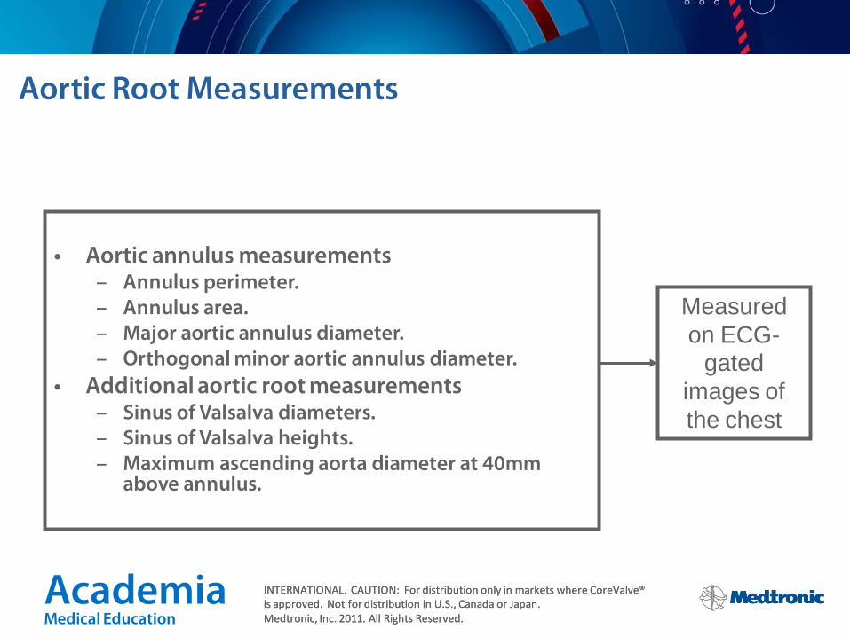
• Aortic annulus measurements – Annulus perimeter. – Annulus area. – Major aortic annulus diameter. – Orthogonal minor aortic annulus diameter.
• Additional aortic root measurements – Sinus of Valsalva diameters. – Sinus of Valsalva heights. – Maximum ascending aorta diameter at 40mm
above annulus.
Measured on ECG-
gated images of the chest
Aortic Root Measurements

Academia Medical Education
INTERNATIONAL. CAUTION: For distribution only in markets where CoreValve® is approved. Not for distribution in U.S., Canada or Japan. Medtronic, Inc. 2011. All Rights Reserved.
INTERNATIONAL. CAUTION: For distribution only in markets where CoreValve® is approved. Not for distribution in U.S., Canada or Japan. Medtronic, Inc. 2011. All Rights Reserved.
Perimeter: linear distance of tracing around the aortic annulus
Area: area contained within tracing around the aortic annulus
Major & Orthogonal Minor Diameters: linear distances through the center of the aortic annulus Mean Diameter: Calculated mean of major and minor diameters
Step 1: Aortic Annulus Measurements

Academia Medical Education
INTERNATIONAL. CAUTION: For distribution only in markets where CoreValve® is approved. Not for distribution in U.S., Canada or Japan. Medtronic, Inc. 2011. All Rights Reserved.
INTERNATIONAL. CAUTION: For distribution only in markets where CoreValve® is approved. Not for distribution in U.S., Canada or Japan. Medtronic, Inc. 2011. All Rights Reserved.
The Aortic Annulus – A Critical & Measurable Parameter for Device Selection
• The annulus is easily definable and measurable. • It represents the constriction between LVOT and aortic root – a
major anchoring point for the inflow of the CoreValve frame • It has a multitude of shapes:
– Non-circular and non-elliptical (most common) – Elliptical – Circular (rare)
Any single diameter may not necessarily
approximate the annulus “size” appropriately due to ellipticity or irregularity in shape.

Academia Medical Education
INTERNATIONAL. CAUTION: For distribution only in markets where CoreValve® is approved. Not for distribution in U.S., Canada or Japan. Medtronic, Inc. 2011. All Rights Reserved.
INTERNATIONAL. CAUTION: For distribution only in markets where CoreValve® is approved. Not for distribution in U.S., Canada or Japan. Medtronic, Inc. 2011. All Rights Reserved.
Step 2: Sinus of Valsalva Diameters
Scroll to the widest portion of the sinus in the reformatted double oblique axial images. Measure from commissure through center to opposite sinus. Complete for all three sinuses.
Right Coronary
Left Coronary
Non Coronary

Academia Medical Education
INTERNATIONAL. CAUTION: For distribution only in markets where CoreValve® is approved. Not for distribution in U.S., Canada or Japan. Medtronic, Inc. 2011. All Rights Reserved.
INTERNATIONAL. CAUTION: For distribution only in markets where CoreValve® is approved. Not for distribution in U.S., Canada or Japan. Medtronic, Inc. 2011. All Rights Reserved.
Left coronary sinus height
Non coronary sinus height
Right coronary sinus height
Measurements should be completed on the oblique coronal and sagittal images and taken from the aortic annulus (shown as the red line) to the highest point of the
sinus, along the direction of the sinus.
Step 3: Sinus of Valsalva Heights
Academia Medical Education
INTERNATIONAL. CAUTION: For distribution only in markets where CoreValve® is approved. Not for distribution in U.S., Canada or Japan. Medtronic, Inc. 2011. All Rights Reserved.
INTERNATIONAL. CAUTION: For distribution only in markets where CoreValve® is approved. Not for distribution in U.S., Canada or Japan. Medtronic, Inc. 2011. All Rights Reserved.
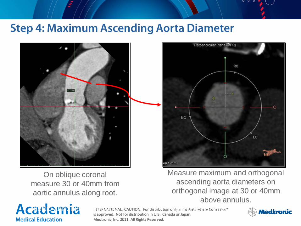
Measure maximum and orthogonal ascending aorta diameters on
orthogonal image at 30 or 40mm above annulus.
On oblique coronal measure 30 or 40mm from aortic annulus along root.
Step 4: Maximum Ascending Aorta Diameter
Measure maximum and orthogonal ascending aorta diameters on
orthogonal image at 30 or 40mm above annulus.
On oblique coronal measure 30 or 40mm from aortic annulus along root.

Academia Medical Education
INTERNATIONAL. CAUTION: For distribution only in markets where CoreValve® is approved. Not for distribution in U.S., Canada or Japan. Medtronic, Inc. 2011. All Rights Reserved.
INTERNATIONAL. CAUTION: For distribution only in markets where CoreValve® is approved. Not for distribution in U.S., Canada or Japan. Medtronic, Inc. 2011. All Rights Reserved.
Step 5: Device Size Selection Sinus of Valsalva and Ascending Aorta Ranges
Sinus of Valsalva Diameter (mm)
Sinus of Valsalva Height (mm)
Ascending Aorta Maximum Diameter (mm)
≥ 25 ≥ 15 ≤ 34
≥ 27 ≥ 15 ≤ 40
≥ 29 ≥ 15 ≤ 43
≥ 29 ≥ 15 ≤ 43 31
23
26
29

Academia Medical Education
INTERNATIONAL. CAUTION: For distribution only in markets where CoreValve® is approved. Not for distribution in U.S., Canada or Japan. Medtronic, Inc. 2011. All Rights Reserved.
INTERNATIONAL. CAUTION: For distribution only in markets where CoreValve® is approved. Not for distribution in U.S., Canada or Japan. Medtronic, Inc. 2011. All Rights Reserved.
Step 5: Device Size Selection Aortic Annulus Ranges
Diameter Range (mm) Perimeter Range (mm) Area Range (mm2)
18 - 20 56.5 - 62.8 254.5 - 314.2
20 - 23 62.8 - 72.3 314.2 - 415.5
23 - 27 72.3 - 84.8 415.5 - 572.6
26 - 29 81.7 - 91.1 530.9 – 660.5 31
23
26
29
Recent evidence supports perimeter as the recommended
method for TAVI sizing

Academia Medical Education
INTERNATIONAL. CAUTION: For distribution only in markets where CoreValve® is approved. Not for distribution in U.S., Canada or Japan. Medtronic, Inc. 2011. All Rights Reserved.
INTERNATIONAL. CAUTION: For distribution only in markets where CoreValve® is approved. Not for distribution in U.S., Canada or Japan. Medtronic, Inc. 2011. All Rights Reserved.
Comparison of MSCT Annulus Measurements Ao
rtic
Reg
urgi
tatio
n Pe
rcen
t (%
)
Imaging modality
0
5
10
15
20
25
30
Echo MSCT mean MSCT area MSCT perimeter
25
20 18
14
Retrospective analysis suggests that perimeter would have the lowest % of patients with AR ≥ 2
Retrospective analysis. Courtesy of Dr. Piazza and Prof. Lange, German Heart Center, Munich Germany

Academia Medical Education
INTERNATIONAL. CAUTION: For distribution only in markets where CoreValve® is approved. Not for distribution in U.S., Canada or Japan. Medtronic, Inc. 2011. All Rights Reserved.
INTERNATIONAL. CAUTION: For distribution only in markets where CoreValve® is approved. Not for distribution in U.S., Canada or Japan. Medtronic, Inc. 2011. All Rights Reserved.
Hypothetical Sizing Example #1
20 mm
20.3 mm 27.6 mm
24.6 mm
Modality Device Size
Echo 26 mm
CT Minor Diameter 26 mm
CT Major Diameter 31 mm
CT Perimeter 29 mm
CT Area 29 mm
CT Mean 29 mm
20 mm
1. Tzikas A, et al. Catheter Cardiovasc Intervent. 2011;77(6):868-75. Reprinted with the permission of John Wiley and Sons.
Academia Medical Education
INTERNATIONAL. CAUTION: For distribution only in markets where CoreValve® is approved. Not for distribution in U.S., Canada or Japan. Medtronic, Inc. 2011. All Rights Reserved.
INTERNATIONAL. CAUTION: For distribution only in markets where CoreValve® is approved. Not for distribution in U.S., Canada or Japan. Medtronic, Inc. 2011. All Rights Reserved.
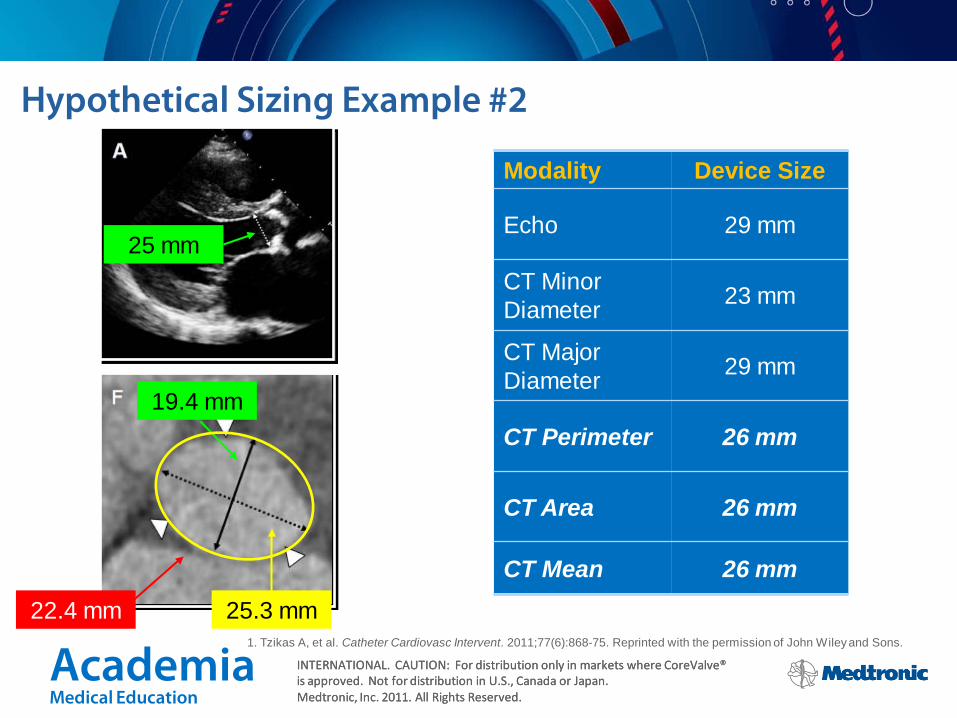
Hypothetical Sizing Example #2
25 mm
19.4 mm
25.3 mm
Modality Device Size
Echo 29 mm
CT Minor Diameter 23 mm
CT Major Diameter 29 mm
CT Perimeter 26 mm
CT Area 26 mm
CT Mean 26 mm 22.4 mm
1. Tzikas A, et al. Catheter Cardiovasc Intervent. 2011;77(6):868-75. Reprinted with the permission of John Wiley and Sons.

Academia Medical Education
INTERNATIONAL. CAUTION: For distribution only in markets where CoreValve® is approved. Not for distribution in U.S., Canada or Japan. Medtronic, Inc. 2011. All Rights Reserved.
INTERNATIONAL. CAUTION: For distribution only in markets where CoreValve® is approved. Not for distribution in U.S., Canada or Japan. Medtronic, Inc. 2011. All Rights Reserved.
Clinical Implications: Paravalvular Leak
Sizing and calcification are being investigated as major determinants of TAVI outcomes, for both Medtronic CoreValve® & Edwards Sapien®
Academia Medical Education
INTERNATIONAL. CAUTION: For distribution only in markets where CoreValve® is approved. Not for distribution in U.S., Canada or Japan. Medtronic, Inc. 2011. All Rights Reserved.
INTERNATIONAL. CAUTION: For distribution only in markets where CoreValve® is approved. Not for distribution in U.S., Canada or Japan. Medtronic, Inc. 2011. All Rights Reserved.
MSCT – Complete Pre-Implant TAVI Planning Tool
• 3D • Multiple measurements possible (area, perimeter,
diameters) • Assessment of calcification • Assessment of entire device landing zone • Assessment of peripheral access routes • High reproducibility and spatial resolution

Academia Medical Education
INTERNATIONAL. CAUTION: For distribution only in markets where CoreValve® is approved. Not for distribution in U.S., Canada or Japan. Medtronic, Inc. 2011. All Rights Reserved.
INTERNATIONAL. CAUTION: For distribution only in markets where CoreValve® is approved. Not for distribution in U.S., Canada or Japan. Medtronic, Inc. 2011. All Rights Reserved.
MSCT is Recommended
• The aortic annulus is non-circular and exhibits variability in shape across the patient population
• MSCT sizing has been linked to a reduction in paravalvular leakage
• Calcification location and burden have been linked to paravalvular leakage – MSCT is the only method available to properly assess calcification
• MSCT allows for complete patient assessment in one exam