sjl dystrophic mice express a significant amount of human
TRANSCRIPT

Author contributions: N.V.: Conception and design, Collection and/or assembly of data, data analysis and interpretation, manuscript writing; C.B..: Conception and design, data analysis and interpretation; V.B.: Provision of study material or patients; L.M.: Conception and design, Collection and/or assembly of data, Data analysis and interpretation, Manuscript writing; E.Z.: Collection and/or assembly of data, Data analysis and interpretation; M.S.: Collection and/or assembly of data, Data analysis and interpretation; M.F.S..: Provision of study material or patients; M.C.: Conception and design, Provision of study material or patients; P.B.: Provision of study material or patients; P.C.B.: Conception and design, manuscript writing; M.V.: Conception and design, Final approval of manuscript; M.Z.: Conception and design, Final approval of manuscript. Address correspondence to: Mayana Zatz, Human Genome Research Center, Department of Genetic and Evolutive Biology, University of São Paulo, Rua do Matão, n.106 - Cidade Universitária, São Paulo – SP, Brasil - CEP: 05508-090 , Phone / fax number: (55) (11) 3091-7966, E-mail: [email protected]. Received January 14, 2008; accepted for publication June 11, 2008; first published online in Stem Cells Express June 26, 2008. ©AlphaMed Press 1066-5099/4/08/$30.00/0 doi: 10.1634/stemcells.2008-0043
STEM CELLS®
TISSUE-SPECIFIC STEM CELLS Sjl Dystrophic Mice Express A Significant Amount Of Human Muscle Proteins Following Systemic Delivery Of Human Adipose-Derived Stromal Cells Without Immunosupression Natássia M. Vieira1, Carlos R. Bueno, Jr.2, Vanessa Brandalise1, Luciana V. Moraes4, Eder Zucconi1, Mariane Secco1, Miriam F. Suzuki3, Maristela M. Camargo4, Paolo Bartolini3, Patricia C. Brum2, Mariz Vainzof1, Mayana Zatz1 1Human Genome Research Center, Biosciences Institute, University of Sao Paulo; 2School of Physical Education and Sport, University of São Paulo; 3Biotechnology Department, National Nuclear Energy Commission-IPEN-CNEN, São Paulo; 4Department of Immunology, Instituto de Ciências Biomédicas, University of São Paulo - Brazil Key words. human adipose stromal cells • xenotransplantion • muscular dystrophy • therapy ABSTRACT Limb-girdle muscular dystrophies (LGMD) are a heterogeneous group of disorders characterized by progressive degeneration of skeletal muscle caused by the absence or defective muscular proteins. The murine model for Limb-Girdle Muscular Dystrophy 2B (LGMD2B), the sjl mice, carry a deletion in the dysferlin gene that causes a reduction in the protein levels to 15% of normal. The mice show muscle weakness that begins at 4–6 weeks and is nearly complete by 8 months of age. The possibility to restore the defective muscle protein and improve muscular performance by cell therapy is a promising approach for the treatment of LGMD or other forms of progressive muscular dystrophies (PMD). Here we
have injected human adipose stromal cells (hASCs) in the sjl mice, without immunosupression, aiming to assess their ability to: engraft into recipient dystrophic muscle after systemic delivery; form chimeric human/mouse muscle fibers; express human muscle proteins in the dystrophic host and improve muscular performance. We show for the first time that hASCs are not rejected after systemic injection even without immunosupression, are able to fuse with the host muscle, express a significant amount of human muscle proteins and improve motor ability of injected animals. These results may have important applications for future therapy in patients with different forms of muscular dystrophies.
INTRODUCTION Progressive muscular dystrophies (PMD) are a clinically and genetically heterogeneous group of disorders caused by deficiency or abnormal muscular proteins and characterized by progressive degeneration and loss of skeletal
muscle. The limb girdle muscular dystrophies (LGMDs) constitute a sub-group characterized by the involvement of the pelvic and shoulder girdle musculature. Until now, 20 different forms have been identified, 7 with autosomal dominant, LGMD1A to LGMD1G and 13 with autosomal recessive inheritance
Stem Cells Express, published online June 26, 2008; doi:10.1634/stemcells.2008-0043
Copyright © 2008 AlphaMed Press
by Mayana Z
atz on August 17, 2008
ww
w.Stem
Cells.com
Dow
nloaded from

Human proteins in the SJL muscle following systemic delivery of hASCs
2
(http://www.musclegenetable.org). Among the autosomal recessive forms, one of the most prevalent is caused by mutations in the dysferlin gene resulting in two phenotypes: Miyoshi myopathy (MM) which affects distal muscles at onset, with preferential early involvement of the gastrocnemius and LGMD2B with a more pronounced limb-girdle involvement [1]. Dysferlin expression is reduced or absent in these patients [2,3]. A 171-bp deletion in the murine dysferlin gene was detected in a mouse model, the sjl mice, with a corresponding reduction in dysferlin levels to 15% of normal. The spontaneous myopathy of SJL mice begins at 4–6 weeks of age and is nearly complete by 8 months of age showing a progressive inflammatory change in muscle [4]. The mutation in sjl mice is in-frame and is probably one of the reasons why its phenotype is milder than that of patients with dysferlinopathy, as in the case of Becker type muscular dystrophy as compared to Duchenne type [5]. Adult skeletal muscle has the potential to regenerate new muscle fibers by activating a population of mononucleated precursors, which otherwise remain in a quiescent and non-proliferative state [6]. However, the continuous and gradual muscle degeneration in progressive muscular dystrophies leads to a depletion of satellite cells and, consequently, the capacity to restore the skeletal muscle is lost [7,8]. The possibility to repair the defective muscle through cell therapy is a promising approach for the potential treatment of PMD. Different sources of stem/progenitor cells (SC) that show extended proliferation in vitro and also have the ability to generate normal muscle fibers in vitro and in vivo were recently described [9-15]. Recently it was reported two different strategies attempting cell therapy for PMD in a dystrophic dog: transplantation of autologous genetically corrected cells or transplantation of normal donor cells. Apparently, the second approach showed better results [12].
An abundant and accessible source of stem cells is adipose tissue. The ability of human adipose-derived stromal cells (hASCs) to differentiate into skeletal muscle when in contact with dystrophic muscle cells was demonstrated by us in vitro [17] and in vivo [18]. This last study showed that implantation of hASCs direct in the muscle of mdx mice restored dystrophin expression in the area nearby the injected place. However, the mdx mice have no evident muscular weakness and therefore are not a good model to assess potential functional effects of SC therapy. Here we have injected hASCs, without any immunosupression, in the sjl mouse model to assess: a) if hASCs cells can reach and engraft into recipient dystrophic muscle cells after systemic delivery, without immunosupression; b) form chimeric human/mouse muscle fibers, c) express human muscle proteins in the dystrophic host and d) improve muscular function. In addition, since it had been suggested that cells already committed to the myogenic phenotype and therefore able to autonomously differentiate into skeletal muscle in vitro, possess the highest regenerating potential in vivo when injected locally [19] we also analyzed the engraftment potential of hASCs previously exposed to myogenic induction media through systemic transplantation. We showed for the first time the successful engraftment and myogenic differentiation of undifferentiated hASCs through systemic delivery without immunosupression which resulted in a significant improvement in the muscular function of the injected mice.
RESULTS Characterization of ASCs ASCs were previously characterized [17] by flow cytometry for the expression of 12 cell surface proteins (HLA-DR, HLA-ABC, CD13, CD29, CD31, CD34, CD44, CD45, CD73, CD90, CD105 and CD117). Cell viability was above 96% by Guava ViaCount reagent (Guava Technologies).
by Mayana Z
atz on August 17, 2008
ww
w.Stem
Cells.com
Dow
nloaded from

Human proteins in the SJL muscle following systemic delivery of hASCs
3
At passage 4, hASCs did not express endothelial markers (CD31- PECAM1) nor hematopoietic markers (CD34, CD45 and CD117-c-kit). The majority of ASCs expressed high levels of CD13, CD44, adhesion markers (CD29-integrin ß1, CD90-Thy-1) and mesenchymal stem cell marker CD73 (SH3). Expression of some markers, such as CD105 (SH2), was variable among the donors. ASCs were negative for HLA-class II (HLA-DR), but positive for HLA-class I (HLA-ABC) (data not shown). The plasticity of hASCs was assessed three weeks after lineage induction [17]. Myogenic, adipogenic, chondrogenic and osteogenic differentiation was demonstrated by the expression of myogenic markers (myosin and desmin), lipid vacuoles, mucopolysaccharide-rich extracellular matrix and calcium deposits, respectively. These results confirmed the mesenchymal nature of the isolated cells as well as their multipotent potential (data not shown). ` hASCs capacity to reach and engraft at the host muscle In order to assess the potential of hASCs to reach and colonize the host muscle we injected undifferentiated hASCs, previously characterized by flow cytometry and differentiation potential [17] into the caudal vein of sjl mice (n=7). The PCR method as previously reported by Pelz et al. (2005 [20]) was used to evaluate the presence of human cells in different tissues of the injected sjl mice. Human DNA was found in the foreleg and hindleg muscles of all seven injected mice (Fig. 1A). One among the seven animals showed human DNA only in the proximal muscles while in the remaining six the cells were found in both distal and proximal muscles. Among other analyzed tissues human DNA was found in the liver, lung and kidney of the injected animals (Fig.1B). Muscle differentiation in the host muscle To explore the myogenic differentiation followed by the engraftment of hASCs we analyzed the expression of dysferlin and human-dystrophin in the host muscle.
The NCL-hamlet antibody against dysferlin showed poor labeling on human and mice muscle sections through immunofluorescence (IF) analysis (Fig.2A) and it also recognizes the SJL mutant dysferlin in IF (Fig.2A) and western blot (WB) analysis (Fig 2C). Therefore, we evaluated dysferlin expression through Real-Time PCR. The expression of exogen dysferlin was variable among the seven injected animals, ranging from 14% to 26% (Fig.3). To evaluate the presence of human muscle proteins in the host muscle, we assessed the presence of human-dystrophin, using human-dystrophin antibody [21]. Through WB analysis, human-dystrophin bands were found in the proximal and distal muscles of foreleg and hindleg of the injected animals (Fig.2B). IF analysis revealed that in the hASCs injected mice, approximately 50 + 2% (p = 3.623 E-13, t-Student test, n=7) of the fibers showed a positive labeling while no positive labeled fibers were observed in the muscles of non injected animals (Fig.2A and Fig.4A). These labeled fibers were seen both in clusters and spread out through the muscle tissue with some totally labeled and others partially positive for human-dystrophin (Fig.4B and C). Capacity of hASCs to reach and engraft at the host muscle after myogenic commitment In order to assess if cells already committed to the myogenic phenotype possess a higher or lower regenerating potential in vivo, we evaluated the effect of in vitro differentiation in the migration ability of hASCs in a second group of animals. In seven mice hASCs were transplanted following in vitro exposure to myogenic differentiation media. Interestingly, after myogenic induction, we did not find human DNA in the muscle of any of the injected animals (Supplemental Fig.1 online). Lymphocyte T Infiltration The evaluation of Lymphocyte T infiltration in the host muscle expressing human muscle proteins was performed by WB analysis for anti-CD3. Positive bands were detected in both
by Mayana Z
atz on August 17, 2008
ww
w.Stem
Cells.com
Dow
nloaded from

Human proteins in the SJL muscle following systemic delivery of hASCs
4
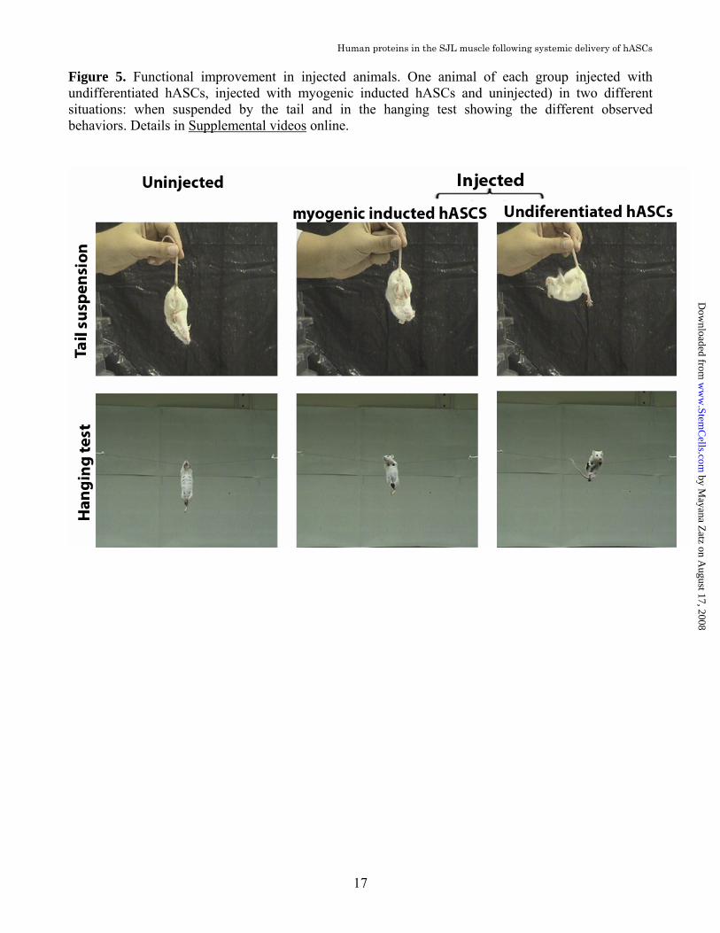
injected and uninjected animals with different intensities in samples from both groups. This data indicates that the presence of hASCs cells neither reduce nor increase the inflammatory changes typically found in the SJL dystrophic muscle (Supplemental Fig.2 online). Skin graft survival In order to evaluate if SJL naïve mice are immunocompetent, we performed allogeneic skin transplantation and tested if these mice were able to reject allografts. Our data show that SJL mice rejected skin from C57Bl/6 donors at the same period as immunocompetent BALB/c mice. Acceptance of syngeneic skin grafts suggests that SJL mice are also capable of maintaining tolerance to self-antigens (Supplemental Fig.3 online). Together these data indicate that these mice are capable of driving normal immune responses as well as modulating reactions towards self. Functional assessment We performed standardized motor ability tests [22] and compared the performance of each sjl mouse (injected and uninjected) before (2-months of age) and after (9-months of age) the injections period. The treated group showed an improvement of 15.2 +7.0% in their performance while the untreated worsened their performance in 6.12 + 6.0% (p = 0.013, t-Student test, n=7). The performance of the animals submitted to hASCs transplant after myogenic induction was very similar to the control group showing a worsening of motor ability in 7.21 + 7.0% (p = 0.449, t-Student test, n=7). (Fig.4 and Supplemental video 1,2 and 3 online) A typical behaviour of the SJL mice is observed when they are suspended by the tail. The first reflex of normal mice is to spread their limbs and digits while the trunk is held in an extended position. However the SJL mice, in contrast, keep their limbs in an adducted and flexed position and tend to curl in on themselves, grasping at their own fur. They often fail in attempts to extend their trunk and they are not
able to turn round and reach the suspending hand, indicating trunk muscle weakness [4]. We also observed a different behavior in injected mice when they were suspended by the tail. Injected animals were able to spread their limbs and digits with their trunk held in an extended position while the uninjected ones showed the typical reflex found in sjl mice. (Fig. 5 and Supplemental video 4, 5 and 6 online)
DISCUSSION Two different authors reported that ASCs are able to differentiate in vivo when injected directly into the muscle of the mdx mice [18,19]. However the potential of these cells to reach the muscle through systemic transplant as well as their potential functional effect in treated mice were not evaluated. The successful use of stem cells for clinical applications in cell therapy for PMD requires the investigation of the capacity of these cells, after systemic injections, to reach the target, engraft and restore the defective protein in the dystrophic muscle. A significant obstacle in designing cell therapy for PMD is the necessity to reach the entire body musculature, a problem that cannot be easily overcome unless systemic cell delivery methods are proved to be effective. Here we show for the first time that systemic delivery of hASCs into the tail vein of the sjl mice resulted in human-dystrophin and dysferlin expression in the host muscles and amelioration of functional parameters in the treated animals after 6-months of injections. In addition, we demonstrate that these positive results were obtained without the use of any immunosupression, which has been reported by local injection of hASCs into the mdx mice muscle [18] but is described here for the first time with systemic transplantation. Sampaolesi et al (2006 [12]) reported that systemic injections of normal dog mesoangioblasts to the muscle of dystrophic dogs resulted in the restoration of dystrophin expression. The mesoangioblasts show
by Mayana Z
atz on August 17, 2008
ww
w.Stem
Cells.com
Dow
nloaded from
Human proteins in the SJL muscle following systemic delivery of hASCs
5
similarities with ASCs in cell surface protein analysis, proliferation and differentiation capacity. However, all transplanted dogs were maintained on steroids as standard treatment and received immunosuppressant drugs, which makes difficult to evaluate functional results, since it is known that immunosuppressive and anti-inflammatory drugs can ameliorate the phenotype in muscular dystrophy patients [23]. More recently, Benchaouir et al. (2007 [24]) reported that exon-skipping-engineered Duchenne muscular dystrophy (DMD) stem cells restored human dystrophin when transplanted into dystrophic mice. In their experiment, the authors performed both intramuscular and intra-arterial delivery of genetically corrected CD133 cells expressing myogenic progenitors isolated from the blood and muscle of DMD patients into the scid/mdx mice. According to the authors, a significant recovery of muscle morphology, function, and dystrophin expression was observed. These results show that autologous transplantation of muscle-derived CD133+ cells may represent a promising approach to treat DMD. However the authors highlighted that the use of gene therapy raises some problems, specifically in terms of biosafety, since they used lentivirus vectors to correct the genetic defect into DMD stem cells prior to in vivo transplantation. They also called the attention for the fact that hazardous integration of the provirus can theoretically disturb the control of some housekeeping genes or tumor suppressor genes, and for clinical purposes, this risk of developing a tumor after in vivo injection has to be carefully addressed. In addition, differently from our approach, the possibility to transform a DMD patient into a less severe form of Becker muscular dystrophy, through the restoration of the RNA reading frame, is not applicable to all cases since it depends on specific mutation-deletions. Here, we show through real-time PCR, WB and IF analysis, that 6-months of systemic injections of non-engineered hASCs resulted in the expression of human muscle proteins in the host
muscle. Interestingly the IF analysis revealed the presence of labeled fibers either in clusters or spread out through the muscle tissue with some totally labeled and others partially positive for human-dystrophin. This pattern of labeling might be explained by the fusion of hASCs nuclei into host muscle fiber segments, demonstrating nuclear domains along the fibers [25]. On the other hand, Di Rocco et al. (2006 [19]) also observed dystrophin positive fibers organized in clusters following local transplantation of mouse ASCs. These results are in agreement with our findings and reinforce these authors suggestion of clonal proliferation of donor cells in the host muscle. Leriche-Guérin et al. (2002) [26] reported the myoblast transplantation into the SJL mice muscle with immunossupresion. The percentage of positive labeled fibers obtained in their study was lower than the percentage of dystrophin-positive fibers usually observed following the transplantation of normal myoblasts in mdx mice (30–90% [27]). Since the immunossupressive drug efficiently controlled the humoral and cellular immune reactions, the authors concluded that the immune rejection is not the cause of the low myoblast transplantation success in the SJL mice. It has been also suggested that cells already committed to the myogenic phenotype and therefore able to autonomously differentiate into skeletal muscle in vitro, possess the highest regenerating potential in vivo when injected locally [19]. In order to address this issue, which has important implications for therapeutic approaches, we assessed the effect of in vitro differentiation in the migration ability of hASCs. Surprisingly, in opposition to these authors, we did not find human DNA in the muscle of any of the injected animals after myogenic induction through systemic delivery. Our findings are in agreement with other authors [28] who observed that cultured myogenic progenitor cells injected intra-arterially into mdx mice resulted in only 1% of engraftment which led them to suggest that these cells extravasate poorly from the
by Mayana Z
atz on August 17, 2008
ww
w.Stem
Cells.com
Dow
nloaded from

Human proteins in the SJL muscle following systemic delivery of hASCs
6
circulation to the muscle locally. Alternatively, it is possible that differentiated cells behave differently or are rejected by the recipient animals following systemic transplantation. On this respect, it might be interesting to assess the effect of local injections of myogenic committed cells in combination with systemic transfer of undifferentiated stem cells for therapeutic purposes in PMD. The functional ability was evaluated by standardized motor ability tests [22] that were already shown to be efficient for other rodent models [29,30,31]. With these selected tests we could assess the skeletal muscle function of each mouse before and after the cell treatment. Finally our results revealed that muscle engraftment and the body-wide distribution of hASCs determined a significant (+15%) functional improvement in the injected animals. However, it is relevant that although hASCs muscle engraftment was widespread, it did not reach the entire population of muscle cells. Since the functional recovery was considerable this may indicate that partial muscle colonization is enough for a significant functional amelioration. These results are of clinical relevance in the development of appropriate stem cell approaches for human patients, suggesting that it might not be necessary to correct the entire muscle to gain a functional benefit. It is also important to highlight that it is not known if this clinical improvement is due to the enhanced expression of the faulty muscle proteins derived from hASCs or do their known immunomodulatory effect [32]. However the lymphocyte T infiltration data, where no difference was found between injected and uninjected animal, suggests that the presence of hASCs does not reduce significantly the inflammatory changes typically found in the SJL dystrophic muscle. Our data indicating that systemic delivery of hASCs to the muscle can be done efficiently without immunosupression may be explained because hASCs in vitro: a) do not induce proliferation of allogeneic T cells, b) suppress
the proliferation of T cells induced either by mitogens or allogeneic cells, c) secrete a soluble factor(s) that inhibit the production of inflammatory cytokines (TNF-α, IFN-γ and IL-12) of T cells stimulated by non-specific mitogenic and by allogeneic stimuli and d) maintain these properties even after differentiation [32,33]. This is supported by the present work since human cells were found in the host muscles for at least 2-months after the last injection. In short, here we show for the first time that hASCs are not rejected when transferred systematically to the sjl mice without any immunosupression, are able to fuse with the host muscle cell, express human skeletal muscle proteins and ameliorate motor ability of affected animals. Although the sjl mice have a mild phenotype, and further in vivo studies in different animal models, which are currently underway, will be essential to corroborate the present observations, the observed functional recovery after 6-months in vivo treatment is encouraging. These results open new avenues for pre-clinical researches which may have important applications for future therapy with the advantages that: a) it may be applicable to patients affected with different forms of progressive muscular dystrophies, regardless of their specific disease causing mutation; b) human liposuctioned fat is available in large quantities and hASCs can be easily obtained without any in vitro genetic modification or induction [34]. Although it remains to be seen if allogenic transplantation can be also done in humans without any immunosupression, this simple approach here reported may represent a great step toward clinical application for the future therapy of different forms of progressive muscular dystrophies.
MATERIALS AND METHODS All experiments were approved by the research ethics committee of the Biosciences Institute, University of São Paulo. All human samples were obtained after written informed consent
by Mayana Z
atz on August 17, 2008
ww
w.Stem
Cells.com
Dow
nloaded from
Human proteins in the SJL muscle following systemic delivery of hASCs
7
from the donors. All researches were carried out in the Human Genome Research Center, at the Biosciences Institute, University of São Paulo. ASC isolation and expansion Human adipose tissue was obtained from elective liposuction procedures. Cells were isolated using methods previously described [17]. Briefly, the unprocessed lipoaspirate was washed extensively with equal volumes of phosphate-buffered saline (PBS) containing antibiotics (100 U/ml of penicillin and 100g/ ml of streptomycin; Gibco) and then dissociated with 0,075% collagenase (Sigma). Enzyme activity was neutralized with Dulbecco’s modified Eagle’s media - high glucose (DMEM-HG – HG; Gibco) containing 10% Fetal Bovine Serum (FBS; Gibco). The infranatant was centrifuged at 1200 x g for 5 min to pellet the cells. The cells from the pellet SVF were filtered to remove debris and seeded in tissue culture plates (NUNC) at 1,000–3,500 cells/cm [2] in DMEM-HG 10% FBS. Cultures were washed with PBS 24-48 hours after plating to remove unattached cells and fed with fresh media. The cultures were maintained at 37°C with 5% CO2, in growth media (GM- DMEM-HG 10% FBS). When they achieved about 70% confluence, the cells were trypsinised (0,025%, TrypLE Express; Gibco) and plated at a density of 5000 per cm2. Cultures were passaged repeatedly after achieving a density of 70%-80% until passage 4. The remaining cells were cryopreserved in cryopreservation media (10% dimethylsulfoxide, 10% DMEM-HG, 80% FBS), frozen at -80°C in isopropanol-jacked closed container and stored in liquid nitrogen the next day. Flow Cytometry The flow cytometry was performed on Guava EasyCyte System (Guava Technologies) using a blue laser (488 nm). Cells were pelleted, resuspended in PBS at concentration of 1 x 10⁵
cells/ml and stained with saturating concentration of antibodies. Cells were incubated in the dark for 45 minutes at room
temperature. After incubation, cells were washed three times with PBS and resuspended in 0.25 ml of cold PBS. Cell viability was accessed with Guava ViaCount reagent (Guava Technologies). ASCs were incubated with the following primary antibodies: HLA-DR-PE, HLA-ABC-FITC, CD13-PE, CD29-PECy5, CD31-PE, CD34-PerCP, CD44-FITC, CD45-FITC, CD73, CD90-PE, CD105 e CD117-PE (Becton Dickinson). Unconjugated markers were reacted with anti-mouse PE secondary antibody (Guava Technologies). Flow cytometer gates were set using unstained cells. Cells were gated by forward scatter to eliminate debris. To eliminate the possible autofluorescence of ASCs, we removed the contribuition of unstained cells in the measurement channel. A minimum of 10,000 events was counted for each analysis. Multilineage Differentiation Cells were analyzed for their capacity to differentiate toward adipogenic, osteogenic, chondrogenic and myogenic lineages as described in Zuk et al (2001). Myogenic differentiation induction hASCs were cultured in GM supplemented with 0,1 µM dexamethasone (Sigma), 50 µM hydrocortisone (Sigma) and 5% horse serum (Gibco) for 10 days before the transplantation to the sjl mice. At this stage, hASCs cells express MyoD [35]. Transplantation Sjl mice were purchased from the Jackson Laboratory. Animal care and experiments were performed in accordance with animal research ethics committee of the Biosciences Institute, University of São Paulo. Two-months sjl mice were divided into three groups (n = 7) for the transplantation experiments (groups A, B, and C). Each animal was injected in the tail vein with 1 ✕ 106 of either hASCs (A group) or myogenic induced
by Mayana Z
atz on August 17, 2008
ww
w.Stem
Cells.com
Dow
nloaded from

Human proteins in the SJL muscle following systemic delivery of hASCs
8
hASCs (B group). The animals were injected for 6 months, weekly in the first month and then monthly. Animals were analyzed blindly in these three groups (A, B, and C); the code for each of the mice groups was disclosed only after completion of all the studies. Two months after the last cell transplantation, the animals were euthanatized using a CO2 chamber. Human DNA analysis The DNA was obtained using DNeasy Blood & Tissue Kit (Qiagen). The presence of human DNA in the host samples were evaluated as described in Pelz et al (2005 [20]). Centromeric region of human chromosome 7 and mice chromosome 8 was amplified by PCR (35 cycles, annealing at 59ºC). The PCR products were separated by electrophoresis on 2% agarose gels and stained with ethidium bromide. Nonsaturated digital images were obtained using an ImageQuant imaging system (GE HealthCare). Immunofluorescence Proximal and distal muscle of the foreleg and hindleg of sjl mice were removed and frozen in liquid N2. Serial muscle crio-sections were fixed in 4% paraformaldehyde in PBS for 20 min at 4°C, permeabilized in 0,05% Triton X-100 in PBS for 5 minutes. Non-specific binding was blocked with 10% FBS in PBS for 1 hour at room temperature. Muscle sections were incubated with primary antibody overnight at 4°C and with secondary antibody for 1 hour at room temperature. The following primary antibodies were used: anti-dysferlin NCL-hamlet (1:300; Novocastra) and anti-human-dystrophin MANEX 1216E2 G10 (1:100), kindly provided by Dr. Glenn E. Morris at Center for Inherited Neuromuscular Diseases, Oswestry, Shropshire, UK; combined with rabbit anti-mouse IgG secondary antibody Cy3-conjugated (1:200; Chemicon) or FITC-Conjugated (1:100; Chemicon). We visualized nuclei with 4',6'-diamidino-2-phenylindole (DAPI; Sigma). The fluorescence signal was examined in Axiovert 200 (Carl Zeiss) and in AxioImager Z1 (Carl Zeiss).
Western Blot Analysis Muscle sample proteins were extracted through treatment with a buffer containing 10mM Tris-HCl (pH 8.0), 150mM NaCl, 5mM EDTA, 1% Triton X-100 and 60mM octyl glucoside. Samples were centrifuged at 13,000 x g for 10 minutes to remove insoluble debris. Soluble proteins were resolved by 6% sodium dodecyl sulfate-polyacrylamide gel electrophoresis (SDS-PAGE), and transferred to nitrocellulose membranes (Hybond; Amersham). All membranes were stained with Ponceau (Sigma) to evaluate the amount of loaded proteins. Blots were blocked for 1 hour in Tris-buffered saline Tween (TBST) containing 5% powdered skim milk and reacted overnight with the following primary antibodies: anti mouse-CD3 (1:1000, Becton Dickinson), anti human-dystrophin MANEX 12/16E2 G10 (1:100) kindly provided by Dr. Glenn E. Morris at Center for Inherited Neuromuscular Diseases, Oswestry, Shropshire, UK and anti-dysferlin NCL - Hamlet (1:25; Novocastra). Blots were incubated one hour with secondary antibodies and immunoreactive bands were detected with ECL chemiluminescence detection system (GE Healthcare). Reverse Transcription Reactions and Quantitative Real-time PCR To test the presence of human cDNA in the host muscle, total RNA from skeletal gastrocnemius of all uninjected, injected with undifferentiated hASCs animals and controls (five normal Swiss mice and human muscle) was extracted. Total RNA (0.2 µg) was reverse transcribed into cDNA using Superscript III reverse transcription kit (Invitrogen). Quantitative real-time PCR (qRT-PCR) was performed using 50 ng of cDNA and SYBR Green PCR master mix in an ABI Prism 7100 system (Applied Biosystems). The PCR conditions were: 94°C for 15 s, 58°C for 30 s, and 72°C for 30 s for 40 cycles. Since the host lacks a 171pb region in the cDNA, primers were designed to amplify cDNA fragment from inside the SJL deleted region, amplifying the normal mice and the human samples. The endogenous control (beta-actin)
by Mayana Z
atz on August 17, 2008
ww
w.Stem
Cells.com
Dow
nloaded from

Human proteins in the SJL muscle following systemic delivery of hASCs
9
primers were designed to amplify SJL, mice and human cDNAs. The authenticity of the PCR products was verified by melt-curve analysis and
agarose gel electrophoresis. Primers sequences were as follows: Dysferlin: sense GCCTCTGGAGAAGGACCTAAAG; antisense: ACAGCGAGCCCCAAACTTG (accession no. NM_003494, GenBank); Beta-actin: sense: GGCTGTATTCCCCTCCATCG; antisense: CCAGTTGGTAACAATGCCATGT (accession no. NM_007393, GenBank). Samples were run in triplicates, and the threshold suggested by the instrument software was used to calculate Ct. To normalize the readings we used Ct values from the beta-actin as internal standard in each run, obtaining a delta Ct value for dysferlin. Skin transplantation Allogeneic or syngeneic tail skin was grafted on the back of SJL or BALB/c recipients. Skin was removed from the donor tail, cut into 1cm2 pieces and kept in PBS at room temperature until use. Grafts were placed on a bed prepared by removing an area on the back dermis of the recipient, sutured and covered with plaster. Donor skin was considered rejected when 90% of the area was destructed. Functional assessment In order to verify whether injected hASCs would improve motor ability in sjl injected mice, we performed motor ability tests before and after 6 months of SC injection period. Mice were examined, weighed, and submitted to the following tests: (a) the inclined plane test evaluated by measuring the maximal angle of a wood board on which the animal was placed until it slipped; (b) the wire hanging test to determine the ability of the mouse suspended on
a horizontal thread by its forelegs, to reach it with its hindlegs and the length of time they were able to stay hanging; (c) the ambulation test which was performed to determine the mean length of a step measured in hindfoot ink prints while mice freely run in a corridor (length, 50 cm; width, 8 cm; height of lateral walls, 20 cm) [22]. Statistical analysis Observations were quantified blindly by two authors (Vieira N and Bueno Junior CR) independently from one another. Numerical data are the mean sd (standard deviation). The statistical analysis of the equivalence between the injected and uninjected mice was achieved by the one-tailed t-student test, at the significance level of p=0.05 and the results were expressed by the percentage variation between their performance before and after SC transfer period. We gratefully acknowledge our colleagues Tatiana Jazedje, Maria Denise Carvalho, Agnes Nishimura, Natale Cavaçana, Miguel Mitne-Neto, Monize Lazar, Viviane Abreu Nunes, Constancia Urbani, David Schlesinger, Daniela Bueno, Roberto Fanganiello, Antonia M P Cerqueira, Bruno Lima, Rafaella Nascimento, Marta Canovas, Paula Onofre and Dr. Maria Rita Passos-Bueno for helpful suggestions as well as the two anonymous reviewers. We would like to thank Dr. Glenn Morris from the Center for Inherited Neuromuscular Disease (CIND), RJAH Orthopaedic Hospital, Oswestry, Shropshire, UK for providing anti-human dystrophin antibody. This research was supported by FAPESP-CEPID (Fundação de Amparo à Pesquisa do Estado de São Paulo - Centro de Pesquisa, Inovação e Difusão), CNPq (Conselho Nacional de Desenvolvimento Científico e Tecnológico).
by Mayana Z
atz on August 17, 2008
ww
w.Stem
Cells.com
Dow
nloaded from

REFERENCES 1. Mahjneh I, Passos-Bueno MR, Zatz M, et al. The
phenotype of chromosome 2p-linked limb-girdle muscular dystrophy. Neurom. Disord. 1996; 6:483-490.
2. Liu J, Aoki M, Illa I, et al. Dysferlin, a novel skeletal muscle gene, is mutated in Miyoshi myopathy and limb girdle muscular dystrophy. Nat Genet. 1998; 20:31–36.
3. Matsuda C, Aoki M, Hayashi YK, et al. Dysferlin is a surface membrane-associated protein that is absent in Miyoshi myopathy. Neurology. 1999; 53:1119–1122.
4. Bittner RE, Anderson LV, Burkhardt E, et al. Dysferlin deletion in SJL mice (SJL-Dysf) defines a natural model for limb girdle muscular dystrophy 2B. Nat Genet. 1999; 23:141–142.
5. Chelly J, Gilgenkrantz H, Lambert M, et al. Effect of dystrophin gene deletions on mRNA levels and processing in Duchenne and Becker muscular dystrophies. Cell. 1990; 63:1239-48.
6. Schultz E, McCormick KM. Skeletal muscle satellite cells. Rev Physiol Biochem Pharmacol. 1994; 123:213-257.
7. Heslop L, Morgan JE, Partridge TA. Evidence for a myogenic stem cell that is exhausted in dystrophic muscle. J Cell Sci. 2000; 113:2299-2308.
8. Laguens R. Satellite cells of skeletal muscle fibers in human progressive muscular dystrophy. Virchows Arch Pathol Anat Physiol Klin Med. 1963; 336:564-569.
9. Gronthos S, Mankani M, Brahim J, et al. Postnatal human dental pulp stem cells DPSCs in vitro and in vivo. Proc Natl Acad Sci USA. 2000; 97:13625-30.
10. Zuk PA, Zhu M, Mizuno H, et al. Multilineage cells from human adipose tissue: Implications for cell-based therapies. Tissue Eng. 2001; 7:211-228.
11. Gussoni E, Soneoka Y, Strickland CD, et al. Dystrophin expression in the mdx mouse restored by stem cell transplantation. Nature. 1999; 401:390-394.
12. Sampaolesi M, Blot S, D’Antona G, et al. Mesoangioblast stem cells ameliorate muscle function in dystrophic dogs. Nature. 2006; 444:574-579.
13. Secco M, Zucconi E, Vieira NM, et al. Multipotent Stem Cells from Umbilical Cord: Cord is Richer than Blood! Stem Cells. 2008; 26:146-50.
14. Chan J, Waddington SN, O'Donoghue K, et al. Widespread Distribution and Muscle Differentiation of Human Fetal Mesenchymal Stem Cells After Intrauterine Transplantation in Dystrophic mdx Mouse. Stem Cells. 2007; 25: 875 – 884.
15. Kong KY, Ren J, Kraus M, et al. Human Umbilical Cord Blood Cells Differentiate into Muscle in sjl Muscular Dystrophy Mice. Stem Cells. 2004; 22: 981 – 993.
16. Salah-Mohellibi N, Millet G, André-Schmutz I, et al. Bone Marrow Transplantation Attenuates the Myopathic Phenotype of a Muscular Mouse Model
of Spinal Muscular Atrophy. Stem Cells. 2006; 24: 2723 – 2732.
17. Vieira NM, Brandalise V, Zucconi E, et al. Human multipotent adipose derived stem cells restore dystrophin expression of Duchenne skeletal muscle cells in vitro. Biol Cell. 2008; 100:231-41.
18. Rodriguez AM, Pisani D, Dechesne CA, et al. Transplantation of a multipotent cell population from human adipose tissue induces dystrophin expression in the immunocompetent mdx mouse. J Exp Med. 2005; 201:1397-1405.
19. Di Rocco G, Iachininoto MG, Tritarelli A, et al. Myogenic potential of adipose-tissue-derived cells. J Cell Sci. 2006; 119:2945-2952.
20 .Pelz O, Wu M, Nikolova T, et al. Duplex polymerase chain reaction quantification of human cells in a murine background. Stem Cells. 2005; 23:828-833.
21. Thanh LT, Neguyen TM, Helliwell TR, et al. Characterization of revertant muscle fibers in Duchenne Muscular Dystrophy using exon-specific monoclonal antibodies against dystrophin. Am J Hum Genet. 1995; 56:725-731.
22. Kennel PF, Fonteneau P, Martin E, et al. Electromyographical and motor performance studies in the pmn mouse model of neurodegenerative disease. Neurobiol Dis 1996; 3:137-47.
23. Davies KE, Grounds MD. Treating muscular dystrophy with stem cells? Cell. 2006; 127:1304-1306.
24. Benchaouir R, Meregalli M, Farini A, et al. Restoration of Human Dystrophin Following Transplantation of Exon-Skipping-Engineered DMD Patient Stem Cells into Dystrophic Mice. Cell Stem Cell. 2007; 1:646-657.
25. Karpati G, Zubrzycka-Gaarn EE, Carpenter S, et al. Age-related conversion of dystrophin-negative to -positive fiber segments of skeletal but not cardiac muscle fibers in heterozygote mdx mice. J Neuropathol Exp Neurol. 1990; 49:96-105.
26. Leriche-Guérin K, Anderson LV, Wrogemann K, et al. Dysferlin expression after normal myoblast transplantation in SCID and in SJL mice. Neuromuscul Disord. 2002; 12:167-173.
27. Partridge T, Morgan JE, Coulton RG, et al. Conversion of mdx myofibres from dystrophin-negative to -positive by injection of normal myoblasts. Nature. 1989; 337:176–179.
28. Bachrach E, Perez AL, Choi YH, et al. Muscle engraftment of myogenic progenitor cells following intraarterial transplantation. Muscle Nerve. 2006; 34:44-52.
29. Groshong JS, Spencer MJ, Bhattacharyya BJ, et al. Calpain activation impairs neuromuscular transmission in a mouse model of the slow-channel myasthenic syndrome J Clin Invest. 2007;117:2903-2912.
30. Simon D, Seznec H, Gansmuller A, et al. Friedreich ataxia mouse models with progressive cerebellar and sensory ataxia reveal autophagic neurodegeneration in dorsal root ganglia. J Neurosci. 2004; 24:1987-95.
by Mayana Z
atz on August 17, 2008
ww
w.Stem
Cells.com
Dow
nloaded from

Human proteins in the SJL muscle following systemic delivery of hASCs
11
31. Yonemori F, Yamaguchi T, Yamada H, et al. Evaluation of a motor deficit after chronic focal cerebral ischemia in rats. J Cereb Blood Flow Metab. 1998;18:1099-106.
32. Yañez R, Lamana ML, García-Castro J, et al. Adipose Tissue-Derived Mesenchymal Stem Cells Have In Vivo Immunosuppressive Properties Applicable for the Control of the Graft-Versus-Host Disease Stem Cells 2006; 24: 2582 – 2591.
33. Niemeyer P, Kornacker M, Mehlhorn A, et al.
Comparison of immunological properties of bone marrow stromal cells and adipose tissue-derived stem
cells before and after osteogenic differentiation in vitro. Tissue Eng 2007; 13: 111-121.
34. Schäffler A, Büchler C. Concise Review: Adipose Tissue-Derived Stromal Cells—Basic and Clinical Implications for Novel Cell-Based Therapies. Stem Cells 2007; 25: 818 – 827.
35. Mizuno H, Zuk PA, Zhu M, et al. Myogenic differentiation by human processed lipoaspirate cells. Plast Reconstr Surg 2002; 109:199-209.
by Mayana Z
atz on August 17, 2008
ww
w.Stem
Cells.com
Dow
nloaded from

Human proteins in the SJL muscle following systemic delivery of hASCs
12
Figure 1. Polymerase chain reaction analysis for human chromosome 7 α-satellite sequences H7), mouse chromosome 8 centromeric repeat sequence M8) of sjl mice. a) Muscles of the injected and uninjected sjl mice. b) Various tissues of the injected animal. Tissues were harvested at day 60 postinjection. Samples shown are the following: 1) Distal foreleg muscle; 2) Proximal foreleg muscle; 3) Distal hindleg muscle; 4) Proximal hindleg muscle; 5) Intestine; 6) Urinary bladder; 7) Heart; 8) Brain; 9) Liver; 10) Spleen; 11) Lung; 12) Kidney; 13) Inguinal fat; 14) Tail; 15) Diaphragm; H) Human DNA; M) Mouse DNA.
by Mayana Z
atz on August 17, 2008
ww
w.Stem
Cells.com
Dow
nloaded from

Human proteins in the SJL muscle following systemic delivery of hASCs
13
Figure 2. Expression of dysferlin and human-dystrophin in the muscle from human and mouse (C57B) normal controls, in one sjl mouse of injected and one of the uninjected group. a) Immunofluorescence analysis of dysferlin and human-dystrophin of the gastrocnemius muscle of the injected and uninjected animal. Histopathological analysis in skeletal muscles from both injected and uninjected sjl mice showed comparable alterations including size variation among individual muscle fibers, fiber splitting, small regenerated basophilic fibers, numerous fibers with centrally located myonuclei and significant connective tissue replacement. b) Western blot analysis for human-dystrophin and dysferlin of the muscles of injected and uninjected animal. Samples shown are the following: 1) Distal foreleg muscle; 2) Proximal foreleg muscle; 3) Distal hindleg muscle; 4) Proximal hindleg muscle; myosin = myosin band detected in the Ponceau S pre-stained blot, for the evaluation of loaded muscle proteins; H) Human muscle; M) Mouse muscle.
by Mayana Z
atz on August 17, 2008
ww
w.Stem
Cells.com
Dow
nloaded from

Human proteins in the SJL muscle following systemic delivery of hASCs
14
by Mayana Z
atz on August 17, 2008
ww
w.Stem
Cells.com
Dow
nloaded from

Human proteins in the SJL muscle following systemic delivery of hASCs
15
Figure 3. Quantitative real-time polymerase chain reaction (qPCR) analysis for exogen dysferlin expression. a) Primers (in red) were designed to amplify cDNA fragment from inside the SJL 171pb deleted region, amplifying the normal mice and human samples. b) Relative Quantification of exogen dysferlin expression in all injected (2-8) and one uninjected animal (1) compared with normal Swiss mice expression (9). Data are normalized to beta-actin expression and expressed as means ± SEM.
by Mayana Z
atz on August 17, 2008
ww
w.Stem
Cells.com
Dow
nloaded from

Human proteins in the SJL muscle following systemic delivery of hASCs
16
Figure 4. Human-dystrophin positive labeled fibers in the gastrocnemius muscle of the injected sjl mice. a) Global distribution of the positively labeled fibers and details of partially positive fibers in b) longitudinal section and c) transversal section.
by M
ayana Zatz on A
ugust 17, 2008 w
ww
.StemC
ells.comD
ownloaded from
Human proteins in the SJL muscle following systemic delivery of hASCs
17
Figure 5. Functional improvement in injected animals. One animal of each group injected with undifferentiated hASCs, injected with myogenic inducted hASCs and uninjected) in two different situations: when suspended by the tail and in the hanging test showing the different observed behaviors. Details in Supplemental videos online.
by Mayana Z
atz on August 17, 2008
ww
w.Stem
Cells.com
Dow
nloaded from

DOI: 10.1634/stemcells.2008-0043 published online Jun 26, 2008; Stem Cells
Patricia C. Brum, Mariz Vainzof and Mayana Zatz Zucconi, Mariane Secco, Miriam F. Suzuki, Maristela M. Camargo, Paolo Bartolini,
Natássia M. Vieira, Carlos R. Bueno, Jr., Vanessa Brandalise, Luciana V. Moraes, Eder Immunosupression
Following Systemic Delivery Of Human Adipose-Derived Stromal Cells Without Sjl Dystrophic Mice Express A Significant Amount Of Human Muscle Proteins
This information is current as of August 17, 2008
& ServicesUpdated Information
http://www.StemCells.comincluding high-resolution figures, can be found at:
Supplementary Material http://www.StemCells.com/cgi/content/full/2008-0043/DC1
Supplementary material can be found at:
by Mayana Z
atz on August 17, 2008
ww
w.Stem
Cells.com
Dow
nloaded from