sjogren’s ss-a and ss-b antigens robert fox, md scripps memorial-ximed la jolla, ca
TRANSCRIPT

Sjogren’s SS-A and SS-B antigens
Robert Fox, MDScripps Memorial-Ximed
La Jolla, CA
Different name, same molecule
• Ro is name of patient who had a particular antibody detected by immune diffusion
• La is a different patient who donated antibody• Subsequent exchange of sera indicated the
identical nature of Ro=SS-A and La=SS-B
SS-A
• Serum containing autoantibodies directed against the Ro/SSA antigens may recognize one or both of two cellular proteins with molecular weights of approximately 52 and 60
• These autoantigens are referred to as “Ro52” and “Ro60,” respectively.
• The two autoantigens were originally thought to interact with each other, but subsequent studies have shown that the two proteins reside in distinct cellular compartments
• Ro60 localized to the nucleus and nucleolus and with Ro52 localized to the cytoplasm

History of Ro-60
• Ro60 was shown to bind small, non-coding RNAs termed “Y RNAs.”
• The Ro60/hYRNA complex originally identified by Steitz as a chaperone for adenoviral RNA
• Subsequently other viruses also found to use hYRNA as their chaperone
• In particular, EBV RNA’s may use hYRNA• Recently, miRNA predicted bind Ro60
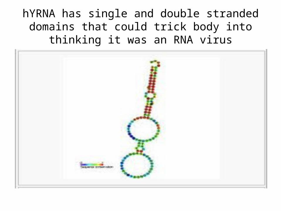
hYRNA has single and double stranded domains that could trick body into thinking it was an RNA virus

SS-A binds to hYRNA
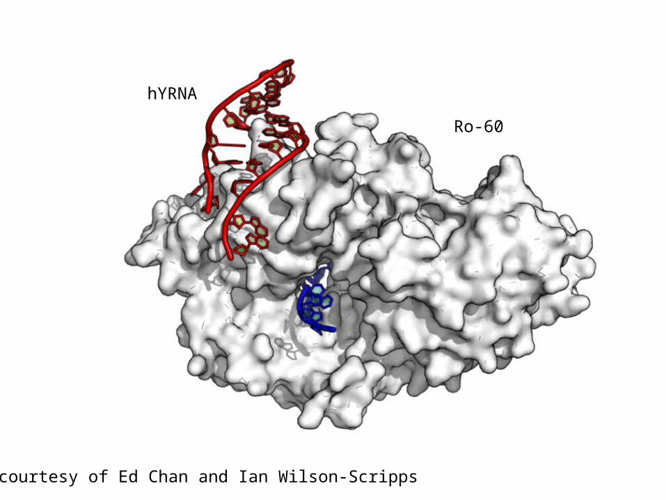
courtesy of Ed Chan and Ian Wilson-Scripps
Ro-60
hYRNA


hYRNA
• Although the function of Y RNAs is unknown, consensus report suggests that Y RNAs may be a source of micro (mi)RNA molecules, which have a role in the regulation of messenger (m)RNA stability and translation.
• The crystal structure of Ro60 suggests that the protein can bind to both single- and double-stranded RNA.
• The protein may function as a “RNA chaperone” that binds to misfolded pre-5S ribosomal RNA and may hasten the degradation of the defective molecule
• Ro60 may also bind to other cellular and viral RNAs in the cell, including Epstein-Barr virus (EBV) early RNA 1 (EBER1)

To avoid this “autoimmune” catastrophy of activation by every RNA
• The body puts the receptors for the RNA viruses (known as Toll receptors 7 and 9) intra-cellularly
TLR
Fc-receptor
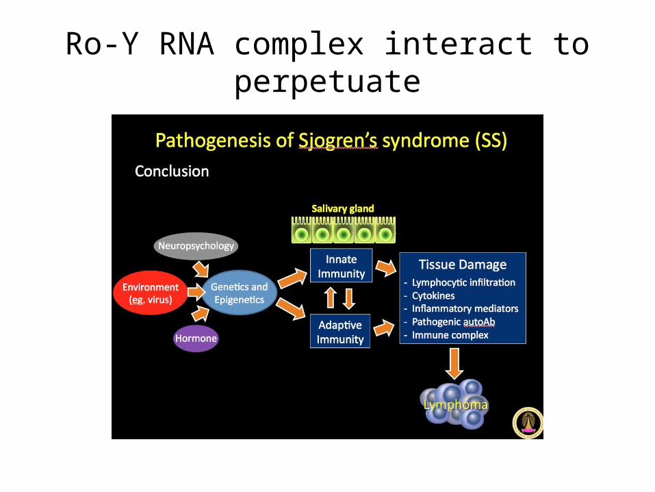
Ro-Y RNA complex interact to perpetuate

microRNA

Cascade of TLR signals

Cascade of Lymphoma
Key point
• SS-A lacks site for apoptotic digestion• Mediated by caspaces• Thus, SS-A is not degraded during apoptotic
death• It migrates into the apoptotic bleb on cell
surface

Immunogenicity
• The SS-A would normally be harmless• If the patient has antibody to SS-A, then the
SS-A antigen forms an immune complex • The microenvironment now contains• SS-A bound to hYRNA and • the antibody to SS-A

Key point
• The antibody to SS-A has an Fc portion• The Fc portion of the SS-A binds to Fc receptor
on the phagocytic dendritic cells (the normal garbage collectors)
• The SS-A is thus dragged into the lysosome• However, the hYRNA is still attached and
stimulates the TLR receptors in the lysozome• This stimulates type I IFN which perpetuates
more antibody
Another proposed mechanism
• Mice that lack the functional gene encoding Ro60 develop an autoimmune syndrome characterized by antiribosome and antichromatin antibodies, as well as glomerulonephritis, suggesting that Ro60 may have an important role in preventing systemic autoimmune disease
• Thus, antibodies that neutralize SS-A could play a role.

SS-B or La antigen
• The amino acid sequence of the Ro52 autoantigen was reported in 1991.
• Ro52 is an interferon-inducible protein that belongs to the “tripartite motif” family of proteins.
• The protein localizes to the cytoplasm and functions as an E3 ubiquitin ligase, an enzyme that adds ubiquitin molecules to target proteins

SS-A or La 52 KD
• Ro52 may also have important roles in the regulation of inflammation.
• Ro52 adds an ubiquitin molecule to activated inhibitor of nuclear factor kappa-B kinase subunit beta (IKKB)
Other roles in mice
• Ro52 also inhibits inflammation by targeting interferon regulatory factors (IRF) 3 and 7 for ubiquitin-mediated degradation
• Consistent with the effects of Ro52 on NFKB signaling and IRF stability, mice that lack Ro52 develop an autoimmune disease, which is characterized by hypergammaglobulinemia and renal disease

SS-B OR La Antigen• Autoantibodies directed against the La/SSB autoantigen (La) interact
with a 47-kD protein, which shuttles between the nucleus and cytoplasm but which is predominantly found in the nucleus.
• The N-terminus of the protein contains a “La” RNA-binding domain and an adjacent RNA recognition motif (RRM), which cooperate to bind RNA.
• The C-terminus of the protein contains a second RRM followed by a short basic motif (“SBF” domain) and a nuclear localization sequence.
• The N-terminal portion of La mediates the interaction with the 3’ end of RNA polymerase III transcripts.
• La participates in the processing of small, noncoding RNAs such as ribosomal 5S RNA.
Additional roles for SS-B
• La also binds cellular transfer RNAs (tRNAs), as well as viral RNA molecules, including those produced by EBV, Hepatitis A and C, respiratory syncytial virus, adenovirus, and human immunodeficiency virus (HIV)
• La may regulate the translation of viral RNAs by enhancing internal ribosomal entry site function.
• A report suggests that La has a critical role in regulating RNA interference (RNAi) by enhancing RNAi turnover in the RNA-induced silencing complex (RISC).
• If confirmed, this result would suggest that La has a role in the regulation of mRNA stability and translation. A mouse model that lacks the La protein has not been reported.
DETECTION OF ANTI-Ro AND ANTI-La ANTIBODIES
• Solid phase assays — In the past, antibodies directed against the Ro and La antigens were detected by immunodiffusion or by counter-immunoelectrophoresis. Most laboratories have subsequently adopted solid phase assays, including enzyme-linked immunosorbent assays (ELISA) or antigen-coated fluorescent microsphere and flow cytometry-based assays. Solid phase assays use either native or recombinant protein as substrate. These assays have advantages over other techniques including decreased cost, increased sensitivity for the detection of the corresponding autoantibodies, and the ability to quantify the amount of antibody. The increased sensitivity of solid phase assays comes at the cost of decreased specificity for the diagnosis of autoimmune diseases. The reported prevalence of anti-Ro and anti-La autoantibodies in patients with autoimmune diseases depends upon the method used to detect the autoantibodies.
Detection• Immunofluorescence staining on HEp-2 substrates — Patients in whom the only autoantibodies present are anti-Ro
antibodies may have a falsely negative antinuclear antibody (ANA) test using the traditional human epithelial cell line-2 (HEp-2) cell substrate, because Ro60 immunoreactivity may be lost during the preparation of the cells and because Ro52 is a cytoplasmic, rather than nuclear, autoantigen.
• The ability to detect anti-Ro60 antibodies by indirect immunofluorescence has been enhanced by use of a modified HEp-2 cell substrate (“HEp-2000”), in which transfection of a complementary DNA (cDNA) encoding Ro-60 was used to over-express the protein in 10 to 20 percent of the cells
• Anti-Ro60 antibodies produce nucleolar and nuclear speckled staining patterns in transfected cells on the Hep-2000 substrate. Only 10 to 20 percent of the cells will produce this staining pattern if anti-Ro60 antibodies are the only autoantibodies present in a patient’s serum. However, only about 25 percent of clinical and commercial laboratories in the United States use the HEp-2000 substrate, while, in Europe and Australia, Hep-2000 is used as the ANA substrate in about 75 percent of such laboratories. In laboratories using the traditional HEp-2 cell substrate, solid phase assays are the most reliable approach to detecting anti-Ro60 antibodies.
• In contrast to anti-Ro60, anti-Ro52 antibodies can be detected by indirect immunofluorescence using the traditional HEp-2 cell substrate. Under normal tissue culture conditions, Ro52 localizes to the cell cytoplasm . Because Ro52 is not a nuclear autoantigen, autoantibodies directed against this antigen may not be reported by those commercial laboratories that only report the presence of antinuclear (but not anticytoplasmic) antibodies using the HEp-2 cell substrate.
• Although La has been shown to shuttle between the nucleus and cytoplasm, the protein is predominantly found in the nucleus of HEp-2 cells. Autoantibodies directed against La produce a speckled nuclear staining pattern

CLINICAL SIGNIFICANCE• — Anti-Ro, detected by solid phase immunoassays, which have
traditionally detected both Ro60 and Ro52, have been found in patients with a range of autoimmune disorders, including systemic lupus erythematosus (SLE, 32 percent), Sjögren’s syndrome (59 percent), idiopathic inflammatory myopathies (IIM, 19 percent), systemic sclerosis (21 percent), and rheumatoid arthritis (RA, 15 percent), as well as primary biliary cirrhosis (PBC) and undefined connective tissue disease (UCTD,infrequently).
• In contrast to anti-Ro antibodies, anti-La antibodies are relatively specific for the diagnosis of SLE and Sjögren’s syndrome. In addition, as with anti-Ro antibodies, anti-La antibodies may be detected in the mothers of children who are born with neonatal lupus syndrome. These women may or may not have evidence of systemic autoimmune disease.

Clinical Significance
• Women with anti-Ro antibodies (with or without anti-La and with or without autoimmune disease) are at increased risk for having a child with neonatal lupus syndrome. Anti-Ro antibodies may be the only autoantibodies present in more than half of the patients with “antinuclear antibody (ANA)-negative” SLE. Anti-Ro antibodies may also be the first detectable autoantibodies that precede the development of SLE in asymptomatic individuals

Anti-Ro preceding autoimmune disease
• Autoantibodies may precede the diagnosis of SLE by years, if not decades.
• A study using serum samples of 130 patients with known SLE from the Department of Defense Serum Repository to test for the presence of autoantibodies (by enzyme-linked immunosorbent assays [ELISA]) prior to the diagnosis detected anti-Ro antibodies in 48 percent of the patients.
• The mean interval between the dates of the stored serum sample in which anti-Ro antibodies were present and of the diagnosis of SLE was longer than 3.6 years.
• Relative to all other autoantibodies tested, anti-Ro antibodies were the first to develop in patients who eventually were diagnosed with SLE. The number of normal individuals who are anti-Ro antibody positive but who never develop an autoimmune disease is unknown.
Summary
• Serum containing autoantibodies directed against the Ro/SSA autoantigens may recognize one or both of two cellular proteins with molecular weights of approximately 52 and 60 kD;
• the autoantigens are referred to as “Ro52” and “Ro60,” respectively. Ro60 is localized to the nucleus and nucleolus, and Ro52 is localized to the cytoplasm

Summary
• Solid phase assays, including enzyme-linked immunosorbent assays (ELISA) and flow cytometry-based assays, with either native or recombinant protein as substrate, are generally used to detect antibodies directed against the Ro and La antigens. Anti-Ro antibodies may not be detected by indirect immunofluorescence using a traditional Hep-2 cell substrate.

• Anti-Ro antibodies may be present in patients with a range of autoimmune disorders, including systemic lupus erythematosus (SLE), Sjögren’s syndrome, dermatomyositis or polymyositis, systemic sclerosis, primary biliary cirrhosis (PBC), and rheumatoid arthritis (RA).
• Women with anti-Ro (with or without co-occurring anti-La) autoantibodies are at increased risk for having a child with neonatal lupus syndrome. Anti-Ro antibodies may be the only autoantibodies present in more than half of the patients with “antinuclear antibody (ANA)-negative” SLE. The combination of anti-Ro and anti-La antibodies is relatively specific for the diagnoses of SLE and Sjögren’s syndrome.