skeletal anchorage in orthodontics – basics and … artikel/grundlagen...areas of application for...
TRANSCRIPT
443J Orofac Orthop 2007 · No. 6 © Urban & Vogel
Journal of Orofacial OrthopedicsFortschritte der Kieferorthopädie Review ArticleJournal of Orofacial OrthopedicsFortschritte der Kieferorthopädie
Skeletal Anchorage in Orthodontics – Basics and Clinical ApplicationSkelettale Verankerung in der Kieferorthopädie – Grundlagen und klinische AnwendungHeiner Wehrbein1, Peter Göllner2
1 Department of Orthodontics, Johannes Gutenberg University Hospital, Mainz, Germany,
2 Orthodontist (SSO) in private practice, Berne, Switzerland.
Received: July 5, 2007; accepted: October 8, 2007
Abstract This review article describes the basics and clinical applications of skeletal anchorage in orthodontics, namely: areas of indica-tion, anchorage devices, insertion areas, indications, potential complications, and their use in growing patients.
The areas for skeletal anchorage include orthodontic-pros-thetic anchorage, orthodontic anchorage, and skeletal anchorage in orthopedic therapy procedures. The anchorage devices current-ly available are: prosthetic implants, mini screws, palatal im-plants, onplants, bone anchors, zygoma wires and skeletally-sup-ported distractors. The insertion areas described so far (accor-ding to therapeutic procedure and bone supply available) include edentulous jaw sections, the interdental septum, infra-apical and supra-apical areas, the palate (median, paramedian, lateral), the retromolar area, and the zygomatic bone. Force systems are ap-plied (direct or indirect anchorage) according to surgical and orthodontic or orthopedic requirements. Skeletal anchorage de-vices should be selected according to the following criteria. Is the anchorage task unifunctional or multifunctional? How many anchorage devices are required for the therapy in question? What is the success rate of the various anchorage devices; what are the applicable biomechanics and soft tissue or hard tissue conditions in the insertion area? The success rates for miniscrews are cur-rently between 80% and 90%, and over 90% for palatal im-plants.
The potential of skeletal anchorage is broadening the cur-rent orthodontic treatment spectrum, guaranteeing the practitio-ner absolute control of anchorage by avoiding the unpredictable reactions of periodontal anchorage, leading to a reduction in un-wanted side effects.
Key Words: Skeletal anchorage · Miniscrews · Palatal im-plants · Bone anchors · Bone-supported RME · Skeletal maxillofacial protraction
Zusammenfassung Der vorliegende Artikel beschreibt die Grundlagen und die klinische Anwendung der skelettalen Verankerung in der Kieferorthopädie: Indikationsbereiche, Verankerungsmittel, Insertionsbereiche, Indi-kationen, potentielle Komplikationen und die Nutzung beim noch wachsenden Patienten.
Als Indikationsbereiche der skelettalen Verankerung sind heu-te zu nennen: orthodontisch-prothetische Verankerung, orthodon-tische Verankerung und skelettale Verankerung bei kieferorthopä-dischen Behandlungsaufgaben. Als Verankerungsmittel stehen heute zur Verfügung: prothetische Implantate, Minischrauben, Gaumenimplantate, Onplants, Bone Anchors, Zygoma-Drähte und skelettal abgestützte Distraktoren. Je nach Behandlungsaufgabe und dem Knochenangebot wurden folgende Insertionsbereiche be-schrieben: zahnloser Kieferabschnitt, interdentales Septum, infra- und supraapikaler Bereich, Gaumen (median, paramedian, lateral), retromolarer Bereich sowie der Bereich des Os zygomaticum. Die Applikation des Kraftsystems (direkte/indirekte Verankerung) rich-tet sich nach den chirurgischen und orthodontischen/orthopä-dischen Erfordernissen. Die Selektion des skelettalen Verankerungs-mittels sollte nach folgenden Kriterien erfolgen: uni- bzw. multi-funktionelle Verankerungsaufgaben, Anzahl der notwendigen Verankerungselemente für die jeweilige Behandlungsaufgabe, Er-folgsrate der diversen Verankerungselemente, der zu applizierenden Biomechanik und der Weichgewebe- sowie Hartgewebebedin-gungen im Insertionsbereich. Die Erfolgsraten liegen heute bei Mi-nischrauben bei 80–90%, bei Gaumenimplantaten bei über 90%.
Die heutigen Möglichkeiten der skelettalen Verankerung er-weitern das kieferorthopädische Behandlungsspektrum, garantie-ren eine absolute Kontrolle der Verankerung durch den Behandler, umgehen die unvorhersagbaren Reaktionen der desmodontalen Verankerung und führen zu einer Reduktion unerwünschter Neben-wirkungen.
Schlüsselwörter: Skelettale Verankerung · Minischrau-ben · Gaumenimplantate · Bone Anchors · Knochen-gestützte GNE · Skelettale maxillofaziale Protraktion
J Orofac Orthop 2007;68:443–461
DOI 10.1007/s00056-007-0725-y

Wehrbein H, Göllner P. Skeletal Anchorage
444 J Orofac Orthop 2007 · No. 6 © Urban & Vogel
IntroductionAnchorage is a fundamental problem in the treatment of malocclusions and dysgnathias. The loading of the anchor-age unit is based on the conditions of static balance (ac-tion = reaction) as defined by Newton in 1687 (Diedrich [23]).
When teeth are used for anchorage purposes, their reac-tive load is determined according to the force magnitude, direction, type, and duration of the moments and forces ex-erted through the biomechanical force system for active tooth movement. This problem must be taken into account.when planning each and every anchorage.
If the patient’s periodontal anchorage potential fails to accommodate the ultimate treatment goal, additional intra-oral or extra-oral anchorage devices must be employed to prevent side effects [23].
Every type of auxiliary anchorage devices such as head-gear or intermaxillary elastics entail characteristic potential disadvantages, i.e. visibility, compliance-dependency, and the risk of unwanted side effects. Class-II elastics, for exam-ple, can lead to canting of the occlusal plane in the cw direc-tion, protrusion of the incisors in the maxilla, and tooth ex-trusion [23].
The aim of this article is to illustrate the basics of skele-tal anchorage and describe the clinical aspects of the skeletal anchorage elements presently available.
Skeletal AnchorageSkeletal anchorage is largely based on the principle of os-seointegration as described in 1977 by Brånemark [12]. Os-seointegration implies direct contact between living bone and an implant visible through an optical microscope. Rob-erts [55] expanded the concept of osseointegration to in-clude no fibrous tissue between implant and bone, high tone on percussion, absence of physiological drift, no move-ment under orthodontic load, and the functional equivalent of a dental ankylosis (Figure 1).
At present endosseous implants are predominantly used for skeletal anchorage purposes, as case reports, prospective clinical research, and animal experimental studies have shown that osseointegrated implants remain stable under orthodontic and orthopedic load conditions [3, 6, 11, 14, 19, 22, 23, 25, 26, 32, 35, 42, 45, 50, 51, 54–58, 62–66, 68]. They can therefore be used as skeletal anchorage elements for orth-odontic and orthopedic treatment purposes.
Areas of Application for Skeletal Anchorage in Orthodontics
Current areas of application for skeletal anchorage in or-thodontics can be classified as [22, 34, 56, 66]:– orthodontic-prosthetic anchorage – orthodontic anchorage – skeletal anchorage for orthopedic procedures
EinleitungVerankerung ist ein grundlegendes Problem bei der Be-handlung von Malokklusionen und Dysgnathien. Die Be-lastung der Verankerungseinheit basiert auf den Bedin-gungen des statischen Gleichgewichtes (actio = reactio), wie bereits von Newton 1687 definiert (siehe Diedrich [23]).
Wenn Zähne für Verankerungszwecke genutzt werden, wird deren reaktive Belastung hinsichtlich Kraftgröße, Rich-tung, Art und Dauer durch die Kräfte und Momente deter-miniert, die durch das biomechanische Kraftsystem zur ak-tiven Zahnbewegung ausgeübt wird. Dieses Problem muss bei jeder Verankerungsplanung berücksichtigt werden.
Wenn das parodontale Verankerungspotential im Hin-blick auf das angestrebte Behandlungsziel inadäquat ist, sollten (müssen) zusätzliche intra- und/oder extraorale Ver-ankerungshilfen eingesetzt werden, um Nebeneffekte zu vermeiden [23].
Zusätzliche Verankerungshilfen wie der Headgear und intermaxilläre Elastics sind je nach Art durch einige potenti-elle Nachteile charakterisert: Sichtbarkeit, Compliance-Ab-hängigkeit und das Risiko von unerwünschten Nebeneffek-ten. Klasse-II-Elastics können z. B. zur Kippung der Okklusi-onsebene im cw-Sinn, Protrusion der Inzisiven im Unterkiefer und Extrusion von Zähnen führen [23].
Das Ziel dieses Artikels ist, die Grundlagen der skelet-talen Verankerung aufzuzeigen und die klinischen Aspekte der momentan verfügbaren skelettalen Verankerungsele-mente darzustellen.
Skelettale Verankerung Die skelettale Verankerung basiert überwiegend auf dem Prinzip der Osseointegration, das Brånemark 1977 [12] be-schrieben hat. Osseointegration bedeutet ein unter dem Lichtmikroskop sichtbarer direkter Kontakt zwischen leben-dem Knochen und einem Implantat. Roberts [55] hat den Begriff der Osseointegration erweitert: kein fibröses Binde-
Figure 1. Physiological periodontal ligament (left), dental ankylosis (center), osseointegrated implant (right). (Photo center: by courtesy of Prof. Diedrich, Aachen, Germany)
Abbildung 1. Physiologisches parodontales Ligament (links), dentale Ankylose (Mitte), osseointegriertes Implantat (rechts). (Bild Mitte: Prof. Diedrich, Aachen)

Wehrbein H, Göllner P. Skelettale Verankerung
445J Orofac Orthop 2007 · No. 6 © Urban & Vogel
Skeletal Orthodontic-prosthetic AnchorageIn skeletal orthodontic-prosthetic anchorage, the prosthetic implants are initially used as orthodontic anchorage ele-ments and then later as abutments upon which to attach a fixed prosthetic replacement [66] (Figure 2). The insertion site is the edentulous alveolar bone of the upper or lower jaw. After insertion and application of a supraconstruction, a bracket or other orthodontic attachments can be applied to a provisional crown or to a commercially-obtainable bonding base. The orthodontic force system thus functions between the tooth to be moved and the implant. The orth-odontic tooth movement’s reactive forces and moments are thus transferred directly to the implant. Biomechanically
gewebe zwischen Implantat und Knochen, hoher Ton auf Perkussion, Abwesenheit physiologischer Drift, keine Bewe-gung gegenüber orthodontischer Belastung und funktio-nelles Äquivalent einer dentalen Ankylose (Abbildung 1).
Heute werden überwiegend enossale Implantate für ske-lettale Verankerungszwecke genutzt, denn Fallberichte, pros-pektive klinische Untersuchungen und tierexperimentelle Studien haben gezeigt, dass osseointegrierte Implantate unter orthodontischen und orthopädischen Belastungsbedingungen positionsstabil bleiben [3, 6, 11, 14, 19, 22, 23, 25, 26, 32, 35, 42, 45, 50, 51, 54–58, 62–66, 68]. Demnach können sie als skelet-tale Verankerungselemente für orthodontische sowie ortho-pädische Behandlungsaufgaben genutzt werden.
Einsatzbereich der skelettalen Verankerung in der Kieferorthopädie
Die heutigen Einsatzbereiche der skelettalen Verankerung in der Kieferorthopädie können eingeteilt werden in [22, 34, 56, 66]:– Orthodontisch-prothetische Verankerung – Orthodontische Verankerung– Skelettale Verankerung bei kieferorthopädischen Maß-
nahmen
Skelettale orthodontisch-prothetische Verankerung Bei der skelettalen orthodontisch-prothetischen Veranke-rung werden grundsätzlich prothetische Implantate verwen-det, welche zunächst als orthodontisches Verankerungsele-ment und späterer als Pfeiler für die Aufnahme eines fest-sitzenden Zahnersatzes genutzt werden [66] (Abbildung 2). Der Insertionsort ist der zahnlose Alveolarknochen des Ober- bzw. Unterkiefers. Nach der Insertion und Applika-tion einer Suprakonstruktion kann ein Bracket oder ande-res orthodontisches Attachment an einer provisorischen Krone oder an einer kommerziell erhältlichen Klebebasis angebracht werden. Das orthodontische Kraftsystem wirkt demnach zwischen den zu bewegenden Zähnen und dem Implantat. Die reaktiven Kräfte und Momente für die ortho dontische Zahnbewegung werden also direkt auf das Implantat übertragen. Biomechanisch gesehen, ist dies der Typ einer direkten orthodontischen Implantatverankerung.
Skelettale orthodontisch-prothetische Verankerungen können bei teilbezahnten Patienten indiziert sein wie z.B. bei: – dysgnath stehenden Frontzähnen mit einseitiger oder
beidseitiger Freiendsituation oder– dysgnath stehenden Front- und/oder Seitenzähnen in
Verbindung mit Zahnlücken (zwei oder mehr fehlende Zähne), meistens im posterioren Bereich des Unter- und/oder Oberkiefers.
Residuales Wachstum und orthodontisch-prothetische Implantatverankerung mit Alveolarfortsatzimplantaten
Enossale Implantate sollten nicht vor Abschluss des Wachs-tums in den Alveolarknochen eingesetzt werden, da sie die
Figure 2. Orthodontic-prosthetic implant anchorage; two prosthetic im-plants were inserted in an edentulous jaw section (missing teeth: 46 and 47). They served first as orthodontic anchorage elements for molar up-righting (uprighting spring according to Burstone) and then as a pillar to accommodate a fixed prosthetic replacement. Advantage: the anterior dentition was not used for anchorage tasks, thus avoiding side effects.
Abbildung 2. Orthodontisch-prothetische Implantatverankerung; zwei prothetische Implantate wurden in einen zahnlosen Kieferabschnitt inse-riert (fehlende Zähne: 46 und 47). Sie dienten zunächst als orthodon-tische Verankerungselemente zur Molarenaufrichtung (Aufrichtefeder nach Burstone) und danach als Pfeiler zur Aufnahme eines festsitzenden prothetischen Ersatzes. Vorteil: Die anteriore Dentition wurde nicht für Verankerungsaufgaben genutzt. Dadurch wurden Nebenwirkungen ver-mieden.

Wehrbein H, Göllner P. Skeletal Anchorage
446 J Orofac Orthop 2007 · No. 6 © Urban & Vogel
speaking, this is the model for direct orthodontic implant anchorage.
Skeletal orthodontic-prosthetic anchorage can be indi-cated for partially-edentulous patients, for instance:– for malpositioned anterior teeth with a free-end situation
on one or both sides, or– for malpositioned anterior teeth and/or lateral teeth in
conjunction with tooth gaps (two or more missing teeth) primarily in the posterior upper and/or lower jaw.
Residual Growth and Orthodontic-prosthetic Implant Anchorage with Alveolar Bone Implants
Endosseous implants should not be inserted in the alveolar bone before growth has ceased, since they can interfere with growth of the surrounding bone and even the eruption of adjacent teeth [50, 59]. Insertion of prosthetic implants in the alveolar process can lead to infraposition of the implant and open-bite development if the patient is still growing.
Skeletal Orthodontic AnchoragePatients who undergo exclusively orthodontic treatment have complete dentitions or extraction sites that should be closed. Hence orthodontic anchorage elements cannot be inserted into an edentulous area in the jaw.
The following anatomical regions have been described concerning the application of skeletal orthodontic anchoring elements:– the interradicular septum of the dentulous alveolar pro-
cess [15, 17, 26, 28, 36, 43, 47] – the supra-apical and infrazygomatical area [18, 43, 47] – the retromolar area of the lower and upper jaw [37, 56] – the median or paramedian anterior palate [3, 5, 6, 7, 9, 19,
25, 31, 38, 67, 69, 72].
Since the above-mentioned insertion sites contain distinctly less horizontal or vertical bone support than the edentulous area of the jaw, special skeletal anchorage devices were de-veloped. Diameter-reduced screws (miniscrews) are proba-bly the most frequently used, followed by length-reduced screws (e.g. palatal implants), so as to maintain the integrity of neighboring anatomical structures.
The following skeletal orthodontic anchorage devices have been described and applied:– miniscrews [10, 11, 17, 18, 26–28, 36, 43, 44, 47, 52, 74] – palatal implants [3, 5, 6, 14, 19, 61, 66, 67, 72] – onplants [9, 38] – bone anchors [20, 24] – zygoma ligatures [46].
Indication for Skeletal Orthodontic Anchorage DevicesSkeletal orthodontic anchorage devices are indicated when stationary anchorage is required involving diverse anchor-age tasks. In particular, these can include [3, 6, 7, 9, 14, 18, 24, 26, 28, 40, 43, 44, 47, 52, 61, 67, 69, 71, 74]:
Entwicklung des umliegenden Knochens und sogar die Eruption der angrenzenden Zähne beeinträchtigen können [50, 59]. Das Einsetzen von prothetischen Implantaten in den Alveolarfortsatz kann bei noch vorhandenem Wachs-tum zu einer Infraokklusion des Implantates und Entwick-lung eines offenen Bisses führen.
Skelettale orthodontische VerankerungAusschließlich kieferorthopädisch zu behandelnde Pati-enten haben geschlossene Zahnreihen oder Extraktionslü-cken, die geschlossen werden sollen. Daher können ortho-dontische Verankerungselemente nicht in einen zahnlosen Kieferabschnitt inseriert werden.
Die folgenden anatomischen Regionen wurden für den Einsatz von skelettalen orthodontischen Verankerungsele-menten beschrieben:– das interradikuläre Septum des bezahnten Alveolarfort-
satzes [15, 17, 26, 28, 36, 43, 47], – der supraapikale und infrazygomatikale Bereich [18, 43,
47], – der retromolare Bereich des Unterkiefers [37, 56] und – der mediane oder paramediane anteriore Gaumen [3, 5,
6, 7, 9, 19, 25, 31, 38, 67, 69, 72].
Da die oben genannten Insertionsbereiche im Vergleich zum zahnlosen Alveolarfortsatz deutlich weniger horizon-tales und/oder vertikales Knochenangebot aufweisen, wur-den spezielle skelettale Verankerungsmittel entwickelt. Am häufigsten werden heute vermutlich durchmesserreduzierte Schrauben (Minischrauben) gefolgt von längenreduzierten Schrauben (z. B. Gaumenimplantat) verwendet, um die In-tegrität benachbarter anatomischer Strukturen nicht zu be-einträchtigen.
Folgende skelettale orthodontische Verankerungsmit-tel wurden beschrieben und angewendet:
Figure 3. Miniscrews from various producers (Ø 1.1–2.2 mm/length: 8–12 mm). Predominantly derived from osteosynthesis technology, machine-polished smooth surface, mode of insertion: self-drilling, self-cutting.
Abbildung 3. Minischrauben verschiedener Hersteller (Ø 1,1–2,2 mm/Länge: 8–12 mm). Überwiegend aus Osteosynthesetechnik abgeleitet, maschinenpolierte glatte Oberfläche, Insertionsmodus: selbstbohrend, -schneidend.

Wehrbein H, Göllner P. Skelettale Verankerung
447J Orofac Orthop 2007 · No. 6 © Urban & Vogel
– space closure from mesial – space closure from distal – intrusion and extrusion (anterior and posterior teeth) – distalization, mesialization, and midline corrections – molar uprighting.
MiniscrewsMiniscrews are largely a product of osteosynthesis technol-ogy. They have a smooth, machine-polished surface. The mode of insertion is self-drilling or self-cutting. Most mini-screws feature a special head to accommodate orthodontic force systems or wires. Miniscrews can be loaded immedi-ately after insertion. It is advisable to take an intraoral peri-apical view prior to insertion in the interdental area to mark the insertion site in the mucosa. A variety of systems (Figure 3) is currently available [44].
The advantages of miniscrews over other skeletal an-chorage concepts are:– simple insertion and explantation technique – relative affordability – application in various anatomical areas (upper and lower
jaw).
Figures 4 to 7 depict miniscrew application possibility in the frontal tooth area of the upper jaw.
Potential disadvantages of the miniscrew are: various insertion sites (each insertion site has special characteristics and their associated risks: e.g. retromolar maxilla, lingual mandible, unattached mucosa), rotational instability, need to use several screws for multifunctional anchorage tasks, screw migration, screw fracture upon removal, impairment of the anchorage concept if a screw is lost where several are
Figure 4. 62-year-old patient with deep bite, pathological migration of the anterior teeth and gingival retraction owing to a periodontal disease. Therapy: MB treatment with skeletal anchorage for intrusion of the ante-rior tooth segment so as not to extrusively load the periodontally-com-promised posterior teeth.
Abbildung 4. 62-jähriger Patient mit Tiefbiss, pathologischer Migration der Frontzähne und Gingivaretraktion aufgrund einer Parodontalerkran-kung. Therapie: MB-Behandlung mit skelettaler Verankerung zur Intrusi-on des Frontzahnsegmentes, um die parodontal geschädigten Seitenzäh-ne nicht extrusiv zu belasten.
Figure 5. Detail from panoramic radiograph: insertion of two miniscrews regio 12 (missing tooth 12) and interdentally between 21 and 22. The se-lection of the screw positions occurred according to surgical (bone support, attached gingiva) and orthodontic criteria (action line of force/center of resistance of the anterior tooth segment to be intruded: 11 to 22). The teeth were intruded with NiTi coil springs or elastic chains, fixed between the miniscrews and the segmented arch (direct implant anchorage).
Abbildung 5. Ausschnitt aus Orthopantomogramm: Insertion von zwei Minischrauben Regio 12 (fehlender 12) und interdental zwischen 21 und 22. Die Selektion der Position der Schrauben erfolgte nach chirurgischen (Knochenangebot, angewachsene Gingiva) und orthodontischen Kriterien (Kraftwirkungslinie/Widerstandszentrum des zu intrudierenden Front-zahnsegmentes: 11 bis 22). Die Zähne wurden mit NiTi-Coil-Springs bzw. elastischen Ketten intrudiert, welche zwischen den Mini-Schrauben und dem Segmentbogen fixiert wurden (direkte Implantatverankerung).
Figure 6. Finding after intrusion from 11 to 21. Retention phase, screws still in situ. The soft tissue around the screws is relatively free of inflam-mation. Insertion occurred in attached gingiva.
Abbildung 6. Zustand nach Intrusion von 11 bis 21. Retentionsphase, Schrauben noch in situ. Das Weichgewebe um die Schrauben ist relativ entzündungsfrei. Die Insertion erfolgte in der angewachsenen Gingiva.

Wehrbein H, Göllner P. Skeletal Anchorage
448 J Orofac Orthop 2007 · No. 6 © Urban & Vogel
required, mucosa irritation or overgrowth where screw head is located in unattached mucosa (Figure 8), and the danger of injury to the root of the tooth when inserted interdentally [15, 17, 26, 28, 43, 47, 52, 74]. In an animal experimental study, however, Asscherickx et al. [2] documented that if tooth roots are injured superficially the periodontal struc-tures (root cement, periodontal ligament space, lamina dura) heal completely once the screw is removed. Screw insertion in the periodontal space, however, is under discussion as one cause of screw loss [2].
The success rate (positional stability through osseointe-gration, no implant loss) of miniscrews is between 51 and 100% according to the literature [15, 17, 26, 36, 52, 74]. By using the appropriate insertion protocol, avoiding insertion in certain regions (e.g. the lingual mandibula), avoiding re-gions with unattached mucosa [15, 36, 52, 74] and selecting the appropriate screw [76], the chances of success can be sig-
– Minischrauben [10, 11, 17, 18, 26–28, 36, 43, 44, 47, 52, 74] – Gaumenimplantate [3, 5, 6, 14, 19, 61, 66, 67, 72] – Onplants [9, 38]– Bone Anchors [20, 24]– Zygoma-Ligaturen [46].
Indikation skelettaler orthodontischer Verankerungsmittel
Die Indikation skelettaler orthodontischer Verankerungs-mittel liegt in der Notwendigkeit einer stationären Veran-kerung bei diversen Verankerungsaufgaben. Diese können im Einzelnen umfassen [3, 6, 7, 9, 14, 18, 24, 26, 28, 40, 43, 44, 47, 52, 61, 67, 69, 71, 74]:– Lückenschluss von mesial – Lückenschluss von distal – In- und Extrusion (Front- und Seitenzähne)– Distalisierung/Mesialisierung/Mittellinienkorrekturen– Molarenaufrichtung.
MinischraubenMinischrauben sind überwiegend aus der Osteosynthese-technik abgeleitet und weisen eine glatte maschinenpolierte Oberfläche auf. Der Insertionsmodus ist selbstbohrend oder selbstschneidend. Der überwiegende Teil der Minischrauben weist einen speziellen Kopf auf, um orthodontische Kraftsys-teme oder Drähte aufzunehmen. Minischrauben können un-mittelbar nach der Insertion belastet werden. Vor der Inser-tion im interdentalen Bereich wird die Anfertigung eines Zahnfilms mit Markierungsdraht empfohlen, um die Inserti-onsstelle in der Mukosa zu markieren. Eine Vielzahl diverser Systeme (Abbildung 3) ist heute erhältlich [44].
Vorteile der Minischrauben gegenüber anderen skelet-talen Verankerungskonzepten sind:– einfache Insertions- und Explantationstechnik, – relativ günstiger Preis und – Anwendung in diversen anatomischen Bereichen (Ober-
und Unterkiefer).
Die Abbildungen 4 bis 7 zeigen eine Anwendungsmöglich-keit von Minischrauben im Oberkieferfrontzahnbereich.
Als potentielle Nachteile der Minischrauben sind zu werten: variierender Insertionsort (jeder Insertionsort hat spezielle Charakteristika und damit auch Risiken: z.B. retro-molare Maxilla, lingualer Unterkiefer, bewegliche Schleim-haut), Rotationsinstabilität, Notwendigkeit der Nutzung mehrerer Schrauben bei multifunktionellen Verankerungs-aufgaben, Schraubenwanderung, Schraubenfraktur bei Ent-fernung, Beeinträchtigung des Verankerungskonzeptes bei Verlust einer Schraube, wenn mehrere Schrauben erforder-lich sind, Mukosairritation/Schleimhautüberwucherung bei Lage des Schraubenkopfes in der nicht angewachsenen Schleimhaut (Abbildung 8) sowie die Gefahr der Zahnwur-zelverletzung bei interdentaler Insertion [15, 17, 26, 28, 43, 47, 52, 74]. In einer tierexperimentellen Studie konnte allerdings
Figure 7. Situation after debonding, screw removal and prosthetic thera-py. Arrows: former insertion site. Note amount of intrusion and improve-ment in the periodontal findings (compare with Figure 4).
Abbildung 7. Zustand nach Debonding, Schraubenentfernung und pro-thetischer Versorgung. Pfeile: ehemaliger Insertionsort. Beachte: Aus-maß der Intrusion und Verbesserung des Parodontalbefundes (vgl. mit Abbildung 4).
Figure 8. Miniscrew insertion in mobile mucosa: mucosal irritation and overgrowing of the screw head by mucosal proliferation.
Abbildung 8. Minischraubeninsertion in beweglicher Mukosa: Schleim-hautirritation und Überwachsen des Schraubenkopfes durch Mukosaproli-feration.

Wehrbein H, Göllner P. Skelettale Verankerung
449J Orofac Orthop 2007 · No. 6 © Urban & Vogel
nificantly enhanced. One recent study established a cumula-tive success rate of 86% [74]. In principle, it is assumed that application of various skeletal anchorage devices (mini-screws, palatal implants, bone anchors, etc.) is associated with a learning curve, and that the success rate will increase as the user gains experience.
Palatal ImplantsPalatal implants (Figure 9) are length-reduced screws with a rough surface; they are a product of implant technology. Owing to their short length, they are especially well-suited for regions with a low vertical bone supply (such as the pal-ate). They retain their positional stability under the applica-tion of orthodontic force. The orthodontic supraconstruction is rigidly attached to the implant abutment (to the implant via a screw-fixed abutment to which a transpalatal arch or quad-pendulum appliance is laser-welded). The vertical bone support is evaluated on the cephalogram [71]. The insertion and explantation modus of palatal implants is slightly more extensive than that of miniscrews.
The advantages of palatal implants over other skeletal-anchorage procedures are:– multifunction anchorage tasks with only one implant
through modification of the supraconstruction,– rigid 3-D anchorage control,– rotational stability,– standardized insertion and explantation protocol,– standardized insertion site (median or paramedian palate,
attached mucosa), and– no danger of injury to roots.
That last point applies to miniscrews as well when they are inserted in the median or paramedian palate.
von Asscherickx et al. [2] nachgewiesen werden, dass bei su-perfizieller Wurzelverletzung die parodontalen Strukturen (Wurzelzement, Desmodont, Lamina dura) nach Entfernung der Schraube wieder völlig ausheilen. Die Insertion einer Schraube in den Parodontalspalt hinein wird jedoch als Ursa-che für Schraubenverluste diskutiert [2].
Die Erfolgsrate (Positionsstabilität durch Osseointegra-tion, kein Implantatverlust) von Minischrauben liegt nach Literaturangaben zwischen 51 und 100% [15, 17, 26, 36, 52, 74]. Durch Anwendung eines adäquaten Insertionsproto-kolls, Vermeidung der Insertion in bestimmten Regionen (z.B. linguale Mandibula), Umgehung von Regionen mit be-weglicher Mukosa [15, 36, 52, 74] und Selektion einer ad-äquaten Schraube [76] kann der Erfolg signifikant gesteigert werden. In einer aktuellen Studie wurde eine kumulative Er-folgsrate von 86% angegeben [74]. Prinzipiell ist davon aus-zugehen, dass die Anwendung diverser skelettaler Veranke-rungsmittel (Minischrauben, Gaumenimplantate, Bone An-chors etc.) mit einer Lernkurve verbunden ist und die Erfolgsrate mit zunehmender Erfahrung des jeweiligen Be-handlers ansteigt.
GaumenimplantateGaumenimplantate (Abbildung 9) sind längenreduzierte Schrauben, welche aus der Implantattechnik abgeleitet sind und eine raue Oberfläche aufweisen. Durch ihre geringe Länge sind sie besonders für Regionen mit geringem ver-tikalem Knochenangebot (z.B. Gaumen) geeignet. Unter
Figure 9. Palatal implant (Orthosystem®, Straumann, Basel, Switzer-land), endosseous implant length: 4 mm. Rough surface (right part of the picture) to increase microretention. Insertion mode: self-cutting.
Abbildung 9. Gaumenimplantat (Orthosystem®, Straumann, Basel, Schweiz), enossale Implantatlänge: 4 mm. Raue Oberfläche (rechter Bild-teil) zur Erhöhung der Mikroretention. Insertionsmodus: selbstschnei-dend.
Figure 10. 15-year-old patient with harmonious profile.
Abbildung 10. 15 Jahre alter Patient mit harmonischem Profil.

Wehrbein H, Göllner P. Skeletal Anchorage
450 J Orofac Orthop 2007 · No. 6 © Urban & Vogel
Figures 9 to 18 illustrate treatment with a multifunction-al anchorage task in which only one palatal implant and one supraconstruction (TPA) were used.
The disadvantages compared to other skeletal anchorage techniques are: 1) slightly more demanding insertion and ex-plantation techniques (in comparison to miniscrews, yet sim-pler in comparison to bone anchors), 2) the necessity of a supra-construction-plus-laboratory procedure when used with indi-rect anchorage (Figure 13), 3) for the most part, the restriction of anchorage to the maxilla. A success rate of over 90% has been reported by several authors after unloaded healing (ca. 12 weeks) [3, 5, 6, 70, 72] and under delayed immediate loading as well (one week after insertion) [19]. This 90% success rate was also attained under conditions in clinical practice [14].
orthodontischer Kraftapplikation bleiben sie positionssta-bil. Die orthodontische Suprakonstruktion wird rigide mit dem Implantatabutment verbunden (am Implantat mittels Schraube fixiertes Abutment, an welches z. B. ein Trans-/Palatinalbügel oder eine Quad-Pendulum-Apparatur ange-lasert wird). Die Evaluation des vertikalen Knochenange-botes erfolgt am Fernröntgenseitenbild [71]. Der Inser-
Figures 11a to 11c. Aplasia of all second premolars, deep bite, palatally positioned tooth 23, Angle Class II, ¼ cusp in width. Treatment goal: extraction of the four second deciduous molars, stationary anchorage of the anterior dentition (no profile change!), space closure exclusively from distal.
Abbildungen 11a bis 11c. Nichtanlage aller zweiten Prämolaren, Tiefbiss, Palatinalstand 23, Angle-Klasse II, ¼ PM Breite. Behandlungsziel: Extraktion der vier zweiten Milchmolaren, stationäre Verankerung der anterioren Dentition (keine Profilveränderung!), Lückenschluss ausschließlich von distal.
Figure 12. Lateral cephalogram: Situation after insertion of a palatal im-plant for stationary anchorage of the anterior dentition in the maxilla.
Abbildung 12. Fernröntgenseitenbild: Zustand nach Insertion eines Gau-menimplantates zur stationären Verankerung der anterioren Dentition im Oberkiefer.
Figure 13. Clinical finding after integration of an implant-supported TPA. The anterior segment is anchored by a palatal implant and serves a multifunctional anchorage task: mesialization of the maxillary lateral teeth (vestibular and palatal traction: arrows) and the interception of Class-II elastics to mesialize the mandibular lateral teeth. Alternatively, four miniscrews would have been required for this anchorage task.
Abbildung 13. Klinischer Befund nach Integration eines implantatge-stützten TPA. Das frontale Segment wird durch ein Implantat skelettal verankert und dient einer multifunktionellen Verankerungsaufgabe: der Mesialisierung der Oberkieferseitenzähne (vestibuläre und palatinale Traktion: Pfeile) sowie dem Abfangen von Klasse-II-Gummizügen zur Me-sialisierung der Unterkieferseitenzähne. Alternativ wären für diese Veran-kerungsaufgabe vier Minischrauben erforderlich.
a b c

Wehrbein H, Göllner P. Skelettale Verankerung
451J Orofac Orthop 2007 · No. 6 © Urban & Vogel
tions- und Explantationsmodus ist im Vergleich zu Mi-nischrauben geringfügig aufwendiger.
Vorteile der Gaumenimplantate gegenüber anderen skelettalen Verankerungssystemen sind:– multifunktionelle Verankerungsaufgaben mit nur einem
Implantat durch Modifikation der Suprakonstruktion– rigide 3-D Verankerungskontrolle – Rotationsstabilität– standardisiertes Insertions- und Explantationsprotokoll– standardisierter Insertionsort (medianer/paramedianer
Gaumen, attached mucosa)– keine Gefahr der Verletzung von Zahnwurzeln.
Der letztgenannte Punkt trifft auch für Minischrauben zu, wenn sie in den medianen oder paramedianen Gaumen in-seriert werden.
Die Abbildungen 9 bis 18 zeigen eine Behandlung mit einer multifunktionellen Verankerungsaufgabe, bei der nur ein Gaumenimplantat und eine Suprakonstruktion (TPA) zur Anwendung kamen.
Die Nachteile gegenüber anderen skelettalen Veranke-rungstechniken liegen in: geringfügig aufwendigere Inser-tions- und Explantationstechnik (im Vergleich zu Mi-nischrauben, jedoch weniger aufwendig im Vergleich zu Bone Anchors), Erfordernis einer Suprakonstruktion mit Laborprozess bei Anwendung der indirekten Verankerung (siehe Abbildung 13), vorwiegende Beschränkung auf Ver-ankerungsaufgaben in der Maxilla. Die Erfolgsrate wird von verschiedenen Autoren nach unbelasteter Einheilung (ca. 12 Wochen) wie auch unter verzögerter Sofortbelastung (1 Woche post insertionem) [19] mit über 90% angegeben [3, 5, 6, 70, 72]. Diese Erfolgsrate (90%) wurde auch unter Praxisbedingungen erzielt [14].
Das von Block & Hoffman [9] und Hoffman [38] beschriebene Onplant nimmt einen Sonderstatus ein. Im Hinblick auf das Design ist diese Fixtur kein enossales Im-plantat, sondern ein subperiostales mit Hydroxylapatit be-schichtetes scheibenförmiges orthodontisches Veranke-rungselement. Vorwiegender Insertionsbereich ist der medi-ane Gaumen.
The onplant described by Block & Hoffman [9] and Hoffman [38] has assumed special status. With regard to de-sign, this device is not an endosseous implant, but a subperi-osteal, disk-form orthodontic anchorage element covered with hydroxyapatite. Its main insertion area is the median palate.
Bone AnchorsBone anchors are modified osteosynthesis plates derived from osteosynthesis technology whose design has been adapted to orthodontic needs. They are usually affixed to the zygomatic bone or basal mandibular body with two or three screws [20, 24]. The application and removal of bone anchors is more complex and time-consuming than that of miniscrews and palatal implants, involving transmucosal ac-cess, bone-surface preparation, and fastening or removal of two or three screws per bone anchor. There are numerous potential uses offered by the 3D-fixation of wires to the transmucosal posts. Bilateral anchorage requires two bone
Figure 14. Class-II elastics to mesialize the mandibular posterior teeth against the implant-supported maxillary anterior teeth.
Abbildung 14. Klasse-II-Gummizüge zur Mesialisierung der Unterkiefer-seitenzähne gegen die implantatgestützten Oberkieferfrontzähne.
Figures 15a to 15c. Lateral and frontal occlusal views after debonding and deep-bite correction; Angle Class I occlusion.
Abbildungen 15a bis 15c. Laterale und frontale Ansichten der Okklusion nach Debonding und Tiefbisskorrektur; Angle-Klasse-I-Verzahnung.
a b c

Wehrbein H, Göllner P. Skeletal Anchorage
452 J Orofac Orthop 2007 · No. 6 © Urban & Vogel
anchors (on the right and left). No information is currently available on the success rate of bone anchors.
Clinical Aspects Concerning the Use of Orthodontic Anchorage ScrewsSelection and Position of Skeletal Anchorage Units
When selecting skeletal orthodontic anchorage devices and their position, surgical and orthodontic aspects must be considered.
Among the surgical aspects to be considered are soft tis-sue conditions in the insertion area (attached vs unattached gingiva or mucosa), bone quality and quantity, and potential root crowding. The insertion of one or two miniscrews in the mid-palate (e.g. for fastening an implant-supported pendu-lum appliance) with low vertical bone support can lead to perforation of the nasal cavity. It is our opinion that clini-cians should not subject their patients to such a risk. Rather, a length-reduced screw (palatal implant) is the device of choice in such a situation. There is a risk of peri-implant mucosal infections, peri-implantitis and soft tissue over-growth in patients whose attached gingiva presents a low apico-coronal width [52] (Figure 8). Those factors are often quoted as reasons for implant loss [52].
The orthodontic aspects comprise [66]: indirect or direct skeletal anchorage, action line of force in relation to the cen-ter of resistance, and number and position of required an-chorage elements. Should for example direct anchorage be effected with one or several miniscrews, the position of the screw or screws must correspond to the required action line of force. If it is impossible to position the screw(s) in the de-sired anatomical areas for surgical reasons (unattached gin-giva or root crowding), they must be inserted in other, ana-tomically more favorable areas. A supraconstruction must,
Bone AnchorsBone Anchors sind aus der Osteosynthesetechnik abgelei-tete modifizierte Osteosyntheseplatten, deren Design an kieferorthopädische Bedürfnisse angepasst wurde. Sie wer-den im Os zygomaticum oder basalen Unterkieferkörper mit mehreren (meist zwei bis drei) Schrauben fixiert [20, 24]. Die Applikation und Entfernung der Bone Anchors ist aufwendiger im Vergleich zu den Minischrauben und Gau-menimplantaten: transmukosaler Zugang, Präparation der Knochenoberfläche, Fixierung bzw. Entfernung von zwei bis drei Schrauben. Durch die Möglichkeit der 3-D Fixie-rung von Drähten am transmukosalen Pfosten ergibt sich eine Vielzahl von Nutzungsmöglichkeiten. Für bilaterale Verankerungsaufgaben in einem Kiefer sind zwei Bone Anchors (rechts/links) erforderlich. Zu Erfolgsraten von Bone Anchors liegen momentan noch keine Angaben vor.
Klinische Aspekte bei der Anwendung orthodontischer Verankerungsschrauben Selektion und Position der skelettalen Verankerungseinheit(en)
Bei Selektion des skelettalen orthodontischen Veranke-rungsmittels sowie dessen Position sind sowohl chirurgische als auch orthodontische Aspekte zu berücksichtigen.
Figure 16. Situation 2 months after palatal-implant removal (arrow: for-mer insertion site), retention phase.
Abbildung 16. Zustand 2 Monate nach Explantation des Gaumenimplan-tates (Pfeil: ehemaliger Insertionsort), Retentionsphase.
Figure 17. Lateral cephalogram after space closure and palatal-implant removal. Compare the position of the anterior teeth (maxilla) with that of the initial lateral caphalogram (Figure 12).
Abbildung 17. Fernröntgenseitenbild nach Lückenschluss und Entfer-nung des Gaumenimplantates. Vergleiche die Position der Frontzähne (Maxilla) mit denen im Anfangs-Fernröntgenseitenbild (Abbildung 12).

Wehrbein H, Göllner P. Skelettale Verankerung
453J Orofac Orthop 2007 · No. 6 © Urban & Vogel
however, then be affixed to the anchorage element to guar-antee required action line of force.
A further possibility consists in the use of indirect an-chorage technique: teeth are connected by a wire to the skel-etal anchorage element (forming a periodontal-skeletal an-chorage unit). Teeth or tooth groups can then be orthodonti-cally moved against this unit. Owing to the small dimensions of the wire being attached to miniscrews (e.g. 0.016 × 0.022 inch), one must assume that some deformation will occur upon the application of force, which can involve anchorage loss, especially when the lever arm is long. A deformation of 0.5–1 mm has been observed under clinical conditions in a palatally implant-supported 0.032 × 0.032 inch TPA [72]. This deformation is, however, clinically irrelevant.
Positional Stability Despite Small DimensionsAnchorage screws should maintain their position under orthodontic load despite their small dimensions. Because of the possibility of implant dislocation or loss under applica-tion of force owing to a connective-tissue encapsulation, os-seointegration should be a fundamental prerequisite.
Histological studies of miniscrews explanted from hu-mans have shown that osseointegration was maintained dur-ing orthodontic loading of long duration under clinical con-ditions [10, 70]. Such results have also been demonstrated in animal experiments (miniscrews) examining translatory and extrusive forces [27].
The percentage of direct bone contact with the implant surface of palatal implants removed after treatment lay be-tween 34% and 93%, with a mean of 75% [70]. The implant
Zu den chirurgischen Aspekten gehören: Schleimhaut im Insertionsbereich (attached vs. non-attached Gingiva/Mukosa), Knochenqualitat und -quantität und potentieller Wurzelengstand. Die Insertion einer oder zweier Minischraube(n) im mittleren Gaumen (z.B. zur Fixierung einer implantatgestützten Pendulumapparatur) mit einem geringen vertikalen Knochenangebot kann zu einer Perfora-tion des Cavum nasi führen. Dies sollte nach Ansicht der Autoren nicht billigend in Kauf genommen werden. Hier ist eine längenreduzierte Schraube das Mittel der Wahl (Gau-menimplantat). Bei Patienten mit einer geringen apiko-ko-ronalen Breite der angewachsenen Gingiva sind periimplan-täre Mukosairritationen, Peri-Implantitis und Überwach-sungen möglich (siehe Abbildung 8) [52]. Diese werden als Grund für Implantatverluste angegeben [52].
Die orthodontischen Aspekte umfassen [66]: indirekte oder direkte skelettale Verankerung, Kraftwirkungslinie in Relation zum Widerstandszentrum sowie Anzahl und Positi-on der erforderlichen Verankerungselemente. Soll z. B. eine direkte Verankerung mit einer oder mehreren Minischraube(n) genutzt werden, muss die Position der Schraube(n) der erfor-derlichen Kraftwirkungslinie entsprechen. Ist die Positionie-rung in den gewünschten anatomischen Bereichen aus chirur-gischen Gründen nicht möglich (non-attached Gingiva/Wur-zelengstand), muss die Schraube in anderen anatomisch günstigeren Bereichen inseriert werden. Eine Suprakonstruk-tion muss dann allerdings am Verankerungselement fixiert werden, um die erforderliche Kraftwirkungslinie zu erzielen.
Eine weitere Möglichkeit besteht in der Anwendung der indirekten Verankerungstechnik: durch einen Draht werden Zähne mit einem skelettalen Verankerungselement verbunden (Bildung einer desmodontal-skelettalen Veran-kerungseinheit). Gegen diese Einheit können dann Zähne oder Zahngruppen orthodontisch bewegt werden. Aufgrund der geringen Dimension der an Minischrauben zu fixierenden Drähte (z. B. 0,016 × 0,022 inch) ist jedoch von einer Defor-mation unter Kraftapplikation auszugehen, womit ein Ver-ankerungsverlust verbunden sein kann – insbesondere dann, wenn der Hebelarm lang ist. Bei gaumenimplantatgestütz-tem TPA (Dimension: 0,032 × 0,032 inch) wurde unter kli-nischen Bedingungen eine Deformation von 0.5–1 mm ange-geben [72]. Diese Deformation ist jedoch klinisch nicht rele-vant.
Positionsstabilität trotz geringer DimensionTrotz ihrer geringen Dimension sollten Verankerungs-schrauben ihre Position unter orthodontischer Belastung aufrechterhalten. Da durch eine bindegewebige Ummante-lung eine Implantatdislokation oder ein -verlust unter Kraft-applikation möglich ist, sollte die Osseointegration eine grundlegende Voraussetzung sein.
Histologische Untersuchungen von explantierten Mi-nischrauben und Gaumenimplantaten beim Menschen zeigten, dass Osseointegration während lang andauernder
Figure 18. Superimposed tracings of lateral cephalogram from before (black) and after (red) treatment: the anterior tooth position has been retained; the profile has not changed.
Abbildung 18. Überlagerte Durchzeichnungen der Fernröntgenseiten-bilder vor (schwarz) und nach (rot) der Behandlung: Die Frontzahnposi-tion wurde beibehalten, und das Profil hat sich nicht verändert.

Wehrbein H, Göllner P. Skeletal Anchorage
454 J Orofac Orthop 2007 · No. 6 © Urban & Vogel
surface seems to play a key role in this context. Implants with a rough surface exhibit much higher removal torque than those with a smooth surface, and significantly higher inter-face shear strength between implant and bone [13, 75]. This may have a positive effect on the necessarily-shorter orth-odontic anchorage implant required by the presence of low vertical bone support (e.g. palate). One disadvantage of im-plants with a rough surface (e.g. palatal implants) lies in the fact that they must be explanted with a trephine drill. On the other hand, thanks to their smooth surfaces, miniscrews can usually be removed from the bone by simply unscrewing them [15, 17, 26, 36, 43, 52]. However, if there is too much bone-implant contact (percentage of osseointegration), re-moval by unscrewing can be difficult, and the screw can even break. This is a genuine problem associated with interdental insertion. In a clinical study involving a total of 227 mini-screws, five broke during explantation [52]. There has been no study to date establishing how much osseointegration is required to both provide positional stability under orthodon-tic loading while still permitting simple removal (unscrew-ing) of miniscrews. Moreover the question arises as to whether the percentage of osseointegration can be controlled at all, and if so, how this is feasible. It is a (probably) unsolv-able problem under clinical conditions.
Minimizing Strain on the PatientPatients are generally subjected to a minimal degree of stress when a miniscrew is implanted or explanted. How-ever, if several miniscrews must be inserted to achieve an-chorage, that advantage is compromised. Such is the case for example when distalization must be performed in the maxilla. The following measures may be required in such a case: bilateral insertion of two vestibular miniscrews in the anterior area for distalization, and later insertion of two vestibular miniscrews in the posterior area for retraction of the anterior dentition (a multifunctional anchorage task). If a screw is lost, anchorage cannot be realized, and another screw must be inserted. A palatal implant is preferable in such a case. One implant suffices, by modifying the supra-construction, to effect complete anchorage.
Another aspect in this context is the dynamics of implant loss (loss rate over time). Palatal implants are lost during the healing phase predominantly [19]. Miniscrews can be lost at any time during treatment [74], a factor that can detract from the success of a course of treatment.
Another example of a case is when, owing to tooth agen-esis in all four quadrants (second permanent premolars) the gaps are closed from distal only. In this case, four miniscrews may be necessary in the upper jaw alone (two vestibular and two palatal: bilateral vestibular and palatal action lines of force). A palatal implant with a rigid TPA (e.g. stainless steel Ø 0.9 mm) is preferable in this case as well, because it handles multifunctional anchorage tasks with just one implant. The anchorage occurs bilaterally-vestibular through the implant-
orthodontischer Belastung unter klinischen Bedingungen aufrechterhalten wurde [10, 70]. Im Tierexperiment (Mi-nischrauben) konnte dies ebenfalls für translatorische und extrusive Kräfte nachgewiesen werden [27].
Der Prozentsatz an direktem Knochenkontakt an der Implantatoberfläche bei nach der Behandlung entnom-menen Gaumenimplantaten lag zwischen 34 und 93%, bei einem Mittelwert von 75% [70]. In diesem Zusammenhang scheint der Implantatoberfläche eine wichtige Bedeutung zuzukommen. Implantate mit rauer Oberfläche wiesen im Vergleich zu denen mit glatter Oberfläche einen deutlich hö-heren „removal torque“ und eine signifikant höhere „inter-face shear strength“ zwischen Implantat und Knochen auf [13, 75]. Dies kommt möglicherweise der erforderlichen Re-duktion der Länge aufgrund eines geringen vertikalen Kno-chenangebotes orthodontischer Verankerungsimplantate zugute (Beispiel: Gaumen). Ein Nachteil von Implantaten mit einer rauen Oberfläche (z.B. Gaumenimplantat) liegt allerdings darin, dass sie mit einem Trepanbohrer explan-tiert werden müssen. Minischrauben können hingegen auf-grund ihrer glatten Oberfläche in der überwiegenden An-zahl der Fälle durch einfaches Herausschrauben aus dem Knochen entfernt werden [15, 17, 26, 36, 43, 52]. Wenn der Knochen-Implantat-Kontakt (Prozentsatz der Osseoin-tegration) bei Minischrauben allerdings zu hoch ist, kann es schwierig werden, diese durch einfaches Herausdrehen zu entfernen oder sie frakturieren. Dies ist bei interdentaler In-sertion als kritisch anzusehen. In einer klinischen Studie frakturierten von insgesamt 227 inserierten Minischrauben fünf bei der Explantation. [52]. Bis heute gibt es keine Stu-die, die belegt, wie viel Osseointegeration erforderlich ist, um einerseits der Positionsstabilität bei orthodontischer Be-lastung und andererseits dem einfachen Herausdrehen von Minischrauben Rechnung zu tragen. Zudem stellt sich die Frage, ob und wenn ja wodurch der Anteil der Osseointegra-tion überhaupt steuerbar ist. Dieses Problem ist vermutlich unter klinischen Bedingungen kaum zu lösen.
Minimale Belastung für den PatientenPrinzipiell ist bei Minischrauben die Belastung für den Pati-enten bei der Im- und Explantation einer Schraube als ge-ring einzustufen. Müssen jedoch zur Realisierung eines Ver-ankerungskonzeptes mehrere Minischrauben inseriert wer-den, relativiert sich dieser Vorteil. Dies ist z. B. der Fall, wenn in der Maxilla distalisiert werden soll. Folgende Maß-nahmen können dabei erforderlich sein: bilaterale Insertion von zwei vestibulären Minischrauben im anterioren Bereich zur Distalisierung und später Insertion von zwei vestibu-lären Minischrauben im posterioren Bereich zur Retraktion der anterioren Dentition (multifunktionelle Verankerungs-aufgabe). Wenn eine Schraube verloren geht, kann zudem das Verankerungskonzept nicht realisiert werden oder eine erneute Schraubeninsertion wird erforderlich. In diesem Fall ist ein Gaumenimplantat zu favorisieren. Durch Modi-

Wehrbein H, Göllner P. Skelettale Verankerung
455J Orofac Orthop 2007 · No. 6 © Urban & Vogel
supported teeth, and bilaterally-palatal via the implant-supported TPA (Figures 10 to 18).
Patient stress can be reduced by employing atraumatic surgical technology when inserting palatal implants [13, 25]. By using a mucosal punch during insertion, or a standardized system-compatible trephine drill during explantation, the ex-tent of intervention is minimized in the soft and hard tissue areas [14, 72]. Surgical intervention is minimally invasive dur-ing miniscrew- and palatal implantation in general. Minimal invasiveness in the surgical sense is defined as buttonhole or keyhole surgery. Transmucosal insertion, or insertion after removal of the mucosal membrane with a mucosal punch, cor-responds therefore to the criterion of minimal invasiveness, since flap preparation with bone exposure is not required.
Skeletal Anchorage in Adolescents In still-growing patients, the insertion of skeletal anchorage elements in still highly growth-active areas must be ap-proached with great caution. This applies to the dentulous alveolar process in particular, as is illustrated by the exam-ple of the maxilla.
Growth changes in the maxilla occur through displace-ment and cortical drift. Björk [8] established, in specific areas of the maxilla, mean vertical growth changes in the maxillary complex from the fourth year of life until the completion of growth: displacement of the maxilla by cranial-suture growth (11.2 mm), apposition (drift) on the orbital floor (6.4 mm), apposition in the infrazygomatical area (2.5 mm), lowering of the nasal floor (drift; 4.6 mm) and increase in alveolar bone height (14.6 mm). If we speculate that a third of growth must still occur from the twelfth year of life to the conclusion of growth, this means that 1.5 mm of residual vertical growth can be anticipated in the palate and 5 mm in the alveolar pro-cess from the twelfth year of life. During that same period of time, any anchorage element inserted in the palate would thus remain 1.5 mm, and an anchorage implant inserted in the alveolar process 5 mm behind the growth of the surrounding bone, with the associated risk of impairing tooth eruption or development. Reliable data on the insertion of anchoring screws in the dentulous alveolar process in still-growing pa-tients are, however, not available at present.
Initial experience with paramedianally-inserted palatal anchoring screws in adolescents has, however, revealed that this insertion site can be used in adolescents from age twelve. Our investigations have shown that anchorage screws insert-ed in the paramedian palate shifted caudally or anterocau-dally with the maxillary complex. Partial sinking of the an-chorage elements due to bone drift of the maxillary complex was not observed. This reflects a potential difference from fixtures inserted in the alveolar process of still-growing per-sons, since the growth potential still existing in adolescents is higher in the alveolar process than in the palate [8]. The paramedian area (directly parasutural) is the insertion site of choice in adolescents. This is necessary so as not to nega-
fikation der Suprakonstruktion reicht ein Implantat aus, um das gesamte Verankerungskonzept zu realisieren.
Ein anderer Aspekt in diesem Zusammenhang ist die Dynamik des Implantatverlustes (Verlustrate über die Zeit). Gaumenimplantate gehen überwiegend in der Einheilungs-phase verloren [19]. Minischrauben können zu jeder Zeit während einer Behandlung verloren gehen [74]. Dies kann den Behandlungsablauf beeinträchtigen.
Ein anderer Fall ist zum Beispiel, wenn aufgrund von Nichtanlagen in allen vier Quadranten (zweite Prämolaren) die Lücken ausschließlich von distal geschlossen werden sollten. In diesem Fall können allein im Oberkiefer vier Mi-nischrauben erforderlich sein (zwei vestibulär und zwei pala-tinal: bilaterale vestibuläre und palatinale Kraftwirkungsli-nien). Auch in diesem Fall ist ein Gaumenimplantat mit einem rigiden TPA (z.B. stainless steel, Ø 0,9 mm) zu favori-sieren aufgrund der Bewältigung multifunktioneller Veran-kerungsaufgaben mit nur einem Implantat. Die Veranke-rung erfolgt bilateral vestibulär über die implantatgestützten Zähne und bilateral palatinal durch den implantatgestützten TPA (siehe Abbildungen 10 bis 18).
Die Belastung für den Patienten kann bei der Implanta-tion von Gaumenimplantaten reduziert werden, indem eine atraumatische chirurgische Technik angewandt wird [13, 72]. Durch Nutzung einer Mukosastanze während der Insertion oder eines standardisierten systemkompatiblen Trepanboh-rers während der Explantation wird der Umfang der Inter-vention im Bereich des Weich- und Hartgewebes minimiert [14, 72]. Prinzipiell ist der chirurgische Eingriff bei Mi-nischrauben und Gaumenimplantaten als minimalinvasiv einzustufen. Minimalinvasivität im chirurgischen Sinn ist de-finiert als Knopfloch- oder Schlüsselchirurgie. Die transmu-kosale Insertion oder Insertion nach Entfernung der Schleimhaut mit einer Mukosastanze entspricht demnach dem Kriterium für Minimalinvasivität, da eine Lappenprä-paration mit Knochenfreilegung nicht erforderlich ist.
Skelettale Verankerung bei JugendlichenBeim noch wachsenden Patienten ist eine Insertion skelet-taler Verankerungselemente in noch (stark) wachstumsak-tive Bereiche als kritisch zu bewerten. Zu diesen gehört insbesondere der bezahnte Alveolarfortsatz. Dies sei am Beispiel der Maxilla verdeutlicht.
Die Wachstumsveränderungen der Maxilla geschehen durch Displacement und kortikale Drift. In spezifischen Be-reichen der Maxilla wurden von Björk [8] die mittleren verti-kalen Wachstumsveränderungen des maxillären Komplexes vom 4. Lebensjahr bis Abschluss des Wachstums ermittelt: Displacement der Maxilla durch Wachstum kranialer Suturen 11,2 mm, Apposition (Drift) am Orbitaboden 6,4 mm, Appo-sition im infrazygomatikalen Bereich 2,5 mm, Absenkung des Nasenbodens (Drift) 4,6 mm und Zunahme der alveolären Knochenhöhe 14,6 mm. Wenn spekuliert wird, dass ein Drit-tel dieses Wachstums vom 12. Lebensjahr bis zum Wachstum-
Wehrbein H, Göllner P. Skeletal Anchorage
456 J Orofac Orthop 2007 · No. 6 © Urban & Vogel
tively influence residual growth processes in the midpalatal suture (transversal growth) [1].
Skeletal Anchorage with Dentofacial-orthopedic Measures
The following skeletal anchorage devices have been suc-cessfully applied in patients:– transpalatal distractors for rapid palatal expansion [29,
48] – implant-fixed Hyrax screws for rapid palatal expansion
[34, 35] – bone anchors for maxillary protraction [21].
The advantages of using skeletal (bone-supported) anchor-age for skeletal treatment are obvious. No teeth are in-cluded in the anchorage; the forces are transferred directly to the facial skeleton, and there is no dental compensation.
Skeletally-supported Rapid Maxillary Expansion (Sutural Expansion)
Below are the potential side effects of dentally-supported rapid maxillary expansion (RME):– compression of the periodontal ligament space – root
resorption – bony fenestrations [4, 41, 60] – tilting movements of the teeth and alveolar process [16,
73] – tendency to relapse [30, 39, 53].
One problem associated with dentally-supported palatal expansion is buccal tilting of the teeth during transversal expansion, with a corresponding tendency to relapse. Hence it is recommended to overcorrect the transversal width so as to compensate for any relapse [30, 39, 53]. An overcorrection can, however, amplify unwanted periodon-tal reactions (recessions, attachment loss). These disadvan-tages can be minimized or even avoided with skeletally-an-chored appliances.
Cambiotti et al. [16] found 6° buccal tilting of the upper molars in 11-year-old patients treated with a dentally-sup-ported RME appliance. When a NiTi-expansion appliance
sende noch realisiert werden muss, bedeutet dies, dass im Gaumen ab dem 12. Lebensjahr ein residuales vertikales Wachstum von noch 1,5 mm und im Alveolarfortsatz von noch 5 mm zu erwarten ist. In derselben Zeiteinheit würde ein in den Gaumen inseriertes Verankerungselement demnach um nur 1,5 mm und ein in den Alveolarfortsatz inseriertes Verankerungsimplantat allerdings um 5 mm hinter dem Wachstum des umgebenden Knochens zurückbleiben und möglicherweise die Zahnentwicklung/-eruption beeinträchti-gen. Belastbare Daten zum Einsatz von Verankerungsschrau-ben in den bezahnten Alveolarfortsatz beim noch wachsen-den Patienten liegen jedoch bis heute nicht vor.
Erste Erfahrungen mit paramedian inserierten palati-nalen Verankerungsschrauben bei Jugendlichen zeigen hin-gegen, dass dieser Insertionsort beim Jugendlichen ab dem 12. Lebensjahr genutzt werden kann. Eigene Untersu-chungen zeigen, dass in den paramedianen Gaumen inse-rierte Verankerungsschrauben mit dem maxillären Komplex nach kaudal bzw. anterokaudal verlagert werden. Eine par-tielle Versenkung der Verankerungselemente durch Kno-chendrift des maxillären Komplexes konnte nicht beobach-tet werden. Hier liegt möglicherweise ein Unterschied zu den im Alveolarfortsatz inserierten Fixturen beim wachsen-den Menschen vor, da das beim Jugendlichen noch vorhan-dene Wachstumspotential im Alveolarfortsatz deutlich hö-her ist als im Gaumen [8]. Beim Jugendlichen ist der parame-diane Bereich (direkt parasutural) der Insertionsort der Wahl. Dies ist erforderlich, um residuale Wachstumsvorgän-ge in der Sutura palatina mediana (tranversales Wachstum) nicht negativ zu beeinflussen [1].
Skelettale Verankerung bei kieferorthopädischen Maßnahmen
Folgende skelettale Verankerungsmittel sind bis heute er-folgreich beim Patienten angewendet worden:– transpalatinale Distraktoren zur schnellen Gaumennaht-
erweiterung [29, 48]– implantatfixierte Hyraxschrauben zur schnellen Gaumen-
nahterweiterung [34, 35]– Bone Anchors zur maxillären Protraktion [21].
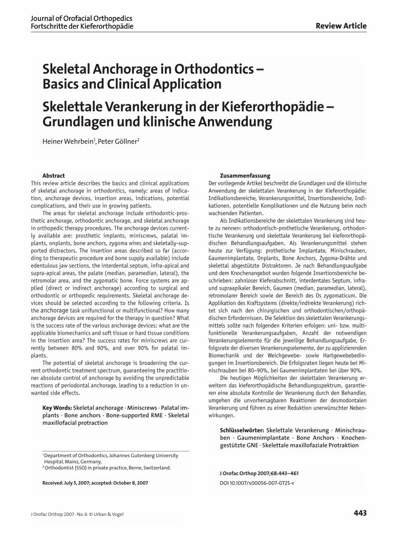
Figure 19. Palatal implant-sup-ported Rapid Maxillary Expansion (RME) for transversal expansion. No-te inflammation-free peri-implant conditions. Further tooth eruption during the retention phase (arrow), since the tooth eruption is not hin-dered by a dentally-fixed RME appli-ance.
Abbildung 19. Gaumenimplantatge-stützte schnelle maxilläre Expansion (GNE) zur transversalen Erweiterung.
Beachte die entzündungsfreien periimplantären Verhältnisse. Während der Retentionsphase erfolgt der weitere Zahndurchbruch (Pfeil), da der Zahndurch-bruch nicht durch eine dental fixierte GNE-Appararur behindert wird.
a b

Wehrbein H, Göllner P. Skelettale Verankerung
457J Orofac Orthop 2007 · No. 6 © Urban & Vogel
was used, buccal tilting even increased to 11.68°. The teeth tip more than does the alveolar process when dentally-sup-ported palatal expansion appliances are employed. The op-posite is true when using skeletally-supported palatal expan-sion appliances. Recent investigations with the Dresden dis-tractor show that the tilting of the teeth by an average of 3.5° to 7° was less than that of the alveolar process [33].
The disadvantage of the skeletally-supported palatal ex-pansion appliance, as opposed to the dentally-supported ap-pliance, is that its application requires a surgical interven-tion. Here, too, a minimally-invasive approach is mandatory, as with the other skeletal anchorage devices.
We anticipate that distractors will be applied which ful-fill this criterion. Plate-supported distractors [29, 48] (lateral palate) require more extensive soft-tissue surgical measures. On the other hand, application of an implant-supported dis-tractor (e.g. the Dresden distractor) can be considered to be minimally invasive. Figure 19 illustrates the application of implant-supported rapid maxillary expansion (RME).
Skeletally-supported Maxillary ProtractionSkeletal anchorage units have been used in animal experi-ments for maxillary protraction. Smalley et al. [58] demon-strated in an experiment with monkeys that dentally-sup-ported (dentition with splint) maxillary protraction led to tilting of the teeth toward anterior, development of vestibu-lar bony dehiscences, and a slight reaction in the temporo-
Die Vorteile der Nutzung einer skelettalen (knochenge-stützten) Verankerung für skelettale Behandlungsaufgaben liegen auf der Hand. Es werden keine Zähne in die Veran-kerung einbezogen, die Kräfte werden direkt auf das Ge-sichtsskelett übertragen und eine dentale Kompensation findet nicht statt.
Skelettal abgestützte schnelle maxilläre Expansion (Gaumennahterweiterung)
Als potentielle Nebenwirkungen einer dentalen Abstüt-zung bei der schnellen maxillären Expansion (GNE) wur-den beschrieben:– Kompression des Parodontalspaltes/Wurzelresorptionen /
knöcherne Fenestrationen [4, 41, 60], – kippende Bewegung der Zähne und der Alveolarfortsätze
[16, 73] und – Rezidivneigung [30, 39, 53].
Ein Problem bei der dental abgestützten Gaumennahterwei-terung ist die Bukkalkippung der Zähne während der trans-versalen Erweiterung mit anschließender Rezidivtendenz. Daher wird empfohlen, die transversale Breite überzukorri-gieren, um die Rezidivtendenz zu kompensieren [30, 39, 53]. Eine Überkorrektur kann jedoch unerwünschte parodontale Reaktionen (Rezessionen, Attachmentverlust) verstärken. Diese Nachteile können durch eine skelettal verankerte Ap-paratur reduziert oder sogar vermieden werden.
Figures 20a to 20c. Skeletally-supported maxillary protraction with a Delaire mask in an adolescent patient with maxillary hypoplasia. Two palatal implants (Orthosystem®, length: 4 or 6 mm) were inserted under local anesthesia in the anterior area of the zygomatic bone (a). A sterilized, individually-bent wire with hooks was fixed to the implants and led through the mucosa. After the soft tissue healed, the implants were loaded. The hooks provided a means upon which to hang the elastics (b and c). Figure 20b: the radiologic finding 6 months after the start of protraction.
Abbildungen 20a bis 20c. Skelettal abgestützte maxilläre Protraktion mit einer Delaire-Maske bei einem Jugendlichen mit maxillärer Hypoplasie. Zwei Gaumenimplantate (Orthosystem®, Länge: 4 bzw. 6 mm) wurden in Lokalanästhesie in den anterioren Bereich des Os zygomaticum inseriert (a). Ein steri-lisierter, individuell gebogener Draht mit Häkchen wurde an den Implantaten fixiert und durch die Mukosa geführt. Nach der Weichteilabheilung wurden die Implantate belastet. Die Häkchen dienten zum Einhängen der Gummizüge (b und c). Abbildung 20b zeigt den radiologischen Befund 6 Monate nach Beginn der Protraktion.
a b c

Wehrbein H, Göllner P. Skeletal Anchorage
458 J Orofac Orthop 2007 · No. 6 © Urban & Vogel
zygomatic suture. Skeletally-supported maxillary protrac-tion (the bilateral zygoma implants were 5 mm long) led however, to exclusively skeletal effects – distinctive sutural expansion (temporozygomatic suture) and no dental side effects. Our experiences show that this procedure is possi-ble in patients (Figures 20a to 20c). Owing to the invasive-ness of this intervention, this procedure is not employed routinely at present.
De Clerck [21] showed, impressively, how the adoles-cent maxilla can be protracted without dental side effects – with four bone anchors (two bizygomatic und two in the an-terior mandible, application of strong Class-III elastics). It remains questionable whether this procedure will gain broad acceptance in routine therapy because of its invasiveness.
ConclusionsThe possibilities of skeletal anchorage broaden our current spectrum of treatments. Anchorage is controlled by the cli-nician, is independent of patient compliance, and is not left to the incalculable reactions of the periodontal ligament. Clinically-relevant anchorage losses are practically unheard of in the presence of positional stability (osseointegration), thus making treatment results easier to predict. In some sit-uations one can work temporarily or during the entire treatment with reduced orthodontic appliances, which in turn enhances wearing comfort and reduces esthetic impair-ment.
The disadvantages or risks of skeletal anchorage are: surgical intervention when applying and removing the an-chorage element, anchorage element loss, infection around the anchorage element, and root injury with interdental ap-plication. Moreover, the patient must cover the additional expenses associated with skeletal anchorage, although these costs are, depending on findings, usually justified by the ad-vantages.
Prerequisites for high acceptance of this relatively new technology are: minimum strain on the patient, risk reduc-tion, simple use, comprehensive biomechanical possibilities in conjunction with the fewest anchorage elements, and high success rates of the one or the other method under clinical conditions.
As Hoffman [38] wrote 12 years ago in his article on skeletal anchorage using the onplant: “Even though an orthodontist´s dream of absolute anchorage control and pure orthopedic intervention has been realized, we set our sights higher to consider matters beyond our horizons”.
Cambiotti et al. [16] stellten eine 6°-Bukkalkippung der oberen Molaren bei Patienten von 11 Jahren fest, die mit einer dental abgestützten forcierten GNE-Apparatur behandelt wurden. Bei Verwendung einer NiTi-Erweiterungsapparatur stieg die Bukkalkippung sogar auf 11,68° an. Bei der Anwen-dung dental abgestützter GNE-Apparaturen ist die Kippung der Zähne größer als die der Alveolarfortsätze. Diese Relati-on wird durch skelettal abgestützte GNE-Apparaturen umge-kehrt. Neuere Untersuchungen mit dem Dresden-Distraktor zeigen, dass die Kippung der Zähne um durchschnittlich 3,5°–7° geringer war als die der Alveolarfortsätze [33].
Die skelettal abgestützte GNE-Apparatur hat gegen-über der dental abgestützten den Nachteil, dass deren Appli-kation mit einem chirurgischen Eingriff verbunden ist. Auch hier ist wie bei den anderen skelettalen Verankerungsmit-teln eine Minimalinvasivität zu fordern.
Zukünftig werden voraussichtlich jene Distraktoren zur Anwendung kommen, die dieses Kriterium erfüllen. Plat-tengestützte Distraktoren (lateraler Gaumen) erfordern ausgedehntere weichteilchirurgische Maßnahmen. Hinge-gen ist die Applikation eines implantatgestützten Distrak-tors (z. B. Dresden-Distraktor) als minimalinvasiv einzustu-fen. Abbildung 19 zeigt die Anwendung einer implantatge-stützten schnellen maxillären Expansion (RME: Rapid Maxillary Expansion).
Skelettal abgestützte maxilläre ProtraktionFerner wurden skelettale Verankerungseinheiten tierexpe-rimentell zur maxillären Protraktion genutzt. Smalley et al. [58] konnten im Tierexperiment am Affen zeigen, dass eine dental abgestützte (Dentition mit Splint) maxilläre Protrak-tion zu folgenden Effekten führte: Kippung der Zähne noch anterior, Ausbildung von vestibulären knöchernen Dehis-zenzen und geringe Reaktion in der Sutura temporozygo-matica. Die skelettal abgestützte maxilläre Protraktion (bi-laterale Zygomaimplantate von 5 mm Länge) führte hinge-gen ausschließlich zu skelettalen Effekten: ausgeprägte su-turale Erweiterung (Sutura temporozygomatica) und keine dentalen Nebeneffekte. Eigene Erfahrungen beim Patienten zeigen (Abbildungen 20a bis 20c), dass dieses Verfahren beim Patienten möglich ist. Aufgrund der Invasivität des Eingriffes ist dieses Verfahren in der Routinebehandlung momentan nicht gebräuchlich.
De Clerck [21] zeigte eindrucksvoll, wie beim Jugend-lichen mit vier Bone Anchors (zwei bizygomatikal und zwei in der anterioren Mandibula, Anwendung von starken Klas-se-III-Gummizügen) die Maxilla skelettal abgestützt und ohne dentale Nebenwirkungen protrahiert werden kann. Die Frage ist, ob sich dieses Verfahren aufgrund seiner In-vasivität in der Routinebehandlung durchsetzen wird.
SchlussfolgerungenDie Möglichkeiten der skelettalen Verankerung erweitern unser heutiges Behandlungsspektrum. Die Verankerung

Wehrbein H, Göllner P. Skelettale Verankerung
459J Orofac Orthop 2007 · No. 6 © Urban & Vogel
References1. Asscherickx K, Hansen JL, Wehrbein H, Sabzevar MM. Orthodontic
anchorage implants inserted in the median palatal suture and nor-mal transverse maxillary growth in growing dogs. A biometric and radiographic study. Angle Orthod 2005;75:826–31.
2. Asscherickx K, Vannet BV, Wehrbein H, Sabzevar MM. Root repair after injury from mini-screw. Clin Oral Implants Res 2005;16:575–8.
3. Bantleon HP, Bernhard T, Crismani AG, Zachrisson BJ. Stable orth-odontic anchorage with palatal osseointegrated implants. World J Orthod 2002;3:109–6.
4. Barber AF, Sims MR. Rapid maxillary expansion and external root resorption in man. A scanning electron microscope study. Am J Or-thod 1981;79:630–52.
5. Bernhard T, Vollgruber A, Gahleitner A, et al. Alternative to the median region of the palate for placement of an orthodontic im-plant. Clin Oral Implants Res 2000;11:595–601.
6. Bernhard T, Dörtbudak O, Wehrbein H, et al. Das Gaumenimplantat. Inf Orthod Kieferorthop 2000;32:209–29.
7. Bernhard T, Freudenthaler J, Dortbudak O, et al. Short epithetic implants for orthodontic anchorage in the paramedian region of the palate. A clinical study. Clin Oral Implants Res 2001;12:624–31.
8. Björk A. Growth of the maxilla in three dimensions as revealed ra-diographically by the implant method. Br J Orthod 1977;4:53–64.
9. Block MS, Hoffman DR. A new device for absolute anchorage for or-thodontics. Am J Orthod Dentofacial Orthop 1995;107:251–8.
10. Böhm B, Fuhrmann R. Clinical application and histological examina-tion of the FAMI screw for skeletal anchorage – a pilot study. J Orofac Orthop 2006;67:175–85.
11. Bousquet F, Bousquet P, Mauran G, Parquel P. Use of an impacted post for anchorage. J Clin Orthod 1996;30:261–5.
12. Brånemark PI, Hansson B, Adell R, et al. Osseointegrated implants in the treatment of the edentulous jaw. Experience from a 10-year period. Stockholm: Almqvist and Wiksell, 1977.
13. Buser D, Nydegger T, Hirt HP, et al. Removal torque values of tita-nium implants in the maxilla of miniature pigs. A direct comparison of sandblasted and acid-etched with machined and acid-etched screw implants. Int J Oral Maxillofac Implants 1998;13:611–9.
14. Chhatwani B, Schneider B. Maximum anchorage in orthodontics with the palatal implant. J Orofac Orthop 2006;6:459–70.
15. Cheng SJ, Tseng IY, Lee JJ, Kok SH. A prospective study of the risk factors associated with failure of mini-implants used for orthodon-tic anchorage. Int J Oral Maxillofac Implants 2004;19:100–6.
16. Ciambotti C, Ngan P, Durkee M, et al. A comparison of dental and dentoalveolar changes between rapid palatal expansion and nickel-titanium palatal expansion appliances. Am J Orthod Dentofacial Orthop 2001;119:11–20.
17. Costa A, Raffini M, Melsen B. Miniscrews as orthodontic anchorage. A preliminary report. Int J Adult Orthod Orthognath Surg 1998; 13:201–9.
18. Ceekmore TD, Eklund MK. The possibility of skelettal anchorage. J Clin Orthod 1983;17:266–9.
19. Crismani AG, Bernhart T, Schwarz K, et al. Ninety percent success in palatal implants loaded 1 week after placement. A clinical evaluation by resonance frequency analysis. Clin Oral Implants Res 2006;17:445–50.
20. De Clerck H, Geerinckx V, Siciliano S. The zygoma anchorage system. J Clin Orthod 2002;36:455–9.
21. De Clerck H. Maxillofacial protraction with the zygoma anchorage system. Vortrag auf dem Symposium: Implantate in der Kieferor-thopädie – aktueller Stand der Wissenschaft und klinische Ein-satzmöglichkeiten, Düsseldorf 9. März 2007.
22. De Pauw GAM, Dermaut L, De Bruin H, Johansson C. Stability of implants as anchorage for orthopedic traction. Angle Orthod 1999;69:401.
wird vom Kliniker kontrolliert, ist complianceunabhängig und nicht den unkalkulierbaren Reaktionen des Desmo-donts überlassen. Klinisch relevante Verankerungsverluste treten bei Positionsstabilität (Osseointegration) quasi nicht auf. Dadurch wird das Behandlungsergebnis besser vorher-sagbar. In einigen Situationen kann temporär oder während der Gesamtbehandlung mit reduzierter orthodontischer Ap-paratur gearbeitet werden, wodurch der Tragekomfort ver-bessert und die ästhetische Beeinträchtigung gemildert wird.
Die Nachteile bzw. Risiken der skelettalen Veranke-rung sind chirurgische Intervention bei der Applikation so-wie bei der Entfernung des jeweiligen Verankerungsele-mentes, Verlust des Verankerungselementes, Infektion um das Verankerungselement und Wurzelverletzung bei inter-dentaler Applikation. Außerdem kommen bei der Anwen-dung der skelettalen Verankerung zusätzliche Kosten auf den Patienten zu. Diese werden allerdings zum Teil je nach Befund durch die Vorteile relativiert.
Voraussetzung für eine hohe Akzeptanz dieser relativ neuen Technologie sind minimale Belastung der Patienten, Risikominimierung, einfache Nutzung, umfassende biome-chanische Möglichkeiten mit der geringsten Anzahl von Verankerungselementen und hohe Erfolgsrate der ein oder anderen Methode unter klinischen Bedingungen.
Wie schrieb Hoffman [38] vor nun schon 12 Jahren in sei-nem Artikel zur skelettalen Verankerung mit dem Onplant: „Obwohl ein Traum des Kieferorthopäden von absoluter Ver-ankerungskontrolle und ausschließlich orthopädischer Inter-vention realisiert wurde, werden wir unsere Ziele höher ste-cken, um Dinge hinter unseren Horizonten zu betrachten.“
Wehrbein H, Göllner P. Skeletal Anchorage
460 J Orofac Orthop 2007 · No. 6 © Urban & Vogel
23. Diedrich P. Verschiedene orthodontische Verankerungssysteme. Eine kritische Betrachtung. Fortschr Kieferorthop 1993;54:156–71.
24. Erverdi N, Baysal B, Cakirer B. Intrusion of overerupted molars by zygomatic implant anchorage in an adult orthoprosthetic patient. Orthodontics 2003;1:99–105.
25 Feifel H, Wehrbein H, Jänicke S, Riediger R. Surgical experience with the Orthosystem, a new palatal implant for orthodontic anchorage. (Abstract) J Cranio-Maxillofac Surg 1998;26(Suppl. 1):90–1.
26. Freudenthaler JW, Haas R, Bantleon HP. Bicortical titanium screw for cortical orthodontic anchorage in the mandible. A preliminary report on clinical applications. Clin Oral Implants Res 2001;12:358–68.
27. Fritz U, Diedrich P, Kinzinger G, Al-Said M. The anchorage quality of mini-implants towards translatory and extrusive forces. J Orofac Or-thop 2003;64:293–304.
28. Fritz U, Ehmer A, Diedrich P. Clinical suitability of Titanium micro-screws for orthodontic anchorage – preliminary experiences. J Oro-fac Orthop 2004;65:410–8.
29. Gerlach KL, Zahl C. Transversal expansion using a palatal distractor. J Orofac Orthop 2003;64:443–9.
30. Glassman AS, Nahigian SJ, Medway JM, Aronowitz HI. Conservative surgical orthodontic adult rapid palatal expansion, sixteen cases. Am J Orthod 1984;86:207–13.
31. Glatzmaier J, Wehrbein H, Diedrich P. Biodegradable implants for orthodontic anchorage. A preliminary biomechanical study. Eur J Orthod 1996;18:465–9.
32. Haanaes HR, Stenvik A, Beyer-Olson ES, et al. The efficacy of two-stage titanium implants as orthodontic anchorage in the preprosth-odontic correction of third molars in adults. A report of three cases. Eur J Orthod 1991;13:287–96.
33. Hansen L. 3-D-Evaluation der chirurgisch unterstützten, implantat-getragenen, forcierten Gaumennahterweiterung mit dem Dresden Distraktor. Med Diss, Dresden 2007.
34. Harzer W, Schneider M, Gedrange T. Direct bone placement of the Hyrax fixation screw for surgically assisted rapid palatal expansion (SARPE). J Oral Maxillofac Surg 2006;64:1313–7.
35. Harzer W, Schneider M, Gedrange T. Rapid maxillary expansion with palatal anchorage of a hyrax screw. Pilot study with case presenta-tion. J Orofac Orthop 2004;65:419–22.
36. Hermann RJ, Currier GF, Miyake A. Mini-implant anchorage for max-illary canine retraction. A pilot study. Am J Orthod Dentofacial Or-thop 2006;130:228–35.
37. Higuchi KW, Slack JM. The use of titanium fixtures for intraoral an-chorage to facilitate orthodontic tooth movement. Int J Oral Maxil-lofac Implants 1991;6:338–44.
38. Hoffman D. Implants and orthodontics. In: Block MS, Kent JK, eds. Endosseous implants for maxillofacial reconstruction. Philadel-phia—London–Toronto–Sydney–Tokyo: Saunders, 1995:382–99.
39. Jacobs JD, Bell WH, Williams CE, Kennedy JW. Control of the trans-verse dimension with surgery and orthodontics. Am J Orthod 1980;77:248–306.
40. Kanomi R. Mini-implants for orthodontic anchorage. J Clin Orthod 1997;11:763–7.
41. Langford SR. Root resorption extreme resulting from clinical RME. Am J Orthod 1982;81:371–7.
42. Linkow LI. Implanto-orthodontics. J Clin Orthod 1970;4:685–705.43. Liou EJW, Pai BCJ, Lin JCY. Do miniscrews remain stationary under
orthodontic forces? Am J Orthod Dentofacial Orthop 2004;126:42–7.44. Ludwig B: Mini-Implantate in der Kieferorthopädie. Berlin–Chicago–
Tokio u.a.: Quintessenz, 2007.45. Majzoub Z, Finotti M, Miotti F, et al. Bone response to orthodontic
loading of endosseous implants in the rabbit calvaria: early con-tinuous distalizing forces. Eur J Orthod 1999;21:223–30.
46. Melsen B, Petersen JK, Costa A. Zygoma ligatures. An alternative form of maxillary anchorage. J Clin Orthod 1998;32:154–8.
47. Melsen B. Mini-implants. Where are we? J Clin Orthod 2005;39: 539–47.
48. Mommaerts MY. Transpalatal distraction as a method of maxillary expansion. Br J Oral Maxillofac Surg 1999;37:268–72.
49. Ödman J, Lekholm U, Jemt T, et al. Osseointegrated titanium im-plants. A new approach in orthodontic treatment. Eur J Orthod 1988;10:98–105.
50. Ödman J, Gröndahl K, Lekholm U, Thilander B. The effect of osseo-integrated implants on the dento-alveolar development. A clinical and radiological study in growing pigs. Eur J Orthod 1991;13:279–86.
51. Ödman J, Lekholm U, Jemt T, Thilander B. Osseointegrated implants as orthodontic anchorage in treatment of partially endentulous adult patients. Eur J Orthod 1994;3:198–201.
52. Park HS, Jeong SH, Kwon OW. Factors affecting the clinical success of screw implants used as orthodontic anchorage. Am J Orthod Den-tofacial Orthop 2006;130:18–25.
53. Progrell MA, Kaban LB, Vargervik K, Baumrind S. Surgically assisted maxillary rapid maxillary expansion in adults. Int J Orthod Orthog-nath Surg 1992;7:37–41.
54. Roberts WE, Smith RK, Silberman Y, et al. Osseous adaptation to continuous loading of rigid endosseous implants. Am J Orthod 1984;86:95–111.
55. Roberts WE, Helm FR, Marshal KJ, Gongloff RK. Rigid endosseous implants for orthodontic and orthopedic anchorage. Angle Orthod 1990;59:247–56.
56. Roberts WE, Marshall KJ, Mozsary PG. Rigid endosseous implant uti-lized as anchorage to protract molars and close an atrophic extrac-tion site. Angle Orthod 1990;60:135–52.
57. Shapiro PA, Kokich VG: Uses of implants in orthodontics. Dent Clin N Am 1988;32:539–50.
58. Smalley W, Shapiro PA, Hohl TH, et al. Osseointegrated titanium implants for maxillofacial protraction in monkeys. Am J Orthod 1988;94:285–95.
59. Thilander B, Ödman J, Gröndahl K, Lekholm U. Osseointegrated im-plants in adolescents. An alternative in replacing missing teeth? Eur J Orthod 1994;16:84–95.
60. Timms DJ, Moss JP. A histological investigation into the effects of rapid maxillary expansion on the teeth and their supporting tissues. Trans Eur Orthod Soc 1971;263–71.
61. Triaca A, Antonini M, Wintermantel E. Ein neues Titan-Flach-schraubenimplantat zur Verankerung am anterioren Gaumen. Inf Orthod Kieferorthop 1992;24:251–7.
62. Turley PK, Shapiro PA, Moffett BC. The loading of bioglass coated aluminium oxide implants to produce sutural expansion of the max-illary complex in pigtail (Macaca nemestrina). Arch Oral Biol 1980;25:459–69.
63. Turley PK, Kean C, Schnur J, et al. Orthodontic force application to titanium endosseous implants. Angle Orthod 1988;58:151–62.
64. Van Roeckel NB. The use of Branemark System implants for orth-odontic anchorage. Report of a case. Int J Oral Maxillofac Implants 1989;4:341–4.
65. Wehrbein H, Diedrich P. Endosseous titanium implant during and after orthodontic load. An experimental study in dog. Clin Oral Im-plants Res 1993;4:76–82.
66. Wehrbein H: Enossale Titanimplantate als orthodontische Veranke-rungselemente. Experimentelle Untersuchungen und klinische An-wendung. Fortschr Kieferorthop 1994;55:236–50.
67. Wehrbein H, Glatzmaier J, Mundwiller U, Diedrich P. The Orthosys-tem. A new implant system for orthodontic anchorage in the palate. J Orofac Orthop 1996;57:142–53.
68. Wehrbein H, Glatzmaier J, Yildirim M. Anchorage capacity of short titanium screw implants in the maxilla. An experimental study in the dog. Clin Oral Impl Res 1997;8:131–41.

Wehrbein H, Göllner P. Skelettale Verankerung
461J Orofac Orthop 2007 · No. 6 © Urban & Vogel
69. Wehrbein H, Merz BR. Aspects of use of endosseous palatal implants in orthodontic therapy. J Esth Dent 1998;6:315–24.
70. Wehrbein H, Merz BR, Hämmerle CHF, Lang PN. Bone-to-implant contact of orthodontic implants in humans subjected to horizontal loading. Clin Oral Implants Res 1998;9:348–53.
71. Wehrbein H, Merz BR, Diedrich P. Palatal bone support for orthodon-tic implant anchorage reinforcement. A clinical and radiological study. Eur J Orthod 1999;27:65–70.
72. Wehrbein H, Feifel H, Diedrich P. Palatal implant anchorage rein-forcement of posterior teeth. A prospective study. Am J Orthod Den-tofacial Orthop 1999;116:678–86.
73. Wertz RA. Skelettal and dental changes accompanying rapid mid-palatal suture opening. Am J Orthod 1970;58:41–66.
74. Wiechmann D, Meyer U, Blüchter U. Success rate of mini- and micro-implants used for orthodontic anchorage. A prospective clinical study. Clin Oral Implants Res 2007;18:263–7.
75. Wilke HJ, Claes L, Steinemann S. The influence of various implant surfaces on the interface shear strength between implant and bone. In: Heinke G, Soltesz U, Lee AJC, eds. Advances in biomaterials. Amsterdam: Elsevier, 1990.
76. Wilmes B, Rademacher C, Olthoff G, Drescher D. Parameters affect-ing primary stability of orthodontic mini-implants. J Orofac Orthop 2006;67:162–74.
Correspondence AddressUniv.- Prof. Dr. med. Dr. med. dent. Heiner WehrbeinDepartment of OrthodonticsJohannes Gutenberg University Hospital Augustusplatz 255131 MainzGermanyPhone: (+49/6131) 17-3024, Fax -5569e-mail: [email protected]