skeletal parasympathetic innervation - pluto huji ac il
TRANSCRIPT
Skeletal parasympathetic innervation communicatescentral IL-1 signals regulating bone mass accrualAlon Bajayoa, Arik Bara, Adam Denesb,c, Marilyn Bachara, Vardit Krama, Malka Attar-Namdara, Alberta Zalloned,Krisztina J. Kovácsc, Raz Yirmiyae, and Itai Baba,1
aBone Laboratory and eDepartment of Psychology, Hebrew University of Jerusalem, Jerusalem 91120, Israel; bFaculty of Life Sciences, University ofManchester, Manchester M13 9PT, United Kingdom; cLaboratory of Molecular Neuroendocrinology, Institute of Experimental Medicine, Budapest H-1450,Hungary; and dDepartment of Human Anatomy and Histology, University of Bari, Bari 70124, Italy
Edited by Hector F. DeLuca, University of Wisconsin, Madison, WI, and approved August 6, 2012 (received for review April 10, 2012)
Bone mass accrual is a major determinant of skeletal mass, governedby bone remodeling, which consists of bone resorption by osteo-clasts and bone formation by osteoblasts. Bone mass accrual isinhibited by sympathetic signaling centrally regulated throughactivation of receptors for serotonin, leptin, and ACh. However,skeletal activity of the parasympathetic nervous system (PSNS)has not been reported at the bone level. Here we report skeletalimmune-positive fibers for the PSNS marker vesicular ACh trans-porter (VAChT). Pseudorabies virus inoculated into the distalfemoral metaphysis is identifiable in the sacral intermediolateralcell column and central autonomic nucleus, demonstrating PSNSfemoral innervation originating in the spinal cord. The PSNS neu-rotransmitter ACh targets nicotinic (nAChRs), but not muscarinicreceptors in bone cells, affecting mainly osteoclasts. nAChR agonistsup-regulate osteoclast apoptosis and restrain bone resorption. Micedeficient of the α2nAChR subunit have increased bone resorptionand low bone mass. Silencing of the IL-1 receptor signaling in thecentral nervous system by brain-specific overexpression of the hu-man IL-1 receptor antagonist (hIL1raAst
+/+ mice) leads to very lowskeletal VAChT expression and ACh levels. These mice also exhibitincreased bone resorption and low bone mass. In WT but not inhIL1raAst
+/+ mice, the cholinergic ACh esterase inhibitor pyridostig-mine increases ACh levels and bone mass apparently by inhibitingbone resorption. Taken together, these results identify a previouslyunexplored key central IL-1–parasympathetic–bone axis that antag-onizes the skeletal sympathetic tone, thus potently favoring bonemass accrual.
autonomic nervous system | postnatal skeletal development
In vertebrates, bone mass is the major factor affecting skeletalstrength. Bone mass is determined by a continuous remodeling
process consisting of bone resorption by osteoclasts and boneformation by osteoblasts. Skeletal remodeling during bone massaccrual in early life favors bone formation. Age-related bone lossresults from a net increase in bone resorption (1). Bone mass isregulated by an intricate system including central cues, so farshown to be transmitted mainly via the sympathetic nervoussystem (SNS), which tonically restrains bone formation andstimulates bone resorption, thus downregulating bone mass ac-crual (2, 3). Increased sympathetic output mediates low bonemass in depression (4), whereas decreased skeletal sympatheticsignaling mediates traumatic brain injury-induced increase inbone formation (5).In most biological systems (e.g., heart, eye pupil, digestive
track, glucose metabolism, and exocrine glands) the SNS is an-tagonized or complemented by the parasympathetic nervoussystem (PSNS) (6–10). These two arms of the autonomic nervoussystem traverse distinct anatomical routes. Preganglionic cells ofthe SNS extend from the first thoracic spinal segment to the thirdlumbar segment. The cell bodies of these neurons are foundwithin the spinal cord, primarily within the intermediolateralgray matter (11). The preganglionic neurons of the PSNS thatinnervate the rostral part of the body project parasympathetic
cranial nerves originating from the brainstem. Sacral pre-ganglionic neurons, which innervate the caudal part of the body,occupy the central autonomic nuclei and the sacral extention ofthe intermediolateral column (11).The main neurotransmitter in use by the SNS is norepineph-
rine, whereas that of the PSNS is ACh, the first identified neu-rotransmitter (12). ACh is biosynthesized in presynaptic neuronsby acetylation of choline, a process catalyzed by choline acetyl-transferase (13). For release into the synaptic space, ACh ispackaged in presynaptic vesicles through the action of anotherenzyme, vesicular ACh transporter (VAChT). The concentrationof VAChT increases progressively with proximity to the nerveterminals: it is thus considered a hallmark and a selective markerof parasympathetic innervation (14, 15). ACh targets nicotinicACh receptors (nAChR), which are homopentamers consistingof α-subunits and heteropentamers comprised of α-, β-, γ-, δ-,and ε-subunits. nAChR function primarily as ion channels (16).In addition, ACh binds to and activates seven transmembranedomain muscarinic receptors (17). Another critical componentof the PSNS is ACh esterase (AChE), which is expressed bypostsynaptic cells and degrades ACh, thus limiting its action (18).It has been shown in the central nervous system that the
skeletal SNS activity is down-regulated through the activation ofmuscarinic receptors (19). In addition, expression of receptorsfor ACh have been reported in bone cells (20), which raises theprospect that the PSNS antagonizes or complements sympatheticactivity at the bone level.In health, central IL-1 signaling has been implicated in mod-
ulating sleep patterns (21), learning, and memory (22). Pre-viously, we showed that central nervous system-targeted silencingof the IL-1 receptor by brain-specific overexpression of the hu-man IL-1 receptor antagonist (hIL1raAst
+/+ mice) inhibits bonemass accrual (23). Because this activity mirrors the SNS skeletalactivity, we reasoned that the PSNS communicates central IL-1cues to the skeleton. Indeed, we show here that bone is in-nervated by the PSNS, leading to a positive tone, which up-regulates bone mass accrual. We further demonstrate that PSNSoutputs are critically regulated by central IL-1 signaling.
ResultsSkeletal Parasympathetic Innervation. Because PSNS regulationhas not been previously demonstrated at the bone level, we firstsought to characterize the cholinergic pathway innervating theskeleton. Indeed, immunohistochemical staining of trabecular
Author contributions: A. Bajayo, A. Bar, R.Y., and I.B. designed research; A. Bajayo, A. Bar,A.D., M.B., V.K., and M.A.-N. performed research; A.D., A.Z., and K.J.K. contributed newreagents/analytic tools; A. Bajayo, A. Bar, M.B., R.Y., and I.B. analyzed data; and A. Bajayo,R.Y., and I.B. wrote the paper.
The authors declare no conflict of interest.
This article is a PNAS Direct Submission.1To whom correspondence should be addressed. E-mail: [email protected].
This article contains supporting information online at www.pnas.org/lookup/suppl/doi:10.1073/pnas.1206061109/-/DCSupplemental.
www.pnas.org/cgi/doi/10.1073/pnas.1206061109 PNAS Early Edition | 1 of 6
MED
ICALSC
IENCE
S
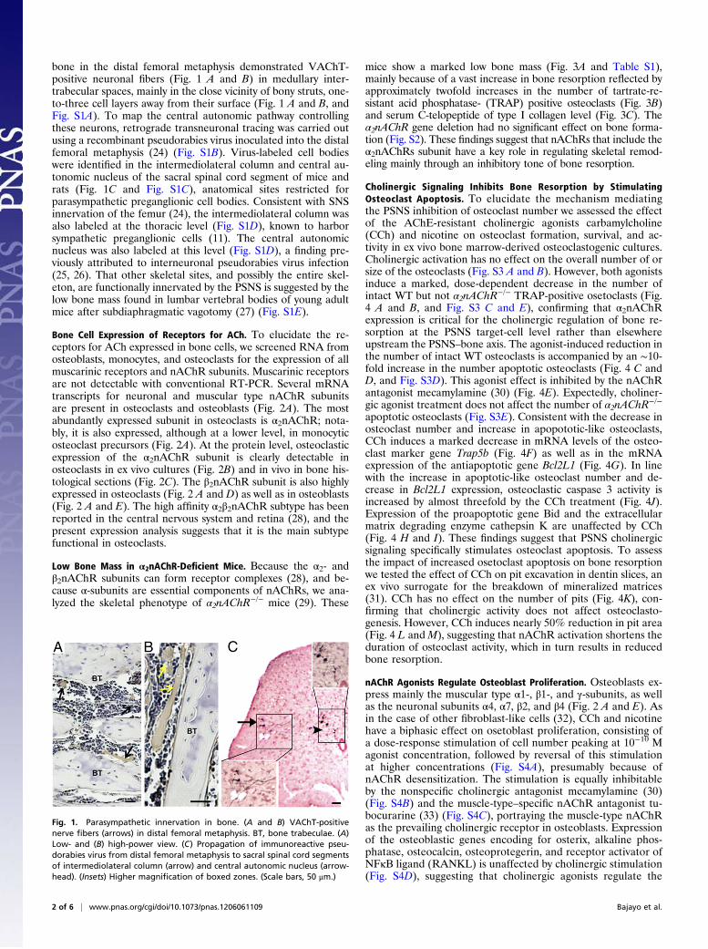
bone in the distal femoral metaphysis demonstrated VAChT-positive neuronal fibers (Fig. 1 A and B) in medullary inter-trabecular spaces, mainly in the close vicinity of bony struts, one-to-three cell layers away from their surface (Fig. 1 A and B, andFig. S1A). To map the central autonomic pathway controllingthese neurons, retrograde transneuronal tracing was carried outusing a recombinant pseudorabies virus inoculated into the distalfemoral metaphysis (24) (Fig. S1B). Virus-labeled cell bodieswere identified in the intermediolateral column and central au-tonomic nucleus of the sacral spinal cord segment of mice andrats (Fig. 1C and Fig. S1C), anatomical sites restricted forparasympathetic preganglionic cell bodies. Consistent with SNSinnervation of the femur (24), the intermediolateral column wasalso labeled at the thoracic level (Fig. S1D), known to harborsympathetic preganglionic cells (11). The central autonomicnucleus was also labeled at this level (Fig. S1D), a finding pre-viously attributed to interneuronal pseudorabies virus infection(25, 26). That other skeletal sites, and possibly the entire skel-eton, are functionally innervated by the PSNS is suggested by thelow bone mass found in lumbar vertebral bodies of young adultmice after subdiaphragmatic vagotomy (27) (Fig. S1E).
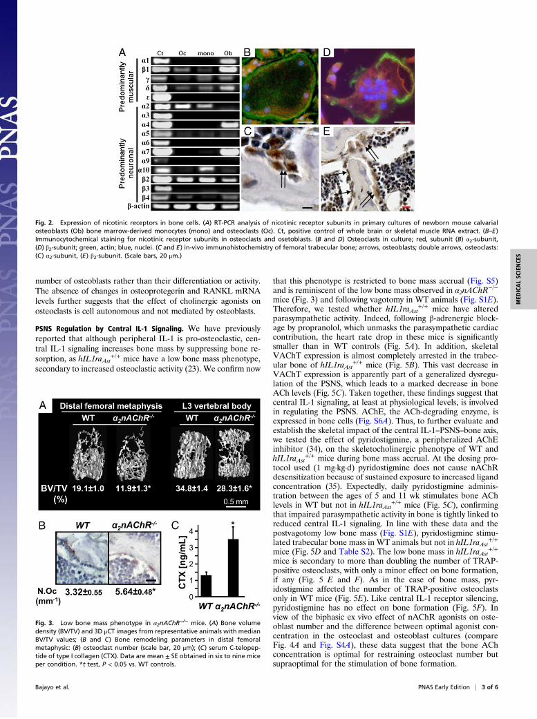
Bone Cell Expression of Receptors for ACh. To elucidate the re-ceptors for ACh expressed in bone cells, we screened RNA fromosteoblasts, monocytes, and osteoclasts for the expression of allmuscarinic receptors and nAChR subunits. Muscarinic receptorsare not detectable with conventional RT-PCR. Several mRNAtranscripts for neuronal and muscular type nAChR subunitsare present in osteoclasts and osteoblasts (Fig. 2A). The mostabundantly expressed subunit in osteoclasts is α2nAChR; nota-bly, it is also expressed, although at a lower level, in monocyticosteoclast precursors (Fig. 2A). At the protein level, osteoclasticexpression of the α2nAChR subunit is clearly detectable inosteoclasts in ex vivo cultures (Fig. 2B) and in vivo in bone his-tological sections (Fig. 2C). The β2nAChR subunit is also highlyexpressed in osteoclasts (Fig. 2 A and D) as well as in osteoblasts(Fig. 2 A and E). The high affinity α2β2nAChR subtype has beenreported in the central nervous system and retina (28), and thepresent expression analysis suggests that it is the main subtypefunctional in osteoclasts.
Low Bone Mass in α2nAChR-Deficient Mice. Because the α2- andβ2nAChR subunits can form receptor complexes (28), and be-cause α-subunits are essential components of nAChRs, we ana-lyzed the skeletal phenotype of α2nAChR
−/− mice (29). These
mice show a marked low bone mass (Fig. 3A and Table S1),mainly because of a vast increase in bone resorption reflected byapproximately twofold increases in the number of tartrate-re-sistant acid phosphatase- (TRAP) positive osteoclasts (Fig. 3B)and serum C-telopeptide of type I collagen level (Fig. 3C). Theα2nAChR gene deletion had no significant effect on bone forma-tion (Fig. S2). These findings suggest that nAChRs that include theα2nAChRs subunit have a key role in regulating skeletal remod-eling mainly through an inhibitory tone of bone resorption.
Cholinergic Signaling Inhibits Bone Resorption by StimulatingOsteoclast Apoptosis. To elucidate the mechanism mediatingthe PSNS inhibition of osteoclast number we assessed the effectof the AChE-resistant cholinergic agonists carbamylcholine(CCh) and nicotine on osteoclast formation, survival, and ac-tivity in ex vivo bone marrow-derived osteoclastogenic cultures.Cholinergic activation has no effect on the overall number of orsize of the osteoclasts (Fig. S3 A and B). However, both agonistsinduce a marked, dose-dependent decrease in the number ofintact WT but not α2nAChR
−/− TRAP-positive osetoclasts (Fig.4 A and B, and Fig. S3 C and E), confirming that α2nAChRexpression is critical for the cholinergic regulation of bone re-sorption at the PSNS target-cell level rather than elsewhereupstream the PSNS–bone axis. The agonist-induced reduction inthe number of intact WT osteoclasts is accompanied by an ∼10-fold increase in the number apoptotic osteoclasts (Fig. 4 C andD, and Fig. S3D). This agonist effect is inhibited by the nAChRantagonist mecamylamine (30) (Fig. 4E). Expectedly, choliner-gic agonist treatment does not affect the number of α2nAChR
−/−
apoptotic osteoclasts (Fig. S3E). Consistent with the decrease inosteoclast number and increase in apopototic-like osteoclasts,CCh induces a marked decrease in mRNA levels of the osteo-clast marker gene Trap5b (Fig. 4F) as well as in the mRNAexpression of the antiapoptotic gene Bcl2L1 (Fig. 4G). In linewith the increase in apoptotic-like osteoclast number and de-crease in Bcl2L1 expression, osteoclastic caspase 3 activity isincreased by almost threefold by the CCh treatment (Fig. 4J).Expression of the proapoptotic gene Bid and the extracellularmatrix degrading enzyme cathepsin K are unaffected by CCh(Fig. 4 H and I). These findings suggest that PSNS cholinergicsignaling specifically stimulates osteoclast apoptosis. To assessthe impact of increased osetoclast apoptosis on bone resorptionwe tested the effect of CCh on pit excavation in dentin slices, anex vivo surrogate for the breakdown of mineralized matrices(31). CCh has no effect on the number of pits (Fig. 4K), con-firming that cholinergic activity does not affect osteoclasto-genesis. However, CCh induces nearly 50% reduction in pit area(Fig. 4 L andM), suggesting that nAChR activation shortens theduration of osteoclast activity, which in turn results in reducedbone resorption.
nAChR Agonists Regulate Osteoblast Proliferation. Osteoblasts ex-press mainly the muscular type α1-, β1-, and γ-subunits, as wellas the neuronal subunits α4, α7, β2, and β4 (Fig. 2 A and E). Asin the case of other fibroblast-like cells (32), CCh and nicotinehave a biphasic effect on osetoblast proliferation, consisting ofa dose-response stimulation of cell number peaking at 10−10 Magonist concentration, followed by reversal of this stimulationat higher concentrations (Fig. S4A), presumably because ofnAChR desensitization. The stimulation is equally inhibitableby the nonspecific cholinergic antagonist mecamylamine (30)(Fig. S4B) and the muscle-type–specific nAChR antagonist tu-bocurarine (33) (Fig. S4C), portraying the muscle-type nAChRas the prevailing cholinergic receptor in osteoblasts. Expressionof the osteoblastic genes encoding for osterix, alkaline phos-phatase, osteocalcin, osteoprotegerin, and receptor activator ofNFκB ligand (RANKL) is unaffected by cholinergic stimulation(Fig. S4D), suggesting that cholinergic agonists regulate the
Fig. 1. Parasympathetic innervation in bone. (A and B) VAChT-positivenerve fibers (arrows) in distal femoral metaphysis. BT, bone trabeculae. (A)Low- and (B) high-power view. (C) Propagation of immunoreactive pseu-dorabies virus from distal femoral metaphysis to sacral spinal cord segmentsof intermediolateral column (arrow) and central autonomic nucleus (arrow-head). (Insets) Higher magnification of boxed zones. (Scale bars, 50 μm.)
2 of 6 | www.pnas.org/cgi/doi/10.1073/pnas.1206061109 Bajayo et al.
number of osteoblasts rather than their differentiation or activity.The absence of changes in osteoprotegerin and RANKL mRNAlevels further suggests that the effect of cholinergic agonists onosteoclasts is cell autonomous and not mediated by osteoblasts.
PSNS Regulation by Central IL-1 Signaling. We have previouslyreported that although peripheral IL-1 is pro-osteoclastic, cen-tral IL-1 signaling increases bone mass by suppressing bone re-sorption, as hIL1raAst
+/+ mice have a low bone mass phenotype,secondary to increased osteoclastic activity (23). We confirm now
that this phenotype is restricted to bone mass accrual (Fig. S5)and is reminiscent of the low bone mass observed in α2nAChR
−/−
mice (Fig. 3) and following vagotomy in WT animals (Fig. S1E).Therefore, we tested whether hIL1raAst
+/+ mice have alteredparasympathetic activity. Indeed, following β-adrenergic block-age by propranolol, which unmasks the parasympathetic cardiaccontribution, the heart rate drop in these mice is significantlysmaller than in WT controls (Fig. 5A). In addition, skeletalVAChT expression is almost completely arrested in the trabec-ular bone of hIL1raAst
+/+ mice (Fig. 5B). This vast decrease inVAChT expression is apparently part of a generalized dysregu-lation of the PSNS, which leads to a marked decrease in boneACh levels (Fig. 5C). Taken together, these findings suggest thatcentral IL-1 signaling, at least at physiological levels, is involvedin regulating the PSNS. AChE, the ACh-degrading enzyme, isexpressed in bone cells (Fig. S6A). Thus, to further evaluate andestablish the skeletal impact of the central IL-1–PSNS–bone axis,we tested the effect of pyridostigmine, a peripheralized AChEinhibitor (34), on the skeletocholinergic phenotype of WT andhIL1raAst
+/+ mice during bone mass accrual. At the dosing pro-tocol used (1 mg·kg·d) pyridostigmine does not cause nAChRdesensitization because of sustained exposure to increased ligandconcentration (35). Expectedly, daily pyridostigmine adminis-tration between the ages of 5 and 11 wk stimulates bone AChlevels in WT but not in hIL1raAst
+/+ mice (Fig. 5C), confirmingthat impaired parasympathetic activity in bone is tightly linked toreduced central IL-1 signaling. In line with these data and thepostvagotomy low bone mass (Fig. S1E), pyridostigmine stimu-lated trabecular bone mass in WT animals but not in hIL1raAst
+/+
mice (Fig. 5D and Table S2). The low bone mass in hIL1raAst+/+
mice is secondary to more than doubling the number of TRAP-positive osteoclasts, with only a minor effect on bone formation,if any (Fig. 5 E and F). As in the case of bone mass, pyr-idostigmine affected the number of TRAP-positive osteoclastsonly in WT mice (Fig. 5E). Like central IL-1 receptor silencing,pyridostigmine has no effect on bone formation (Fig. 5F). Inview of the biphasic ex vivo effect of nAChR agonists on oste-oblast number and the difference between optimal agonist con-centration in the osteoclast and osteoblast cultures (compareFig. 4A and Fig. S4A), these data suggest that the bone AChconcentration is optimal for restraining osteoclast number butsupraoptimal for the stimulation of bone formation.
Fig. 2. Expression of nicotinic receptors in bone cells. (A) RT-PCR analysis of nicotinic receptor subunits in primary cultures of newborn mouse calvarialosteoblasts (Ob) bone marrow-derived monocytes (mono) and osteoclasts (Oc). Ct, positive control of whole brain or skeletal muscle RNA extract. (B–E)Immunocytochemical staining for nicotinic receptor subunits in osteoclasts and osetoblasts. (B and D) Osteoclasts in culture; red, subunit (B) α2-subunit,(D) β2-subunit; green, actin; blue, nuclei. (C and E) in-vivo immunohistochemistry of femoral trabecular bone; arrows, osteoblasts; double arrows, osteoclasts:(C) α2-subunit, (E) β2-subunit. (Scale bars, 20 μm.)
Fig. 3. Low bone mass phenotype in α2nAChR−/− mice. (A) Bone volume
density (BV/TV) and 3D μCT images from representative animals with medianBV/TV values; (B and C) Bone remodeling parameters in distal femoralmetaphysic: (B) osteoclast number (scale bar, 20 μm); (C) serum C-telopep-tide of type I collagen (CTX). Data are mean ± SE obtained in six to nine miceper condition. *t test, P < 0.05 vs. WT controls.
Bajayo et al. PNAS Early Edition | 3 of 6
MED
ICALSC
IENCE
S

DiscussionThis study uncovers skeletal innervation by the PSNS consistingof functional cholinergic nerve fibers, production of the PSNSneurotransmitter ACh, as well as bone cell expression of AChreceptors and its rate-limiting enzyme, AChE. We provide fur-ther evidence for a previously unexplored regulation of the PSNSby central IL-1 signaling.That this IL-1–PSNS–bone axis involves the entire skeleton is
suggested by the low bone mass in both axial (vertebrae) andappendicular (femora) skeletal parts of hIL1raAst
+/+ mice andfailure of pyridostigmine and ACh to induced high bone mass inthese mice. As in the case of other caudal PSNS targets, such asthe lower gastrointestinal tract and genitalia (11), the PSNS in-nervation to the femur originates in the spinal cord, as suggestedby the retrograde tracing of pseudorabies virus from this bone tothe sacral intermediolateral column and central autonomic
nucleus. The vagus is the cranial nerve that carries PSNS in-nervation to the rostral part of the trunk (11). Therefore, the lowbone mass in the lumbar vertebrae following subdiaphragmaticvagotomy suggests that, like the heart and eye, PSNS innervationof the rostral part of the skeleton is via cranial nerves. Ourfindings in the heart further suggest that this regulation is rathergeneralized involving the skeleton as well as nonskeletal tissuesand organs.Although the SNS targets primarily osteoblasts (36), our
previous findings in hIL1raAst+/+ mice (23) and the present data
Fig. 4. nAChR activation enhances osteoclast apoptosis and inhibits min-eralized matrix resorption. (A–J) Effect of CCh in bone marrow derivedosteoclastogenic cultures. (A and B) Intact osteoclasts derived from WT(black bars) and α2nAChR
−/− (gray bars) mice. (C) WT apoptotic osteoclasts.(D) In situ images of intact and apoptotic osteoclasts in respective control andCCh-treated cultures. (Scale bar, 500 μm.) (E) Apoptotic osteoclasts treatedwith CCh and mecamylamine. (F–I) Real-time RT-PCR analysis of osteoclasticgenes in intact osteoclasts. (F) Trap5b; (G) Bcl2L1; (H) Bid; (I) Cathepsin K; (J)caspase 3 activity. pNA, p-nitroaniline. (K–M) Pit formation in osteoclastogeniccultures of intact cells grown on dentin slices. (K) Pit number, (L) average pitsize, (M) toluidine blue staining of vehicle and CCh treated dentin slices. (Scalebar, 100 μm.) Data are mean± SE obtained in five to six (A–E, K, and L) or three(F–J) culture wells per condition. *P < 0.05, vs. WT vehicle-treated controls(Veh). (A, C, E, K, and L) one-way ANOVA; (B) two-way ANOVA; (F–J) t test.
Fig. 5. Cardiac and skeletal parasympathetic signaling is regulated by centralIL-1 receptor activity. (A–F) Reduced parasympathetic tone in hIL1raAst
+/+ mice.(A) Percentage of pretreatment heart rate following propranolol administra-tion; (B) VAChT mRNA levels in distal femoral metaphysis. (C–F) Cholinergic andbone remodeling parameters in vehicle and pyridostigmine-treated mice: (C)ACh levels in distal femoral metaphysis; (D) μCT representative images of distalfemoral metaphyses (Upper), and L3 vertebral bodies (Lower) from animalswith median volume density (BV/TV) values and actual BV/TV determination. (Eand F) Analysis of bone resorption and bone formation in distal femoral met-aphysis. (E) Osteoclast number (Oc.N/BS) and representative images of TRAP-stained sections from WT (Upper) and hIL1raAst
+/+ (Lower) mice treated withvehicle (Left) or pyridostigmine (Right). (Scale bar, 80 μm.) (F) Bone formationrate (BFR). Data are mean ± SE obtained in 5–10 mice per condition. *P < 0.05vs. WT vehicle treated controls. (A and B) t test; (C and F) two-way ANOVA.
4 of 6 | www.pnas.org/cgi/doi/10.1073/pnas.1206061109 Bajayo et al.
suggest that osteoclast apoptosis is the main process affected bythe central IL-1–PSNS–bone axis. Although this and otherreports show that osteoblasts express several nAChRs subunits(37), no significant changes in bone formation were found fol-lowing central IL-1 receptor silencing or stimulation of skeletalACh levels by pyridostigmine treatment. Although low concen-trations of nAChRs agonists are mitogenic to osteoblasts, thiseffect is reversed by higher concentrations, shown to induce os-teoclast apoptosis. It thus appears that physiologically AChregulates only bone resorption and that the cholinergic regula-tion of osteoblast number becomes effective only when AChlevels are reduced in an attempt to attenuate the noradrenegicinhibition of bone formation (4) in situations such as stress,depression, and pain disorders (38).The nAChR subunit profile demonstrated here in osteoblasts
is reminiscent of previously reported expression profiles (37).Although muscarinic receptors have been recently described inhuman bone samples and in in vitro osteoblast models (39), weand others were unable to show significant levels of thesereceptors in bone cells and their conditional deletion in osteo-blasts has no skeletal effects (19). In line with our mRNA andhistochemical analyses, we show that deletion of the α2nAChRsubunit leads to a marked low bone mass during the de-velopmental phase of the skeleton, because of a vast increase inosteoclast number and bone resorption. We also show that themain βnAChR subunit is β2, thus portraying the α2β2nAChRsubtype as necessary for the cholinergic antiresorptive tone. Highlyrelevant to the central control of the PSNS is the close similaritybetween the hIL1raAst
+/+ and α2nAChR−/− skeletal phenotypes.
ACh is released from PSNS nerve endings as well as fromnonneuronal cells, thus functioning in an autocrine or paracrinemanner (40). Indeed, VAChT and choline acetyltransferasemRNAs have been reported in murine osteoblasts (41). There-fore, ACh might act in an autocrine manner to enhance cellproliferation in bone. However, the protein level of at leastVAChT is probably very low in nonneuronal bone and bone
marrow cells compared with its level in PSNS neurons inasmuchas our immunohistochemical staining for VAChT failed to showpositive cells other than neurons. In addition, the central IL-1receptor silencing resulted in almost complete absence of skeletalVAChT mRNA expression and a marked reduction in bone AChlevels, stressing the predominance of neuronal-derived ACh.The present findings add important components to the model of
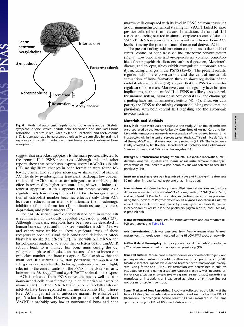
central control of bone mass via the autonomic nervous system(Fig. 6). Low bone mass and osteoporosis are common comorbid-ities of neuropsychiatric disorders, such as depression, Alzheimer’sdisease, and epilepsy, which exhibit dysregulated autonomic activ-ity, including changes in the PSNS (42–45). The present results,together with these observations and the central muscarinicstimulation of bone formation through down-regulation of theskeletal adrenergic tone (19), suggest that the PSNS is a masterregulator of bone mass. Moreover, our findings may have broaderimplications, as the identified IL-1–PSNS axis likely also controlsthe immune system, inasmuch as both central IL-1 and cholinergicsignaling have anti-inflammatory activity (46, 47). Thus, our dataportray the PSNS as the missing component linking osteo-immunephysiology with both central IL-1 signaling and the autonomicnervous system.
Materials and MethodsMice. Male mice were used throughout the study. All animal experimentswere approved by the Hebrew University Committee of Animal Care and Use.Mice with homozygous transgenic overexpression of the secreted human IL-1rain astrocytes within the central nervous system (hIL1raAst
+/+) and mice deficientof the α2nAChR subunit were reported previously (23, 29). The latter werekindly provided by Jim Boulter, Department of Psychiatry and BiobehavioralSciences, University of California, Los Angeles, CA).
Retrograde Transneuronal Tracing of Skeletal Autonomic Innervation. Pseu-dorabies virus was injected into mouse or rat distal femoral metaphyses.Progression of immunoreactive pseudorabies virus was detected as describedpreviously (24).
Heart Function. Heart rate was determined inWT and hIL1raAst+/+ before and30 min after intraperitoneal propranolol administration.
Immunohisto- and Cytochemistry. Decalcified femoral sections and culturedishes were reacted with anti-VAChT (Abcam), anti-α2nAChR (Santa Cruz),and anti-β2nAChR (Santa Cruz) antibodies. Sections were further processedusing the SuperPicture Polymer detection Kit (Zymed Laboratories). Cultureswere further reacted with anti-mouse Cy-3 conjugated antibody (ChemiconInternational), fluorescein-labeled phalloidin (Sigma-Aldrich) and DAPI (48)(Sigma-Aldrich).
mRNA Determination. Primer sets for semiquantitative and quantitative RT-PCR are reported in Table S3.
ACh Determination. ACh was extracted from freshly frozen distal femoralmetaphyses. Its levels were measured using HPLC/MS/MS spectrometry (49).
In Vivo Skeletal Phenotyping. Histomorphometry and qualitative/quantitativeμCT analyses were carried out as reported previously (23).
Bone Cell Cultures.Mouse bone marrow-derived ex vivo osteoclastogenic andprimary newborn calvarial osteoblast cultures were as reported recently (50).Nicotinic receptor ligands were added together with macrophage colony-stimulating factor and RANKL. Pit formation was determined in culturesincubated on bovine dentin slices (30). Caspase-3 activity was measured us-ing the CaspACE Assay System (Promega; catalog no. G7220) according tomanufacturer instructions and expressed as release of p-nitroaniline permicrogram of protein per hour.
Serum Markers of Bone Remodeling. Blood was collected retro-orbitally at thetime of killing. Serum osteocalcin was determined using a two-site EIA kit(Biomedical Technologies). Mouse serum CTX was measured in the samespecimens using an EIA kit (Wuhan EIAab Sciences).
Fig. 6. Model of autonomic regulation of bone mass accrual. Skeletalsympathetic tone, which inhibits bone formation and stimulates boneresorption, is centrally regulated by leptin, serotonin, and acetylcholine(19). It is antagonized by parasympathetic activity controlled by brain IL-1signaling and results in enhanced bone formation and restrained boneresorption.
Bajayo et al. PNAS Early Edition | 5 of 6
MED
ICALSC
IENCE
S

Statistical Analysis. A t test was used when two samples were compared.Multiple means were compared by ANOVA. When significant differences
were indicated by ANOVA, group means were compared using the Student
Newman–Keuls test for pair-wise comparisons.
ACKNOWLEDGMENTS. We thank Dr. Zsolt Boldogkoi for providing therecombinant pseudorabies virus and to Dr. Jim Boulter for the α2nAChR-deficientmice. This study was supported by German Israeli Foundation for Scientific Re-search and Development Grant I-843-207.11/2004 and the Israel Science Founda-tion Grant 222/10.
1. Rodan GA, Martin TJ (2000) Therapeutic approaches to bone diseases. Science 289:1508–1514.
2. Elefteriou F, et al. (2005) Leptin regulation of bone resorption by the sympatheticnervous system and CART. Nature 434:514–520.
3. Katayama Y, et al. (2006) Signals from the sympathetic nervous system regulate he-matopoietic stem cell egress from bone marrow. Cell 124:407–421.
4. Yirmiya R, et al. (2006) Depression induces bone loss through stimulation of thesympathetic nervous system. Proc Natl Acad Sci USA 103:16876–16881.
5. Tam J, et al. (2008) The cannabinoid CB1 receptor regulates bone formation bymodulating adrenergic signaling. FASEB J 22:285–294.
6. Johnson RH, Spaulding JM (1974) Disorders of the autonomic nervous system. Chapter3. The nervous control of the circulation and its investigation. Contemp Neurol Ser 11:33–58.
7. Kawasaki A (1999) Physiology, assessment, and disorders of the pupil. Curr OpinOphthalmol 10:394–400.
8. Kiba T (2004) Relationships between the autonomic nervous system and the pancreasincluding regulation of regeneration and apoptosis: Recent developments. Pancreas29:e51–e58.
9. Manabe N, Tanaka T, Hata J, Kusunoki H, Haruma K (2009) Pathophysiology un-derlying irritable bowel syndrome—From the viewpoint of dysfunction of autonomicnervous system activity. J Smooth Muscle Res 45:15–23.
10. Young JA, Van Lennep EW (1977) Morphology and physiology of salivary my-oepithelial cells. Int Rev Physiol 12:105–125.
11. Kandel ER, Schwartz JH, Jessell TM (2000) Principles of Neural Science (McGraw-Hill,New York), 4th Ed.
12. Loewi O (1921) Über humorale Übertragbarkeit der Herznervenwirkung [About hu-moral transmission of cardiac nerves effect]. Pflugers Arch, 189:239–242. German.
13. Nachmansohn D, John HM (1945) On the formation of acetylcholine in the nerveaxon. Science 102:250–251.
14. Schäfer MK, Weihe E, Varoqui H, Eiden LE, Erickson JD (1994) Distribution of thevesicular acetylcholine transporter (VAChT) in the central and peripheral nervoussystems of the rat. J Mol Neurosci 5:1–26.
15. Weihe E, Tao-Cheng JH, Schäfer MK, Erickson JD, Eiden LE (1996) Visualization of thevesicular acetylcholine transporter in cholinergic nerve terminals and its targeting toa specific population of small synaptic vesicles. Proc Natl Acad Sci USA 93:3547–3552.
16. Dani JA, Bertrand D (2007) Nicotinic acetylcholine receptors and nicotinic cholinergicmechanisms of the central nervous system. Annu Rev Pharmacol Toxicol 47:699–729.
17. Caulfield MP, Birdsall NJ (1998) International Union of Pharmacology. XVII. Classifi-cation of muscarinic acetylcholine receptors. Pharmacol Rev 50:279–290.
18. Taylor P, Radi�c Z (1994) The cholinesterases: From genes to proteins. Annu RevPharmacol Toxicol 34:281–320.
19. Shi Y, et al. (2010) Signaling through the M(3) muscarinic receptor favors bone massaccrual by decreasing sympathetic activity. Cell Metab 11:231–238.
20. En-Nosse M, et al. (2009) Expression of non-neuronal cholinergic system in osteoblast-like cells and its involvement in osteogenesis. Cell Tissue Res 338:203–215.
21. Krueger JM, et al. (1998) Sleep. A physiologic role for IL-1 beta and TNF-alpha. Ann NY Acad Sci 856:148–159.
22. Avital A, et al. (2003) Impaired interleukin-1 signaling is associated with deficits inhippocampal memory processes and neural plasticity. Hippocampus 13:826–834.
23. Bajayo A, et al. (2005) Central IL-1 receptor signaling regulates bone growth andmass. Proc Natl Acad Sci USA 102:12956–12961.
24. Dénes A, et al. (2005) Central autonomic control of the bone marrow: Multisynaptictract tracing by recombinant pseudorabies virus. Neuroscience 134:947–963.
25. Trotter RN, Stornetta RL, Guyenet PG, Roberts MR (2007) Transneuronal mapping ofthe CNS network controlling sympathetic outflow to the rat thymus. Auton Neurosci131:9–20.
26. Cano G, Sved AF, Rinaman L, Rabin BS, Card JP (2001) Characterization of the centralnervous system innervation of the rat spleen using viral transneuronal tracing. J CompNeurol 439:1–18.
27. Tien D, Ohara PT, Larson AA, Jasmin L (2003) Vagal afferents are necessary for theestablishment but not the maintenance of kainic acid-induced hyperalgesia in mice.Pain 102:39–49.
28. Marritt AM, et al. (2005) Nicotinic cholinergic receptors in the rat retina: Simple andmixed heteromeric subtypes. Mol Pharmacol 68:1656–1668.
29. Whiteaker P, et al. (2009) Pharmacological and immunochemical characterization ofalpha2* nicotinic acetylcholine receptors (nAChRs) in mouse brain. Acta PharmacolSin 30:795–804.
30. Martin BR, Onaivi ES, Martin TJ (1989) What is the nature of mecamylamine’s an-tagonism of the central effects of nicotine? Biochem Pharmacol 38:3391–3397.
31. Suda T, Jimi E, Nakamura I, Takahashi N (1997) Role of 1 alpha,25-dihydroxyvitaminD3 in osteoclast differentiation and function. Methods Enzymol 282:223–235.
32. Rothem DE, Rothem L, Soudry M, Dahan A, Eliakim R (2009) Nicotine modulates bonemetabolism-associated gene expression in osteoblast cells. J Bone Miner Metab 27:555–561.
33. Pedersen SE, Cohen JB (1990) d-Tubocurarine binding sites are located at alpha-gamma and alpha-delta subunit interfaces of the nicotinic acetylcholine receptor.Proc Natl Acad Sci USA 87:2785–2789.
34. Masson P (2011) Evolution of and perspectives on therapeutic approaches to nerveagent poisoning. Toxicol Lett 206:5–13.
35. Buisson B, Bertrand D (2002) Nicotine addiction: The possible role of functional up-regulation. Trends Pharmacol Sci 23:130–136.
36. Qin W, BaumanWA, Cardozo CP (2010) Evolving concepts in neurogenic osteoporosis.Curr Osteoporos Rep 8:212–218.
37. Romano SJ, Pugh PC, McIntosh JM, Berg DK (1997) Neuronal-type acetylcholine re-ceptors and regulation of alpha 7 gene expression in vertebrate skeletal muscle. JNeurobiol 32:69–80.
38. Maletic V, Raison CL (2009) Neurobiology of depression, fibromyalgia and neuro-pathic pain. Front Biosci 14:5291–5338.
39. Liu PS, Chen YY, Feng CK, Lin YH, Yu TC (2011) Muscarinic acetylcholine receptorspresent in human osteoblast and bone tissue. Eur J Pharmacol 650:34–40.
40. Proskocil BJ, et al. (2004) Acetylcholine is an autocrine or paracrine hormone syn-thesized and secreted by airway bronchial epithelial cells. Endocrinology 145:2498–2506.
41. Sato T, et al. (2010) Functional role of acetylcholine and the expression of cholinergicreceptors and components in osteoblasts. FEBS Lett 584:817–824.
42. Yirmiya R, Bab I (2009) Major depression is a risk factor for low bone mineral density:A meta-analysis. Biol Psychiatry 66:423–432.
43. Pack AM, Reddy DS, Duncan S, Herzog A (2011) Neuroendocrinological aspects ofepilepsy: Important issues and trends in future research. Epilepsy Behav 22:94–102.
44. Mineur YS, Picciotto MR (2010) Nicotine receptors and depression: Revisiting andrevising the cholinergic hypothesis. Trends Pharmacol Sci 31:580–586.
45. Miwa JM, Freedman R, Lester HA (2011) Neural systems governed by nicotinic ace-tylcholine receptors: Emerging hypotheses. Neuron 70:20–33.
46. Sullivan GM, et al. (1997) Intracerebroventricular injection of interleukin-1 suppressesperipheral lymphocyte function in the primate. Neuroimmunomodulation 4:12–18.
47. Rosas-Ballina M, et al. (2008) Splenic nerve is required for cholinergic antiin-flammatory pathway control of TNF in endotoxemia. Proc Natl Acad Sci USA 105:11008–11013.
48. Colucci S, Colaianni G, Mori G, Grano M, Zallone A (2002) Human osteoclasts expressoxytocin receptor. Biochem Biophys Res Commun 297:442–445.
49. Brown OM (1976) Cat heart acetylcholine: Structural proof and distribution. Am JPhysiol 231:781–785.
50. Smoum R, et al. (2010) Oleoyl serine, an endogenous N-acyl amide, modulates boneremodeling and mass. Proc Natl Acad Sci USA 107:17710–17715.
6 of 6 | www.pnas.org/cgi/doi/10.1073/pnas.1206061109 Bajayo et al.