skeletal system. bone classification a.long bones b.short bones c.flat bones d.irregular bones...
TRANSCRIPT

Skeletal System
Skeletal System
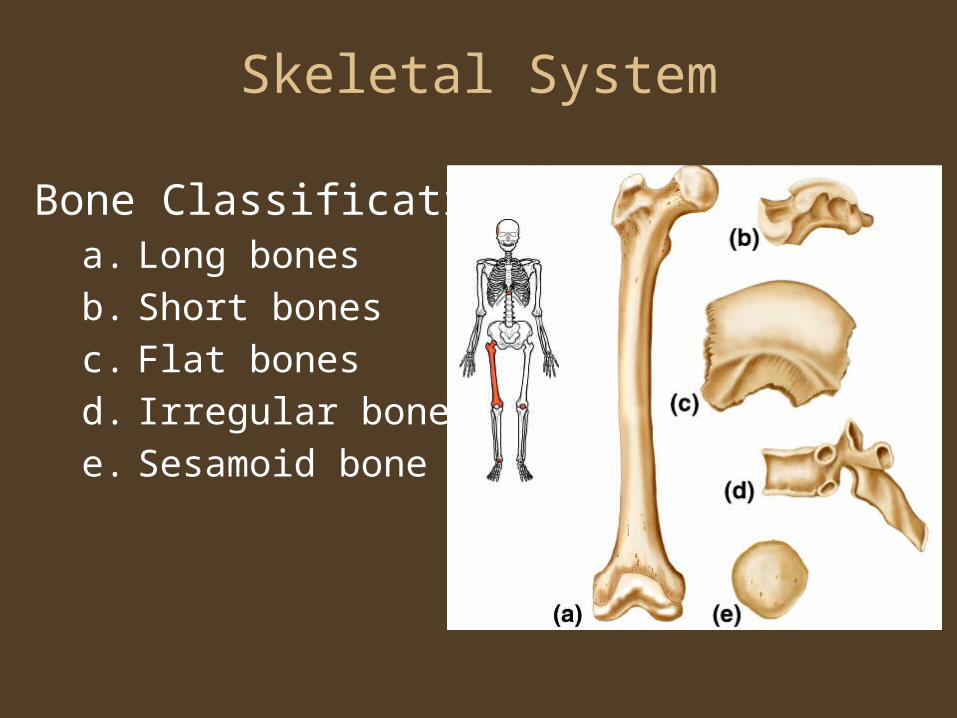
Bone Classificationa. Long bones
b. Short bones
c. Flat bones
d. Irregular bones
e. Sesamoid bone
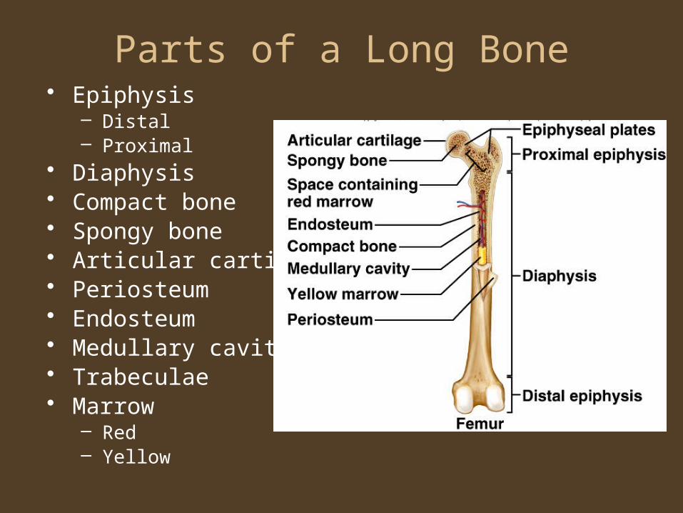
Parts of a Long Bone• Epiphysis
– Distal– Proximal
• Diaphysis• Compact bone• Spongy bone• Articular cartilage• Periosteum• Endosteum• Medullary cavity• Trabeculae• Marrow
– Red– Yellow
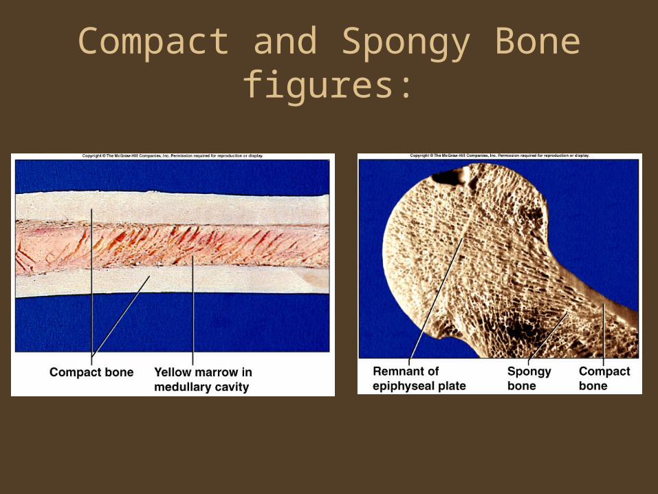
Compact and Spongy Bonefigures:

Microscopic Structureof Compact Bone
• Osteon• Central canal• Perforating canal• Osteocyte• Lacuna• Bone matrix• canaliculus

Bone Development• Intramembranous Ossification
– Bones originate within sheetlike layers of connective tissues
– Broad, flat bones– Skull bones (except mandible)– Intramembranous bones
• Endochondral Ossification– Bones begin as hyaline cartilage– Most bones of the skeleton– Endochondral bones
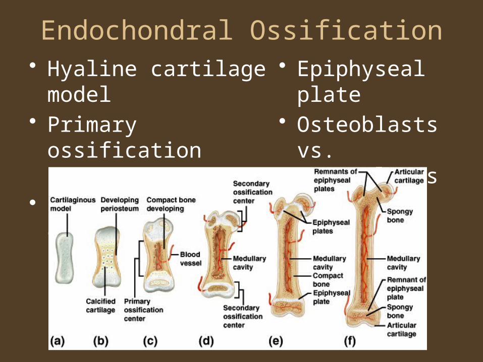
Endochondral Ossification• Hyaline cartilage model• Primary ossification
center• Secondary ossification
centers
• Epiphyseal plate
• Osteoblasts vs. Osteoclasts
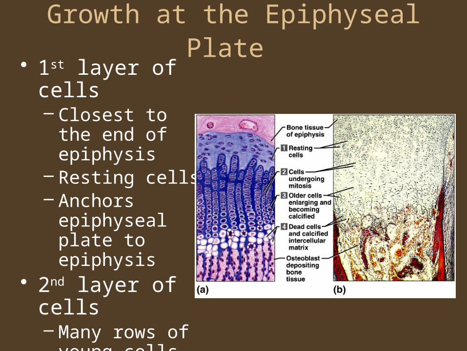
Growth at the Epiphyseal Plate • 1st layer of cells
– Closest to the end of epiphysis
– Resting cells– Anchors
epiphyseal plate to epiphysis
• 2nd layer of cells– Many rows of
young cells– Undergoing
mitosis
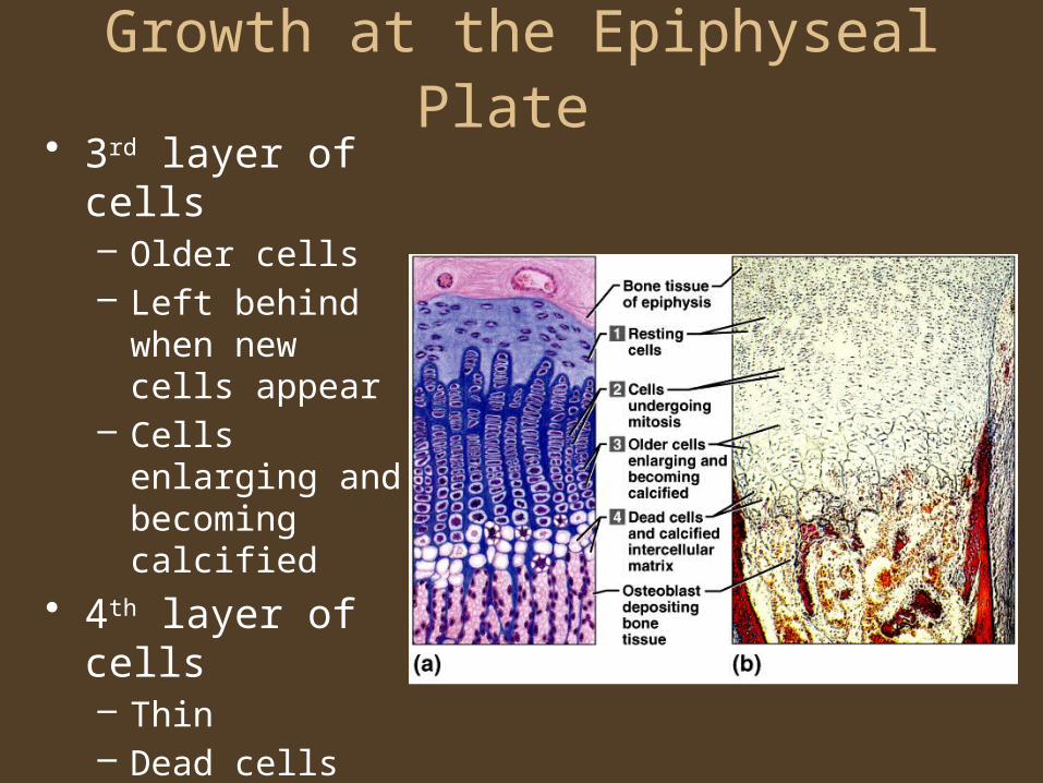
Growth at the Epiphyseal Plate • 3rd layer of cells
– Older cells– Left behind when
new cells appear– Cells enlarging
and becoming calcified
• 4th layer of cells– Thin– Dead cells– Calcified
intercellular substance

Homeostasis of Bone Tissue
• Bone Resorption – action of osteoclasts and parathyroid hormone
• Bone Deposition – action of osteoblasts and calcitonin
Factors Affecting Bone Development, Growth, and Repair
• Deficiency of Vitamin A – retards bone development
• Deficiency of Vitamin C – results in fragile bones• Deficiency of Vitamin D – rickets, osteomalacia• Insufficient Growth Hormone – dwarfism • Excessive Growth Hormone – gigantism,
acromegaly• Insufficient Thyroid Hormone – delays bone
growth• Sex Hormones – promote bone formation;
stimulate ossification of epiphyseal plates• Physical Stress – stimulates bone growth

Bone Function
• Support and Protection– Gives shape to
head, etc.– Supports body’s
weight– Protects lungs, etc.
• Body Movement– Interacts with
muscles– Bones act as rigid
bar of a lever
• Blood Cell Formation– Hematopoiesis– Red marrow
• Inorganic Salt Storage– Calcium– Phosphate– Magnesium– Sodium– potassium

Levers• Four basic components
1. Rigid bar- bones2. Fulcrum- point on which bar moves; joint3. Object moved against resistance4. Force – supplies energy for movement;
muscles

Levers and Movement
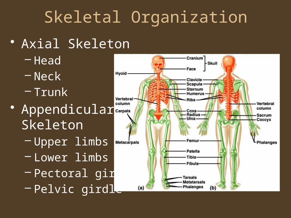
Skeletal Organization
• Axial Skeleton– Head– Neck– Trunk
• Appendicular Skeleton– Upper limbs– Lower limbs– Pectoral girdle– Pelvic girdle
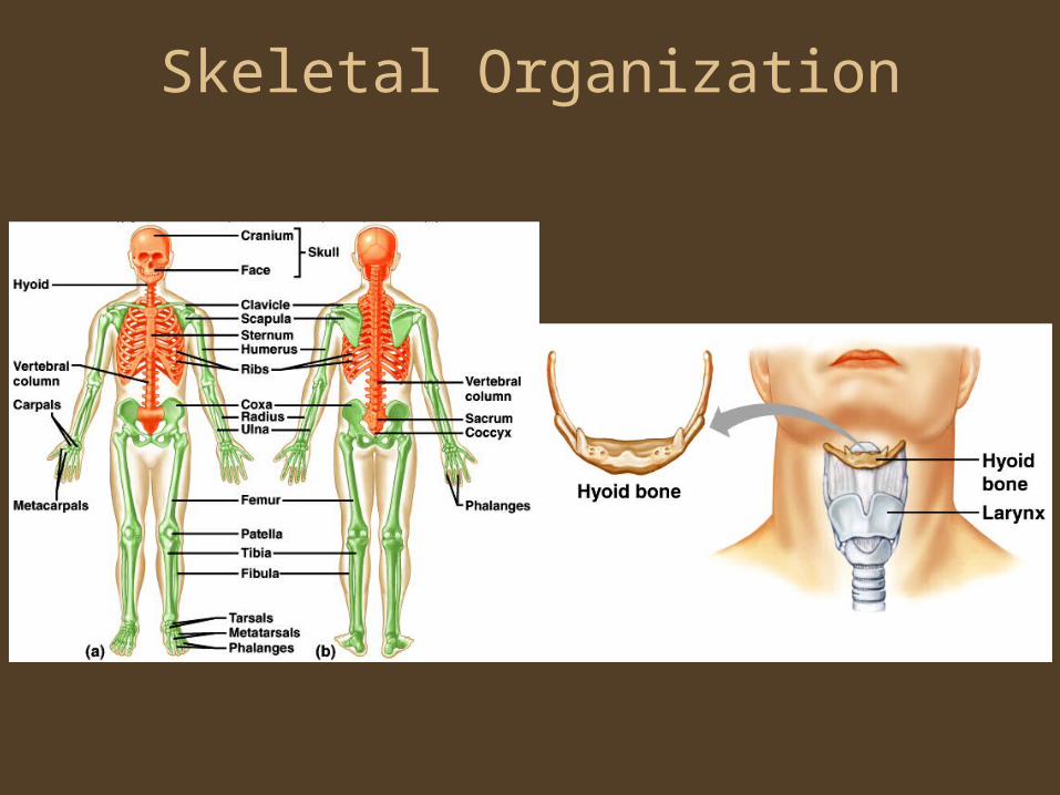
Skeletal Organization

Skull
• Frontal (1)– Forehead– Roof of nasal
cavity– Roofs of
orbits– Frontal
sinuses– Supraorbital
foramen– Coronal
suture

Skull
• Parietal (2)– Side walls
of cranium– Roof of
cranium– Sagittal
suture

Skull• Temporal (2)
– Wall of cranium– Floor of cranium– Floors and sides of
orbits– Squamosal suture– External acoustic
meatus– Mandibular fossa– Mastoid process– Styloid process– Zygomatic process
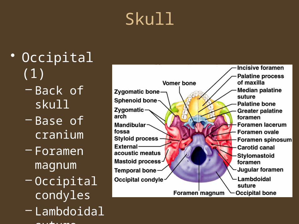
Skull
• Occipital (1)– Back of skull– Base of
cranium– Foramen
magnum– Occipital
condyles– Lambdoidal
suture
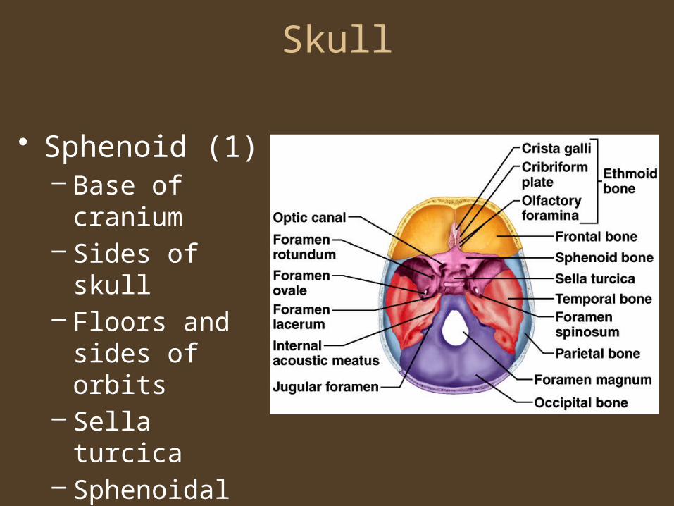
Skull
• Sphenoid (1)– Base of cranium– Sides of skull– Floors and sides
of orbits– Sella turcica– Sphenoidal
sinuses
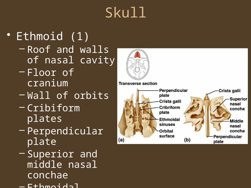
Skull
• Ethmoid (1)– Roof and walls of
nasal cavity– Floor of cranium– Wall of orbits– Cribiform plates– Perpendicular plate– Superior and middle
nasal conchae– Ethmoidal sinuses– Crista gallis

Facial Skeleton• Maxillary (2)
– Upper jaw– Anterior roof of
mouth– Floors of orbits– Sides of nasal
cavity– Floors of nasal
cavity– Alveolar
processes– Maxillary
sinuses– Palatine
process
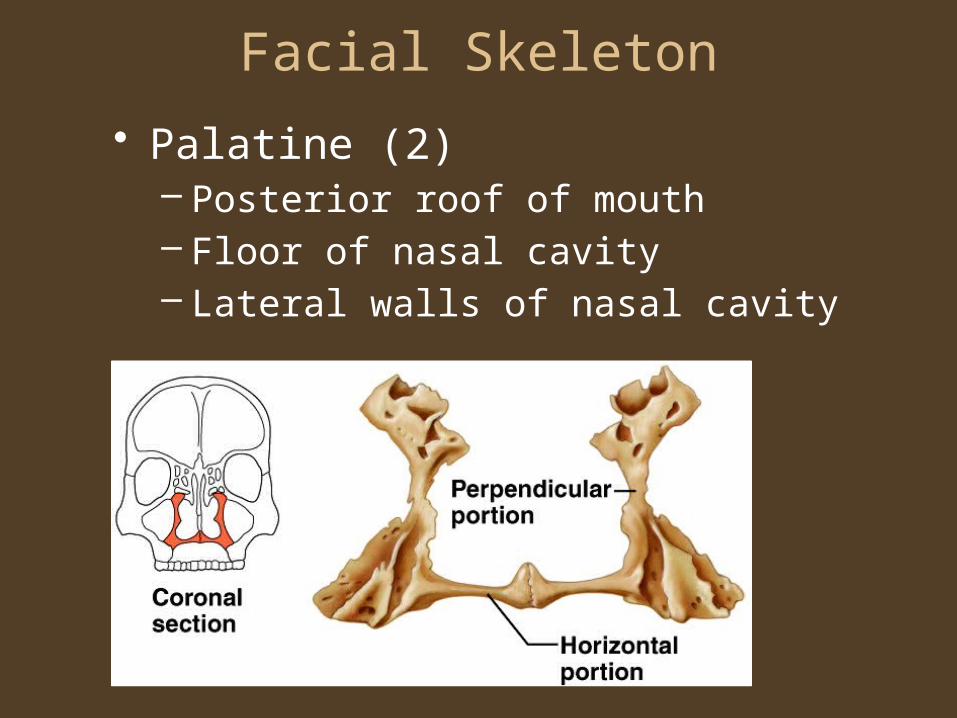
Facial Skeleton
• Palatine (2)– Posterior roof of mouth– Floor of nasal cavity– Lateral walls of nasal cavity

Facial Skeletion
• Zygomatic (2)– Prominences
of cheeks– Lateral walls
of orbits– Floors of
orbits– Temporal
process
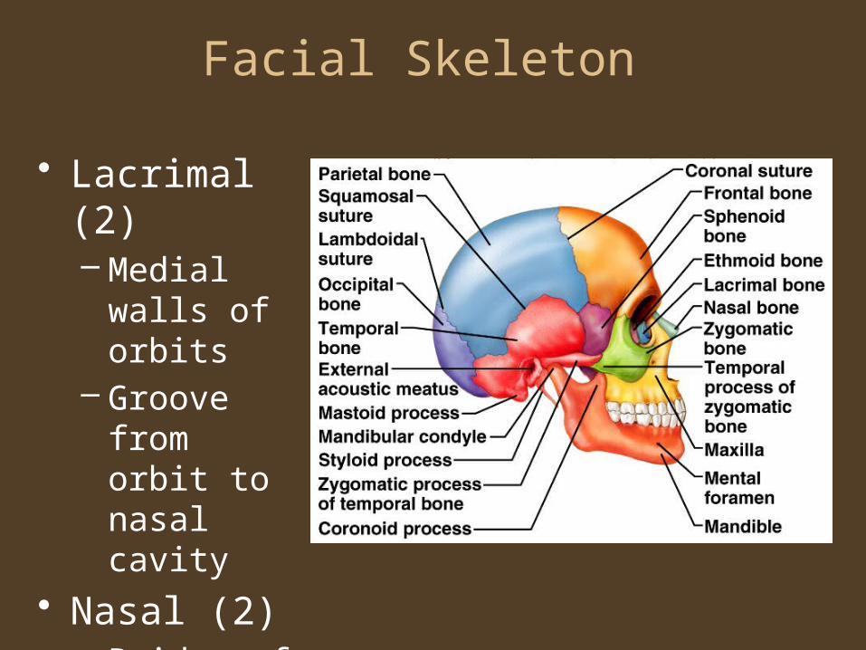
Facial Skeleton
• Lacrimal (2)– Medial walls
of orbits– Groove from
orbit to nasal cavity
• Nasal (2)– Bridge of
nose

Facial Skeleton
• Vomer (1)– Inferior
portion of nasal septum

Facial Skeleton
• Inferior Nasal Conchae (2)– Extend from
lateral walls of nasal cavity

Facial Skeleton
• Mandible (1)– Lower jaw– Body– Ramus– Mandibular
condyle– Coronoid process– Alveolar process– Mandibular
foramen– Mental foramen

Infantile Skull
• Fontanels – fibrous membranes

Vertebral Column
• Cervical vertebrae (7)
• Thoracic vertebrae (12)
• Lumbar vertebrae (5)
• Sacrum• coccyx

Vertebral Column
• Cervical curvature• Thoracic
curvature• Lumbar curvature• Pelvic curvature• Rib facets• Vertebra
prominens• Intervertebral
discs• Intervertebral
foramina

Cervical Vertebrae• Atlas – 1st; supports head• Axis – 2nd; dens pivots to turn head• Transverse foramina• Bifid spinous processes• Vertebral prominens – useful landmark
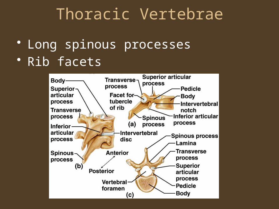
Thoracic Vertebrae
• Long spinous processes• Rib facets

Lumbar Vertebrae• Large bodies• Thick, short spinous processes

Sacrum
• Five fused bertebrae
• Median sacral crest
• Dorsal sacral foramina
• Posterior wall of pelvic cavity
• Sacral promontory
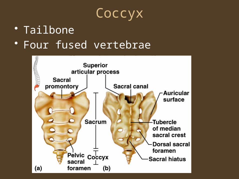
Coccyx• Tailbone• Four fused vertebrae
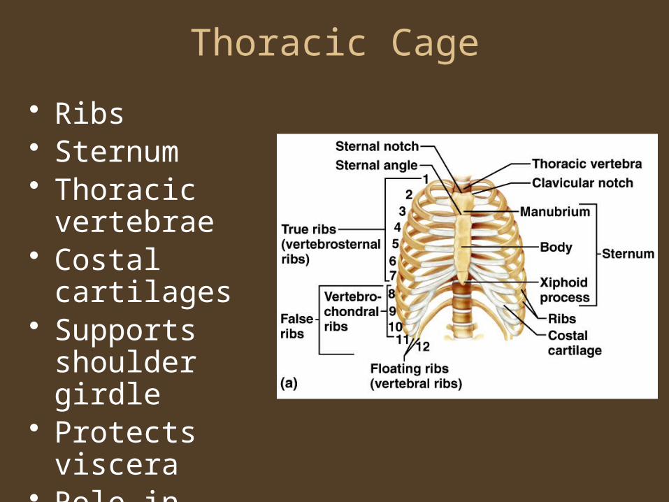
Thoracic Cage
• Ribs• Sternum• Thoracic
vertebrae• Costal cartilages• Supports
shoulder girdle• Protects viscera• Role in
breathing
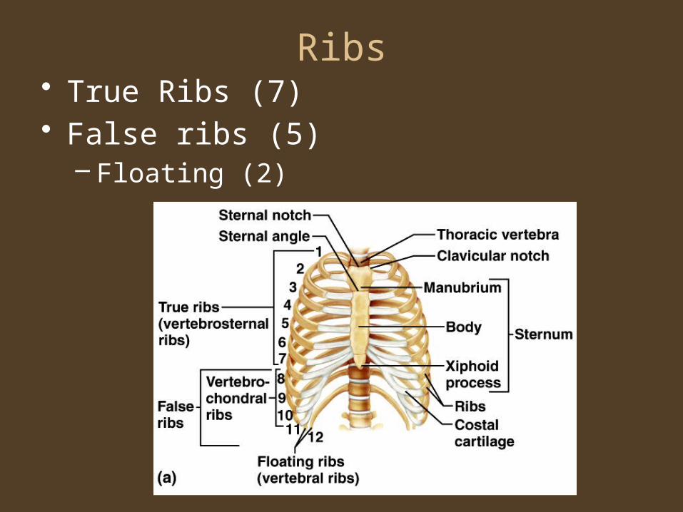
Ribs• True Ribs (7)• False ribs (5)
– Floating (2)
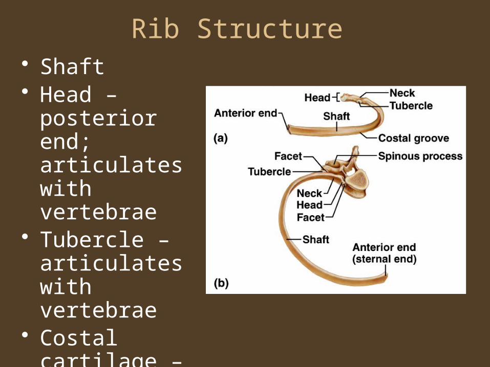
Rib Structure • Shaft• Head –
posterior end; articulates with vertebrae
• Tubercle – articulates with vertebrae
• Costal cartilage – hyaline cartilage

Sternum• Manubrium• Body• Xiphoid Process
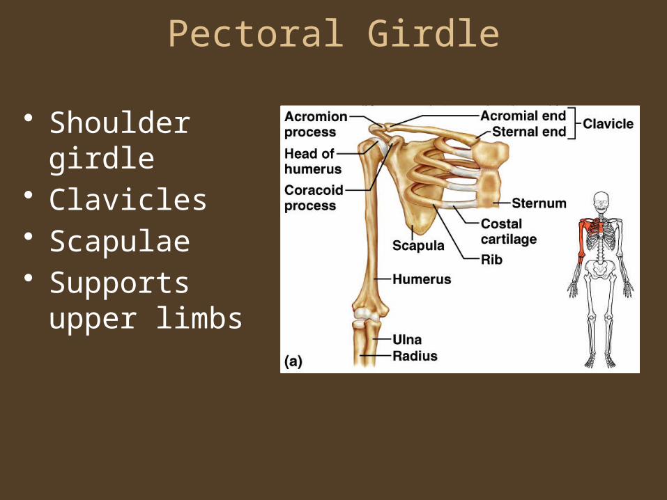
Pectoral Girdle
• Shoulder girdle• Clavicles• Scapulae• Supports upper
limbs

Clavicles• Articulate with manubrium• Articulate with scapulae (acromion process)

Scapulae• Spine• Supraspinous
fossa• Infraspinous fossa
• Acromion process• Coracoid process • Glenoid cavity
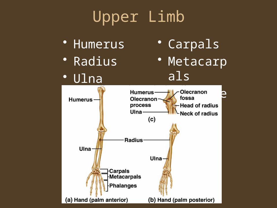
Upper Limb
• Humerus• Radius• Ulna
• Carpals• Metacarpal
s• Phalanges
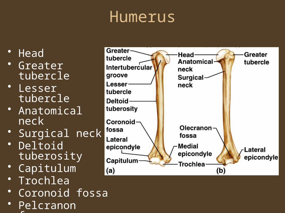
Humerus
• Head• Greater tubercle• Lesser tubercle• Anatomical neck• Surgical neck• Deltoid tuberosity• Capitulum• Trochlea• Coronoid fossa• Pelcranon fossa

Radius• Lateral forearm
bone• Head
• Radial tuberosity• Styloid process
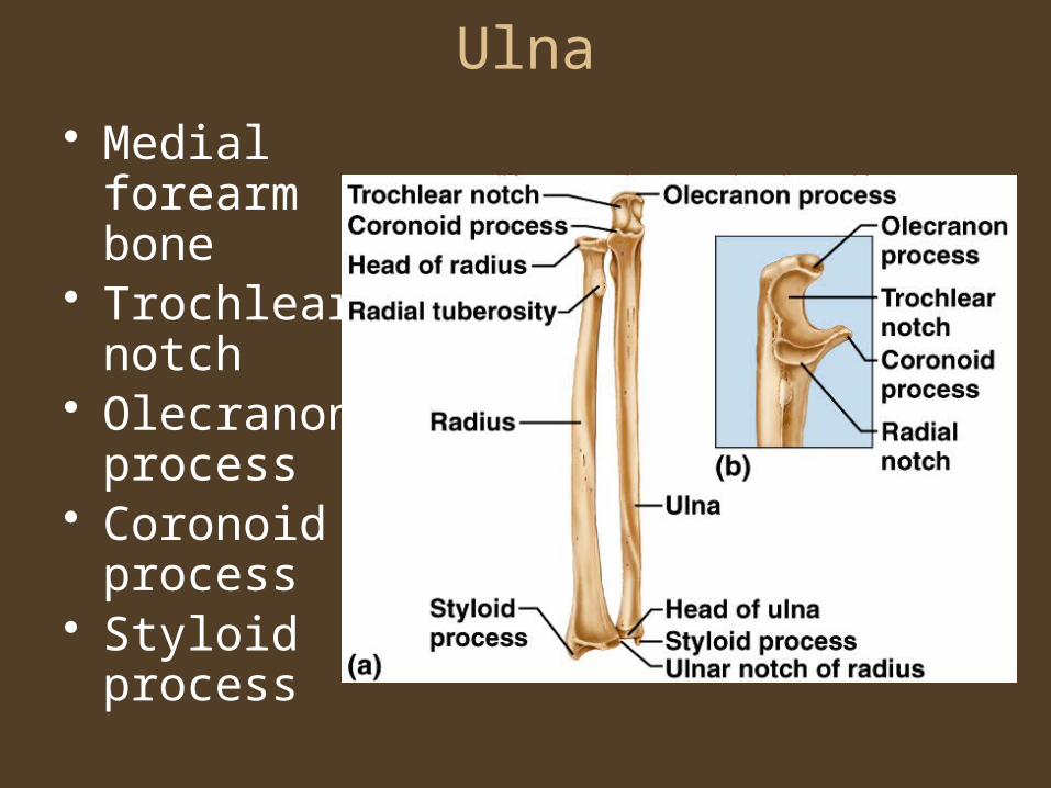
Ulna
• Medial forearm bone
• Trochlear notch
• Olecranon process
• Coronoid process
• Styloid process

Wrist and Hand• Carpals (16)
– Trapezium– Trapezoid– Capitate– Scaphoid– Pisiform– Triquetrum– Bamate– Lunate
• Metacarpals – (10)
• Phalanges (28)– Proximal phalanx– Middle phalanx– Distal phalanx