skin colour changes in ornamental koi (cyprinus
TRANSCRIPT

SKIN COLOUR CHANGES IN ORNAMENTAL KOI
(Cyprinus carpio) FED DIFFERENT DIETARY CAROTENOID SOURCES
TAN PHAIK SHIANG
UNIVERSITI SAINS MALAYSIA 2006

SKIN COLOUR CHANGES IN ORNAMENTAL KOI (Cyprinus carpio) FED DIFFERENT DIETARY
CAROTENOID SOURCES
by
TAN PHAIK SHIANG
Thesis submitted in fulfillment of the requirements for the degree
of Master of Science
March 2006

viii
TABLE OF CONTENTS PRELIMINARIES Page ACKNOWLEDGEMENTS
i
ABSTRAK ii ABSTRACT v TABLE OF CONTENTS viii LIST OF TABLES xii LIST OF FIGURES xiv LIST OF PLATES xv CHAPTER 1 INTRODUCTION 1 CHAPTER 2 LITERATURE REVIEW 4 2.1 Carotenoids 4
2.1.1 Structure of carotenoids 8
2.1.2 Carotenoid sources 13
2.1.3 Function of carotenoids 19
2.1.3.1 Biological Functions 19
2.1.3.2 Pigmentation Functions 22
2.2 Koi 27
2.2.1 History of Koi 27
2.2.2 Physiology of Koi 30
2.2.3 Koi Keeping 32
2.2.4 Kohaku – Colour and pattern 33
2.3 Chromatophores 36
2.3.1 Shapes of Chromatophores 38

ix
2.3.2 Koi Coloration 39
2.3.3 Colour enhancement 40
CHAPTER 3 METHODOLOGY 42 3.1 Scales Chromatophore analysis 42
3.2 Proximate Analysis 42
3.3 Carotenoid Sources 43
3.3.1 Astaxanthin source @ Carophyll® Pink 8% 43
3.3.2 Astaxanthin source @ NatuRoseTM 1.5% 45
3.3.3 Canthaxanthin source @ Lucanthin® Red 10% 46
3.3.4 Canthaxanthin source @ Carophyll® Red 10%
47
3.3.5 Natural Carotenoids Source @ Spirulina PacificaTM 48
3.3.6 Palm Carotenoid Source @ CarosolTM 3% 49
3.3.7 Palm Carotenoid Source @ CarominTM 10% & 20% 49
3.4 Experimental Diets 51
3.4.1 Experiment I – Efficacy of Various Dietary Carotenoid Sources for Small Koi
52
3.4.2 Experiment II – Effect of Different Dietary Levels of Carotenoid Sources for Large Koi
54
3.4.3 Experiment III – Effect of Feeding Period on Pigmentation in Koi Fed Various Dietary Carotenoid Sources
57
3.5 Fish and experimental design 59
3.5.1 Experiment I – Efficacy of Various Dietary Carotenoid Sources for Small Koi
59
3.5.2 Experiment II – Effect of Different Dietary Levels of Carotenoid Sources for Large Koi
62
3.5.3 Experiment III – Effect of Feeding Period on Pigmentation in Koi Fed Various Dietary Carotenoid Source
64
3.6 Sample Collection 67
3.6.1 Experiment I – Efficacy of Various Dietary Carotenoid
Sources for Small Koi 67

x
3.6.2 Experiment II – Effect of Different Dietary Levels of Carotenoid Sources for Large Koi
67
3.6.3 Experiment III – Effect of Feeding Period on Pigmentation in Koi Fed Various Dietary Carotenoid Sources
68
3.7 Growth Parameters and Feed Efficiency Calculation 69
3.8 Condition Index 70
3.9 Colour measurement and analysis 70
3.10 Total carotenoid analysis 73
3.11 High Performance Liquid Chromatography - HPLC system and conditions 74
3.12 Statistical Analysis 75
CHAPTER 4 RESULTS 77 4.1 Chromatophores Observation 77
4.2 Experiment I – Efficacy of Various Dietary Carotenoid Sources for Small Koi
80
4.2.1 Growth Parameters and Feed Utilization Efficiency 80
4.2.2 Condition Index 84
4.2.3 Total Carotenoids in Koi Tissues 85
4.2.4 Astaxanthin, β-Carotene and Canthaxanthin Concentrations 88
4.2.5 Chromatophores Observation 91
4.3 Experiment II – Effect of Different Dietary Levels of Carotenoid Sources for Large Koi
95
4.3.1 Growth Parameters and Feed Utilization Efficiency 95
4.3.2 Quantitative Measurement of Skin Colour Changes 98
4.3.3 Visual Observation of Skin Colour Changes 101
4.3.4 Total Carotenoids in Koi Blood and Gonads 106
4.3.5 Astaxanthin, β-Carotene and Canthaxanthin Concentrations 108
4.4 Experiment III – Effect of Feeding Period on Pigmentation in Koi Fed Various Dietary Carotenoid Sources
112

xi
4.4.1 Growth Parameters and Feed Utilization Efficiency 112
4.4.2 Visual Observation of Skin Colour Changes 113
4.4.3 Condition Index 115
4.4.4 Quantitative Measurement of Skin Colour Changes 117
4.4.5 Total Carotenoids in Koi Skin 118
4.4.6 Astaxanthin, β-Carotene and Canthaxanthin Concentrations 119
CHAPTER 5 DISCUSSION 122
5.1 Chromatophores observation and analysis 122
5.2 Effect of carotenoid on growth rate and feed utilization efficiency
123
5.3 Effect of carotenoid on pigment deposition 126
5.4 Effect of carotenoid on skin coloration 132
5.5 Effect of background colour 134
CHAPTER 6 CONCLUSION 136 BIBLIOGRAPHY 138
APPENDICES 148
APPENDIX I – Proximate Analysis 149
APPENDIX II – Formulation for Mineral and Vitamin Premix 153
APPENDIX III - Growth Parameters and Feed Utilization Efficiency 154
APPENDIX IV - Total Carotenoids in Koi Tissues
158
APPENDIX V - The standard curve for Astaxanthin, β-carotene and Canthaxanthin analysis
163
APPENDIX VI - Colour parameters (L*, a*,b*)/(L*,c*,h*) in skin (mg kg-1)
of Koi (Cyprinus carpio) fed the experimental diets1 (Experiment III)
165

xii
LIST OF TABLES
Table 3.1 Technical characteristics of Carophyll® Pink 8% 44
Table 3.2 Specifications of NatuRoseTM 1.5% 45
Table 3.3 Technical characteristics of Lucanthin® Red 10% 46
Table 3.4 Specifications of Carophyll® Red 10% 47
Table 3.5 Technical characteristics of Spirulina 48
Table 3.6 Typical chemical properties of CarosolTM 3% 50
Table 3.7 Technical specifications of CarominTM 10% & 20% 50
Table 3.8 Feed composition and proximate analysis for diets (g 100g-1 dry matter) for Experiments I
53
Table 3.9 Feed composition and proximate analysis for diets (g 100g-1 dry matter) for Experiments II
56
Table 3.10 Feed composition and proximate analysis for diets (g 100g-1 dry matter) for Experiments III
58
Table 4.1 The evaluation of growth parameters and feed utilization efficiency for koi (Cyprinus carpio) in Experiment I
83
Table 4.2 Hepatosomatic Index (H.S.I) and Haematocrit Index of koi (Cyprinus carpio) fed various carotenoid sources for 10 weeks
84
Table 4.3 Astaxanthin, β-carotene and Canthaxanthin concentration (µg g-1) in various koi (Cyprinus carpio) tissues in Experiment I
90
Table 4.4 Visual observation of the morphology of chromatophores on the scales of koi (Cyprinus carpio) fed with different diets in Experiment I
92
Table 4.5 The evaluation of growth parameters and feed utilization efficiency for koi (Cyprinus carpio) in Experiment II
97
Table 4.6 Colour parameters (L*,c*,h*) of fish skin for koi (Cyprinus carpio) fed the experimental diets in Experiment II
100
Table 4.7 Astaxanthin, β-carotene and Canthaxanthin
concentration (µg g-1) in various koi (Cyprinus carpio) tissues in Experiment II
111
Table 4.8 The evaluation of growth parameters and feed utilization efficiency for koi (Cyprinus carpio) in Experiment III
112
Table 4.9 Hepatosomatic Index (H.S.I) and Haematocrit Index of koi (Cyprinus carpio) fed various carotenoid sources for 12 weeks in Experiment III
116

xiii
Table 4.10 Colour parameters (L*, a*, b*)/ (L*, c*, h*) and Total Carotenoids (TC) in skin (mg kg-1) for Koi (Cyprinus carpio) fed the experimental diets in Experiment III
118
Table 4.11 Carotenoid content (µg g-1) in carotenoid supplemented experimental diet and experimental koi (Cyprinus carpio) in Experiment III
121
xiv
LIST OF FIGURES
Figure 2.1 Postulated pathways of carotenoid conversions for red carp type fish modified from Simpson, 1982
7
Figure 2.2 β- carotene 8
Figure 2.3 Three stereoisomers of astaxanthin 11
Figure 2.4 Canthaxanthin 12
Figure 2.5 Stages comprising the melanophore index of Hogben and Slome
38
Figure 3.1 Flow chart for the processes involved in experiment I 61
Figure 3.2 Flow chart for the processes involved in experiment II 63
Figure 3.3 Flow chart for the processes involved in experiment III 66
Figure 3.4 The framework of the CIELAB (1976) colour space diagram.
72
Figure 3.5 The colour attributes in the CIELCH space (CIELCH, 1976) showing the relationship between Lightness, Chroma and hue
73
Figure 4.1 Weight gain (%) of experimental koi fed different diet with various dietary carotenoid sources in Experiment I
82
Figure 4.2 Total carotenoids in the skin of koi (Cyprinus carpio) fed with different diets measured at 490 nm wavelength in Experiment I
85
Figure 4.3 Total carotenoids in the muscle of koi (Cyprinus carpio) fed with different diets measured at 490 nm wavelength in Experiment I
86
Figure 4.4 Total carotenoids in the liver of koi (Cyprinus carpio) fed with different diets measured at 490 nm wavelength in Experiment I
86
Figure 4.5 Comparison of Specific Growth Rate, SGR (%/day) of experimental koi (Cyprinus carpio) fed different diets in Experiment II
95
Figure 4.6 Total carotenoids in the blood serum of koi fed different diet measured at 490 nm wavelength in Experiment II
107
Figure 4.7 Total carotenoids in the gonad of koi fed different diet measured at 490 nm wavelength in Experiment II
107
Figure 4.8 Total carotenoids in the skin of koi fed different diet measured at 490 nm wavelength in Experiment III a (D1, Control) b (D2, Carophyll Pink) c (D3, Carophyll Red) d (D4, Caromin)
120

xv
LIST OF PLATES
Plate 2.1 Kohaku 33
Plate 3.1 The high power microscope used for chromatophores observation
43
Plate 3.2 Experimental diets for Experiment I 55
Plate 3.3 Experimental diets for Experiment II 55
Plate 3.4 Experimental diets for Experiment III 57
Plate 3.5 Experimental aquarium used for Experiment I 60
Plate 3.6 Experimental tanks for Experiment II 65
Plate 3.7 Experimental system for Experiment III 65
Plate 3.8 One of the Experiment II koi with arrow pointing at the colour measurement region showing R-red, O-orange and W-white
72
Plate 3.9 Light absorption or UV-visible spectrophotometer 76
Plate 3.10 High Performance Liquid Chromatography (HPLC) 76
Plate 4.1ab Chromatophores found in black scales with silver background
78
Plate 4.2ab Chromatophores found in black scales with white background
78
Plate 4.3ab Chromatophores found in black scales with orange background
79
Plate 4.4ab Chromatophores found in red scales 79
Plate 4.5ab Chromatophores found in orange scales 79
Plate 4.6 Chromatophores on the scale of koi fed Diet 1 - Control, 0ppm
93
Plate 4.7 Chromatophores on the scale of koi fed Diet 2 - Carophyll Pink, 100ppm
93
Plate 4.8 Chromatophores on the scale of koi fed Diet 3 - Lucanthin Red, 100ppm
93
Plate 4.9 Chromatophores on the scale of koi fed Diet 4 - NatuRose, 100ppm
93
Plate 4.10 Chromatophores on the scale of koi fed Diet 5 - Spirulina, 100ppm
93
Plate 4.11 Chromatophores on the scale of koi fed Diet 6 - Carosol, 100ppm
93

xvi
Plate 4.12 Chromatophores on the scale of koi fed Diet 1 - Control, 0ppm
94
Plate 4.13 Chromatophores on the scale of koi fed Diet 2 - Carophyll Pink, 100ppm
94
Plate 4.14 Chromatophores on the scale of koi fed Diet 3 - Lucanthin Red, 100ppm
94
Plate 4.15 Chromatophores on the scale of koi fed Diet 4 - NatuRose, 100ppm
94
Plate 4.16 Chromatophores on the scale of koi fed Diet 5 - Spirulina, 100ppm
94
Plate 4.17 Chromatophores on the scale of koi fed Diet 6 - Carosol, 100ppm
94
Plate 4.18 Fish T1F1 on week 0 before fed Diet 1 (Control) 102
Plate 4.19 Fish T1F1 fed Diet 1 (Control) after 12 weeks 102
Plate 4.20 Fish T1F2 on week 0 before fed Diet 1 (Control) 102
Plate 4.21 Fish T1F2 fed Diet 1 (Control) after 12 weeks 102
Plate 4.22 Fish T1F3 on week 0 before fed Diet 1 (Control) 102
Plate 4.23 Fish T1F3 fed Diet 1 (Control) after 12 weeks 102
Plate 4.24 Fish T2F2 on week 0 before fed Diet 2 (Carophyll Pink 250ppm)
103
Plate 4.25 Fish T2F2 fed Diet 2 (Carophyll Pink 250ppm) after 12 weeks
103
Plate 4.26 Fish T2F3 on week 0 before fed Diet 2 (Carophyll Pink 250ppm)
103
Plate 4.27 Fish T2F3 fed Diet 2 (Carophyll Pink 250ppm) after 12 weeks
103
Plate 4.28 Fish T3F2 on week 0 before fed Diet 3 (Carophyll Pink 500ppm)
103
Plate 4.29 Fish T3F2 fed Diet 3 (Carophyll Pink 500ppm) after 12 weeks
103
Plate 4.30 Fish T3F3 on week 0 before fed Diet 3 (Carophyll Pink 500ppm)
103
Plate 4.31 Fish T3F3 fed Diet 3 (Carophyll Pink 500ppm) after 12 weeks
103
Plate 4.32 Fish T4F1 on week 0 before fed Diet 4 (Spirulina 250ppm)
104
Plate 4.33 Fish T4F1 fed Diet 4(Spirulina 250ppm) after 12 weeks 104
Plate 4.34 Fish T4F2 on week 0 before fed Diet 4 (Spirulina 250ppm)
104

xvii
Plate 4.35 Fish T4F2 fed Diet 4(Spirulina 250ppm) after 12 weeks 104
Plate 4.36 Fish T5F1 on week 0 before fed Diet 5 (Spirulina 500ppm)
104
Plate 4.37 Fish T5F1 fed Diet 5 (Spirulina 500ppm) after 12 weeks 104
Plate 4.38 Fish T5F2 on week 0 before fed Diet 5 (Spirulina 500ppm)
104
Plate 4.39 Fish T5F2 fed Diet 5 (Spirulina 500ppm) after 12 weeks 104
Plate 4.40 Fish T5F3 on week 0 before fed Diet 5 (Spirulina 500ppm)
104
Plate 4.41 Fish T5F3 fed Diet 5 (Spirulina 500ppm) after 12 weeks 104
Plate 4.42 Fish T6F1 on week 0 before fed Diet 6 (Caromin 500ppm)
105
Plate 4.43 Fish T6F1 fed Diet 6 (Caromin 500ppm) after 12 weeks 105
Plate 4.44 Fish T6F2 on week 0 before fed Diet 6 (Caromin 500ppm)
105
Plate 4.45 Fish T6F2 fed Diet 6 (Caromin 500ppm) after 12 weeks 105
Plate 4.46 Fish T6F3 on week 0 before fed Diet 6 (Caromin 500ppm)
105
Plate 4.47 Fish T6F3 fed Diet 6 (Caromin 500ppm) after 12 weeks 105
Plate 4.48 Gonad inside koi before dissecting out 110
Plate 4.49 Comparison on koi gonad fed D1, D3, D5 and D6 110
Plate 4.50 Photo taken on pre week 114
Plate 4.51 Photo taken on week 0 114
Plate 4.52a Koi on week 0 before feed with Diet 1 (Control) 114
Plate 4.52b Koi fed with Diet 1 (Control) after week 12 114
Plate 4.53a Koi on week 0 before feed with Diet 2 (Carophyll Pink 250ppm)
114
Plate 4.53b Koi fed with Diet 2 (Carophyll Pink 250ppm) after week 12
114
Plate 4.54a Koi on week 0 before feed with Diet 3 (Carophyll Red 250ppm)
114
Plate 4.54b Koi fed with Diet 3 (Carophyll Red 250ppm) after week 12
114
Plate 4.55a Koi on week 0 before feed with Diet 4 (Caromin 250ppm)
114
Plate 4.55b Koi fed with Diet 4 (Caromin 250ppm) after week 12 114

i
ACKNOWLEDGEMENTS
First of all, I would like to extend my sincere appreciation and gratitude to
my supervisors, Associate Professor Dr. Ng Wing Keong and Professor Dr.
Boey Peng Lim. I really appreciate their invaluable guidance, patience,
kindness, understanding, encouragement and time throughout the period of my
postgraduate studies.
I would like to convey my appreciation and thanks to the School of
Biological Sciences and School of Chemistry for providing a conducive research
environment, USM for providing invaluable financial support through the
Graduate Assistance Scheme, and the Institute of Postgraduate Studies (IPS)
for providing assistance throughout the study.
My appreciation to Aquaculture Station for supplying the experimental
fishes Cyprinus carpio. My thanks also reach out to all laboratory assistants
especially Mr. Bahrim, Mr. Patchamuthu Ramasamy, Mr. Johari and Mr. Sobri
for their technical assistance and help.
Furthermore, I would like to thank all my friends especially Ms. Jillian Lim
Phaik Kin, Mr. Wang Yan, Mr. Chong Cheong Yew, Mr. Lee Kuan Shern and
Ms. Jacqueline Liew for their help, support and encouragement given to me
during my postgraduate studies. Last but not least; I would like to express my
most sincere and warmest gratitude to my family and friends for their love and
encouragement.

ii
PERUBAHAN WARNA KULIT PADA IKAN KOI HIASAN (Cyprinus carpio) YANG DIBERI MAKAN DENGAN PELBAGAI SUMBER DIET
BERKAROTENOID
ABSTRAK
Harga ikan koi meningkat sejajar dengan keamatan warna kulitnya.
Warna and pigmentasi ikan disebab oleh penyerapan dan penempatan
karotenoid. Ikan tidak berupaya untuk sintesis karotenoid sendiri, oleh itu,
karotenoid mesti dimasukkan ke dalam diet mereka.
Tiga eksperimen telah dijalankan untuk menyiasat perubahan warna kulit
ikan koi hiasan (Kohaku) dengan memberi diet yang diperkaya dengan
karotenoid daripada alga (Haematococcus pluvialis dan Spirulina) dan kelapa
sawit (dalam bentuk serbuk dan minyak). Diet yang ditambah dengan
karotenoid sintetik (astaxanthin dan kantaxanthin) dan satu diet kawalan yang
tidak ditambah sebarang karotenoid digunakan untuk perbandingan.
Dalam eksperimen pertama, ikan koi (12.89 + 0.02g) diberi diet yang
ditambah dengan sumber karotenoid sebanyak 100ppm. Eksperimen ini telah
dijalankan dengan sistem air yang statik dalam jangkamasa 10 minggu. Ikan
koi yang diberi diet karotenoid tambahan menunjukkan pewarnaan badan yang
lebih daripada ikan diet kawalan yang tiada penambahan karotenoid.
Parameter tumbesaran ikan koi dipengaruhi oleh sumber karotenoid. Ikan koi
yang diberi diet yang ditambah dengan spirulina atau Carosol masing-masing,
mendapat parameter tumbesaran yang lebih tinggi daripada ikan koi yang diberi
diet yang lain. Terdapat perbezaan yang signifikan (P< 0.05) dalam jumlah
karotenoid dan jenis karotenoid yang terdapat pada kulit, otot dan hati ikan koi.

iii
Jumlah karotenoid yang terdapat dalam kulit ikan yang diberi diet yang
ditambah dengan spirulina atau Carophyll Pink menunjukkan nilai tinggi yang
signifikan, 12.08mg kg-1 dan 11.76mg kg-1, masing-masing. Pemerhatian
morfologi kromatofor dalam sisik ikan koi yang diberi diet berlainan dijalankan
dengan mengguna mikroskop cahaya berkuasa tinggi.
Dalam eksperimen kedua, ikan koi (254.89 + 16.82g) diberi diet yang
ditambah dengan sumber karotenoid pada 250ppm atau 500ppm masing-
masing. Eksperimen ini telah dijalankan dalam sistem air statik untuk
jangkamasa 8 minggu. Parameter tumbesaran ikan koi tidak dipengaruhi oleh
penambahan karotenoid ke dalam diet mereka. Ikan koi yang diberi diet
karotenoid tambahan menunjukkan pewarnaan badan yang lebih baik daripada
ikan koi yang diberi diet kawalan. Parameter warna seperti L*, c* dan h* diukur
dengan menggunakan Minolta Colour Reader (CR-100) pada tiga bahagian
yang terdapat warna seperti putih, jingga dan merah. Berdasarkan skala warna
CIELCH (CIE, 1976), L* menunjukkan kecahayaan, C* menunjukkan Kroma
dan h* menunjukkan sudut warna. Ikan koi yang diberi diet yang ditambah
dengan 500ppm Carophyll Pink atau spirulina telah menambahkan kemerahan.
Sebahagian ikan koi yang diberi diet karotenoid tambahan 500ppm, bahagian
kepala menjadi kekuningan dan ini menujukkan bahawa karotenoid telah
melebihi batasan. Terdapat perbezaan signifikan (P< 0.05) dalam jumlah
karotenoid dan jenis karotenoid individu yang disimpan di dalam gonad dan
serum darah. Nilai jumlah karotenoid yang terdapat dalam serum darah dan
gonad ikan koi yang diberi diet 250ppm spirulina tambahan merupakan nilai
tertinggi.

iv
Dalam eksperimen ketiga, ikan koi (49.36 + 0.32g) diberi diet yang
ditambah dengan sumber karotenoid sebanyak 250ppm. Eksperimen ini telah
dijalankan dalam system air yang bergerak untuk jangkamasa selama 12
minggu. Parameter tumbesaran ikan koi tidak dipengaruhi oleh penambahan
karotenoid pada diet mereka. Tiada perbezaan signifikan (P> 0.05) pada
jumlah karotenoid dalam kulit semua ikan koi. Bagaimanapun, terdapat
perbezaan yang signifikan pada jenis karotenoid individu untuk ikan koi yang
diberi diet berlainan. Kandungan astaxantin dalam ikan koi diberi diet yang
ditambah dengan 250ppm Carophyll Pink adalah lebih tinggi daripada diet yang
ditambah dengan 250ppm Carophyll Red atau Caromin sepanjang tempoh
pemberian makanan. Parameter warna (L*, a*, b*, c* and h*) diukur pada
sebekah kiri bahagian dorsal. L* adalah kecahayaan, a* menunjukkan merah-
hijau chromaticity dan b* kuning-biru chromatociti (CIELAB, 1976). Terdapat
perbezaan yang signifikan (P<0.05) bagi nilai L* dan b* dalam kulit ikan koi.
Secara kesimpluan, diet yang ditambah dengan sebarang karotenoid boleh
mempengaruhi atau meninggikan parameter tumbesaran dan pewarnaan dalam
ikan koi. Pigmentasi ikan koi bergantung pada sumber karotenoid yang sesuai
(sintetik atau alga) dengan kepekatan yang sesuai (100-250 ppm) dan
jangkamasa pemeliharaan yang sesuai (8-12 minggu) adalah faktor yang
penting untuk pewarnaan dan parameter tumbesaran ikan koi yang lebih baik.

v
SKIN COLOUR CHANGES IN ORNAMENTAL KOI (Cyprinus carpio) FED DIFFERENT DIETARY CAROTENOID SOURCES
ABSTRACT
Koi value increases with intensity of skin colour. Fish coloration and
pigmentation is due to absorption and deposition of carotenoids. Fishes are
unable to synthesize their own carotenoids and therefore these must be
included in their diet.
Three trials were undertaken to investigate skin colour changes in
ornamental koi (Kohaku) by feeding a dietary carotenoid supplement of algal
(Haematococcus pluvialis or Spirulina) or palm carotenoid (powder and oil
form). Diets containing synthethic carotenoids (Astaxanthin or Canthaxanthin)
and a control diet with no carotenoids added were used for comparison.
In the first experiment, koi (12.89 + 0.02g) were fed diets supplemented
with carotenoid sources at a level of 100ppm. The experiment was conducted
in a static water system for a period of 10 weeks. Koi fed with diets
supplemented with carotenoids exhibited better body coloration than those fed
the control diet without added carotenoids. Growth performance of koi were
affected by the dietary carotenoids source. Koi fed diets supplemented with
spirulina or Carosol respectively, showed higher growth than koi fed other diets.
There were differences (P< 0.05) in skin total carotenoid and individual
carotenoid types in the skin, muscle and liver of koi. The total carotenoids on
the skin of koi fed with Spirulina or Carophyll Pink supplemented diets showed
significantly higher value, 12.08mg kg-1 and 11.76mg kg-1, respectively. Visual

vi
observations of the morphology of chromatophores on the scale of koi fed with
different diet were done under high power light microscope.
In the second experiment, koi (254.89 + 16.82g) were fed diets fortified
with carotenoids at a level of 250ppm or 500ppm, respectively. The experiment
was conducted in static water system for a period of 8 weeks. Growth
performances of koi were not affected by the addition of carotenoids to their
diet. Koi fed added dietary carotenoids exhibited better body coloration than koi
fed the control diet with no added carotenoids. Colour parameter such as L*, c*
and h* were measured using a Minolta Colour Reader (CR-100) at three
different colour regions (white, orange and red) on the koi body. Based on the
CIELCH colour scale (CIE, 1976), L* indicates Lightness, C* indicates chroma
and h* indicates the hue angle. Koi fed diets supplemented with 500ppm
Carophyll Pink or Spirulina increased their redness. Some of the head region of
koi fed carotenoids supplemented at 500ppm turned yellowish and this
indicated an overdose of dietary carotenoids. There were significant differences
(P< 0.05) in total carotenoid and individual carotenoid types deposited in koi
gonad and blood serum. The total carotenoids concentrations in blood serum
and gonad of the koi fed 250ppm Spirulina supplemented diet were the highest.
In the third experiment, koi (49.36 + 0.32g) were fed diets fortified with
carotenoid sources at a level of 250ppm in a water flow through system for a
period of 12 weeks. Growth performances of the koi were not affected by the
addition of carotenoids to their diet. There were no significant differences (P>
0.05) in total carotenoid on the skin of all the koi. However, there were

vii
significant differences (P< 0.05) in individual carotenoid types of koi fed different
diets. The astaxanthin content of koi fed diets supplemented with 250ppm
Carophyll Pink was significantly higher than diet supplemented with 250ppm
Carophyll Red or Caromin throughout the feeding trial. Colour parameter (L*,
a*, b*, c* and h*) were measured at the left dorsal regions. L* indicated
Lightness, a* indicates red-green chromaticity and b* indicates yellow-blue
chromaticity (CIELAB, 1976). There were significant differences (P<0.05) for L*
and b* of koi skin. In conclusion, certain dietary carotenoids may affect or
increase the growth performances and colouration in koi. Koi pigmentation
depends on proper carotenoid source (synthetic or algal) with suitable
carotenoid levels (100-250ppm) and appropriate feeding period (8-12 weeks)
which are the crucial factors for better colouration and growth performances of
koi.

1
CHAPTER 1 INTRODUCTION
The culture and export of ornamental fishes in Malaysia has expanded
rapidly in recent years. The requirement of feed has increased following the
high production of ornamental fishes. Nutrition is the most important factor in
keeping ornamental fishes healthy. In order to develop efficient and economical
feed formulas for aquaculture, basic information of nutrient requirement and
chemical composition of feed ingredients in relation to their acceptability and the
ability of fish to digest and utilise nutrients from various sources is required.
Thus, it is necessary to conduct research and development work to develop
effective diets which are of lower cost for the industry.
Differences in species, varieties, colours and patterns of ornamental
fishes play an important role in attracting home owners to keep these fishes in
their house. Ornamental fishes also derive the name as “life jewels” because of
its ornamental values. That means it must be beautiful and decorative as it
helps to beautify our living environment. In other words, coloration of
ornamental fish plays the most important role.
Coloration and hence, pigmentation plays an important role in both
ornamental and food fishes such as goldfish, koi, salmon, trout, sea bream and
prawns. Normally, the “quality” of the fish is based on the pigmentation. The
colour of fish might also bring some other meanings for certain people. For
example, red-coloured fishes such as koi and goldfish will be more attractive
2
and higher priced as the Chinese, Taiwanese and Japanese consider them
more auspicious.
Ornamental carp or koi is now available in many countries spanning
Europe, USA and the Far East. Availability of koi is maintained by local
producers in these countries, but trade of high quality koi plays a major role in
meeting a growing world-wide demand. European countries import large
quantities of koi, mainly from Israel, at an annual return of about US$ 10 million.
However, top-quality koi are imported mainly from Japan. Their price may
reach several thousand US dollar for each fish. Colours and patterns are the
major factors in making a high quality koi. Based on growing demand, it seems
that global koi production can still increase in both quantity and quality.
Koi is an economically important species of ornamental fish cultured in
Malaysia. A major challenge towards meeting the growing demand for koi is to
understand the genetic control of colour traits, but so far very few studies have
been carried out. Other ways to enhance the coloration of koi are hormone
stimulation, natural environment stimulation, feeding dietary pigment enriched
feed and injection of dyes. However, the most economical and practical way is
through feeding of pigment enriched feeds. Fishes are given special diets
containing additional pigments to trigger and enhance coloration of fishes and
this has lead to enhanced quality of the coloured ornamental koi.
The cost of each pigment and carotenoid source is different. Carophyll
Pink TM is the most effective pigment for colour enhancement tested so far. Due

3
to this reason, Carophyll Pink TM is also the most expensive among the various
commercially available pigment sources. Therefore, a lot of researches have
been carried out in food fish to replace Carophyll Pink with other cheaper
carotenoid sources but very few studies have been conducted in ornamental
fish such as koi. Even though small amounts of carotenoids are added in
commercial feeds, it is very expensive and sometimes can amount to 15% or
more of ornamental fish feed cost.
The palm-based carotenoid source is cheaper and is currently only used
in food applications. To date, no research has been done using the palm-based
carotenoid in fish diets for pigmentation purposes and this is the first study.
This study had been carried out to compare the efficacy of carotenoids
extracted from palm oil with that of established carotenoid sources currently
used in aquafeeds.
The present study has been designed to determine a proper dose-
response relationship regarding the pigmentation of koi. The aim was to benefit
feed formulators and the production of ornamental koi by improving koi colour
quality. Therefore, the objective for this study was to determine the effects of
different levels and various sources of carotenoid on the colour enhancement,
pigmentation efficiency and growth rate of Japanese koi (Cyprinus carpio L.).
4
CHAPTER 2 LITERATURE REVIEW
2.1 Carotenoids
According to the Britannica Encyclopaedia, carotenoids refer to any
group of non-nitrogenous and bio-chromes yellow, orange or red pigments that
are almost universally distributed in living organisms. Carotenoids can be
synthesized by bacteria, fungi, lower algae and green plants. They are most
conspicuous in the petals, pollen and fruit (e.g., carrots, tomatoes, sweet
potatoes and citrus fruits) of the flowering plants. Carotenoids can also be seen
in the autumn foliage of deciduous shrubs and trees. Carotenoids in the leaves
of green plants are serving as accessory pigments in photosynthesis. They trap
the solar energy and pass it to the primary photosynthetic pigment, chlorophyll.
Carotenoids play a major role in the biological colouration of animals.
Carotenoids are an important group of natural lipid soluble pigments that
are found in all families of the plant and animal kingdoms. There are about 600
different natural carotenoids that have been identified (Lorenz 1998a; Latscha,
1991). Carotenoids derive their names from the fact that they constitute the
major pigment in the carrot according to a trivial name system and an IUPAC
system (Bauernfeind & Klaui, 1981; Simpson, 1982). Carotenoids represent the
most widespread and structurally diverse pigmenting agents. Carotenoids and
their derivatives are important in animals as the basis of the visual pigments
responsible for light detection and colour discrimination (Britton, 1983).
Carotenoids are responsible for many of the brilliant yellow to red colours in

5
plants and animals, as well as the variety of bluish, greenish, purplish, brownish
and blackish colours seen in many fish and crustaceans (Latscha, 1990).
Carotenoids are structurally related to retinol and β-carotene, the main
sources of vitamin A for animals (Latscha, 1991). β-carotene can be converted
to two identical molecules of retinol and vitamin A (Boileau et al., 1999). The
beautiful yellow, orange and red colours found in the skin of ornamental fish are
the result of a group of carotenoid pigments, although colourless pteridines may
also be present (Chan et al., 1990). Fish, in the same way as other animals,
must ingest carotenoids as precursors for vitamin A, as well as depositing
pigments. Aquatic animals are unable to perform a de novo synthesis of
carotenoids in their body and therefore they must acquire these pigments from
their diet (Bagnara & Hadley, 1973; Simpson, 1982; Lorenz, 1998b; Latscha,
1991). In nature, fish gain their colour through their natural diets such as algae
because only plants and protists are able to synthesize carotenoids.
Bicyclic carotenoids like β–carotene or xanthophylls are ingested from
plants and may be converted to astaxanthin, the most commonly occurring
carotenoids in aquatic animals (Simpson et al., 1981; Goodwin, 1986;
Torrissen, 1989; Latscha, 1990). Based on this, carotenoid synthesised from
plants and algae can be added into diets for colour enhancement. Fish differ
greatly in their ability to ingest and transform various carotenoids. The
specificity of the carotenoid pattern of fish and the selectivity of carotenoids
uptake has been studied and it is known that they concentrate carotenoids in
their skin, ovary, liver, muscle, and other tissues (Bagnara & Hadley, 1973).

6
Dietary carotenoids are widely used to provide pigmentation of fish in
cases where the fish can no longer obtain it from natural feeds. Although the
usage of carotenoids in improving pigmentation is commonly adopted, the
proper type of carotenoids, the correct amounts and the feeding period are still
a research issue for different species of fishes in their various growing stage. In
intensive modern aquaculture systems, dietary carotenoids must be included as
part of the diets formulation, especially in diets for ornamental fishes.
Katayama et al. (1973) proposed that aquatic animals can be divided into three
classes based on their biosynthetic capabilities:
Group I. Red carp type: Animals that can convert lutein, zeaxanthin or
intermediates to astaxanthin, but β-carotene is not the major
precursor of astaxanthin. They can store astaxanthin in the diet
directly to their body. Goldfish, red carp and fancy red carp belongs
to this group.
Group II. Prawn type: Animals that can convert β-carotene and zeaxanthin to
astaxanthin. Generally crustaceans belong to this group.
Group III. Sea bream type: Animals that cannot convert β-carotene, lutein or
zeaxanthin to astaxanthin but can transfer pigments from diet to
their body tissue pigment, as free form or esterified. Sea bream and
red sea bream are the examples of this group.

7
Simpson (1982) reported that a number of recent studies on pigmented
carp have shown the conversion to astaxanthin of β-carotene, of lutein, of
zeaxanthin and of isocrytoptoxanthin, echinenone, and canthaxanthin (Figure
2.1). Astaxanthin is mainly responsible for the bright reddish colour of
ornamental fishes. Figure 2.1 shows the carotenoid conversions that have
been postulated for red carp. Koi is listed under this group.
������� β-Carotene
Isocryptoxanthin
Lutein
Echinenone Zeaxanthin
��������������������������������������������������������������������������������� α-Doradexanthin 4-Hydroxy-4’-keto- β-carotene
Canthaxanthin β-Doradexanthin
Phoenicoxanthin
Astaxanthin
8
Figure 2.1 Postulated pathways of carotenoid conversions for red carp type fish modified from Simpson, 1982.
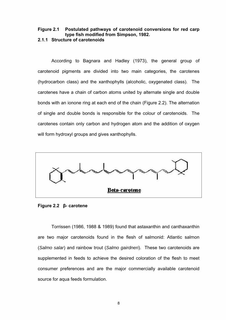
2.1.1 Structure of carotenoids
According to Bagnara and Hadley (1973), the general group of
carotenoid pigments are divided into two main categories, the carotenes
(hydrocarbon class) and the xanthophylls (alcoholic, oxygenated class). The
carotenes have a chain of carbon atoms united by alternate single and double
bonds with an ionone ring at each end of the chain (Figure 2.2). The alternation
of single and double bonds is responsible for the colour of carotenoids. The
carotenes contain only carbon and hydrogen atom and the addition of oxygen
will form hydroxyl groups and gives xanthophylls.
Figure 2.2 β- carotene
Torrissen (1986, 1988 & 1989) found that astaxanthin and canthaxanthin
are two major carotenoids found in the flesh of salmonid: Atlantic salmon
(Salmo salar) and rainbow trout (Salmo gairdneri). These two carotenoids are
supplemented in feeds to achieve the desired coloration of the flesh to meet
consumer preferences and are the major commercially available carotenoid
source for aqua feeds formulation.

9
Astaxanthin
Astaxanthin, like other carotenoids, has been shown to have biological
and nutritional functions in fish (Torrissen, 1984; Torrissen et al. 1989b;
Torrissen & Christiansen, 1995a). Astaxanthin is the carotenoid responsible for
the pink-red pigmentation of wild fishes and shrimps. Astaxanthin is the main
carotenoid pigment of red and pink coloured aquatic animals (Simpson et al.
1981; Torrissen, 1986). According to the technical report of Aquasearch (2000),
astaxanthin has chemical features that result in the existence of several forms
of astaxanthin that can be found in nature, such as stereoisomers, geometric
isomers and free or esterified forms (Figure 2.3).
Stereoisomers - Astaxanthin has two chiral or asymmetric centres. These are
the carbons numbered 3 and 3’ on the two rings in the structure. Chemists
identify chiral centres as being either R or S. R and S are from the Latin words
rectus and sinister, meaning right and left. The two chiral centres in
astaxanthin, carbons 3 and 3’ can each exist either in the R or the S form.
Thus, there are a total of three stereoisomers: 3S, 3’S; 3R, 3’S or 3R, 3’R.
Geometric isomers - Carbon-carbon double bonds can have the atoms
attached to them arranged in different ways. If the two groups are attached on
the opposite side of the double bond, they are termed E. If the two groups are
attached on the same side of the double bond, they are termed Z. E and Z are

10
from the German words entgegen and zusammen, meaning together and
opposed. Older texts may refer to Z as “cis” and E as “trans”.
Free or esterified – Astaxanthin has two hydroxyl (OH) groups, one on each
terminal ring. These can be “free” (unreacted) hydroxyls, or can react with an
acid to form an ester. If one hydroxyl reacts with a fatty acid, the result is
termed as a mono-ester. If both hydroxyl groups are reacted with fatty acids,
the result is termed as a di-ester. Adding of a fatty acid to form an ester makes
the esterified end of the molecule more hydrophobic. The hydrophobicity
(difficulty in dissolving in water) of di-esters are higher than mono-ester followed
by the “free” form.
Difference between synthetic astaxanthin and natural astaxanthin
Synthetic astaxanthin is produced as the free (unesterified) xanthophylls
and as a 1:2:1 mixture of the three stereoisomers. Astaxanthin from natural
sources tends to occur predominantly as either the 3S, 3’S; or 3R, 3’R (all – E)
isomers, while the 3R, 3’S (meso) isomer is the most abundant in synthetic
astaxanthin.
Astaxanthin is deposited in different forms, level and tissues by different
fish species. Free astaxanthin is deposited mainly in the flesh, blood serum and
other internal organs, while esterified astaxanthin predominates in the skin,
teguments and eggs of salmon (Torrisen et al., 1989b).

11
Figure 2.3 Three stereoisomers of astaxanthin.

12
Canthaxanthin
Canthaxanthin is a naturally occurring carotenoid found in nature in
tissues of various bird species, fungal species Cantharellus cinnabarinus
(chanterelle), insects, fish, crustacean, fungi, algae and many other organisms.
Canthaxanthin is widely applied as a feed additive delivering red pigmentation
(Figure 2.4). Canthaxanthin is used in poultry pigmentation to impart a red
colour to egg yolks and to broiler skin. Used in conjunction with yellow
pigments, canthaxanthin increases yolk colour intensity to meet market
demands for golden-orange yolks (Roche). In the pigmentation of salmonid
fishes, canthaxanthin is supplied in the feed in order to impart a desirable
colouration to the flesh. Addition of this pigment to grow-out salmonid feeds
ensures that flesh products attain the colour according to consumer
expectations (Storebakken et al. 1987). As well as having vitamin A activity,
canthaxanthin is a well known free-radical scavenger.
Canthaxanthin

13
Figure 2.4 Canthaxanthin 2.1.2 Carotenoid Sources
Hoffman La Roche (Basel, Switzerland) started commercial production of
synthetic canthaxanthin under the trade name “Carophyll Red” in 1964 for
colouring food and feeds. The other manufacturer, BASF group, also produced
synthetic canthaxanthin under the trade name Lucanthin® Red. Beside this
synthetic canthaxanthin, free astaxanthin also been synthesized by Hoffman La
Roche under the trade name “Carophyll Pink”. Over 6000kg of carotenoid
pigments were used in salmonid culture at a cost over $1000 U.S. per kg for
synthesized product (Torrissen et al. 1989b).
Some of the commercial aquafeed producer is currently adding Carophyll
Pink into their dry-pelleted diets (Torrissen et al. 1989b). A group of scientific
researchers did several studies on Atlantic salmon and rainbow trout using
Carophyll Pink and Carophyll Red to improve flesh pigmentation (Choubert &
Storbakken, 1989; Bjerkeng & Berge, 2000; Baker et al., 2002). Based on the
results of studies on salmonids, astaxanthin seems to be absorbed and
deposited better than canthaxanthin. (Torrissen, 1986; Storbakken et al., 1986;
Storbakken et al., 1987; Foss et al., 1987b). Hatlen (1995) also tested the
pigmentation on Arctic charr using Carophyll Pink in different concentrations.
Harpaz et al. (1998) compared the effect of three different carotenoid
sources (dried algal cells Dunaliella salina, Carophyll Pink and alfalfa meal) on

14
growth and pigmentation of crayfish and found that crayfish fed feeds fortified
with carotenoids exhibited better coloration. Growth and survival of the crayfish
were not affected by carotenoids.
According to the NatuRose Technical Bulletin published by Cyanotech
Corporation, NatuRoseTM is a natural source of astaxanthin derived from a
unique strain of the microalga, Haematococcus pluvialis grown under controlled
conditions on the island of Hawaii. The largest percentage of carotenoid
fraction of NatuRose consists of astaxanthin, with about 15% of the remaining
fraction consisting of canthaxanthin, lutein and beta-carotene (Lorenz, 2000).
This is the algae first studied by the scientist Girod Chantrans two hundred
years ago and has recently received much attention due to its capability to
synthesize and accumulates large amounts of astaxanthin during and at the end
of the growth phase.
NatuRoseTM is used as a pigmentation enhancer and as a nutrition
source for a variety of freshwater and marine species and poultry animals
(Lorenz, 2000). The astaxanthin ester composition of Haematococcus algae
meal is similar to that of crustaceans (Lambertsen and Braekken, 1971, Maoka
et al., 1985, Foss et al., 1987a). The market price of alga products of
astaxanthin is about US$300 per kg and β-carotene is about US$600 per kg
(Borowitzka, 1994).
Turujman et al. (1997) indicated that farmed salmon should be fed a diet
containing natural astaxanthin to achieve the same astaxanthin profile as their

15
wild counterparts. This study showed that farmed salmon could be easily
distinguished from the wild salmon. The farmed salmon are fed synthetic
astaxanthin, which contains primarily the 3R-3'S isomer and unable to convert it
to the more common and natural 3S-3'S form. This study also concluded that
the 3S-3'S is the main form found in wild Pacific and Atlantic salmon species
and this is the same form found in H. pluvialis.
Scientific studies of Haematococcus microalgae by research institutions
and international feed companies have concluded that this microalgae is a safe
and effective natural alternative for pigmenting aquatic animals (Lorenz, 2000).
Miki (1991) found that the more stable esterified form of astaxanthin is believed
to be an adaptive feature to be able to store astaxanthin in tissues without
excessive oxidation. Esterified astaxanthin is the main form found in H.
pluvialis. Naturose has been successfully used as a source of pigments in
aquaculture in such species as shrimp, rainbow trout, coho and Atlantic salmon.
Scientific research done by Torrissen (1989) compared two different
pigment sources (Carophyll Pink and Haematococcus astaxanthin) also
indicated that the algal astaxanthin can be used as an alternative pigment
source for Atlantic salmon (Lorenz, 2000). Choubert & Heinrich, (1993) used H.
pluvialis in comparison with synthetic astaxanthin and canthaxanthin to test
pigmentation on rainbow trout, Oncorhynchus mykiss. Their results indicated
that physical colour measurements in fish fed with algal incorporation diets
showed that increased pigmentation of the trout muscle caused an increase in
Chroma (C*) and a reduction in hue (h*) and lightness (L*). Recent studies on

16
Haematococcus indicated that properly disrupted cells have similar
bioavailability as commercially formulated astaxanthin (Barbosa et al., 1999).
NatuRose has produced demonstrable results in the pigmentation of koi
and many varieties of ornamental fish (Lorenz, 2000). Koi breeders carried out
fish trials to determine the ability of the koi to assimilate xanthophylls from
Haematococcus algae and they found that koi fed with astaxanthin from
Haematococcus algal meal had a dark red coloration, whereas the control
group that had no astaxanthin had a pale orange skin coloration (Lorenz, 2000).
Based on the unpublished data, a group of fish breeder from Aquarium Center,
Inc. of Randallstown Maryland, Reef Propagations Inc. and fish farms in Hawaii
have added or mixed NatuRose into the ornamental fish feeding diets. A
significant improvement in colour and pigmentation could be seen in freshwater
and marine ornamental fish, such as tetras, cichlids, gouramis, goldfish, koi,
danios, swordtails, Rosy Bards, rainbow fish, discus and clown anemone fish
(Lorenz, 2000).
Spirulina is a multicellular, filamentous blue-green algae belonging to the
phylum Cyanophyceae (Torrissen et al. 1989b). Spirulina can be considered as
the most concentrated natural sources of nutrition known for both terrestrial and
aquatic animals. Wagener and Rebello (1987) stated that spirulina which
contain valuable ingredients of protein required in animal feed is an excellent
substitute for animal protein in human nutrition. Animal feed grade spirulina
powder is used as a feed ingredient for pigmentation of prawns, marine fish and
ornamental fish. Spirulina have relatively expensive market prize at around $17

17
to $25 per kg depends on the quality (Henson, 1990). The commercially
produced microalgae, Spirulina platensis or S. maxima are a remarkably rich
source of amino acids, carotenoids and vitamin (Henrikson, 1989 cited in
Henson, 1990).
Henson (1990) stated that Japanese fish farmers are discovering the
potential benefits of using spirulina algae as a feed supplement. They
discovered five key benefits of using spirulina, such as better growth rates,
increased survival rates, improvement in quality and enhanced coloration of the
cultured fish while reduced medication requirements and waste in the effluent
(wastewater treatment). They found that spirulina increases feed palatability
while providing essential nutrients.
Producers of fish fry starter feeds in Japan are including spirulina in their
premium feeds. Results show that the palatability and flavour of spirulina trigger
the feeding response of the fish fingerlings therefore allowing it to get the
nutrition it needs to survive. Taiyo Fisheries has conducted a sixty days feeding
trial with yellowtail fingerlings with results showing both an increase in growth
and decrease in mortality rates with a 37% increase in harvested tons (Kato,
1988). Fish fed with spirulina have improved flavour (Hirano, 1985; Suyama,
1984), flesh texture (Henson, 1990), firm flesh and bright skin colours (Mori,
1987). Japanese growers of ornamental koi carp, mackerel, yellowtail and sea
bream observed colour improvement with spirulina supplemented diets
(Matsuno, 1979). Fish with firm texture and sharp colours can command a
higher selling price.

18
Spirulina reduces toxicity of medications and may itself have anti-viral
properties (Henson, 1990). Many medications can cause kidney damage
resulting in reduced vitality or even death. Spirulina supplemented diets reduce
pollution of the environment by increasing feed utilization due to uneaten and
undigested feed. Moreover, spirulina also help to reduce wastewater pollutants,
and finally eliminating costly treatment systems. Overall, spirulina will increase
the effectiveness of existing systems.
Boonyaratpalin and Unprasert (1989) indicated that spirulina influence
the pigmentation of red tilapia. Likewise, spirulina added in the diet of
ornamental fish such as goldfish and fancy red carp enhanced pigmentation
(Miki et al., 1986; Borowitzka, 1994), whereas Choubert (1979) did not observe
pigmentation in rainbow trout fed diets containing spirulina. Gouveia et al.
(2003) compared various microalgal biomass carotenoid sources (Chlorella
vulgaris, H. pluvialis and Spirulina maxima) with synthetic astaxanthin
(Carophyll Pink) and the found that microalgal biomass, especially C. vulgaris,
may contribute to enhanced skin colour of koi and goldfish.
CarosolTM 3% is in a spray dried water dispersible form of natural mixed
carotenes which has been extracted and purified from fruits of the oil palm tree.
CarosolTM 3% contains natural alpha-carotene, beta-carotene, gamma-
carotene, lycopene and other carotenoids. It is a unique mixture in natural
proportions and commonly found in fruits and vegetables. Concentrated
CarominTM is an opaque reddish vegetable oil suspension of natural occurring
mixture of carotenoids, extracted and concentrated from fruits of the oil palm

19
tree. CarominTM has been used to formulate colour emulsions by food colorant
manufacturers worldwide. Besides being a natural food colorant, CarominTM
has vitamin A activity and confer protection against free radical damages to the
human cells. There is a lack of scientific studies on these two carotenoid
sources in aquatic animals. As far as we know, these two carotenoid sources
have not been evaluated for use as a pigmentation agent in fish feeds.
2.1.3 Function of carotenoids
2.1.3.1 Biological Functions
Czeczuga (1979) reported that fishes with higher level of carotenoids
were more resistant to bacterial and fungal diseases than fishes with low
carotenoid levels. Torrissen (1984) showed that carotenoids supplied in the diet
increased the growth rate in Atlantic salmon. This strongly indicated that
carotenoids have a biological function. Segner et al. (1989) found an improved
liver histology and performance in fish fed high astaxanthin levels in the diet.
Other carotenoids functions include (Torrissen 1989):
1) Fertilization hormone
2) Source of pigments for chromatophores
3) Function in respiration
4) Protection from light
5) Resistance to elevated temperature and ammonia
6) Provitamin A

20
Other than pigmentation, studies indicate that carotenoids also have a
biological function involved in growth and reproduction. Several researchers
have reported positive effects on growth for different fishes fed diets
supplemented with carotenoids, especially astaxanthin (Torrisen, 1984 and
1986; Christiansen et al., 1994). The mobilization of carotenoids from the flesh
to the skin and ovaries during maturation also shows that carotenoids may have
a function in reproduction. Torrissen and Torrissen (1985) detected a decrease
in the carotenoids content of the flesh of Atlantic salmon about six months prior
to spawning and proved the mobilization of carotenoids from the flesh to the
skin and ovaries during maturation. Torrissen (1984, 1989b) showed the
presence of astaxanthin and canthaxanthin in the plasma of feeding rainbow
trout, indicating that serum is the transport medium.
Astaxanthin has also been shown to increase egg survival and
percentage of fertilized eggs, to protect eggs against extreme conditions (Craik,
1985) and to stimulate growth (Torrissen, 1984). Craik (1985) assumed that the
colour of eggs provides an indication of the quality of eggs. It has been
suggested that carotenoids may have a respiratory function because the eggs
of species that undergo development in poorly oxygenated water have more
pigment than those which develop in well-oxygenated water and large eggs
tend to have higher carotenoid concentration than small eggs. It has been
proposed that astaxanthin has a positive effect on reproduction. Carotenoid
content has also been linked to the ability of the egg to tolerate harsh
environment conditions.

21
A number of studies published by Christiansen in Norway has examined
the effects of dietary astaxanthin supplementation on fertilization and egg
survival in Atlantic salmon (Christiansen & Torrissen, 1997), growth and survival
of Atlantic salmon juveniles (Christiansen & Torrissen, 1996), and first-feeding
fry (Christiansen et al., 1995a), antioxidant status and immunity in Atlantic
salmon (Christiansen et al., 1995b) and effects of astaxanthin and vitamin A on
growth and survival during the first feeding of Atlantic salmon (Christiansen et
al., 1994). A recent ground-breaking study (Christiansen & Torrissen, 1995b;
Christiansen et al., 1996) demonstrated that Atlantic salmon fry have a definitive
growth and survival requirement for astaxanthin in their diet. Fish fed diets with
astaxanthin below 5.3ppm were found to have marginal growth; those fed levels
above 5.3ppm had significantly higher lipid levels accompanied by lower
moisture levels. When fry were fed astaxanthin concentrations below 1ppm,
survival rates plummeted. More than 50% of the fry fed diets with less than
1.0ppm astaxanthin died during the experimental period and survival of those
groups receiving higher concentrations had survival rates greater than 90%.
Thus, Atlantic salmon have the distinction as being the first salmonid species for
which astaxanthin has been shown to be an essential vitamin, with absolute
minimum levels being about 5.1ppm. Higher astaxanthin levels of 13.7ppm in
the feed continued to improve the fish lipid levels. Their results also strongly
suggested a provitamin A function for astaxanthin over the same fry-feeding
period (Christiansen et al., 1994). Furthermore the results indicated astaxanthin
as a fertilization hormone and photo protective element (Christiansen &
Torrissen, 1997). Astaxanthin also has function in respiration and stress
protection from elevated temperatures or ammonia (Christiansen et al., 1995b).
22
2.1.3.2 Pigmentation Functions
Studies have shown that there are many sources of dietary carotenoids
that can be used for the coloration of cultured fish. Carrot and hibiscus
(Shahreza, 1994), astaxanthin and canthaxanthin (Torrissen, 1986; Ito et al.,
1986; Storbakken et al., 1987; Choubert & Storbakken, 1989; Choubert &
Heinrich, 1993; Lim, 1999; Barbosa et al., 1999; Baker et al., 2002) and
microalgae (Harpaz et al., 1998; Law, 2000, Gouveia et al., 2003) can be used
in fish diets to enhance the colours of fishes.
Synthetic astaxanthin has long been used in aquaculture to enhance the
flesh colour of cultured rainbow trout and salmon (Torrissen, 1984, 1986 and
1989; Choubert & Storbakken, 1989; Choubert & Heinrich, 1993; Barbosa et al.,
1999; Baker et al., 2002). Torrissen (1989) had shown evidence pointing to the
vital role of carotenoids in the physiology and health of plants and animals. This
study observed higher growth rates in Atlantic salmon. Astaxanthin has also
been incorporated into feeds for ornamental fishes.
Ito et al. (1986) tested the effects of feeding red sea bream (Pagrus
major) different astaxanthin sources. In the group fed free astaxanthin, the
carotenoid content of the skin improved for 1 month, but reached a saturation
point and did not increase further. In the group fed astaxanthin ester, the
carotenoids in the skin was significantly higher after the first and second
sampling periods. After two months, the group fed astaxanthin esters had 1.7-
fold higher astaxanthin content in the skin than the group fed free astaxanthin

23
(13.23 mg kg-1 compared to 7.94 mg kg-1, respectively). Thus, dietary
astaxanthin esters are more efficiently utilized than free astaxanthin for
deposition and coloration of skin of red sea bream.
The findings of Smith et al. (1992) on coho salmon suggested that
feeding low astaxanthin concentrations throughout the grow-out period from fry
to market size resulted in the most efficient use of pigment and produced
uniform pigmentation. In addition, an increased dietary dose has been shown
to have no significant effect on the variation in pigmentation in Atlantic salmon
(Torrissen et al., 1995), while an increase in the duration of feeding carotenoids
has been reported to reduce inter-fish variation in pigmentation in Atlantic
salmon (Torrissen et al., 1995), rainbow trout and Chinook salmon (March and
MacMillan, 1996).
Storebakken et al. (1987) also showed similar results in the study on
pigmentation of Atlantic salmon. The retention coefficients decreased as the
pigment dose in the diet increase and this phenomenon showed that there is an
optimal level of dietary carotenoids which maximised deposit accumulation of
carotenoid in the fish. The excess carotenoid given would be a waste and it will
increase the cost of production.
Storebakken et al. (1987) also reported that astaxanthin tends to be
better deposited than canthaxanthin in Atlantic salmon. Recently, published
data showed that canthaxanthin can be equally as effective as astaxanthin, if
not better, in pigmenting Atlantic salmon (Buttle et al., 2001). Given the

24
relatively lower market price for canthaxanthin, this has attracted enormous
interest. On the other hand, canthaxanthin has been perceived as being less
“natural” than astaxanthin, primarily because astaxanthin is by far the most
abundant carotenoid in the flesh of wild Atlantic salmon (Shahidi et al., 1998).
Stability of canthaxanthin through secondary processing (freezing/smoking) has
also been of concern (Sheehen et al., 1998).
Torrissen (1989) assumed that pink to red pigmentation in flesh could be
achieved by pigmentation of astaxanthin and canthaxanthin. The study of
Choubert and Storebakken (1989) on dose response to astaxanthin and
canthaxanthin pigmentation of rainbow trout fed various dietary carotenoids
showed that astaxanthin is more efficiently utilized and the flesh pigmentation
increased by increasing the dietary carotenoids concentration in different ratio
of different pigment.
Due to lack of scientific studies, most of the information on ornamental
fish, especially koi, is unpublished data, university project reports and
testimonies from hobbyists, aquaculturists and fish farmers.
Shahreza (1994) added hibiscus flower and carrot into goldfish practical
diet to test the skin colour enhancement and the result indicated that
carotenoids accumulated in the body of goldfish especially in the head region.
The study on the effect of Carophyll Pink on the skin coloration of Angelfish
(Pterophyllum scalare), Japanese carp (Cyprinus carpio) and goldfish
(Carassius auratus) also showed the same result (Lim, 1999). Paripatananont