sleep-wake research in the netherlands 26 · sleep-wake research in the netherlands volume 26, 2015...
TRANSCRIPT
500445-L-bw-NSWO500445-L-bw-NSWO500445-L-bw-NSWO500445-L-bw-NSWO
NSWO 26, 2015 1
SLEEP-WAKE Research in the Netherlands
Volume 26, 2015
26 This publication was sponsored by UCB Pharma BV Ipskamp Drukkers BV, Enschede ISBN 978-90-73675-23-0
500445-L-bw-NSWO500445-L-bw-NSWO500445-L-bw-NSWO500445-L-bw-NSWO
NSWO 26, 2015 2
Board H.L. Hamburger president K.E. Schreuder secretary O.J.M. Vogels treasurer E. Møst member, chair scientific committee K.B. van der Heijden member, chair PR committee I.H. Philippens member G.J. Lammers member, chair Education committee Scientific committee E. Møst chair, coordinator autumn meeting V. van Kasteel co-chair, secretary P. Meerlo member, co-coordinator spring meeting F. van Oosterhout member C. Leenaars member J. Verbraecken member, co-coordinator spring meeting © 2015 Dutch Society for Sleep-Wake Research Founded at Leiden, the Netherlands, June 7, 1990 ISBN 978-90-73675-23-0
500445-L-bw-NSWO500445-L-bw-NSWO500445-L-bw-NSWO500445-L-bw-NSWO
NSWO 26, 2015 3
SLEEP-WAKE Research in the Netherlands
Volume 26, 2015 Published by Dutch Society for Sleep-Wake Research Edited by Els Møst Philips Research, Eindhoven Viviane van Kasteel Sein, Zwolle Cathalijn Leenaars Radboud Universitair Medisch Centrum, Nijmegen Peter Meerlo University of Groningen, Groningen Floor van Oosterhout MC Slotervaart, Amsterdam Johan Verbraecken University of Antwerp, Antwerp
500445-L-bw-NSWO500445-L-bw-NSWO500445-L-bw-NSWO500445-L-bw-NSWO
NSWO 26, 2015 4
CONTENTS
Preface HL Hamburger
8
Editorial Note E Møst
10
PHD THESES
Don’t let the bedbugs bite. Sleep’s beneficial effects on childhood cognition Rebecca Astill
12
Neuropsychiatric studies of sleep and the 24-hour activity rhythm: a population-based approach Annemarie Luik
15
A good laugh and a long sleep: Insights from perspective and ambulatory assessments about the importance of positive affect and sleep in mental health. Jessica A. Hartmann
18
Sleep in Parkinson’s Disease - A focus on nocturnal movements Maartje Louter
23
On the analysis and classification of sleep stages from cardiorespiratory activity Xi Long
26
Neuroplasticity in the mammalian clock: the effect of aging and seasons Sahar Farajnia
28
Retinal and neuronal mechanisms of circadian photoreception Hester van Diepen
32
RESEARCH PAPERS
Activation of the endocannabinoid CB1 RECEPTOR Alters the stability of waking transitions in rats A. Ahnaou and W.H.I.M. Drinkenburg
36
Synaptic strength expressed in EPSP size is downscaled in sleep: evidence of the synaptic homeostasis hypothesis Anton Coenen
41
Effects of mild cortical electronical stunning on the EEG and the level of unconsciousness of chicken Anton Coenen and Gerard van Oijen
44
500445-L-bw-NSWO500445-L-bw-NSWO500445-L-bw-NSWO500445-L-bw-NSWO
NSWO 26, 2015 5
A pilot study of team communication in simulated navy watch schedules Amy R. Sparrow, Kip Smith, Elena Skornyakov, Nita L. Shattuck, Panagiotis Matsangas and Hans P.A. Van Dongen
48
The effectiveness of mandibulair reposition appliances (MRA) in treatment of obstructive sleep apnea Karel Schreuder, Lisette Rohling, Ron Tijdens, Xander de Vries, Aarnoud Hoekema, Katrina Finnema and Al de Weerd
52
ABSTRACTS
Sleep maintenance, spindling excessive beta and impulse control: an RDoC arousal and regulatory systems approach? Martijn Arns , Ron J. Swatzyna , Jay Gunkelman and Sebastian Olbrich
58
Quantitative motor performance and sleep benefit in Parkinson’s disease Merel M. van Gilst, Petra van Mierlo, Bastiaan R. Bloem and Sebastiaan Overeem
59
Prospective assessment of subjective sleep benefit in Parkinson’s disease Merel M. van Gilst , Bastiaan R. Bloem and Sebastiaan Overeem
60
Fragmentation and stability of circadian activity rhythms predict mortality: the Rotterdam study Lisette A. Zuurbier, Annemarie I. Luik, Albert Hofman, Oscar H. Franco, Eus J.W. Van Someren and Henning Tiemeier
61
Reduced influence of satiation on food choices in human narcolepsy Sebastiaan Overeem, Ruth J. van Holst, Lisa van der Cruijssen, Gert Jan Lammers, Roshan Cools and Esther Aarts
62
Online self-management of sleep problems: a training for women suffering from breast cancer A. van Straten
63
Guided internet-delivered cognitive behavioral treatment for insomnia: a randomized trial A. van Straten, J. Emmelkamp, J. de Wit, J. Lancee, G. Andersson, E.J.W. van Someren and P. Cuijpers
64
I-Sleep: guided online CBT for insomnia – a randomized clinical trial in the general practice T. van der Zweerde, A. van Straten, J. Lancee and P. Cuijpers
65
Noise sensitive persons are more likely to have sleep problems W.F. Hofman
66
Time variant spindle dynamics using statistical signal analysis A. Kumar, W.F. Hofman and L.M. Talamini
67
500445-L-bw-NSWO500445-L-bw-NSWO500445-L-bw-NSWO500445-L-bw-NSWO
NSWO 26, 2015 6
Sleep, napping and food choice in a Dutch student population C.H.C. Leenaars, A. Aussems, N. Borger, V. Faatz , A. Hak, E .Houben, J .Ramackers and D. Snackers
68
Transiently increasing camp levels selectively in hippocampal excitatory neurons during sleep deprivation prevents memory deficits caused by sleep loss Robbert Havekes, Vibeke M. Bruinenberg, Jennifer C. Tudor, Sarah L. Ferri, Arnd Baumann, Peter Meerlo and Ted Abel
69
Oxalic acid and diacylglycerol 36:3 are cross-species markers of sleep debt Aalim M. Weljie, Peter Meerlo, Namni Goel, Arjun Sengupta, Matthew S. Kayser, Ted Abel, Morris J. Birnbaum, David F. Dinges and Amita Sehgal
70
Deep sleep after social stress: NREMsleep slow-wave activity is enhanced in both winners and losers of a conflict Jeanine Kamphuis, Marike Lancel, Jaap M. Koolhaas and Peter Meerlo
71
Efficacy of cognitive behavioral therapy for insomnia in adolescents: a randomized controlled trial with internet therapy, group therapy and a waiting list condition E.J. de Bruin, S.M. Bögels, F.J. Oort and A.M. Meijer
72
Differential effects of online insomnia treatment on executive functions in adolescents E.J. de Bruin, J.F. Dewald-Kaufmann, F.J. Oort, S.M. Bögels andA.M. Meijer
73
Extreme violation of sleep hygiene: sleeping against the biological clock during a multiday relay event A. van Maanen, B. Roest, M. Moen, F. Oort, P. Vergouwen, I. Paul, P. Groenenboom and M. Smits
74
Melatonin treatment and classical conditioning in children with delayed sleep phase A. van Maanen, A.M. Meijer, M.G. Smits and F.J. Oort
75
Depression, schizophrenia, and sleep disorders: their traditional oriental medicine equivalents Peggy Bosch, Peter de Rover, Sabina Lim, Heike Staudte and Maurits van den Noort
76
Two neurology patients with nocturnal hypercapnic hypoventilation Al de Weerd and Karel Schreuder
78
Is the circadian rhythmicity in periodic leg movements affected by pharmacological treatment? R. Tijdens and A.W. de Weerd
79
500445-L-bw-NSWO500445-L-bw-NSWO500445-L-bw-NSWO500445-L-bw-NSWO
NSWO 26, 2015 7
BOOK ABSTRACTS Sleep and adult neurogenesis: implications for cognition and mood Anka D. Mueller, Peter Meerlo, Dennis McGinty and Ralph E. Mistlberger
84
Animal studies on the role of sleep in memory: from behavioral performance to molecular mechanisms Robbert Havekes, Peter Meerlo and Ted Abel
85
Stress, arousal and sleep Larry D. Sanford, Deborah Suchecki and Peter Meerlo
86
Chronically restricted or disrupted sleep as a causal factor in the development of depression Peter Meerlo, Robbert Havekes and Axel Steiger
87
Sleep, neuronal plasticity and brain function Peter Meerlo, Ruth M. Benca and Ted Abel (editors)
88
SOS Slaap Johan Verbraecken and Tine Bergen
89
Het slapende brein Ton Coenen
90
DUTCH SOCIETY FOR SLEEP-WAKE RESEARCH - MEMBERS
Honorary Members
94
Regular Members
95
500445-L-bw-NSWO500445-L-bw-NSWO500445-L-bw-NSWO500445-L-bw-NSWO
NSWO 26, 2015 8
PREFACE The passed year was characterized by an increasing interest from the media in Sleep and Sleep Medicine. No week passed without a sleep topic in the newspapers, glossies, on radio or TV. Topics concerning sleep medication and health issues due to sleep loss were the most prominent. Recently the free use of melatonin resulted in a court case in which the judge regulated in favour of the free sale of this drug. Interestingly the court, in order to support this decision, gathered no scientific information. The absence of an information site to obtain the necessary background knowledge to support such a case, is in this respect indicative for this failure. NSWO considers this as a serious void. Recent years have shown an increased fragmentation concerning the representation of Sleep Medicine. A rise in often competiting bodies concerning different topics in sleep and sleep medicine, makes it difficult for outsiders to decide where the centre of knowledge is situated. Furthermore it is not yet decided how these groups and societies will work together. What is clear however, is that more cooperation would benefit all, not only all members of these societies but also the public, the press and governmental agencies. From the start, NSWO has been an open society for all researchers and workers in the field of sleep medicine. It would therefore be a logical step to unite all those scientists, medical workers and others with interest in “Sleep” into one large “Dutch Sleep Society”. This is also advocated by the European societies ESRS and ANSS. The NSWO board has considered it as its duty to accomplish this difficult task. During a meeting of a newly formed body of specialists in Sleep Medicine it was proposed to jointly organize a Dutch conference on Sleep Medicine in November 2016. NSWO will actively cooperate to make this a successful event. Together with the team of SWS-Neurology (werkgroep slaap-waak stoornissen van de Nederlandse vereniging voor neurologie) a joint meeting is organized on the use of melatonin. More intense cooperation between all groups interested in sleep has a lot of advantages such as better PR to media, better and faster social media, more and adequate information for governmental agencies, IGZ, health care professionals, health care insurance companies and many others. This will result in easier accessible fund raising for research projects and conveying new knowledge during conferences. Without changing its acronym, NSWO could stand for “Nederlandse vereniging voor Slaapgeneeskunde en Wetenschappelijk Onderzoek” or the “Netherlands Society for Sleep Medicine and Sleep Research”. National Sleep Week The passed year has seen again a prominent role of our PR committee during the National Sleep Week, starting together with the International Sleep Day. With help from several large newspapers, we could reach many respondents, resulting in a large body of data. Sleepy driving has shown to be an important topic for many. However, whether this increased attention will result in safer traffic is questionable. In the near future we should focus more on follow-up of these items. This might be feasible if our society would be larger.
500445-L-bw-NSWO500445-L-bw-NSWO500445-L-bw-NSWO500445-L-bw-NSWO
NSWO 26, 2015 9
Training and education The International Sleep Medicine Course for training somnologist has shown to be quite successful. Many participants have successfully taken the recent examination to become a somnologist, the European title for expert in sleep medicine. Since sleep is such an important topic for health in general we express the whish that in the near future there will be a more prominent place for sleep education in the medical curriculum and training. Present and future In the near future we will witness an explosion of knowledge on sleep and its influence on body and mind. We know now that not only our brain needs sleep but all our organs and tissues. Despite the fact that we live in a 24/7 society in which most people consider sleep as overrated, there is an increasing trend in monitoring bodily functions. Apps on smartphones with or without peripherals are widely used to monitor footsteps, heart rate, body temperature and also sleep. This trend should be seen as a positive one. That these applications are not yet validated should be seen as a challenge for our society. We hope NSWO will be able to join all those in the field of sleep, from basic science, chrono-biology to sleep medicine, in order to make knowledge available not only for its members but for the greater public. The NSWO board members express their wish that in the years to come, we will be able to unite all those working in the broader field of sleep. Hans Hamburger Chair NSWO, Amsterdam September 2015

500445-L-bw-NSWO500445-L-bw-NSWO500445-L-bw-NSWO500445-L-bw-NSWO
NSWO 26, 2015 10
EDITORIAL NOTE After almost a decade serving the Dutch Sleep Wake Organization, Tom de Boer stepped down as chair of the scientific committee and as editor of the yearbook at the end of last year. As my first ‘official’ task as new chair, I would like to take this opportuni ty to thank him for his longstanding dedication to the NSWO and his relentless efforts in organizing and publishing our yearbook. I'm sure we can expect great things from his efforts now spent towards the ESRS. Before he left, Tom assured me that being chair of the scientific committee would be an easy going task, with just two main priorities: the half yearly meetings and the yearbook. Of course, he didn’t have to mention that scientists are a special breed, extremely focused on their research and deadline prone. As such it can still be a challenge to have them find the time to either speak or write something dedicated to the NSWO, and especially them being forehanded. This year, I found, was no exception. Past the first deadline there was hardly material enough for a thin booklet. But with only hours until the extended deadline exited we had enough material for another nice publication. I would like to thank all authors for their contributions. I would also like to draw your attention to the large number of thesis synopses published this year. Over the last couple of years there has been quite an increase in the number of PhD students graduating in the field of sleep science or medicine. This broadens and strengthens our research field, as well as we gain renewed input and ideas from ‘fresh’ scientists. The scientific committee congratulates the young doctors with their accomplishments and thanks the authors contributing with the thesis comments. Lastly, this yearbook was not put together by me alone, but by the scientific committee as a whole and I would like to express my gratitude for my colleagues’ efforts, as I was conducting my own little sleep experiment while on maternity leave. Els Møst Eindhoven, September 2015
500445-L-bw-NSWO500445-L-bw-NSWO500445-L-bw-NSWO500445-L-bw-NSWO
NSWO 26, 2015 11
SLEEP-WAKE Research in The Netherlands
Volume 26, 2015
PhD Theses
500445-L-bw-NSWO500445-L-bw-NSWO500445-L-bw-NSWO500445-L-bw-NSWO
NSWO 26, 2015 12
DON’T LET THE BEDBUGS BITE.
SLEEP’S BENEFICIAL EFFECTS ON CHILDHOOD COGNITION
Rebecca Astill
Netherlands Institute for Neuroscience
The present thesis focuses on the association between sleep and cognitive functions in the developing human brain during the childhood and adolescent years. The importance of sleep for learning and memory processes has been established firmly in a large number of studies in adults. These studies have shown that sleep contributes to efficient consolidation of both declarative memory—the memory for facts and events—and procedural memory—the memory for skills and procedures, and that sleep also supports cognitive functions other than memory, as well as emotional functioning. Furthermore, experimentally restricting sleep in healthy adult participants resulted in a host of negative consequences for cognitive, emotional and motor performance. In contrast to the large number of studies on sleep and cognition in adults, a much smaller number of studies investigated the role of sleep and its associated oscillations in cognition across childhood. Yet there are valid applied and fundamental scientific reasons to investigate this relationship earlier in development. The work presented in this thesis therefore addresses the following questions: · Is sleep (duration, efficiency) related to cognition or behavioural performance in children? More specifically, which consequences can we expect following sleep restriction? · To what extent does children’s sleep change in response to shortening its duration? Do children show a similar compensatory sleep response to sleep restriction as adults? Does this compensatory response persist in the face of prolonged stress? · Looking at children’s sleep in more depth, can we find evidence of wakelike cognitive activity, specifically during slow waves? A potential mechanism behind sleep-dependent memory consolidation is the co-occurrence of electrophysiologically measured wake-like cortical high frequency (gamma) oscillatory activity and sleep’s slow oscillations. This mechanism has been difficult to verify in adults, due to the low amplitude of their cortical gamma activity. Children may provide an optimal situation to investigate this, due to the larger amplitude of their cortical oscillations across the frequency spectrum. · Can we find direct evidence for a relationship between sleep and memory performance in children? Furthermore, can we find evidence for a relationship between sleep spindles or slow waves and cognitive performance, similar to that previously suggested in adults? These questions we addressed by means of a meta-analysis on existing studies (Chapter 2), an observational study (Chapter 3), and an experimental study (Chapters 4 and 5). We briefly summarise the answers to these questions below. Chapter 2 describes a meta-analysis on all past scientific studies relating children’s sleep (duration or efficiency) to cognition and behavioural problems.
500445-L-bw-NSWO500445-L-bw-NSWO500445-L-bw-NSWO500445-L-bw-NSWO
NSWO 26, 2015 13
A total of 86 studies on 35,936 children (5–12 years old) was found suitable for inclusion and provided a number of insights, of which the most important are summarised here. Children who sleep longer show slightly, but significantly, better cognitive performance. This association is most prominent for cognitive tasks that required executive functioning, for tasks that addressed multiple cognitive domains, and for school performance. Children who sleep longer, moreover, show slightly less internalising and externalising behavioural problems. Quite unlike typical findings in adults, sleep duration is not associated with sustained attention or memory performance, whilst these are the two domains most severely affected by sleep restriction in adults. Both methodological issues and brain developmental immaturities were proposed to underlie the marked differences between the findings in children and adults. A practical implication of these findings is that it may be valuable to research whether interventions aimed at increasing sleep duration in school-age children can improve complex cognitive functions, executive functions, and school performance, whilst ameliorating internalising and externalising behavioural problems. Chapter 3 examines the interaction between sleep restriction and stress in adolescents. When sleep duration is restricted in adults, a homeostatic response ensures that sleep will become more consolidated, more efficient. Chapter 3 aims to determine whether sleep restriction will lead to a similar compensatory sleep efficiency response in adolescents. Furthermore, it aims to investigate whether this compensatory mechanism can persist when faced with chronic stress? These questions were addressed in a naturalistic ecologically valid quasi-experimental repeated-measures study, in which we evaluated sleep during a week’s holidays (low-stress extended-sleep), during a regular week of school (low-stress restricted-sleep), and during stressful examination weeks (high-stress restricted-sleep). The findings suggest that when adolescents’ s leep is challenged—as a consequence of school attendance—by a reduction in its duration, it responds by an increase in its efficiency. However, when adolescents experience chronic stress—due to examinations—in addition to sleep restriction, they fail to maintain this seemingly compensatory increase in efficiency. A practical implication of these findings is that it might prove valuable to investigate whether a more dispersed schedule of examinations would interfere less with sleep and its supporting role for cognitive performance. Chapter 4 evaluates the possibility to investigate the supportive role of sleep for memory consolidation, without having to rely on invasive and costly methodologies. Animal studies, as well as a handful of intracranial and magnetoencephalography studies in humans, have shown that during the slow waves of sleep bouts of high-frequency (gamma band) electrical activity occurs. These bouts resemble the cortical activity underlying cognition in wakefulness. They have therefore been proposed to represent very brief periods of wakefulness to support cognition. Although it would be most interesting to study the role of phasic gamma-band activity in relation to daytime cognition, gamma oscillations are of such small amplitude that they are difficult to measure reliably in the electroencephalography (EEG) of adults. We noted that sleep-EEG studies in 11-year-olds might provide an interesting opportunity to study this phenomenon, as during this developmental stage oscillations are most pronounced and thus lead to a better signal-to-noise ratio than is the case in adults. Indeed, using time-frequency analyses on the sleep EEG
500445-L-bw-NSWO500445-L-bw-NSWO500445-L-bw-NSWO500445-L-bw-NSWO
NSWO 26, 2015 14
obtained in 30 children, we found a remarkable modulation of gamma power along the time course of a slow wave. Furthermore, for the first time in children, we found a direct link between sleep’s slow waves and spindles. A practical implication of these findings is that children provide a unique opportunity to conduct non-invasive and affordable investigations into the role of gamma—during sleep—for daytime cognition. Chapter 5 focuses on the association of motor skill performance and sleep in children. Similar to that previously found in adults, the accuracy of children’s motor skill performance increased only if the consolidation period includes a period of sleep. However, children increased the speed of their performance no matter whether the interval included a period of sleep or wakefulness. Moreover, we showed that the dominant frequencies of the two most characteristic sleep-EEG events (i.e., spindles and slow waves) were strongly predictive of individual motor skill performance levels. Children with a lower density of slow spindles, a higher density of fast spindles, and a faster slow wave frequency perform better. Those children with a higher density of slow sleep spindles and a slower average frequency of slow waves show lower initial and lower overall performance, yet the greatest overnight improvement in accuracy. Slower spindle and slow wave frequencies may thus reflect immaturity of the neuronal networks involved in motor skill learning. A first practical implication of the findings is that studies on the role of spindles in overnight memory consolidation should be aware of the confounding effects of initial differences in baseline performance onto the investigated parameters. An intriguing second implication of these findings is that it would be of great value to study why children are able to increase their motor skill speed without training, and why this capacity disappears in adulthood. In summary, the studies described in this thesis have added a valuable contribution to our knowledge of the role of sleep in cognition and behavioural problems in children. This thesis shows it is important to consider sleep in our understanding of individual differences in cognition and behavioural expressions in children. Chapters 2 and 3 suggest that it is timely to evaluate whether interventions aimed at improving sleep in children may improve cognitive performance—including school performance—and ameliorate behavioural problems. Chapters 4 and 5 indicate that it may be of particular relevance to study the role of sleep in cognitive performance across different developmental stages, and not just in young adults. It appears timely to consider large-scale multivariate follow-up studies to disentangle individual traits from developmental aspects in the supportive role of sleep for cognition and behaviour.
500445-L-bw-NSWO500445-L-bw-NSWO500445-L-bw-NSWO500445-L-bw-NSWO
NSWO 26, 2015 15
NEUROPSYCHIATRIC STUDIES ON SLEEP AND 24-HOUR ACTIVITY RHYTHMS: A POPULATION-BASED APPROACH
Annemarie I. Luika
Promotores: Henning Tiemeiera, b, c and Eus J.W. van Somerend, e
a Department of Epidemiology, Erasmus University Medical Center, Rotterdam, the Netherlands b Department of Child and Adolescent Psychiatry, Erasmus Medical Center, Rotterdam, the
Netherlands c Department of Psychiatry, Erasmus Medical Center, Rotterdam, the Netherlands
d Department of Sleep and Cognition, Netherlands Institute for Neuroscience, an Institute of the Netherlands Royal Academy of Arts and Sciences, Amsterdam, the Netherlands
e Departments of Integrative Neurophysiology and Medical Psychology, Center for Neurogenomics and Cognitive Research, Neuroscience Campus Amsterdam, VU University and Medical Center,
Amsterdam, the Netherlands
We spend roughly a third of our life sleeping, but much is still unknown about this behavior in the population. Sleep is thought to be the consequence of the interaction of two processes, the sleep pressure, or sleep propensity, and the circadian component. The circadian component reflects a clocklike mechanism that is basically independent of prior sleep and waking and determines the approximately 24-hour rhythm of the sleep-wake pattern. Sleep is measured most accurately with polysomnography (PSG), which consists of a multitude of electrophysiological measurements. However, PSG is not suited to measure the 24-hour organization of the rhythm, as it is not feasible to wear the equipment continuously for multiple days. Therefore, I studied 24-hour rhythms by measuring activity patterns with an accelerometer. Over 2000 participants of the Rotterdam Study, a large population-based study of middle aged and elderly persons in the Netherlands, were asked to wear an accelerometer for 7 consecutive days and nights. In addition, sleep was studied in over 900 participants with a full, ambulant PSG recording at their own homes for one night. Disturbances in sleep and the 24-hour rhythm can occur as a single problem, but can also be comorbid with other disorders. Specifically neuropsychiatric symptoms and disorders are often related to sleep and rhythm disturbances. The goal of this thesis was to assess the variation of sleep and the 24-hour activity rhythm in middle-aged and elderly persons of the general population and to study how this variation is related to neuropsychiatric problems. In chapter 2 of this thesis, correlates of the 24-hour activity rhythm in 1734 middle-aged and elderly persons of the Rotterdam Study are described. First, the associations of demographics, lifestyle and sleep parameters on the 24-hour activity rhythm are reported. The results indicate that older age is associated with more stable and more fragmented rhythms. With older age the 24-hour activity rhythm becomes more rigid, while the ability to maintain either an active or inactive state for a longer period of time is compromised. In addition, less healthy behavior, such as a higher body mass index and smoking, are also associated with more rhythm disturbances. And, while actigraphic estimated sleep parameters are associated with 24-hour rhythm parameters, they cannot be used as proxies for each other. Disturbed 24-hour rhythms can also have detrimental effects on health; we
500445-L-bw-NSWO500445-L-bw-NSWO500445-L-bw-NSWO500445-L-bw-NSWO
NSWO 26, 2015 16
tested the effect of disturbed rhythms on mortality. After a mean follow-up of 7.3 years, both a more fragmented rhythm and a less stable rhythm increase the mortality risk, independent of age and other health behaviors. Disturbed 24-hour activity rhythms might thus be viewed as an indicator of disease. The associations of disturbed rhythms and actigraphic estimates of sleep with neuropsychiatric problems are described in chapter 3. First, the associations of 24-hour rhythms and sleep with five cognitive tests were reported. Cognitive functions do not only change with age, but also due to alterations in sleep and rhythms. Our results demonstrate that disturbances in sleep are mainly associated with tasks related to memory and verbal capacities, while disturbances in the rhythm relate to worse performance on tasks that tap highly on executive functioning and perceptual speed. We also report on the relation of disturbed rhythms and sleep with two common psychiatric disorders, depression and anxiety. Depression, and in a lesser extent anxiety, is closely related to sleep and bidirectional associations have been suggested. The relation of depressions and anxiety with 24-hour rhythms are much less explored in our population. Our results show that fragmented rhythms are related with both depression and anxiety, while actigraphic sleep estimates are not related to depression and anxiety in the population. Perceived sleep quality is also associated with anxiety and depression. It thus seems that, instead of sleep per se, disturbed rhythms and perceived sleep quality are related to depression and anxiety in a population of middle-aged and elderly persons. Possibly, associations between mood and sleep can be related via deficits in the functioning of the HPA axis. Therefore we studied sleep and 24-hour rhythms in relation to the negative feedback loop of the hypothalamic-pituitary-adrenal (HPA) axis with a very low dose dexamethasone test (0.25 mg) in 493 persons. The results demonstrate that both sleep, the stability of the rhythm and a poor perceived sleep quality are related to the change in cortisol levels after the intake of a very low-dose of dexamethasone. In chapter 4 the first results of a polysomnography (PSG) sleep study in the Rotterdam Study are reported. These results report on the first 500 PSG recordings in the Rotterdam Study. Alterations in rapid eye movement (REM)-sleep have been consistently related to depression in clinical studies, but evidence from population-based studies has been limited. Our study suggests that REM-density is a marker of depressive symptoms in the general population, while REM-latency and REM-duration are not related to depressive symptoms in the population. However, the associations of REM-sleep are modified by the use of medication, REM-density was stronger associated with depressive symptoms in persons who did not report the use of sleep medication, psycholeptics or psychoanaleptics. Lastly, the interrelation of sleep apnea, depressive symptoms and fatigue was assessed. Our results suggest that the apnea hypopnea index (AHI) and depressive symptoms are not related in the middle-aged and elderly population. Other mechanisms, rather than the severity of the hypoxic events, might explain the high prevalence of depressive disorder in sleep apnea patients. In addition, both AHI and depressive symptoms are related to fatigue, severe fatigue might obscure the association of sleep apnea and depressive symptoms.
500445-L-bw-NSWO500445-L-bw-NSWO500445-L-bw-NSWO500445-L-bw-NSWO
NSWO 26, 2015 17
To conclude, in a middle-aged and elderly population, associations of the 24-hour rhythms and physical and mental health are prominent, next to the associations of sleep with health. Both sleep and undisturbed 24-hour rhythms are thus important for our health, and they should receive not only attention as a comorbidity of other diseases, but should also be addressed as a disease on its own or a possible causal factor in the disease process. ACKNOWLEDGEMENTS The research described in this thesis was performed within the framework of the Rotterdam Study. The contribution of the study participants, the staff from the Rotterdam Study and the participating general practitioners and pharmacists is gratefully acknowledged. The research was supported by a Netherlands Organization for Scientific Research grant (NWO-VIDI: 017.106.370) awarded to H. Tiemeier. The Rotterdam Study is funded by Erasmus Medical Center and Erasmus University, Rotterdam, Netherlands Organization for the Health Research and Development (ZonMw), the Research Institute for Diseases in the Elderly (RIDE), the Ministry of Education, Culture and Science, the Ministry for Health, Welfare and Sports, the European Commission (DG XII), and the Municipality of Rotterdam.
500445-L-bw-NSWO500445-L-bw-NSWO500445-L-bw-NSWO500445-L-bw-NSWO
NSWO 26, 2015 18
A GOOD LAUGH AND A LONG SLEEP: INSIGHTS FROM PROSPECTIVE AND AMBULATORY ASSESSMENTS ABOUT THE IMPORTANCE OF
POSITIVE AFFECT AND SLEEP IN MENTAL HEALTH
Jessica A. Hartmanna,b
a Orygen, the National Centre of Excellence in Youth Mental Health, University of Melbourne, Melbourne, Australia
b Department of Psychiatry and Psychology, Maastricht University Medical Centre, Maastricht, The Netherlands;
INTRODUCTION A good laugh and a long sleep are the best cures in a doctor’s book This proverb, originating in Ireland, emphasizes the significance of two concepts to our (mental) health: positive affect (PA), closely linked to laughter, and a good night’s sleep. PA refers to mood states such as enthusiasm, cheerfulness, and contentment, reflecting a reward-oriented affective dimension.1 Mental ill-health, with major depressive disorder (MDD) as a vanguard, represents the leading cause of disability worldwide.2 Only a fraction of affected individuals receive adequate treatment; of those who receive treatment, a large proportion relapses or their depression remains resistant.2-4 This situation calls for more research into the mechanisms underlying the development and course of MDD, as well as the development of innovative, evidence-based and cost-effective interventions. As with the majority of psychiatric disorders, MDD is characterized by disturbed sleep; however, it is also uniquely defined by low levels of PA.5 The present dissertation investigated the underlying mechanisms linking sleep and (positive/negative) affect regulation in relation to depressive symptoms, and tested a novel intervention aimed at modifying levels of PA in patients with MDD. In both approaches, momentary assessment technology (herein referred to as: Experience Sampling Methodology, ESM) was employed, allowing for the prospective, repeated and ecological valid assessment of variables of interest at a micro-level.6 In part I of the present dissertation, ESM was used as a tool to get insight into underlying dynamics and mechanisms of sleep-and affect regulation in depression. In part II, the potential to use ESM as a tool to intervene in depression by providing personalized feedback, was explored. PART I Before investigating the associations between subjective sleep quality and affect regulation, the construct of ‘subjective sleep quality’ was explored.7 This study investigated the role of psychiatric illness in subjective sleep quality reports as assessed with (a) a retrospective measure (the Pittsburgh Sleep Quality Index, PSQI),8 versus (b) a prospective, ambulatory
500445-L-bw-NSWO500445-L-bw-NSWO500445-L-bw-NSWO500445-L-bw-NSWO
NSWO 26, 2015 19
measure (a sleep diary). Results showed that the association between the prospective sleep diary and the retrospective PSQI was dependent on psychiatric status: the association was weaker in insomnia patients with a comorbid psychiatric condition compared to patients without comorbidity. Furthermore, patients with a psychiatric comorbidity scored significantly higher on the PSQI than those without, a difference which did not remain significant after controlling for anxiety. The insomnia patients, with or without psychiatric comorbidity, did not differ in their sleep quality ratings based on the sleep diary. It was concluded that (1) psychiatric patients may be more biased in their retrospective sleep quality ratings, and (2) the PSQI total score may partially reflect sleep-related distress.7 In a further study, we aimed at disentangling potential mechanisms by which sleep may be related to depression, using ESM in combination with sleep diaries.9 First, we zoomed in to the ‘micro-level’ of within-person day-to-day patterns of subjective sleep and momentary affect. Second, we zoomed out to the ‘macro-level’ by investigating the association between baseline sleep and future depression in a population-based female sample. Results showed that subjective sleep was associated with affect during the next day, particularly PA, while affect during the day was not, or only weakly, associated with subsequent night-time sleep. Furthermore, sleep reported at baseline predicted depressive symptoms across the follow-up period. These findings suggest that the subtle, repetitive impact of sleep on (positive) affect the subsequent day may play an underlying role in the development of depression.9 Subsequently, we investigated the potential biological underpinnings of this sleep-affect-depression link.10 Serotonin is associated with the regulation of both affective and sleep-related processes. A functional polymorphism in the serotonin transporter gene (5-HTTLPR) has been associated with serotonergic functioning.11 Accordingly, we investigated whether allelic variation of this gene moderates the previously identified association between subjective sleep and next-day PA. Results showed that the association between subjective sleep quality and PA was dependent on the 5-HTTLPR polymorphism: The association was stronger in carriers of at least one copy of the S-allele compared to homozygous L-carriers.10 This finding supports the theory that serotonin may play a role in the association between sleep and affect. Studies have demonstrated that PA and negative affect (NA) unfold differentially across the day: while NA remains relatively stable, PA shows a quadratic course (‘inverted u-curve’).12 There is preliminary evidence that the diurnal variation of PA and NA may be deviant in depression. However, it has yet to be systematically investigated and it is not known if this (potentially) deviant pattern is specific for depression. In this study, the diurnal variation of PA and NA was examined and compared among patients with depression, psychosis and a general population sample using ESM. We identified a quadratic-like course of PA for all three groups. However, the depressed patients showed an attenuated decrease of PA at the end of the day. Furthermore, during the afternoon and evening, NA decreased significantly more in the depressed patients compared to the general population sample. Patients with psychosis showed a course of affect that did not significantly differ from the general population sample. The results confirmed the hypothesis that the diurnal variation of affect may be deviant in patients with depression, which seemed specific for this disorder. This could be related to an altered functioning of the internal biological clock in depression.
500445-L-bw-NSWO500445-L-bw-NSWO500445-L-bw-NSWO500445-L-bw-NSWO
NSWO 26, 2015 20
PART II In part II of the thesis, the feasibility and clinical effectiveness of a randomized controlled trial (RCT) using ESM as a therapeutic application was investigated.13, 14 In this RCT, an ESM-intervention was examined in which depressed patients collected ESM data over a 6-week period on an electronic ESM device (‘PsyMate’). They received weekly feedback on daily life context (i.e., social context, current activities, physical activity etc.) in relation to their momentary emotional responses, with a focus on the experience of PA. The study consisted of three arms: ESM self-monitoring combined with feedback sessions (experimental group); ESM self-monitoring combined with sessions without feedback (pseudo-experimental group); and treatment as usual (control group). The aim was to give patients more insight into functional and dysfunctional behaviors. Regarding the tolerability and feasibility, the study yielded positive results: Although some aspects of the PsyMate were experienced as demanding, the overall feasibility of this ESM-based intervention seemed to be promising and it was generally well tolerated.15 Subsequently, we investigated if the ESM-based intervention was effective, i.e., if the experimental intervention was associated with an increase in momentary PA during or shortly after the intervention. Results showed that the experimental group did not differ in PA increase compared to the pseudo-experimental or control group.13 Thus, the feedback did not significantly impact on daily PA during or shortly after the intervention. These results contrasted with the parent study, showing that the experimental intervention was associated with a reduction in depressive symptoms.16 However, a this reduction in depressive symptoms unveiled itself only after several weeks, it is conceivable that the effects on daily life PA evolve slowly and therefore were not captured by the experience sampling procedure immediately after treatment. Therefore, the period of ESM measures may have been of insufficient length to assess these gradually evolving chang es in PA. DISCUSSION In the present dissertation, two unique characteristics of ambulatory assessment techniques were utilized to (1) measure micro-level mechanisms potentially underlying the sleep-depression link, and (2) intervene on a micro-level in depression. In the context of the existing literature, the findings of part I of this dissertation can be integrated into a model of micro-mechanisms potentially underlying depression (Figure 1). Although it could not be directly demonstrated that the PA-focused intervention was effective in bringing about short-term changes in PA, the RCT presented in part II of the dissertation provide previously unavailable evidence that the joined monitoring (patient and clinician) of affective experiences is a feasible and potentially effective new way to intervene in depression and other mental disorders. Exploration of the use of ESM as intervention is just one example of the opportunities offered by the advent of rapidly developing electronic ambulatory techniques. These ambulatory techniques include a plethora of wearable electronic sensors in the form of wristbands, smart watches, pendants or smart clothing. Among the possible research and therapeutic applications of this new technology is the integration of several ambulatory procedures such as: ESM, actigraphy, monitoring of physiological variables (e.g. temperature, heart rate, hormonal levels), luxometry, and registry of geographic location.
500445-L-bw-NSWO500445-L-bw-NSWO500445-L-bw-NSWO500445-L-bw-NSWO
NSWO 26, 2015 21
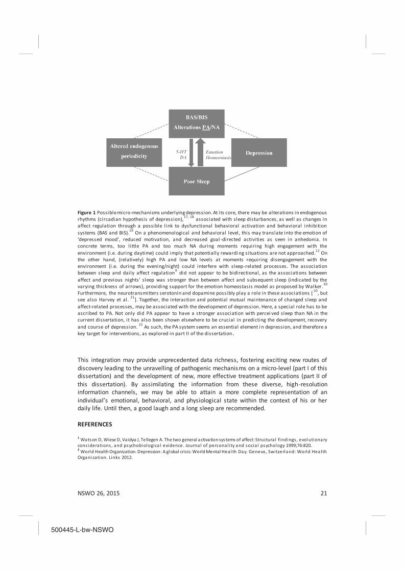
Figure 1 Possible micro-mechanisms underlying depression. At its core, there may be alterations in endogenous rhythms (circadian hypothesis of depression),17, 18 associated with sleep disturbances, as well as changes in affect regulation through a possible l ink to dysfunctional behavioral activation and behavioral inhibition systems (BAS and BIS).19 On a phenomenological and behavioral level, this may translate into the emotion of ‘depressed mood’, reduced motivation, and decreased goal -directed activities as seen in anhedonia. In concrete terms, too little PA and too much NA during moments requiring high engagement with the environment (i.e. during daytime) could imply that potentially rewardi ng situations are not approached.12 On the other hand, (relatively) high PA and low NA levels at moments requiring disengagement with the environment (i.e. during the evening/night) could interfere with sleep-related processes. The association between sleep and daily affect regulation9 did not appear to be bidirectional, as the associations between affect and previous nights’ sleep was stronger than between affect and subsequent sleep (indicated by the varying thickness of arrows), providing support for the emotion homeostasis model as proposed by Walker.20 Furthermore, the neurotransmitters serotonin and dopamine possibly play a role in these associations [ 10, but see also Harvey et al. 21]. Together, the interaction and potential mutual maintenance of changed sleep and affect-related processes, may be associated with the development of depression. Here, a special role has to be ascribed to PA. Not only did PA appear to have a stronger association with percei ved sleep than NA in the current dissertation, it has also been shown elsewhere to be crucial in predicting the development, recovery and course of depression. 22 As such, the PA system seems an essential element i n depression, and therefore a key target for interventions, as explored in part II of the dissertation .
This integration may provide unprecedented data richness, fostering exciting new routes of discovery leading to the unravelling of pathogenic mechanisms on a micro-level (part I of this dissertation) and the development of new, more effective treatment applications (part II of this dissertation). By assimilating the information from these diverse, high-resolution information channels, we may be able to attain a more complete representation of an individual’s emotional, behavioral, and physiological state within the context of his or her daily life. Until then, a good laugh and a long sleep are recommended. REFERENCES 1 Watson D, Wiese D, Va idya J, Tellegen A. The two general activation systems of affect: Structura l findings , evolutionary cons iderations , and psychobiologica l evidence. Journal of personal i ty and socia l psychology 1999;76:820. 2 World Health Organization. Depression: A global crisis: World Mental Health Day. Geneva, Switzerland: World Health Organization. Links 2012.
500445-L-bw-NSWO500445-L-bw-NSWO500445-L-bw-NSWO500445-L-bw-NSWO
NSWO 26, 2015 22
3 González HM, Vega WA, Wi lliams DR, Tarraf W, West BT, Neighbors HW. Depression care in the United States: too little for too few. Archives of Genera l Psychiatry 2010;67:37-46. 4 Hirschfeld R. The epidemiology of depress ion and the evolution of treatment. J Cl in Psychiatry 2012;73:5 -9. 5 Watson D, Clark LA, Carey G. Positive and negative affectivity and their relation to anxiety and depress ive disorders . Journal of abnormal psychology 1988;97:346. 6 Cs ikszentmihalyi M, Larson R. Validity and reliability of the Experience-Sampling Method. J Nerv Ment Dis 1987;175:526-36. 7 Hartmann JA, Carney CE, Lachowski A, Edinger JD. Exploring the construct of subjective s leep qual i ty in patients with insomnia. J Cl in Psychiatry 2015;76:e768-73. 8 Buysse DJ, Reynolds CF, 3rd, Monk TH, Berman SR, Kupfer DJ. The Pi ttsburgh Sleep Qual i ty Index: a new instrument for psychiatric practice and research. Psychiatry Res 1989;28:193-213. 9 de Wi ld-Hartmann JA, Wichers M, van Bemmel AL, et al. Day-to-day associations between subjective s leep and affect in regard to future depress ion in a female population -based sample. Br J Psychiatry 2013;202:407-12. 10 Hartmann JA, Wichers M, van Bemmel AL, et a l. The serotonin transporter 5-HTTLPR polymorphism in the association between s leep qual i ty and affect. Eur Neuropsychopharmacol 2014;24:1086-90. 11 Monti JM. Serotonin control of s leep-wake behavior. Sleep Med Rev 2011;15:269-81. 12 Murray G, Nicholas CL, Kleiman J, et al. Nature's clocks and human mood: the ci rcadian system modulates reward motivation. Emotion 2009;9:705-16. 13 Hartmann JA, Wichers M, Menne-Lothmann C, et a l. Experience sampling-based personal ized feedback and pos i tive affect: a randomized control led tria l in depressed patients . PLoS One 2015;10:e0128095. 14 Wichers M, Simons CJ, Kramer IM, et al. Momentary assessment technology as a tool to help patients with depress ion help themselves . Acta Psychiatrica Scandinavica 2011;124:262-72. 15 Wichers M, Hartmann JA, Kramer IM, et a l. Translating assessments of the film of daily l ife into person-tailored feedback interventions in depress ion. Acta Psychiatrica Scandinavica 2011;123:402-3. 16 Kramer I, Simons CJ, Hartmann JA, et a l. A therapeutic application of the experience sampling method in the treatment of depress ion: a randomized control led tria l . World Psychiatry 2014;13:68-77. 17 Germain A, Kupfer DJ. Ci rcadian rhythm dis turbances in depress ion. Human Psychopharmacology: Cl ini ca l and Experimental 2008;23:571-85. 18 McClung CA. How might circadian rhythms control mood? Let me count the ways . Biologica l psychiatry 2013;74:242-9. 19 Has ler BP, Al len JJ, Sbarra DA, Bootzin RR, Bernert RA. Morningness-eveningness and depression: pre l iminary evidence for the role of the behaviora l activation system and pos i tive affect. Psychiatry research 2010;176:166 -73. 20 Goldstein AN, Walker MP. The role of s leep in emotional bra in function. Annual review of cl inica l psychology 2014;10:679-708. 21 Harvey AG, Murray G, Chandler RA, Soehner A. Sleep disturbance as transdiagnostic: consideration of neurobiologica l mechanisms. Cl inica l psychology review 2011;31:225-35. 22 Dunn BD. Helping depressed clients reconnect to positive emotion experience: current ins ights and future di rections . Cl inica l Psychology & Psychotherapy 2012;19:326-40.
500445-L-bw-NSWO500445-L-bw-NSWO500445-L-bw-NSWO500445-L-bw-NSWO
NSWO 26, 2015 23
SLEEP IN PARKINSON’S DISEASE A focus on nocturnal movements
Maartje Louter
Radboud University Nijmegen
In clinical practice it is assumed that the motor symptoms of PD have a negative influence on sleep quality. Nocturnal hypokinesia could lead to difficulties turning around in bed which could cause sleep initiation or sleep maintenance problems. Scientific proof whether there is an actual relation between complaints of impaired mobility, number of body position changes and sleep quality is lacking. This thesis addresses this underexposed part of sleep in PD. By studying both the subjective complaints as well as the objective signs of nocturnal movements and their influence on sleep parameters we addressed most of these questions. Furthermore, we studied if possible changes in nocturnal movement could serve as a pre-clinical marker in the early detection of PD. In Chapter 2.1 we describe that sleep disorders are not only common but also very diverse: often more than one sleep problem is present in a patient with PD. This could lead to difficulties in the recognition and separation of sleep disorders being present. In Chapter 2.2 we studied the importance of sleep compared to other symptoms and daily limits for the PD patient by using a priority list. Seventy percent of the 153 PD patients had disturbed sleep. About a third of them (37.9%) prioritized sleep as an item they wanted to discuss during their visit to the movement disorder specialist. Sleep was the 6th item on the list of 23 potential items. Patients who prioritized sleep had significantly worse sleep quality (PSQI 9.3±3.8 vs. 5.5±2.8, p < 0.001), however, patients who prioritized sleep had exactly the same Epworth Sleepiness Scale scores as patients who did not (both 8.0±5.2, p = .996). Impaired bed mobility in Parkinson’s disease Although important for many PD patients, the subject of impaired bed mobility (IBM) and sleep problems have so far received little attention in research. In Chapter 3.1 we showed a clear detrimental influence of difficulties turning around in bed on the quality of nocturnal sleep. We studied a large cohort of 240 PD patients, focusing specifically on the relation between sleep quality on one hand, and the presence and frequency of impaired bed mobility on the other. Impaired bed mobility was present in 56.3% of patients. The prevalence of poor sleep was higher in patients with impaired bed mobility, as reflected by significantly higher mean PSQI scores (PD+IBM 7.7±4.1 vs.PD-IBM 6.1±3.4, p = .001). When we corrected the results for age, disease duration, H&Y stage and LED, presence of impaired bed mobility still had a significant effect on PSQI total score (R-squared = 0.066, standardized-beta = 0.163, p=0.026). The relation between the frequency of difficulties turning around in bed and sleep quality showed a linear trend (contrast estimate 1.9, p=0.001). We studied the influence of impaired bed mobility on objective sleep parameters in Chapter 3.2. We compared the influence of subjective impaired bed mobility on objective sleep
500445-L-bw-NSWO500445-L-bw-NSWO500445-L-bw-NSWO500445-L-bw-NSWO
NSWO 26, 2015 24
quality in patients with PD. We found that PD patients who complain of subjective impaired bed mobility had a significantly diminished sleep efficiency (PD+IBM 63.5% (26.2-85.6) vs. PD-IBM 78.4% (54.8-92.6), p<.001) and a shorter total sleep time compared to patients without complaints (PD+IBD 298.0 min (103.0-419.0) vs. PD-IBM 379.6 min (243.0-530.0), p = .001). Our studies were the first to find an association between sleep disruption and impaired bed mobility within a group of PD patients. We showed that both subjective sleep quality measured with questionnaires as well as objective sleep quality based on PSG findings are worse in PD patients with complaints of impaired bed mobility. Body position changes in PD In Chapter 3.2 we also objectified the complaint of impaired bed mobility. We compared actual body position changes between 24 PD patients with and 20 without complaints of impaired bed mobility and 44 healthy controls. Our results showed only a marginally significant difference between turns during the total night (PD 7.6/h (0.0-19.1) vs. controls 8.8/h (2.0-47.6), p = .046). Furthermore, no significant difference was found in number of turns between PD patients with and without complaints of impaired bed mobility (PD+IBM 6.6/h (0.3-15.0) vs. PD-IBM 8.1/h (0.0-19.1), p = .099). When focusing on actual body position changes during sleep alone the results did show a reduced frequency in patients with impaired bed mobility (PD+IBM 0.4/h (0.0-1.8) vs. PD-IBM 1.4/h (0.0-4.6), p=.015). There was a broad range in the frequency of body position changes however, and even subjects with almost no shifts did not necessarily have impaired sleep quality. More importantly, no correlation was found between turns during sleep and sleep efficiency (PD+IBM R squared = 0.043, p = .900). Nocturnal movements in the preclinical phase of Parkinson’s disease Although previous studies suggested differences in nocturnal mobility between PD patients and controls, the study of Chapter 3.2 did not show increased activity levels during the night in PD patients, and also no decreased frequency of nocturnal turns. Body position changes during the night may, however, have different patterns compared to controls. These changes may also precede the onset of PD and could therefore be interesting to study as an early PD marker. In Chapter 4 we compared nocturnal movements in 11 PD patients and 13 healthy controls and 33 non-PD individuals with a potential high risk for future development of the disease (HR-PD). All patients were investigated within the framework of the PMPP study (Progression Markers in the Premotor Phase of Parkinson’s Disease). The results show that with respect to general movement assessment, mean acceleration was lower in PD patients compared to controls. Again the frequency of axial turns did not significantly differ between both groups, but the distribution and pattern of the axial turns did: total size of axial turns was smaller (PD 32.6° (17.5-46.9) vs. controls 46.72° (22.7-73.9), p < .001) and duration of turns was shorter (PD 5.7 s (4.1-8.8) vs. controls 6.96 s (5.6-10.2), p = .001) in PD patients. No differences were found between mean acceleration in HR-PD patients and controls. Furthermore, characteristics of axial turns were not different.
500445-L-bw-NSWO500445-L-bw-NSWO500445-L-bw-NSWO500445-L-bw-NSWO
NSWO 26, 2015 25
Actigraphy as a diagnostic tool in REM sleep behavior disorder Assessing the presence of RBD in PD patients based on the clinical interview alone often results in misdiagnosis. According to the current diagnostic criteria, the diagnosis of RBD requires a clinical interview and video polysomnography (v-PSG). In clinical practice, however, this is not always feasible, since it is time consuming and expensive. Therefore there is a need for less expensive, easy to use devices to diagnose RBD. In Chapter 5 we studied the use of actigraphy as a diagnostic tool for RBD in PD patients. We studied 45 PD patients with and without the clinical diagnosis of RBD. The diagnosis was bas ed on the clinical interview accompanied with v-PSG, according to the ISCD-II criteria. The main outcome measure was the total number of bouts classified as “wake”, compared between patients with (PD+RBD) and without (PD- RBD) RBD. The total number of wake bouts was significantly higher in RBD patients (PD+RBD 73.2±40.2 vs. PD-RBD 48.4±23.3, p = .016). A cut off of 95 wake bouts per night resulted in a specificity of 95.5%, a sensitivity of 20.1% and a positive predictive value of 85.7%. Based on the clinical interview, seven patients were suspected of having RBD, but they did not fulfill the full ICSD-II criteria. All but one of these patients had sleep initiation or sleep maintenance problems (insomnia); two were diagnosed with obstructive sleep apnea syndrome, two had restless legs syndrome, three showed an increased level of periodic leg movements, and one reported nocturnal hallucination. Six of the patients had a wake bout count lower 95. Our results show that using actigraphy, the number of bouts classified as “wake” is significantly higher in PD patient with RBD compared to PD patients without. Accordingly, we show that actigraphy has a high specificity and a good positive predictive value for diagnosing RBD in PD patients. In our study we also focused on the use of actigraphy in clinical practice. Our results show that using an epoch length of 0.25 min and a cut-off of 95 wake bouts per night, actigraphy is a highly specific tool, albeit with a low sensitivity. Based on a semi-structured clinical interview alone, we found seven patients incorrectly suspected of having RBD. Of these, only one patients scored above the threshold of 95 wake bouts per night. Therefore, these results show an additional value of using actigraphy next to a clinical interview in the diagnostic trajectory of RBD to exclude its presence.
500445-L-bw-NSWO500445-L-bw-NSWO500445-L-bw-NSWO500445-L-bw-NSWO
NSWO 26, 2015 26
ON THE ANALYSIS AND CLASSIFICATION OF SLEEP STAGES FROM CARDIORESPIRATORY ACTIVITY
Xi Longa,b
aDepartment of Electrical Engineering, Eindhoven University of Technology, Postbus 513,
5600 MB, Eindhoven, The Netherlands bDepartment of Personal Health, Philips Group Innovation Research, High Tech Campus,
5656 AE, Eindhoven, The Netherlands
Sleep is a state of reversible disconnection from the environment and plays an exceptionally essential role in maintaining internal homeostasis, memory consolidation, energy conservation, and cognitive and behavioral performance. Nowadays, problems in sleeping are widely prevalent around the world with increasing sleep complaints. Historically, such problems have been less common because the regulation of sleep is synchronized with the external environment through a biological circadian rhythm. However, since we are now living in a modern industrialized society with artificial environments where lighting, heat, and food are available at any moment, sleep disturbances and disorders have reached epidemic levels. People experience the symptoms of disturbed sleep such as fatigue, increased impulsiveness, and agitation without being aware of the link between thes e issues and their sleeping patterns. In order to have a healthy condition in body and mind, people should be empowered with the ability to monitor sleep easily and without disturbing the sleep, to assess sleep quality or sleep-related problems and to be able to adjust their sleep habits accordingly. However, the traditional sleep monitoring method, known as polysomnography (PSG), has the problems that the monitoring is usually accomplished in a sleep laboratory with costly facilities, and many sleep-disturbing devices with electrodes and wires have to be attached to the body. Furthermore, the measurements of such devices can only be interpreted by highly trained sleep clinicians. Therefore, although PSG is currently considered the gold standard and common practice for sleep monitoring, it is very unfit for daily use in a home scenario by people without specialized training, and will introduce undesired sleep disturbances. This has motivated the investigation of alternative sensors and methods that allow for monitoring sleep in an unobtrusive manner, preferably inexpensive and with no requirement of training. Objective sleep assessment is often based on monitoring sleep stages throughout the night. In the past decades, cardiorespiratory signals have attracted more and more attention in the context of sleep staging or sleep stage classification. Cardiorespiratory activity has been shown to associate with sleep stages through the regulation of the autonomic nervous system. More importantly, cardiorespiratory signals can be acquired unobtrusively using advanced technologies such as microwave Doppler radar, ballistocardiography, photoplethysmography, pressure-sensitive bed sheets, acoustic devices, and near-infrared cameras. Thus, investigating cardiac and respiratory characteristics in different sleep stages
500445-L-bw-NSWO500445-L-bw-NSWO500445-L-bw-NSWO500445-L-bw-NSWO
NSWO 26, 2015 27
is important for providing a reliable performance in sleep stage classification, with which a more adequate sleep assessment can be delivered. This thesis first exploits characteristics of cardiac/respiratory activity and their interaction during sleep using several signal analysis methods. These are: frequency band adaptation on heart rate variability (Chapter 2), dynamic time/frequency warping and uniform scaling (measuring self-dissimilarity) for respiration (Chapter 3 and Chapter 4 respectively), analysis of breathing depth and volume (Chapter 5), and visibility graph analysis in complex networks for cardiorespiratory interaction (Chapter 6). Based on these methods, novel cardiorespiratory features (expressing certain physiological properties) are proposed to classify sleep stages. Results show that these features can help to profoundly improve performance of sleep stage classification. In addition, an interesting finding is demonstrated in Chapter 7, which is that there is a time delay between the changes in brain activity and autonomic variations during sleep transitions. It appears that the cardiac changes consistently precede the variations in brain activity during light-deep sleep and sleep-wake transitions. In Chapter 8, this finding is utilized to detect deep sleep (i.e., slow wave sleep) by using the feature values from with a preceding time interval of a few minutes before, which can help to significantly improve the detection results. Furthermore, the major challenge of sleep stage classification based on cardiorespiratory activity is discussed in Chapter 9. It is found that the classification performance is mainly limited by the between- and within-subject variations in autonomic physiology as well as subject demographics. Therefore, methods of feature normalization and feature smoothing over the entire night are proposed in Chapter 10, which serve to reduce these variations between and within subjects that are observed in the cardiorespiratory features. As a result, marked improvements in sleep stage classification are observed. In summary, this thesis focuses on objectively analyzing and classifying sleep stages using cardiorespiratory signals. It shows that by extracting novel features from the signals, post-processing features using normalization and smoothing, and applying new findings regarding autonomic-brain time delay, the sleep stage classifiers can be substantially improved with reliable results being ultimately achieved.
500445-L-bw-NSWO500445-L-bw-NSWO500445-L-bw-NSWO500445-L-bw-NSWO
NSWO 26, 2015 28
NEUROPLASTICITY IN THE MAMMALIAN CLOCK: THE EFFECT OF AGING AND SEASONS
Sahar Farajnia
Department of Neurophysiology Leiden University Medical Center
Many organisms, from unicellular to humans, have developed an internal timing system to cope with the environmental daily and seasonal cycles. In mammals, a central circadian clock (circa: around, dies: day, about a day) controls rhythms in behavior and physiology with a period length of about 24h. The master clock is located in the suprachiasmatic nuclei (SCN) of the anterior hypothalamus and consists of approximately 20000 neurons located above the optic chiasm and bilateral to the 3rd ventricle. To use the SCN as a reliable time reference in the body, its circadian rhythm has to be synchronized to the environmental cycles of exactly 24 h. The most important environmental time cue or zeitgeber is light, which is received and processed by specialized, photosensitive ganglion cell in the retina projecting directly to the SCN via the retinohypothalamic tract. Most of the SCN neurons are oscillator cells capable of generating an autonomous rhythm in the frequency of action potentials with the peak during the middle of the day. The coupled network of SCN neurons produce a strong, high amplitude rhythm in electrical activity and neurohormone release that serve as output signals. The signal generated by the SCN is distributed to so-called peripheral oscillators in other brain areas and target organs to control the timing of many physiological functions and behavior, and synchronize their rhythms to the environment. Many lines of evidence indicate that the electrical activity plays a major role in the output of the SCN. The rhythm in SCN electrical activity originates from circadian controlled ionic conductances which regulate cell excitability and action potential generation. The waveform of the SCN’s unified electrical output changes over the seasons and in aging. In the case of seasonal modifications the SCN signal encodes the day length and distributes this information. However, in aging the non-reversible changes result in a severe reduction in the amplitude of the signal, which is presumably insufficient to control rhythms of peripheral oscillators. In this thesis, the possible cellular and interneuronal mechanisms influencing the waveform of the SCN’s circadian rhythm in electrical activity were investigated with various techniques. A summary of our current understanding of the effect of aging on SCN function is given in chapter 2. The elderly in today’s society suffer from age-related disorders which affect their brain function, behavior and social life. Aging also impairs the circadian rhythms of many physiological functions such as sleep-wake cycle and disturbs the accurate function of the SCN. Recently, a great deal of evidence has been presented that link the circadian clock and its proper function to mental and physical health. The aging related deficits in clock function therefore aggravate the health problems of the elderly, hence improvement of clock
500445-L-bw-NSWO500445-L-bw-NSWO500445-L-bw-NSWO500445-L-bw-NSWO
NSWO 26, 2015 29
function can aid healthy aging. Deficits in the function of the aged SCN have been indicated in many studies, but the underlying mechanisms are not known yet. In chapter 3, first a longitudinal study of running wheel behavior from mice between 3 and 30 month of age revealed a distinct time course of age related changes in period length, duration of activity and fragmentation of the locomotor activity. The question if the SCN is determining this behavioral phenotype was addressed with multiunit recordings of the SCN electrical activity in vitro in young (3-6 month) and old mice (>24 month). The distribution of electrical activity within the SCN network was greatly disturbed with a population of neurons active during the night, which is in anti-phase to the main cluster of neurons active in the middle of the day. This resulted in a reduced amplitude of the ensemble electrical activity rhythm. Similar broad phase distribution of SCN neuronal activity patterns has previously been shown to reduce the phase shifting capacity of the SCN. In aging consequently, I demonstrated that the phase shifting capacity to a light-pulse is reduced. Patch-clamp recordings of membrane properties, voltage-dependent K currents and GABAergic postsynaptic currents in SCN neurons revealed the highest degree of age-induced dysfunctions at the cellular level. The circadian control of voltage-dependent K+ currents was selectively affected by age. I found a lack of circadian modulation in fast-delayed rectifier K+ current (FDR) and the transient K+ current (IA), which may contribute to the increase in neuronal activity during the night and to the decrease in firing frequency during the day respectively. The results of this chapter suggest that modification in cellular characteristics and intercellular communication may cause the alterations in the network level and lead to a defective behavioral function such as sleep problems and fragmented locomotor activity. Remarkably, the adverse age-related cellular changes are partly compensated on the network level. To further investigate the role of the circadian controlled K+ currents in attenuation of the SCN electrical rhythm amplitude during aging, I measured the large conductance calcium activated potassium currents (BK) in chapter 4. BK currents are essential for normal SCN electrical activity pattern, and proper behavioral and physiological rhythms. BK channel deficient mice show a similar phenotype as aged mice in some aspects such as the reduction in total behavioral activity, a reduced stability and precision of behavioral rhythm, a moderate lengthening of the circadian period and an altered SCN electrical activity. Patch clamp and calcium imaging recordings in old mice indicated a loss in rhythmic modulation of BK currents and a reversed rhythm in intracellular calcium concentration ([Ca2+]i). The BK current was reduced in magnitude at night compared with the young group. Bk channels in many neurons contribute to action potential waveform and can change [Ca 2+]i. The decrease in BK current in aged SCN neurons at night was indeed found to be associated with a depolarization of the membrane potential, a prolonged action potential repolarization, a reduced afterhyperpolarization potential and an elevated [Ca2+]i. These data indicate that age-related reduction of BK currents at night modifies the action potential waveform in SCN neurons. The accurate shape of action potential determines the amount of Ca2+ influx through voltage gated Ca2+ channels during neuronal activity. A prolonged spike repolarization longer activates the voltage gated Ca2+ channels or a reduced AHP delays the deactivation of Ca2+ channels and cause an increased Ca2+ entry and elevated [Ca2+]i. The results of this study
500445-L-bw-NSWO500445-L-bw-NSWO500445-L-bw-NSWO500445-L-bw-NSWO
NSWO 26, 2015 30
suggest that changes of BK currents and subsequently in action potential waveform can contribute to increased [Ca2+]i at night and to the aged SCN phenotype. Chapter 5 discusses the cellular basis of seasonal adaptation by the SCN. The SCN encodes seasonal changes in day-length by modulating the phase distribution of electrical activity patterns of individual neurons. The collective electrical pattern of the SCN determines the waveform of the SCN electrical activity, and hence the duration of behavioral activity. In a nocturnal animal adaptation to a long-day photoperiod, results in a more distributed electrical pattern and a short duration of behavioral activity while exposure to a short-day photoperiod enhanced phase synchrony among neurons results in longer activity pattern. In this way, the SCN, in concert with other brain regions involved in seasonal adaptation, e.g. pineal gland, controls the seasonal behavior. While some of the mechanisms underlying this neuronal phase adjustment in different photoperiods in the SCN, like a role for VIP, have been described, little is known about the cellular mechanisms. Using patch clamp technique, passive and active membrane properties of single neurons such as firing frequency, membrane potential and input resistance were measured in long-day (LD16:8) and short-day (LD 8:16) photoperiods. The results of this study indicate that these cellular properties are similar in the different photoperiods. Remarkably, among the various K+ currents that we measured, only the FDR current was influenced by changes in photoperiod. The FDR current is known to influence behavioral activity rhythms and modulate electrical activity rhythms. It also affects photic information processing within the SCN. There is evidence that the magnitude of the FDR current is affected by light-mediated stimuli and is necessary for photic regulation of gene expression within the SCN. In chapter 5 I have shown that the FDR current is elevated during the night in long-day photoperiod and the daily rhythm in FDR current is reversed in this photoperiod. However, the functional role of the FDR current in photoperiodic adaptation is not clear. In the SCN, long-range synchronization between dorsal and ventral parts of the SCN was suggested to be weakened in long-day photoperiod, which may allow for a wider phase distribution within the SCN network. The FDR current has a potential role in intercellular synchronization in the SCN as it was shown for other brain regions (i.e. neocortex). The precise role of FDR currents in photoperiodic adaptation needs further investigations. Consistent with previous studies, the electrical output and membrane properties of single cells do not change in the photoperiodic adaptation. Photoperiodic phase adjustment therefore, is more likely to be caused by a modified intercellular communications. GABA is one of the main neurotransmitters within the SCN. To understand the role of GABA in seasonal adaptation, GABAergic signaling in the SCN was investigated after adaptation to long- and short-day photoperiods. Chapter 6 describes GABAergic activity and responses, using patch clamp and calcium imaging recordings, in suprachiasmatic neurons of mice exposed to long-day or short-day photoperiod. Exposure to short-day photoperiod decreased the frequency of spontaneous GABAergic synaptic events compared to long -day photoperiod. Importantly, we found enhanced GABAergic excitatory responses in circadian clock neurons of mice exposed to long days (40%) as compared to short days (28%). A precise balance between GABAergic excitation and inhibition in the SCN neuronal network may play a considerable role in adjusting the phase distribution to encode and convey photoperiodic information in the SCN. Photoperiod seems to have influence on basic
500445-L-bw-NSWO500445-L-bw-NSWO500445-L-bw-NSWO500445-L-bw-NSWO
NSWO 26, 2015 31
biophysical properties of the clock cell physiology, changing the concentration of intracellular Cl- by regulating activity of Cl- cotransporter, which subsequently will lead to a shift in the GABA equilibrium potential. Environmental cues such as daylight can thus affect the function of GABA as a key neurotransmitter in the SCN. Daylight or other environmental signals may also influence the function of GABA or other neurotransmitters and thereby the balance between excitation and inhibition in the central nervous system. Plasticity in the circadian clock organization is needed for adaptation to environmental challenges. But, this thesis also emphasizes the importance of the circadian system for health. Life style in modern societies influence our brain in different ways than the natural environment would. Artificial prolonged light duration can affect the neurotransmitter system in the brain. This has been demonstrated for the SCN in this thesis and for other hypothalamic nuclei in a previous study, observing a switch in neurotransmitter content with a correlated change in anxiety behavior. The neuronal strategies which the SCN uses to adapt to various photoperiods, has been evolved in mammals to help the organisms to anticipate seasonal changes and adjust their behavior to the corresponding environmental challenges. However artificial prolonged light in the evening may keep our clocks in a continuous mode of long summer days, which may have detrimental effects on our physiology and behavior. While the effect of seasonal changes in photoperiod are reversible, aging irreversibly affects different levels of the clock machinery from molecular rhythms, intracellular signaling and membrane properties to intercellular communication, neuronal network synchronization and behavioral function. As a result, the amplitude of the circadian timing signal is reduced, peripheral oscillators are weakened and the accuracy of daily rhythms in physiology and behavior is decreased. Restoration of the deficiencies in cellular and intercellular functions may help to achieve healthy aging and mitigate age-related diseases provoked by clock dysfunction.
500445-L-bw-NSWO500445-L-bw-NSWO500445-L-bw-NSWO500445-L-bw-NSWO
NSWO 26, 2015 32
RETINAL AND NEURONAL MECHANISMS OF CIRCADIAN PHOTORECEPTION
Hester van Diepen
Department of Neurophysiology Leiden University Medical Center
A twenty-four hour rhythm is a major characteristic of the temporal profile of many mammalian species. These rhythms are regulated by a biological clock, which resides in the suprachiasmatic nucleus (SCN) of the hypothalamus. The SCN is a bilateral brain structure and contains about 10,000 neurons in each hemisphere. It is located immediately above the crossing of the optic nerves. The SCN generates rhythms with a period of about 24 hours, which have adaptive significance when appropriately synchronized to the environmental light-dark cycle. The SCN receives light information through the retino-hypothalamic tract (RHT). Photic cues are sensed by photosensitive ganglion cells in the inner retina containing the photopigment melanopsin. These photosensitive ganglion cells receive input from the classical rod and cone photoreceptors in the outer retina. Each class of photoreceptors has its peak sensitivity to a specific wavelength of light. The mouse retina is dichromatic and contains two classes of cone photoreceptors which are the short wavelength sensitive cones and the mid wavelength sensitive cones. The finding that mice lacking rod and cone photoreceptors as well as mice lacking melanopsin are able to entrain to an external light:dark cycle revealed that all classes of photoreceptors can regulate photoentrainment.
Chapter 2 gives an overview of the light signaling pathway from the retina to the SCN and discusses the different photoreceptors of the mouse retina and the effects of retinal illumination on photoreceptor electrical activity. The second part of this chapter descri bes how photic information affects the release of neurotransmitters in the SCN and how it changes SCN neuronal activity. Chapter 2 concludes with an overview of the contribution of the different photoreceptors to photoentrainment. The relative contribution of each class of photoreceptors to photoreception by the SCN was determined in chapters 3, 4 and 5. In these chapters we made use of various retinal mutant mouse models to unravel the photoreceptor origin of the response kinetics in SCN neuronal activity. Light exposure typically induces a fast transient increase in electrical impulse frequency of SCN neurons followed by a sustained component for the duration that the lights were on. It was long thought that the sustained component in SCN electrical activity is mainly dependent on melanopsin, whereas the fast response characteristics would originate from cone photoreceptors. In this thesis the degree of the contribution of the classical photoreceptors to the response kinetics of SCN neurons was investigated and the major conclusion is that classical photoreceptors can mediate sustained responses in SCN electrical activity.
In chapter 3 results of experiments are described that elucidate the effects of ultraviolet ( UV) light on the circadian system and sleep. UV wavelength of 360 nm was chosen to maximally stimulate short wavelength sensitive cones. The data report an effect of UV light on SCN electrical activity that was indistinguishable from the effect of white light. To
500445-L-bw-NSWO500445-L-bw-NSWO500445-L-bw-NSWO500445-L-bw-NSWO
NSWO 26, 2015 33
investigate the contribution of the classical photoreceptors to the response kinetics in SCN neuronal activity induced by UV light, similar experiments were performed in mice lacking the photopigment melanopsin. Surprisingly, UV light exposure leads to similar response characteristics in SCN neuronal activity demonstrating a role for classical photoreceptors in mediating this response. Moreover, UV light still elicited an enhancement in SCN neuronal activity, when all photoreceptors except the UV-sensitive cones were desensitized using bright white light. These findings indicate a role for UV-sensitive cones in mediating this response.
The response characteristics of SCN neurons during exposure to longer wavelengths of light were investigated in chapter 4. The light-induced increase in SCN neuronal activity described in this chapter in response to both short and long wavelength light were indistinguishable in melanopsin-deficient mice compared to wild type mice. These findings elucidate a role for classical photoreceptors in irradiance detection by the SCN during exposure to both short and long wavelength light. Similar recordings in mice lacking rods and cones in their retina revealed a contribution of melanopsin to photic transmission to the SCN, especially at higher light intensities and with a relatively long response latency.
In chapter 5 the specific contribution of short and long wavelength-sensitive cone photoreceptors was determined by performing SCN in vivo electrophysiological recordings in mice having cones as the only functional photoreceptors in the retina. In response to relatively short light exposure (up to 1 minute), a sustained light-induced increase in SCN neuronal activity was recorded. Interestingly, the SCN electrical discharge rates decayed to baseline levels after the initial phase of light exposure. These experimental outcomes reveal an important contribution of cone photoreceptors in the initial phase of light detection, whereas cone photoreceptors are unable to generate sustained responses in SCN electrical activity for prolonged durations of light exposure. UV light induced significantly larger changes in SCN electrical activity compared to longer wavelengths of light.
The ability of cone photoreceptors in mediating the transmission of photic information to the SCN does not preclude a role for the other classes of photoreceptors in this process. Our findings indicate the likelihood that the different classes of photoreceptor fulfill additive functions to circadian photoreception. The relative contribution of melanops in, rod and cone photoreceptors is most likely dependent on the intensity and wavelength composition of the light source.
In chapter 6 the effect of disturbed sleep and caffeine on light signaling to the SCN was investigated. Behavioral activity recordings showed that sleep deprivation in mice led to a decrease in the light-induced phase-shifting capacity of the circadian system. To unravel the mechanism causing this reduction, the effect of prolonged wakefulness on light-induced changes in SCN neuronal activity was investigated. In accordance with the effect on behavioral activity, light-induced increases in SCN neuronal activity were attenuated after prolonged wakefulness. The attenuation of SCN electrical discharge rates were restored when the non-selective adenosine antagonist caffeine was administrated after prolonged wakefulness and prior to light exposure. Furthermore, caffeine administration enhanced period lengthening in constant light. These results suggest a role for adenosine and adenosine antagonists in modulating light sensitivity of the circadian system.
500445-L-bw-NSWO500445-L-bw-NSWO500445-L-bw-NSWO500445-L-bw-NSWO
NSWO 26, 2015 34
The impact of the loss of another neurotransmitter, vasoactive intestinal peptide (VIP), on photic regulation of the circadian system was investigated in chapter 7. The hypothesis was tested that VIP plays a major role in the transmission of photic information within the SCN. The light-induced changes in SCN neuronal activity did not differ in VIP mutant mice compared to wild type controls. Similarly, N-methyl-D-aspartate (NMDA) enhanced firing rates of ventral SCN neurons of both VIP mutant and wild type mice. Stimulation of the RHT was used to simulate light exposure and calcium transients were recorded. In response to light exposure and glutamate release, calcium level transients are evoked. RHT stimulation evoked calcium transients in the ventral SCN of both VIP mutant and wild type mice, but exhibited a weaker response in the dorsal SCN in the absence of VIP. Furthermore, light-induced gene expression revealed a reduction after 60 minutes of light exposure especially in the dorsal region of VIP mutant mice. Together these data show an important role for VIP in communication of photic information within the SCN, more specifically in transmission of light information from the ventral to the dorsal SCN region.
In chapter 8 the effects of behavioral activity on the circadian system were investigated. Whereas light exposure enhances SCN neuronal activity, behavioral activity leads to acute suppressions in SCN electrical activity levels. In chapter 8 the hypothesis was tested that enhanced levels of behavioral activity lead to an increase in the amplitude of the SCN electrical activity rhythm. Behavioral activity levels were enhanced by running-wheel activity to determine the influence of exercise on the SCN rhythm in electrical activity. The amplitude of the SCN rhythm in electrical activity was significantly enhanced when behavioral activity levels were increased. Exercise also leads to an increase in rhythm strength. These findings indicate an important role for exercise as a non-invasive intervention for improving the circadian system.
The studies described in this thesis are reviewed in chapter 9. The different photoreceptors in the retina each have their contribution to photoentrainment most l ikely depending on the light intensity and the specific wavelength of light. The experiments described in this thesis reveal an unexpected contribution of the classical photoreceptors to circadian photoreception. We also showed that photic transmission to SCN neurons can be affected downstream of the retina by sleep deprivation and caffeine administration. Furthermore, the transmission of photic information within the SCN is disrupted in the absence of vasoactive intestinal peptide. Taken together these studies demonstrate that light input to the circadian system can be affected both in the retina as well as at the level of the SCN. Finally an important influence of exercise on enhancing the amplitude and increase the rhythm strength of the waveform in SCN electrical activity was elucidated. These findings indicate a role for exercise as a valuable intervention for deficits in circadian rhythms.
500445-L-bw-NSWO500445-L-bw-NSWO500445-L-bw-NSWO500445-L-bw-NSWO
NSWO 26, 2015 35
SLEEP-WAKE Research in the Netherlands
Volume 26, 2015
Research papers
500445-L-bw-NSWO500445-L-bw-NSWO500445-L-bw-NSWO500445-L-bw-NSWO
NSWO 26, 2015 36
ACTIVATION OF THE ENDOCANNABINOID CB1 RECEPTOR ALTERS THE STABILITY OF WAKING TRANSITIONS IN RATS
A. Ahnaou and W.H.I.M. Drinkenburg
Dept. of neurosciences, Janssen Research & Development, a Division of Janssen Pharmaceutica
NV.Turnhoutseweg 30, B-2340 Beerse, Belgium INTRODUCTION The endocannabinoid system has recently emerged as an important modulator of multiple physiological functions and is therefore considered a potential target for the treatment of psychiatric and metabolic conditions. Endocannabinoids (ECs) are a class of atypical neurotransmitters such as anandamide and 2-arachidonoylglycerol, which are synthesized and released from the post-synaptic membrane during periods of enhanced cellular activity. They activate the central (CB1) and peripheral (CB2) cannabinoid receptors. A high density of CB1 receptors was found in brain areas such as thalamus, hypothalamus, cortex, hippocampus, limbic system, basal ganglia and brainstem1. The CB1 receptor is present in pre-synaptic terminals where they decrease synaptic transmission for instance in GABAergic and glutamatergic networks, whereas the presence of the receptor in post-synaptic terminals increase synaptic activity for instance in acetylcholinergic, serotonergic and noradrenergic networks2. This indicates an important physiological role in the modulation of diverse behaviours such as sleep. Prior studies have shown that Δ9-tetrahydrocannabinol and anandamide enhance sleep through CB1 receptors in man and animals 3,4,5. In contrast, the CB1 antagonist SR141716A promotes waking. Thus, targeting the cannabinoid system is a potentially effective approach to the prevention and management of sleep disturbances such as insomnia or excessive sleepiness. There is a general consensus that sleep plays a crucial role in synaptic plasticity. Plastic changes occurring during waking produce coherent oscillatory changes during subsequent sleep states, which favor consolidation of memory and learning. Activation of the CB1 receptor by the potent CB1 receptor agonist WIN 55,212-2 had detrimental effects on short term memory in delayed match- and non-match-to-sample (DNMS) tasks6. The strong bidirectional relationship between sleep and memory was the motivation of the present study to evaluate effects of lower doses of WIN 55,212-2 on sleep-wake architecture in freely moving rats chronically instrumented with EEG/EMG electrodes. METHODS Animals and EEG recording All experimental procedures conformed to the guidelines of the Association for Assessment and Accreditation of Laboratory Animal Care International (AAALAC) and the European Communities Council Directive of 24 November 1986 (86/609/EEC). Male Sprague Dawley rats (Harlan, Netherlands) weighing 250–300 g at the time of surgery were used in this polysomnographic study. Animals were maintained under controlled environmental conditions throughout the study: 22 °C ± 2 °C ambient temperature, relative humidity 60%, standard 12:12 light cycle regime (illumination intensity: 100 lx, lights on at 12:00 AM. Electrodes were chronically implanted to record the frontal and parietal EEG (AP + 2 mm, L -
500445-L-bw-NSWO500445-L-bw-NSWO500445-L-bw-NSWO500445-L-bw-NSWO
NSWO 26, 2015 37
2 mm, and AP - 6 mm, L +3 mm from Bregma), EOG (peri-ocular muscles) and EMG (nuchal muscles) activities. Vigilance states anlaysis Polysomnograms were recorded for 20-hrs following the subcutaneous administration of saline (n=8) and WIN 55,212-2 (0.03, 0.1, 0.3 mg/kg; n=8 for each dose). Offline, six vigilance states were distinguished automatically in each artefact-free 2s epoch as being active wake, passive wake, light sleep, deep sleep, intermediate stage or rapid eye movement (REM) sleep. Time spent in each vigilance state, transitions, and latencies for deep sleep and REM sleep onsets (defined as the time between the beginning of the recordings and the appearance of the first sleep period lasting at least 30 s) are presented here. Statistical analysis A mixed-model ANOVA with time (intervals of 4 hrs post-administration) as the within-subjects factor and dose (WIN 55,212-2 at 0.03, 0.1, 0.3 mg/kg) as the between-subjects factor, followed by a post-hoc Dunnett’s test, was used to compare responses for each dose group to that of the vehicle group. For graphics, time spent in each vigilance state was averaged over the first 4-hrs following the administration of WIN 55,212-2 and of vehicle. Data are presented as mean values ± SEM, and value of P < 0.05 was considered to be significant. RESULTS WIN 55,212-2 at the doses of 0.03, 0.1 mg/kg had no major effect on sleep-wake cycle, however WIN 55,212-2 at the 0.3 mg/kg significantly increased passive waking during the 4-hrs post-administration (treatment x time effect, F(3,205) = 6.12, p=0.0005, Figure 1A). Consequently, significant reductions in deep sleep and REM sleep were observed. No homeostatic rebound followed the initial sleep suppression during the subsequent dark phase (data not shown). The wake promoting effects of WIN 55,212-2 (0.3 mg/kg) are consistent with lengthened sleep onset latencies (Figure 1B). The enhanced passive waking was due to increased mean duration (treatment x time effect: F(21,189) = 2.02, p=0.007) and number of periods (treatment x time effect: F(21,189) = 3.00, p=0.005) of passive waking. However, reduction in deep sleep and REM sleep derived solely from shorter mean duration of these vigilance states (treatment x time effect for deep sleep: F(21,189) = 1.62, p=0.040; treatment x time effect for REM sleep: F(21,189) = 1.60, p=0.049). Remarkably, WIN 55,212-2 at 0.3 mg/kg increased the frequency of bidirectional transitions from active to passive waking (treatment x time effect: F(9,205) = 3.20, p=0.0012) and from passive to active waking (treatment x time effect: F(9,205) = 3.00, p=0.002, Figure 1C). DISCUSSION WIN 55,212-2 significantly promoted passive waking at the expense of deep sleep and REM sleep. A deregulation of the endocannabinoid system has been proposed to be at the basis of several neuropsychiatric disorders. CB1, fatty acid amide hydrolase (FAAH) and monoacylglycerol (MAGL) lipase, involved in synthesis and degradation of endocannabinoids anandamide and 2-arachidonoylglycerol respectively, are the ECs most studied so far7.
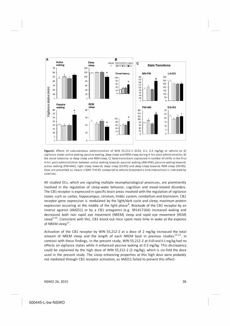
500445-L-bw-NSWO500445-L-bw-NSWO500445-L-bw-NSWO500445-L-bw-NSWO
NSWO 26, 2015 38
Figure1: Effects of subcutaneous administration of WIN 55,212-2 (0.03, 0.1, 0.3 mg/kg) or vehicle on A/ vigilance states active waking, passive waking, deep sleep and REM sleep during 4 -hrs post-administration, B/ the onset latencies to deep sleep and REM sleep, C/ State transitions expressed in number of shifts in the first 4-hrs post-administration between active waking towards passive waking (AW>PW), passive waking towards active waking (PW>AW), l ight sleep towards deep sleep (LS>DS) and deep sleep towards REM sleep (DS>RS). Data are presented as means ± SEM. P<0.05 compared to vehicle (treatment x time interaction) is indicated by asterisks. All studied ECs, which are signaling multiple neurophysiological processes, are prominently involved in the regulation of sleep-wake behavior, cognition and mood-related disorders. The CB1 receptor is expressed in specific brain areas involved with the regulation of vigilance states such as cortex, hippocampus, striatum, limbic system, cerebellum and brainstem. CB1 receptor gene expression is modulated by the light/dark cycle and sleep; maximum protein expression occurring at the middle of the light phase8. Blockade of the CB1 receptor by an inverse agonist (AM251) or by a CB1 antagonist (e.g. SR141716A) increased waking and decreased both non rapid eye movement (NREM) sleep and rapid eye movement (REM) sleep9,10. Consistent with this, CB1 knock-out mice spent more time in wake at the expense of NREM sleep11.
Activation of the CB1 receptor by WIN 55,212-2 at a dose of 2 mg/kg increased the total amount of NREM sleep and the length of each NREM bout in previous studies 12,13. In contrast with these findings, in the present study, WIN 55,212-2 at 0.03 and 0.1 mg/kg had no effects on vigilance states while it enhanced passive waking at 0.3 mg/kg. This discrepancy could be explained by the high dose of WIN 55,212-2 (2 mg/kg), which is six-fold the dose used in the present study. The sleep enhancing properties at this high dose were probably not mediated through CB1 receptor activation, as AM251 failed to prevent this effect.
500445-L-bw-NSWO500445-L-bw-NSWO500445-L-bw-NSWO500445-L-bw-NSWO
NSWO 26, 2015 39
In addition, animals receiving WIN 55,212-2 (0.3 mg/kg) experienced more bidirectional transitions between active and passive waking than did animals treated with vehicle. The ratio of transitions from passive waking to light sleep, from light sleep to deep sleep and from deep sleep to REM sleep did not significantly alter. The exact biological significance of WIN 55,212-2-induced changes in the direction of these states transitions remains unclear, but the changes may have important implications for the organization of the sleep-wake cycle and sleep-dependent memory processes. The destabilized state transitions may be the mechanism underlying the increased latency to sleep onset; animals seem to have difficulties falling asleep and reduced sleep efficiency. The CB1 receptors are localized widely spread in multiple brain regions targeted by excitatory and inhibitory efferent involved in the regulation of sleep-wake cycle. This implies that activation of CB1 may modulate network-excitability via multiple neurotransmitter systems. The following effects of CB1 activation have been reported in literature: decreased release of both GABA and glutamate in the cortex14, enhanced release of acetylcholine15, increased firing rate of noradrenergic neurons16 and enhanced activity of the serotonergic and dopaminergic neurons17,18. In addition, CB1-orexinergic (OX1R) heteromers can be formed in the hypothalamic area119,20, which may influence the flip-flop switch mechanisms to cause more transitions between (waking) vigilance states. Therefore, exogenous activation of CB1 receptors could affect network mechanisms regulating both excitatory and inhibitory neurotransmission in local cortical areas to trigger instability of vigilance states. In addition, WIN 55,212-2 at lower doses (0.10-0.50 mg/kg) as those used in the present work, were shown to produce a delay-dependent deficit in the DNMS task, and thus alter short term memory in a similar way as hippocampal lesions 6. Because of the strong relationship between the quality of sleep and memory processes, we hypothesize that the high frequency of transitions between active and passive waking associated with shorter sleep bouts affects sleep related memory consolidation processes. Overall, the data presented here are in agreement with studies showing that an exogenous CB1 agonist modulates vigilance states and the frequency of transitions between active and passive waking states. The findings suggest that activation of CB1 may interfere with the neuronal centers that coordinate and stabilize the brain’s sleep-wake states and flip-flop mechanisms, which may consequently affect sleep continuity and associated memory processes. ACKNOWLEDGEMENTS We thank Ms. H. Huysmans for technical assistance. REFERENCES 1Mackie K. Distribution of cannabinoid receptors in the central and peripheral nervous system. Handb Exp Pharmacol. 2005; 168:299-325. 2Xu JY, Chen C. Endocannabinoids in synaptic plasticity and neuroprotection. Neuroscientist 2015 ; 21:152 -68 3Buonamici M, Young GA, Khazan N, Effects of acute Δ9-THC administration of EEG and EEG power spectra in the rat. Neuropharmacology 1982; 21: 825-829 4Feinberg I, Jones R, Walker JM. Effects of high dosage delta 9 tetrahydrocannabinol on sleep patterns in man. Clinical Pharmacology and Therapeutics 1975; 17: 458-466.
500445-L-bw-NSWO500445-L-bw-NSWO500445-L-bw-NSWO500445-L-bw-NSWO
NSWO 26, 2015 40
5Muril lo-Rodríguez E, Sánchez-Alavez M, Navarro L, Martínez-González D, Drucker-Colín R, Prospéro-García O. Anandamide modulates sleep and memory in rats. Brain Res. 1998; 812: 270 -4. 6Hampson RE, Deadwyler SA. Cannabinoids, hippocampal function and memory. Life Sci. 1999;65:715 -23. 7Janero DR, Vadivel SK, Makriyannis A. Pharmacotherapeutic modulation of the endocannabinoid signalling system in psychiatric disorders: drug-discovery strategies. Int Rev Psychiatry 2009; 21: 122-33 8Martínez-Vargas M, Muril lo-Rodríguez E, González-Rivera R, Landa A, Méndez-Díaz M, Prospro-García O, Navarro L. Sleep modulates cannabinoid receptor 1 expression in the pons of rats. Neuroscience. 2003; 117: 197-201. 9Méndez-Díaz M, Caynas-Rojas S, Arteaga Santacruz V, Ruiz-Contreras AE, Aguilar-Roblero R, Prospéro-García O. Entopeduncular nucleus endocannabinoid system modulates sleep-waking cycle and mood in rats. Pharmacol Biochem Behav. 2013; 107:29-35. 10Santucci V, Storme JJ, Soubrie P, Le Fur G. Arousal -enhancing properties of the CB1 cannabinoid receptor antagonist SR 141716A in rats as assessed by electroencephalographic spectral and sleep-waking cycle analysis. Life sciences 1996; 58: PL103–110. 11Pava MJ, den Hartog CR, Blanco-Centurion C, Shiromani PJ, Woodward JJ. Endocannabinoid modulation of cortical up-states and NREM sleep. PLoS One. 2014; 9:e88672. 12Goonawardena AV, Plano A, Robinson L, Platt B, Hampson RE, Riedel G. A Pilot Study into the Effects of the CB1 Cannabinoid Receptor Agonist WIN55,212-2 or the Antagonist/Inverse Agonist AM251 on Sleep in Rats. Sleep Disord. 2011: 178469. 13Goonawardena AV, Plano A, Robinson L, Ross R, Greig I, Pertwee RG, Hampson RE, Platt B, Riedel G. Modulation of food consumption and sleep-wake cycle in mice by the neutral CB1 antagonist ABD459. Behav Pharmacol. 2015; 26:289-303. 14Fortin DA, Levine ES. Differential effects of endocannabinoids on glutamatergic and GABAergic inputs to layer 5 pyramidal neurons. Cereb Cortex 2007; 17: 163–174. 15Acquas E, Pisanu A, Marrocu P, Di Chiara G. Cannabinoid CB1 receptor agonist increase rat cortical and hippocampal acetylcholine release in vivo. Eur J Pharmacol 2000;401:179 -85. 16Muntoni AL, Pil lolla G, Melis M, Perra S, Gessa GL, Pistis M. Cannabinoids modulate spontaneous neuronal activity and evoked inhibition of locus coeruleus noradrenergic neurons. Eur J Neurosci 2006; 23:2385 –94. 17Bambico FR, Katz N, Debonnel G, Gobbi G. Cannabinoids elicit antidepressant-like behavior and activate serotonergic neurons through the medial prefrontal cortex. J Neurosci. 2007; 27: 11700 -11. 18Fitzgerald ML, Shobin E, Pickel VM. Cannabinoid modulation of the dopaminergic circuitry: implications for l imbic and striatal output. Prog Neuropsychopharmacol Biol Psychiatry. 2012; 38: 21-9. 19Saper CB, Scammell TE, Lu J. Hypothalamic regulation of sleep and circadian rhythms. Nature. 2005; 437: 1257-63. 20Flores A, Maldonado R, Berrendero F. Cannabinoid-hypocretin cross-talk in the central nervous system: what we know so far. Front Neurosci. 2013; 20; 7:256.
500445-L-bw-NSWO500445-L-bw-NSWO500445-L-bw-NSWO500445-L-bw-NSWO
NSWO 26, 2015 41
SYNAPTIC STRENGTH EXPRESSED IN EPSP SIZE IS DOWNSCALED IN SLEEP: EVIDENCE FOR THE SYNAPTIC HOMEOSTASIS HYPOTHESIS
Anton Coenen
Department of Biological Psychology
Donders Centre for Cognition, Radboud University Nijmegen
INTRODUCTION The synaptic homeostasis hypothesis of sleep claims that during sleep the synaptic strength is downscaled1,2. This is energetically sustainable and beneficial for memory consolidation during sleep. During wakefulness a net increase in synaptic strength in the brain is necessary for an adequate processing of information, leading to proper behavioral responses. However, synaptic upscaling needs energy. To save energy during sleep, brain activity is reduced by synaptic downscaling and homeostasis is restored. A disconnection from the brain to the environment is the result. The concept of the transfer ratio during varying levels of alertness was developed by Coenen and Vendrik3. The transfer ratio, the ratio between the incoming sensory information and the ultimate arriving of this information to perceiving brain areas, is high during waking and low during sleep. This implicates that the flow of information through the brain is strongly reduced during sleep. The partial disconnection of the brain from the environment, due to a lowering of brain activity, might protect the process of sleep. It is suggested by the synaptic homeostasis hypothesis that the function of sleep is to reduce the synaptic strength, associated with an uncoupling of the brain from the environment and a decrease of the flow of brain information. In this way, the transfer ratio concept of sleep of Coenen and Vendrik 3 is reminiscent to the synaptic homeostasis hypothesis of Tononi and Cirelli2. Circumstantial evidence was already obtained by Coenen and Vendrik (1972) that the process leading to the decrease of synaptic strength might be the reduction of the size of the excitatory postsynaptic potentials (EPSPs) during sleep3. Since the mechanism behind synaptic up- and downscaling is not fully understood, which is regarded as a minor point 4, the original data of Coenen and Vendrik were reanalyzed to find whether the size of the EPSPs 3, regarded as an adequate measure for synaptic strength, is modulated according to the levels of alertness during waking and sleeping. METHODS In the experiment of Coenen and Vendrik the effect of the level of alertness on evoked neuronal activity by a visual stimulus was determined by electrical recordings of cells in the lateral geniculate nucleus of a cat 3. In a data base, these neuronal recordings along with the cortical EEGs of the cat were stored. The cat’s alertness, determined by EEG criteria fluctuated between wakefulness, drowsiness and (slow wave) sleep. Though most recordings were of extracellular origin, it was sometimes succeeded to perform intracellular recordings. A long stable intracellular recording was made of a neuron (unit 75-4) during all levels of
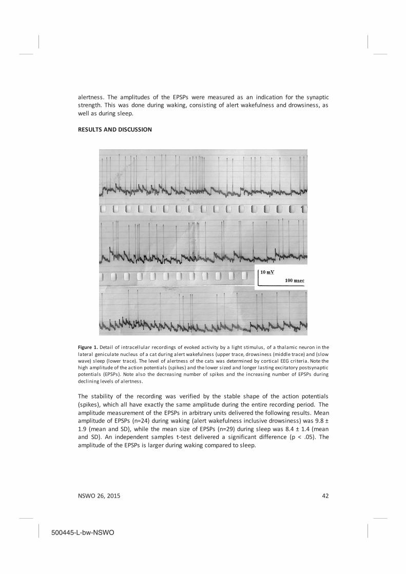
500445-L-bw-NSWO500445-L-bw-NSWO500445-L-bw-NSWO500445-L-bw-NSWO
NSWO 26, 2015 42
alertness. The amplitudes of the EPSPs were measured as an indication for the synaptic strength. This was done during waking, consisting of alert wakefulness and drowsiness, as well as during sleep. RESULTS AND DISCUSSION
Figure 1. Detail of intracellular recordings of evoked activity by a l ight stimulus, of a thalamic neuron in the lateral geniculate nucleus of a cat during alert wakefulness (upper trace, drowsiness (middle trace) and (slow wave) sleep (lower trace). The level of alertness of the cats was determined by cortical EEG criteria. Note the high amplitude of the action potentials (spikes) and the lower sized and longer lasting excitatory postsynaptic potentials (EPSPs). Note also the decreasing number of spikes and the increasing number of EPSPs during declining levels of alertness. The stability of the recording was verified by the stable shape of the action potentials (spikes), which all have exactly the same amplitude during the entire recording period. The amplitude measurement of the EPSPs in arbitrary units delivered the following results. Mean amplitude of EPSPs (n=24) during waking (alert wakefulness inclusive drowsiness) was 9.8 ± 1.9 (mean and SD), while the mean size of EPSPs (n=29) during sleep was 8.4 ± 1.4 (mean and SD). An independent samples t-test delivered a significant difference (p < .05). The amplitude of the EPSPs is larger during waking compared to sleep.
500445-L-bw-NSWO500445-L-bw-NSWO500445-L-bw-NSWO500445-L-bw-NSWO
NSWO 26, 2015 43
In Figure 1 the intracellular recordings of the evoked activity are presented. The recordings during alert waking, drowsiness and sleep are shown. For waking, the recordings for alert wakefulness and drowsiness are combined. This was done for two reasons. First, the distinction between alert waking and drowsiness is not always obvious, and secondly, the number of subthreshold EPSPs during waking is relatively small. The fact that even less EPSPs are visible during alert waking than during drowsiness 3,5, is presumably due to the fact that the EPSPs are than still larger compared to drowsiness, and more easily reaching the threshold and converted into spikes. If the number of action potentials and EPSPs are added up, then in all three levels of alertness, the same numbers were found. In the details of the recording periods presented in Figure 1 there are 34 spikes and 8 EPSPs (total 42) during alert waking, 29 spikes and 12 EPSPs (total 41) during drowsiness and 16 spikes and 24 EPSPs (total 40) during sleep. The sensory input to this cell is equal, but the output to perceptive areas is reduced. It is speculated that a mechanism such as presynaptic inhibition might be responsible for this reduction in EPSP size and so regulating the synaptic strength. In this way the flow of activity through the brain as well as the general brain action is considerably reduced during sleep, or, in the conception of the synaptic homeostasis hypothesis, sleep is necessary for a reduction of brain activity by synaptic downscaling. CONCLUSIONS The synaptic strength expressed in the amplitude of the EPSPs, is modulated according to the level of alertness. The synaptic strength is stronger during waking compared to sleep. This is in favor of the synaptic homeostasis hypothesis of sleep of Tononi and Cirelli (2006). It shows that synaptic downscaling decreases the amount of brain activity during sleep. REFERENCES 1 Tononi, G., Cirell i , C.: Sleep function and synaptic homeostasis. Sleep Medicine Reviews 2006; 10: 49 -62. 2 Tononi, G., Cirell i , C.: Sleep and the price of plasticity: from synaptic and cellular homeostasis to memory consolidation and integration. Neuron 2014; 81: 12-34.
3 Coenen, A.M.L., Vendrik, A.J.H.: Determination of the transfer ratio of cat’s geniculate neurons through quasi -intracellular recordings and the relation with the level of alertness. Experimental Brain Research 1972; 14: 227 -242. 4 Frank, M.G.: Why I am not SHY: a replay to Tononi and Cirell i . Neural Plasticity 2013: ID394946, 3 pages http://dx.doi.org/10.1155/2013/394946 5 Coenen, A.M.L: Neuronal activities underlying the electroencephalogram and evoked potentials of sleeping and waking: implications for information processing. Neuroscience and Biobehavioral Reviews 1995; 19: 447 -463
500445-L-bw-NSWO500445-L-bw-NSWO500445-L-bw-NSWO500445-L-bw-NSWO
NSWO 26, 2015 44
EFFECTS OF MILD ELECTRICAL STUNNING ON THE EEG AND THE LEVEL OF UNCONSCIOUSNESS OF CHICKENS
Anton Coenen and Gerard van Oijen
Department of Biological Psychology, Donders Centre for Cognition
Radboud University Nijmegen
INTRODUCTION Electrical water bath stunning is a common technique for stunning of chickens and is generally used in poultry slaughter houses. Treatments with high voltage and low frequencies ensure a good stunning of birds. However, heavy muscular contractions producing meat quality defects are main adverse effects of this stunning technique. To avoid these negative effects, a two-phase stunning procedure is in development in which in the first phase a low voltage (15 V) pulsed DC current of 550 Hz is applied to anesthetize and immobilize the animal, and a higher voltage AC current of 50-60 Hz in the second phase to finally kill the bird. Preliminary assessments of this technique have shown that the muscular convulsions and meat quality defects following this two-phase stunning procedure are strongly reduced compared to traditional single phase stunning. However, the stunning efficiency of this electrical set-up is markedly reduced. It is assumed that the low voltage DC as used in the first phase does not induce full unconsciousness in the birds, giving rise to a less effective second phase. In the present study it is investigated what the minimal electrical parameters of the current used in the first phase must be to induce a light, but sufficient, anesthesia and a proper immobilization. To that aim chickens were mildly stunned with high frequency DC currents ranging from 10 to 90 mA (10 - 80 V). To assess the degree of unconsciousness of the birds the EEG was recorded before and after stunning. METHODS Approval for this experiment was given by the animal ethical committee (DEC) of the Radboud University Nijmegen. A total of 50 Ross chickens of 40 days old, with a mean body weight of 2295 g were used. They were randomly attributed to five different stunning treatments: 10, 20, 40, 60 and 80 V of pulsed DC current with a duty cycle of 25:75 (on-off time respectively) at 550 Hz. Afterwards the birds were killed in a box filled with carbon dioxide. In the experiment the bird’s feet were fixed in a slaughter shackle and the breast placed on an inclined board. The birds were fixed with elastic bands and the wings were slightly tied to prevent robust wing flapping during stunning to enable undisturbed EEG recordings. The heads of the birds were fixed in the chicken EEG clamp ‘CHEC’ (Coenen et al., 2007), a non-invasive EEG recording device for chickens. The voltage was supplied across the chicken’s body, where metal straps over the top of the head formed the positive electrode while the feet were fixed in grounded metal shackles. This enabled immediate recording of the EEG after termination of the current flow. The baseline EEG of every chicken was recorded for 30 seconds. Thereafter, the stunning current was applied for 10 seconds. The rms current applied to each bird was measured by a Fluke Multimeter 189. Immediately after the current was switched off, the chicken clamp was used to record the EEG for a
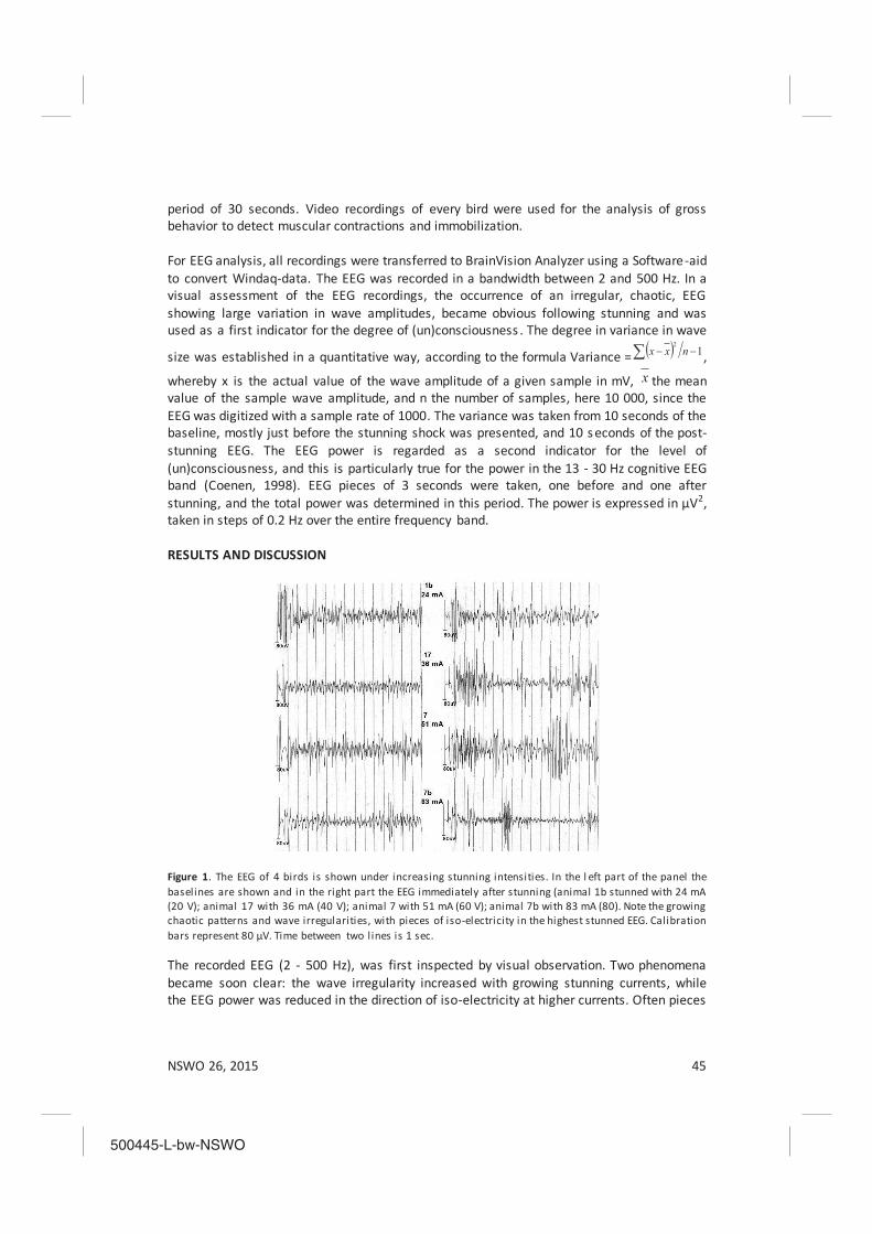
500445-L-bw-NSWO500445-L-bw-NSWO500445-L-bw-NSWO500445-L-bw-NSWO
NSWO 26, 2015 45
period of 30 seconds. Video recordings of every bird were used for the analysis of gross behavior to detect muscular contractions and immobilization. For EEG analysis, all recordings were transferred to BrainVision Analyzer using a Software-aid to convert Windaq-data. The EEG was recorded in a bandwidth between 2 and 500 Hz. In a visual assessment of the EEG recordings, the occurrence of an irregular, chaotic, EEG showing large variation in wave amplitudes, became obvious following stunning and was used as a first indicator for the degree of (un)consciousness . The degree in variance in wave
size was established in a quantitative way, according to the formula Variance = 12nxx ,
whereby x is the actual value of the wave amplitude of a given sample in mV, x the mean value of the sample wave amplitude, and n the number of samples, here 10 000, since the EEG was digitized with a sample rate of 1000. The variance was taken from 10 seconds of the baseline, mostly just before the stunning shock was presented, and 10 s econds of the post-stunning EEG. The EEG power is regarded as a second indicator for the level of (un)consciousness, and this is particularly true for the power in the 13 - 30 Hz cognitive EEG band (Coenen, 1998). EEG pieces of 3 seconds were taken, one before and one after stunning, and the total power was determined in this period. The power is expressed in μV2, taken in steps of 0.2 Hz over the entire frequency band. RESULTS AND DISCUSSION
Figure 1. The EEG of 4 birds is shown under increasing stunning intensities. In the l eft part of the panel the baselines are shown and in the right part the EEG immediately after stunning (animal 1b stunned with 24 mA (20 V); animal 17 with 36 mA (40 V); animal 7 with 51 mA (60 V); animal 7b with 83 mA (80). Note the growing chaotic patterns and wave irregularities, with pieces of iso-electricity in the highest stunned EEG. Calibration bars represent 80 μV. Time between two lines is 1 sec. The recorded EEG (2 - 500 Hz), was first inspected by visual observation. Two phenomena became soon clear: the wave irregularity increased with growing stunning currents, while the EEG power was reduced in the direction of iso-electricity at higher currents. Often pieces
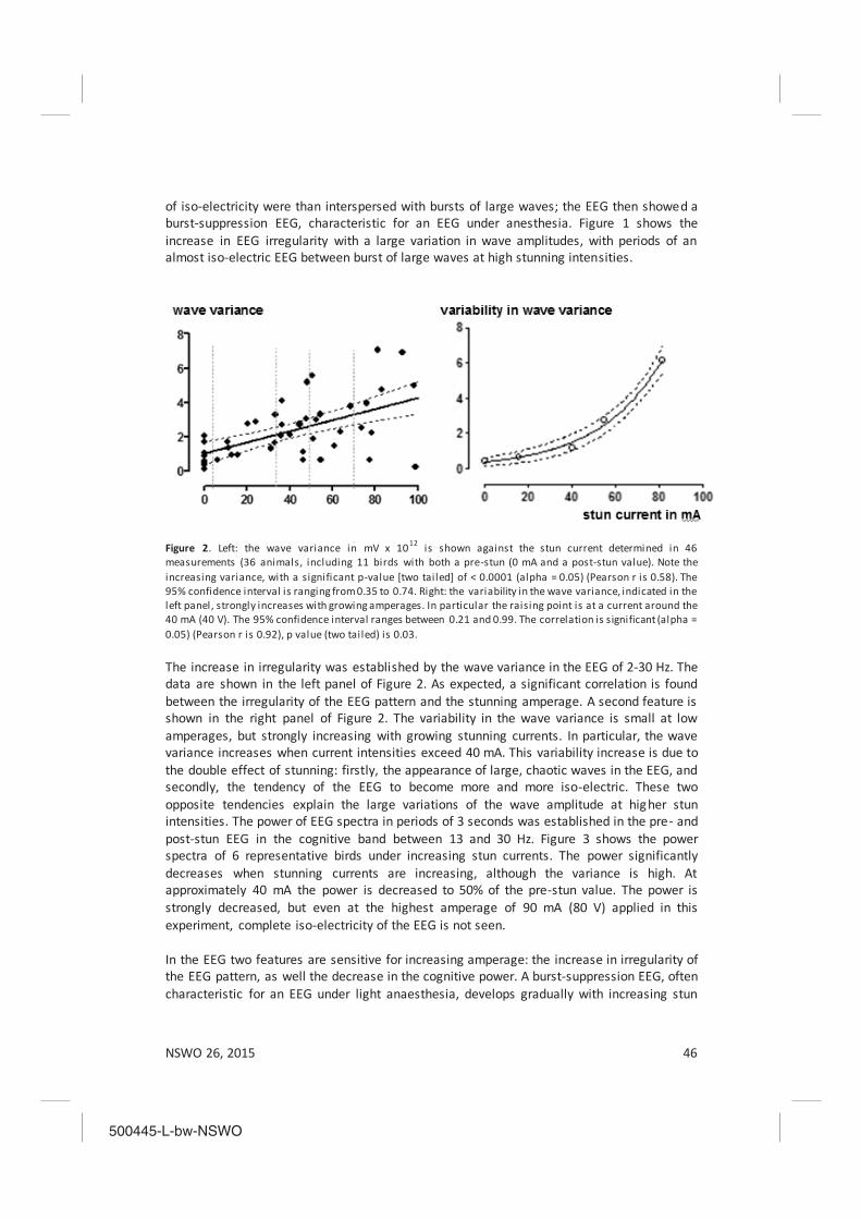
500445-L-bw-NSWO500445-L-bw-NSWO500445-L-bw-NSWO500445-L-bw-NSWO
NSWO 26, 2015 46
of iso-electricity were than interspersed with bursts of large waves; the EEG then showed a burst-suppression EEG, characteristic for an EEG under anesthesia. Figure 1 shows the increase in EEG irregularity with a large variation in wave amplitudes, with periods of an almost iso-electric EEG between burst of large waves at high stunning intensities.
Figure 2. Left: the wave variance in mV x 1012 is shown against the stun current determined in 46 measurements (36 animals, including 11 birds with both a pre-stun (0 mA and a post-stun value). Note the increasing variance, with a significant p-value [two tailed] of < 0.0001 (alpha = 0.05) (Pearson r is 0.58). The 95% confidence interval is ranging from 0.35 to 0.74. Right: the variability in the wave variance, indicated in the left panel, strongly increases with growing amperages. In particular the raising point is at a current around the 40 mA (40 V). The 95% confidence interval ranges between 0.21 and 0.99. The correlation is significant (alpha = 0.05) (Pearson r is 0.92), p value (two tailed) is 0.03. The increase in irregularity was established by the wave variance in the EEG of 2-30 Hz. The data are shown in the left panel of Figure 2. As expected, a significant correlation is found between the irregularity of the EEG pattern and the stunning amperage. A second feature is shown in the right panel of Figure 2. The variability in the wave variance is small at low amperages, but strongly increasing with growing stunning currents. In particular, the wave variance increases when current intensities exceed 40 mA. This variability increase is due to the double effect of stunning: firstly, the appearance of large, chaotic waves in the EEG, and secondly, the tendency of the EEG to become more and more iso-electric. These two opposite tendencies explain the large variations of the wave amplitude at higher stun intensities. The power of EEG spectra in periods of 3 seconds was established in the pre- and post-stun EEG in the cognitive band between 13 and 30 Hz. Figure 3 shows the power spectra of 6 representative birds under increasing stun currents. The power significantly decreases when stunning currents are increasing, although the variance is high. At approximately 40 mA the power is decreased to 50% of the pre-stun value. The power is strongly decreased, but even at the highest amperage of 90 mA (80 V) applied in this experiment, complete iso-electricity of the EEG is not seen. In the EEG two features are sensitive for increasing amperage: the increase in irregularity of the EEG pattern, as well the decrease in the cognitive power. A burst-suppression EEG, often characteristic for an EEG under light anaesthesia, develops gradually with increasing stun
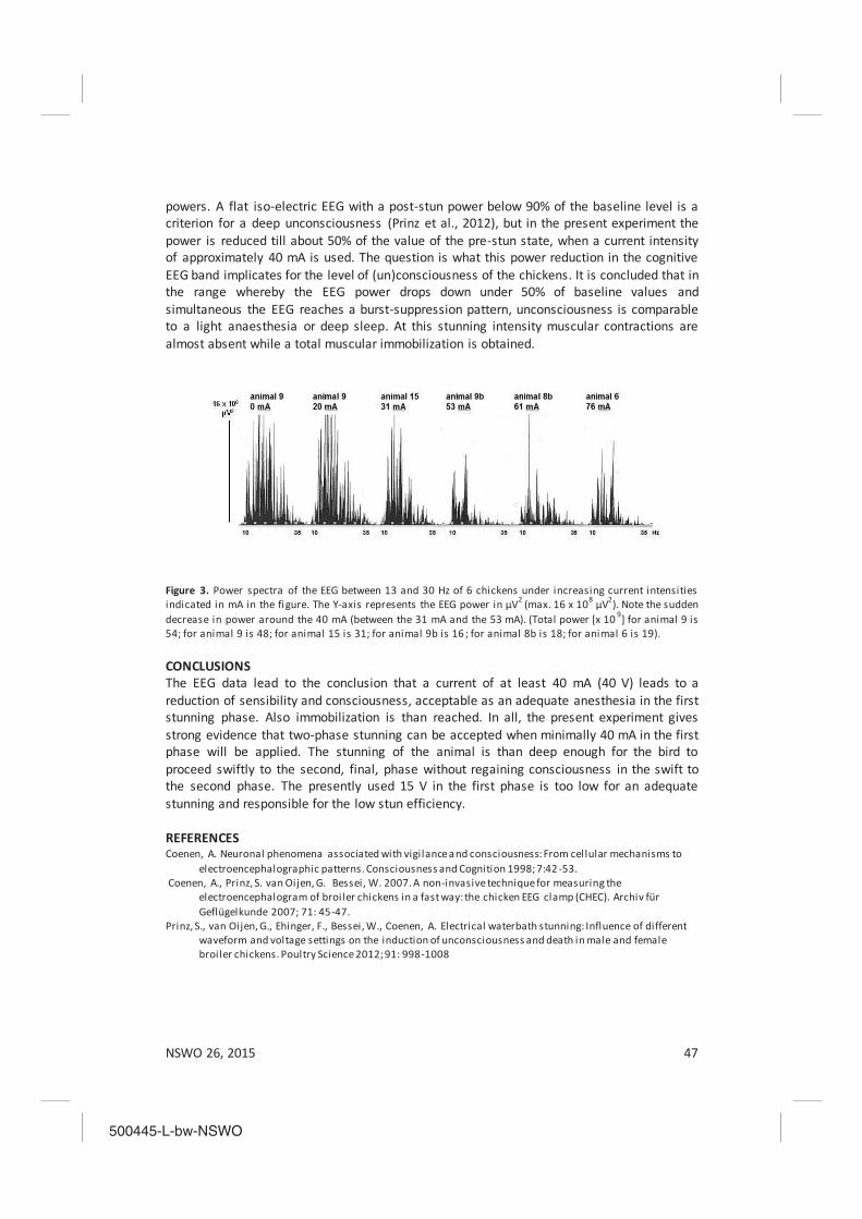
500445-L-bw-NSWO500445-L-bw-NSWO500445-L-bw-NSWO500445-L-bw-NSWO
NSWO 26, 2015 47
powers. A flat iso-electric EEG with a post-stun power below 90% of the baseline level is a criterion for a deep unconsciousness (Prinz et al., 2012), but in the present experiment the power is reduced till about 50% of the value of the pre-stun state, when a current intensity of approximately 40 mA is used. The question is what this power reduction in the cognitive EEG band implicates for the level of (un)consciousness of the chickens. It is concluded that in the range whereby the EEG power drops down under 50% of baseline values and simultaneous the EEG reaches a burst-suppression pattern, unconsciousness is comparable to a light anaesthesia or deep sleep. At this stunning intensity muscular contractions are almost absent while a total muscular immobilization is obtained.
Figure 3. Power spectra of the EEG between 13 and 30 Hz of 6 chickens under increasing current intensities indicated in mA in the figure. The Y-axis represents the EEG power in μV2 (max. 16 x 108 μV2). Note the sudden decrease in power around the 40 mA (between the 31 mA and the 53 mA). (Total power [x 10 9] for animal 9 is 54; for animal 9 is 48; for animal 15 is 31; for animal 9b is 16 ; for animal 8b is 18; for animal 6 is 19). CONCLUSIONS The EEG data lead to the conclusion that a current of at least 40 mA (40 V) leads to a reduction of sensibility and consciousness, acceptable as an adequate anesthesia in the first stunning phase. Also immobilization is than reached. In all, the present experiment gives strong evidence that two-phase stunning can be accepted when minimally 40 mA in the first phase will be applied. The stunning of the animal is than deep enough for the bird to proceed swiftly to the second, final, phase without regaining consciousness in the swift to the second phase. The presently used 15 V in the first phase is too low for an adequate stunning and responsible for the low stun efficiency. REFERENCES Coenen, A. Neuronal phenomena associated with vigilance and consciousness: From cellular mechanisms to
electroencephalographic patterns. Consciousness and Cognition 1998; 7:42 -53. Coenen, A., Prinz, S. van Oijen, G. Bessei, W. 2007. A non-invasive technique for measuring the
electroencephalogram of broiler chickens in a fast way: the chicken EEG clamp (CHEC). Archiv für Geflügelkunde 2007; 71: 45-47.
Prinz, S., van Oijen, G., Ehinger, F., Bessei, W., Coenen, A. Electrical waterbath stunning: Influence of different waveform and voltage settings on the induction of unconsciousness and death in male and female broiler chickens. Poultry Science 2012; 91: 998-1008
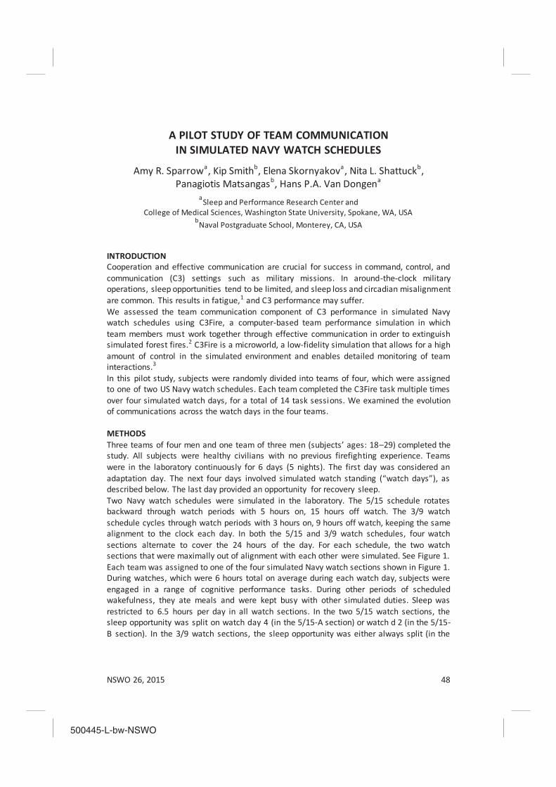
500445-L-bw-NSWO500445-L-bw-NSWO500445-L-bw-NSWO500445-L-bw-NSWO
NSWO 26, 2015 48
A PILOT STUDY OF TEAM COMMUNICATION IN SIMULATED NAVY WATCH SCHEDULES
Amy R. Sparrowa, Kip Smithb, Elena Skornyakova, Nita L. Shattuckb, Panagiotis Matsangasb, Hans P.A. Van Dongena
aSleep and Performance Research Center and College of Medical Sciences, Washington State University, Spokane, WA, USA
bNaval Postgraduate School, Monterey, CA, USA
INTRODUCTION Cooperation and effective communication are crucial for success in command, control, and communication (C3) settings such as military missions. In around-the-clock military operations, sleep opportunities tend to be limited, and sleep loss and circadian misalignment are common. This results in fatigue,1 and C3 performance may suffer. We assessed the team communication component of C3 performance in simulated Navy watch schedules using C3Fire, a computer-based team performance simulation in which team members must work together through effective communication in order to extinguish simulated forest fires.2 C3Fire is a microworld, a low-fidelity simulation that allows for a high amount of control in the simulated environment and enables detailed monitoring of team interactions.3 In this pilot study, subjects were randomly divided into teams of four, which were assigned to one of two US Navy watch schedules. Each team completed the C3Fire task multiple times over four simulated watch days, for a total of 14 task sessions. We examined the evolution of communications across the watch days in the four teams. METHODS Three teams of four men and one team of three men (subjects’ ages: 18–29) completed the study. All subjects were healthy civilians with no previous firefighting experience. Teams were in the laboratory continuously for 6 days (5 nights). The first day was considered an adaptation day. The next four days involved simulated watch standing (“watch days”), as described below. The last day provided an opportunity for recovery sleep. Two Navy watch schedules were simulated in the laboratory. The 5/15 schedule rotates backward through watch periods with 5 hours on, 15 hours off watch. The 3/9 watch schedule cycles through watch periods with 3 hours on, 9 hours off watch, keeping the same alignment to the clock each day. In both the 5/15 and 3/9 watch schedules, four watch sections alternate to cover the 24 hours of the day. For each schedule, the two watch sections that were maximally out of alignment with each other were simulated. See Figure 1. Each team was assigned to one of the four simulated Navy watch sections shown in Figure 1. During watches, which were 6 hours total on average during each watch day, subjects were engaged in a range of cognitive performance tasks. During other periods of scheduled wakefulness, they ate meals and were kept busy with other simulated duties. Sleep was restricted to 6.5 hours per day in all watch sections. In the two 5/15 watch sections, the sleep opportunity was split on watch day 4 (in the 5/15-A section) or watch d 2 (in the 5/15-B section). In the 3/9 watch sections, the sleep opportunity was either always split (in the
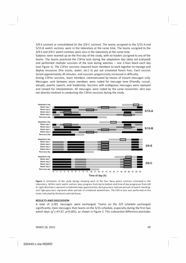
500445-L-bw-NSWO500445-L-bw-NSWO500445-L-bw-NSWO500445-L-bw-NSWO
NSWO 26, 2015 49
3/9-S section) or consolidated (in the 3/9-C section). The teams assigned to the 5/15-A and 5/15-B watch sections were in the laboratory at the same time. The teams assigned to the 3/9-S and 3/9-C watch sections were also in the laboratory at the same time. Subjects were teamed up on the first day of the study, with no leaders assigned to any of the teams. The teams practiced the C3Fire task during the adaptation day (data not analyzed) and performed multiple sessions of the task during watches – one 2-hour block each day (see Figure 1). The C3Fire sessions required team members to work together to manage and deploy resources (fire trucks, water, etc.) to put out simulated forest fires. Each session lasted approximately 30 minutes, and sessions progressively increased in difficulty. During C3Fire sessions, team members communicated by means of instant messages only. Messages sent between team members were coded for message tone (friendly, casual, abrupt), puerile speech, and leadership. Sessions with ambiguous messages were replayed and viewed for interpretation. All messages were coded by the same researcher, who was not directly involved in conducting the C3Fire sessions during the study.
ı
Watch day 2Watch day 3
30
Watch day 3
Watch day 1
13 14 2316 17 18 19 20 21 226 10 118 97 151 2 124 5Recovery day
Adaptation day
Adaptation dayWatch day 1
Watch day 4
Watch day 2
Watch day 1
Watch day 3
Adaptation dayWatch day 1
Recovery day
Adaptation day
Watch day 4Recovery day
Watch day 4
Watch day 2
Watch day 2
Watch day 4Watch day 3
Recovery day
Time of day (h)
5/15-A
5/15-B
3/9-S
3/9-C
Figure 1. Schematic of the study design showing each of the four Navy watch sections simulated in the laboratory. Within each watch section, days progress from top to bottom and time of day progresses from left to right. Black bars represent scheduled sleep opportunities, dark gray bars indicate periods of watch standing, and light gray bars represent other periods of scheduled wakefulness. The C3Fire task was performed at th e times indicated by the black outlined boxes. RESULTS AND DISCUSSION A total of 2,281 messages were exchanged. Teams on the 3/9 schedule exchanged significantly more messages than teams on the 5/15 schedule, especially during the first two watch days (χ2
3=47.67, p<0.001), as shown in Figure 2. This substantial difference precludes
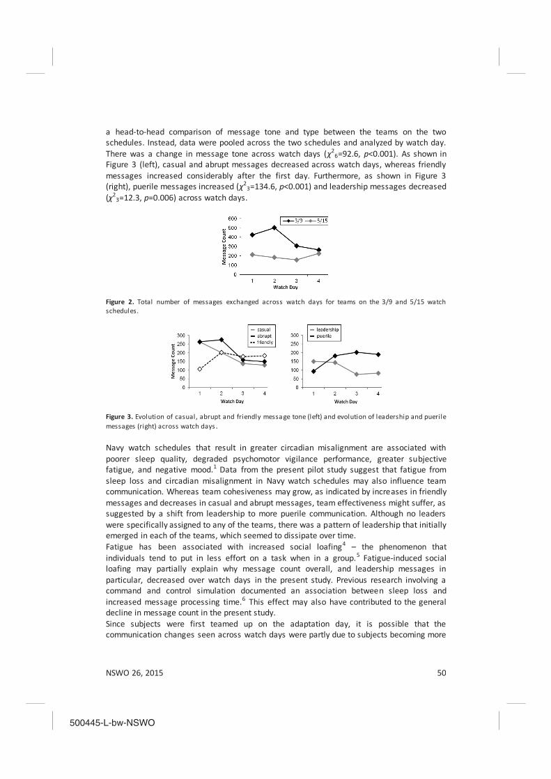
500445-L-bw-NSWO500445-L-bw-NSWO500445-L-bw-NSWO500445-L-bw-NSWO
NSWO 26, 2015 50
a head-to-head comparison of message tone and type between the teams on the two schedules. Instead, data were pooled across the two schedules and analyzed by watch day. There was a change in message tone across watch days (χ2
6=92.6, p<0.001). As shown in Figure 3 (left), casual and abrupt messages decreased across watch days, whereas friendly messages increased considerably after the first day. Furthermore, as shown in Figure 3 (right), puerile messages increased (χ2
3=134.6, p<0.001) and leadership messages decreased (χ2
3=12.3, p=0.006) across watch days.
Figure 2. Total number of messages exchanged across watch days for teams on the 3/9 and 5/15 watch schedules.
Figure 3. Evolution of casual, abrupt and friendly messa ge tone (left) and evolution of leadership and puerile messages (right) across watch days . Navy watch schedules that result in greater circadian misalignment are associated with poorer sleep quality, degraded psychomotor vigilance performance, greater subjective fatigue, and negative mood.1 Data from the present pilot study suggest that fatigue from sleep loss and circadian misalignment in Navy watch schedules may also influence team communication. Whereas team cohesiveness may grow, as indicated by increases in friendly messages and decreases in casual and abrupt messages, team effectiveness might suffer, as suggested by a shift from leadership to more puerile communication. Although no leaders were specifically assigned to any of the teams, there was a pattern of leadership that initially emerged in each of the teams, which seemed to dissipate over time. Fatigue has been associated with increased social loafing4 – the phenomenon that individuals tend to put in less effort on a task when in a group.5 Fatigue-induced social loafing may partially explain why message count overall, and leadership messages in particular, decreased over watch days in the present study. Previous research involving a command and control simulation documented an association between sleep loss and increased message processing time.6 This effect may also have contributed to the general decline in message count in the present study. Since subjects were first teamed up on the adaptation day, it is possible that the communication changes seen across watch days were partly due to subjects becoming more
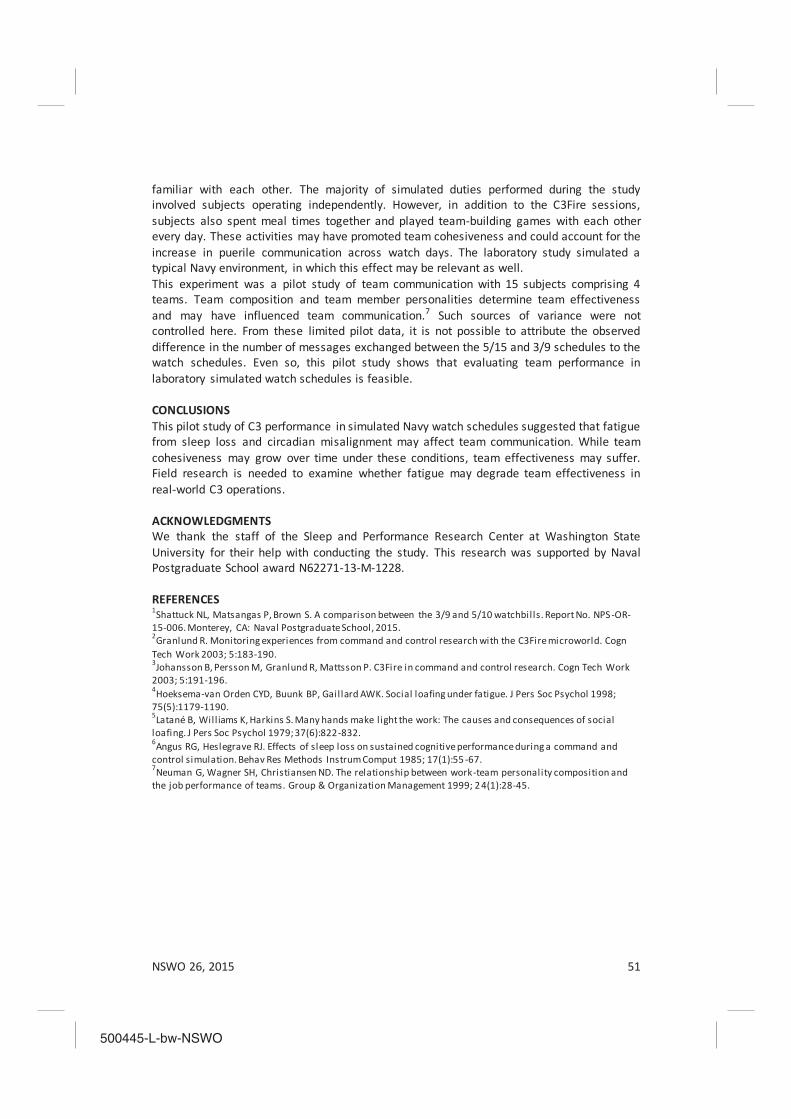
500445-L-bw-NSWO500445-L-bw-NSWO500445-L-bw-NSWO500445-L-bw-NSWO
NSWO 26, 2015 51
familiar with each other. The majority of simulated duties performed during the study involved subjects operating independently. However, in addition to the C3Fire sessions, subjects also spent meal times together and played team-building games with each other every day. These activities may have promoted team cohesiveness and could account for the increase in puerile communication across watch days. The laboratory study simulated a typical Navy environment, in which this effect may be relevant as well. This experiment was a pilot study of team communication with 15 subjects comprising 4 teams. Team composition and team member personalities determine team effectiveness and may have influenced team communication.7 Such sources of variance were not controlled here. From these limited pilot data, it is not possible to attribute the observed difference in the number of messages exchanged between the 5/15 and 3/9 schedules to the watch schedules. Even so, this pilot study shows that evaluating team performance in laboratory simulated watch schedules is feasible. CONCLUSIONS This pilot study of C3 performance in simulated Navy watch schedules suggested that fatigue from sleep loss and circadian misalignment may affect team communication. While team cohesiveness may grow over time under these conditions, team effectiveness may suffer. Field research is needed to examine whether fatigue may degrade team effectiveness in real-world C3 operations. ACKNOWLEDGMENTS We thank the staff of the Sleep and Performance Research Center at Washington State University for their help with conducting the study. This research was supported by Naval Postgraduate School award N62271-13-M-1228. REFERENCES 1Shattuck NL, Matsangas P, Brown S. A comparison between the 3/9 and 5/10 watchbills. Report No. NPS-OR-15-006. Monterey, CA: Naval Postgraduate School, 2015. 2Granlund R. Monitoring experiences from command and control research with the C3Fire microworld. Cogn Tech Work 2003; 5:183-190. 3Johansson B, Persson M, Granlund R, Mattsson P. C3Fire in command and control research. Cogn Tech Work 2003; 5:191-196. 4Hoeksema-van Orden CYD, Buunk BP, Gaillard AWK. Social loafing under fatigue. J Pers Soc Psychol 1998; 75(5):1179-1190. 5Latané B, Will iams K, Harkins S. Many hands make light the work: The causes and consequences of social loafing. J Pers Soc Psychol 1979; 37(6):822-832. 6Angus RG, Heslegrave RJ. Effects of sleep loss on sustained cognitive performance during a command and control simulation. Behav Res Methods Instrum Comput 1985; 17(1):55 -67. 7Neuman G, Wagner SH, Christiansen ND. The relationship between work-team personality composition and the job performance of teams. Group & Organization Management 1999; 2 4(1):28-45.
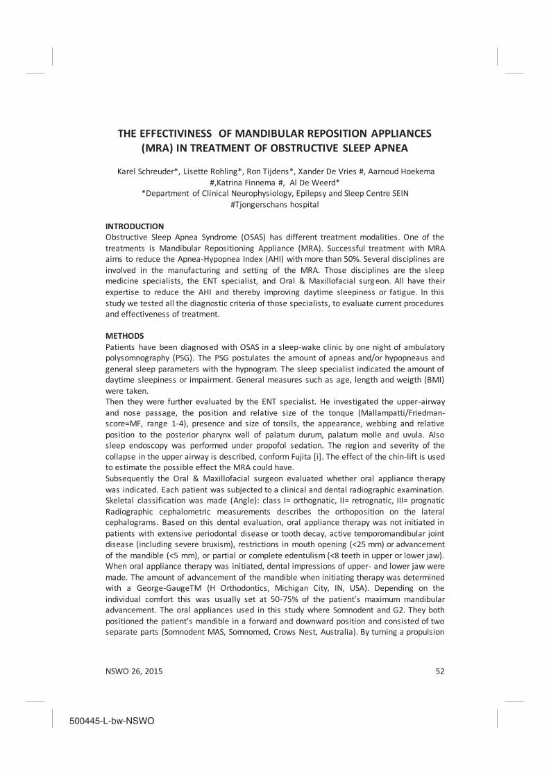
500445-L-bw-NSWO500445-L-bw-NSWO500445-L-bw-NSWO500445-L-bw-NSWO
NSWO 26, 2015 52
THE EFFECTIVINESS OF MANDIBULAR REPOSITION APPLIANCES (MRA) IN TREATMENT OF OBSTRUCTIVE SLEEP APNEA
Karel Schreuder*, Lisette Rohling*, Ron Tijdens*, Xander De Vries #, Aarnoud Hoekema
#,Katrina Finnema #, Al De Weerd* *Department of Clinical Neurophysiology, Epilepsy and Sleep Centre SEIN
#Tjongerschans hospital INTRODUCTION Obstructive Sleep Apnea Syndrome (OSAS) has different treatment modalities. One of the treatments is Mandibular Repositioning Appliance (MRA). Successful treatment with MRA aims to reduce the Apnea-Hypopnea Index (AHI) with more than 50%. Several disciplines are involved in the manufacturing and setting of the MRA. Those disciplines are the sleep medicine specialists, the ENT specialist, and Oral & Maxillofacial surg eon. All have their expertise to reduce the AHI and thereby improving daytime sleepiness or fatigue. In this study we tested all the diagnostic criteria of those specialists, to evaluate current procedures and effectiveness of treatment. METHODS Patients have been diagnosed with OSAS in a sleep-wake clinic by one night of ambulatory polysomnography (PSG). The PSG postulates the amount of apneas and/or hypopneaus and general sleep parameters with the hypnogram. The sleep specialist indicated the amount of daytime sleepiness or impairment. General measures such as age, length and weigth (BMI) were taken. Then they were further evaluated by the ENT specialist. He investigated the upper-airway and nose passage, the position and relative size of the tonque (Mallampatti/Friedman-score=MF, range 1-4), presence and size of tonsils, the appearance, webbing and relative position to the posterior pharynx wall of palatum durum, palatum molle and uvula. Also sleep endoscopy was performed under propofol sedation. The region and severity of the collapse in the upper airway is described, conform Fujita [i]. The effect of the chin-lift is used to estimate the possible effect the MRA could have. Subsequently the Oral & Maxillofacial surgeon evaluated whether oral appliance therapy was indicated. Each patient was subjected to a clinical and dental radiographic examination. Skeletal classification was made (Angle): class I= orthognatic, II= retrognatic, III= prognatic Radiographic cephalometric measurements describes the orthoposition on the lateral cephalograms. Based on this dental evaluation, oral appliance therapy was not initiated in patients with extensive periodontal disease or tooth decay, active temporomandibular joint disease (including severe bruxism), restrictions in mouth opening (<25 mm) or advancement of the mandible (<5 mm), or partial or complete edentulism (<8 teeth in upper or lower jaw). When oral appliance therapy was initiated, dental impressions of upper- and lower jaw were made. The amount of advancement of the mandible when initiating therapy was determined with a George-GaugeTM (H Orthodontics, Michigan City, IN, USA). Depending on the individual comfort this was usually set at 50-75% of the patient’s maximum mandibular advancement. The oral appliances used in this study where Somnodent and G2. They both positioned the patient’s mandible in a forward and downward position and consisted of two separate parts (Somnodent MAS, Somnomed, Crows Nest, Australia). By turning a propulsion
500445-L-bw-NSWO500445-L-bw-NSWO500445-L-bw-NSWO500445-L-bw-NSWO
NSWO 26, 2015 53
screw incorporated in both sides of the upper appliance, patients could adjust mandibular advancement in 0.1 mm increments. After patients became accustomed to the oral appliance during a two-week period, they returned for a check-up visit. At this stage patients were instructed to advance the mandible until symptoms abated (i.e., snoring, apneas, hypopneas, or excessive sleepiness) or until further advancement caused discomfort. After 2 to 3 months of oral appliance use patients were referred back to the sleep clinic for a polysomnographic assessment of therapeutic efficacy. RESULTS 23 patients of 25 were evaluated, of which 18 were male and 5 female. The preliminary results of 23 patients, mean age =51.5 (33-66) yrs), mean BMI= 28.3, sd= 3.7 (23.1-39.6), indicated that the AHI reduced significantly during the MRA therapy from AHI=17.23, sd=7.49, to AHI=9.79, sd=8.12 (p<0.001) .
Patients with Mallampatti/Friedmanscore MF=3 had higher values of AHI at diagnosis (AHI=19.3; sd=2.2) than MF=2 (AHI=13.2; sd=2.6), the reducing effect was similar and there was no interaction effect between the Friedman-Mallampati score and AHI (p=0.197).
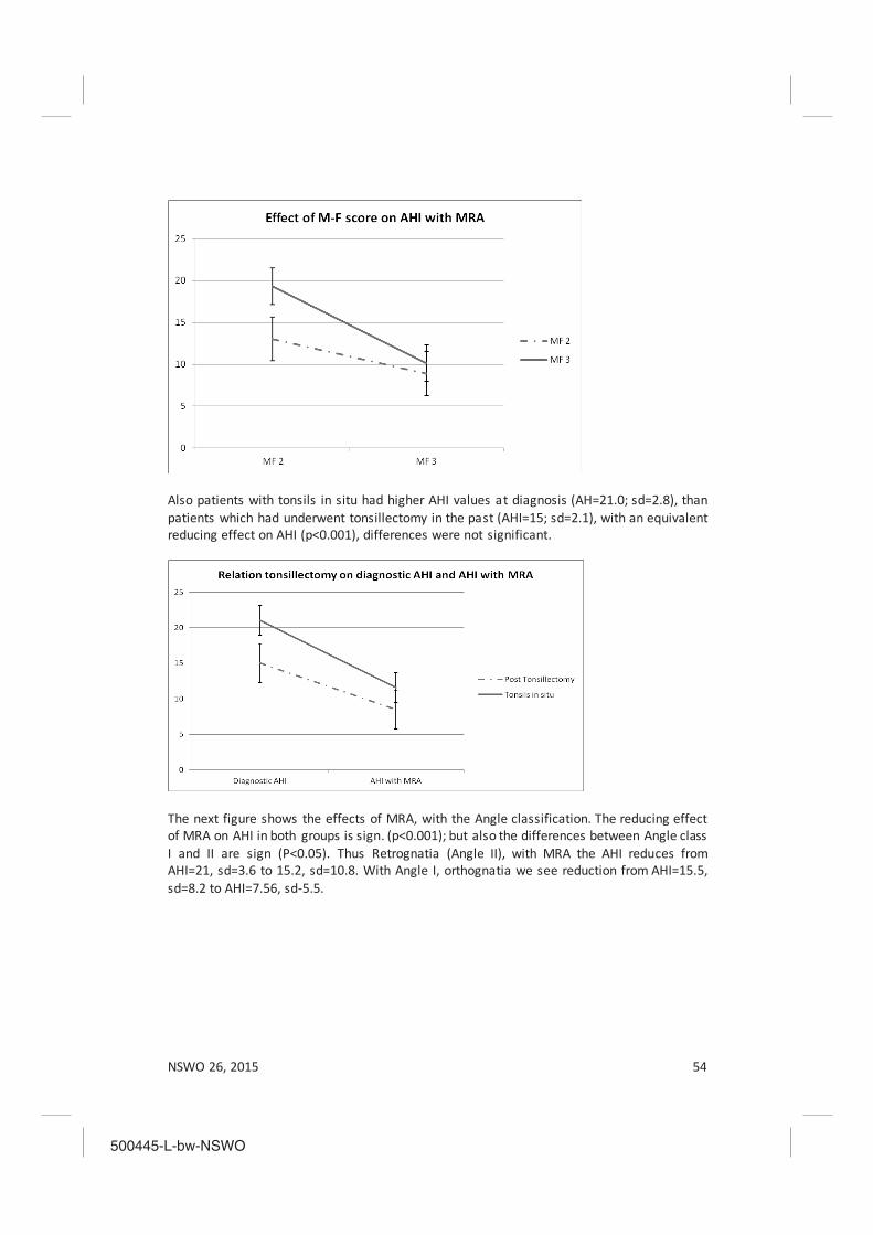
500445-L-bw-NSWO500445-L-bw-NSWO500445-L-bw-NSWO500445-L-bw-NSWO
NSWO 26, 2015 54
Also patients with tonsils in situ had higher AHI values at diagnosis (AH=21.0; sd=2.8), than patients which had underwent tonsillectomy in the past (AHI=15; sd=2.1), with an equivalent reducing effect on AHI (p<0.001), differences were not significant.
The next figure shows the effects of MRA, with the Angle classification. The reducing effect of MRA on AHI in both groups is sign. (p<0.001); but also the differences between Angle class I and II are sign (P<0.05). Thus Retrognatia (Angle II), with MRA the AHI reduces from AHI=21, sd=3.6 to 15.2, sd=10.8. With Angle I, orthognatia we see reduction from AHI=15.5, sd=8.2 to AHI=7.56, sd-5.5.
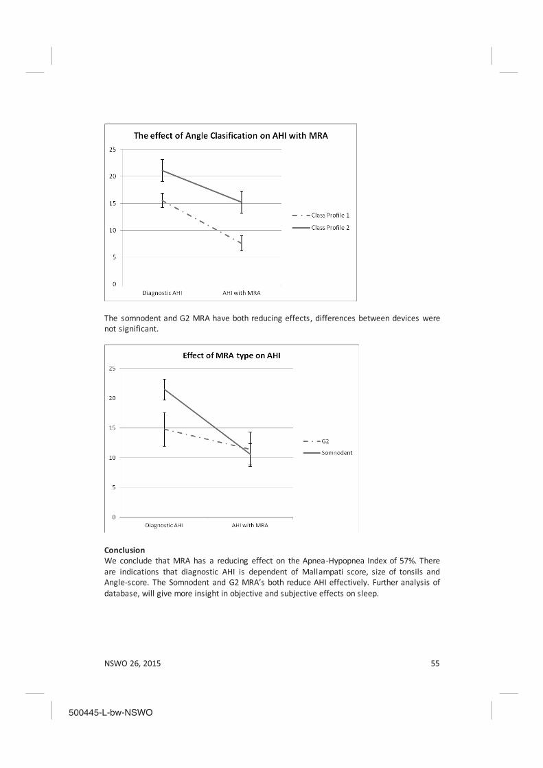
500445-L-bw-NSWO500445-L-bw-NSWO500445-L-bw-NSWO500445-L-bw-NSWO
NSWO 26, 2015 55
The somnodent and G2 MRA have both reducing effects, differences between devices were not significant.
Conclusion We conclude that MRA has a reducing effect on the Apnea-Hypopnea Index of 57%. There are indications that diagnostic AHI is dependent of Mallampati score, size of tonsils and Angle-score. The Somnodent and G2 MRA’s both reduce AHI effectively. Further analysis of database, will give more insight in objective and subjective effects on sleep.

500445-L-bw-NSWO500445-L-bw-NSWO500445-L-bw-NSWO500445-L-bw-NSWO
NSWO 26, 2015 56
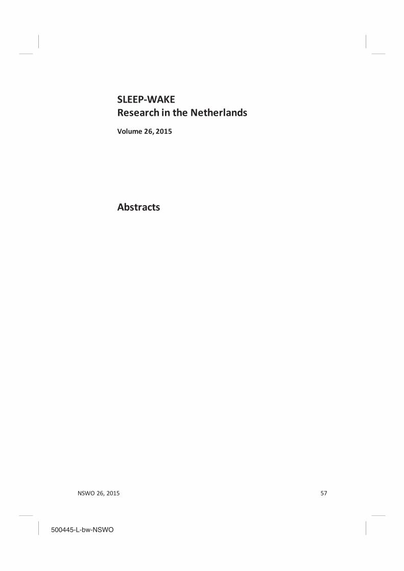
500445-L-bw-NSWO500445-L-bw-NSWO500445-L-bw-NSWO500445-L-bw-NSWO
NSWO 26, 2015 57
SLEEP-WAKE Research in the Netherlands
Volume 26, 2015
Abstracts
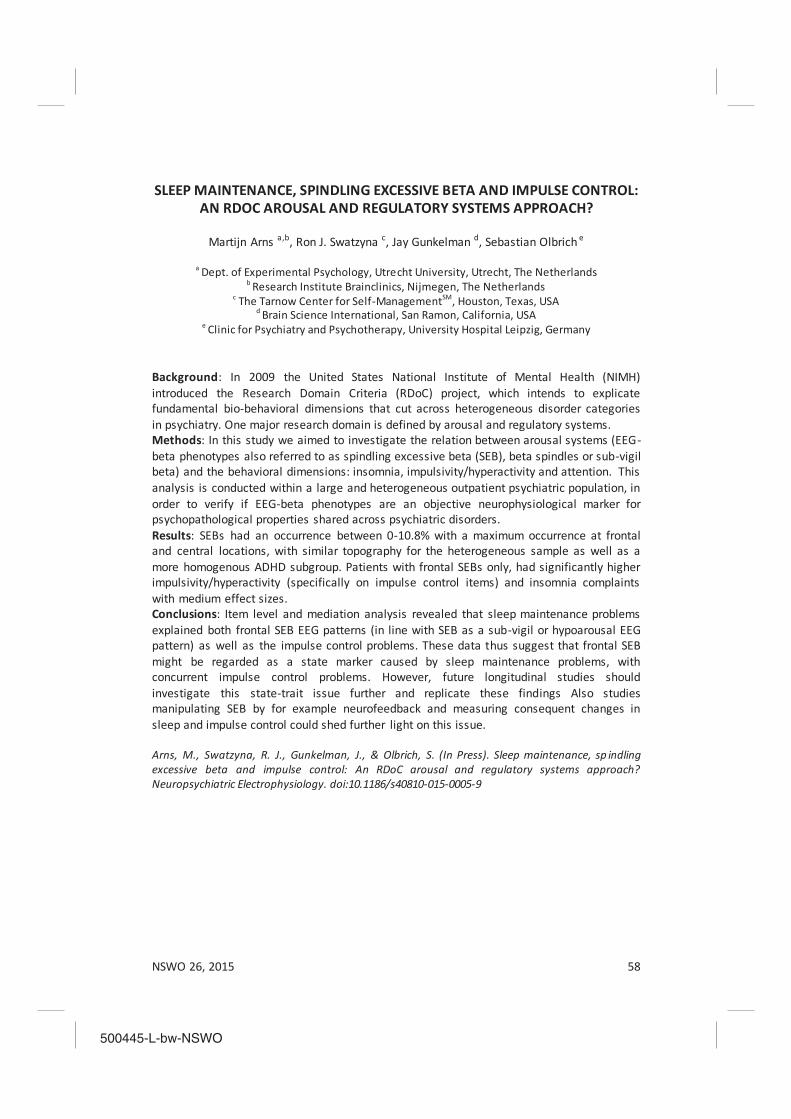
500445-L-bw-NSWO500445-L-bw-NSWO500445-L-bw-NSWO500445-L-bw-NSWO
NSWO 26, 2015 58
SLEEP MAINTENANCE, SPINDLING EXCESSIVE BETA AND IMPULSE CONTROL: AN RDOC AROUSAL AND REGULATORY SYSTEMS APPROACH?
Martijn Arns a,b, Ron J. Swatzyna c, Jay Gunkelman d, Sebastian Olbrich e
a Dept. of Experimental Psychology, Utrecht University, Utrecht, The Netherlands
b Research Institute Brainclinics, Nijmegen, The Netherlands c The Tarnow Center for Self-ManagementSM, Houston, Texas, USA
d Brain Science International, San Ramon, California, USA e Clinic for Psychiatry and Psychotherapy, University Hospital Leipzig, Germany
Background: In 2009 the United States National Institute of Mental Health (NIMH) introduced the Research Domain Criteria (RDoC) project, which intends to explicate fundamental bio-behavioral dimensions that cut across heterogeneous disorder categories in psychiatry. One major research domain is defined by arousal and regulatory systems. Methods: In this study we aimed to investigate the relation between arousal systems (EEG-beta phenotypes also referred to as spindling excessive beta (SEB), beta spindles or sub-vigil beta) and the behavioral dimensions: insomnia, impulsivity/hyperactivity and attention. This analysis is conducted within a large and heterogeneous outpatient psychiatric population, in order to verify if EEG-beta phenotypes are an objective neurophysiological marker for psychopathological properties shared across psychiatric disorders. Results: SEBs had an occurrence between 0-10.8% with a maximum occurrence at frontal and central locations, with similar topography for the heterogeneous sample as well as a more homogenous ADHD subgroup. Patients with frontal SEBs only, had significantly higher impulsivity/hyperactivity (specifically on impulse control items) and insomnia complaints with medium effect sizes. Conclusions: Item level and mediation analysis revealed that sleep maintenance problems explained both frontal SEB EEG patterns (in line with SEB as a sub-vigil or hypoarousal EEG pattern) as well as the impulse control problems. These data thus suggest that frontal SEB might be regarded as a state marker caused by sleep maintenance problems, with concurrent impulse control problems. However, future longitudinal studies should investigate this state-trait issue further and replicate these findings Also studies manipulating SEB by for example neurofeedback and measuring consequent changes in sleep and impulse control could shed further light on this issue. Arns, M., Swatzyna, R. J., Gunkelman, J., & Olbrich, S. (In Press). Sleep maintenance, sp indling excessive beta and impulse control: An RDoC arousal and regulatory systems approach? Neuropsychiatric Electrophysiology. doi:10.1186/s40810-015-0005-9
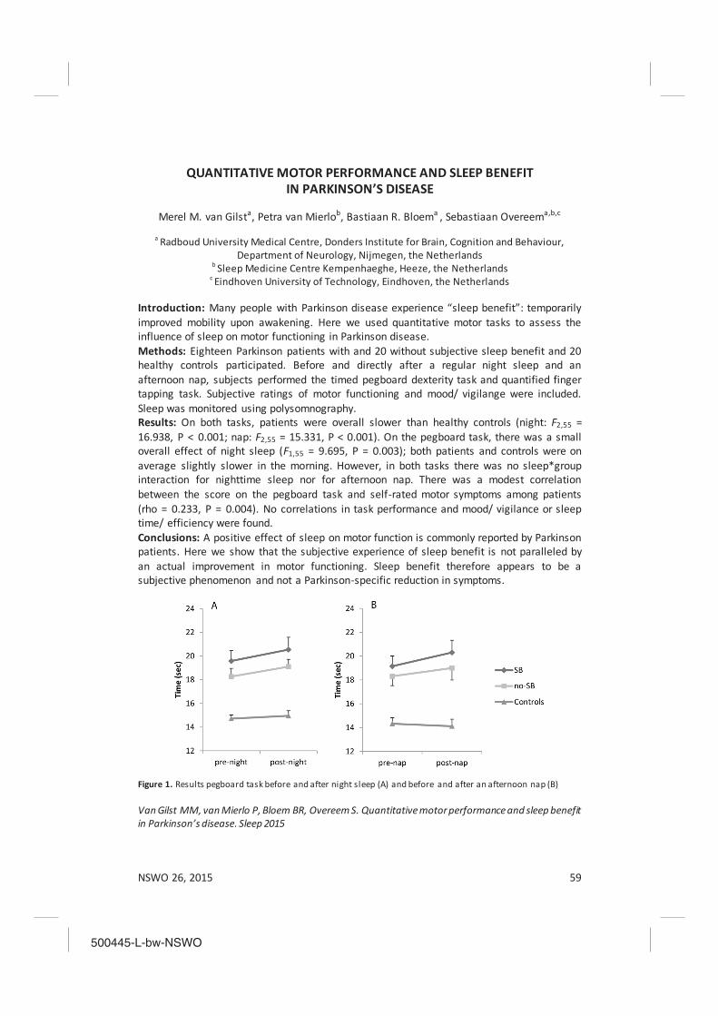
500445-L-bw-NSWO500445-L-bw-NSWO500445-L-bw-NSWO500445-L-bw-NSWO
NSWO 26, 2015 59
QUANTITATIVE MOTOR PERFORMANCE AND SLEEP BENEFIT IN PARKINSON’S DISEASE
Merel M. van Gilsta, Petra van Mierlob, Bastiaan R. Bloema , Sebastiaan Overeema,b,c
a Radboud University Medical Centre, Donders Institute for Brain, Cognition and Behaviour,
Department of Neurology, Nijmegen, the Netherlands b Sleep Medicine Centre Kempenhaeghe, Heeze, the Netherlands
c Eindhoven University of Technology, Eindhoven, the Netherlands Introduction: Many people with Parkinson disease experience “sleep benefit”: temporarily improved mobility upon awakening. Here we used quantitative motor tasks to assess the influence of sleep on motor functioning in Parkinson disease. Methods: Eighteen Parkinson patients with and 20 without subjective sleep benefit and 20 healthy controls participated. Before and directly after a regular night sleep and an afternoon nap, subjects performed the timed pegboard dexterity task and quantified finger tapping task. Subjective ratings of motor functioning and mood/ vigilange were included. Sleep was monitored using polysomnography. Results: On both tasks, patients were overall slower than healthy controls (night: F2,55 = 16.938, P < 0.001; nap: F2,55 = 15.331, P < 0.001). On the pegboard task, there was a small overall effect of night sleep (F1,55 = 9.695, P = 0.003); both patients and controls were on average slightly slower in the morning. However, in both tasks there was no sleep*group interaction for nighttime sleep nor for afternoon nap. There was a modest correlation between the score on the pegboard task and self-rated motor symptoms among patients (rho = 0.233, P = 0.004). No correlations in task performance and mood/ vigilance or sleep time/ efficiency were found. Conclusions: A positive effect of sleep on motor function is commonly reported by Parkinson patients. Here we show that the subjective experience of sleep benefit is not paralleled by an actual improvement in motor functioning. Sleep benefit therefore appears to be a subjective phenomenon and not a Parkinson-specific reduction in symptoms.
Figure 1. Results pegboard task before and after night sleep (A) and before and after an afternoon nap (B) Van Gilst MM, van Mierlo P, Bloem BR, Overeem S. Quantitative motor performance and sleep benefit in Parkinson’s disease. Sleep 2015
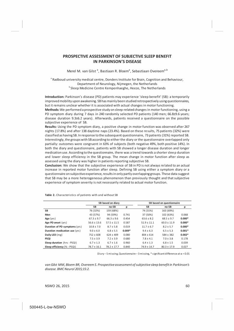
500445-L-bw-NSWO500445-L-bw-NSWO500445-L-bw-NSWO500445-L-bw-NSWO
NSWO 26, 2015 60
PROSPECTIVE ASSESSMENT OF SUBJECTIVE SLEEP BENEFIT IN PARKINSON’S DISEASE
Merel M. van Gilst a, Bastiaan R. Bloema, Sebastiaan Overeema,b
a Radboud university medical centre, Donders Institute for Brain, Cognition and Behaviour,
Department of Neurology, Nijmegen, the Netherlands b Sleep Medicine Centre Kempenhaeghe, Heeze, The Netherlands
Introduction: Parkinson’s disease (PD) patients may experience ‘sleep benefit’ (SB): a temporarily improved mobility upon awakening. SB has mainly been studied retrospectively using questionnaires, but it remains unclear whether it is associated with actual changes in motor functioning. Methods: We performed a prospective study on sleep-related changes in motor functioning, using a PD symptom diary during 7 days in 240 randomly selected PD patients (140 men; 66.8±9.6 years; disease duration 9.3±6.2 years). Afterwards, patients received a questionnaire on the possible subjective experience of SB. Results: Using the PD symptom diary, a positive change in motor function was observed after 267 nights (17.8%) and after 138 daytime naps (23.4%). Based on these results, 75 patients (32%) were classified as having SB. In response to the subsequent questionnaire, 73 patients (31%) reported SB. Interestingly, the groups with SB according to either the diary or the questionnaire overlapped only partially: outcomes were congruent in 63% of subjects (both negative 49%, both positive 14%). In both the diary and questionnaire, patients with SB showed a longer disease duration and longer medication use. According to the questionnaire, there was a trend towards a shorter slee p duration and lower sleep efficiency in the SB group. The mean change in motor function after sleep as assessed using the diary was higher in patients reporting subjective SB. Conclusion: We show that the subjective experience of SB in PD is not always re lated to an actual increase in reported motor function after sleep. Defining SB using either a symptom diary or a questionnaire on subjective experience, results in only partly overlapping groups. These data suggest that SB may be a more heterogeneous phenomenon than previously thought and that subjective experience of symptom severity is not necessarily related to actual motor function. Table 2. Characteristics of patients with and without SB
Diary – 5 missing, Questionnaire – 3 missing, * s ignificant difference at α = 0.01 van Gilst MM, Bloem BR, Overeem S. Prospective assessment of subjective sleep benefit in Parkinson's disease. BMC Neurol 2015;15:2.
SB based on diary SB based on questionnaire SB no-SB p SB no-SB p SB 76 (32%) 159 (68%) 74 (31%) 163 (69%) Men 43 (57%) 94 (59%) 0.741 37 (50%) 102 (63%) 0.068 Age (yrs ) 67.3 ± 9.7 66.3 ± 9.6 0.454 63.6 ± 8.2 68.5 ± 9.7 0.000* Age PD onset (yrs ) 56.6 ± 13.6 57.5 ± 11.5 0.587 51.9 ± 11.1 60.0 ± 11.9 0.000* Duration of PD symptoms (yrs ) 10.8 ± 7.0 8.7 ± 5.8 0.019 11.7 ± 6.7 8.2 ± 5.7 0.000* Duration medication use (yrs ) 9.0 ± 6.0 6.8 ± 5.5 0.009* 9.4 ± 6.3 6.5 ± 5.3 0.001* Daily LED (mg) 752 ± 608 624 ± 409 0.080 804 ± 614 584 ± 382 0.002* PSQI 7.5 ± 3.9 7.2 ± 3.9 0.680 7.8 ± 4.1 7.0 ± 3.8 0.178 Sleep duration (hrs - PSQI) 6.7 ± 1.3 6.7 ± 1.6 0.960 6.4 ± 1.3 6.8 ± 1.5 0.039 Sleep efficiency (% - PSQI) 78.7 ± 16.1 78.2 ± 17.7 0.840 74.9 ± 14.7 80.3 ± 17.9 0.027
500445-L-bw-NSWO500445-L-bw-NSWO500445-L-bw-NSWO500445-L-bw-NSWO
NSWO 26, 2015 61
FRAGMENTATION AND STABILITY OF CIRCADIAN ACTIVITY RHYTHMS PREDICT MORTALITY: THE ROTTERDAM STUDY
Lisette A. Zuurbiera, Annemarie I. Luika, Albert Hofmana, Oscar H. Francoa, Eus J.W. Van
Somerenb,c, Henning Tiemeiera,d,e
aDepartment of Epidemiology, Erasmus Medical Center, Rotterdam, The Netherlands
bDepartment of Sleep and Cognition, Netherlands Institute for Neuroscience, an Institute of the Netherlands Royal Academy of Arts and Sciences, Amsterdam, The Netherlands
cDepartments of Integrative Neurophysiology and Medical Psychology, Center for Neurogenomics and Cognitive Research, Neuroscience Campus Amsterdam, VU University and Medical Center,
Amsterdam, The Netherlands dDepartment of Child and Adolescent Psychiatry, Erasmus Medical Center, Rotterdam, The
Netherlands eDepartment of Psychiatry, Erasmus Medical Center, Rotterdam, The Netherlands
Introduction: Circadian rhythms and sleep patterns change as people age. Little is known about the associations between circadian rhythms and mortality rates. We investigated whether 24-hour activity rhythms and sleep characteristics independently predicted mortality. Methods: Actigraphy was used to determine the stability and fragmentation of the 24-hour activity rhythm in 1,734 persons (aged 45–98 years) from the Rotterdam Study (2004–2013). Sleep was assessed objectively using actigraphy and subjectively using sleep diaries to estimate sleep duration, sleep onset latency, and waking after sleep onset. Results: The mean follow-up time was 7.3 years; 154 participants (8.9%) died. Sleep measures were not related to mortality after adjustment for health parameters. In contrast, a more stable 24-hour activity rhythm was associated with a lower mortality risk (per 1 standard deviation, hazard ratio = 0.83, 95% confidence interval: 0.71, 0.96), and a more fragmented rhythm was associated with a higher mortality risk (per 1 standard deviation, hazard ratio = 1.22, 95% confidence interval: 1.04, 1.44). Conclusion: Low stability and high fragmentation of the 24-hour activity rhythm predicted all-cause mortality, whereas estimates from actigraphy and sleep diaries did not. Disturbed circadian activity rhythms reflect age-related alterations in the biological clock and could be an indicator of disease. L.A. Zuurbier and A.I. Luik were supported by a Netherlands Organization for Scientific Research grant (NOW-VIDI: 017.106.370) awarded to H.T. Zuurbier, L.A., Luik, A.I., Hofman, A., Franco, O.H., Van Someren, E.J.W., & Tiemeier H. (2015). Fragmentation and stability of circadian rhythms predict mortality: The Rotterdam Study. American Journal of Epidemiology, 181 (1), 54-63.
500445-L-bw-NSWO500445-L-bw-NSWO500445-L-bw-NSWO500445-L-bw-NSWO
NSWO 26, 2015 62
REDUCED INFLUENCE OF SATIATION ON FOOD CHOICES IN HUMAN NARCOLEPSY
Sebastiaan Overeema,b, Ruth J. van Holsta,e, Lisa van der Cruijssena, Gert Jan Lammersc,
Roshan Coolse,f, Esther Aartse.
aRadboud university medical center, department of neurology, Nijmegen, the Netherlands b Sleep Medicine Center Kempenhaeghe, Heeze, the Netherlands
c Sleep-Wake Center SEIN, Heemstede, the Netherlands dDonders Institute for brain, cognition and behavior, Nijmegen, The Netherlands
eRadboud university medical center, department of psychiatry, Nijmegen, the Netherlands Introduction: Narcolepsy with cataplexy is a chronic sleep disorder caused by a hypocretin (orexin) deficiency. Besides sleep regulation, hypocretin signaling is important for reward motivational processes, including appetite regulation. As obesity is a common symptom in narcolepsy, we explored food-related choices and their dependence on satiation in patients with type I narcolepsy (n=20) compared with idiopathic hypersomnia (n=13) as well as healthy matched controls (n=18). Methods: After fasting for at least 5 hours, subjects were first trained on a concurrent choice task to earn their favorite savory and sweet snack before one of the snack outcomes was devalued by sensory-specific specific satiation. Subsequently, choice for the savory or sweet snack was tested, without feedback (i.e. in extinction). Goal-directed behavior was measured by the selective reduction in button presses associated with the devalued outcome, e.g. satiation on a savory snack, leading to less button presses associated with obtaining that savory snack in the extinction test relative to the training phase. After the tests, we measured how many calories subjects consumed spontaneously from ad-libitum available food when they were completing food-related questionnaires. Results: After satiation, all groups reported less wanting for the satiated snack than before satiation. However, while controls and idiopathic hypersomnia patients showed goal -directed behavior, patients with narcolepsy still chose the satiated snack as often as before satiation. Narcolepsy patients also spontaneously consumed more calories when completing the questionnaires, although not reporting to eat more in daily life. Conclusion: While narcolepsy patients do report less wanting after being satiated, they do not adjust their behavior accordingly. This discrepancy between self-report and actual behavior was also evident in their spontaneous caloric intake. We conclude that narcolepsy patients exhibit reduced goal-directed control of behavior towards food. This might contribute to the development of obesity. This study was supported by a VIDI research grant from the Netherlands Organization for Scientific Research awarded to S. Overeem (grant no. 016.116.371).
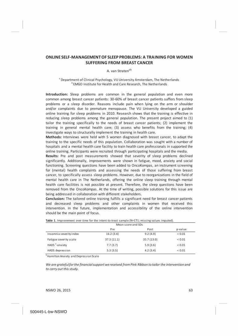
500445-L-bw-NSWO500445-L-bw-NSWO500445-L-bw-NSWO500445-L-bw-NSWO
NSWO 26, 2015 63
ONLINE SELF-MANAGEMENT OF SLEEP PROBLEMS: A TRAINING FOR WOMEN SUFFERING FROM BREAST CANCER
A. van Stratenab
a Department of Clinical Psychology, VU University Amsterdam, The Netherlands
b EMGO Institute for Health and Care Research, The Netherlands Introduction: Sleep problems are common in the general population and even more common among breast cancer patients: 30-60% of breast cancer patients suffers from sleep problems or a sleep disorder. Reasons include pain when lying on the arm or shoulder and/or complaints due to premature menopause. The VU University developed a guided online training for sleep problems in 2010. Research shows that the training is effective in reducing sleep problems among the general population. The present project aimed to (1) tailor the training specifically to the needs of breast cancer patients; (2) implement the training in general mental health care; (3) assess who benefits from the training; (4) investigate ways to structurally implement the training in health care. Methods: Interviews were held with 5 women diagnosed with breast cancer, to adapt the training to the specific needs of this population. Collaboration was sought with a number of hospitals and a mental health care facility to train health care professionals in supported the online training. Participants were recruited through participating hospitals and the media. Results: Pre and post measurements showed that severity of sleep problems declined significantly. Additionally, improvements were shown in fatigue, mood, anxiety and social functioning. Screening questions have been added to OncoKompas, an instrument screening for (mental) health complaints and assessing the needs of those suffering from breast cancer, to specifically assess sleep problems. However, due to reorganizations in the field of mental health care in The Netherlands, offering the online sleep training through mental health care facilities is not possible at present. Therefore, the sleep questions have been removed from the OncoKompas. At the time of writing, possible solutions for this issue are being addressed in collaboration with different stakeholders. Conclusion: The tailored online training fulfills a significant need for breast cancer patients and decreased sleep problems and other complaints in women that received this intervention. In the future, implementation and accessibility of the online intervention should be the main point of focus. Table 1. Improvement over time for the intent-to-treat sample (N=171; missing values imputed).
Mean score and SDs Pre Post p-value Insomnia severity index 16.2 (3.4) 9.2 (4.9) < 0.01
Fatigue severity scale 37.3 (11.1) 35.7 (13.0) < 0.01
HADS a-anxiety 7.7 (3.7) 5.9 (3.6) < 0.01
HADS depression 5.3 (3.5) 4.2 (3.4) < 0.01 a Hamilton Anxiety and Depression Scale We are grateful for the financial support we received from Pink Ribbon to tailor the intervention and to carry out this study.
500445-L-bw-NSWO500445-L-bw-NSWO500445-L-bw-NSWO500445-L-bw-NSWO
NSWO 26, 2015 64
GUIDED INTERNET-DELIVERED COGNITIVE BEHAVIORAL TREATMENT FOR INSOMNIA: A RANDOMIZED TRIAL
A. van Stratenab, J. Emmelkampab, J. de Witab, J. Lanceec, G. Anderssonde, E.J.W. van
Somerenfg, P. Cuijpersab
aDepartment of Clinical Psychology, VU University Amsterdam, The Netherlands
b EMGO Institute for Health and Care Research, The Netherlands c University of Amsterdam (UvA), Department of Clinical Psychology, The Netherlands
d Department of Behavioural Sciences and Learning, Swedisch Institute for Disability Research, Linköping University, Linköping, Sweden
e Department of Clinical Neuroscience, Karolinska Institutet, Stockholm, Sweden f Department of Sleep and Cognition, Netherlands Institute for Neuroscience, Institute of the Royal
Netherlands Academy of Arts and Sciences, Amsterdam, The Netherlands
g Departments of Integrative Neurophysiology and Medical Psychology, Centre for Neurogenomics and Cognitive Research, VU University and Medical Centre, Amsterdam, The Netherlands
Introduction: Insomnia is a prevalent problem with a high burden of disease (e.g. reduced quality of life, reduced work capacity) and a high co-morbidity with other mental and somatic disorders. Cognitive behavioural therapy (CBT) is effective in the treatment of insomnia but is seldom offered. CBT delivered through the Internet might be a more accessible alternative. In this study we examined the effectiveness of a guided Internet-delivered CBT for adults with insomnia using a randomized controlled trial (RCT). Methods: A total of 118 patients, recruited from the general population, were randomized to the 6-week guided Internet intervention (n=59) or to a wait-list control group (n=59). Patients filled out an online questionnaire and a 7-day sleep diary before (T0) and after (T1) the 6-week period. The intervention group received a follow-up questionnaire 3 months after baseline (T2). Results: Almost three-quarters (72.9%) of the patients completed the whole intervention. Intention-to-treat (ITT) analysis showed that the treatment had statistically significant medium to large effects (p<0.05; Cohen’s d between 0.40 and 1.06), and resulted more often in clinically relevant changes, on all sleep and secondary outcomes with the exception of sleep onset latency (SOL) and number of awakenings (NA). There was a non-significant difference in the reduction in sleep medication between the intervention (a decrease of 6.8%) and control (an increase of 1.8%) groups (p=0.20). Data on longer-term effects were inconclusive. Conclusion: This study adds to the growing body of literature that indicates that guided CBT for insomnia can be delivered through the Internet. Patients accept the format and their sleep improves. We are grateful for the financial support we received from Fund NutsOhra (0804-46) to develop the intervention and to carry out this study. Van Straten, A., Emmelkamp, J., De Wit, J., Lancee, J., Andersson, G., van Someren, E. J. W., & Cuijpers, P. (2014). Guided Internet-delivered cognitive behavioural treatment for insomnia: a randomized trial. Psychological medicine,44(07), 1521-1532.

500445-L-bw-NSWO500445-L-bw-NSWO500445-L-bw-NSWO500445-L-bw-NSWO
NSWO 26, 2015 65
I-SLEEP: GUIDED ONLINE CBT FOR INSOMNIA –A RANDOMIZED CLINICAL TRIAL IN THE GENERAL PRACTICE (in preparation)
T. van der Zweerdeab, A. van Stratenab, J. Lanceec, P. Cuijpersab
a Department of Clinical Psychology, VU University Amsterdam, The Netherlands
b EMGO Institute for Health and Care Research, The Netherlands c University of Amsterdam (UvA), Department of Clinical Psychology, The Netherlands
Introduction: Insomnia is a major public health issue because of its high prevalence, substantial impact on patients’ daily life, increased risk of comorbidity, and huge (societal) costs associated with it. The usual treatment for insomnia is sleep medication, even though current general practice guidelines for insomnia (published July 2014) strongly recommend to refrain from this and advocate delivering non-pharmacological treatment. The present study focusses on an online guided CBT treatment, that proved to be feasible, acceptable and effective in a previous trial, recruiting participants through a website on insomnia. The primary objective now is to establish the effectiveness of the guided online CBT intervention I-Sleep for insomnia in comparison to care-as-usual in the general practice in reducing insomnia complaints. Secondary objectives include cost-effectiveness of the intervention. The intervention is guided by psychological wellbeing practitioners (PWPs). Methods: In a randomized controlled trial design, conducted in the general practice, patients with insomnia (>18 years, N=160) are allocated randomly to the I-Sleep or care-as-usual condition. The I-Sleep intervention is a 6 week online CBT program to be completed by the patient at home and consisting of psycho-education, sleep hygiene, sleep restriction, stimulus control, cognitive restructuring and relapse prevention. A psychological wellbeing practitioner (PWP, Dutch: POH-GGZ) will offer guidance and feedback to increase motivation and adherence. Costs, health care use and effects will be measured at baseline and after 2, 6 and 12 months. Data on consultations and medication covering the 1 year trial period will be extracted from the electronic medical records of the GPs. Discussion: Results of this RCT will provide insight into the feasibility and (cost)-effectiveness of the I-Sleep intervention in the general practice. Conclusions based on clinical and economic evaluations will indicate whether implementation of I-Sleep in the general practice would offer a (cost)-effective alternative to care-as-usual. Strengths and possible limitations of the study are discussed. We are grateful for the financial support we received from ZonMw to carry out this study.
500445-L-bw-NSWO500445-L-bw-NSWO500445-L-bw-NSWO500445-L-bw-NSWO
NSWO 26, 2015 66
NOISE SENSITIVE PERSONS ARE MORE LIKELY TO HAVE SLEEP PROBLEMS
W.F. Hofmana
aPsychology, Brain & Cognition, Universiteit van Amsterdam, Amsterdam, The Netherlands
Objectives: Many insomnia patients complain about a disturbance by even minimal sounds from the environment. However, the concept of noise sensitivity is still poorly understood. Noise sensitive people are more affected by a noisy environment, but they do not seem to have an increased auditory sensitivity. Nevertheless noise sensitivity can have a large impact on daily life and in general they seem to have more sleep problems and more psychological problems. In this study noise sensitivity is studied in relation to individual sleep patterns, sleep quality and symptoms of sleep problems. In addition the association between noise sensitivity with personality traits is studied. Methods: A group of 450 first year students in completed a noise sensitivity questionnaire (NoiSeQ) and a questionnaire for general sensitivity to environmental stimuli. In addition individual sleep patterns were measured with a general sleep questionnaire and subjective sleep quality was assessed with the Dutch Sleep Quality Scale. The Sleep50 scale was used to measure presence of symptoms of sleep problems. Two personality tests were administered: the 5 PVT (neuroticism and extraversion) and the ZBV to measure the tendency for anxiety. A group of low noise sensitive persons (LO) and a group of high sensitive persons (HI) were selected from NoiSeQ scores for the sleep subscale. Results: More than a third of this student sample (36.5%) judged their chronotype as ‘extreme evening types’. This can be explained by the fact that these young students were still adolescents. 14.1% of the subjects had subjective symptoms of insomnia, 12.8% had symptoms of apnea, whereas 9.4% claimed to have parasomnias like sleepwalking or nightmares. The HI group had a longer sleep latency (p<.05) and more symptoms of insomnia (p<.000), PLMD (p<.01) and biological clock disorders (p<.05). In addition subjects with higher noise sensitivity had a higher score on neuroticism (p<.000) and were more inclined to feel anxious (p<.000). Although noise sensitivity was significantly correlated with general sensitivity for environmental stimuli, general sensitivity was not correlated with any of the sleep parameters. Conclusion: Noise sensitive persons have more sleep problems and are more anxious than persons who are not noise sensitive. This cannot be explained by a general sensitivity to environmental stimuli. World Sleep 2015, Oct 31 – Nov 3, Istanbul, Turkey
500445-L-bw-NSWO500445-L-bw-NSWO500445-L-bw-NSWO500445-L-bw-NSWO
NSWO 26, 2015 67
TIME VARIANT SPINDLE DYNAMICS USING STATISTICAL SIGNAL ANALYSIS
A. Kumara, W.F. Hofmanb, L.M. Talaminib
aPersonal Health Institute International, Amsterdam, The Netherlands bPsychology, Brain & Cognition, University of Amsterdam, Amsterdam, The Netherlands
Objectives: A spindle reflects neurophysiological mechanisms of interactions between inhibitory cells in the thalamic reticular nucleus (RE) and bursting thalamocortical (TC) relay neurons. A gradual cell recruitment in RE-TC-RE loops is linked to the waxing of spindles. The cause of spindle waning is less clear, but a depolarizing action by the thalamic IH current may be involved. The influence of these neurophysiological mechanisms on spindle morphology inthe scalp EEG is still to be clarified. The transient nature of spindling and the lack of knowledge of the precise mechanism behind spindles require that the analysis of scalp EEG spindles should be performed with a minimum of assumptions and a high temporal resolution. This paper presents a statistical approach for the analysis of scalp EEG to determine the dynamics of spindle power. Methods: The algorithm, developed in the Galaxy sleep system, has a very high time-resolution and deploys a minimum of assumptions. EEG is band-pass filtered (11.0-16.0 Hz) using a FIR-filter. The standard deviation of the signal is computed with a moving window of 0.2 second. The resulting power has a time resolution of the sample rate of the signal. Waxing and waning characteristics of a spindle are represented by the time-variant characteristics of the power. A pattern recognition algorithm detects all waxing/waning couplets. Various characteristics like peak power, total intensity, duration, symmetry, polarization/depolarization speed etc. are calculated for each waxing and waning couplet. In addition power dynamics in different spindle bands are calculated. Results: Sleep EEG data of previously published studies on sleep and memory were reanalyzed to compare the statistics of the waxing/waning dynamics with spindles detected heuristically by visual criteria. The statistical analysis without prior assumptions provided more details of a spindle that could not be detected by visual heuristics (even with automated detection). The power dynamics showed that the power within the slow band (10-13 Hz) increased during the waxing part whereas the power in the fast band (13-16Hz) increased during the waning part of the spindle. Conclusion: Detecting all waxing/waning patterns without prior criteria like amplitude, duration etc. is useful. Subsequent analysis of waxing/waning parameters reveals more details missed by analyzing only heuristically detected spindles. World Sleep 2015, Oct 31 – Nov 3, Istanbul, Turkey
500445-L-bw-NSWO500445-L-bw-NSWO500445-L-bw-NSWO500445-L-bw-NSWO
NSWO 26, 2015 68
SLEEP, NAPPING AND FOOD CHOICE IN A DUTCH STUDENT POPULATION
C.H.C Leenaarsa,b, A Aussemsb, N Borgerb, V Faatzb , A Hakb, E Houbenb, J Ramackersb, D Snackersb.
aDepartment of Anesthesiology, Faculty of Medical and Health Sciences, University of Auckland,
Auckland, New Zealand. bFaculty of Psychology and Neuroscience, Maastricht University, Maastricht, the Netherlands.
Objectives: Previous work shows an increased risk of obesity among short sleepers. Increased energy intake remains the most prevailing explanation for this risk. To find out if sleep duration affects food choice in real-life situations, we performed a correlational analysis on self-reported food intake and sleep. To find out if daytime naps are sufficient to alter food choice, we performed a controlled experiment. Methods: Participants kept a detailed food- and sleep diary for one week. After completing the diaries, a subset of this sample came to the lab twice. We recorded participants’ PSG during 30 min of napping or staying awake before they completed a food questionnaire. Results: 51 students from Maastricht and surroundings participated in the correlational study. Average age was 22.3 years (n=49, SEM = 0.60) and the majority was female (71%). Self-reported sleep duration was on average 7.9h ± 0.1 per night and 8.0h ± 0.1 per 24h. Students that slept longer had a lower caloric intake: ρ = -0.378, p = 0.006. Sleep duration did not correlate with intake of fat (%), saturated fat (%), carbohydrates (%) or protein (%). In the experimental nap condition, 14 participants slept 2.0 - 28.5 min. Only one participant did not sleep and was deleted from further analyses. Napping in the laboratory did not affect the choice frequency of high-fat sweet food items in the food questionnaire: F(1;14)=1.13, p=0.31. Conclusion: Decreased sleep duration does correlate with increased caloric intake. Sleep duration does however not seem to affect food choice qualitatively. Short daytime naps do not alter food preference as measured with a questionnaire. This work was funded by Maastricht University. This work was presented at the 11th Annual Meeting (“The time of your life”) of the Australasian Chronobiology Society on November 14, 2014.
500445-L-bw-NSWO500445-L-bw-NSWO500445-L-bw-NSWO500445-L-bw-NSWO
NSWO 26, 2015 69
TRANSIENTLY INCREASING CAMP LEVELS SELECTIVELY IN HIPPOCAMPAL EXCITATORY NEURONS DURING SLEEP DEPRIVATION
PREVENTS MEMORY DEFICITS CAUSED BY SLEEP LOSS
Robbert Havekesa, Vibeke M Bruinenbergb, Jennifer C Tudora, Sarah L Ferria, Arnd Baumannc, Peter Meerlob, Ted Abela
a Department of Biology, University of Pennsylvania, Philadelphia, PA, USA
b Center for Behavior and Neurosciences, University of Groningen, The Netherlands c Forschungszentrum Julich, Institute of Complex Systems, Julich, Germany
The hippocampus is particularly sensitive to sleep loss. Although previous work has indicated that sleep deprivation impairs hippocampal cAMP signaling, it remains to be determined whether the cognitive deficits associated with sleep deprivation are caused by attenuated cAMP signaling in the hippocampus. Further, it is unclear which cell types are responsible for the memory impairments associated with sleep deprivation. Transgenic approaches lack the spatial resolution to manipulate specific signaling pathways selectively in the hippocampus, while pharmacological strategies are limited in terms of cell -type specificity. Therefore, we used a pharmacogenetic approach based on a virus-mediated expression of a Gαs-coupled Drosophila octopamine receptor selectively in mouse hippocampal excitatory neurons in vivo. With this approach, a systemic injection with the receptor ligand octopamine leads to increased cAMP levels in this specific set of hippocampal neurons. We assessed whether transiently increasing cAMP levels during sleep deprivation prevents memory consolidation deficits associated with sleep loss in an object-location task. Five hours of total sleep deprivation directly following training impaired the formation of object-location memories. Transiently increasing cAMP levels in hippocampal neurons during the course of sleep deprivation prevented these memory consolidation deficits. These findings demonstrate that attenuated cAMP signaling in hippocampal excitatory neurons is a critical component underlying the memory deficits in hippocampus-dependent learning tasks associated with sleep deprivation. Havekes R, Bruinenberg VM, Tudor JC, Ferri SL, Baumann A, Meerlo P, Abel T. Transiently increasing cAMP levels selectively in hippocampal excitatory neurons during sleep deprivation prevents memory deficits caused by sleep loss. Journal of Neuroscience 34: 15715-21, 2014.
500445-L-bw-NSWO500445-L-bw-NSWO500445-L-bw-NSWO500445-L-bw-NSWO
NSWO 26, 2015 70
OXALIC ACID AND DIACYLGLYCEROL 36:3 ARE CROSS-SPECIES MARKERS OF SLEEP DEBT
Aalim M. Weljiea, Peter Meerlob, Namni Goela, Arjun Senguptaa, Matthew S. Kaysera,
Ted Abela, Morris J. Birnbauma, David F. Dingesa, Amita Sehgala
a University of Pennsylvania, Philadelphia, PA,USA b Center for Behavior and Neurosciences, University of Groningen, The Netherlands
Sleep is an essential biological process that is thought to have a critical role in metabolic regulation. In humans, reduced sleep duration has been associated with risk for metabolic disorders, including weight gain, diabetes, obesity, and cardiovascular disease. However, our understanding of the molecular mechanisms underlying effects of sleep loss is only in its nascent stages. In this study we used rat and human models to simulate modern-day conditions of restricted sleep and addressed cross-species consequences via comprehensive metabolite profiling. Serum from sleep restricted rats was analyzed using polar and nonpolar methods in two independent datasets (n = 10 per study, 3,380 measured features, 407 identified). A total of 38 features were changed across independent experiments, with the majority classified as lipids (18 from 28 identified). In a parallel human study, 92 metabolites were identified as potentially significant, with the majority also classified as lipids (32 of 37 identified). Intriguingly, two metabolites, oxalic acid and diacylglycerol 36:3, were robustly and quantitatively reduced in both species following sleep restriction, and recovered to near baseline levels after sleep restriction (P < 0.05, false-discovery rate < 0.2). Elevated phospholipids were also noted after sleep restriction in both species, as well as metabolites associated with an oxidizing environment. In addition, polar metabolites reflective of neurotransmitters, vitamin B3, and gut metabolism were elevated in sleep-restricted humans. These results are consistent with induction of peroxisome proliferator-activated receptors and disruptions of the circadian clock. The findings provide a potential link between known pathologies of reduced sleep duration and metabolic dysfunction, and potential biomarkers for sleep loss. Weljie AM, Meerlo P, Goel N, Sengupta A, Kayser MS, Abel T, Birnbaum MJ, Dinges DF, Sehgal A. Oxalic acid and diacylglycerol 36:3 are cross-species markers of sleep debt. PNAS 112: 2569-2574, 2015.
500445-L-bw-NSWO500445-L-bw-NSWO500445-L-bw-NSWO500445-L-bw-NSWO
NSWO 26, 2015 71
DEEP SLEEP AFTER SOCIAL STRESS: NREM SLEEP SLOW-WAVE ACTIVITY IS ENHANCED IN BOTH WINNERS AND LOSERS OF A CONFLICT
Jeanine Kamphuisa, Marike Lancela, Jaap M. Koolhaasb, Peter Meerlob
a Department of Forensic Psychiatry, Mental Health Services Drenthe, Assen, The Netherlands
b Center for Behavior and Neurosciences, University of Groningen, The Netherlands
Sleep is considered to be a recovery process of prior wakefulness. Not only duration of the waking period affects sleep architecture and sleep EEG, the quality of wakefulness is also highly important. Studies in rats have shown that social defeat stress, in which experimental animals are attacked and defeated by a dominant conspecific, is followed by an acute increase in NREM sleep EEG slow wave activity (SWA). However, it is not known whether this effect is specific for the stress of social defeat or a result of the conflict per se. In the present experiment, we examined how sleep is affected in both the winners and losers of a social conflict. Sleep-wake patterns and sleep EEG were recorded in male wild-type Groningen rats that were subjected to one hour of social conflict in the middle of the light phase. All animals were confronted with a conspecific of similar aggression level and the conflict took place in a neutral arena where both individuals had an equal chance to either win or lose the conflict. NREM sleep SWA was significantly increased after the social conflict compared to baseline values and a gentle stimulation control condition. REM sleep was significantly suppressed in the first hours after the conflict. Winners and losers did not differ significantly in NREM sleep time, NREM sleep SWA and REM sleep time immediately after the conflict. Losers tended to have slightly more NREM sleep later in the recovery period. This study shows that in rats a social conflict with an unpredictable outcome has quantitatively and qualitatively largely similar acute effects on subsequent sleep in winners and losers. Kamphuis J, Koolhaas JM, Meerlo P. Deep sleep after a social stress: NREM sleep slow-wave activity is enhanced in both winners and losers of a conflict. Brain Behavior and Immunity 47: 149-154, 2015.
500445-L-bw-NSWO500445-L-bw-NSWO500445-L-bw-NSWO500445-L-bw-NSWO
NSWO 26, 2015 72
EFFICACY OF COGNITIVE BEHAVIORAL THERAPY FOR INSOMNIA IN ADOLESCENTS: A RANDOMIZED CONTROLLED TRIAL WITH INTERNET
THERAPY, GROUP THERAPY AND A WAITING LIST CONDITION
E.J. de Bruin, S.M. Bögels, F.J. Oort, A.M. Meijer
Research Institute of Child Development and Education, University of Amsterdam, Amsterdam, the Netherland
Study Objectives: To investigate the efficacy of cognitive behavioral therapy for insomnia (CBTI) in adolescents. Design: A randomized controlled trial of CBTI in group therapy (GT), guided internet therapy (IT), and a waiting list (WL), with assessments at baseline, directly after treatment (post-test), and at 2 months follow-up. Setting: Diagnostic interviews were held at the laboratory of the Research Institute of Child Development and Education at the University of Amsterdam. Treatment for GT occurred at the mental health care center UvAMinds in Amsterdam, the Netherlands. Participants: One hundred sixteen adolescents (mean age = 15.6 y, SD = 1.6 y, 25% males) meeting DSM-IV criteria for insomnia, were randomized to IT, GT, or WL. Interventions: CBTI of 6 weekly sessions, consisted of psychoeducation, sleep hygiene, restriction of time in bed, stimulus control, cognitive therapy, and relaxation techniques. GT was conducted in groups of 6 to 8 adolescents, guided by 2 trained sleep therapists. IT was applied through an online guided self-help website with programmed instructions and written feedback from a trained sleep therapist. Measurements and Results: Sleep was measured with actigraphy and sleep logs for 7 consecutive days. Symptoms of insomnia and chronic sleep reduction were measured with questionnaires. Results showed that adolescents in both IT and GT, compared to WL, improved significantly on sleep efficiency, sleep onset latency, wake after sleep onset, and total sleep time at post-test, and improvements were maintained at follow-up. Most of these improvements were found in both objective and subjective measures. Furthermore, insomnia complaints and symptoms of chronic sleep reduction also decreased significantly in both treatment conditions compared to WL. Effect sizes for improvements ranged from medium to large. A greater proportion of participants from the treatment conditions showed high end-state functioning and clinically significant improvement after treatment and at follow-up compared to WL. Conclusions: This study is the first RCT that provides evidence that CBTI is effective for the treatment of adolescents with insomnia, with medium to large effect sizes. There were small differences between internet- and group therapy, but both treatments reached comparable endpoints. De Bruin, E.J., Bögels, S.M., Oort, F.J., & Meijer, A.M. (2015). Efficacy of Cognitive Behavioral Therapy for Insomnia in Adolescents: A Randomized Controlled Trial with Internet Therapy, Group Therapy and a Waiting List Condition. SLEEP. (In press). Presented at European Association for Behavioural and Cognitive Therapies (EABCT) Congress 2014 10 - 13 September 2014, The Hague, The Netherlands.
500445-L-bw-NSWO500445-L-bw-NSWO500445-L-bw-NSWO500445-L-bw-NSWO
NSWO 26, 2015 73
DIFFERENTIAL EFFECTS OF ONLINE INSOMNIA TREATMENT ON EXECUTIVE
FUNCTIONS IN ADOLESCENTS
E.J. de Bruin, J.F. Dewald-Kaufmann, F.J. Oort, S.M. Bögels, A.M. Meijer
Research Institute of Child Development and Education, University of Amsterdam, Amsterdam, the Netherland
Objective: To examine the effects of online Cognitive Behavior Therapy for Insomnia (CBTI) on adolescents’ sleep and cognitive functioning. Methods: 32 adolescents (13–19 years, M = 15.9, SD = 1.6) with DSM-5 insomnia disorder, were randomly assigned to a treatment group (n = 18) or a waiting list (n = 14). Treatment consisted of six guided self-help online CBTI sessions. Both groups were assessed at baseline and post-treatment. Sleep was measured with actigraphy, sleep logs, and questionnaires. Cognitive functioning was assessed with a battery of standard cognitive tests. Results: After CBTI the treatment group showed significant improvements compared to the waiting list group in sleep efficiency from actigraphy and sleep logs. This finding was confirmed by improvements in other sleep variables from sleep logs, and in symptoms of chronic sleep reduction and insomnia. Most participants from the treatment group improved to sub clinical levels of insomnia. Cognitive functioning of the treatment group showed more improvement compared to the waiting list in visuospatial processing, selective attention and phonological working memory, and a trend of improvement in response inhibition and set shifting, letter fluency and sustained attention, but not in declarative memory, visuospatial working memory, category fluency, and general cognitive speed. Changes in sleep appeared to be related to changes in cognitive functioning. Conclusions: These results indicate that CBTI can have positive effects on cognitive functions in adolescents, with notable improvements in visuospatial processing and phonological working memory but not in visuospatial working memory. De Bruin, E.J., Dewald-Kaufmann, J. F., Oort, F. J, Bögels, S.M, & Meijer, A.M. (2015). Differential effects of online insomnia treatment on executive functions in adolescents. Sleep Medicine, doi: 10.1016/j.sleep.2014.12.009. Presented at European Sleep Research Society Congress 2014, Tallinn, Estonia, 16-20 September 2014.
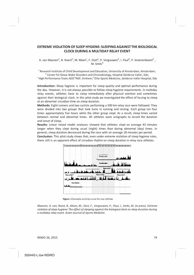
500445-L-bw-NSWO500445-L-bw-NSWO500445-L-bw-NSWO500445-L-bw-NSWO
NSWO 26, 2015 74
EXTREME VIOLATION OF SLEEP HYGIENE: SLEEPING AGAINST THE BIOLOGICAL CLOCK DURING A MULTIDAY RELAY EVENT
A. van Maanena, B. Roestb, M. Moenc, F. Oorta, P. Vergouwend, I. Pauld, P. Groenenboomd ,
M. Smitsb
a Research Institute of Child Development and Education, University of Amsterdam, Amsterdam; b Center for Sleep-Wake Disorders and Chronobiology, Hospital Gelderse Vallei , Ede;
c High Performance Team, NOC*NSF, Arnhem;d Elite Sports Medicine, Gelderse Vallei Hospital, Ede Introduction: Sleep hygiene is important for sleep quality and optimal performance during the day. However, it is not always possible to follow sleep hygiene requirements. In multiday relay events, athletes have to sleep immediately after physical exertion and sometimes against their biological clock. In this pilot study we investigated the effect of having to sleep at an abnormal circadian time on sleep duration. Methods: Eight runners and two cyclists performing a 500 km relay race were followed. They were divided into two groups that took turns in running and resting. Each group ran four times approximately five hours while the other group slept. As a result, sleep times varied between normal and abnormal times. All athletes wore actigraphs to record the duration and onset of sleep. Results: Linear mixed model analyses showed that athletes slept on average 43 minutes longer when they slept during usual (night) times than during abnormal (day) times. In general, sleep duration decreased during the race with on average 18 minutes per period. Conclusion: This pilot study shows that, even under extreme violation of sleep hygiene rules, there still is an apparent effect of circadian rhythm on sleep duration in relay race athletes.
Figure 1 Example activity curve for one athlete
Maanen, A. van, Roest, B., Moen, M., Oort, F., Vergouwen, P., Paul, I., Smits, M. (in press). Extreme violation of sleep hygiene: The effect of sleeping against the biological clock on sleep duration during a multiday relay event. Asian Journal of Sports Medicine.
500445-L-bw-NSWO500445-L-bw-NSWO500445-L-bw-NSWO500445-L-bw-NSWO
NSWO 26, 2015 75
MELATONIN TREATMENT AND CLASSICAL CONDITIONING IN CHILDREN WITH DELAYED SLEEP PHASE
A. van Maanena, A.M. Meijera, M.G. Smitsb, F.J. Oorta
a Research Institute of Child Development and Education, University of Amsterdam, Amsterdam;
b Center for Sleep-Wake Disorders and Chronobiology, Hospital Gelderse Vallei , Ede Introduction: The positive effects of melatonin treatment in children with sleep onset problems usually disappear when treatment is discontinued. We investigated whether classical conditioning might help to preserve treatment effects of melatonin. Methods: After a baseline week, 16 children (mean age 9.78 years) received melatonin treatment for 3 weeks, followed by a week in which they received half the dose, and a stop week. Classical conditioning was applied by having children drink biological lemonade while taking melatonin, and by using a lamp that was turned on when children went to bed. Variables measured were sleep (sleep diaries, actigraphy), Dim Light Melatonin Onset, and health, behavior problems, and parenting stress. The results were compared with a control group consisting of 41 children (mean age 9.43 years) who received melatonin without classical conditioning. Results: Melatonin treatment was effective in advancing DLMO and reducing sleep onset problems, and in addition positive effects were found on sleep quality, health, behavior problems, and parenting stress. After stopping melatonin, sleep returned to baseline levels. Interestingly, for both groups behavior problems and parenting stress remained significantly lower. Results did not differ between the experimental and control group, so we concluded that there was no effect of classical conditioning. However, when we excluded children with comorbid conditions, classical conditioning did seem to subdue the negative effects of withholding melatonin. Conclusion: This study suggests that classical conditioning does not help to preserve the efficacy of melatonin treatment after its discontinuation. However, classical conditioning may be effective in children without comorbid problems. Maanen, A. van, Meijer, A. M., Smits, M. G., & Oort, F. J. (2014). Melatonin treatment and classical conditioning in children with delayed sleep phase. Journal of Sleep Research, 23 (Suppl. 1), 41.
500445-L-bw-NSWO500445-L-bw-NSWO500445-L-bw-NSWO500445-L-bw-NSWO
NSWO 26, 2015 76
DEPRESSION, SCHIZOPHRENIA, AND SLEEP DISORDERS: THEIR TRADITIONAL ORIENTAL MEDICINE EQUIVALENTS
Peggy Boscha,b,c, Peter de Roverd, Sabina Limc, Heike Staudteb, Maurits van den Noortc,e
a Donders Institute for Brain, Cognition, and Behaviour, Centre for Cognition, Radboud University,
Nijmegen, The Netherlands b Psychiatric Research Institute, LVR-Klinik Bedburg-Hau, Bedburg-Hau, Germany
c Research Group of Pain and Neuroscience, Kyung Hee University, Seoul, Republic of Korea d Karel de Grote Hoge School, Antwerp, Belgium
e Brussels Institute of Applied Linguistics, Vrije Universiteit Brussel, Brussels, Belgium
Sleep disorders are comorbid with most psychiatric disorders. A well -known fact is that in depression, sleep disorders occur, but in schizophrenia, they are often underestimated. Psychiatric disorders can be described from both a Western (allopathic) and an Eastern perspective. Especially patients with a Western diagnosis of depression or schizophrenia seem likely to be in Western treatment when they are referred to an acupuncturist for (add-on) treatment, so knowledge on both sides is needed for integrating these treatments. In this study, the different Traditional Oriental Medicine (TOM) diagnostic patterns in patients with a Western diagnosis of depression, schizophrenia, or sleep disorders are described from a literature and a clinical perspective. The data on 30 depression and 30 schizophrenia patients from a German study are presented. All patients were outpatients at the LVR-Klinik Bedburg-Hau, which is a large psychiatric clinic in Germany. The patients with depression were diagnosed with depression F33.2 according to the ICD-10 (the 10th revision of the International Statistical Classification of Diseases and Related Health Problems ), and the patients with schizophrenia were diagnosed with schizophrenia F 20.0 (paranoid schizophrenia – 28 patients) or F20.5 (schizophrenic residuum – 2 patients). As can be seen in Table 1, the results of the PSQI (Pittsburgh Sleep Quality Index) with a cut-off score of 6 showed that of the 30 patients with depression, 24 patients (80.00%) suffered from sleep disorders whereas of the 30 patients with schizophrenia, 19 patients (63.33%) suffered from sleep disorders. In addition, the TOM diagnosis results of our total psychiatric sample (n = 60) show the following main TOM diagnostic patterns for the patients with sleep disorders: Qi & Blood deficiency, Kidney Essence deficiency, Phlegm heat harrassing the mind, and Phlegm misting the mind. Moreover, our results show that if a psychiatric group, sorted in accordance to Western diagnostic principles, is diagnosed on the basis of TOM diagnostic patterns, many different groups of patients arise (see Table 1), which has far-reaching consequences for scientific research. Acupuncture research on patients with depression or schizophrenia is difficult to design, conduct and interpret, and due to the different TOM diagnostic patterns that are treated within the depression and the schizophrenia groups, results differ. Finally, based on the clinical results of our psychiatric sample, we hypothesize that acupuncture treatment effects (symptom reductions) in patients with depression and in patients with schizophrenia might partially be mediated through improved sleep.
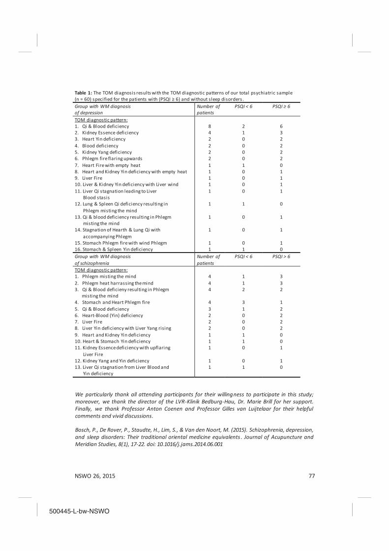
500445-L-bw-NSWO500445-L-bw-NSWO500445-L-bw-NSWO500445-L-bw-NSWO
NSWO 26, 2015 77
Table 1: The TOM diagnosis results with the TOM diagnostic patterns of our total psychiatric sample (n = 60) specified for the patients with (PSQI ≥ 6) and without sleep disorders . Group with WM diagnosis of depression
Number of patients
PSQI < 6 PSQI ≥ 6
TOM diagnostic pattern: 1. Qi & Blood deficiency 2. Kidney Essence deficiency 3. Heart Yin deficiency
8 4 2
2 1 0
6 3 2
4. Blood deficiency 5. Kidney Yang deficiency 6. Phlegm fire flaring upwards 7. Heart Fire with empty heat 8. Heart and Kidney Yin deficiency with empty heat 9. Liver Fire 10. Liver & Kidney Yin deficiency with Liver wind 11. Liver Qi stagnation leading to Liver Blood stasis 12. Lung & Spleen Qi deficiency resulting in Phlegm misting the mind 13. Qi & blood deficiency resulting in Phlegm misting the mind 14. Stagnation of Hearth & Lung Qi with accompanying Phlegm 15. Stomach Phlegm fire with wind Phlegm 16. Stomach & Spleen Yin deficiency
2 2 2 1 1 1 1 1
1
1
1
1 1
0 0 0 1 0 0 0 0
1
0
0
0 1
2 2 2 0 1 1 1 1
0
1
1
1 0
Group with WM diagnosis of schizophrenia
Number of patients
PSQI < 6 PSQI > 6
TOM diagnostic pattern: 1. Phlegm misting the mind 2. Phlegm heat harrassing the mind 3. Qi & Blood deficieny resulting in Phlegm misting the mind 4. Stomach and Heart Phlegm fire 5. Qi & Blood deficiency 6. Heart-Blood (Yin) deficiency 7. Liver Fire 8. Liver Yin deficiency with Liver Yang rising 9. Heart and Kidney Yin deficiency 10. Heart & Stomach Yin deficiency 11. Kidney Essence deficiency with upflaring Liver Fire 12. Kidney Yang and Yin deficiency 13. Liver Qi stagnation from Liver Blood and Yin deficiency
4 4 4
4 3 2 2 2 1 1 1
1 1
1 1 2
3 1 0 0 0 1 1 0
0 1
3 3 2
1 2 2 2 2 0 0 1
1 0
We particularly thank all attending participants for their willingness to participate in this study; moreover, we thank the director of the LVR-Klinik Bedburg-Hau, Dr. Marie Brill for her support. Finally, we thank Professor Anton Coenen and Professor Gilles van Luijtelaar for their helpful comments and vivid discussions. Bosch, P., De Rover, P., Staudte, H., Lim, S., & Van den Noort, M. (2015). Schizophrenia, depression, and sleep disorders: Their traditional oriental medicine equivalents . Journal of Acupuncture and Meridian Studies, 8(1), 17-22. doi: 10.1016/j.jams.2014.06.001
500445-L-bw-NSWO500445-L-bw-NSWO500445-L-bw-NSWO500445-L-bw-NSWO
NSWO 26, 2015 78
TWO NEUROLOGY PATIENTS WITH NOCTURNAL HYPERCAPNIC HYPOVENTILATION
Al de Weerd and Karel Schreuder
Sleep-Wake Center SEIN Zwolle-Groningen
Introduction: The link between neurological disorders and respiratory failure is well known for patients with recent cerebral ischemia. We present two cases with hypoventilation and neurological disease some years before, for whom the causal relationship between both disorders is not common knowledge. Case 1. A 67 y/o male. He has a bilateral diaphragm paralysis probably due to a cervical spinal stenosis 13 years ago. Since 6 months he has a persistent cough and complains of frequent awakenings during the night, mostly after a dream. At that moment he is gasping for air. This feeling disappears quickly after sitting up and 10-20 seconds hyperventilation. The patient does not snore and his partner is not aware of apneas during the night. At polysomnography (PSG) frequent awakenings from REM sleep are seen just after or concurrent with a central apnea leading to short lasting hypercapnia. Over the whole night the pCO2 increases from baseline 40 mm to 52 mm Hg. Discussion and conclusion for this case: in the normal situation REM atonia stops all activity of respiratory muscles except for the diaphragma. The patient has no or very limited neuronal input to the diaphragma and must have had REM related hypoventilation for the last 13 years. Since he has a chronic bronchitis (the cough!) he stops breathing at all. He recovers quickly after awakening and sitting up. We proposed respiratory assistance (CPAP, BiPAP eventually ASV), but first treated the bronchitis. The latter was enough to restore the equilibrium for already 2 years. Case 2. A 37 y/o male. Two years before we met this patient, he had a hemorrhagic accident in his brainstem at the right side of his medulla oblongata just below the pons. He recovered well except for a slight left sided sensory disturbance. Since some months his wife hears respiratory stops during the night. At PSG sleep is fragmented due to frequent central apneas and episodes of low breathing frequency, as low as 8/minute. Similar to case 1, over the whole night he gradually develops hypercapnia as seen during transcutaneous monitoring of CO2. Discussion and conclusion for this case: nocturnal hypoventilation and respiratory dysrhythmia including central apneas, due to malfunction of respiratory nuclei in the medulla oblongata. A low dose of acetozolamid was enough to restore the equilibrium in his total respiratory system. Conclusion: In general, hypercapnic hypoventilation during the night may be due to COPD and other chronic lung disorders, depression (opiates!) or failure of cerebral respiratory nuclei (case 2), neuromuscular disorders as ALS, or dysfunction of the (phrenic) nerve as in case 1, and in patients with extreme obesitas or kyphoscoliosis, Poster presentation annual meeting neurology 2014
500445-L-bw-NSWO500445-L-bw-NSWO500445-L-bw-NSWO500445-L-bw-NSWO
NSWO 26, 2015 79
IS THE CIRCADIAN RHYTHMICITY IN PERIODIC LEG MOVEMENTS AFFECTED BY PHARMACOLOGICAL TREATMENT?
R. Tijdens and A.W. de Weerd
Sleep-Wake Centre SEIN, Zwolle-Groningen, the Netherlands
Introduction: It is well-known that the frequency of periodic leg movements (PLMs) diminishes during the night. Different kinds of medication have different effects on PLMs. Some kinds of medication are known to inhibit the PLMs. Other types of medication induce or exacerbate PLMs. This study examined the possible interaction effect of inhibiting or inducing medication on the circadian rhythmicity of periodic leg movements. Methods: The patient data collected during a year showed that 39 patients got the diagnosis PLMD at the sleep-wake center SEIN at Zwolle-Groningen. The data analysis was done on the amount of PSG’s available per patient. There were two 24 hour PSG’s available from 22 patients and from 17 patients only one PSG. In total the data of 61 PSG’s were analyzed by measuring the PLMs for every specific hour of night sleep from lights off until lights on. The percentages of the total PLMs were calculated for each individual hour, which made a balanced comparison of the PLMs between the patients possible. The percentages of the first 4 hour combined were compared with the PLMS of the second part of the night (up to six hours of sleep). The different kinds of medication, used by the patients during the diagnostic PSG´s, were grouped in PLMs inducing (antidepressants, antipsychotics or antihistamines) or PLMs inhibiting medication (dopaminergic, anticonvulsants, opiates or benzodiazepines). 18 PSG’s were done with PLMs inducing medication and 10 PSG’s with PLMs inhibiting medication. The 8 PSG’s with both kinds of medication and the 24 PSG’s without any kind of medication were excluded from the analysis. Results: When analyzing the data of the total amounts of PSG’s a significant difference was found by comparing the frequency of PLMs during the first part of the night with the second part, t(60)=3.612, p<0.001. These results confirm the existence of nocturnal circadian rhythmicity in periodic leg movements. A 2x2 mixed ANOVA was used to analyze a possible interaction effect between the kind of medication and the circadian rhythmicity of the periodic leg movements (Figure 1). The data showed no significant main effect for the type of medication or for the part of the night. Neither did the data show a significant interaction effect, F(1.26)=1.72, p=0.20. Conclusion: These results indicate that type of medication does not have an effect on the circadian rhythmicity of periodic leg movements. Further research, with an extended patient population, is being performed.
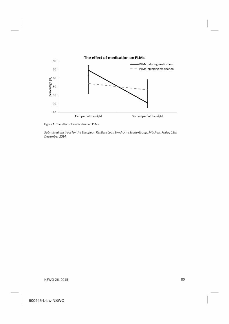
500445-L-bw-NSWO500445-L-bw-NSWO500445-L-bw-NSWO500445-L-bw-NSWO
NSWO 26, 2015 80
Figure 1. The effect of medication on PLMs Submitted abstract for the European Restless Legs Syndrome Study Group. Müchen, Friday 12th December 2014.
500445-L-bw-NSWO500445-L-bw-NSWO500445-L-bw-NSWO500445-L-bw-NSWO
NSWO 26, 2015 81
SLEEP AND PERFORMANCE IN ELITE ATHLETES
Melanie Knufinke, Arne Nieuwenhuys, Sabine A.E. Geurts, Michiel A.J. Kompier
Behavioural Science Institute, Radboud University, The Netherlands
Sleep is assumed to be essential for recovery and skill-learning in athletes (Venter, 2012). Furthermore, there is evidence to suggest that when sleep is restricted, cognitive and physical performance may deteriorate (Fullagar et al., 2014). Still, very few studies investigated sleep characteristics of elite athletes (Lastella et al., 2014; Leeder et al., 2012) and there is no information regarding the extent to which natural variations in elite athlete’s sleep may impact their performance. To this end, the current study aimed to quantify elite athletes’ habitual nocturnal sleep and to extent previous findings by establishing (i) distribution of sleep stages (deep sleep, REM-sleep, light sleep); and (ii) relations between sleep and performance. In the current study 98 Dutch elite athletes (aged 18.78 years ± 3.01; 56 female) from different individual and team sports (Road cycling, Triathlon, Mountain bike, Handball, Volleyball, Soccer) participated. Ambulatory sleep measurements were taken for 10-consecutive days of training, using actigraphy (sleep-wake pattern), one-channel EEG sensors (sleep-staging) and sleep diaries. Furthermore, to establish relations between sleep and performance, psychomotor vigilance (PVT) and sport-specific skill execution (gross and fine motor skills) were tested on day 1, 4, 7 and 10. In line with previous research (Leeder et al., 2012), results revealed a large discrepancy between Time in Bed (TIB; M=8:55 hours, SD=1:10 hours) and Total Sleep Time (TST; M=7:37; SD=1:06 hours), which could mainly be explained by relatively long Sleep Onset Latencies (SOL; M=13.65 minutes, SD=15.42 minutes) and nocturnal awakenings (WASO; M=58.78 minutes, SD=24.27 minutes). Sleep staging, however, revealed a healthy distribution of deep-sleep (M=19.34%, SD=7.34%), REM-sleep (M=25.10%, SD=7.14%) and light-sleep (47.40 ± 8.06%). Finally, with respect to performance, preliminary analyses (273 nights), suggest that a higher Total Sleep Time (TST) relates to faster reaction times and fewer lapses (reaction time > 500ms) on a 10-minute psychomotor vigilance task (PVT). Findings suggest that habitual fluctuation in elite athletes’ sleep may be related to chang es in cognitive performance (PVT), thereby highlighting the importance of developing interventions that increase actual sleep time while lowering nocturnal awakenings. References Fullagar, H. H., Skorski, S., Duffield, R., Hammes, D., Coutts, A. J., & Meyer, T. (2014). Sleep and athletic performance: The effects of sleep loss on exercise performance, and physiological and cognitive responses to exercise. Sports Medicine, 1-26. Lastella, M., Roach, G. D., Halson, S. L., & Sargent, C. (2014). Sleep/wake behaviours of elite athletes from individual and team sports. European Journal of Sport Science, 1-7. Leeder, J., Glaister, M., Pizzoferro, K., Dawson, J., & Pedlar, C. (2012). Sleep duration and quality in elite athletes measured using wristwatch actigraphy. Journal of Sports Sciences, 30(6), 541-545. Venter, R. E. (2012). Role of sleep in performance and recovery of athletes: a review article. South African Journal for Research in Sport, Physical Education and Recreation , 34(1), 167-184. Submitted for a presentation at the Behavioral Science Day 2015, Radboud Univeristy and the WAOP 2015. Presenteerd on the FEPSAC, Bern 2015.

500445-L-bw-NSWO500445-L-bw-NSWO500445-L-bw-NSWO500445-L-bw-NSWO
NSWO 26, 2015 82
500445-L-bw-NSWO500445-L-bw-NSWO500445-L-bw-NSWO500445-L-bw-NSWO
NSWO 26, 2015 83
SLEEP-WAKE Research in the Netherlands
Volume 26, 2015
Book Abstracts
500445-L-bw-NSWO500445-L-bw-NSWO500445-L-bw-NSWO500445-L-bw-NSWO
NSWO 26, 2015 84
SLEEP AND ADULT NEUROGENESIS: IMPLICATIONS FOR COGNITION AND MOOD
Anka D. Muellera, Peter Meerlob, Dennis McGintyc, Ralph E. Mistlbergera
a Department of Psychology, Simon Fraser University, Burnaby, BC, Canada
b Center for Behavior and Neurosciences, University of Groningen, The Netherlands c UCLA/VA Sleep Research Labs, University of California, North Hills, CA, USA
The hippocampal dentate gyrus plays a critical role in learning and memory throughout life, in part by the integration of adult born neurons into existing circuits. Neurogenesis in the adult hippocampus is regulated by numerous environmental, physiological and behavioral factors known to affect learning and memory. Sleep is also important for learning and memory. Here we critically examine evidence from correlation, deprivation, and s timulation studies that sleep may be among those factors that regulate hippocampal neurogenesis. There is mixed evidence for correlations between sleep variables and rates of hippocampal cell proliferation across the day, the year and the lifespan. There is modest evidence that periods of increased sleep are associated with increased cell proliferation or survival. There is strong evidence that disruptions of sleep exceeding 24h, by total deprivation, selective REM sleep deprivation, chronic restriction or fragmentation, significantly inhibit cell proliferation and in some cases neurogenesis. The mechanisms by which sleep disruption inhibits neurogenesis are not fully understood. Although sleep disruption procedures are typically at least mildly stressful, elevated adrenal corticosterone secretion is not necessary for this effect. However, procedures that prevent both elevated corticosterone and interleukin 1 signaling have been found to block the effect of sleep deprivation on cell proliferation. This result suggests that sleep loss impairs hippocampal neurogenesis by the presence of wake-dependent factors, rather than by the absence of sleep-specific processes. This would weigh against a hypothesis that regulation of neurogenesis is a function of sleep. Nonetheless, impaired neurogenesis may underlie some of the memory and mood effects associated with acute and chronic sleep disruptions. Mueller A, Meerlo P, McGinty D, Mistlberger R. Sleep and adult neurogenesis: implications for cognition and mood. Current Topics in Behavioral Neuroscience 25: 151-181, 2015.
500445-L-bw-NSWO500445-L-bw-NSWO500445-L-bw-NSWO500445-L-bw-NSWO
NSWO 26, 2015 85
ANIMAL STUDIES ON THE ROLE OF SLEEP IN MEMORY: FROM BEHAVIORAL PERFORMANCE TO MOLECULAR MECHANISMS
Robbert Havekesa, Peter Meerlob, Ted Abela
a Department of Biology, University of Pennsylvania, Philadelphia, PA, USA
b Center for Behavior and Neurosciences, University of Groningen, The Netherlands Although the exact functions of sleep remain a topic of debate, several hypotheses propose that sleep benefits neuronal plasticity, which ultimately supports brain function and cognition. For over a century, researchers have applied a wide variety of behavioral, electrophysiological, biochemical, and molecular approaches to study how memory processes are promoted by sleep and perturbed by sleep loss. Interestingly, experimental studies indicate that cognitive impairments as a consequence of sleep deprivation appear to be most severe with learning and memory processes that require the hippocampus, which suggests that this brain region is particularly sens itive to the consequences of sleep loss. Moreover, recent studies in laboratory rodents indicate that sleep deprivation impairs hippocampal neuronal plasticity and memory processes by attenuating intracellular cyclic adenosine monophosphate (cAMP)-protein kinase A (PKA) signaling. Attenuated cAMP-PKA signaling can lead to a reduced activity of the transcription factor cAMP response element binding protein (CREB) and ultimately affect the expression of genes and proteins involved in neuronal plasticity and memory formation. Pharmacogenetic experiments in mice show that memory deficits following sleep deprivation can be prevented by specifically boosting cAMP signaling in excitatory neurons of the hippocampus. Given the high incidence of sleep disturbance and sleep restriction in our 24/7 society, understanding the consequences of sleep loss and unraveling the underlying molecular mechanisms is of great importance. Havekes R, Meerlo P, Abel T. Animal studies on the role of sleep in memory: from behavioral performance to molecular mechanisms. Current Topics in Behavioral Neuroscience 25: 183-206, 2015.
500445-L-bw-NSWO500445-L-bw-NSWO500445-L-bw-NSWO500445-L-bw-NSWO
NSWO 26, 2015 86
STRESS, AROUSAL AND SLEEP
Larry D. Sanforda, Deborah Sucheckib, Peter Meerloc
a Department of Pathology and Anatomy, Eastern Virginia Medical School, Norfolk, VA, USA b Departamento de Psicobiologia, Universidade Federal de Sao Paulo, Sao Paulo, Brazil
c Center for Behavior and Neurosciences, University of Groningen, The Netherlands
Stress is considered to be an important cause of disrupted sleep and insomnia . However, controlled and experimental studies in rodents indicate that effects of stress on sleep–wake regulation are complex and may strongly depend on the nature of the stressor. While most stressors are associated with at least a brief period of arousal and wakefulness, the subsequent amount and architecture of recovery sleep can vary dramatically across conditions even though classical markers of acute stress such as corticosterone are virtually the same. Sleep after stress appears to be highly influenced by situational variables including whether the stressor was controllable and/or predictable, whether the individual had the possibility to learn and adapt, and by the relative resilience and vulnerability of the individual experiencing stress. There are multiple brain regions and neurochemical systems linking stress and sleep, and the specific balance and interactions between these systems may ultimately determine the alterations in sleep–wake architecture. Factors that appear to play an important role in stress-induced wakefulness and sleep changes include various monominergic neurotransmitters, hypocretins, corticotropin releasing factor, and prolactin. In addition to the brain regions directly involved in stress response such as the hypothalamus, the locus coeruleus, and the amygdala, differential effects of stressor controllability on behavior and sleep may be mediated by the medial prefrontal cortex. These various brain regions interact and influence each other and in turn affect the activity of sleep–wake controlling centers in the brain. Also, these regions likely play significant roles in memory processes and participate in the way stressful memories may affect arousal and sleep. Finally, stress-induced changes in sleep-architecture may affect sleep-related neuronal plasticity processes and thereby contribute to cognitive dysfunction and psychiatric disorders. Sanford LD, Suchecki D, Meerlo P. Sleep, stress and arousal. Current Topics in Behavioral Neuroscience 25: 379-410, 2015.
500445-L-bw-NSWO500445-L-bw-NSWO500445-L-bw-NSWO500445-L-bw-NSWO
NSWO 26, 2015 87
CHRONICALLY RESTRICTED OR DISRUPTED SLEEP AS A CAUSAL FACTOR IN THE DEVELOPMENT OF DEPRESSION
Peter Meerloa, Robbert Havekesb, Axel Steigerc
a Center for Behavior and Neurosciences, University of Groningen, The Netherlands
b Department of Biology, University of Pennsylvania, Philadelphia, PA, USA c Max Planck Institute of Psychiatry, Munich, Germany
Sleep problems are a common complaint in the majority of people suffering from depression. While sleep complaints were traditionally seen as a symptom of mood disorders, accumulating evidence suggests that in many cases the relationship may be reverse as well. A long list of longitudinal studies shows that sleep complaints often precede the onset of depression and constitute an independent risk factor for the development of the disorder. Additionally, experimental studies in animals show that chronically restricted or disrupted sleep may gradually induce neurobiological changes that are very similar to what has been reported for depressed patients. The mechanisms through which insufficient sleep increases the risk for depression are poorly understood but may include effects of sleep disturbance on neuroendocrine stress systems, serotonergic neurotransmission, and various interacting signaling pathways involved in the regulation of neuronal plasticity and neurogenesis. Because sleep is considered to play a crucial role in regulating neuronal plasticity and synaptic strength, chronically insufficient sleep may contribute to depression through an impairment of these plasticity processes leading to altered connectivity and communication within and between brain regions involved in the regulation of mood. Meerlo P, Havekes R, Steiger A. Sleep disruption as a causal factor in the development of depression. Current Topics in Behavioral Neuroscience 25: 459-481, 2015.
500445-L-bw-NSWO500445-L-bw-NSWO500445-L-bw-NSWO500445-L-bw-NSWO
NSWO 26, 2015 88
SLEEP, NEURONAL PLASTICITY AND BRAIN FUNCTION
Peter Meerloa, Ruth M. Bencab, Ted Abelc (editors)
a University of Groningen, Groningen, The Netherlands b University of Madison, Madison, WI, USA
c University of Pennsylvania, Philadelphia, PA, USA This book reviews current knowledge on the importance of sleep for brain function, from molecular mechanisms to behavioral output, with special emphasis on the question how sleep and sleep loss ultimately affect cognition and mood. It provides an extensive overview of the latest insights in the role of sleep in regulating gene expression, synaptic plasticity and neurogenesis, and how that in turn is linked to learning and memory processes. In addition, readers will learn about the potential clinical implications of insufficient sleep and discover how chronically restricted or disrupted sleep may contribute to age-related cognitive decline and the development of psychiatric disorders such schizophrenia and depression. The book consists of 19 chapters, written by experts in basic sleep research and sleep medicine, which together cover a wide range of topics on the importance of sleep and consequences of sleep disruption. This book will be of interest to students, researchers and clinicians with a general interest in brain function or a specific interest in sleep. Meerlo P, Benca R, Abel T (eds.) Current Topics in Behavioral Neuroscience, Vol 25: Sleep, neuronal plasticity and brain function. Springer, Germany, 2015.
500445-L-bw-NSWO500445-L-bw-NSWO500445-L-bw-NSWO500445-L-bw-NSWO
NSWO 26, 2015 89
SOS SLAAP
Johan Verbraecken and Tine Bergen SOS Slaap is written for the general public and offers a clear insight on sleep and what looks evident but is not the case for everyone. Also some do’s and don’ts are addressed. This book (136 p) consists of 7 chapters. In the introduction, a number of fundamental aspects are discussed, like sleep stages and sleep structure, sleep duration versus sleep need, sleep quality, morning and evening type, naps and staying in bed late, morning mood, the importance of light exposure and sleep during life span. In chapter 2, sleep hygiene is discussed extensively with the necessary boundary conditions in order to get a qualitative sleep. In chapter 3, the different functions of sleep are described, with its influence on cognition, emotions, energy balance, general health and social functioning. Consequently, in chapter 4, the different sleep disorders are discussed, e.g. insomnia, hypersomnia, parasomnia and specifically sleep apnea. Aspects of sleep problems in children are part of chapter 5, addressing the parental role, contributing factors, complaints and solutions. Sleep apnea and ADHD in childhood are also included in this chapter. A separate chapter 6 is attributed to the phenomenon dreaming and nightmares. Finally, a number of frequently asked questions is answered in chapter 7, like “Is snoring a problem ?”, “Is a "nightcap” a good idea ?”, “Is it wrong to use the mobile Phone as an alarm clock ?”. The book can be read in different ways: starting a chapter with the highest personal interest or starting with the FAQ’s.

500445-L-bw-NSWO500445-L-bw-NSWO500445-L-bw-NSWO500445-L-bw-NSWO
NSWO 26, 2015 90
HET SLAPENDE BREIN
Ton Coenen The book written by Ton Coenen, past-president of the NSWO and professor emeritus at the Radboud University in Nijmegen, is a contemplation on the many aspects of sleep. This book can therefore be considered as a real handbook on sleep. Starting from the history of sleep research, Coenen addresses the current state of the art. He focuses on what happens during sleep in the brain. It seems that our brains are not switched off during this nocturnal activity. Many more things happen, including the processing of the scarce information entering the brain. Of course, the “why” of sleep is on the agenda. The Nijmegian professor considers the restaurative function of sleep as the most important activity, given that important recovery processes take place under the influence of hormones. Also, sleep is involved in memory consolidation. Coenen argues that this is due to the very scarce input of signals. Also during wakefulness, we process and store data in our memory, but this storage occurs less well due to disturbing influences of new incoming information. The periodic appearance of sleep related to the dark night is extensively discussed, with the triggers of sleep and wake, especially light intensity and body temperature. Much energy is spent to dreaming, probably the most mysterious and intriguing aspect of the sleep process. The information processing theory, which poses that REM sleep has developed to dream in order to process the acquired information, was tested versus the activation-synthesis hypothesis, which means that REM sleep itself is of importance and that the dream is only a minor side effect. Facts and myths about the dream are furthermore taken into account. Different kinds of sleep disorders, like apnea, narcolepsia and restless legs syndrome are discussed and the existing treatments are pointed out. Much attention is spent to common poor sleep and to the current pharmacological approach of insomnia. The rising non-pharmacological treatment modalities of insomnia are extensively discussed in this context.
500445-L-bw-NSWO500445-L-bw-NSWO500445-L-bw-NSWO500445-L-bw-NSWO
NSWO 26, 2015 91
With the legendary statement “You win the Tour in bed”, the famous former cyclist Joop Zoetemelk has contributed to getting sleep on the agenda. Sleep has become a social issue and even the sporting community has become interested in sleep. By optimizing sleep, well -being and performance of sporters can be improved. With all these considerations around sleep, the book of the Nijmegian sleep professor is an indispensable link between theory and practice. Therefore, Joop Zoetemelk got the first copy of Coenen’s book during the official release on July 3, just before the Tour started in Utrecht. The book, with an obvious Dutch touch, is readably and clearly written for the general public. It is abundantly illustrated; the text is supported by many drawings, graphs and pictures. Coenen states that the knowledge and experience he collected in almost 50 years of sleep research is presented in an accessible way.

500445-L-bw-NSWO500445-L-bw-NSWO500445-L-bw-NSWO500445-L-bw-NSWO
NSWO 26, 2015 92
500445-L-bw-NSWO500445-L-bw-NSWO500445-L-bw-NSWO500445-L-bw-NSWO
NSWO 26, 2015 93
SLEEP-WAKE Research in the Netherlands
Volume 26, 2015
Dutch Society for Sleep-Wake Research Members
500445-L-bw-NSWO500445-L-bw-NSWO500445-L-bw-NSWO500445-L-bw-NSWO
NSWO 26, 2015 94
HONORARY MEMBERS Dr. A.C. Declerck Prof. Dr. R.H. van den Hoofdakker (1934-2012) Prof. Dr. H.A.C. Kamphuisen (1931-2013) Prof. Dr. P. Visser (1919-2009) Dr. G. Ruigt
500445-L-bw-NSWO500445-L-bw-NSWO500445-L-bw-NSWO500445-L-bw-NSWO
NSWO 26, 2015 95
REGULAR MEMBERS Mw. Msc J.A. Aaronson UvA Neuropsychologie AIO Psychologie [email protected] Prof.Dr. R.M. Aarts Philips Research Research Fellow/ Hoogleraar HTC 34.5.001, 5656 AE EINDHOVEN [email protected] +31 (0)40 27 43149 Dhr. R.M. Aleva Máxima Medisch Centrum loc. Eindhoven Longgeneeskunde Longarts Postbus 90052, 5600 PD EINDHOVEN [email protected], [email protected] +31 (0)40 8886100 Mw. W.H. Alings-Bokkestijn (secretaris NVLA) Longfunctieanalist / slaaponderzoek Coordinator longfunctie Van Riebeeckweg 212, 1213 XZ HILVERSUM [email protected] +31 (0)88 753 1630 Dhr PhD D. Alvarez-Estevez Medisch Centrum Haaglanden, Slaapcentrum Physicist Lijnbaan32, 2512 VA DEN HAAG [email protected] +31 (0)6 33456098 Mw.PhD N. Antypa LUMC Klinische Psychologie chronotype slaap en depressie GZ psycholoog/UD ass.Prof Wassenaarseweg 52, 2333 AK LEIDEN [email protected] +31 (0)715276677 Mw. W.C.M. Arends-Derks SEIN Zwolle EEG afdeling en Slaapcentrum KNF laborant Dr. Denekampweg 20, 8025 BV ZWOLLE [email protected] +31 (0)38 4557167
Dhr. Drs. M. Arns Research Institute Brainclinics Psychologenpraktijk Brainclinics Director / Researcher Bijleveldsingel 34, 6524 AD NIJMEGEN [email protected] +31 (0)24 7503505 Dhr. Drs. J. Asin Amphia Ziekenhuis Slaapafhankelijke ademhalingsstoornissen, Slaapgeneeskunde Longarts Postbus 67, 4900 AB OOSTERHOUT [email protected] +31 (0)16 2327053 Mw. Drs. R.G. Astil l MC Slotervaart Amsterdam SlaapWaak Centrum Louwesweg 6, 1066 EC AMSTERDAM rgastil [email protected] Mw. M. Bakker-Boone ApneuVereniging Secretariaat Postbus 299, 1560 AG KROMMENIE [email protected] Dhr.Dr. J.D. Barth Star MDC Cardiologie Researcher [email protected] +31 (0)23 8222855 Mw. Drs. K. Beerhorst Atrium MC Parkstad Polikliniek neurologie Neuroloog Henri Dunantstraat 5, 6419 PC HEERLEN [email protected] +31 (0)45 576 6700 Prof.Dr. D.G.M. Beersma Rijksuniversiteit Groningen Chronobiologie Hoogleraar, Hoofd afd. Chronobiologie Postbus 14, 9750 AA HAREN [email protected] +31 (0)50 3632053
500445-L-bw-NSWO500445-L-bw-NSWO500445-L-bw-NSWO500445-L-bw-NSWO
NSWO 26, 2015 96
Dr. J.S. Benjamins Nederlands Instituut voor Neurowetenschappen Slaap en Cognitie Postdoc Meibergdreef 47, 1105 BA AMSTERDAM ZO [email protected] +31 (0)20 566 5488 Mw.Dr. J.F. van den Berg Parnassia Psychiatrie Onderzoeker KCO Mangostraat 1, 2552 KS DEN HAAG [email protected] +31 (0)88 3576550 Dr. F.W. Bes Psychofysioloog Rhijnestein 61, 1121 VE LANDSMEER [email protected] +31 (0)6 22395179 Dhr. H.J.C. Bindels Atrium MC Parkstad Longfunctie en OSAS-onderzoek en -therapie Praktijkdocent longfunctie-analist Postbus 4446, 6401 CX HEERLEN [email protected] +31 (0)45 5766331 Dhr. J.H.J. Bisschop Isala Ziekenhuis Klinische Neurofysiologie Laborant Postbus 10500, 8000 GM ZWOLLE [email protected] +31 (0)38 4244007 Mw. Drs C.R. Blanker Ziekenhuis Rivierenland Tiel Psychiatrie Psychiater Pres. Kennedylaan 1, 4002 WP TIEL [email protected] +31 (0)344 674911 Dr. T. de Boer Leids Universitair Medisch Centrum, Afd. Moleculaire Celbiologie, sectie Neurofysiologie Bioloog Postbus 9600, 2300 RC LEIDEN [email protected] +31 (0)71 526 9772
Dr. H. Boot Spaarne Gasthuis Poli neurologie Neuroloog Spaarnepoort 1, 2134 TM HOOFDDORP [email protected] +31 (0)23 8908900 Drs. J.B.S. Boringa Meander Medisch Centrum Neurologie Neuroloog Postbus 1502, 3800 BM AMERSFOORT [email protected] +31 (0)33 8501733 Mw.Dr. M. Bornebroek Neurologie Neuroloog [email protected] Mw. R.A.S. van den Bossche SEIN Zwolle EEG afdeling en Slaapwaakcentrum Laborant Postbus 563, 8000 AN ZWOLLE [email protected] Mw. Drs. S.M. Boukamp SEIN Zwolle Slaapwaakcentrum Laborant / Psycholoog Dr. Denekampweg 20, 8025 BV ZWOLLE [email protected] +31 (0)38 4557167 Mw. Drs. M.D. Bourez-Swart SEIN Zwolle Afdeling KNF Neuroloog Dr. Denekampweg 20, 8025 BV ZWOLLE [email protected] +31 (0)38 4557185 Dhr. Drs. W.J. Braam ’s Heeren Loo Midden-Nederland, Loc. Wekerom Arts voor verstandelijk gehandicapten Apeldoornseweg 60, 6733 SC WEKEROM [email protected] +31 (0)318 593562
500445-L-bw-NSWO500445-L-bw-NSWO500445-L-bw-NSWO500445-L-bw-NSWO
NSWO 26, 2015 97
Dhr. MD, PhD G.J.F. Brekelmans St. Elisabethziekenhuis Tilburg KNF, neurologie, epilepsie en slaap Neuroloog, klinisch neurofysioloog, deelopleider Hilvarenbeekseweg 60, 5022 GC TILBURG [email protected] +31 (0)13 5392552 Dr. M.H.M. Breteler EEG Resource Institute Psychologie, Neurofeedback Onderzoeker RU Nijmegen Postbus 31070, 6503 CB NIJMEGEN [email protected] +31 (0)24 3538846 Dhr. Drs. Y.F.P.M. van den Broek De snurkpoli Tandheelkundige slaapgeneeskunde Tandheelkunde Cimbaallaan 49/A, 5402 AX UDEN [email protected] +31 (0)413 255612 Mw. S.E. Bruggink "Nyx" Sherpa Nyx Zorg en Ondersteuning Adviseur / Verpleegkundige Berkellaan 2, 3744 NA BAARN [email protected] +31 (0)6 26796236 Mw. K.R.J. de Bruijckere-Marteijn ZorgSaam Ziekenhuis Zeeuws-Vlaanderen Loc. Antonius KNF / Slaapprofessional Pastoor van Genklaan 6, 4501 AJ OOSTBURG [email protected] +31 (0)115 677197 Dhr. Drs. E.J. de Bruin Neuropsychologie - slaaponderzoek bij adolescenten AIO Nieuwe prinsengracht 130, 1018 VZ AMSTERDAM [email protected] +31 (0)20 5251327 Mw. N. van den Bussche Kempenhaeghe Centrum voor slaapgeneeskunde Arts Postbus 61, 5590 AB HEEZE [email protected] +32 (0)474648193
Prof.Dr. R. Cluijdts Fac Psychologie Vrije Univ. Brussel Psycholoog Pleinlaan 2, 1050 BRUSSEL [email protected] Prof.Dr. A.M.L. Coenen Donders Centre For Cognitition, Radboud University Nijmegen Department of Biological Psychology Bioloog-psycholoog Postbus 9104, 6500 HE NIJMEGEN [email protected] +31 (0)24 3612544 Dr. L. Cohen Psychologenpraktijk Amstelveen Psycholoog Van Nijenrodeweg 898, 1081 BH AMSTERDAM [email protected] +31 (0)848 325870 Mw. Y. Cuppen Catharinaziekenhuis Longziekten OSAS Longfunctielaborante Michel Angelolaan 2, 5623 EJ EINDHOVEN [email protected] Prof. Dr. M.G. Curfs Leopold AZM Dept. Clinical Genetics Postbus 1475, 6201 BL MAASTRICHT [email protected] +31 (0)43 3877858 Dhr. H.J. Dalewijk Nieuwstad 56, 1381 CD WEESP [email protected] +31 (0)294 412747 Dr. A.C. Declerck Neuroloog Acacialaan 52, 8301 KNOKKE HEIST [email protected] +32 (0)032 50608451 Mw. E. Dijkers AMC Longfunctie C2-434 Hoofd Longfunctie Postbus 22660, 1100 DD AMSTERDAM [email protected] +31 (0)20 5663104
500445-L-bw-NSWO500445-L-bw-NSWO500445-L-bw-NSWO500445-L-bw-NSWO
NSWO 26, 2015 98
Dr. Ing. M.H.J. Doff Universitair Medisch Centrum Groningen Kaakchirurgie MKA-chirurg i.o. en wetenschappelijk onderzoeker Hanzeplein 1, 9700 RB GRONINGEN [email protected] +31 (0)50 3613840 Mw. Bsc. M. Dolmans KNF epilepsie en slaap KNF-laborant [email protected] Mw. Drs. J. Dolsma Diaconessenhuis Leiden Longgeneeskunde Longarts Postbus 9650, 2300 LD LEIDEN [email protected] Dr. H.P.A. van Dongen Washington State University Spokane Sleep and Performance Research Center Neurofysioloog 1014 S. Pepper Tree Lane SPOKANE, WA 99224 [email protected] Mw. Drs. C.E.H.M. Donjacour SEIN Slaapwaakcentrum lokatie Zwolle neuroloog Dr. Denekampweg 20, 5025BV ZWOLLE [email protected] +31 (0)38 8457185 Dr. W.H.I.M. Drinkenburg Janssen Pharmaceutica Psycholoog Turnhoutseweg 30, B-2340 BEERSE [email protected] Dhr.Dr. H.A. Droogleever Fortuyn Psychiater, consultatieve psychiatrie [email protected] Mw. M. Droppert-Pellegrom Medisch Centrum Haaglanden Centrum Slaap- en Waakstoornissen Laborant Klinische neurofysiologie Lijnbaan 32, 2512 VA DEN HAAG [email protected] +31 (0)70 3302721
Mw. Drs. N. van Duinen LUMC, C4 R-88 AIO Albinusdreef 2, 2333 ZC LEIDEN [email protected] +31 (0)71 5262646 Mw. A.A.M. Duis Amphia Ziekenhuis Longgeneeskunde, slaapcentrum Physician Assistant Slaap- en Longgeneeskunde Pasteurlaan 9, 4900 AB OOSTERHOUT [email protected], [email protected] +31 (0)76 5955000, dect 5139 Dhr. P.R. Eijkelenboom Arts Venmanshof 3, 6093 DA HEYTHUYSEN [email protected] +31 (0)475 591767 Dhr. M.M.M. Eijsvogel Med.Spectrum Twente, longziekten Longziekten Longarts Postbus 50000, 7500 KA ENSCHEDE [email protected] +31 (0)53 4872629 Dr. M.G. van Erp Kempenhaeghe Neurologie, slaapgeneeskunde Neurofysioloog Postbus 61, 5590 AB HEEZE [email protected] +31 (0)40 2279202 Dhr. Drs. C.A.G. van Esbroeck Lievensberg ziekenhuis Longgeneeskunde Longarts Boerhaaveplein 1, 4624 VT BERGEN OP ZOOM [email protected] +31 (0)164 278884 Mw. E. Felix- van Dijl St.Antoniusziekenhuis Neurologie Physician Assistant Koekoekslaan 1, 3435 CM NIEUWEGEIN [email protected]
500445-L-bw-NSWO500445-L-bw-NSWO500445-L-bw-NSWO500445-L-bw-NSWO
NSWO 26, 2015 99
Dhr. Drs. S.J. Fischer MC Slotervaart Medische psyschologie/ziekenhuispsychiatrie Klinisch psycholoog, afdelingshoofd Postbus 90440, 1006 BK AMSTERDAM [email protected] +31 (0)20 5124261 Dr. R. Fronczek Leiden University Medical Center Afdeling K5Q, Neurologie Postbus 9600, 2300 RC LEIDEN [email protected] +31 (0)71 5263902 Mw. PhD, AiO M. Geerdink Rijksuniversiteit Groningen FWN, Centrum voor Levenswetenschappen Basiseenheid Chronobiologie, kr 3.44 Nijenborgh 7, 9747 AG GRONINGEN [email protected] +31 (0)623239795 Mw.Dr. I.M. van Geijlswijk Universiteit Utrecht Apotheek faculteit Diergeneeskunde Ziekenhuisapotheker / Hoofd apotheek Yalelaan 106, 3584 CM UTRECHT [email protected] +31 (0)30 2532066 Drs. M.E.H. Geuke Medisch Centrum Haaglanden Psychologen Slaapcentrum Psycholoog Postbus 432, 2501 CK DEN HAAG [email protected] +31 (0)70 3302841 Mw. R. Geurts Medisch Centrum Haaglanden Centrum Slaap- en Waakstoornissen KNF Laborant Lijnbaan 32, 2512 VA DEN HAAG [email protected] +31 (0)70 3302841 Dhr. L.C.J.L. Ghijsen Novadic-Kentron soms Loc. Vught Arts verslavingsgeneeskunde Rompertsebaan 28, 5231 GT 's-HERTOGENBOSCH [email protected] +31 (0)73 640 9696
Mw. Drs. E.W. Gieteling SEIN Slaap waak centrum lokatie Heemstede Neuroloog, Klinisch neurofysioloog Achterweg 5, 2103 SW HEEMSTEDE [email protected] +31 (0)50 3616161 Mw. MSc M.M. van Gilst Radboud UMC Neurowetenschappen, hp 935 AIO Postbus 9101, 6500 HB NIJMEGEN [email protected] +31 (0)24-3615213 Mw. MSc M.S. Goelema Philips research neuropsycholoog High Tech Campus 34, 5656 AE EINDHOVEN [email protected] Mw.Dr. H.K.J. Gonnissen Universiteit Maastricht Departement Humane Biologie PO Box 616, 6200 MD MAASTRICHT [email protected] +31 (0)43 3884596 Mw.Dr. M.C.M. Gordijn Rijksuniversiteit Groningen Chronobiologie / Chrono@work Bioloog Postbus 11103, 9700 CC GRONINGEN [email protected] +31 (0)50 36376758 Mw.Dr. G.B. Górnicka Śląskie Centrum Rehabilitacji i Prewencji Hartrevalidatie Psycholoog ul. Zdrojowa 6, 43-450 Ustroń [email protected] +48 33 854 16 32 int. 231 Mw. MSc M.J. Graauwmans Donders Institute for Brain, Cognitition and Behaviour, Radboud University Nijmegen Medical Centre Centre for Neuroimaging Onderzoeksassistent Kapittelweg 29, 6525 EN NIJMEGEN [email protected]
500445-L-bw-NSWO500445-L-bw-NSWO500445-L-bw-NSWO500445-L-bw-NSWO
NSWO 26, 2015 100
Dr. ir. C.P.M. van der Grinten MUMC. afdeling Longziekten Fysioloog Postbus 5800, 6202 AZ MAASTRICHT [email protected] +31 (0)43 3876042 Dr. J.H.M. de Groen Stichting Slaaponderzoek Neuroloog [email protected] Mw. MANP M.B. Gruppelaar ZGT Centrum voor Slaapgeneeskunde Hengelo Verpleegkundig Specialist-GGZ / Slaapgeneeskunde Postbus 546, 7550 AM HENGELO [email protected] +31(0)742905279 Mw. Th. Gutter SEIN Zwolle Onderzoek & Ontwikkeling Onderzoeker Dr. Denekampweg 20, 8025 BV ZWOLLE [email protected] +31 (0)38 8457171 Dr. Ir. R. Haakma Unobtrusive Sleep Monitoring Senior Director Amerlaan 55, 5626 BR EINDHOVEN [email protected] +31 (0)40 2624846 Mw.Dr. P.M.J. Haffmans Parnassia Groep/ PsyQ Medische Chronobiologie Lijnbaan 4, 2512 VA DEN HAAG [email protected] +31 (0)70 3918353 Dhr. M.A.J. van Hal Ziekenhuis Rivierenland Tiel Slaappoli Slaapconsulent / Laborant Klinische Neurofysiologie Pres. Kennedylaan 1, 4002 WP TIEL [email protected] Dr. H.L. Hamburger MC Slotervaart WaakSlaapcentrum Amsterdam Neuroloog Postbus 90440, 1006 BK AMSTERDAM [email protected] +31 (0)20 5124464
Mw. MSc A.B.H. Hamoen Slaappsychologie Psycholoog [email protected] +31 (0)6 49953810 Mw. Drs. J.H.G.M. van Haren-Willems Universitair Longcentrum - ULC Dekkerswald Hoofd Longfunctie en Centrum voor Slaap Afhankelijke Ademhalingsstoornissen Postbus 66, 6560 AB GROESBEEK [email protected] +31 (0)24 6859911 Dhr. L.A. Harten MC Slotervaart Waak Slaap Centrum Amsterdam, KNF Hoofdlaborant Louwesweg 6, 1066 EC AMSTERDAM [email protected] Mw. Drs. R.C.T. Hauzer Reinier van Arkel Groep, GGZ Herlaarhof Psychiater slaapgeneeskundige i.o Brevier 1, 5709 AD HELMOND [email protected] +31 (0)492 349950 Dr. K.B. van der Heijden Universiteit Leiden, Leiden Institute for Brain and Cognition Afdeling Orthopedagogiek Universitair Docent Postbus 9555, Kamer: 4A15 2300 RB LEIDEN [email protected] +31(0)71 52756628 Dhr. Drs. J.C. den Heijer Medisch Centrum Leeuwarden Neurologie Neuroloog Henry Dunantweg 2, 8934 AD LEEUWARDEN [email protected] +31 (0)58 2866211 Dhr. Y.R. Heimans Philips Healthcare Home Healthcare Solutions Clinical Specialist Netherlands Boschdijk 525, Building VB 3 5621 JG EINDHOVEN [email protected] +31 (0)6 10085122
500445-L-bw-NSWO500445-L-bw-NSWO500445-L-bw-NSWO500445-L-bw-NSWO
NSWO 26, 2015 101
Mw. BSc Btr M. Henderson Natural Sciences with Biology and Environmental Sciences [email protected] Mw. Drs. D. Hendriks Medisch Centrum Haaglanden Centrum voor Slaap/Waakstoornissen Orthopedagoog Postbus 432, 2501 CK DEN HAAG [email protected] +31 (0)70 3302458 Dhr. Drs. C.A.M. Henke Kempenhaeghe KNF Neuroloog Postbus 61, 5590 AB HEEZE [email protected] Dhr. Drs. C.S.I. Herwijnen T-products BV Slaap/waakonderzoek Laborant Klinische Neurofysiologie [email protected] Mw. MSc B. van Hest TherapieCulemborg, chronische slaapproblematiek Psycholoog Herenstraat 37, 4101 BS CULEMBORG [email protected] +31 (0)644373537 Dhr. F.E.J.M. Hilhorst Scheper Ziekenhuis KNF Laborant Klinische Neurofysiologie Boermarkeweg 60, 7800 AA EMMEN [email protected] +31 (0)591 691145 Mw. Drs. J. van den Hoed Mediant Psychiater Laan van Driene 101, 7552 EN HENGELO [email protected] + 31 (0)74 2560256 Dhr. J. Hoefnagel Laborant Klinische Neurofysiologie [email protected] Dhr. Drs. A. Hoekema Tjongerschans Ziekenhuis Heerenveen / UMCG Kaakchirurgie Kaakchirurg
Thialffweg 44, 8441 PW HEERENVEEN [email protected] +31 (0)513 685685 Dhr. Dr. J.H. van der Hoeven UMCG Afdeling Neurologie Neuroloog / Hoofd Afd. Klinische Neurofysiologie Postbus 30001, 9700 RB GRONINGEN [email protected] +31 (0)50 3612599 Mw. Dr. W.F. Hofman UvA Psychonomie Psychofysioloog Roetersstraat 15, 1018 WB AMSTERDAM [email protected] +31 (0)20 5256848 Mw. Dr. W.A. Hofstra Neurologie, Slaapgeneeskunde en Epilepsie [email protected] +31 (0)38 4557175 Dhr. Dr. B.E.A. Hol Albert Schweitzer ziekenhuis Dordrecht longziekten Longarts Polkshof 14, 3318 BJ DORDRECHT [email protected] +31 (0)629067465 Mw. K. Hoogland Slotervaart Ziekenhuis Klinische Neurofysiologie Laborant KNF / Slaaplaborant Louwesweg 6, 1066 EC AMSTERDAM [email protected] +31 (0)20 5125123 Dhr. B.P.J. Hoondert Kempenhaeghe Centrum voor slaapgeneeskunde Laborant Klinische Neurofysiologie Sterkselseweg 65, 5591 VE HEEZE [email protected] +31 (0)40 2279208 Mw.Dr. J.W. Hop UMC Utrecht Kinder- en jeugdpsychiatrie Kinder- en jeugdpsychiater [email protected] +31 (0)30 2724849
500445-L-bw-NSWO500445-L-bw-NSWO500445-L-bw-NSWO500445-L-bw-NSWO
NSWO 26, 2015 102
Dhr. Drs. A. Huisman Albert Schweitzer ziekenhuis Slaap-Waak Centrum, medische psychologie Klinisch psycholoog Postbus 444, 3300 AK DORDRECHT [email protected] ; +31 (0)78 6542948 Dhr. Drs. T. Hylkema Slaapcentrum SEIN / Talant / Nyx Slaap en Slaapproblemen bij mensen met een verstandelijke handicap Gezondheidswetenschapper Waterland 33, 9205 GA DRACHTEN [email protected] +31 (0)6 42920858 Mw. D.T. IJsselstein MC Slotervaart KNF en Slaapwaakcentrum. Neuropsychologie Bachelor KNF, Bachelor/Master klin.Neuropsychologie Louwesweg 6, 1066 EC AMSTERDAM [email protected] +31 (0)23 5125123 Dhr. Drs. P.L. Ingenhoest Admiraal de Ruyter Ziekenhuis Klinische Neurofysiologie/ Slaapcentrum; Hoofdlaborant Klinische Neurofysiologie Postbus 3200, 4380 DD VLISSINGEN [email protected] +31 (0)118 425409 Dhr. Ir. E.J.M. Jansen Kempenhaeghe Centrum voor slaapgeneeskunde; Laborant Klinische Neurofysiologie Sterkselseweg 65, 5591 VE HEEZE [email protected] +31 (0)40 2279772 Mw.Bc J.W. Janssen-Poot KNF-laborant Halve Maan 11, 4207 BD GORINCHEM [email protected] Mw.Dr. H.C.J.P. Janssen Kempenhaeghe Centrum voor slaapgeneeskunde; Longarts Postbus 61, 5590 AB HEEZE [email protected] +31 (0)40 2279192
Mw. V. van Kasteel SEIN Slaapwaakcentrum lokatie Zwolle neuroloog Dr. Denekampweg 20, 5025BV ZWOLLE [email protected] +31 (0)38 8457185 Mw. Drs. I.E.M. Kauw MC Slotervaart medische psychologie/ziekenhuispsychiatrie GZ psycholoog Louwesweg 6 /postbus 90440 1006 BK AMSTERDAM [email protected] Mw. L.M.M. Kerckhoffs-Smits UMC Nijmegen Klinische Neurofysiologie, huispostnr 943 Laborant Klinische Neurofysiologie Postbus 9101, 6500 HB NIJMEGEN [email protected] Dhr. B. Kelder Coordinator Slaaponderzoek en Senior Longfunctie Assistant [email protected] +31 (0)24 3567314 Prof. Dr. G.A. Kerkhof Universiteit van Amsterdam Psychofysioloog* Roetersstraat 15, 1018 WB AMSTERDAM [email protected] +31 (0)20 5256739 Drs. B.L.J. Kessels Tweestedenziekenhuis Tilburg Longziekten Longarts Dr. Deelenlaan 5, 5042 AD TILBURG [email protected] +31 (0)13 4655655 Mw. M.S. Klompmaker-Wienke Scheper Ziekenhuis KNF Laborant KNF, Slaap en vaatlab. Boermarkeweg 60, 7800 AA EMMEN [email protected] +31 (0)591 691145 Mw. Drs. C. Kluft Psycholoog Santiagosingel 47, 2548 HM DEN HAAG [email protected] +31 (0)6 21574413
500445-L-bw-NSWO500445-L-bw-NSWO500445-L-bw-NSWO500445-L-bw-NSWO
NSWO 26, 2015 103
Mw. J.V. Knol Medisch Centrum Haaglanden Centrum Slaap- en Waakstoornissen Unitcoordinator Slaapcentrum Lijnbaan 32, 2512 VA DEN HAAG [email protected] +31 (0)70 3302722 Mw. I.L.H. Knottnerus (ev Stevenhagen) Medisch Centrum Twente Neuroloog Postbus 50000, 7500 KA ENSCHEDE [email protected] +31 (0)534 872000 Mw. Msc. M. Knufinke Behavioural Science Institute, Radboud University Nijmegen Dpt. of Work & Organizational Psychology Psycholoog AIO Montessorilaan 3, room A.08.26 6525 HR NIJMEGEN [email protected] +31 (0)24 3615729 Dr. A. Knuistingh Neven KN-Medical Consultancy Huisarts-epidemioloog Westzoom 3, 2931 AZ KRIMPEN A/D LEK [email protected] +31 (0)71 5275318 Mw. Dr. B.C.P. Koch Erasmus MC Rotterdam Ziekenhuisapotheker i.o. Postbus 2040, 3000 CA ROTTERDAM [email protected] Drs. F.H. Koenegracht Psychiater Witte Singel 63, 2311 BM LEIDEN [email protected] +31 (0)172 460300 Mw. Drs. G.M.L.G. Konings AZM, Klinische Genetica Klin.psycholoog Postbus 5800, 6202 AZ MAASTRICHT [email protected] +31 (0)43 3875855 Dr. K.W. van Kralingen Het Van Weel-Bethesda Ziekenhuis Longarts Postbus 153, 3240 AD MIDDELHARNIS [email protected] +31 (0)187 607196
Mw.Dr. C.J. Kramer [email protected] Mw. L. Kranstauber-van Wijngaarden Onze Lieve Vrouwe Gasthuis Klinische Neurofysiologie - B1 Leidinggevende KNF Postbus 95500, 1090 HM AMSTERDAM [email protected] +31 (0)20 5993039 Dhr. Drs. M.R. Kross Slotervaart Ziekenhuis Longziekten longarts Louwesweg 6, 1066 EC AMSTERDAM [email protected] +31 (0)624761886 Mw. Drs. V. Kruiver Brainclinic Psychologenpraktijk Orthopedagoog Bijleveldsingel 34, 6524 AD NIJMEGEN [email protected] +31 (0)24 7503505 Ir. A. Kumar Personal Health Institute International L Braakensiekstraat 94, 1065 HP AMSTERDAM [email protected] +31 (0)64 6104625 Drs. A.G.Y. Kurvers MC Slotervaart Neurologie AIOS neurologie Louwesweg 6, 1066 EC AMSTERDAM [email protected] +31 (0)652667893 Dhr. Drs. M. van de Laar Kempenhaeghe Centrum voor slaapgeneeskunde Psycholoog Postbus 61, 5590 AB HEEZE [email protected] +31 (0)40 2279239 Drs. D.M. Laman St. Lucas Andreas Ziekenhuis, KNF Klinisch neurofysioloog Jan Tooropstraat 164, 1061 AE AMSTERDAM [email protected] +31 (0)20 5108911
500445-L-bw-NSWO500445-L-bw-NSWO500445-L-bw-NSWO500445-L-bw-NSWO
NSWO 26, 2015 104
Dr. G.J. Lammers SEIN Slaap waak centrum, lokatie Heemstede Neuroloog, Klinisch neurofysioloog Achterweg 5, 2103 SW HEEMSTEDE [email protected] +31 (0)71 5262895 Drs. H.M. Lammers-v.d. Holst UvA Afd. Psychologie, Psychonomie Roetersstraat 15, 1018 WB AMSTERDAM +31 (0)20 5256123 Dr. J. Lancee Universiteit van Amsterdam Klinische en Psychologie Klinisch Psycholoog Weesperplein 4, 1018 XA AMSTERDAM [email protected] +31 (0)30 2532387 Mw.Dr. M. Lancel GGZ Drenthe Divisie forensische psychiatrie Senior Onderzoeker Dennenweg 9, 9404 LA ASSEN [email protected] +31 (0)592 334654 Mw.Dr. L.M.L. de Lau MC Slotervaart Neurologie, Slaapgeneeskunde neuroloog Louwesweg 6, 1066 EC AMSTERDAM [email protected] +31 (0)205129333 Dhr. m.sci J.A.J.M. Lebon Longgeneeskunde Physician Assistant Teggert 102, 6367 XP VOERENDAAL [email protected] +31 (0)6 46033143 Mw. Dr. C.H.C. Leenaars CDL Radboud Universitair Medisch Centrum Geert Grooteplein 29, 6525 EZ Nijmegen [email protected]
Drs. W.M.A. van Leeuwen Stockholm University Division for Biological Psychology and Treatment Research Researcher Stockholms universitet 106 91 SE-10691 STOCKHOLM [email protected] +46 8 5537 8917 Mw. F. Lefebvre Snurken en slaapapneu Rue Hougnee 23, B-4260 FUMAL +32 (0)19587504 Mw. H.J. Leijendekker Longfuntie-Assistente Koriander 32, 8252 CK DRONTEN [email protected] +31 (0)321 317700 Dr. T.R.M. Leufkens Philips Corporate Technologies Research Researcher, psychopharmacologie High Tech Campus, HTC-34 5656 AE EINDHOVEN [email protected] Dhr. Drs. M. Liedorp Havenziekenhuis Rotterdam Neurologie Medisch specialist neurologie Postbus 70031, 3000 LN ROTTERDAM [email protected] +31 (0)10 4043300 Dhr. MSc B.H.W. te Lindert Netherlands Insititute for Neuroscience Dept. Sleep and Cognition AIO, thermoregulatie, Bewegingswetenschapper Meibergdreef 47, 1105 BA AMSTERDAM b.te.l [email protected] +31 (0)20 5665495 Prof.Dr. F. Lobbezoo ACTA Afdeling Mondgezondheidswetenschappen Hoogleraar Orofaciale Pijn en Disfunctie Gustav Mahlerlaan 3004, 1081 LA AMSTERDAM [email protected] +31 (0)20 5980412
500445-L-bw-NSWO500445-L-bw-NSWO500445-L-bw-NSWO500445-L-bw-NSWO
NSWO 26, 2015 105
Mw. Drs. M. Louter Westeinde ziekenhuis, Medisch Centrum Haaglanden Neurologie Basisarts, AIO Postbus 432, 2501 CK DEN HAAG [email protected] Dhr. Drs. S.A.P Lucius ChronoMed Psychofysioloog vondellaan 35/A, 2332 AA LEIDEN [email protected] +31 (0)71 572 36 82 Prof.Dr. E.L.J.M. van Luijtelaar Radboud Universiteit Donders institute for Brain , Cognition and Behaviour Psycholoog Montessorilaan 3, 6525 HR NIJMEGEN [email protected] +31 (0)24 3615617 Mw. MSc MA A.I. Luik Erasmus MC Rotterdam Epidemiologie PhD-Student PO Box 2040, Kamer Na 2817 3000 CA ROTTERDAM [email protected] +31 (0)10 7044043 Mw. Dr. A.P.H.M. Maas Universiteit Maastricht GROW, School for Oncology and Developmental Biology Gedragswetenschapper Postbus 616, 6200 MD MAASTRICHT anneke.maas@ maastrichtuniversity.nl +31 (0)6 19506469 Dhr. Drs. J.P. van Maanen MC Slotervaart Poli KNO / OSAS, KNO KNO-arts Louwesweg 6, 1066 EC AMSTERDAM [email protected] +31 (0)6 29621412 Dhr. Drs. H.A. Machiels St. Elisabethziekenhuis Tilburg Longgeneeskunde / Athmen BV Longarts Postbus 90151, 5000 LC TILBURG [email protected] +31 (0)13 5392548
Mw. Drs. E.H.B. Magnée Vincent van Gogh voor GGZ Adviseur Strategie & Ontwikkeling Postbus 5, 5800 AA VENRAY [email protected] +31 (0)6 14032808 Dhr. Dr. Ing R. Maijer Orthodontie Orthodontist Wildenborch 79, 1082 KC AMSTERDAM [email protected] +31 (0)6 50648178 Dr. M.H.J.M. Majoor Ziekenhuis 'De Gelderse Vallei' KNO-arts Postbus 9025, 6710 HN EDE [email protected] Ir. W.L.J. Martens Physion bv Kromstraat 3, 5421 XZ GEMERT [email protected] +31 (0)492 365290 Dr. P. Meerlo Groningen Institute for Evolutionary Life Sciences Sectie Neurobiologie Nijenborgh 7, 9747 AG Groningen [email protected] +31 (0)50 3632334 Mw. Dr. A.M. Meijer UVA Afd. Pedagogische Wetenschappen; Nieuwe Prinsengracht 130 1090 GE AMSTERDAM [email protected] +31 (0)20 5751572 Drs. J.B.C. Mertens RKZ Beverwijk, Psygids Velsen-Zuid Psychiater [email protected] Dhr.Dr. J. Meulstee Canisius Wilhelmina Ziekenhuis, Afd.C86, KNF Postbus 9015, 6500 GS NIJMEGEN [email protected] +31 (0)24 3658335
500445-L-bw-NSWO500445-L-bw-NSWO500445-L-bw-NSWO500445-L-bw-NSWO
NSWO 26, 2015 106
Mw. P.T.C. Mierlo Kempenhaeghe Centrum voor slaapgeneeskunde Onderzoeksverpleegkundige Postbus 61, 5590 AB HEEZE [email protected] +31 (0)40 2279535 Mw.Dr. E.I.S. Møst Philips Research Chronobiologie / Slaapfysiologie Research Scientist High Tech Campus 34, 5.030 5656 AE EINDHOVEN [email protected] +31 (0)6 15052591 Mw. MSc B.P. Mulder Slaapmakend Slaap psycholoog Adriaanstraat 11 bis , 3581 SB UTRECHT [email protected] +31 (0)6 43229770 Dr. J.E. Nagtegaal Meander Medisch Centrum Klinische farmacie, Chronofarmacologie Ziekenhuisapotheker Postbus 1502, 3800 BM AMERSFOORT [email protected] +31(0)33 8501209 Mw. N.E. Neerings-Verberkmoes MC Slotervaart Neurologie/Slaap Centrum Physician Assistant Louwesweg 6, 1066 EC AMSTERDAM [email protected] Mw. MSc K. Nikolakopoulou neuropsycholoog Boterdiepstraat 55 II, 1079 SV AMSTERDAM [email protected] Mw. S. van Nistelrooij Psychologenpraktijk Ubbergen Psychosociale Therapeut Rijksstraatweg 29, 6574 AA UBBERGEN (GLD) [email protected] Mw H.M.T. van Oosten Medisch Centrum Haaglanden (Westeinde ziekenhuis), Slaapcentrum Laborant Klinische Neurofysiologie Postbus 432, 2501 CK DEN HAAG [email protected] +31 (0)70 330 2721
Mw. Dr. F. van Oosterhout MC Slotervaart Slaap Centrum Amsterdam Chronobioloog Louwesweg 6, 1066 EC AMSTERDAM [email protected] Dr. A. Oosterhuis AMOK , Psychologenpraktijk Medische psychologie Psycholoog Kanaalweg 35, 2903 LR CAPELLE A/D IJSSEL [email protected] +31 (0)10 2270710 Dr. S. Overeem Donders Institute for Brain, Cognitition and Behaviour, Radboud University Nijmegen Medical Centre Department of Neurology Somnoloog PO Box 9101, 6500 HB NIJMEGEN [email protected] +31 (0)24 3615202 Dhr. H. R. Pasma MCL Leeuwarden Longarts Henry Dunantweg 2, 8934 AD LEEUWARDEN [email protected] Dhr.Dr. J.W. Pasman Radboud umc Adfeling KNF 920 Neuroloog / klinisch neurofysioloog Postbus 9101, 6500 HB NIJMEGEN [email protected] +31(0)243613491 Mw. Drs. E.A.J. Peeters Medisch Centrum Haaglanden Loc. Westeinde Ziekenhuis Kinderneuroloog / neuroloog Lijnbaan 32, 2512 VA DEN HAAG [email protected] +31 (0)70 3302000 (sein 106) +31 (0)70 3127200 (sein 7457) Mw. J.M.M. Peeters-Leenen Kempenhaeghe Afdeling KNF Laborant Klinische Neurofysiologie en slaap Sterkselseweg 65, 5591 VE HEEZE [email protected] +31 (0)40 2279201

500445-L-bw-NSWO500445-L-bw-NSWO500445-L-bw-NSWO500445-L-bw-NSWO
NSWO 26, 2015 107
Dhr. Drs. S.M. Pennings De Waag Forensische psychiatrie voor jongeren Ambulant psychiater Casuaristraat 3, 2511 VB DEN HAAG [email protected] +31 (0)70 3115890 Dhr. Prof. D.A.A. Pevernagie Kempenhaeghe Centrum voor slaapgeneeskunde Postbus 61, 5590 AB HEEZE [email protected] +31 (0)40 2279224 Mw. Dr. I.H.C.H.M. Philippens Biomedical Primate Research Centre Postbus 3306, 2280 GH RIJSWIJK [email protected] +31 (0)15 2842699 Drs. G. Piantoni Nederlands Instituut voor Neurowetenschappen Slaap en Cognitie AIO Meibergdreef 47, 1105 BA AMSTERDAM-ZO [email protected] Mw.Dr. W.C.T.M. Pijpers Slaapgeneeskunde / Neurologie Neuroloog [email protected] Mw. Dr. S Pil len Kempenhaeghe Kinderarts, Kinderslaapgeneeskunde Medisch Specialist, Kinderneuroloog Sterkselseweg 65, 5591 VE HEEZE pil [email protected] +31 (0)40 2279022 Dhr. Drs. R.E. Plaat Medisch Centrum Leeuwarden KNO-Heelkunde KNO-arts Henry Dunantweg 2, 8934 AD LEEUWARDEN [email protected] +31 (0)58 2866200 Mw. F.C. Pleijsier-Slomp Centraal Militair Hospitaal, MPC 55 O Afdeling KNF en Slaap KNF laborant Postbus 90.000, 3509 AA UTRECHT [email protected] +31 (0)30 2502009
Drs. H.M.M. Pluis Praktijk voor SlaapTherapie Architect Luthmannstraat 12, 2552 ZH DEN HAAG [email protected] +31 (0)70 3971702 De heren JN & JW van Proosdij Actiwatch, FYPRO Trudostraat 6, 5386 BH GEFFEN [email protected] +31 (0)73 5321154 Dhr. Dr. R.J.E.M. Raymann Neurowetenschapper [email protected] Mw. Drs. E.M. Redlich SEIN Zwolle Slaap-Waakcentrum Psycholoog Dr. Denekampweg 20, 8025 BV ZWOLLE [email protected] +31 (0)38 8457185 Mw. Rs. L.J.A. Reichman Ziekenhuisgroep Twente Neurologie Neuroloog [email protected] Dhr. P. Reijngoud Stichting Restless Legs Secretaris [email protected] Dhr. C.G. Rense MediluX B.V. Zicht 16, 3781 CE VOORTHUIZEN [email protected] +31 (0)23 5396179 Dhr.Dr. T.W. Rentmeester Kempenhaeghe Centrum voor slaapgeneeskunde Neuroloog Postbus 61, 5590 AB HEEZE [email protected] +31 (0)40 2279490 Mw.Dr. R.F. Riemersma-van der Lek UCP-UMCG Afd. polikliniek bipolaire stoornissen Psychiater Postbus 30.001, 9700 RB GRONINGEN [email protected] +31 (0)50 361 0930
500445-L-bw-NSWO500445-L-bw-NSWO500445-L-bw-NSWO500445-L-bw-NSWO
NSWO 26, 2015 108
Mw. Drs. K.J. Rijn van Medisch Centrum Haaglanden, SLC/PBS Psychologie, behandeling slaap- en waakstoornissen GZ-Psycholoog Postbus 432, 2501 CK DEN HAAG [email protected] Mw.Dr. R.M. Rijsman Medisch Centrum Haaglanden Neurologie, KNFlinische Neurofysiologie, Centrum voor Slaap en Waakstoornissen Neuroloog / Hoofd Centrum Slaap- en Waakstoornissen en KNF afd. Postbus 432, 2501 CK DEN HAAG l.ri [email protected] +31 (0)70 3302000 Mw. Drs. I. Ritmeijer Tweestedenziekenhuis Tilburg Polikliniek slaapstoornissen GZ-Psycholoog [email protected] Mw. MSc L.C. Rohling SEIN Zwolle KNF/Slaapcentrum Technisch Geneeskundige, Slaapgeneeskunde Dr. Denekampweg 20, 8025 BV ZWOLLE [email protected] +31 (0)38 845 7185 Dhr. Drs. N. Romeijn Nederlands Instituut voor Neurowetenschappen OIO, slaap & cognitie in de mens Meibergdreef 47, 1105 BA AMSTERDAM [email protected] +31 (0)20 5665488 Dr. G.S.F. Ruigt EEG, Neurofarmacologie [email protected] Dr. M. Sastry CIRO Hornerheide Academisch slaapcentrum, expertisecentrum voor chronisch orgaanfalen Internist/Longarts/Slaapgeneeskunde Hornerheide 1, 6085 NM HORN [email protected] +31 (0)475 587646
Mw. Drs. N. van der Schaaf SEIN Slaapcentrum Zwolle/Groningen Polikliniek voor mensen met een verstandelijke beperking Arts verstandel ijk gehandicapten Dr. Denekampweg 20, 8025 BV ZWOLLE [email protected] +31 (0)38 8457185 Drs. A.M. van Schagen Stichting Centrum '45 Klinische Psychologie Klinisch psycholoog, Psychotherapeut Rijnzichtweg 35, 2342 AX OEGSTGEEST [email protected] +31 (0)71 5191500 Dhr. R.L.L.A.M. Schellens Elisabeth Ziekenhuis KNF Opleider KNF [email protected] +31 (0)76 5228128 Mw. J. Scheper SEIN Zwolle Slaap-waakcentrum, KNF Slaaplaborant Dr. Denekampweg 20, 8025 BV ZWOLLE [email protected] +31 (0)38 8457167 Mw. Drs M.K.M. van Schie Diamantlaan 14 2332 GP Leiden [email protected] Dr. R.J. Schimsheimer Medisch Centrum Haaglanden Centrum voor Slaap/Waakstoornisssen Neuroloog Postbus 432, 2501 CK DEN HAAG [email protected] +31 (0)70 3302722 Dhr. Dr. L.J.M. Schlangen Senior application scientist Phil ips Lighting Fleminglaan 20, 5644 DH EINDHOVEN [email protected] Mw. Bc. K. Schmüll-de Jong Albert Schweitzer ziekenhuis Dordrecht Slaap-Waak Centrum, Klinische Neurofysiologie Laborant Klinische Neurofysiologie Postbus 444, 3300 AK DORDRECHT [email protected] +31 (0)78-6547185
500445-L-bw-NSWO500445-L-bw-NSWO500445-L-bw-NSWO500445-L-bw-NSWO
NSWO 26, 2015 109
Dhr. MSc B. Schottert SEIN Zwolle slaapgeneeskunde KNF laborant Dr. Denekampweg 20, 8025 BV ZWOLLE [email protected] +31 (0)388457185 Dhr. Drs. K.E. Schreuder SEIN, Slaap-waakcentrum, lokatie Zwolle Arts, Somnoloog Postbus 563, 8000 AN ZWOLLE [email protected] +31 (0)38 8457185 Mw. G.J.M. Schrooten KNF/slaaponderzoeken [email protected] Dr. W.J. Schuiling Medisch Centrum Leeuwarden (MCL) Secretariaat Neurologie Neuroloog Postbus 888, 8901 BR LEEUWARDEN [email protected] +31 (0)58 2866136 Secretariaat NVN Postbus 275 7400 AG DEVENTER Mw. Drs. M. Sernee Medisch Centrum Haaglanden Centrum Slaap- en Waakstoornissen, afd. PBS Psycholoog Postbus 10631, 2501 HP DEN HAAG [email protected] +31 (0)70 3302463 Drs. J.A.M.L. Sie Ziekenhuis De Tjongerschans Neurologie Neuroloog Postbus 10500, 8440 MA HEERENVEEN [email protected] +31 (0)513685685 Drs. P.J. Simons Kempenhaeghe Centrum voor slaapgeneeskunde Neuroloog Postbus 61, 5590 AB HEEZE [email protected] +31 (0)162 481800
Mw. H.M.S. Smit MC Slotervaart Waak-slaap/KNF Bachelor KNF, laborant Louwesweg 6, 1066 EC AMSTERDAM [email protected] +31 (0)205125123 Dhr W. Smit KNF laborant io Leerling laborant [email protected] Mw. Drs. A.E. Smits Talant / Medische dienst AVG (Arts voor Verstandelijk Gehandicapten), Slaapgeneeskunde De Velden 3, 9204 WH DRACHTEN [email protected] + 31 (0)512 580524 Dr. M.G. Smits Ziekenhuis 'De Gelderse Vallei' Neurologie, Slaapgeneeskunde Neuroloog, Hoofd Slaapcentrum Postbus 9025, 6710 HN EDE [email protected] +31 (0)318 435016 Prof. Dr. E. van Someren Netherlands Insititute for Neuroscience Meibergdreef 47, 1105 BA AMSTERDAM [email protected] +31 (0)20 5665500 Mw. Drs. L.M. Soors d'Ancona SEIN Zwolle, Lokatie Heemstaede AVG (Arts voor Verstandelijk Gehandicapten) Dr. Denekampweg 20, 8025 BV ZWOLLE [email protected] +31 (0)38 8457171 Drs. R.P. van Steenwijk AMC, longfunctie C2 Longziekte en TBC Longarts Postbus 22660, 1100 DD AMSTERDAM [email protected] +31 (0)365 375103 Mw. M.J.L. Steinbusch MC Slotervaart KNF/Waak Slaap Centrum Laborant Klinische Neurofysiologie Postbus 90440, 1006 BK AMSTERDAM [email protected] +31 (0)20 5125123
500445-L-bw-NSWO500445-L-bw-NSWO500445-L-bw-NSWO500445-L-bw-NSWO
NSWO 26, 2015 110
Dhr. Drs. R.J. Stoffelsen de Bascule Kinder en jeugdpsychiatrie Kinder- en jeugdpsychiater Rijksstraatweg 145, 1115 AP DUIVENDRECHT [email protected] +31 (0)20 8901900 Mw. Drs. N.J. Storm Divisie forensische psychiatrie Psychiater Dennenweg 9, 9404 LA ASSEN [email protected] +31 (0)592 334800 Mw. Prof.dr. A. van Straten VU Medisch Centrum FPP afd. Klinische Psychologie hoogleraar klinische psychologie/basis GGZ van Boechorststraat 1, 1018 BT AMSTERDAM [email protected] +31 (0)20 5988970 Dr. R.L.M. Strijers VU Medisch Centrum Neuroloog Postbus 7057, 1007 MB AMSTERDAM [email protected] +31 (0)20 4440731 Dhr. T.J. Tacke Ziekenhuisgroep Twente Slaapcentrum ZGT, Hengelo/Almelo Neuroloog/klinisch neurofysioloog Postbus 546, 7550 AM HENGELO [email protected] +31(0)74 2905279 Mw. P. Terpstra MC Slotervaart Waak Slaap Centrum Amsterdam Laborant Klinische Neurofysiologie Louwesweg 6, 1066 EC AMSTERDAM [email protected] +31 (0)20 5124834 Mw. Dr. L.L. Teunissen Antonius Ziekenhuis Neurologie en klinische neurofysiologie, slaap Postbus 2500, 3430 EM NIEUWEGEIN [email protected] +31 (0)30 6092452
Mw. M.F.J.C. Teunissen Catharinaziekenhuis Longziekten Physician Assistant i.o. Michel Angelolaan 2, 5623 EJ EINDHOVEN [email protected] +31(0)40 2398750 Dhr. MSc R.R. Tijdens SEIN Zwolle Neuropsychologie Slaaplaborant Dr.Denekampweg 20, 8025 BV ZWOLLE [email protected] +31 (0)6 15345364 Dhr. Drs. Th.P.J. Timmerhuis Jeroen Bosch ziekenhuis Klinisch neuroloog Hervensebaan 4, 5232 JL S-HERTOGENBOSCH [email protected] +31 (0)73 6996025 Mw. M. Turci MC Slotervaart KNF, Slaap Centrum Amsterdam Laborant Klinische Neurofysiologie Louwesweg 6, 1066 EC AMSTERDAM [email protected] +31 (0)20 5125123 Dhr. MSc B. Urbanus SEIN Heemstede Slaap-Waakcentrum, Insomnie Psycholoog Achterweg 5, 2103 SW HEEMSTEDE [email protected] +31 (0)6 52022426 Mw. I.W. van Velzen Medisch Centrum Haaglanden Centrum voor Slaap/Waakstoornissen Slaaplaborant EEG/Epilepsie Lijnbaan 32, 2512 VA DEN HAAG [email protected] +31 (0)70 3302721 Mw. Drs. L.N. Venekamp Kempenhaeghe Centrum voor slaapgeneeskunde Longarst, Slaapgeneeskunde Postbus 61, 5590 AB HEEZE [email protected] +31 (0)40 2279202
500445-L-bw-NSWO500445-L-bw-NSWO500445-L-bw-NSWO500445-L-bw-NSWO
NSWO 26, 2015 111
Mw. Dr. H.M.J.C. Verbeek Kempenhaeghe Centrum voor slaapgeneeskunde Medisch biologe Postbus 61, 5590 AB HEEZE [email protected] +31 (0)40 2279239 Prof. Dr. J.A.S. Verbraecken Universitair Ziekenhuis Antwerpen Multidisciplinair Slaapcentrum Longarts Wilrijkstraat 10, B-2650 EDEGEM [email protected] +32 38213800 Mw. J.G. Verdoorn Laborant Klinische Neurofysiologie [email protected] Mw. MSc S. Verkooijen UMC Afd. Psychiatrie AIO, Bipolaire Stoornis Huispostnr. A01.146, Heidelberglaan 100 3584 CX UTRECHT [email protected] +31 (0)88 7556366 Mw. Dr. A. Vermeeren Universiteit Maastricht Faculteit Psychologie en Neurowetenschappen, Sectie Neuropsychologie en Psychofarmacologie Postbus 616, 6200 MD MAASTRICHT [email protected] +31 (0)43 3881952 Mw. Drs. F.H. Vermeij Sint Franciscus Gasthuis Neurologie Neuroloog Kleiweg 500, 3043 PM ROTTERDAM [email protected] +31 (0)10 4616142 Mw. M.C.M. Vermeulen Universiteit Leiden en NIN Orthopedagogiek AIO, slaap & orthopedagogiek [email protected] Mw. A. Vermeulen-Born Laborant KNF, bachelor Laborant Klinische Neurofysiologie, bachelor [email protected]
Mw. Dr. J.C. Vis SleepVision Nassaulaan 38, 6571 AD BERG EN DAL [email protected] +31 (0)24 6842234 Mw. BSc T.C. Visser Wavemedical Slaapdiagnostiek Kempenaerssingel 10-31 8442 LZ HEERENVEEN [email protected] +31 (0)6 15235997 Mw.Dr. M.H.M. Vlak Medisch Centrum Haaglanden Neurologie, Klinische Neurofysiologie, Centrum voor Slaap en Waakstoornissen Neuroloog Postbus 432, 2501 CK DEN HAAG [email protected] +31 (0)70 3302000 Dhr. C.J.M. Vleer Vleer Medisch Advies Slaapgeneeskunde Slaaparts Fielepien 10, 5422 DL GEMERT [email protected] +31 (0)492 524033 Mw. Dr. J.A. van Vliet Neurologie, hoofdpijn en slaap Neuroloog [email protected] +31 (0)314 354089 Dhr. Drs. W. Voet Diakonessenhuis Utrecht Neurologie Neuroloog Bosboomstraat 1, 3583 KE UTRECHT [email protected] Dr. O.J.M. Vogels Antonius Ziekenhuis Neurologie, KNF Neuroloog Koekoekslaan 1, 3435 CM NIEUWEGEIN [email protected] +31 (0)30 6099111 sein 273
500445-L-bw-NSWO500445-L-bw-NSWO500445-L-bw-NSWO500445-L-bw-NSWO
NSWO 26, 2015 112
Dr. R.L.C. Vogels MCA Slaapkliniek Alkmaar Neuroloog, Coordinator Slaapkliniek Alkmaar (MCA) Wilhelminalaan 12, 1815 JD ALKMAAR [email protected] +31 (0)72 5483000 Mw.Dr. P.J.E. Vos Rijnstate ZH Longgeneeskunde Longarts Postbus 9555, 6800 TA ARNHEM [email protected] +31 (0)88 0058888 Dhr. Drs. M. de Waal Albert Schweitzer ziekenhuis Neuroloog Postbus 444, 3300 AK DORDRECHT [email protected] +31 (0)78 6542663 Mw. I. Warnaar SEIN Zwolle Slaap-Waakcentrum Laborant Klinische Neurofysiologie Dr. Denekampweg 20, 8025 BV ZWOLLE [email protected] +31 (0)38 8457167 Dr. D. Waterman Sleepfactory Amsterdam, 1ste l i jns psycholoog SPEL Almere Psycholoog De Lairessestraat 99, 1071 NX AMSTERDAM [email protected] +31 (0)20 5730301 Dr. A.W. de Weerd SEIN Zwolle-Groningen, KNF/Slaapwaakcentrum SEIN lokatie Zwolle Medisch hoofd Slaapcentrum Postbus 563, 8000 AN ZWOLLE [email protected] +31 (0)38 8457185 Dhr.Dr. Y. van der Werf Academisch Ziekenhuis VU Klinische Neurofysiologie, Polikliniek, receptie C Postdoc, neuroimaging/neuropsychologie De Boelelaan 1117, 1081 HV AMSTERDAM [email protected] +31 (0)20 4440731
Mw. Drs. M. van de Werken Universiteit Groningen Chronobiologie AIO, chronobiologie Postbus 14, 9750 AA HAREN [email protected] +31 (0)50 3632026 Dhr. Drs. J. Westbroek Ziekenhuis De Tjongerschans Longgeneeskunde Longarts Thialffweg 44, 8441 PW HEERENVEEN [email protected] +31 (0)6 51256162 Mw. Drs. B.N.J. van Wetten Albert Schweitzer ziekenhuis, Locatie Zwijndrecht Slaapwaakcentrum Neuroloog Langeweg 336, 3331LZ ZWIJNDRECHT [email protected] +31 (0)78 6545353 Drs. G.A.A.M. Wetzer GGZ Regio Helmond Psychiater Postbus 51, 5700 AB HELMOND [email protected] Dhr. Drs. T.E.J. Weysen Philips Research Slaap en Thermoregulatie AIO High Tech Campus 34, Rm 4.013 5656 AE EINDHOVEN [email protected] +31 (0)40 27 47188 Mw. C.M.E Wichard-Holla MC Slotervaart KNF / Waak Slaap Centrum Amsterdam Teamleider Louwesweg 6, 1066 EC AMSTERDAM [email protected] +31 (0)20 5125123 Mw. MSc J. Wild-Hartmann de Maastricht University Department for Psychiatry and Psychology, School for Mental Health and Neuroscience AIO Psychologie, Sociale psychiatrie P.O.Box 616, 6200 MD MAASTRICHT [email protected] +31 (0)6 34899945
500445-L-bw-NSWO500445-L-bw-NSWO500445-L-bw-NSWO500445-L-bw-NSWO
NSWO 26, 2015 113
Mw. Drs. N.G.J. Wolters Ziekenhuisgroep Twente Slaapcentrum Kinderarts Geerdinksweg 141 Postbus 546, 7550 AM HENGELO [email protected] +31 (0)74 2905315 Mw.Dr. E.G.J. Zandbergen Rijnstate Ziekenhuis neurologie neuroloog slaapstoornissen / oncologie Postbus 9555, 6800 TA ARNHEM [email protected] +31 (0)88 0058877 Mw. MSc P.S. de Zeeuw Chronomed psycholoog/slaaptherapeut Prinses Annalaan 61, 2263 XA LEIDSCHENDAM [email protected] +31 (0)6 33687547 Mw. BSc A. Zeijlemaker SEIN Heemstede Slaap-Waakcentrum KNF-laborant Achterweg 5, 2103 SW HEEMSTEDE [email protected] +31 (0)630777881 L.A. Zuurbier Erasmus MC Rotterdam Epidemiologie PhD Student Na-2818, Postbus 2040, 3000 CA ROTTERDAM [email protected] +31 (0)10 7044043 Mw. P.H. Zwartkruis-Pelgrim Philips Research Baby slaapmetingen (niet medisch) Topic Owner Mother & Child Care High Tech Campus 34, Rm 4.063 5656 AE EINDHOVEN [email protected] +31 (0)6 31628185 Prof.Dr. M.J. Zwarts Kempenhaege Neuroloog Postbus 61, 5590 AB HEEZE [email protected]

500445-L-bw-NSWO500445-L-bw-NSWO500445-L-bw-NSWO500445-L-bw-NSWO

500445-L-bw-NSWO500445-L-bw-NSWO500445-L-bw-NSWO500445-L-bw-NSWO

500445-L-bw-NSWO500445-L-bw-NSWO500445-L-bw-NSWO500445-L-bw-NSWO