slicerdmri: open source diffusion mri software for brain
TRANSCRIPT

HAL Id: hal-01564088https://hal.inria.fr/hal-01564088
Submitted on 1 Aug 2017
HAL is a multi-disciplinary open accessarchive for the deposit and dissemination of sci-entific research documents, whether they are pub-lished or not. The documents may come fromteaching and research institutions in France orabroad, or from public or private research centers.
L’archive ouverte pluridisciplinaire HAL, estdestinée au dépôt et à la diffusion de documentsscientifiques de niveau recherche, publiés ou non,émanant des établissements d’enseignement et derecherche français ou étrangers, des laboratoirespublics ou privés.
SlicerDMRI: Open Source Diffusion MRI Software forBrain Cancer Research
Isaiah Norton, Ibn Essayed, Fan Zhang, Sonia Pujol, Alex Yarmarkovich,Alexandra Golby, Gordon Kindlmann, Demian Wassermann, Raul San José
Estepar, Yogesh Rathi, et al.
To cite this version:Isaiah Norton, Ibn Essayed, Fan Zhang, Sonia Pujol, Alex Yarmarkovich, et al.. SlicerDMRI: OpenSource Diffusion MRI Software for Brain Cancer Research. Cancer Research, American Associationfor Cancer Research, 2017, �10.1158/0008-5472.CAN-17-0332�. �hal-01564088�

SlicerDMRI: Open Source Diffusion MRI
Software for Brain Cancer Research
Isaiah Norton [1], Walid Ibn Essayed [1], Fan Zhang [1], Sonia Pujol [1], Alex
Yarmarkovich [4], Alexandra J. Golby [1], Gordon Kindlmann [2], Demian Wasserman
[3], Raul San Jose Estepar [1], Yogesh Rathi [1], Steve Pieper [4], Ron Kikinis [1], Hans
J. Johnson [5], Carl-Fredrik Westin [1], Lauren J. O’Donnell [1]
[1] Brigham & Women’s Hospital and Harvard Medical School [2] University of Chicago
[3] INRIA Sophia-Antipolis [4] Isomics, Inc. [5] University of Iowa
Corresponding author: Lauren O’Donnell ([email protected])
Running Title: “SlicerDMRI: Diffusion MRI Software for Brain Cancer Research”
The authors declare no potential conflicts of interest.
The authors gratefully acknowledge the support of NIH NCI ITCR grant U01CA199459 (Open
Source Diffusion MRI Technology For Brain Cancer Research), NIH P41EB015898 (National
Center for Image Guided Therapy, NCIGT), and NIH P41EB015902 (Neuroimaging Analysis
Center, NAC). We are also thankful for other grant support over the lifetime of 3D Slicer,
including NIH R01MH074794, NIH R01MH097979, and NIH U54EB005149 (National Alliance
for Medical Image Computing, NA-MIC).

1
Abstract
Diffusion magnetic resonance imaging (dMRI) is the only non-invasive method for mapping
white matter connections in the brain. We describe SlicerDMRI, a software suite that enables
visualization and analysis of dMRI for neuroscientific studies and patient-specific anatomical
assessment. SlicerDMRI has been successfully applied in multiple studies of the human brain in
health and disease, and here we especially focus on its cancer research applications. As an
extension module of the 3D Slicer medical image computing platform, the SlicerDMRI suite
enables dMRI analysis in a clinically relevant multimodal imaging workflow. Core SlicerDMRI
functionality includes diffusion tensor estimation, white matter tractography with single and
multi-fiber models, and dMRI quantification. SlicerDMRI supports clinical DICOM and research
file formats, is open-source and cross-platform, and can be installed as an extension to 3D
Slicer (www.slicer.org). More information, videos, tutorials, and sample data are available at
dmri.slicer.org.
Introduction
Diffusion MRI (dMRI) extends MRI beyond static contrast techniques (e.g. T1- or T2-weighted
imaging) to measure the molecular motion (diffusion) of water molecules [1]. dMRI is applicable
to a number of physical questions requiring understanding of tissue structure and
compartmentation. In the brain, the cellular membranes and myelin of axonal bundles in the
white matter directionally impede water diffusion with aggregate effects measurable at MRI
scale (mm). dMRI is thus the only non-invasive technique that can map the brain’s white matter
fiber tracts (brain connections). dMRI also enables modeling and quantification of tissue
2
microstructure. In cancer research, dMRI has two main applications: tractography to map white
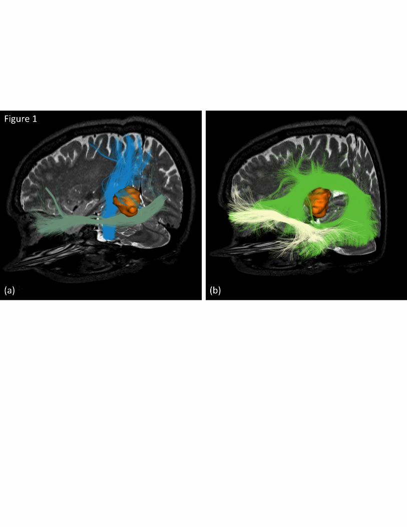
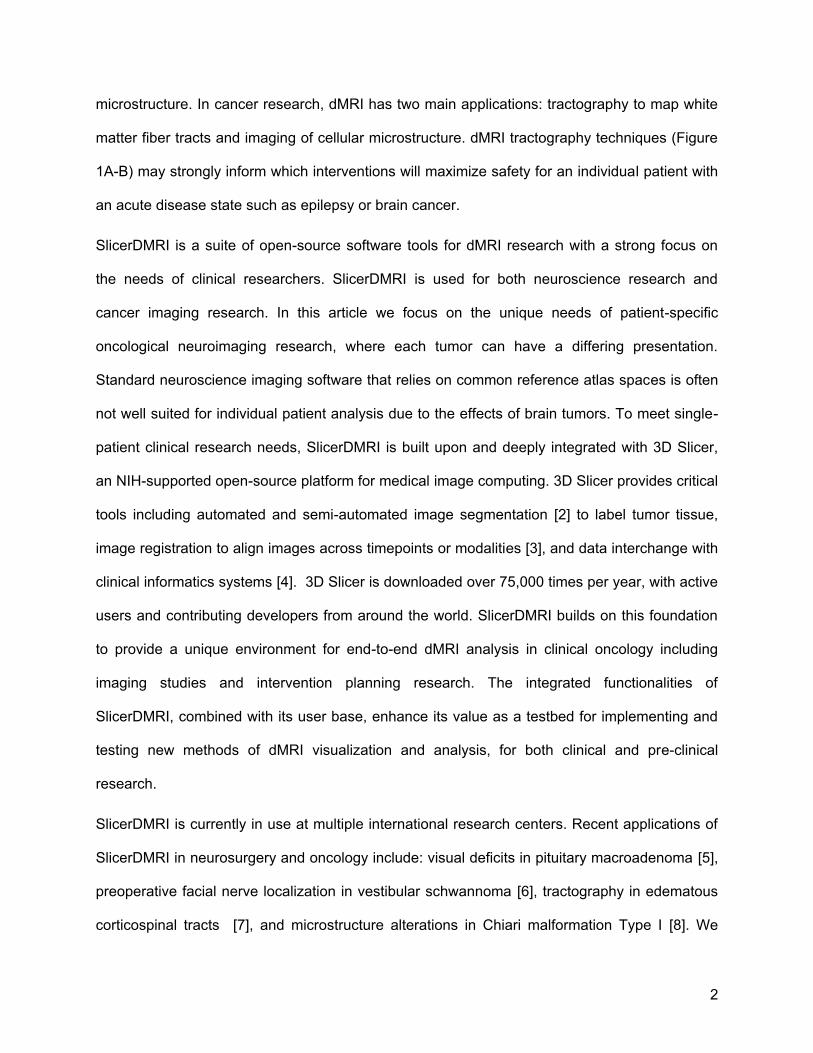
matter fiber tracts and imaging of cellular microstructure. dMRI tractography techniques (Figure
1A-B) may strongly inform which interventions will maximize safety for an individual patient with
an acute disease state such as epilepsy or brain cancer.
SlicerDMRI is a suite of open-source software tools for dMRI research with a strong focus on
the needs of clinical researchers. SlicerDMRI is used for both neuroscience research and
cancer imaging research. In this article we focus on the unique needs of patient-specific
oncological neuroimaging research, where each tumor can have a differing presentation.
Standard neuroscience imaging software that relies on common reference atlas spaces is often
not well suited for individual patient analysis due to the effects of brain tumors. To meet single-
patient clinical research needs, SlicerDMRI is built upon and deeply integrated with 3D Slicer,
an NIH-supported open-source platform for medical image computing. 3D Slicer provides critical
tools including automated and semi-automated image segmentation [2] to label tumor tissue,
image registration to align images across timepoints or modalities [3], and data interchange with
clinical informatics systems [4]. 3D Slicer is downloaded over 75,000 times per year, with active
users and contributing developers from around the world. SlicerDMRI builds on this foundation
to provide a unique environment for end-to-end dMRI analysis in clinical oncology including
imaging studies and intervention planning research. The integrated functionalities of
SlicerDMRI, combined with its user base, enhance its value as a testbed for implementing and
testing new methods of dMRI visualization and analysis, for both clinical and pre-clinical
research.
SlicerDMRI is currently in use at multiple international research centers. Recent applications of
SlicerDMRI in neurosurgery and oncology include: visual deficits in pituitary macroadenoma [5],
preoperative facial nerve localization in vestibular schwannoma [6], tractography in edematous
corticospinal tracts [7], and microstructure alterations in Chiari malformation Type I [8]. We

3
maintain a list of publications using 3D Slicer diffusion functionality such as tensor estimation,
fiber tractography, fiber selection, and tractography visualization at dmri.slicer.org/citations.
Computer Resource: SlicerDMRI Functionality
SlicerDMRI includes clinician-accessible tools for end-to-end diffusion MRI analysis, including
computation of tissue microstructure models (e.g. diffusion tensors), tracing of fiber tracts
(tractography), and quantitative analysis of diffusion-derived measures. See Video 1 for a quick
tutorial covering the installation and basic usage of SlicerDMRI. Technical computing expertise
is not required to use the graphical user interface of SlicerDMRI. For studies in which multiple
datasets will be processed in the same way (batch analysis), SlicerDMRI supports scripting of
most computations.
Core Functionality
Here we describe the main functionality of SlicerDMRI, along with brief examples of usage in
clinical cancer research, as well as relevant software modules in italics.
Support for clinical and research file formats
Support of data interchange is crucial for integration in clinical research workflows. SlicerDMRI
is compatible with clinical Digital Imaging and Communications in Medicine (DICOM) diffusion-
weighted imaging formats as well as research imaging formats such as nrrd and NIfTI.
SlicerDMRI has recently introduced support for the new DICOM tractography interchange
format.
Integrated DTI estimation and visualization
Diffusion tensor imaging (DTI) is the most widely-used method for analyzing diffusion MRI data.
SlicerDMRI provides a graphical user interface to calculate diffusion tensor images from

4
diffusion-weighted image sequences, with several options for brain masking and tensor fitting.
Options for visualization in both 2D and 3D include tractography, diffusion tensors (as
ellipsoids), and derived quantitative measures such as FA (fractional anisotropy) and MD (mean
diffusivity) images. An example application in cancer research is visualization of FA to give
insight into the location and condition of the white matter in proximity to a tumor. Slicer 4.6
modules in this workflow include DWIConvert, DiffusionBrainMasking,
DiffusionTensorEstimation and Volumes.
Tracing of brain connections via fiber tractography
The process of computationally tracing white matter fiber tracts using dMRI is called
tractography. Slicer supports both single-fiber (DTI) tractography and multi-fiber tractography.
DTI tractography: After tensor estimation, tractography may be seeded (initiated) in two clinically
relevant ways: from a region of interest created with the Slicer editing tools, or from an
interactively visualized 3D object that can be manipulated by the user. After seeding, individual
tracts may be isolated via region selection tools. All settings and selection objects may be
interactively adjusted, in both 2D slice and 3D view windows, with tractography results updated
immediately to match. Interactivity allows for rapid isolation of tracts of specific interest. Slicer
4.6 modules in this workflow include TractographySeeding and TractographyDisplay.
Multi-fiber UKF tractography: In comparison with the single-tensor DTI model, which cannot
model anatomical crossing of fibers, modern multi-fiber tracking approaches are better able to
trace important tracts such as the corticospinal tract and arcuate fasciculus. SlicerDMRI
includes the UKFTractography module [9], providing an expanded range of multi-fiber models,
including multi-tensor and multi-compartment models, along with a tractography tracing
technique based on the unscented Kalman filter that uses information from prior steps to
stabilize fiber tracking. See Video 2 for visualization of UKF multi-fiber diffusion MRI
tractography in a neurosurgical case.

5
Quantification of dMRI in cancer research
SlicerDMRI provides tools to measure diffusion-derived values, both in regions of interest
(DiffusionScalarMaps and LabelStatistics) and in fiber tracts (TractographyMeasurements). An
example application in cancer research is measurement of FA to study tissue microstructure.
White matter tract identification in patients
Interactive selection of tractography data can be difficult for a busy clinician. One approach to
simplification of tractography is to cluster (group) tracts using measures of shape similarity. The
SlicerDMRI WhiteMatterAnalysis package includes tools to create data-driven tractography
cluster atlases, and we have recently demonstrated automatic identification of fiber tracts in
patients with brain tumors [10].
Integration into the operating room
Building on 3D Slicer’s capabilities for integration with surgical navigation devices [11,12],
SlicerDMRI has been used in the operating room under research committee oversight to
develop new surgical visualization methods [13,14]. The advantage of this hybrid platform
approach is that clinicians may rely on reliable commercial systems for core navigation
guidance, while also gaining access to, and providing feedback for, the development of novel
visualization and selection techniques in the operative environment.
Software access
SlicerDMRI installation and user support
The SlicerDMRI extension is available from the 3D Slicer Extension Manager, an “app store”
that enables developers to independently implement and distribute software extensions.
SlicerDMRI installation instructions, tutorials, sample data, and introductory and clinical
6
application videos can be found at dmri.slicer.org. SlicerDMRI is cross-platform and can be
used on Windows, Mac, and Linux. Community support for SlicerDMRI users is available
through the 3D Slicer forums (more information at www.slicer.org). The SlicerDMRI developers
actively monitor and respond to diffusion-related topics, and participation in the larger Slicer
community provides access to experts with a breadth of knowledge on many imaging topics.
SlicerDMRI software information
Originally developed starting in 2001 at the MIT AI Lab in collaboration with researchers at the
Surgical Planning Laboratory at Harvard Medical School [15], the SlicerDMRI source code is
now freely available with a BSD-like license that permits unrestricted use, with all code
downloadable at www.github.com/SlicerDMRI. Interested developers may freely include or
extend SlicerDMRI functionality in their own extensions for public distribution through Slicer or
for private use. Information about Slicer extension development is available in the Slicer
developer manual on slicer.org. We welcome potential code contributions to SlicerDMRI via the
GitHub pull request system. Although end-users do not need any programming experience to
install or use the software, interested developers and collaborators are welcome to request or
submit features on the GitHub issue tracker at
https://github.com/SlicerDMRI/SlicerDMRI/issues. Software issues may also be reported on the
issue tracker.
Conclusion
SlicerDMRI provides a platform for diffusion imaging research in a clinical oncology setting with
tools for end-to-end diffusion image analysis as well as interoperation with clinical imaging
systems.

7
References
1. O’Donnell LJ, Westin C-F. An introduction to diffusion tensor image analysis. Neurosurg
Clin N Am. 2011;22: 185–96, viii.
2. Pinter C, Lasso A, Wang A, Sharp GC, Alexander K, Jaffray D, et al. Performing radiation
therapy research using the open-source SlicerRT toolkit. In: Jaffray DA, editor. World
Congress on Medical Physics and Biomedical Engineering, June 7-12, 2015, Toronto,
Canada. Springer International Publishing; 2015. pp. 622–625.
3. Johnson H, Harris G, Williams K, Others. BRAINSFit: mutual information rigid registrations
of whole-brain 3D images, using the insight toolkit. Insight J. 2007;57. Available:
http://www.insight-journal.org/download/pdf/8169/BRAINSFit.pdf
4. Fedorov A, Beichel R, Kalpathy-Cramer J, Finet J, Fillion-Robin J-C, Pujol S, et al. 3D
Slicer as an image computing platform for the Quantitative Imaging Network. Magn Reson
Imaging. Elsevier; 2012;30: 1323–1341.
5. Au KH, Zadeh G. Optic Nerve Tractography Prediction of Visual Deficit in Pituitary
Macroadenoma. J Neurol Surg B Skull Base. 2016;77: P060.
6. Song F, Hou Y, Sun G, Chen X, Xu B, Huang JH, et al. In vivo visualization of the facial
nerve in patients with acoustic neuroma using diffusion tensor imaging-based fiber tracking.
J Neurosurg. 2016;125: 787–794.
7. Chen Z, Tie Y, Olubiyi O, Zhang F, Mehrtash A, Rigolo L, et al. Corticospinal tract modeling
for neurosurgical planning by tracking through regions of peritumoral edema and crossing
fibers using two-tensor unscented Kalman filter tractography. Int J Comput Assist Radiol
Surg. Springer Berlin Heidelberg; 2016;11: 1475–1486.

8
8. Krishna V, Sammartino F, Yee P, Mikulis D, Walker M, Elias G, et al. Diffusion tensor
imaging assessment of microstructural brainstem integrity in Chiari malformation Type I. J
Neurosurg. 2016;125: 1112–1119.
9. Malcolm JG, Shenton ME, Rathi Y. Filtered multitensor tractography. IEEE Trans Med
Imaging. IEEE; 2010;29: 1664–1675.
10. O’Donnell LJ, Suter Y, Rigolo L, Kahali P, Zhang F, Norton I, et al. Automated white matter
fiber tract identification in patients with brain tumors. Neuroimage Clin. 2017;13: 138–153.
11. Ungi T, Lasso A, Fichtinger G. Open-source platforms for navigated image-guided
interventions. Med Image Anal. 2016;33: 181–186.
12. Tokuda J, Fischer GS, Papademetris X, Yaniv Z, Ibanez L, Cheng P, et al. OpenIGTLink:
an open network protocol for image-guided therapy environment. Int J Med Robot. John
Wiley & Sons, Ltd.; 2009;5: 423–434.
13. Golby AJ, Kindlmann G, Norton I, Yarmarkovich A, Pieper S, Kikinis R. Interactive diffusion
tensor tractography visualization for neurosurgical planning. Neurosurgery. NIH Public
Access; 2011;68: 496–505.
14. Elhawary H, Liu H, Patel P, Norton I, Rigolo L, Papademetris X, et al. Intraoperative real-
time querying of white matter tracts during frameless stereotactic neuronavigation.
Neurosurgery. 2011;68: 506–16; discussion 516.
15. Talos I-F, O’Donnell L, Westin C-F, Warfield SK, Iii WW, Yoo S-S, et al. Diffusion Tensor
and Functional MRI Fusion with Anatomical MRI for Image-Guided Neurosurgery. In: Ellis
RE, Peters TM, editors. Medical Image Computing and Computer-Assisted Intervention -
MICCAI 2003. Springer; 2003. pp. 407–415.

9
Figure Caption
Figure 1: Example visualization of a neurosurgical case using SlicerDMRI, with tracts from the
UKF two-tensor tractography method. The patient presented with a history of right side
paresthesia and aphasia, leading to the diagnosis of a left insular glioblastoma. (a) The
corticospinal tract (blue) wraps around the medial and superior aspect of the lesion and the
inferior fronto-occipital fasciculus (pastel green) is relatively close to the inferior pole of the
tumor. (b) The arcuate fasciculus (green) fibers spread along the superior surface of the tumor,
lateral to the corticospinal tract, and the uncinate fasciculus (white) is distant from the lesion.