sliding motility in mycobacteria - jb.asm.orgjb.asm.org/content/181/23/7331.full.pdf · sliding...
TRANSCRIPT
JOURNAL OF BACTERIOLOGY,0021-9193/99/$04.0010
Dec. 1999, p. 7331–7338 Vol. 181, No. 23
Copyright © 1999, American Society for Microbiology. All Rights Reserved.
Sliding Motility in MycobacteriaASUNCION MARTINEZ, SANDRA TORELLO, AND ROBERTO KOLTER*
Department of Microbiology and Molecular Genetics, Harvard Medical School, Boston, Massachusetts 02115
Received 14 June 1999/Accepted 15 September 1999
Mycobacteria are nonflagellated gram-positive microorganisms. Previously thought to be nonmotile, weshow here that Mycobacterium smegmatis can spread on the surface of growth medium by a sliding mechanism.M. smegmatis spreads as a monolayer of cells which are arranged in pseudofilaments by close cell-to-cellcontacts, predominantly along their longitudinal axis. The monolayer moves away from the inoculation pointas a unit with only minor rearrangements. No extracellular structures such as pili or fimbriae appear to beinvolved in this process. The ability to translocate over the surface correlates with the presence of glycopep-tidolipids, a mycobacterium-specific class of amphiphilic molecules located in the outermost layer of the cellenvelope. We present evidence that surface motility is not restricted to M. smegmatis but is also a property ofthe slow-growing opportunistic pathogen M. avium. This form of motility could play an important role insurface colonization by mycobacteria in the environment as well as in the host.
Although most mycobacteria are free-living saprophytic or-ganisms, much of the research on this genus has focused onthose species that are pathogenic to humans. These includeobligate pathogens such as the leprosy bacillus, M. leprae, andthe tubercule bacillus, M. tuberculosis, which kills more than 3million people per year and infects one-third of the worldpopulation (8, 23). Others are opportunistic pathogens whichoccur naturally in the environment but can occasionally causedisease, especially in immunocompromised individuals. Themost important of the opportunistic pathogens are the mem-bers of the M. avium-M. intracellulare complex, which are aleading cause of bacteremia in AIDS patients (21).
One of the most striking characteristics of mycobacteria isthe enormous complexity of their cell envelope (reviewed inreferences 9 and 14). Extensive chemical analyses have shownthat the cell wall of mycobacteria consists of three components.The outside layer is composed of mycolic acids, a complexmixture of long-chain a-branched b-hydroxy fatty acids whichare arranged as a densely packed monolayer. The mycolic acidsare covalently linked to arabinogalactan, which is in turn at-tached to the peptidoglycan layer. This complex cell wall issurrounded by a capsule of noncovalently bound polysaccha-rides, proteins, and a small amount of lipids, which include thespecies- and type-specific glycopeptidolipids (GPLs) and phe-nolic glycolipids. This unusual envelope provides mycobacteriawith remarkable impermeability to external substances, a crit-ical virulence determinant for these organisms.
While much effort has been placed on studying the functionsof cell wall components in pathogenesis, little attention hasbeen focused on the biological significance of the cell wallarchitecture for free-living mycobacteria. In nature most bac-teria are associated with surfaces (12). The type of interactionbetween a bacterium and a surface, whether it attaches to it ormoves on it, is largely determined by the nature of the bacterialcell surface. Bacteria have evolved a wide array of surfacetranslocation modes (20), all of which require special surfacestructures or components, including flagella, pili and fimbriae,surfactants, slime, and capsules. Here we report for the first
time that the fast-growing saprophytic species M. smegmatisand the slow-growing opportunistic pathogen M. avium havethe ability to translocate on solid surfaces by a flagellum-independent spreading mechanism known as sliding (20).Spreading appears to require the presence of GPLs on the cellsurface since rough strains of both species, which lack GPLs,do not exhibit this form of translocation. This form of motilityis likely to play a significant role in the ability of mycobacteriato colonize surfaces in the environments as well as in the host.
MATERIALS AND METHODS
Strains and growth media. M. smegmatis mc2155 (35) and its morphologicalvariants were routinely grown in M63 salts medium (31) supplemented with 1mM MgCl2, glucose (0.2 or 2%), Casamino Acids (0.5%), FeCl2 (10 mM), and amicronutrient solution (28), as indicated. Middlebrook 7H9 and 7H10 media(Difco) supplemented with ADC (22) were used to grow M. avium. M. avium2151-SmD, SmT, Rg-0, and Rg-4 (7) were provided by J. Belisle.
Surface spreading assays. M63 or 7H9 medium supplemented as indicatedwere solidified with 0.3% agar (Difco) or 0.1 to 0.8% ultrapure SeaKem LEagarose (FMC Bioproducts). Twenty-five milliliters of sterile medium that hadbeen cooled to 65°C was dispensed per plate (9-cm diameter). Plates wereallowed to sit at room temperature overnight prior to inoculation and wereinoculated from single colonies by poking with a sterile toothpick or from liquidcultures after cells had been washed in M63 salts. Spreading was evaluatedvisually after incubation of parafilm-sealed plates at 37°C in a humidified incu-bator (50% relative humidity) for the indicated period of time.
Phase-contrast microscopy. Cells on the surface of the growth medium werevisualized with a Nikon Diaphot 200 inverted microscope. The images werecaptured with a black and white CCD72 camera integrated with a power Macin-tosh 8600-300 computer with video capability (Cupertino). Images were pro-cessed using Scion Image (Scion Corporation) and Photoshop 4.0.1 (Adobe)software.
Electron microscopy. Formvar carbon-coated copper grids were gently placedon the surface of the solid growth medium directly over the spreading cells. After1 min, the grids were carefully removed, rinsed twice in distilled water, andstained with 1% uranyl acetate or 2% phosphotungstic acid, as indicated, for 1min. Negatively stained cells were visualized by using a JEOL 1200 EX, 80-kVtransmission electron microscope.
Mixing experiments with GFP-labeled cells. M. smegmatis mc2155 was trans-formed by standard procedures (22) with pGFP, a vector carrying a promoterlessgfp gene (38) cloned into the shuttle vector pMVI203 (11a) or pGFP/O, aplasmid carrying a transcriptional fusion to gfp which results in detectable levelsof green fluorescent protein (GFP) expression (25a). Four-day-old cultures ofcells grown in M63–0.2% glucose–kanamycin (25 mg/ml) medium were mixed1:100 (1 GFP-labeled cell per every 100 unlabeled cells), centrifuged, and washedtwice in M63 salts. Twenty-five microliters of a 1024 dilution was inoculated ontothe surface of 0.3% agarose–M63 salts and –7H9 basal medium (without glyc-erol) plates. Phase-contrast and fluorescence microscopy analyses (2003 mag-nification) were performed using a Nikon microscope equipped with episcopic-fluorescence attachment EFD-3 and a fluorescein isothiocyante filter. Imageswere captured with an Optronics DEI-750 color camera and processed withScion Image and Photoshop software.
* Corresponding author. Mailing address: Department of Microbi-ology and Molecular Genetics, Harvard Medical School, 200 Long-wood Ave., Boston, MA 02115. Phone: (617) 432-1776. Fax: (617)738-7664. E-mail [email protected].
7331
on August 6, 2018 by guest
http://jb.asm.org/
Dow
nloaded from
Isolation of GPLs and TLC. GPLs were isolated from cells grown on thesurface of 7H9–ADC–0.3% agarose plates as previously described (10). GPLprofiles were analyzed by thin-layer chromatography (TLC) on silica plates(Alltech), using as developing solvent chloroform-methanol-water (90:10:1 byvolume). After chromatography, lipids were visualized by spraying with 10%H2SO4 in ethanol and heating at 120°C. M. smegmatis GPLs were identified bycomparison with published patterns of GPLs analyzed under the same conditions(17).
RESULTS
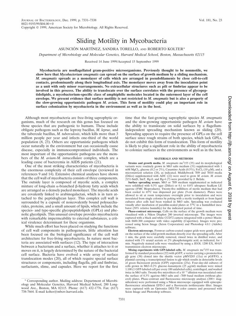
M. smegmatis spreads on the surface of semisolid agarplates. When M. smegmatis mc2155 was inoculated on a semi-solid motility agar plate (0.3% agar) containing low levels ofnutrients (such as M63 salts or 7H9 basal medium with noadded carbon source), two distinct phases of growth were ob-served. Initially, bacterial growth occurred on the surface atthe point of inoculation, as expected from nonswimming bac-teria. After 3 to 4 days, however, a striking change occurred.Finger-like extensions appeared in the periphery of the colonyand spread outwards from the initial inoculation point (Fig. 1).Phase-contrast microscopy revealed that the tips of the spread-ing fingers consist of a monolayer of cells translocating on thesurface as a compact group. No discrete movement of individ-ual cells was observed, in contrast to the jerky movements oftwitching Pseudomonas aeruginosa or the forward/backwardsmovement of gliding Myxococcus xanthus (20). Concentrationsof agar above 0.6% completely inhibited the spreading of my-cobacteria, while replacing the agar by ultrapure agarose al-lowed reproducible spreading under a variety of conditions.Therefore, we used agarose as a solidifying agent in the me-dium for all the subsequent experiments. The ability of myco-bacteria to translocate over surfaces had not been previouslyreported.
The extent of spreading of M. smegmatis on the surface ofagarose plates depends on the degree of wetness. M. smegmatiswas able to spread on the surface of M63 salts plates (with no
added carbon source) over a wide range of agarose concentra-tions (0.1 to 0.8%). In 0.1% agarose, the spreading cells ap-peared to sink into grooves on the soft surface and expanded asfingers reminiscent of those observed in motility agar plates(data not shown). Plates prepared with concentrations of aga-rose equal or above 0.2% were rigid enough to allow thespreading fronts to extend over a large surface, eventuallysurrounding the inoculation site with a circular halo (Fig. 2A).The diameter of the halo was inversely related to the agaroseconcentration, indicating that the wetness of the medium is acritical parameter affecting surface spreading of M. smegmatis.Indeed, the use of freshly prepared plates and a humidifiedincubator are critical for optimum spreading. The diameter ofthe halo correlates with the density at which cells are packedwithin the monolayer. Phase microscopy revealed that the ha-los produced in 0.3 and 0.8% agarose plates consist entirely ofa monolayer of cells arranged as pseudofilaments which aremore tightly packed at 0.8% agarose (Fig. 2B). A phase-brightslime covering the spreading halo was particularly noticeable inthe high-percentage agarose plate. Perhaps the slime extractsmoisture from the medium to create an appropriate surface forthe cells to slide on, as has been proposed for other surface-translocating bacteria (11, 36).
A more detailed view of the arrangement of the spreadingcells was obtained by electron microscopical analysis of gridsthat had been placed directly over the halo. Spreading cells arearranged in pseudofilaments by end-to-end connections alongtheir longitudinal axis (Fig. 2C). However, the contact pointsbetween cells do not always coincide with the cell poles aswould be expected if septum separation after cell division hadnot been complete. Rods are frequently curved, and no surfacestructures such as pili or fimbriae are observed. Rather, thewhole mass of cells seems to be encased in an electron-lightlayer within which an amorphous material connecting groupsof cells can be occasionally observed.
Spreading of M. smegmatis on a solid surface is accompa-nied by growth. In order to address questions regarding growthrates and movement of individual cells within an expandinghalo, we performed a series of mixing experiments in which aminority of the cells used as inoculum were labeled with GFP,allowing their identification by fluorescence microscopy. M.smegmatis cells were transformed with either pGFP, a plasmidcontaining a promotorless gfp, or pGFP/O, a plasmid with atranscriptional fusion of an M. smegmatis gene to gfp thatresults in detectable GFP expression. Cells containing pGFP/Oexhibited uniform detectable GFP levels in all the cells of thepopulation when growing as a halo in 0.3% agarose–M63 saltsplates (data not shown). GFP-labeled cells were mixed 1:100with unlabeled cells and plated in triplicate on 0.3% agarose–M63 and –7H9 (without glycerol or ADC) plates. Immediatelyafter inoculation, GFP-labeled cells were present mostly assingle cells within the inoculum, although small clumps (two tofour cells) were also visible (data not shown). After 2 days ofincubation the diameters of the halos were 2.4 6 0.1 cm in M63and 3.2 6 0.3 cm in 7H9. At that time (Fig. 3), no singlefluorescent cells were observed, but instead green cells werearranged in discrete small groups within the monolayers, indi-cating that growth had occurred in both media. On average thegroups of green cells contained approximately 16 to 20 cells inM63 and 50 to 70 cells in 7H9, which corresponds to four andsix doublings, respectively, after 2 days of incubation. A smallnumber of larger groups of fluorescent cells, probably resultingfrom the growth of the clumps in the inoculum, were alsoobserved. These results show that the formation of halos isaccompanied by growth and that the faster growth observed in7H9 correlates with a higher spreading rate. The carbon and
FIG. 1. Macroscopic morphology of M. smegmatis mc2155 strain spreadingon the surface of a motility agar plate. mc2155 was grown in 7H10, and a singlecolony was transferred with a toothpick to the center of a 0.3% agar platecontaining 7H9 basal medium without any added carbon source. The plate wassealed with parafilm and incubated at 37°C for 2 weeks.
7332 MARTINEZ ET AL. J. BACTERIOL.
on August 6, 2018 by guest
http://jb.asm.org/
Dow
nloaded from

FIG. 2. Macroscopic and microscopic analysis of mc2155 spreading on the surface of agarose plates. (A) Halo formation on 0.3% (left) and 0.8% (right) agaroseplates containing M63 salts with no added source of carbon. Plates were inoculated by poking a single colony from a 0.2% glucose M63 agar plate and transferring itto the center of the plate. The photograph was taken after 5 days of incubation at 37°C. (B) Phase-contrast images of the edges of the spreading halos shown in panelA. Bar, 25 mm. (C) Electron micrograph of cells spreading on a 0.3% agarose–M63 salts plate. A Formvar carbon-coated grid was placed directly over the spreadinghalo and cells were stained with 1% uranyl acetate. Bar, 2 mm.
VOL. 181, 1999 SLIDING MOTILITY IN MYCOBACTERIA 7333
on August 6, 2018 by guest
http://jb.asm.org/
Dow
nloaded from

energy sources supporting this growth are unknown but areunlikely to consist of carried-over liquid medium components,since the cells used in these experiments were washed repeat-edly in M63 buffer prior to inoculation. These results also showthat cells within the monolayer remain in the vicinity of theirsiblings. This very limited rearrangement of the spreading cellsmarkedly contrasts with the high fluidity of cell-cell interac-tions in swarming Serratia liquefaciens or gliding M. xanthus,where similar mixing experiments showed isolated GFP-la-beled cells within the moving population (16, 37).
M. smegmatis colony morphology variants exhibit alteredspreading phenotypes. The capacity of the cells to spread overthe growth surface is likely to be determined in part by thesurface properties of the cells. Since differences in colony mor-phology in many bacterial species are associated with changesin cell surface components, we analyzed the spreading pheno-types of a collection of uncharacterized spontaneous M. smeg-matis mutants previously isolated in our laboratory on the basisof their altered colony appearance. We chose two clones, Sm-1and Rg-1, as representatives of the most severe morphologicalchanges (Fig. 4A). The original M. smegmatis strain, mc2155,appears rugose but moist in 7H10 agar plates. In contrast,under the same conditions Sm-1 is moist and smooth whileRg-1 is rough and extremely dry. A comparison of the spread-ing phenotypes of these strains in M63 salts–0.3% agaroseplates with no added carbon source is shown in Fig. 4B. Sm-1was able to spread on the surface, producing halos that were
very similar to those of mc2155. In contrast, Rg-1 was com-pletely unable to spread and grew at the inoculation point as adensely packed mass of clumped cells.
Addition of nutrients (2% glucose and 0.5% Casamino Ac-ids) had profound effects on the spreading behavior (Fig. 4C).While Rg-1 grew but did not spread, mc2155 and Sm-1 main-tained their spreading capacity but the spreading zone wasmultilayered. While mc2155 appeared smooth and uniform, astar-like pattern irradiating from the inoculation point ap-peared in the Sm-1 halos, which were consistently larger thanthose of mc2155. By the time nutrients had been exhausted, theplate was covered by very dense masses of the spreadingstrains, while the growth of the nonspreading Rg-1 had beenseverely limited (results not shown). These results indicate firstthat the surface properties of the cells can severely affect theirability to spread on solid surfaces. In addition, they demon-strate that the ability to spread confers competitive advantagefor surface colonization and access to nutrients since all threestrains grow at similar rates in standard 2% agar plates of thesame composition (which do not allow spreading).
Time-lapse movies of spreading halos under phase micros-copy. Addition of high levels of all nutrients to M63 plates(glucose, Casamino Acids, and iron and other micronutrients)led to a faster halo expansion. Under these conditions, it waspossible to record time-lapse movies of phase-contrast imagesof the edge of a spreading halo. These movies (one of which isavailable at http://gasp.med.harvard.edu/smegmatis/sliding.html)
FIG. 3. Growth accompanies mycobacterial spreading. A 1:100 mix of GFP-labeled (light) and unlabeled (dark) mc2155 cells grown as described in Materials andMethods were plated on the surface of 0.3% M63 salts– (A) and 7H9 (with no added carbon source)– (B) agarose plates. Photographs were taken after 2 days ofincubation at 37°C. Phase-contrast images showing the continuous spreading halo are on the left, and fluorescent micrographs of the same fields showing the locationsof GFP-labeled cells are on the right. Bars, 25 mm.
7334 MARTINEZ ET AL. J. BACTERIOL.
on August 6, 2018 by guest
http://jb.asm.org/
Dow
nloaded from
clearly show a compact mass of cells sliding over the agarsurface away from the inoculation point, with only minor re-arrangements. The approximate speeds of spreading were 1.6mm/min for mc2155 and 2.5 mm/min for Sm-1.
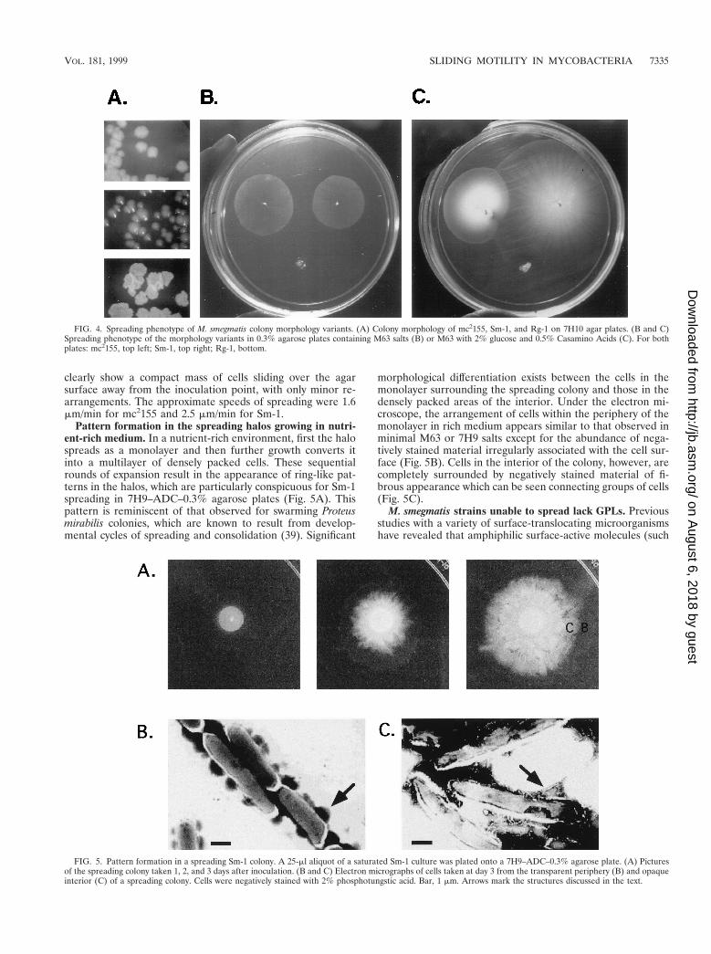
Pattern formation in the spreading halos growing in nutri-ent-rich medium. In a nutrient-rich environment, first the halospreads as a monolayer and then further growth converts itinto a multilayer of densely packed cells. These sequentialrounds of expansion result in the appearance of ring-like pat-terns in the halos, which are particularly conspicuous for Sm-1spreading in 7H9–ADC–0.3% agarose plates (Fig. 5A). Thispattern is reminiscent of that observed for swarming Proteusmirabilis colonies, which are known to result from develop-mental cycles of spreading and consolidation (39). Significant
morphological differentiation exists between the cells in themonolayer surrounding the spreading colony and those in thedensely packed areas of the interior. Under the electron mi-croscope, the arrangement of cells within the periphery of themonolayer in rich medium appears similar to that observed inminimal M63 or 7H9 salts except for the abundance of nega-tively stained material irregularly associated with the cell sur-face (Fig. 5B). Cells in the interior of the colony, however, arecompletely surrounded by negatively stained material of fi-brous appearance which can be seen connecting groups of cells(Fig. 5C).
M. smegmatis strains unable to spread lack GPLs. Previousstudies with a variety of surface-translocating microorganismshave revealed that amphiphilic surface-active molecules (such
FIG. 4. Spreading phenotype of M. smegmatis colony morphology variants. (A) Colony morphology of mc2155, Sm-1, and Rg-1 on 7H10 agar plates. (B and C)Spreading phenotype of the morphology variants in 0.3% agarose plates containing M63 salts (B) or M63 with 2% glucose and 0.5% Casamino Acids (C). For bothplates: mc2155, top left; Sm-1, top right; Rg-1, bottom.
FIG. 5. Pattern formation in a spreading Sm-1 colony. A 25-ml aliquot of a saturated Sm-1 culture was plated onto a 7H9–ADC–0.3% agarose plate. (A) Picturesof the spreading colony taken 1, 2, and 3 days after inoculation. (B and C) Electron micrographs of cells taken at day 3 from the transparent periphery (B) and opaqueinterior (C) of a spreading colony. Cells were negatively stained with 2% phosphotungstic acid. Bar, 1 mm. Arrows mark the structures discussed in the text.
VOL. 181, 1999 SLIDING MOTILITY IN MYCOBACTERIA 7335
on August 6, 2018 by guest
http://jb.asm.org/
Dow
nloaded from
as polysaccharides, peptidolipids, and sulfonolipids) secretedinto the medium or present on the cell surface are often re-quired for movement (1, 18, 26). We hypothesized that someamphiphilic substance produced by mc2155 and Sm-1 but miss-ing in Rg-1 could be required for the spreading behavior. GPLswere possible candidates for this function (for reviews seereferences 9 and 14). The general structure of M. smegmatisGPLs is as follows:
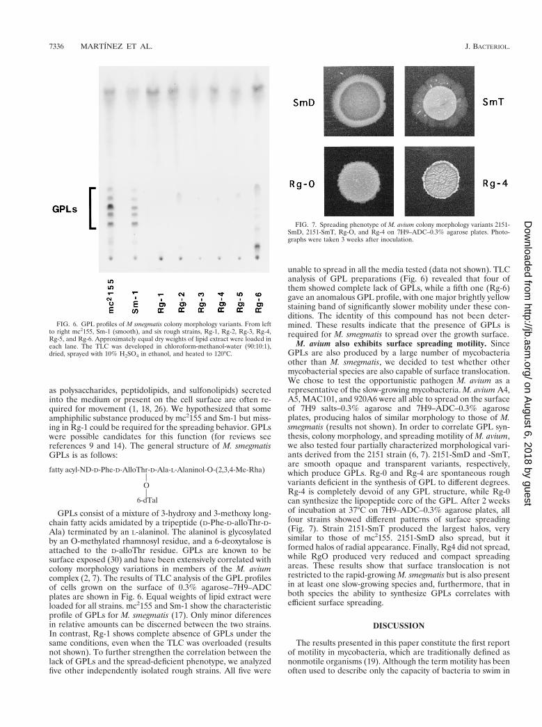
fatty acyl-ND-D-Phe-D-AlloThr-D-Ala-L-Alaninol-O-(2,3,4-Me-Rha)POP
6-dTal
GPLs consist of a mixture of 3-hydroxy and 3-methoxy long-chain fatty acids amidated by a tripeptide (D-Phe-D-alloThr-D-Ala) terminated by an L-alaninol. The alaninol is glycosylatedby an O-methylated rhamnosyl residue, and a 6-deoxytalose isattached to the D-alloThr residue. GPLs are known to besurface exposed (30) and have been extensively correlated withcolony morphology variations in members of the M. aviumcomplex (2, 7). The results of TLC analysis of the GPL profilesof cells grown on the surface of 0.3% agarose–7H9–ADCplates are shown in Fig. 6. Equal weights of lipid extract wereloaded for all strains. mc2155 and Sm-1 show the characteristicprofile of GPLs for M. smegmatis (17). Only minor diferencesin relative amounts can be discerned between the two strains.In contrast, Rg-1 shows complete absence of GPLs under thesame conditions, even when the TLC was overloaded (resultsnot shown). To further strengthen the correlation between thelack of GPLs and the spread-deficient phenotype, we analyzedfive other independently isolated rough strains. All five were
unable to spread in all the media tested (data not shown). TLCanalysis of GPL preparations (Fig. 6) revealed that four ofthem showed complete lack of GPLs, while a fifth one (Rg-6)gave an anomalous GPL profile, with one major brightly yellowstaining band of significantly slower mobility under these con-ditions. The identity of this compound has not been deter-mined. These results indicate that the presence of GPLs isrequired for M. smegmatis to spread over the growth surface.
M. avium also exhibits surface spreading motility. SinceGPLs are also produced by a large number of mycobacteriaother than M. smegmatis, we decided to test whether othermycobacterial species are also capable of surface translocation.We chose to test the opportunistic pathogen M. avium as arepresentative of the slow-growing mycobacteria. M. avium A4,A5, MAC101, and 920A6 were all able to spread on the surfaceof 7H9 salts–0.3% agarose and 7H9–ADC–0.3% agaroseplates, producing halos of similar morphology to those of M.smegmatis (results not shown). In order to correlate GPL syn-thesis, colony morphology, and spreading motility of M. avium,we also tested four partially characterized morphological vari-ants derived from the 2151 strain (6, 7). 2151-SmD and -SmT,are smooth opaque and transparent variants, respectively,which produce GPLs. Rg-0 and Rg-4 are spontaneous roughvariants deficient in the synthesis of GPL to different degrees.Rg-4 is completely devoid of any GPL structure, while Rg-0can synthesize the lipopeptide core of the GPL. After 2 weeksof incubation at 37°C on 7H9–ADC–0.3% agarose plates, allfour strains showed different patterns of surface spreading(Fig. 7). Strain 2151-SmT produced the largest halos, verysimilar to those of mc2155. 2151-SmD also spread, but itformed halos of radial appearance. Finally, Rg4 did not spread,while RgO produced very reduced and compact spreadingareas. These results show that surface translocation is notrestricted to the rapid-growing M. smegmatis but is also presentin at least one slow-growing species and, furthermore, that inboth species the ability to synthesize GPLs correlates withefficient surface spreading.
DISCUSSION
The results presented in this paper constitute the first reportof motility in mycobacteria, which are traditionally defined asnonmotile organisms (19). Although the term motility has beenoften used to describe only the capacity of bacteria to swim in
FIG. 6. GPL profiles of M smegmatis colony morphology variants. From leftto right mc2155, Sm-1 (smooth), and six rough strains, Rg-1, Rg-2, Rg-3, Rg-4,Rg-5, and Rg-6. Approximately equal dry weights of lipid extract were loaded ineach lane. The TLC was developed in chloroform-methanol-water (90:10:1),dried, sprayed with 10% H2SO4 in ethanol, and heated to 120°C.
FIG. 7. Spreading phenotype of M. avium colony morphology variants 2151-SmD, 2151-SmT, Rg-O, and Rg-4 on 7H9–ADC–0.3% agarose plates. Photo-graphs were taken 3 weeks after inoculation.
7336 MARTINEZ ET AL. J. BACTERIOL.
on August 6, 2018 by guest
http://jb.asm.org/
Dow
nloaded from

liquid media, it is now recognized that the ability to move onsolid surfaces is widespread among bacteria. Swarming of P.mirabilis, gliding of myxobacteria and cyanobacteria, andtwitching of pseudomonads are well-known examples of bac-terial surface translocation. The mode of mycobacterial surfacetranslocation reported here should be classified as sliding asdefined by Henrichsen (20): “a kind of surface translocationproduced by the expansive forces in a growing culture in com-bination with special surface properties of the cells resulting inreduced friction between cell and substrate. The micromor-phological pattern is that of a uniform sheet of closely packedcells in a single layer. The sheet moves slowly as a unit.”Examples of sliding bacteria include members of the generaAlcaligenes, Flavobacterium, Acinetobacter, Streptococcus, andCorynebacterium (20). The mechanism underlying sliding inthese organisms has not been characterized in detail. In addi-tion, the flagellum-independent surface spreading of Serratia(26) also satisfies the definition of sliding given by Henrichsen(20).
The arrangement of the translocating mycobacteria as asliding sheet is most clearly shown in the time-lapse movies,where it is evident that cohesive groups of cells are pushedaway from the inoculation site. Importantly, movement of in-dividual cells relative to others, which is one of the maindifferences between sliding and twitching or gliding, is notobserved. The restricted fluidity within the monolayer is alsoconfirmed by the close grouping of siblings in the GFP labelingexperiments. The nature of the cell-to-cell contacts within themonolayer is not known. Groups of cells appear to be arrangedas pseudofilaments, mostly along their longitudinal axis, butcontacts are not restricted to the cell poles. Extracellular struc-tures such as pili or fimbriae, which have been implicated in avariety of cell-to-cell contacts (34, 38), were not observed inpreparations of spreading cells negatively stained with uranylacetate or phosphotugstic acid.
Bacterial translocation over surfaces requires reduced fric-tion between the cells and the substratum. In particular, move-ment of cells over the surface of an agar or agarose plateshould be facilitated by a reduction of the hydrophilic interac-tions between cells and the surface. Bacterial surface-activecompounds are known to have a profound effect in the inter-action of bacteria with interfaces, and a variety of these com-pounds, such as lipopeptides, sulfonolipids, and polysaccha-rides, have been implicated in surface translocation in severalsystems (29). By analogy, we hypothesized that sliding motilityof mycobacteria was therefore likely to involve some kind ofsurface-active compound. The capsular GPLs (reviewed in ref-erences 9 and 14) could play such a role since they are surface-exposed amphiphilic molecules whose absence has been exten-sively correlated with rough colony morphology, the phenotypeof strains deficient for surface spreading. Our data show thatsix independently isolated rough mutants of M. smegmatistested were unable to spread on agarose plates and were de-fective in GPLs (five completely lack GPLs, and one showed anobviously altered profile). Furthermore, the two rough strainsof M. avium we have tested, which are also GPL2, exhibitedspreading-deficient phenotypes. Therefore, there is a strongcorrelation between the lack of GPL and the inability to moveon a surface.
The involvement of GPLs in surface motility in mycobacteriais reminiscent of the role of serrawettings in Serratia spreading(reviewed in reference 16). Serrawettings are a family of cycliclipopeptides with surfactant activity required for flagellum-dependent and -independent surface translocation (25, 26).They are secreted into the medium, where they form a hydro-phobic conditioning film over the hydrophilic agar surface,
thereby reducing the interactions at the interface and promot-ing spreading. Mycobacterial GPLs, however, are present onthe cell surface of intact M. smegmatis and M. avium (30) andhave been found to be the major components of the superficiallayer of smooth variants of M. avium and M. intracellulare (2,3). Freeze fracture analysis of intramacrophagic M. avium hasshown that the bacilli are surrounded by a discontinuous mul-tilamellar capsule-like structure where each lamella is made upof paralell fibers of GPL (33). We have observed discontinuouscapsular structures in negatively stained preparations of M.smegmatis strains spreading in rich medium (Fig. 5B). Thesestructures are present in the GPL-producing strains mc2155and Sm-1 but are absent in Rg-1, the GPL2 strain, and mighttherefore represent accumulations of GPL in the surface oftranslocating bacteria. GPLs could render the bacterial surfacemore hydrophobic and therefore decrease interactions with theagarose surface, facilitating spreading growth. GPLs might alsobe released in some proportion, creating a conditioning film onthe agarose surface for the cells to slide on, as is the case forSerratia.
GPLs are likely not to be the only components affectingmycobacterial spreading motility. For example, spreading bac-teria appear to be surrounded by a mucoid clear material orslime layer of unknown composition. In addition, two of our M.smegmatis strains, mc2155 and Sm-1, which in our analysisappear similar in their GPL components, show differences intheir spreading phenotypes in rich media, where Sm-1 spreadsfaster and forms halos with a complex radial pattern absent inmc2155. There are also obvious differences between thespreading phenotypes of M. avium 2151-SmD and 2151-SmT,both of which produce GPLs (7). These strains differ in theamount of capsular polysaccharide, which is decreased in theSmD strain (32).
On rich medium plates, the morphology of the spreadingcolony becomes complex. What in poor medium is a fairlyuniform spreading of cells as a monolayer, in rich mediumappears to turn into cycles of spreading followed by conversionof the monolayer into a dense cell mass. This switch is accom-panied by changes in the appearance of the cell surface: fibersconnecting groups of cells are present in the densely packedareas but missing in the spreading front. The result is a serieson concentric zones of growth surrounded in the periphery bya monolayer of cells. The cause of the switch between forms ofgrowth is unlikely to be starvation since we observed it in smallisolated microcolonies growing in small numbers on very richmoist plates, but could be due to cell density. Similar successiverounds of expansion have been reported in swarming coloniesof Bacillus subtilis (27) and P. mirabilis (reviewed in reference4). In the case of P. mirabilis it is well documented that theterraces are the result of rounds of swarming followed byconsolidation, where cells “dedifferentiate” into the nonmotilevegetative cells. The mechanism that synchronizes thesechanges is not completely understood. A membrane sensorhistidine kinase has been recently found to be involved in theprocess (5), and differences in fimbria and pilus expressionlevels have been observed among areas of a colony (24). Sim-ilarly, differential gene expression within an expanding colonyis likely to cause the cycles observed in the expansion of amycobacterial colony in rich medium.
The most obvious advantage of surface translocation is thatit results in fast colonization of the available surface by themotile bacteria. We have shown that under conditions thatallow spreading, motile strains of M. smegmatis quickly colo-nize the growth surface and outcompete the nonmotile strainsfor access to the available nutrients. Thus, surface transloca-tion is likely to play an important role in the evolutionary
VOL. 181, 1999 SLIDING MOTILITY IN MYCOBACTERIA 7337
on August 6, 2018 by guest
http://jb.asm.org/
Dow
nloaded from

success of free-living mycobacteria in the environment as mostbacterial growth is likely to occur on a surface (12). In addition,surface translocation could play another role for M. avium.Infections by this opportunistic pathogen are acquired throughthe gastrointestinal and respiratory tracts (21). The capacity ofM. avium strains to spread over surfaces might play an impor-tant role in mucosal colonization and thus could be a virulencedeterminant. Interestingly, fresh isolates of M. avium strainsfrom patients are SmT (13, 15), and under our conditions,strain 2151-SmT showed the most pronounced spreading phe-notype.
We have shown that mycobacterial spreading motility is notrestricted to M. smegmatis but also occurs with M. avium.Interestingly, GPLs are synthesized by a large number of my-cobacterial species, and other classes of amphiphilic lipids thatcould play a similar role are present in the outermost layer ofother mycobacteria (14). The ability to translocate over sur-faces might thus be a general characteristic of mycobacteria.
ACKNOWLEDGMENTS
We thank John Belisle and Michael Starnbach for providing M.avium strains, Maria Ericsson for assistance with the electron micro-scope, and members of the Kolter lab for valuable discussions andcomments on the manuscript.
This work was supported by a postdoctoral fellowship to A.M. fromthe Heiser Program for Research in Leprosy and Tuberculosis andNIH grant GM58213 to R.K.
REFERENCES
1. Abbanat, D. R., E. R. Leadbetter, W. Godchaux III, and A. Escher. 1986.Sulphonolipids are molecular determinants of gliding motility. Nature 324:367–369.
2. Barrow, W. W., and P. J. Brennan. 1982. Isolation in high frequency of roughvariants of Mycobacterium intracellulare lacking C-mycoside glycopeptidol-ipid antigens. J. Bacteriol. 150:381–384.
3. Barrow, W. W., B. P. Ullom, and P. J. Brennan. 1980. Peptidoglycolipidnature of the superficial cell-wall sheath of smooth-colony-forming myco-bacteria. J. Bacteriol. 144:814–822.
4. Belas, R. 1996. Proteus mirabilis and other swarming bacteria. 183–219. InJ. A. and D. Shapiro M (ed.), Bacteria as multicellular organisms. OxfordUniversity Press, Oxford, England.
5. Belas, R., R. Schneider, and M. Melch. 1998. Characterization of Proteusmirabilis precocious swarming mutants: identification of rsbA, encoding aregulator of swarming behavior. J. Bacteriol. 180:6126–6139.
6. Belisle, J. T., K. Klaczkiewicz, P. J. Brennan, W. R. Jacobs, Jr., and J. In-amine. 1993. Rough morphological variants of Mycobacterium avium. Char-acterization of genomic deletions resulting in the loss of glycopetidolipidexpression. J. Biol. Chem. 268:10517–10523.
7. Belisle, J. T., M. R. McNeil, D. Chatterjee, J. Inamine, and P. J. Brennan.1993. Expression of the core lipopetide of the glycopeptidolipid surfaceantigens in rough mutants of Mycobacterium avium. J. Biol. Chem. 268:10510–10516.
8. Bloom, B. R., and C. J. L. Murray. 1992. Tuberculosis: commentary on areemergent killer. Science 257:1055–1064.
9. Brennan, P. J., and H. Nikaido. 1995. The envelope of Mycobacteria. Annu.Rev. Biochem. 64:29–63.
10. Brennan, P. J., M. Souhrada, B. Ullom, J. K. McClatchy, and M. B. Goren.1978. Identification of atypical mycobacteria by thin-layer chromatographyof their surface antigens. J. Clin. Microbiol. 8:374–379.
11. Burchard, R. P. 1981. Gliding motility of prokaryotes: ultrastructure, phys-iology and genetics. Annu. Rev. Microbiol. 35:497–529.
11a.Connel, N., and B. Jacobs. Unpublished results.12. Costerton, J. W., D. E. Lewandowski, D. E. Cladweil, D. R. Korber, and
H. M. Lappin-Scott. 1995. Microbial biofilms. Annu. Rev. Microbiol. 49:711–745.
13. Crowle, A. J., A. Y. Tsang, A. E. Vatter, and M. H. May. 1986. Comparisonof 15 laboratory and patient-derived strains of Mycobacterium avium forability to infect and multiply in cultured human macrophages. J. Clin. Mi-crobiol. 24:812–821.
14. Daffe, M., and P. Draper. 1998. The envelope layers of Mycobacteria withreference to their pathogenicity. Adv. Microb. Physiol. 39:131–203.
15. Dunbar, F. P., I. Pejovic, R. Cacciatore, L. Peric-Golia, and E. H. Runyon.1968. Mycobacterium intracellulare maintenance of pathogenicity in relation-ship to lyophilization and colony form. Scand. J. Respir. Dis. 49:153–162.
16. Eberl, L., S. Molin, and M. Givskov. 1999. Surface motility of Serratialiquefaciens MG1. J. Bacteriol. 181:1703–1712.
17. Eckstein, T. M., F. S. Silbaq, D. Chaterjee, N. J. Kelly, P. J. Brennan, andJ. T. Belisle. 1998. Identification and recombinant expression of a Mycobac-terium avium rhamnosyltransferase gene (rtfA) involved in glycopeptidolipidbiosynthesis. J. Bacteriol. 180:5567–5573.
18. Godchaux, W., III, M. A. Lynes, and E. R. Leadbetter. 1991. Defects ingliding motility in mutants of Cytophaga johnsonae lacking a high-molecular-weight cell surface polysaccharide. J. Bacteriol. 173:7607–7614.
19. Goodfellow, M., and T. Cross. 1983. Classification, p. 8–99. In M. Goodfel-low, M. Mordarski, and S. T. Williams (ed.), The biology of actinomycetes.Academic Press, London, England.
20. Henrichsen, J. 1972. Bacterial surface translocation: a survey and classifica-tion. Bacteriol. Rev. 36:478–503.
21. Inderlied, C. B., C. A. Kemper, and L. E. Bermudez. 1993. The Mycobacte-rium avium complex. Clin. Microbiol. Rev. 6:266–310.
22. Jacobs, W. R., Jr., G. V. Kalpana, J. D. Cirillo, L. Pascopella, S. B. Snapper,R. A. Udani, W. Jones, R. G. Barletta, and B. R. Bloom. 1991. Geneticsystems for mycobacteria. Methods Enzymol. 204:537–555.
23. Kochi, A. 1991. Government intervention programs in HIV/tuberculous in-fection. Outline of guidelines for national tuberculosis control programs inview of the HIV epidemic. Bull. Int. Union Tuberc. Lung Dis. 66:33–66.
24. Latta, R. K., A. Grondin, H. C. Jarrel, G. R. Nichols, and L. R. Berube. 1999.Differential expression of nonagglutinating fimbriae and MR/P pili in swarm-ing colonies of Proteus mirabilis. J. Bacteriol. 181:3220–3225.
25. Lindum, P. W., C. Anthoni, C. Christoffersen, L. Eberl, S. Molin, and M.Givskov. 1998. N-Acyl-L-homoserine lactone autoinducers control produc-tion of an extracellular lipopeptide biosurfactant required for swarmingmotility of Serratia liquefaciens MG1. J. Bacteriol. 180:6384–6388.
25a.Martinez, A., S. Torello, and R. Kolter. Unpublished results.26. Matsuyama, T., K. Kaneda, Y. Nakagawa, K. Isa, H. Hara-Hotta, and I.
Yano. 1992. A novel extracellular cyclic lipopeptide which promotes flagel-lum-dependent and -independent spreading growth of Serratia marcesens. J.Bacteriol. 174:1769–1776.
27. Mendelson, N. H., and B. Salhi. 1996. Patterns of reporter gene expressionin the phase diagram of Bacillus subtilis colony forms. J. Bacteriol. 178:1980–1989.
28. Neidhardt, F. C., P. L. Bloch, and D. F. Smith. 1974. Culture medium forenterobacteria. J. Bacteriol. 119:736–747.
29. Neu, T. R. 1996. Significance of bacterial surface-active compounds in inter-action of bacteria with interfaces. Microbiol. Rev. 60:151–166.
30. Ortalo-Magne, A., A. Lemassu, M. Laneelle, F. Bardou, G. Silve, P. Gounon,G. Marchal, and M. Daffe. 1996. Identification of surface-exposed lipids onthe cell envelopes of Mycobacterium tuberculosis and other mycobacterialspecies. J. Bacteriol. 178:456–461.
31. Pardee, A. B., F. Jacob, and J. Monod. 1959. The genetic control andcytoplasmic expression of “inducibility” in the synthesis of b-galactosidase inE. coli. J. Mol. Biol. 1:165–178.
32. Rastogi, N., C. Frehel, A. Ryter, H. Ohanyon, M. Lesourd, and H. L. David.1981. Multiple drug resistance in Mycobacterium avium: is the wall architec-ture responsible for the exclusion of antimicrobial agents? Antimicrob.Agents Chemother. 20:666–667.
33. Rulong, S., A. P. Aguas, P. Pinto Da Silva, and M. T. Silva. 1991. Intram-acrophagic Mycobacterium avium bacilli are coated by a multiple lamellarstructure: freeze fracture analysis of infected mouse liver. Infect. Immun.59:3895–3902.
34. Shimkets, L. J. 1990. Social and developmental biology of the myxobacteria.Microbiol. Rev. 54:473–501.
35. Snapper, S., L. Lugosi, A. Jekkel, R. Melton, T. Kieser, B. R. Bloom, andW. R. Jacobs, Jr. 1988. Lysogeny and transformation in Mycobacteria: stableexpression of foreign genes. Proc. Natl. Acad. Sci. USA 85:6987–6991.
36. Stahl, S. J., K. R. Stewart, and F. D. Williams. 1983. Extracellular slimeassociated with Proteus mirabilis during swarming. J. Bacteriol. 154:930–937.
37. Wall, D., and D. Kaiser. 1998. Alignment enhances the cell-to-cell transfer ofpilus phenotype. Proc. Natl. Acad. Sci. USA 95:3054–3058.
38. Whittaker, C. J., C. M. Klier, and P. E. Kolenbrander. 1996. Mechanisms ofadhesion by oral bacteria. Annu. Rev. Microbiol. 50:513–552.
39. Williams, F. D., and R. H. Schwarzhoff. 1978. Nature of the swarmingphenomenon in Proteus. Annu. Rev. Microbiol. 32:101–122.
7338 MARTINEZ ET AL. J. BACTERIOL.
on August 6, 2018 by guest
http://jb.asm.org/
Dow
nloaded from