sma nanci yuan, md lpch/stanford medical center may 17, 2006
TRANSCRIPT

SMASMA
Nanci Yuan, MDNanci Yuan, MD
LPCH/Stanford Medical CenterLPCH/Stanford Medical Center
May 17, 2006May 17, 2006
Spinal Muscular Atrophy (SMA)Spinal Muscular Atrophy (SMA)
Spinal muscular atrophy (SMA) is a genetic, Spinal muscular atrophy (SMA) is a genetic, motor neuron disease caused by progressive motor neuron disease caused by progressive degeneration of motor neurons in the entire degeneration of motor neurons in the entire spinal cord and in select brainstem motor nuclei spinal cord and in select brainstem motor nuclei (nuclei of cranial nerves V, VII, IX, and XII). (nuclei of cranial nerves V, VII, IX, and XII).
The disorder causes weakness and wasting of The disorder causes weakness and wasting of the voluntary muscles. the voluntary muscles.
Weakness is often more severe in the legs than Weakness is often more severe in the legs than in the arms. in the arms.


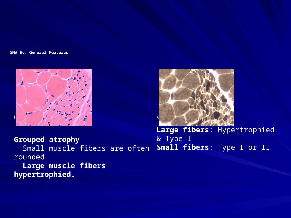
SMA 5q: General Features
H&E stain
ATPase pH 9.4 stain
Grouped atrophy Small muscle fibers are often rounded Large muscle fibers hypertrophied.
Large fibers: Hypertrophied & Type ISmall fibers: Type I or II

SMASMA
SMA is the second most common autosomal SMA is the second most common autosomal recessive disease in the US after cystic fibrosis.recessive disease in the US after cystic fibrosis.Incidence:Incidence:– Type 1: 1 per 10,000 live birthsType 1: 1 per 10,000 live births– Types II and III: 1 per 24,000 birthsTypes II and III: 1 per 24,000 births– SMA types I and III each account for about one fourth SMA types I and III each account for about one fourth
of cases, whereas SMA type II is the largest group of cases, whereas SMA type II is the largest group and accounts for one half of all cases and accounts for one half of all cases
Worldwide 7.8-10 cases per 100,000 live births Worldwide 7.8-10 cases per 100,000 live births ? M:F predominance or M>F? M:F predominance or M>FNo ethnic predominance.No ethnic predominance.
SMASMA
The genetic defects associated with SMA The genetic defects associated with SMA types I-III are localized on chromosome types I-III are localized on chromosome 5q11.2-13.3.5q11.2-13.3.Mutations in the Mutations in the SMN SMN gene result in a loss gene result in a loss of function of the SMN protein. of function of the SMN protein. Many classification systems based on Many classification systems based on inheritance, clinical, and genetic criteria. inheritance, clinical, and genetic criteria. International SMA Consortium (ISMAC, International SMA Consortium (ISMAC, 1994) systems is the most common.1994) systems is the most common.


SMA Type 1SMA Type 1
SMA type I,SMA type I, (Werdnig-Hoffmann acute infantile, (Werdnig-Hoffmann acute infantile, non-sitters), occur birth – 6 months (95% by 3 non-sitters), occur birth – 6 months (95% by 3 months)months)Severe, progressive muscle weakness and Severe, progressive muscle weakness and flaccid or reduced muscle tone (hypotonia). flaccid or reduced muscle tone (hypotonia). Bulbar dysfunction includes poor suck ability, Bulbar dysfunction includes poor suck ability, reduced swallowing, and respiratory failure. reduced swallowing, and respiratory failure. Patients have no involvement of the extraocular Patients have no involvement of the extraocular muscles, and facial weakness is often minimal or muscles, and facial weakness is often minimal or absent.absent. They have no evidence of cerebral involvement, They have no evidence of cerebral involvement, and infants appear alert.and infants appear alert.

SMA Type 1SMA Type 1
Impaired fetal movements are observed in 30% Impaired fetal movements are observed in 30% of casesof cases60% of infants with SMA type I are floppy babies 60% of infants with SMA type I are floppy babies at birth. Prolonged cyanosis may be noted at at birth. Prolonged cyanosis may be noted at delivery. delivery. In some instances, the disease can cause In some instances, the disease can cause fulminant weakness in the first few days of life. fulminant weakness in the first few days of life. Such severe weakness and early bulbar Such severe weakness and early bulbar dysfunction -> mean survival of 5.9 months.dysfunction -> mean survival of 5.9 months.Affected children never sit or stand.Affected children never sit or stand. In 95% of cases, infants die from complications In 95% of cases, infants die from complications of the disease by 18 months. of the disease by 18 months.


SMA Type 2SMA Type 2
SMA type IISMA type II (chronic infantile, sitters) usually (chronic infantile, sitters) usually begin between 6 - 18 months.begin between 6 - 18 months.
Most common form of SMAMost common form of SMA
Most common manifestation is developmental Most common manifestation is developmental motor delay. Infants with SMA type II often have motor delay. Infants with SMA type II often have difficulties with sitting independently or failure to difficulties with sitting independently or failure to stand by 1 year of age. stand by 1 year of age.
These children may learn to sit but will never be These children may learn to sit but will never be able to stand or walk.able to stand or walk.

SMA Type 2SMA Type 2
An unusual feature of the disease is a postural An unusual feature of the disease is a postural tremor affecting the fingers. This is thought to be tremor affecting the fingers. This is thought to be related to fasciculations in the skeletal musclesrelated to fasciculations in the skeletal muscles
Pseudohypertrophy of the gastrocnemius Pseudohypertrophy of the gastrocnemius muscle, musculoskeletal deformities, and muscle, musculoskeletal deformities, and respiratory failure can occur.respiratory failure can occur.
The lifespan of patients with SMA type II varies The lifespan of patients with SMA type II varies from 2 years to the third decade of life. from 2 years to the third decade of life. Respiratory infections account for most deaths. Respiratory infections account for most deaths.


SMA Type 3SMA Type 3
SMA type III (Kugelberg-Welander, SMA type III (Kugelberg-Welander, chronic chronic juvenile, walkersjuvenile, walkers)) appear 18 months – adult. appear 18 months – adult.Slowly progressive proximal weakness. Most Slowly progressive proximal weakness. Most can stand and walk but have trouble with motor can stand and walk but have trouble with motor skills, such as going up and down stairs.skills, such as going up and down stairs.Bulbar dysfunction occurs late in the disease. Bulbar dysfunction occurs late in the disease. Patients may show evidence of Patients may show evidence of pseudohypertrophy.pseudohypertrophy.The disease progresses slowly, and the overall The disease progresses slowly, and the overall course is mild. Many patients have normal life course is mild. Many patients have normal life expectancies. expectancies.


SMASMA
Kennedy syndromeKennedy syndrome or or progressive spinobulbar progressive spinobulbar muscular atrophymuscular atrophy may occur between 15 and 60 may occur between 15 and 60 years of age. Features of this type may include years of age. Features of this type may include weakness of muscles in the tongue and face, weakness of muscles in the tongue and face, difficulty swallowing, speech impairment, and difficulty swallowing, speech impairment, and excessive development of the mammary glands excessive development of the mammary glands in males. The course of the disorder is usually in males. The course of the disorder is usually slowly progressive. Kennedy syndrome is an X-slowly progressive. Kennedy syndrome is an X-linked recessive disorder, which means that linked recessive disorder, which means that women carry the gene, but the disorder only women carry the gene, but the disorder only occurs in men. occurs in men.

Bulbo-Spinal Muscular Atrophy (BSMA; Kennedy's Syndrome; X-linked)
SMASMA
Congenital SMA with arthrogryposisCongenital SMA with arthrogryposis (persistent contracture of joints with fixed (persistent contracture of joints with fixed abnormal posture of the limb) is a rare abnormal posture of the limb) is a rare disorder. Manifestations include severe disorder. Manifestations include severe contractures, curvature of the spine, chest contractures, curvature of the spine, chest deformity, respiratory problems, an deformity, respiratory problems, an unusually small jaw, and drooping upper unusually small jaw, and drooping upper eyelids. eyelids.


Pulmonary NeedsPulmonary Needs
Is NOT Duchenne’s Muscular DystophyIs NOT Duchenne’s Muscular DystophyDiaphragm NOT involved and IS the Diaphragm NOT involved and IS the primary muscle of breathingprimary muscle of breathingDiaphragm function better when flat or in Diaphragm function better when flat or in TrendelenburgTrendelenburgIntercostals are weak/ineffectiveIntercostals are weak/ineffectiveChest wall compliance is increased -> Chest wall compliance is increased -> chest wall shape changes (pulled down chest wall shape changes (pulled down chest appearance) -> then decreaseschest appearance) -> then decreases
Pulmonary NeedsPulmonary Needs
Pulmonary compliance increases then Pulmonary compliance increases then decreasesdecreases
HypoventilationHypoventilation
Oxygen therapy does not address Oxygen therapy does not address hypoventilationhypoventilation
Non-invasive positive pressure ventilation Non-invasive positive pressure ventilation (NIPPV) – BiPAP NOT CPAP or non-(NIPPV) – BiPAP NOT CPAP or non-invasive volume ventilator invasive volume ventilator
NIPPV GoalsNIPPV Goals
From Mehta and Hill From Mehta and Hill Short – termShort – term– Relieve symptomsRelieve symptoms– Reduce WOBReduce WOB– Improve/stabilize blood gasImprove/stabilize blood gas– Optimize patient comfortOptimize patient comfort– Good patient-ventilator synchronyGood patient-ventilator synchrony– Minimize riskMinimize risk– Avoid intubationAvoid intubation
NIPPV GoalsNIPPV Goals
From Mehta and Hill From Mehta and Hill
Long – termLong – term– Improve sleep duration and qualityImprove sleep duration and quality– Maximize quality of lifeMaximize quality of life– Enhance functional statusEnhance functional status– Prolong survivalProlong survival

NIPPVNIPPV
More effective in decreasing work of More effective in decreasing work of breathing than CPAP or negative pressurebreathing than CPAP or negative pressure
Increasing tidal volume -> decreases Increasing tidal volume -> decreases respiratory raterespiratory rate
More effective gas exchangeMore effective gas exchange
Reduces daytime PCO2Reduces daytime PCO2– ?resets PCO2 set point, improves ?resets PCO2 set point, improves
microatelectasis, rests fatigued musclesmicroatelectasis, rests fatigued muscles

BiPAPBiPAP
High IPAP (PIP) -> goal is ventilation NOT to High IPAP (PIP) -> goal is ventilation NOT to overcome obstruction overcome obstruction – Want to rest muscles/decrease WOBWant to rest muscles/decrease WOB
Low EPAP (PEEP) -> LOW 3-6 cm H20 -> Low EPAP (PEEP) -> LOW 3-6 cm H20 -> compromise ability to exhale compromise ability to exhale Spontaneous – Timed mode -> want high Spontaneous – Timed mode -> want high respiratory rate that will capture their respiratory respiratory rate that will capture their respiratory effort and rest patient (goal patient synchrony effort and rest patient (goal patient synchrony and rest) – ex. 30and rest) – ex. 30– May also overdrive them if problems with synchrony May also overdrive them if problems with synchrony
and then decreaseand then decrease
BiPAPBiPAP
I – time based on patient age and I – time based on patient age and respiratory raterespiratory rate
Rise time (speed of breath delivery) Rise time (speed of breath delivery) usually medium setting and on room air (at usually medium setting and on room air (at home)home)

NIPPVNIPPV
Respironics Synchrony Respironics Synchrony
ResMed VPAP IIIResMed VPAP III
Puritan BennettPuritan Bennett
Need appropriate interfaceNeed appropriate interface
Heated or pass-over cool mist Heated or pass-over cool mist humidificationhumidification
Non-invasive VentilatorNon-invasive Ventilator
Pressure versus volume NO difference on Pressure versus volume NO difference on outcomesoutcomes
Volume – breaths on demand during dayVolume – breaths on demand during day– Usually tidal volume 13-20 ml/kg/on nasal Usually tidal volume 13-20 ml/kg/on nasal
maskmask

IntubatedIntubated
Extubate to BiPAP settingsExtubate to BiPAP settings
Do NOT wean to low rates or T-pieceDo NOT wean to low rates or T-piece
Extubate when already on room air so not Extubate when already on room air so not to mask atelectasis with supplemental to mask atelectasis with supplemental oxygenoxygen

Respiratory AidsRespiratory Aids
Cough AssistCough AssistChest physiotherapyChest physiotherapyIPVIPVHome suctionHome suctionPulse oximeterPulse oximeterAmbubagAmbubagNebulizer Nebulizer – ?bronchodilators, mucolytics, anticholinergics?bronchodilators, mucolytics, anticholinergics
Gtube and Nissen fundoplicationGtube and Nissen fundoplication
“No DMD person should ever require a trach tube or develop any respiratory complications. If people follow closely what we describe here, respiratory difficulties can be eliminated.”
Dr. John
R.
Bach
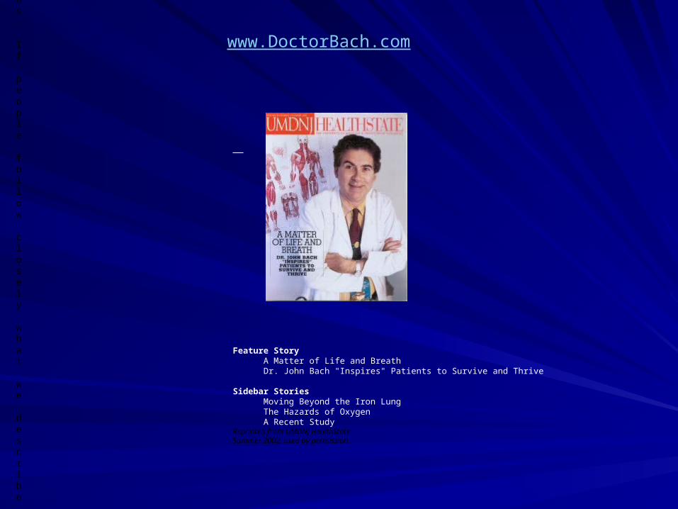
www.DoctorBach.com
Feature Story A Matter of Life and Breath Dr. John Bach "Inspires" Patients to Survive and Thrive
Sidebar Stories Moving Beyond the Iron Lung The Hazards of Oxygen A Recent Study Reprinted from UMDNJ HealthState Summer 2002, used by permission.

Conventional[Top]
1. Oxygen administrated arbitrarily in concentrations that maintain SaO2 well above 95%.
2. Frequent airway suctioning via the tube. 3. Supplemental oxygen increased when desaturations occur.
4. Ventilator weaning attempted at the expense of hypercapnia. 5. Extubation not attempted unless the patient appears to be ventilator weaned.
6. Extubation to CPAP or low span bi-level positive airway pressure and continued oxygen therapy.
7. Deep airway suctioning by catheterizing the upper airway along with postural drainage and chest physical therapy.
8. With increasing CO2 retention or hypoxia supplemental oxygen is increased and ultimately the patient is reintubated.
9. Following re-intubation tracheostomy is thought to be the only long-term option ...or following successful extubation bronchodilators and ongoing routine chest
physical therapy are used. 10. Eventually discharged home with a tracheostomy, often following a rehabilitation
stay for family training.

Protocol[Top]
1. Oxygen administration limited only to approach 95% SaO2. 2. Mechanical insufflation-exsufflation used via the tube at 25 to 40 cm H2O to -25 to -40 cm H2O pressures up to every 10 minutes as
needed to reverse oxyhemoglobin desaturations due to airway mucus accumulation and when there is auscultatory evidence of
secretion accumulation. Abdominal thrusts are applied during exsufflation. Tube and upper airway are suctioned following use of
expiratory aids as needed. 3. Expiratory aids used when desaturations occur.
4. Ventilator weaning attempted without permitting hypercapnia. 5. Extubation attempted whether or not the patient is ventilator
weaned when meeting the following: A. Afebrile
B. No supplemental oxygen requirement to maintain SaO2 >94% C. Chest radiograph abnormalities cleared or clearing
D. Any respiratory depressants discontinued E. Airway suctioning required less than 1-2x/eight hours

F. Coryza diminished sufficiently so that suctioning of the nasal orifices is required less than once every 6 hours (important to facilitate use of nasal
prongs/mask for post-extubation nasal ventilation) 6. Extubation to continuous nasal ventilation and no supplemental oxygen.
7. Oximetry feedback used to guide the use of expiratory aids, postural drainage, and chest physical therapy to reverse any desaturations due to
airway mucus accumulation. 8. With CO2 retention or ventilator synchronization difficulties nasal interface leaks were eliminated, pressure support and ventilator rate increased or the
patient switched from BiPAP-ST™ to using a volume cycled ventilator. Persistent oxyhemoglobin desaturation despite eucapnia and aggressive use
of expiratory aids indicated impending respiratory distress and need to re-intubate.
9. Following re-intubation the protocol was used for a second trial of extubation to nasal ventilation
...or following successful extubation bronchodilators and chest physical therapy were discontinued and the patient weaned to nocturnal nasal
ventilation. 10. Discharge home after the SaO2 remained within normal limits for 2 days
and when assisted coughing was needed for less than 4 times per day.

Mary Schroth, MDAssociate ProfessorUW Hospital - Clinical Science CenterOffice Suite K4/942Office: (608) 263-8555Email: [email protected] Dr. Mary Schroth's Full CV