snai2 upregulation is associated with an aggressive ... · (continued from previous page)...
TRANSCRIPT

RESEARCH ARTICLE Open Access
SNAI2 upregulation is associated with anaggressive phenotype in fulvestrant-resistant breast cancer cells and is anindicator of poor response to endocrinetherapy in estrogen receptor-positivemetastatic breast cancerCarla L. Alves1* , Daniel Elias1, Maria B. Lyng1, Martin Bak2 and Henrik J. Ditzel1,3,4*
Abstract
Background: Endocrine resistance in estrogen receptor-positive (ER+) breast cancer is a major clinical problem andis associated with accelerated cancer cell growth, increased motility and acquisition of mesenchymal characteristics.However, the specific molecules and pathways involved in these altered features remain to be detailed, and maybe promising therapeutic targets to overcome endocrine resistance.
Methods: In the present study, we evaluated altered expression of epithelial-mesenchymal transition (EMT) regulators inER+ breast cancer cell models of tamoxifen or fulvestrant resistance, by gene expression profiling. We investigated thespecific role of increased SNAI2 expression in fulvestrant-resistant cells by gene knockdown and treatment with aSNAIL-p53 binding inhibitor, and evaluated the effect on cell growth, migration and expression of EMT markers.Furthermore, we evaluated SNAI2 expression by immunohistochemical analysis in metastatic samples from two cohortsof patients with breast cancer treated with endocrine therapy in the advanced setting.
Results: SNAI2 was found to be significantly upregulated in all endocrine-resistant cells compared to parental cell lines,while no changes were observed in the expression of other EMT-associated transcription factors. SNAI2 knockdown withspecific small interfering RNA (siRNA) converted the mesenchymal-like fulvestrant-resistant cells into an epithelial-likephenotype and reduced cell motility. Furthermore, inhibition of SNAI2 with specific siRNA or a SNAIL-p53 bindinginhibitor reduced growth of cells resistant to fulvestrant treatment. Clinical evaluation of SNAI2 expression in twoindependent cohorts of patients with ER+ metastatic breast cancer treated with endocrine therapy in the advancedsetting (N = 86 and N = 67) showed that high SNAI2 expression in the metastasis correlated significantly with shorterprogression-free survival on endocrine treatment (p = 0.0003 and p = 0.004).
(Continued on next page)
* Correspondence: [email protected]; [email protected] of Cancer and Inflammation Research, Institute of MolecularMedicine, University of Southern Denmark, J.B. Winsløwsvej 25, 5000 OdenseC, DenmarkFull list of author information is available at the end of the article
© The Author(s). 2018 Open Access This article is distributed under the terms of the Creative Commons Attribution 4.0International License (http://creativecommons.org/licenses/by/4.0/), which permits unrestricted use, distribution, andreproduction in any medium, provided you give appropriate credit to the original author(s) and the source, provide a link tothe Creative Commons license, and indicate if changes were made. The Creative Commons Public Domain Dedication waiver(http://creativecommons.org/publicdomain/zero/1.0/) applies to the data made available in this article, unless otherwise stated.
Alves et al. Breast Cancer Research (2018) 20:60 https://doi.org/10.1186/s13058-018-0988-9
(Continued from previous page)
Conclusions: Our results suggest that SNAI2 is a key regulator of the aggressive phenotype observed inendocrine-resistant breast cancer cells, an independent prognostic biomarker in ER+ advanced breast cancertreated with endocrine therapy, and may be a promising therapeutic target in combination with endocrinetherapies in ER+ metastatic breast cancer exhibiting high SNAI2 levels.
Keywords: Endocrine resistance, Epithelial-mesenchymal transition, Estrogen receptor-positive breast cancer,Fulvestrant, SNAI2
BackgroundApproximately 80% of all breast tumors are positive for es-trogen receptor (ER+), which is an indicator of potentialresponsiveness to endocrine therapy both in the adjuvantand advanced settings [1]. Despite the efficacy of endocrinetherapy for treatment of ER+ breast cancer, a significantnumber of patients develop resistance to these drugs.There is considerable evidence suggesting that acquisitionof endocrine resistance is accompanied by acceleratedtumor growth and increased metastatic propensity, and isassociated with morphological changes characteristic ofcells undergoing epithelial-mesenchymal transition (EMT)[2]. However, key questions remain regarding the centralmolecules controlling the EMT process during develop-ment of endocrine resistance, which may be promisingtherapeutic targets in combination with endocrine therapy.EMT is a complex process characterized by loss of epi-
thelial features, such as downregulation of the E-cadherinand occludins, and acquisition of mesenchymal properties,including upregulation of vimentin and fibronectin, andcytoskeleton reorganization [3]. EMT has been associatedwith increased cell migration capacity and invasiveness, andis a prominent hallmark of cancer progression [4, 5]. Epi-thelial tumor cells may acquire a mesenchymal-like pheno-type to facilitate migration and invasion and then possiblyreverse to an epithelial state through mesenchymal-epithe-lial transition (MET) to form organized tumorigenic nod-ules at the lodgment sites [6]. EMT and MET are regulatedby signals from the stroma associated with tumors, such astransforming growth factor (TGF)-β, and by a series ofEMT-inducing transcription factors, including SNAI1,SNAI2, TWIST, ZEB1 and ZEB2 [7].The role of EMT in endocrine resistance was first re-
ported in studies on ER-depleted breast cancer cells,which were found to convert the non-invasive epithelialfeatures into a mesenchymal-like phenotype with invasivecharacteristics [6, 8]. Moreover, EMT has been shown tomediate endocrine resistance through the action of EMTtranscription factors. SNAI family members were found todirectly repress ER [9, 10] and enhance the anti-apoptoticbehavior of cancer cells, contributing to resistance totherapy [11]. A large body of evidence supports the im-portance of EMT in sustaining cancer stem cells (CSCs),which can be intrinsically resistant to treatment [12].
Furthermore, several growth factor receptors, such as epi-dermal growth factor receptor (EGFR), insulin-like growthfactor 1 receptor (IGF-1R) and fibroblast growth factor 1receptor (FGFR1), which are involved in the EMT process,are highly expressed in ER- breast tumor cells, supportingthe link between EMT and insensitivity to endocrine ther-apy [2]. The emerging role of EMT as a mediator of endo-crine resistance in breast cancer has raised interest intherapeutic strategies based on reversing EMT to preventtumor progression and re-sensitizing tumor cells toendocrine therapy [13]. One promising pharmacologicalapproach involves the development of specific inhibitorsof EMT-associated transcription factors to therapeuticallyinhibit EMT induction or target the mesenchymal celltype [14].In this study, we investigated the altered expression of
various EMT regulators in MCF-7-based breast cancercell models of endocrine resistance by gene array. Weobserved upregulation of SNAI2 in fulvestrant-resistantand tamoxifen-resistant cells compared to the parentalcell lines, while other EMT-associated transcriptionfactors were not altered. Inhibition of SNAI2 inducedepithelial characteristics, reduced cell motility, and im-paired growth of fulvestrant-resistant breast cancer cells.High levels of SNAI2 in ER+ metastatic tumor samplesfrom two cohorts of patients treated with endocrinetherapy in the advanced setting correlated significantlywith poor clinical outcome. Our findings indicatedSNAI2 as an independent prognosis biomarker in ER+metastatic breast cancer patients treated with endocrinetherapy and a potential novel therapeutic target thatmay contribute to reversing EMT and re-sensitizingbreast cancer cells to endocrine therapy.
MethodsCell lines and culture conditionsThe original MCF-7 cell line was obtained from the BreastCancer Task Force Cell Culture Bank, Mason ResearchInstitute. MCF-7 cells were gradually adapted to grow inlow serum concentration [15], and this subline, MCF-7/S0.5,was used to establish two fulvestrant-resistant cell models,MCF-7/182R (including 182R-1 and 182R-6 cell lines) andMCF-7/164R (including 164R-1 and 164R-4 cell lines),by extended treatment with 100 nM of fulvestrant
Alves et al. Breast Cancer Research (2018) 20:60 Page 2 of 12
(ICI 182,780) or ICI 164,384, respectively [16].Tamoxifen-resistant (TamR) cell lines, includingTamR-1, TamR-4, TamR-7 and TamR-8 cells, wereestablished from MCF-7/S0.5 by long-term treatmentwith 1 μM of tamoxifen [17]. The MCF-7/S0.5 cellline was routinely propagated in phenol red-freeDulbecco’s modified Eagle medium (DMEM)/F12(Gibco) supplemented with 1% glutamine (Gibco), 1%heat-inactivated fetal bovine serum (FBS; Gibco) and6 ng/ml insulin (Sigma-Aldrich). Fulvestrant-resistantand tamoxifen-resistant cell lines were maintained inthe same growth medium as MCF-7/S0.5 supple-mented with 100 nM fulvestrant (Tocris) or 1 μMtamoxifen (Sigma-Aldrich), respectively. Cells were grownin a humidified atmosphere of 5% CO2 at 37 °C and thegrowth medium was renewed every second or third day.To reduce variability between experiments, cells weremaintained at low passage numbers (< 10 passages)throughout the experiments. All cell lines underwentDNA authentication using Cell ID™ System (Promega)before the described experiments to ensure consistentcell identity.
Global gene expression profilingMCF-7/S0.5, tamoxifen-resistant and fulvestrant-resistantcell lines were grown to 70–80% confluence for total RNApurification using a RNA kit (Qiagen) and arrayed separ-ately in Affymetrix Gene Chip® Human Genome U133plus 2.0 arrays (Affymetrix), as described [18]. Data wereanalyzed using Partek Genomic Suite (Partek Inc.). Genesfrom the data set that exhibited twofold or greateralteration in expression and false discovery rate (FDR)cutoff < 0.05 were considered as significantly alteredregulated.
RNA isolation and reverse transcription (RT)-quantitative(q)PCR (RT-qPCR)Total RNA was extracted using Isol-Lysis Reagent,TRIzol® (Life technologies) followed by chloroform andisopropyl alcohol (Sigma-Aldrich) for separation andprecipitation of RNA. Concentration and purity weremeasured using the NanoDrop-1000 spectrophotometer(Saveen). Complementary DNA (cDNA) was synthesizedusing RevertAid Premium Reverse Transcriptase kit(Fermentas). Relative quantification of gene expressionwas performed using SYBR® Green PCR Mastermix(Applied Biosystems) according to manufacturer’s instruc-tions. The following primers purchased from Qiagen wereused: SNAI2 (QT00044128), CDH1 (QT00080143), andPUM1 (QT00029421) QuantiTect® Primer. PUM1 wasused as the reference gene for normalization. RT-qPCR re-actions were performed on a StepOnePlus™ Real-TimePCR system from Applied Biosystems and data wereobtained from StepOne Software Version 2.1. Relative
expression levels were calculated using the comparativethreshold method [19].
Western blottingWhole cell extracts were obtained using radioimmuno-precipitation assay (RIPA) buffer (50 mM Tris HCl(pH 8), 150 mM NaCl (pH 8), 1% IgePAL 630, 0.5% so-dium dioxycholate, 0.1% SDS) containing protease andphosphatase inhibitors (Roche). The protein concentra-tion of the lysate samples was determined using Piercebicinchoninic acid (BCA) Protein Assay Kit (ThermoFisher Scientific) and the optical density (OD) was mea-sured at 562 nm in the microplate reader SunriseTM 500ELISA-reader (Tecan). 10–20 μg of total protein lysatewas loaded on a 4–20% SDS-PAGE gel (Biorad) underreducing conditions and electroblotted onto a polyvinyli-dene difluoride (PVDF) transfer membrane. Prior toprimary antibody incubation, membranes were blockedin Tris-buffered saline (TBS), 0.1% Tween-20 (Sigma-Al-drich) containing 5% non-fat dry milk powder(Sigma-Aldrich) or 5% bovine serum albumin(Sigma-Aldrich). The following antibodies were used ac-cording to the manufacturer’s protocol: anti-E-cadherin(#3195, Cell Signaling), anti-SNAI2 (#9585, Cell Signaling);anti-vimentin (#6630, Sigma-Aldrich); anti-ERα antibody(#9101, Thermo Fisher Scientific); anti-SOX2 (#AF2018,R&D Systems); anti-β-actin (#6276, Abcam) as loadingcontrol; horseradish peroxidase (HRP)-conjugated goatanti-mouse (#P0447, Dako); HRP-conjugated goatanti-rabbit (#P0448, Dako); HRP-conjugated donkeyanti-goat (#sc-2020, Santa Cruz Biotechnology). Themembrane was developed with Enhanced Chemilu-minescence (ECL) Prime Western Blotting DetectionReagents (GE Healthcare) and visualized using theFusion-Fx7–7026 WL/26MX instrument (Vilbaer).
siRNA-mediated gene knockdownCells were transfected with siRNA against SNAI2(s13127; Life Technologies) or SOX2 (D-011778-01;Dharmacon) using an Electroporation Ingenio kit (MirusBio) in a Nucleofector™ II device (Amaxa, Lonza) orLipofectamine 3000 reagent (Thermo Fischer Scientific),respectively, according to manufacturers’ instructions.Mission siRNA Universal Negative Control (SIC001)(Sigma-Aldrich) was used as control. Transfected cellswere seeded in 24-well plates (5 × 10⁴ cells/well) toevaluate gene knockdown efficiency 48 h followingtransfection, by RT-qPCR. Transfected cells were seededin T25 flasks (5 × 10⁵ cells) and incubated for 96 h toassess protein expression by western blotting.
Cell growth assayTransfected cells were seeded (2.5–5 × 10⁴ cells/well) in24-well plates and incubated for 24 and 96 h at 37 °C in
Alves et al. Breast Cancer Research (2018) 20:60 Page 3 of 12

5% CO2 for evaluation of cell growth using crystalviolet-based colorimetric assay [20]. For growth assayswith the chemical inhibitor, cells were seeded (3 × 10⁴
cells/well) in 24-well plates in the presence of 3 μMSNAIL-p53 binding inhibitor GN25 (Millipore) or itssolvent (DMSO, Sigma-Aldrich), and cell growth wasmeasured 72 h after seeding using crystal violet-basedcolorimetric assay. The OD was analyzed at 570 nm in aSunrise™ 500 absorbance reader (Tecan).
Cell migration assayA total of 1 × 10⁵ cells, starved overnight, were harvestedin serum-free medium and seeded in the upper chamberof 8-μm-pore polystyrene membrane chamber-insertTranswell® apparatus (Corning, Costar) in 24-well plateswith 10% FBS medium, according to the manufacturer’sinstructions. Cells were incubated for 96 h at 37 °C in5% CO2. Cells on the top surface of the insert were re-moved with a cotton swab, and cells that migrated tothe bottom face of the insert were fixed and stained withcrystal violet in methanol solution. To determine thenumber of migrated cells, five random fields were usedto count cells at the microscope. To determine the totalnumber of cells that migrated in one insert, the averagenumber of cells counted was divided by the area of themicroscope viewing field and then multiplied by the en-tire area of the Transwell insert (0.3 cm2). Normalizationof migration according to growth rate was performedusing crystal violet staining.
Cell invasion assayCell invasion was evaluated using a QCM ECMatrix24-well kit (Chemicon ECM550) according to the manu-facturer’s instructions. Cells were seeded in serum-freemedium in the upper chamber of an insert in 24-wellplates with 10% FBS medium, and incubated for 96 h at37 °C in 5% CO2. Invading cells were detached, lysed,stained with dye, and measured by fluorescent lightemission (480 nm/520 nm) using a Victor3™ 1420 coun-ter (Perkin Elmer Wallac). Fluorescent measurementswere reported as relative fluorescent unit (RFU) values.Light emission was normalized to cell growth ratemeasured by crystal violet colorimetric assay.
Immunocytochemical analysisMCF-7/S0.5 and fulvestrant-resistant cells were fixed in4% formalin, paraffin-embedded, and mounted in 4-μmsections on glass slides. Antigen retrieval was performedby boiling sections in T-EG solution/TRS buffer (Dako).Sections were incubated with anti-SOX2 antibody(#AF2018, R&D Systems) for one hour at roomtemperature. PowerVision Poly-HRP was used as the de-tection system. Microscopy of cells was performed usinga Leica DMLB microscope (× 100 or × 200/numerical
aperture (NA) 1.25, Leica Microsystems) using LasV3.6acquisition software.
Clinical samplesFormalin-fixed, paraffin-embedded (FFPE) metastatictumor samples from patients with ER+ breast cancertreated with endocrine therapy in the advanced settingwere selected by database extraction from the archivesof the Department of Pathology at Odense UniversityHospital (OUH) encompassing the period 2004–2013(N = 165; cohort 1) and 2013–2016 (N = 128; cohort 2).Patients eligible for inclusion were those with ER+ breastcancer with metastatic disease, who had undergone sur-gery or biopsy at OUH, and for whom complete clinicalinformation and pathological verification that the meta-static lesion was of breast cancer origin were available.Exclusion criteria were competing cancer(s), cytologicalbiopsies, or insufficient material in the FFPE block.These parameters yielded 86 (cohort 1) and 67 (cohort2) metastatic lesions from patients with advanced breastcancer treated with endocrine therapy. The metastaticbiopsies used for evaluation of SNAI2 expression wereobtained prior to treatment with endocrine therapy.Tumors were defined as ER+ if ≥ 1% of the tumor cellsstained positive.
Immunohistochemical stainingWhole FFPE sections of metastatic lesions were incubatedwith anti-SNAI2 (#sc-15391, Santa Cruz Biotechnology)and immunostained using the HRP-conjugated Power-Vision+™ system on the autostainer TechMateTM 500(Dako), as described [21]. A Leica DMLB microscope(× 100/numerical aperture 1.25, Leica Microsystems) andLasV3.6 acquisition software were used for tissue micros-copy. Evaluation of the staining was performed by an ex-perienced breast pathologist in a blinded setup. SNAI2expression was observed in the cell nucleus and cytoplasmand tumors were scored based on the staining intensityscore (0–3). The cutoff value for high versus low SNAI2(intensity score = 2) was determined and optimized in co-hort 1 employing the web-based tool Cutoff Finder [22]with the “survival significance” function, and the samecutoff was then applied to cohort 2.
Clinical endpointsProgression-free survival (PFS) was defined as the timefrom the date of initiation of endocrine treatment untildisease progression within 5 years. Patients without pro-gression within a 5-year period were censored at thedate of database retrieval from the registry or 5 yearsfrom the date of endocrine treatment initiation, which-ever came first.
Alves et al. Breast Cancer Research (2018) 20:60 Page 4 of 12

Statistical analysisOne-way analysis of variance (ANOVA) was used basedon twofold or greater change in expression and aFDR of < 0.05 to select genes differentially expressedbetween MCF-7-based endocrine-resistant and endocrine-sensitive cell lines. The two-tailed t test was used to com-pare data between groups from RT-qPCR, cell growth andmigration assays. Association between SNAI2 expressionand patient clinicopathological parameters was deter-mined by Fisher’s exact and the chi-square (χ2) test. Multi-variate analysis was performed using a Cox proportionalhazard regression model to assess the adjusted hazard ra-tio (HR) of PFS by SNAI2 expression and clinicopatholog-ical characteristics, including age at metastasis, site ofrelapse and human epidermal growth factor receptor 2(HER2) status. Survival curves were generated usingKaplan-Meier estimates from the log-rank test to evaluatethe correlation between SNAI2 expression and PFS.STATA v14.0 (STATACorp) and GraphPad Prism v5.0(GraphPad Software, Inc.) were used for statisticalanalysis. P values < 0.05 were considered statisticallysignificant.
ResultsThe EMT transcription regulator SNAI2 is upregulated inendocrine-resistant breast cancer cellsTo identify EMT-associated genes involved in theendocrine-resistant phenotype of breast cancer cells, weevaluated whether several EMT regulators exhibited al-tered expression in our gene expression profiling data fromMCF-7-based fulvestrant-resistant and tamoxifen-resistantcell lines [18, 23]. Among the transcription factors involvedin the regulation of EMT, SNAI2 was found to be signifi-cantly upregulated in fulvestrant-resistant (FulvR) andtamoxifen-resistant (TamR) cells compared to their paren-tal cell lines (twofold or greater alteration in expression,p < 0.05) (Fig. 1a and b). Altered expression of SNAI2 wasverified in two fulvestrant-resistant cell line models (182Rand 164R), each containing two cell lines, and fourtamoxifen-resistant cell lines, at messenger RNA (mRNA)(Fig. 1c and d) and protein levels (Fig. 1e and f) usingRT-qPCR and western blotting, respectively. We previouslydemonstrated the functional importance of SNAI2 intamoxifen resistance [24], therefore the present studyfocused on the role of this EMT-associated tran-scription factor in the mechanisms of resistance tofulvestrant.
Fulvestrant-resistant cells show increased motility andhigher expression of mesenchymal markers compared tofulvestrant-sensitive cell linesTo investigate whether SNAI2 upregulation infulvestrant-resistant cells is associated with a mesenchy-mal phenotype, we evaluated cell migration, invasion, and
expression of EMT markers in the two fulvestrant-resist-ant models. All four fulvestrant-resistant cell lines had sig-nificantly greater ability to migrate compared withfulvestrant-sensitive cell lines (Fig. 2a and b). However,neither fulvestrant-resistant nor fulvestrant-sensitive cellswere able to invade extracellular matrix (ECM)-coatedmembrane (data not shown). Further, fulvestrant-resistantcells exhibited higher expression of vimentin and lowerexpression of E-cadherin, consistent with a mesenchymalphenotype, whereas parental fulvestrant-sensitive cells ex-hibited epithelial-like features, including high E-cadherin
Fig. 1 Upregulation of SNAI2 in endocrine-resistant cells compared toparental endocrine-sensitive cell lines. Evaluation of the gene expressionof epithelial-mesenchymal transition (EMT) transcription regulators inMCF-7-based fulvestrant-resistant (FulvR) vs. fulvestrant-sensitive (a) andtamoxifen-resistant (TamR) vs. tamoxifen-sensitive (b) cells by gene array.Verification of altered expression of SNAI2 in four fulvestrant-resistant (c)and four tamoxifen-resistant cell lines (d), by reverse transcription (RT)-quantitative (q)PCR (RT-qPCR). Gene expression was normalized usingPUM1 and depicted as the relative expression in endocrine-resistant vs.endocrine-sensitive cells: *p < 0.05. Data are shown with error barsrepresenting mean ± standard deviation. SNAI2 protein expressionalteration in the same fulvestrant-resistant (e) and tamoxifen-resistantcell lines (f), as determined by western blotting. β-actin was used asloading control. A representative of three independent experimentsis shown
Alves et al. Breast Cancer Research (2018) 20:60 Page 5 of 12

and low vimentin expression (Fig. 2c). The expression ofER was markedly reduced, but still present, in allfulvestrant-resistant cells compared to the parental cellline (Fig. 2c).
SNAI2 knockdown restores epithelial-like features andimpairs growth of fulvestrant-resistant breast cancer cellsTo evaluate the role of SNAI2 in the control of EMTcharacteristics and resistance to fulvestrant, we per-formed gene knockdown studies with specific siRNA tar-geting SNAI2 in the two fulvestrant-resistant breastcancer cell models. Transient transfection of siRNAagainst SNAI2 led to efficient downregulation of SNAI2in both fulvestrant-resistant cells and the parentalMCF-7 cell line, as determined by RT-qPCR (Fig. 3a).Protein levels of SNAI2 following SNAI2 knockdownwere determined by western blotting and are shown inAdditional file 1: Figure S1A. Migration of fulvestrant-resistant cells following SNAI2 downregulation was sig-nificantly reduced compared with cells transfected withcontrol siRNA (Fig. 3b and c). Furthermore, E-cadherinmRNA expression was increased in fulvestrant-resistantcells following SNAI2 knockdown, as determined byRT-qPCR (Fig. 3d), supporting that there is alterationto a more epithelial-like phenotype. Protein levels ofE-cadherin following SNAI2 knockdown were determined
by western blotting and are shown in Additional file 1:Figure S1B. Additionally, inhibition of SNAI2 bysiRNA-mediated knockdown or treatment with SNAIL-p53 binding inhibitor (GN25) reduced growth offulvestrant-resistant cells (Fig. 3e and f, respectively). Theeffect of SNAI2 inhibition in reducing fulvestrant-sensitivecell growth was comparable to that of fulvestrant treat-ment alone (Fig. 3e and f).As SNAI2 has been implicated in breast CSCs by con-
trolling SOX2 transcription [25], we evaluated SOX2levels in FFPE fulvestrant-resistant and parental sensitivecell lines and found that SOX2 was upregulated infulvestrant-resistant compared to fulvestrant-sensitivecells (Fig. 3g). Next, we evaluated the effect of SNAI2knockdown in SOX2 levels and observed a marked reduc-tion in SOX2 protein expression in three of the fourfulvestrant-resistant cell lines (Fig. 3h), suggesting that up-regulation of SOX2 in fulvestrant-resistant cells may bemediated by SNAI2. Finally, we evaluated the relevance ofSOX2 in fulvestrant resistance by siRNA-mediated knock-down (Additional file 2: Figure S2). We found thatreduction of SOX2 expression resulted in decreasedfulvestrant-resistant cell growth comparable to the effectof SNAI2 inhibition, which suggests that the role ofSNAI2 in controlling growth of resistant cells might bedependent on regulation of SOX2 expression.
a
b c
Fig. 2 Fulvestrant-resistant cells exhibit a more motile mesenchymal phenotype compared to parental fulvestrant-sensitive cells. The motility ofMCF-7-based fulvestrant-sensitive and fulvestrant-resistant cells was evaluated by a transwell assay. a Representative micrographs (purple-stainedcells, × 20 magnification) and b column diagram analysis of the percentage of cells that migrated through the membrane: *p < 0.05. Data areshown with error bars representing mean ± standard deviation. c Protein expression levels of E-cadherin, vimentin and estrogen receptor (ER)α bywestern blotting. β-actin was used as loading control. A representative of three independent experiments is shown
Alves et al. Breast Cancer Research (2018) 20:60 Page 6 of 12

SNAI2 expression strongly correlates with clinicaloutcome in patients with ER+ advanced breast cancerTo investigate the clinical relevance of SNAI2, we evaluatedthe expression of this protein by immunohistochemicalanalysis in full sections of ER+ metastatic lesions from aninitial cohort of postmenopausal patients treated withendocrine treatment in the advanced setting (N = 86), in-cluding patients treated with fulvestrant, tamoxifen, andaromatase inhibitors. Clinical and pathological characteris-tics of this cohort are shown in Table 1. Survival analysisshowed that patients with tumors expressing high levels ofSNAI2 exhibited significantly shorter PFS on endocrinetherapy (median time to progression 4.41 months vs.9.90 months, p = 0.0003) (Fig. 4a). To validate the resultsfrom the initial cohort, we analyzed a second cohort ofpostmenopausal patients with ER+ metastatic breast cancertreated with endocrine treatment in the advanced setting
(N = 67), including patients treated with fulvestrant,tamoxifen, and aromatase inhibitors. Clinical andpathological characteristics of the second cohort arealso shown in Table 1. Analysis of the data from thiscohort confirmed significant correlation between highSNAI2 expression and shorter PFS in patients onendocrine therapy (6.57 months vs. 18.67 months,p = 0.004) (Fig. 4b). Furthermore, we tested the correl-ation between SNAI2 expression in metastasis andPFS in patients treated with fulvestrant, from cohort1 (N = 45) and cohort 2 (N = 44) (Additional file 3:Figure S3A and B, respectively). Although there were no sta-tistically significant results, likely due to the small number ofpatients in individual analysis of the two cohorts, there wasseparation of the Kaplan-Meier curves and the median timeto progression was shorter in patients with SNAI2-high thanwith SNAI2-low metastasis in both cohorts (cohort 1,
a b
c d e
f g h
Fig. 3 Inhibition of SNAI2 induces epithelial characteristics and impairs growth of fulvestrant-resistant breast cancer cell lines. a MCF-7-basedfulvestrant-resistant and parental sensitive cells were transfected with small interfering RNA (siRNA) against SNAI2 leading to a reduction at themessenger RNA (mRNA) level, as evaluated by reverse transcription (RT)-quantitative (q)PCR (RT-qPCR). SNAI2 knockdown resulted in a significantreduction in the migration ability of both fulvestrant-resistant and fuvestrant-sensitive cells, as depicted in b representative micrographs (purple-stainedcells, × 20 magnification) and c by column diagram analysis of the percentage of cells that migrated following SNAI2 knockdown. d E-cadherin mRNAexpression after SNAI2 silencing determined by RT-qPCR. Gene expression was normalized using PUM1. e Effect of SNAI2 knockdown on growth offulvestrant-resistant and parental sensitive cells, as measured by crystal violet-based colorimetric assay. f Cell growth of fulvestrant-resistant andparental sensitive cells following treatment with the selective p53-SNAIL binding inhibitor (GN25), fulvestrant, the two drugs in combination or vehicle(control), as measured by crystal violet-based colorimetric assay: *p < 0.05. Data are shown with error bars representing mean ± standarddeviation. g Immunocytochemical analysis of SOX2 protein in formalin-fixed paraffin-embedded fulvestrant-resistant and fulvestrant-sensitive cells(× 20 magnification). h Protein expression levels of SOX2 following SNAI2 knockdown determined by western blotting. β-actin was used as loadingcontrol. A representative of three independent experiments is shown
Alves et al. Breast Cancer Research (2018) 20:60 Page 7 of 12
median time to progression 4.16 vs. 7.56 months, respect-ively, p = 0.07; cohort 2, median time to progression of 3.35vs. 8.12 months, respectively, p = 0.14). To increase the sam-ple size, we performed Kaplan-Meier estimates and thelog-rank test in fulvestrant-treated patients from both co-horts combined (N = 89), which showed shorter PFS inpatients with metastasis exhibiting high SNAI2 comparedwith those with metastasis expressing low SNAI2 (mediantime to progression of 3.52 vs. 7.56 months, respectively,p = 0.03) (Fig. 4c). Representative micrographs of breastcancer sections showing low SNAI2 expression (staining
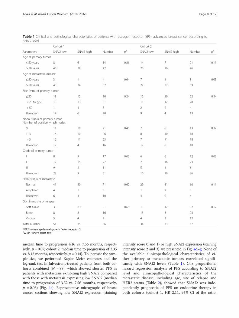
intensity score 0 and 1) or high SNAI2 expression (stainingintensity score 2 and 3) are presented in Fig. 4d–g. None ofthe available clinicopathological characteristics of ei-ther primary or metastatic tumors correlated signifi-cantly with SNAI2 levels (Table 1). Cox proportionalhazard regression analysis of PFS according to SNAI2level and clinicopathological characteristics of themetastatic disease, including age, site of relapse andHER2 status (Table 2), showed that SNAI2 was inde-pendently prognostic of PFS on endocrine therapy inboth cohorts (cohort 1, HR 2.11, 95% CI of the ratio,
Table 1 Clinical and pathological characteristics of patients with estrogen receptor (ER)+ advanced breast cancer according toSNAI2 level
Cohort 1 Cohort 2
Parameters SNAI2 low SNAI2 high Number pa SNAI2 low SNAI2 high Number pa
Age at primary tumor
≤ 50 years 8 6 14 0.86 14 7 21 0.11
> 50 years 43 29 72 20 26 46
Age at metastatic disease
≤ 50 years 3 1 4 0.64 7 1 8 0.05
> 50 years 48 34 82 27 32 59
Size (mm) of primary tumor
≤ 20 18 12 30 0.24 12 10 22 0.34
> 20 to ≤ 50 18 13 31 11 17 28
> 50 1 4 5 2 2 4
Unknown 14 6 20 9 4 13
Nodal status of primary tumorNumber of positive lymph nodes
0 11 10 21 0.46 7 6 13 0.37
1–3 16 10 26 8 10 18
> 3 12 11 23 7 11 18
Unknown 12 4 16 12 6 18
Grade of primary tumor
I 8 9 17 0.06 6 6 12 0.06
II 12 15 27 7 16 23
III 9 2 11 5 1 6
Unknown 22 9 31 16 10 26
HER2 status of metastasis
Normal 41 30 71 0.62 29 31 60 0.11
Amplified 4 1 5 1 2 3
Unknown 6 4 10 4 0 4
Dominant site of relapse
Soft tissue 38 23 61 0.65 15 17 32 0.17
Bone 8 8 16 15 8 23
Viscera 5 4 9 4 8 12
Total number 51 35 86 34 33 67
HER2 human epidermal growth factor receptor 2aχ2 or Fisher’s exact test
Alves et al. Breast Cancer Research (2018) 20:60 Page 8 of 12

1.21–3.66, p = 0.008; cohort 2, HR 1.92, 95% CI ofthe ratio, 1.03–3.59, p = 0.04).
DiscussionThe development of resistance to endocrine therapyinvolves alteration of multiple pathways that may be tar-geted with novel therapeutic agents. Inhibition of growthfactor receptor pathways that cross-talk with ER andblockage of cell cycle progression have been shown to bepromising strategies in ER+ breast cancer treatment. Agrowing body of evidence implicating enrichment of EMTmarkers in breast cancer cells resistant to endocrine treat-ment supports the use of novel pharmacological strategiestargeting EMT for breast cancer. By inhibiting EMT,tumor cells could maintain, or reverse to, an epithelial
state with reduced migratory capacity and re-sensitizationto endocrine therapy. However, the specific molecules in-volved in the regulation of EMT that should be targetedto overcome endocrine resistance remain to be defined.In this study, we show that SNAI2, a mediator of EMT
highly expressed in triple-negative breast cancer [26], isupregulated in endocrine-resistant cells, whereas otherEMT-associated transcription factors, such as SNAI1/3,TWIST1/2, ZEB1/2, FOXC2, and GSC, are unaltered. Wepreviously reported that miRNA-593, which is predictedto target SNAI2, is downregulated in tamoxifen-resistantcell lines, implicating SNAI2 in tamoxifen resistance [24].We also showed that this EMT-inducing transcription fac-tor is a key molecule in the control of tamoxifen-resistantcell growth [24]. In the present study, we focused on the
a b
c d e
f g
Fig. 4 SNAI2 expression correlates with progression-free survival (PFS) in patients with estrogen receptor (ER)+ metastatic breast cancer treatedwith endocrine therapy. Kaplan-Meier plots evaluating PFS according to expression of SNAI2 in ER+ metastatic lesions from a an initial and b asecond cohort of patients with breast cancer treated with endocrine therapy in the advanced setting. c Survival analysis of PFS according to SNAI2levels in fulvestrant-treated patients from cohorts 1 and 2. A two-sided p value (*p < 0.05) was calculated using log-rank testing. Representativemicrographs of breast cancer metastasis sections showing low SNAI2 expression (d and e) or high SNAI2 expression (f and g) (× 20 magnification)
Table 2 Regression analysis of progression-free survival according to SNAI2 level and clinicopathological characteristics
Cohort 1 Cohort 2
Variable Hazard ratio (95% CI) p Hazard ratio (95% CI) p
SNAI2 level 2.11 (1.21–3.66) 0.008 1.92 (1.03–3.59) 0.04
Age at metastasis 2.19 (0.37–12.95) 0.39 0.90 (0.33–2.49) 0.85
Site of relapse 1.47 (0.98–2.21) 0.07 0.85 (0.56–1.31) 0.47
HER2 status of metastasis 1.46 (0.50–4.28) 0.49 1.49 (0.34–6.52) 0.60
HER2 human epidermal growth factor receptor 2
Alves et al. Breast Cancer Research (2018) 20:60 Page 9 of 12

possible role of SNAI2 in fulvestrant resistance andshowed that fulvestrant-resistant cells, which express highlevels of SNAI2, exhibit increased migration, higher ex-pression of the mesenchymal marker vimentin, and reducedlevels of the epithelial marker E-cadherin compared withthe parental fulvestrant-sensitive cell line. Our data concurwith previous studies showing that tamoxifen-resistantMCF-7 breast cancer cells display enhanced motile and in-vasive behavior and EM-like properties compared withtamoxifen-sensitive MCF-7 cells [27, 28].Previous investigations have demonstrated that silen-
cing of ER in ER+ MCF-7 breast cancer cells leads to ac-quisition of endocrine resistance and mesenchymalfeatures, which contribute to tumor aggressiveness andmetastatic ability [6, 29]. Studies have also reported thatER and SNAI2 levels are inversely correlated and thatER directly suppresses SNAI2 transcription, thus regu-lating EMT [30, 31]. Despite decreased ER levels in ourfulvestrant-resistant cells expressing high SNAI2, thesecells remain ER+ and are growth-stimulated by estrogenand inhibited by tamoxifen treatment [16]. Althoughoverexpression of SNAIL in ER+ breast cancer cell lineshas been shown to induce resistance to tamoxifen ac-companied by reduced ER levels, ectopic expression ofER in these cells did not restore sensitivity to tamoxifen,suggesting that SNAIL might promote resistance toanti-estrogens independent of ER signaling [32].We observed that reduction of SNAI2 expression im-
paired cell migration and increased E-cadherin levels intwo fulvestrant-resistant breast cancer cell models,confirming a key role for SNAI2 in the control of cellmotility and maintenance of a mesenchymal phenotypein resistant cells. These findings are in line with previousreports showing increased mesenchymal characteristicsby ectopic expression of SNAI2 in ER+ MCF-7 cells[10]. Additionally, it has been shown that SNAI1/SNAI2can induce drug resistance in breast cancer cells viaalteration of cell survival signaling pathways [24, 32].We demonstrated that siRNA-mediated knockdown ofSNAI2 impairs growth of fulvestrant-resistant cells,which exhibit a high level of SNAI2. In contrast, growthof SNAI2-low breast cancer cells was significantly inhib-ited by fulvestrant alone, and downregulation of SNAI2had no additional effect on decreasing the growth ofthese cells compared to standard endocrine therapy. Therole of SNAI2 in controlling the growth of resistant celllines was further supported by the observation thattreatment with a chemical agent interfering with SNAILbinding to p53 (GN25) markedly decreased their growth.Although it is not clear that the growth inhibitory effectof GN25 was due to specific inhibition of SNAI2, as theagent targets other SNAI proteins such as SNAI1 andSNAI3, it seems plausible since SNAI2 was the onlySNAI-family member that exhibited increased expression
in fulvestrant-resistant cells. These findings suggest thattumor cells exhibiting high levels of SNAI2 may benefitfrom inhibition of SNAI2 in combination with standardfulvestrant treatment, while tumors with low SNAI2 ex-pression can be treated with fulvestrant alone. Previousstudies have demonstrated that SNAI2 increases stemnessin breast cancer cells when co-expressed with SOX9 [33]or through regulation of stem cell markers, includingc-Myc, SOX2 and Oct4 [10], possibly contributing to drugresistance. Interestingly, we found upregulation of SOX2in MCF-7-based fulvestrant-resistant cells compared tofulvestrant-sensitive cells, and SNAI2 knockdowndecreased SOX2 expression in three of the fourfulvestrant-resistant cell lines, suggesting that SNAI2might be involved in the mechanism of regulation of thisstem marker in fulvestrant resistance.Finally, we evaluated the clinical relevance of SNAI2
expression in metastatic lesions from two independentcohorts of patients with ER+ breast cancer treated withendocrine therapy in the advanced setting and showedthat high SNAI2 levels correlated significantly withshorter PFS in patients on endocrine therapy, includingfulvestrant. Correlation between high SNAI2 expressionin primary breast tumors and shorter relapse-free sur-vival has been previously demonstrated in ER+ breastcancer [34]. Studies have also shown that high SNAI2expression in primary ER- breast tumors correlates withpoor prognosis in those patients [35, 36]. Nevertheless,our study is the first to our knowledge to report theprognostic value of SNAI2 in patients with ER+advanced breast cancer treated with endocrine therapy.
ConclusionsIn summary, our data support SNAI2 as a key regulatorof the aggressive phenotype observed in endocrine-re-sistant breast cancer cells and a prognostic biomarker inER+ advanced breast cancer treated with endocrine ther-apy. These findings highlight the role of SNAI2 as a po-tential target for therapeutic strategies against EMT andendocrine resistance.
Additional files
Additional file 1: Figure S1. SNAI2 and E-cadherin protein levels followingsiRNA-mediated SNAI2 knockdown. MCF-7-based fulvestrant-resistant andparental-sensitive cells were transfected with siRNA against SNAI2 and SNAI2(A) and E-cadherin (B) protein levels were evaluated 96 h following transfection,by Western blotting. β-actin was used as loading control. (TIF 357 kb)
Additional file 2: Figure S2. SOX2 knockdown reduces growth offulvestrant-resistant breast cancer cells. (A) 182R-1 fulvestrant-resistantcells were transfected with siRNA against SOX2 leading to a reductionat the mRNA level, as evaluated by RT-qPCR. Gene expression wasnormalized using PUM1. (B) SOX2 knockdown resulted in decreasedgrowth of fulvestrant-resistant cells as measured by crystal violet-basedcolorimetric assay. Cells were grown in medium containing fulvestrant.
Alves et al. Breast Cancer Research (2018) 20:60 Page 10 of 12

Experiment was performed in technical triplicates and results are shownwith error bars representing mean ± standard deviation. (TIF 313 kb)
Additional file 3: Figure S3. Correlation between SNAI2 expression andPFS in patients with ER+ metastatic breast cancer from cohort 1 and 2treated with fulvestrant. Kaplan-Meier plots evaluating PFS according toexpression of SNAI2 in ER+ metastatic lesions from fulvestrant-treatedpatients from cohort 1 (A) and cohort 2 (B). A two-sided p value(*p < 0.05) was calculated using log-rank testing. (TIF 351 kb)
AbbreviationsCI: Confidence interval; CSCs: Cancer stem cells; ECM: Extracellular matrix;EMT: Epithelial-mesenchymal transition; ER +: Estrogen receptor-positivebreast cancer; FBS: Fetal bovine serum; FDR: False discovery rate;FFPE: Formalin-fixed paraffin-embedded; FulvR: Fulvestrant-resistant cells;HER2: Human Epidermal growth factor Receptor 2; HR: Hazard ratio;HRP: Horseradish peroxidase; MET: Mesenchymal-epithelial transition;OD: Optical density; OUH: Odense University Hospital; PFS: Progression-freesurvival; RT-qPCR: Reverse transcription (RT)-quantitative (q)PCR; siRNA: Smallinterfering RNA; TamR: Tamoxifen-resistant cells
AcknowledgementsWe would like to thank Anne E. Lykkesfeldt for providing the tamoxifen-resistantand fulvestrant-resistant cell lines, Lisbet Mortensen and Ole Nielsen at theDepartment of Pathology, Odense University Hospital, for excellent technicalassistance with the immunocytochemical and immunohistochemical staining,and M. Kat Occhipinti for editorial assistance.
FundingThis work was supported by the Danish Cancer Society (H.J. Ditzel), DanishCancer Research Foundation (C.L. Alves), A Race Against Breast Cancer(H.J. Ditzel), Region of Southern Denmark Research Foundation (H.J. Ditzel),Odense University Hospital Research Council (H.J. Ditzel), Region of SouthernDenmark Research Council (H.J. Ditzel), Academy of Geriatric CancerResearch (AgeCare) (H.J. Ditzel), and National Experimental TherapyPartnership (NEXT) Innovation Fund Denmark (H.J. Ditzel).
Availability of data and materialsMicroarray data were deposited and are accessible from the Gene ExpressionOmnibus (GEO) database [GEO:GSE74391]. The other data supporting theconclusions of this article are included within the article.
Authors’ contributionsCLA, DE, and HJD participated in the study design, analysis, and interpretationof data and writing of the manuscript. DE and HJD supervised the study. CLA,MBL, MB, and HJD contributed to data acquisition. All authors read andapproved the final manuscript.
Ethics approval and consent to participateAll clinical samples were coded to maintain patient confidentiality andstudies were approved by the Ethics Committee of the Region of SouthernDenmark (approval no S-2008-0115) and the Danish Data Protection Agency(approval no. 2008–580035(14/10607)).
Competing interestsThe authors declare that they have no competing interests.
Publisher’s NoteSpringer Nature remains neutral with regard to jurisdictional claims inpublished maps and institutional affiliations.
Author details1Department of Cancer and Inflammation Research, Institute of MolecularMedicine, University of Southern Denmark, J.B. Winsløwsvej 25, 5000 OdenseC, Denmark. 2Department of Pathology, Odense University Hospital, 5000Odense, Denmark. 3Department of Oncology, Odense University Hospital,5000 Odense, Denmark. 4Academy of Geriatric Cancer Research (AgeCare),Odense University Hospital, 5000 Odense, Denmark.
Received: 7 November 2017 Accepted: 15 May 2018
References1. Keen JC, Davidson NE. The biology of breast carcinoma. Cancer. 2003;97:825–33.2. Luqmani YA, Alam-Eldin N. Overcoming resistance to endocrine therapy in
breast cancer: new approaches to a nagging problem. Med Princ Pract.2016;25(Suppl 2):28–40.
3. Sarrio D, Rodriguez-Pinilla SM, Hardisson D, Cano A, Moreno-Bueno G,Palacios J. Epithelial-mesenchymal transition in breast cancer relates to thebasal-like phenotype. Cancer Res. 2008;68:989–97.
4. Gupta GP, Massague J. Cancer metastasis: building a framework. Cell.2006;127:679–95.
5. Thiery JP. Epithelial-mesenchymal transitions in tumour progression. Nat RevCancer. 2002;2:442–54.
6. Al Saleh S, Al Mulla F, Luqmani YA. Estrogen receptor silencing inducesepithelial to mesenchymal transition in human breast cancer cells.PLoS One. 2011;6:e20610.
7. Polyak K, Weinberg RA. Transitions between epithelial and mesenchymalstates: acquisition of malignant and stem cell traits. Nat Rev Cancer.2009;9:265–73.
8. Luqmani YA, Al Azmi A, Al Bader M, Abraham G, El Zawahri M. Modificationof gene expression induced by siRNA targeting of estrogen receptor alphain MCF7 human breast cancer cells. Int J Oncol. 2009;34:231–42.
9. Dhasarathy A, Kajita M, Wade PA. The transcription factor snail mediatesepithelial to mesenchymal transitions by repression of estrogen receptor-alpha. Mol Endocrinol. 2007;21:2907–18.
10. Li Y, Wu Y, Abbatiello TC, Wu WL, Kim JR, Sarkissyan M, et al. Slugcontributes to cancer progression by direct regulation of ERalpha signalingpathway. Int J Oncol. 2015;46:1461–72.
11. Voutsadakis IA. Epithelial-mesenchymal transition (EMT) and regulation ofEMT Factors by steroid nuclear receptors in breast cancer: a review and insilico investigation. J Clin Med. 2016;5:11.
12. Dave B, Mittal V, Tan NM, Chang JC. Epithelial-mesenchymal transition,cancer stem cells and treatment resistance. Breast Cancer Res. 2012;14:202.
13. Al Saleh S, Sharaf LH, Luqmani YA. Signalling pathways involved inendocrine resistance in breast cancer and associations with epithelial tomesenchymal transition (Review). Int J Oncol. 2011;38:1197–217.
14. Davis FM, Stewart TA, Thompson EW, Monteith GR. Targeting EMT in cancer:opportunities for pharmacological intervention. Trends Pharmacol Sci.2014;35:479–88.
15. Briand P, Lykkesfeldt AE. Effect of estrogen and antiestrogen on the humanbreast cancer cell line MCF-7 adapted to growth at low serum concentration.Cancer Res. 1984;44:1114–9.
16. Lykkesfeldt AE, Larsen SS, Briand P. Human breast cancer cell lines resistantto pure anti-estrogens are sensitive to tamoxifen treatment. Int J Cancer.1995;61:529–34.
17. Lykkesfeldt AE, Madsen MW, Briand P. Altered expression ofestrogen-regulated genes in a tamoxifen-resistant and ICI 164,384 andICI 182,780 sensitive human breast cancer cell line, MCF-7/TAMR-1.Cancer Res. 1994;54:1587–95.
18. Elias D, Vever H, Laenkholm AV, Gjerstorff MF, Yde CW, Lykkesfeldt AE, et al.Gene expression profiling identifies FYN as an important molecule intamoxifen resistance and a predictor of early recurrence in patients treatedwith endocrine therapy. Oncogene. 2015;34:1919–27.
19. Livak KJ, Schmittgen TD. Analysis of relative gene expression data usingreal-time quantitative PCR and the 2(−Delta Delta C(T)) method. Methods.2001;25:402–8.
20. Lundholt BK, Briand P, Lykkesfeldt AE. Growth inhibition and growthstimulation by estradiol of estrogen receptor transfected human breastepithelial cell lines involve different pathways. Breast Cancer Res Treat.2001;67:199–214.
21. Leth-Larsen R, Lund R, Hansen HV, Laenkholm AV, Tarin D, Jensen ON, et al.Metastasis-related plasma membrane proteins of human breast cancer cellsidentified by comparative quantitative mass spectrometry. Mol CellProteomics. 2009;8:1436–49.
22. Budczies J, Klauschen F, Sinn BV, Gyorffy B, Schmitt WD, Darb-EsfahaniS, et al. Cutoff Finder: a comprehensive and straightforward Webapplication enabling rapid biomarker cutoff optimization. PLoS One.2012;7:e51862.
Alves et al. Breast Cancer Research (2018) 20:60 Page 11 of 12

23. Alves CL, Elias D, Lyng M, Bak M, Kirkegaard T, Lykkesfeldt AE, et al. HighCDK6 protects cells from fulvestrant-mediated apoptosis and is a predictorof resistance to fulvestrant in estrogen receptor-positive metastatic breastcancer. Clin Cancer Res. 2016;22:5514–26.
24. Joshi T, Elias D, Stenvang J, Alves CL, Teng F, Lyng MB, et al. Integrativeanalysis of miRNA and gene expression reveals regulatory networks intamoxifen-resistant breast cancer. Oncotarget. 2016;7:57239–53.
25. Samanta S, Sun H, Goel HL, Pursell B, Chang C, Khan A, et al. IMP3 promotesstem-like properties in triple-negative breast cancer by regulating SLUG.Oncogene. 2016;35:1111–21.
26. Proia TA, Keller PJ, Gupta PB, Klebba I, Jones AD, Sedic M, et al. Geneticpredisposition directs breast cancer phenotype by dictating progenitor cellfate. Cell Stem Cell. 2011;8:149–63.
27. Hiscox S, Jiang WG, Obermeier K, Taylor K, Morgan L, Burmi R, et al.Tamoxifen resistance in MCF7 cells promotes EMT-like behaviour andinvolves modulation of beta-catenin phosphorylation. Int J Cancer.2006;118:290–301.
28. Ward A, Balwierz A, Zhang JD, Kublbeck M, Pawitan Y, Hielscher T, et al.Re-expression of microRNA-375 reverses both tamoxifen resistance andaccompanying EMT-like properties in breast cancer. Oncogene.2013;32:1173–82.
29. Bouris P, Skandalis SS, Piperigkou Z, Afratis N, Karamanou K, Aletras AJ, et al.Estrogen receptor alpha mediates epithelial to mesenchymal transition,expression of specific matrix effectors and functional properties of breastcancer cells. Matrix Biol. 2015;43:42–60.
30. Ye Y, Xiao Y, Wang W, Yearsley K, Gao JX, Barsky SH. ERalpha suppressesslug expression directly by transcriptional repression. Biochem J. 2008;416:179–87.
31. Ye Y, Xiao Y, Wang W, Yearsley K, Gao JX, Shetuni B, et al. ERalpha signalingthrough slug regulates E-cadherin and EMT. Oncogene. 2010;29:1451–62.
32. Jiang Y, Zhao X, Xiao Q, Liu Q, Ding K, Yu F, et al. Snail and Slug mediatetamoxifen resistance in breast cancer cells through activation of EGFR-ERKindependent of epithelial-mesenchymal transition. J Mol Cell Biol.2014;6:352–4.
33. Guo WJ, Keckesova Z, Donaher JL, Shibue T, Tischler V, Reinhardt F, et al.Slug and Sox9 Cooperatively Determine the Mammary Stem Cell State. Cell.2012;148:1015–28.
34. Chimge NO, Baniwal SK, Little GH, Chen YB, Kahn M, Tripathy D, et al.Regulation of breast cancer metastasis by Runx2 and estrogen signaling:the role of SNAI2. Breast Cancer Res. 2011;13:R127.
35. Liu T, Zhang XY, Shang M, Zhang YX, Xia BS, Niu M, et al. Dysregulatedexpression of Slug, vimentin, and E-cadherin correlates with poor clinicaloutcome in patients with basal-like breast cancer. J Surg Oncol.2013;107:188–94.
36. Storci G, Sansone P, Trere D, Tavolari S, Taffurelli M, Ceccarelli C, et al. Thebasal-like breast carcinoma phenotype is regulated by SLUG geneexpression. J Pathol. 2008;214:25–37.
Alves et al. Breast Cancer Research (2018) 20:60 Page 12 of 12