soccer syndrome - common presentations and manual
TRANSCRIPT

American Journal of Sports Science 2014; 2(6): 141-154
Published online November 10, 2014 (http://www.sciencepublishinggroup.com/j/ajss)
doi: 10.11648/j.ajss.20140206.11
ISSN: 2330-8559 (Print); ISSN: 2330-8540 (Online)
Soccer syndrome - Common presentations and manual diagnostic techniques for pelvic malalignment syndrome
Ganesh Elumalai1, *
, Malarvani Thangamani1, Nirmala Palayathan
2, Ajit Kumar
1, Manish Kr Singh
1
1Department of Anatomy, Tribhuvan University, National Medical College, Birgunj, Nepal 2Department of Anatomy, Kathmandu University, Nobel Medical College & Teaching Hospital, Biratnagar, Nepal
Email address: [email protected] (E. Ganesh), [email protected] (T. Malarvani), [email protected] (P. Nirmala),
[email protected] (A. Kumar), [email protected] (Manish Kr Singh)
To cite this article: Ganesh Elumalai, Malarvani Thangamani, Nirmala Palayathan, Ajit Kumar, Manish Kr Singh. Soccer Syndrome - Common Presentations
and Manual Diagnostic Techniques for Pelvic Malalignment Syndrome. American Journal of Sports Science.
Vol. 2, No. 6, 2014, pp. 141-154. doi: 10.11648/j.ajss.20140206.11
Abstract: The pelvic malalignment syndrome is the most common in sports injuries and even in every client who presents
with low back & pelvis pain or dysfunction. In this study, we used simple bony palpation method to assess the various pelvic
malalignments in football players. While there are many methods to determine pelvic symmetry or asymmetry, this method is
very simple one and that consistently works well for me. Our method of assessing the pelvic malalignments, even works well
to the common individuals suffering with low back & pelvis pain or dysfunction due to various pelvic malalignment
presentation. This study includes the healthy football players from National Football Club (NFC) of our institution, who were
training or playing during the period of investigation (September 2012 – September 2014) (n = 40) were eligible for
participation. At entry to the study, this eligible sample had a mean age of 22.2 ± 3.9 years, height of 175.8 ± 6.6 cm, and mass
of 87.5 ± 7.1 kg. The number of eligible participants at each time point was 40 for all the four session time, point 1 (T1; start of
preseason-1), point 2 (T2; end of season-1), point 3 (T3; start of preseason-2), and point 4 (T4; end of preseason-2). We
instructed participants to nominate their leg preference for kicking, which was considered the dominant leg. Among the total
number of eligible players assessed, 12.5% were left-leg dominant, 80% were right-leg dominant, and 7.5% reported no leg
dominance. Subjects were limited to men who had a normal muscle strength and Range of Motion (ROM) of the back and
lower extremities and who had no history of orthopaedic or neurologic disorders. All the individuals are subject to gain access
the malalignment of both Innominate and Sacral bones of the bony pelvis unit. In addition, to justify our diagnosis we access
the muscles of the functional slings and the muscles around the bony pelvis related to the malalignment of the pelvic unit. The
techniques were repeated in different position and also performed for two times per day and continued for fifteen days to
standardize. In this study, we observed, 87.5% i.e., the majority of the soccer's are suffering with multiple pelvic
malalignments , includes innominate & sacral stuck. The soccer's suffering with single component malalignment account for
about 10%, includes innominate or sacral stuck and soccer's who had the symmetry pelvis is only 2.5%. Conclusion: This
method of assessing the pelvic malalignments until then not documented. In this sense, the objective of this study is to analyze
and document the different presentations in pelvic malalignments which are common in soccer players and its simple method
of evaluation is the literature state of the art. The present study may provide useful information to analyze common
presentations of pelvic malalignments in different sports.
Keywords: Pelvic Malalignment, Anterior Pelvic-Tilt, Posterior Pelvic-Tilt, Innominate Upslip
1. Introduction
Very rarely both the right and left sides of the body is same.
In structural integration we often ignore these side-to-side
asymmetries. An understanding of this ‘malalignment
syndrome’ requires the knowledge of common presentations
in malalignment and the techniques used to diagnose and
treat. This study is mainly focused on the common
presentations and manual diagnostic techniques for different
pelvic malalignments. We need to learn and customize our
assessment to diagnose these structural asymmetries (Liz
Gaggini, 2010). This is very important in pelvic asymmetry.

142 E. Ganesh et al.: Soccer Syndrome - Common Presentations and Manual Diagnostic Techniques for
Pelvic Malalignment Syndrome
Because, following the pelvic malalignment, cause the
secondary adaptations in the vertebral alignment, leg length,
postural and movements modifications to overcome these
malalignment.
The pelvic malalignment syndrome is the most common in
sports injuries and even in every client who presents with
low back & pelvis pain or dysfunction. An understanding of
this 'malalignment syndrome' requires knowledge of the
common presentations of malalignment and the techniques
used to diagnose and treat. This study is mainly focused on
the common presentations and manual diagnostic techniques
for pelvic malalignment syndrome. We used simple bony
palpation method to assess the various pelvic malalignments
in football players. While there are many methods to
determine pelvic symmetry or asymmetry, this method is
very simple one and that is consistently works well for me.
Our method of assessing the pelvic malalignments, even
works well to the common individuals suffering with low
back & pelvis pain or dysfunction due to various pelvic
malalignment presentation. To be masterly skilled in our
current techniques of evaluating the pelvic malalignment it is
very simple and essential that you will need to be able to
locate the anterior superior iliac spines (ASIS) and posterior
superior iliac spines (PSIS), able to judge the levelness of
both iliac crests when viewing from the front or rear even in
different postures like standing, lying in supine & prone also
while performing the movements during the assessment.
To justify our diagnosis we access the muscles of the
functional slings and the muscles around the bony pelvis
related to the malalignment of the pelvic unit. The techniques
were repeated in different position and also performed for
two times per day and continued for fifteen days to
standardize.
However, this method of assessing the pelvic
malalignments until then not documented. In this sense, the
objective of this study to analyze and document the different
presentations in pelvic malalignments which are common in
soccer players and its simple method of evaluation is the
literature state of the art. The present study may provide
useful information to analyze common presentations of
pelvic malalignments in different sports.
2. Materials & Methods
The healthy football players from National Football Club
(NFC) of our institution, who were undergoing training or
playing during the period of investigation ( September 2012
– September 2014) (n = 40) were eligible for participation in
the study. At entry to the study, this eligible sample had a
mean age of 22.2 ± 3.9 years, height of 175.8 ± 6.6cm, and
mass of 87.5 ± 7.1 kg. The number of eligible participants at
each time point was 40 for all the four session time, period -
1 (T1; start of preseason - 1), period - 2 (T2; end of season -
1), period - 3 (T3; start of preseason - 2), and period - 4 (T4;
end of season - 2). We instructed participants to nominate
their leg preference for kicking, which was considered the
dominant leg. Among the total number of eligible players
assessed, 12.5% were left-leg dominant, 80% were right-leg
dominant, and 7.5% reported no leg dominance.
Subjects were limited to men who had the normal muscle
strength and normal ROM of the back and lower extremities
and who had no history of orthopaedic or neurological
disorders. All subjects were instructed to restrict excessive
physical activity on the day of testing, such as recreational
running and bicycling, and to wear gym trunks for the tests.
On the day of testing, all subjects reviewed and signed
informed consent forms for the study. On assessment we
taught the subjects how to perform the movements to gain
access to the malalignment of both Innominate and Sacral
bones of the bony pelvis unit. In addition, to justify our
diagnosis we access the muscles of the functional slings and
the muscles around the bony pelvis related to the
malalignment of the pelvic unit (Abdolhamid Daneshjoo,
2013), (Julie Hides, 2012), (Fernando Idoate, 2011). We
encouraged all the subjects to relax and told them that the
movements would not be judged as "good" or "bad." We
believed these verbal cues decreased subjects' anxiety and,
therefore, helped to standardize the testing. The techniques
were repeated in different positions and also performed two
times per day and continued for fifteen days to standardize.
3. Bony Pelvis
The bony pelvis includes Right & Left pelvic bones in
sides & front and the Sacrum & Coccyx behind. They are
articulated at Pubic symphysis & Sacro-Iliac joint (Fig-1). It
usually moves as a unit and less often as particular bone. The
pelvic malalignments includes, Innominate & Sacral stuck.
The Innominate malalignment involves, Rotational stuck &
Upward stuck. The sacral malalignment includes Nutational
stuck, Counter-Nutational stuck & Rotatory stuck.
A. Bony Pelvis - Anterior view
B. Bony Pelvis - Posterior view
Figure 1. Structure of Bony Pelvis

American Journal of Sports Science 2014; 2(6): 141-154 143
4. Movements of Innominate Bone
1. Rotational movement of innominate possible in
‘anterior’ or ‘posterior’ direction, around the coronal axis in
the sagittal plane (Fig-2,3,4,5 & 6). It may occur in one or
both innominate and it moves in relative to the sacrum. If
both rotates, it may be:
a. In the same direction simultaneously, e.g. as occurs
usually on flexion or extension of the trunk or tilting of the
pelvis when sitting
Trunk flexion:
Table 1a. Rotatory movements of Innominate during trunk flexion
Standing (Open Kinematic Chain) Sitting (Closed Kinematic Chain)
Innominates rotates anteriorly Innominates rotates anteriorly
Trunk extension:
Table 1b. Rotatory movements of Innominate during trunk extension
Standing (Open Kinematic Chain) Sitting (Closed Kinematic Chain)
Innominates rotates posteriorly Innominates rotates posteriorly
b. In opposite directions, e.g. as occurs during the normal
gait cycle
Right leg in stance & left leg in swing:
Table 2a. Rotatory movements of Innominate during normal gait cycle
Right Innominate Left Innominate
Innominate rotates Anteriorly Innominate rotates posteriorly
Left leg in stance & right leg in swing:
Table 2b. Rotatory movements of Innominate during normal gait cycle
Right Innominate Left Innominate
Innominate rotates posteriorly Innominate rotates Anteriorly
Figure 2. Anterior pelvic tilt of Innominate bone
Figure 3. Posterior pelvic tilt of Innominate bone
Figure 4. Rotation of Innominate bone
Figure 5. Anterior Rotation Muscles
Figure 6. Posterior Rotation Muscles
2. Rotational movement of innominate is also possible as
‘Outflare’ and ‘Inflare’. It refers as movement of the
innominates outward and inward, respectively, around the
vertical axis in the transverse plane (Fig-7).
It may occur in one or both innominate and it moves in
relation to the sacrum. If both rotate, it may be:
a) In the same direction simultaneously, e.g. as occurs

144 E. Ganesh et al.: Soccer Syndrome - Common Presentations and Manual Diagnostic Techniques for
Pelvic Malalignment Syndrome
usually on flexion or extension of the trunk or tilting of
the pelvis when sitting
Trunk flexion:
Table 3a. Outflare & Inflare of Innominate during trunk flexion
Standing (Open Kinematic Chain) Sitting (Closed Kinematic Chain)
Innominates Outflare Innominates Outflare
Trunk extension:
Table 3b. Outflare & Inflare of Innominate during trunk extension
Standing (Open Kinematic Chain) Sitting (Closed Kinematic Chain)
Innominates Inflare Innominates Inflare
b. In opposite directions, e.g. as occurs during the normal
gait cycle
Right leg in stance & Left leg in swing:
Table 4a. Outflare & Inflare of Innominate during normal gait cycle
Right Innominate Left Innominate
Innominate Outflare Innominate Inflare
Left leg in stance & Right leg in swing:
Table 4b. Outflare & Inflare of Innominate during normal gait cycle
Right Innominate Left Innominate
Innominate Inflare Innominate Outflare
Figure 7. Inflare and Outflare of Right Innominate bone
4.1. Muscles responsible to produce "Outflare & Inflare"
The Outflare & Inflare movements are usually associated
with the anterior and posterior rotation of innominate bone
respectively. The muscles which help to produce the
"Outflare & Inflare" movements are Internal rotators &
External rotators of hip respectively.
3. Innominate "Upslip" (Fig-8 & 9), is usually associated
with superior pubic dysfunction. Normally upslip of
unilateral innominate will occur during stair climbing up or
down. Missing a step and landing with increased force on
one extremity can cause Upslip malalignment of the
innominate.
a) With the leg vertical on impact, the force transmitted
through the hip joint can result in upward displacement
of the innominate relative to the sacrum (a so-called
‘upslip’).
b) Landing with the leg at a hip-flexion angle on impact
results in an ‘anterior rotational’ force on the
innominate which may result in combination with
"upslip & anterior rotation" of innominate.
c) Climbing-up with the leg at a hip-flexion angle on
impact results in a ‘Posterior rotational’ force on the
innominate which may result in combination with
"upslip & posterior rotation" of innominate.
Figure 8. Upslip of Innominate bone
Figure 9. Muscles responsible for Upslip of Innominate bone
5. Movements of Sacrum
The sacrum moves at its joints called Sacroiliac & Lumbo-
Sacral joints, usually it moves along with the Innominate and
/ or Lumbar bones in horizontal axis in the vertical plane
(Nutation & Counter-Nutation) (Fig-10,11,13 & 14) & Right
and Left Oblique axis (Rotation) (Fig-12 & 15).
Trunk flexion:
Table 5a. Sacral movements during Trunk flexion
standing (Open Kinematic Chain) Sitting (Closed Kinematic Chain)
� Innominates moves Anteriorly &
Outflare
� Sacrum undergoes "Nutation"
initially. Once the Interosseus ,
Sacrotuberous & Sacrospinous
ligaments gets tightened, it
undergoes "Counter-Nutation".
� Innominates moves Anteriorly &
Outflare
� Sacrum undergoes "Counter-
Nutation". It will cause tightness
in " Long Dorso-Sacroiliac
ligament". Then it results in
posterior rotation of Innominate
& Nutation of Sacrum.
Trunk extension:

American Journal of Sports Science 2014; 2(6): 141-154 145
Table 5b. Sacral movements during Trunk extension
Standing (Open Kinematic Chain) Sitting (Closed Kinematic Chain)
� Innominates moves Posteriorly &
Inflare
� Sacrum produce "Nutation"
� Innominates, do not move
initially.
� Sacrum produce "Nutation"
� Nutation produce, increased
traction force in Sacrotuberous
ligament below upwards which
cause "anterior rotation' of
innominate.
Sacral movements during Normal gait cycle
Table 6a. Right leg in stance & Left leg in swing
Results in
� Right Innominate - moves Anteriorly & Outflare
� Left Innominate - moves Posteriorly & Inflare
� Sacrum rotates in Right oblique axis
Table 6b. Left leg in stance & Right leg in swing
Results in
� Left Innominate - moves Anteriorly & Outflare
� Right Innominate - moves Posteriorly & Inflare
� Sacrum rotates in Left oblique axis
Figure 10. Sacral Nutation and Counter- nutation in relation to the Bony
pelvis
Figure 11. Sacral Nutation and Counter- nutation in relation to the Trunk movements
Figure 12. Sacral rotation in oblique axis during Normal Gait cycle

146 E. Ganesh et al.: Soccer Syndrome - Common Presentations and Manual Diagnostic Techniques for
Pelvic Malalignment Syndrome
Figure 13. Muscles responsible for nutation of Sacral bone
Figure 14. Muscles responsible for Counter - nutation of Sacral bone
Figure 15. Muscles responsible for Rotation of Sacrum in oblique axis
6. Stabilizers of the Pelvic Unit
The ‘Stabilizers of the pelvic unit’ is made up of oblique
and longitudinal systems of ‘slings’, it is formed by the
interconnecting muscles, tendons, ligaments and fascia. The
four basic sling systems helps to keep the bony pelvis unit in
symmetrical position are:
� Superficial posterior oblique & longitudinal sling
� Deep posterior oblique & longitudinal sling
� Anterior oblique & longitudinal sling
� Lateral longitudinal & oblique sling
6.1. Superficial Posterior Oblique & Longitudinal Sling
The latissimus dorsi connected, by thoracolumbar fascia, to
the contralateral gluteus maximus constitutes the upper part
(oblique) of this system. The lower part (longitudinal) of this
system is comprised of the continuations of gluteus maximus
with the Ilio-Tibial Band (ITB). Concomitant contraction of
Tensor-fascia latae and Vastus lateralis helps in further
increase effectiveness of ITB.
6.2. Deep Posterior Oblique & Longitudinal Sling
This sling formed by the ipsilateral erector-spinae muscle
and contralateral iliocostalis connected (oblique), by way of
the deep lamina of the thoracolumbar fascia. The lower part
(longitudinal) of this system is comprised of the
continuations of iliocostalis with the sacrotuberous ligament
and biceps femoris. Concomitant contraction of peroneus
longus and tibialis anterior helps n further increase the
effectiveness of this sling.
6.3. Anterior Oblique & Longitudinal Sling
The upper part (oblique) of this sling is formed by external
oblique on one side are connected, by way of the anterior
abdominal fascia, to the contralateral internal abdominal
oblique and the lower part (longitudinal) of this system is
comprised of the continuations of internal abdominal oblique
with the adductors of the thigh.
6.4. Lateral Longitudinal & Oblique Sling
The upper part (longitudinal) of this sling is formed by the
ipsilateral quadratus lumborum from above and the same side
gluteus medius, gluteus minimus & tensor fascia latae from
below. The lower part (oblique) of this system is comprised
of contralateral adductors of the thigh.
7. Muscles Responsible to Maintain the
Symmetry of Bony Pelvis
Figure 16. Muscles responsible for symmetry of Pelvic bone
Figure 17. Muscles responsible for lateral stability of Pelvic bone

American Journal of Sports Science 2014; 2(6): 141-154 147
Figure 18. Muscles responsible for symmetry of Sacral bone in Transverse
Axis
Figure 19. Muscles responsible for symmetry of Sacral bone in Oblique axis
8. Malalignments in Innominate Bone
The common presentation may appear in isolation or in
combination with one or both of the others. For example, an
‘upslip’ appears on its own in about 10%, in combination
with either ‘rotational malalignment’ or ‘flare’ or both in
another 10%, for a total of 20% overall. In this study, three
common malalignment presentations are observed in 80 -
95% of individuals. These presentations are:
1. ‘rotational malalignment’ (80-85%)
2. pelvic ‘flare’ - innominate ‘outflare’ or ‘inflare’ (40 -
50%), and the
3. ‘upslip’ (15-20%)
The common ‘Innominate malalignment’ refers to fixation
of an innominate bone relative to the sacrum in excessive
anterior or posterior rotation in the sagittal plane. Such
rotation can affect an innominate on one side only but is more
likely to be seen in association with:
1. Compensatory rotation of the contralateral innominate
around the coronal axis in the sagittal plane.(e.g. Right
side anterior rotation with Left side Posterior rotation)
2. Compensatory rotation of the contralateral innominate
around the vertical axis in the transverse plane. (e.g.
Right side anterior rotation with outflare & Left side
Posterior rotation with inflare)
3. Upslip of innominate with the displacement of the pubic
bone superiorly, relative to each other.
9. Malalignments in Sacrum
The sacral malalignment is usually accompanied with
malalignment of Innominate and / or Lumbar vertebra. The
common sacral malalignment is rotational (oblique axis)
stuck, it is usually associated with rotational malalignment of
innominate bone.
1. If left Innominate stuck posteriorly & inflare, the right
Innominate undergoes compensatory rotational stuck in
anteriorly & outflare and Sacrum undergoes rotatory
stuck in Right oblique axis.
2. If right Innominate stuck posteriorly & inflare, the left
Innominate undergoes compensatory rotational stuck in
anteriorly & outflare and Sacrum undergoes rotatory
stuck in Left oblique axis.
3. The bilateral innominate tilt is usually associated with
the sacrum stuck in either Nutation or Counter-Nutation.
10. Assessment of Malalignment
The initial step in the diagnosis of malalignment is to
establish whether asymmetry is present and, if so, it is caused
by one (or combination) of the following:
1. an anatomical (true) leg length difference (LLD)
2. presence of True or Compensated Trendelenburg's sign
3. one of the three common presentations of Innominate
malalignment, with a functional LLD (seen with an
‘upslip’ and ‘rotational malalignment’)
4. sacral stuck
Examination is preferably carried out on a firm, even
surface in different postures like Standing, Sitting and Lying.
Examination performed on a soft or sagging support, or
across a break in the surface (a feature common to medical
plinths), may affect the assessment and lead to incorrect
conclusions and possibly misdiagnosis. To correct the pelvic
malalignment, determination of the type of pelvic
malalignment present is the utmost importance.
10.1. Measurement of Limb Length Discrepancies:
(Lying-Sitting-Lying Test)
This test affords an individual and those caring for patients
a quick way of establishing whether malalignment is actually
present and, if so, to find out whether there is a ‘rotational
malalignment’, ‘upslip’, ‘flare’ or a combination of these, so
that appropriate treatment can be initiated.
The measurement of leg length is much easier and more
accurate, by comparing the level of the thumbs placed in the
hollow sulcus immediately below the medial malleolus on
each side, directly overlying the medial ankle ligaments.
Point the tip of each thumb straight downward (distal phalanx
vertical) helps to compare the relative level of
interphalangeal joints (i.e. knuckles) which end up closer
together and are more clearly demarcated than the tip
malleoli assessment, also it helps to make side-to-side
comparison more accurate. Remember to hold onto
the ankles lightly - the thumbs are only serving you as a
guide to compare side-to-side leg movement and length on

148 E. Ganesh et al.: Soccer Syndrome - Common Presentations and Manual Diagnostic Techniques for
Pelvic Malalignment Syndrome
sitting and lying. A common mistake is to hold on forcefully,
at the risk of impairing free upward and downward
movement of the legs, making the person to actual discomfort.
The person initially lies supine and is then asked to sit up.
A shift of the pelvis or other error is less likely avoided (to
prevent the activation of leg, pelvic or trunk muscles that can
influence movement of the pelvis). Then the assessment is
repeated with the supine lying, for comparison.
Clinical Correlations:
1 when one leg is shorter by an equal amount in both
sitting-up and lying-down
a. ‘true’ LLD, with all the landmarks are aligned
b. an ‘upslip’, with all the landmarks, both anterior and
posterior, have moved upward on the side of the
‘upslip’.
1 when leg length is equal
a. pelvis is in alignment, with all the landmarks
symmetrical
b. presence of an ‘outflare or inflare’ malalignment
2 when leg length shifts
a. suspect a ‘rotational malalignment’, with landmarks all
asymmetrical. But,
b. rule out the person is not sitting or lying slightly
asymmetrically with a wallet or other object in a back
pocket or on account of a break in the plinth.
10.2. Assessment of True or Compensated Trendelenburg's
(Gait) Sign
Figure 20. True & Compensated Trendelenburg's gait
Asymmetrical weight-bearing plays an important factor on
account of an anatomical (true) or a functional leg length
difference, attempting the pelvic girdle to compensate for
problems to transfer load through the lumbo pelvic- hip
complex in order to overcome the insufficiency. It may be
either by two ways
a) Compensated Trendelenburg (sign) gait
If a person is with the weakness in the left hip abductors, it
makes him difficult for stabilizing the left hip and SI joint for
proper load transfer through the hip, SI joint and up through
the lumbo-sacral junction (Fig-20).
When walking, the person compensates by leaning the
trunk into the impaired left side during mid-stance, moving
the centre of gravity outward from midline and more directly
over top of the hip joint, thereby
� decreasing the need for left abductor muscle action to
achieve stability of the left hip joint
� decreasing vertical shear forces through the left SI joint.
b) True Trendelenburg (sign) gait
A person with the left hip instability caused by the
degeneration (osteoarthritis) of the joint or by inflamed and
painful joint will have difficulty in stabilizing the left hip and
SI joint (Fig-20).
The person may compensate for the impaired ability to
transfer load through the left hip by leaning away from that
side (adducting the pelvis, abducting the left femur) in mid-
stance, thereby
� bringing the centre of gravity closer to midline (away
from the left hip joint and toward the SI joint)
� decreasing stress on the painful left hip joint
� depending more on the strong left hip abductors to
ensure stability of the left hip joint and also of the
pelvic unit.
10.3. Assessment of Pelvic Bone Alignment
It is essential to locate the anterior superior iliac spines
(ASIS) and posterior superior iliac spines (PSIS) (Pelvic
evaluation, 2010/09), to judge the alignment of pelvic girdle
on observing the pelvis from both the front and rear. ASIS
level is best judged by kneeling or standing in front of the
standing or lying client and hooking your thumbs just under
the ASIS, rather than trying to place the thumb on the apex of
the ASIS (Fig-21). Mark the level of ASIS on both the side
using marker to note the side of high or low. PSIS’s are
palpated best by kneeling or standing behind the standing or
lying client, placing the hands so that the sides of the first
finger is resting on the iliac crest (Fig-22) (palms facing
down with the thumbs oriented slightly downward). While
tracing on the Iliac crest for PSIS palpation, the tips of the
thumbs rest on the skin over the PSIS called dimple (of Venus)
(Fig-23); look for these first. On other way, you can palpate
the PSIS by hooking your thumbs just caudal to the PSIS
protuberance. Accurate palpation of the ASIS and PSIS is a
matter of practice, accuracy will improve with practice. It
tends to be more difficult to locate landmarks with heavier
clients and with clients who are very tight or have the high
muscle mass.
Figure 21. Palpation of Anterior superior iliac spine

American Journal of Sports Science 2014; 2(6): 141-154 149
Figure 22. Palpation of Posterior superior iliac spine
Figure 23. Surface landmark for Posterior superior iliac spine
The level of pubic bone on both the side is best assessed
with the patient in supine position, rather than standing or
sitting. Subjects are advised to relax the abdominal muscles,
the examiner approach the pubis by giving gentle palpation
using both the thumbs over the pubic symphysis to assess the
level of body of both the pubis.
Clinical Correlations:
The general guidelines on innominate findings: (in relation
to the Right innominate)
1 Low right ASIS, high right PSIS, near level Intercristal
line:
Anterior rotation of the right innominate or posterior
rotation of the left innominate
Figure 24. Muscles involved in Anterior Innominate stuck
2 High right ASIS, low right PSIS, near level Intercristal
line:
Posterior rotation of the right innominate or anterior
rotation of the left innominate
Figure 25. Muscles involved in Posterior Innominate stuck
3 High right ASIS & PSIS, with right superior pubic:
Right innominate up-slip with supra-pubic dysfunction
Figure 26. Muscles involved in Right Innominate Upslip stuck
4 Level ASIS’s with high right PSIS and high right iliac
crest:
Right innominate anterior rotation with a right up-slip
Figure 27. Muscles involved in Right Innominate Upslip & Anterior
rotational stuck
5 High right ASIS with level PSIS’s and high right crest:
Right innominate posterior rotation with up-slip

150 E. Ganesh et al.: Soccer Syndrome - Common Presentations and Manual Diagnostic Techniques for
Pelvic Malalignment Syndrome
Figure 28. Muscles involved in Right Innominate Upslip & Posterior
rotational stuck
6 Medial right ASIS with Lateral right PSIS
Right Inflare innominate
7 Lateral right ASIS with Medial right PSIS
Right Outflare innominate
The above mentioned presentation may appear in isolation
or in combination with one or both of the others. The
foreseen seven possibilities are applicable also in the left side
innominate bone also.
10.4. Assessment of Sacral Alignment
It is essential to locate the posterior superior iliac spines
(PSIS), to judge the alignment of sacrum. The other finest
method to assess the sacral alignment is to identify the
position of the sacral base as judged by the sacral sulci. The
sulci are formed by the junction of the ala (‘wings’) of the
sacral base with the ilium on either side. Locate the
depression on each side at the junction of L5 and S1with the
tip of an index finger and then run the fingers outward at this
level until they meet the medial edge of the posterior iliac
crest (approximately 1.5-2.5 cm lateral to the midline, often
clearly demarcated by an overlying dimple). The true depth
of the sulcus is approximately 1.0-1.5 cm, usually reduced to
about 0.5-1.0 cm by the overlying skin and subcutaneous
tissues. The depth of the right sulcus should be equal to that
on the left and lie in same level in the coronal (frontal) plane.
Picture 1. Palpation of Sacral Base
The sacral malalignment can be assessed by the following
methods,
1 Standing trunk flexion test: (Wedel , 2013)
Sacrum undergoes "Nutation" initially. Once the
Interosseus, Sacrotuberous & Sacrospinous ligaments gets
tightened, it undergoes "Counter-Nutation".
Picture 2. Standing trunk flexion test
2 Sitting trunk flexion test: (Wedel , 2013)
Sacrum undergoes "Counter-Nutation" initially and it will
cause tightness in "Long Dorso-Sacroiliac ligament". Then
posterior rotation of Innominate occurs along with Nutation
of Sacrum.
Picture 3. Sitting trunk flexion test
3 Standing single leg flexion test:
This is to assess the rotational stuck of sacrum in oblique
axis.
Picture 4. Standing single leg flexion test
American Journal of Sports Science 2014; 2(6): 141-154 151
Clinical Correlations:
The general guidelines on Sacral findings:
1. Both Right & Left Sacral sulci stuck anteriorly and fails
to come back to neutral position during trunk extension
from end flexion.
Figure 29. Muscles involved in Nutational stuck of Sacrum
2 Both Right & Left Sacral sulci stuck posteriorly and
fails to come front to neutral position during trunk
flexion from end extension.
Figure 30. Muscles involved in Counter - Nutational stuck of Sacrum
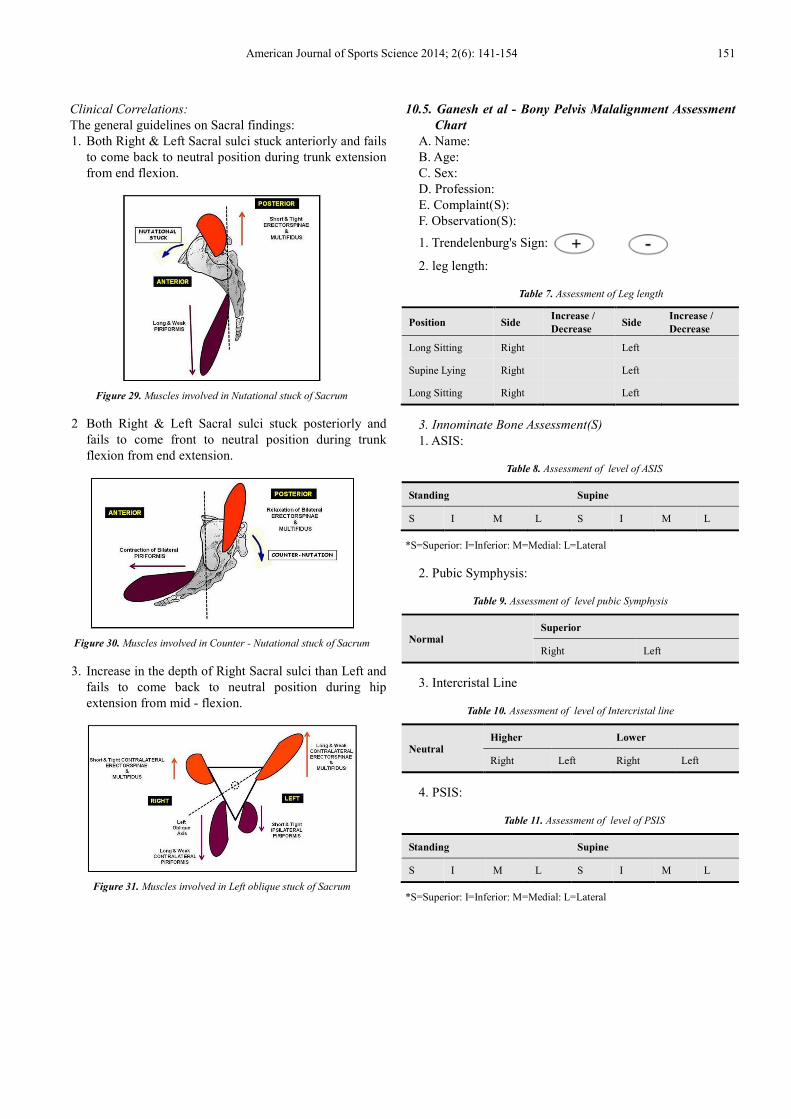
3. Increase in the depth of Right Sacral sulci than Left and
fails to come back to neutral position during hip
extension from mid - flexion.
Figure 31. Muscles involved in Left oblique stuck of Sacrum
10.5. Ganesh et al - Bony Pelvis Malalignment Assessment
Chart
A. Name:
B. Age:
C. Sex:
D. Profession:
E. Complaint(S):
F. Observation(S):
1. Trendelenburg's Sign:
2. leg length:
Table 7. Assessment of Leg length
Position Side Increase /
Decrease Side
Increase /
Decrease
Long Sitting Right Left
Supine Lying Right Left
Long Sitting Right Left
3. Innominate Bone Assessment(S)
1. ASIS:
Table 8. Assessment of level of ASIS
Standing Supine
S I M L S I M L
*S=Superior: I=Inferior: M=Medial: L=Lateral
2. Pubic Symphysis:
Table 9. Assessment of level pubic Symphysis
Normal Superior
Right Left
3. Intercristal Line
Table 10. Assessment of level of Intercristal line
Neutral Higher Lower
Right Left Right Left
4. PSIS:
Table 11. Assessment of level of PSIS
Standing Supine
S I M L S I M L
*S=Superior: I=Inferior: M=Medial: L=Lateral

152 E. Ganesh et al.: Soccer Syndrome - Common Presentations and Manual Diagnostic Techniques for
Pelvic Malalignment Syndrome
Figure 32. Assessment of the level of ASIS Figure 33. Assessment of the level of PSIS
4. Sacral Assessment:
Table 12. Assessment of Sacral stuck
Nutation (Forward Bending) Counter Nutation (Back to Normal) Rotation (Standing Leg Flexion)
Right Left Right Left Right Left
5. Muscle Assessment:
Table 13. Assessment of Muscle strength responsible for pelvic stability
Muscle Group & Its Test Muscles Shortened / Tightened Lengthened / Weakened
Right Left Right Left
Hip Flexors (Modified Thomas Test) Iliopsoas
Rectus Femoris
Hip Extensors
(Reverse Slr)
Gluteus Maximus
Hamstrings
Hip Adductors Adductor Longus
Gracilis
Hip Abductors Gluteus Medius
Tensor Fascia Latae (TFL)
Trunk Flexors Rectus Abdominis
External Oblique
Trunk Extensors Erector Spinae
Multifidus
Trunk Side Flexors Quadratus Lumborum
Gluteus Medius
Sacral Flexors Piriformis
G. Provisional Diagnosis:
11. Observations
Table 14. Period - 1 (Start of preseason - 1)
Malalignment Percentage (%)
Multiple (31) 77.5
Single (07) 17.5
Symmetry Pelvis (02) 05
Table 15. Period - 2 (End of season - 1)
Malalignment Percentage (%)
Multiple (35) 87.5
Single (04) 10
Symmetry Pelvis (01) 02.5
Table 16. Period - 3 (Start of preseason - 2)
Malalignment Percentage (%)
Multiple (36) 90
Single (03) 7.5
Symmetry Pelvis (01) 2.5
Table 17. Period - 4 (End of season - 2)
Malalignment Percentage (%)
Multiple (38) 95
Single (02) 05
Symmetry Pelvis (00) 00
Graph 1. Comparison of pelvic malalignment of Soccer players from period
- 1 to period - 4

American Journal of Sports Science 2014; 2(6): 141-154 153
Table 18. Total analysis of observations
Malalignment Percentage (%)
Multiple (35) 87.5
Single (04) 10
Symmetry Pelvis (01) 2.5
Graph 2. Final analysis of the observations
12. Discussion
The pelvic malalignment syndrome is the most common in
soccer players. An understanding of this 'malalignment
syndrome' requires knowledge of the common presentations
of malalignment and the techniques used to diagnose and
treat.
According to the e-source on pelvic evaluation (2010/09),
there are four common presentations on innominate
malalignment but the current study observed that there are
seven possible innominate malalignment presentations. And
the possible three sacral stucks for professional soccer
players are also observed in the present study which was not
mentioned in the previous studies (Pelvic evaluation, 2010;
Abdolhamid Daneshjoo, 2013; Fernando Idoate, 2011; Julie
Hides, 2012).
The present study shows that Anterior and Posterior
Innominate stuck can occur due to the changes in the strength
of Quadriceps and Hamstring muscles which is similar to the
previous work done by Abdolhamid,2013.
According to Abdolhamid,2013, the physical performance
and movement patterns of Soccer players may change the
balance of strength in both the legs, not particularly to the
dominant kicking leg which is contradict to the current study
in which we identified the muscle imbalance and
malalignment is more common in preferred kicking leg.
Rectus abdominis undergoes hypertrophy in the soccer
players which is a common observation between the current
study and Fernando, 2011.In the present study we observed
that change in the strength and bulk of Rectus abdominis in
the preferred kicking leg results in posterior innominate stuck
in the soccer players but Fernando, 2011 stated that changes
in the strength and bulk of rectus abdominis is bilateral and
asymmetrical.
Julie hides, 2012 stated that the imbalance between
Multifidus and Erector spinae results in Lumbo-pelvic
instability and this adds an additional proof to the reason for
rotational stuck of sacrum in our current study. In the present
study ,some additional observations has been made regarding
the sacral stuck, bilateral tightness of Multifidus and Erector
spinae results in Counter-Nutational stuck of sacrum and
bilateral weakness of the above mentioned muscles results in
Nutational stuck of sacrum.
The above mentioned facts, observations and results
indicated that an imbalance in the key muscles around the
bony pelvis will affect the Sacro-pelvic stability.
13. Conclusion
The present study is mainly focused on the common
presentations and manual diagnostic techniques for pelvic
malalignment syndrome. We used simple bony palpation
method to assess the various pelvic malalignments in football
players. While there are many methods to determine pelvic
symmetry or asymmetry, this method is very simple one and
that is consistently works well for me. Our method of
assessing the pelvic malalignments, even works well to the
common individuals suffering with low back & pelvis pain or
dysfunction due to various pelvic malalignment presentation.
To be masterly skilled in our current techniques of evaluating
the pelvic malalignment it is very simple and essential that
you will need to be able to locate the anterior superior iliac
spines (ASIS) and posterior superior iliac spines (PSIS), able
to judge the levelness of both iliac crests when viewing from
the front or rear even in different postures like standing, lying
in supine & prone also while performing the movements
during the assessment.
The repeated measurement, and analysis proves that the
"asymmetry of pelvis" which may be "ipsilateral" or
"contralateral" to the preferred kicking leg. The results
showed an imbalance of the key muscles associated with
Sacro-pelvic stability. In this study, we observed, 87.5% i.e.,
the majority of the soccer's are suffering with multiple pelvic
malalignments, includes innominate & sacral stuck. The
soccer's suffering with single component malalignment
account for about 10%, includes innominate or sacral stuck
and soccer's who had the symmetry pelvis is only 2.5%.
Noted, the pelvic rotations are relative and it is not always
necessary to know which side is the dysfunctional side if you
are treating them, as you should treat both sides of the pelvis.
To justify our diagnosis we access the muscles of the
functional slings and the muscles around the bony pelvis
related to the malalignment of the pelvic unit. The techniques
were repeated in different position and also performed for
two times per day and continued for fifteen days to
standardize.
However, This method of assessing the pelvic
malalignments until then not documented. In this sense, the
current study is mainly focused on the analysis and
documentation of the different common presentations in
pelvic malalignments which are common in soccer players
and its simple method of evaluation is the literature state of
the art. The present study may provide useful information to
analyze common presentations of pelvic malalignments in
different sports.

154 E. Ganesh et al.: Soccer Syndrome - Common Presentations and Manual Diagnostic Techniques for
Pelvic Malalignment Syndrome
References
[1] Abdolhamid Daneshjoo et al., Bilateral and Unilateral Asymmetries of Isokinetic strength and Flexibility in Male Young Professional Soccer Players. Journal of Human Kinetics.36, 45-53. 2013.
[2] Fernando Idoate et al., Soccer attenuates the Asymmetry of Rectus abdominis Muscle observed in Non- Athletes. PLOS ONE . 6(4), 1-7. 2011.
[3] F.P.Wedel,D.O., Evaluation and Treatment of Sacral Somatic
Dysfunction.Instruction manuals published by Michel Bakker. 2013.
[4] Julie Hides & Warren Stanton., Muscle imbalance among Elite Australian Rules Football Players, A longitudinal study of changes in trunk muscle size. Journal of Athletic training.47(3), 314 - 319. 2012.
[5] Liz Gaggini,M.A.., The asymmetric pelvis. International Association of Structural Integrators. 1-7. 2010
[6] Pelvic Evaluation., www.waltfritz seminars.com/myofascial resource/wp-content/uploads/2010/09.