sodium-carbonate co-substituted hydroxyapatite ceramics 22 02.pdf · powders of sodium-carbonate...
TRANSCRIPT

153
Processing and Application of Ceramics 7 [4] (2013) 153–157
Sodium-carbonate co-substituted hydroxyapatite ceramicsZoltan Z. Zyman*, Mykola V. TkachenkoPhysics of Solids Department, V.N. Karazin Kharkiv National University, 4, Svoboda Sq., Kharkiv 61022, UkraineReceived 9 September 2013; received in revised form 28 November 2013; accepted 8 December 2013
AbstractPowders of sodium-carbonate co-substituted hydroxyapatite, having sodium content in the range of 0.25–1.5 wt.% with a 0.25 wt.% step, were prepared by a precipitation-solid state reaction route. Compacts of the pow-ders were sintered in a CO2 flow (4 mL/min) at 1100 °C for 2 h. The sintered ceramics contained sodium and carbonate ions in the ranges of 0–1.5 wt.% and 1.3–6 wt.%, respectively, which are typical impurity concen-trations in biological apatite. A relationship between sodium and carbonate contents and the type of carbon-ate substitution was found. The total carbonate content progressively increased with the sodium content. The obtained ceramics showed an AB-type carbonate substitution. However, the substitution became more B-type as the sodium content increased. As a result, the carbonation was almost B-type (94 %) for the highest sodium content (1.5 wt.%).
Keywords: sodium-carbonate co-substituted hydroxyapatites, sintering, characterization
I. IntroductionBiological apatite based on hydroxyapatite, HA, has
a number of substitutions in cationic and anionic sub-lattices. The substitutions significantly affect the chem-ical and physico-chemical properties of the apatite, like mineralization and demineralization processes of calci-fied tissues and susceptibility to caries of teeth. Sodium (Na+) and carbonate (CO3
2-) ions are some of the major substituents occurring at levels of ca 0.9 wt.% and 5–8 wt.% [1–4].
As nanosize of crystals and their separation from the organic matrix cause difficulties in the apatite exami-nation, synthetic carbonated hydroxyapatites, CHAs, are typically used as structural models for studying the growth and dissolution processes of biological crystals. Such studies also result in the development of bioactive materials for medical purposes [5–13]. The effect of so-dium substitution in HA has extensively been studied [14–18], however, the simultaneous sodium-carbonate substitutions have not been examined as often. Besides, only limited combinations of the sodium and carbonate concentrations have been reported [19–24].
The sodium and carbonate ions have been intro-duced into HA in various ways. These have been (i) pre-cipitation, with addition of sodium and carbonate into the mother solution containing soluble salts [19,20]; (ii) wet precipitation combined with a preliminary me-chanical activation of parent reactants [24] or with fir-ing powder samples in a wet carbon dioxide atmosphere [23]; (iii) synthesis at a high temperature and moderate pressure [22].
A recent in vivo study revealed that sodium-free CHA enhanced bone in-growth and coverage [25]. However, an in vitro study of sodium-carbonate HA showed that the co-substitution brought little enhance in osteoblast proliferation or phenotype expression compared to HA [25,26]. The processing routes indicated above may re-sult in different physico-chemical characteristics, struc-tural perturbations and, particularly, in CO3
2– distribu-tions in the lattice (A-, AB-, and B-types of substitution [5,10]) of sodium-bearing CHAs designed as the same product. This may cause contradicting biological be-haviour of such compounds.
In this study, a relationship between sodium and car-bonate concentrations in a wide range in biological crys-tals, and modes of carbonate distribution for each designed combination of the substituents was found in substituted apatite ceramics prepared by a simple reliable route.
* Corresponding author: tel: +38 057 707 56 84, e-mail: [email protected] [email protected], [email protected]
DOI: 10.2298/PAC1304153Z

154
Z.Z. Zyman & M.V. Tkachenko / Processing and Application of Ceramics 7 [4] (2013) 153–157
II. Materials and methodsAn initial powder of carbonated hydroxyapatite
(CHA) was prepared through the reaction of calcium carbonate, CaCO3, (Merck, Germany, analytical grade) and a solution of the phosphate acid, H3PO4, (Merck) [2,27]. Sodium dopants were introduced into the CHA powder by soaking the powder in a solution of sodi-um hydrocarbonate, NaHCO3, at 60 °C until the sol-
vent was completely evaporated. The concentration of solution was varied so that the amount of sodium in the ceramics resulted from firing of compacts of such soaked powders was from 0.25–1.5 wt.% with a step of 0.25 wt.%. The compacts in the form of pellets (3 mm height and 10 mm diameter) were prepared in a steel mold by uniaxial pressing under 120 MPa. The compacts were sintered in a muffle in a dry CO2 flow (4 mL/min) at 1100 °C for 2 h. Six batches of sintered compacts were prepared with five samples of each so-dium concentration.
Calcium and sodium amounts in the samples were determined by atomic absorption spectrosco-py (Thermo Electron Corporation, M-series AA spec-trometer). Corresponding amounts of orthophosphate were found by colorimetry using the molybdenum blue method (Varian Cary Win UV spectrophotome-ter, λ = 725 nm). Carbon and hydrogen contents were measured by the burning method (EA 1110 CHNS-0 elemental analyser). Structural analysis was per-formed using a Philips APDW 40C diffractometer and a copper Kα radiation (λ = 0.154 nm) with a nickel fil-ter through 20–70° diffraction angles (2θ). IR spectra were recorded by employing a BIO-RAD 175 spec-trometer at a 2 cm-1 resolution and the KBr technique, operating in the transmittance mode between wave numbers of 400–4000 cm-1.
III. Results and discussionCompositions of the sintered ceramics, with given
sodium concentrations, are listed in Table 1. The piv-otal result is the interrelation between the sodium and carbon concentrations: the higher the value for sodi-um, the higher is the carbon content. The comparison of the XRD patterns for ceramics with no sodium and a high sodium content shows no changes in the crys-tallographic apatite structure (Fig. 1). However, the lattice constants substantially change (Fig. 2). Start-ing from the reference values of HA (the a constant is a little enhanced compared to the reference value be-cause the ceramics was sintered in the CO2 atmosphere [23,28]), the a constant gradually decreased, and the c constant gradually increased as the sodium content in-
Figure 2. Lattice constants of ceramics with varying sodium concentration (the dotted horizontal lines are the
reference values of HA)
Figure 1. XRD patterns of ceramics without sodium (CHA-0) and with 1.25 wt.% of sodium (CHA-5)
Table 1. Elemental analysis of the sodium-substituted carbonated ceramics
Samplenotation
Na[wt.%]
Ca[wt.%]
PO4[wt.%]
C[wt.%]
H[wt.%]
CHA-0 0 40.6 54.6 0.27 0.15CHA-1 0.25 40.3 54.4 0.53 0.15CHA-2 0.50 40.1 53.0 0.78 0.13CHA-3 0.75 39.6 52.6 0.90 0.11CHA-4 1.00 39.5 52.5 1.10 0.11CHA-5 1.25 39.5 52.0 1.08 0.10CHA-6 1.50 38.2 52.0 1.20 <0.1
155
Z.Z. Zyman & M.V. Tkachenko / Processing and Application of Ceramics 7 [4] (2013) 153–157
creased in the ceramics. Most probably, this is not a result of the Na+ for Ca2+ substitution as the effective ionic radii of Na+ (0.99 Å) and Ca2+ (1.00 Å) are very close [29]. The change in lattice constants could be
caused by increased carbonate content in these ceram-ics (Table 1). However, the presence of carbonate may differently affect the lattice constants in CHA depend-ing on the substitution site [5,10].
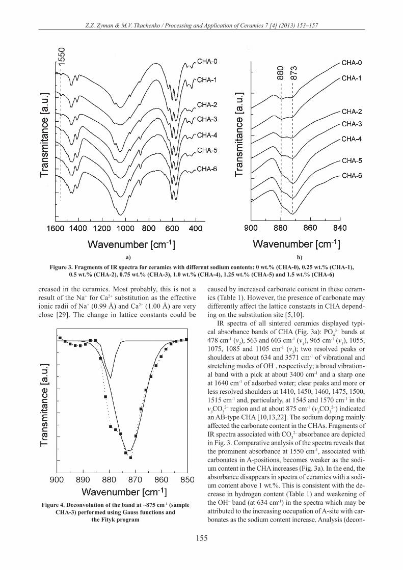
IR spectra of all sintered ceramics displayed typi-cal absorbance bands of CHA (Fig. 3a): PO4
3– bands at 478 cm-1 (v2), 563 and 603 cm-1 (v4), 965 cm-1 (v1), 1055, 1075, 1085 and 1105 cm-1 (v3); two resolved peaks or shoulders at about 634 and 3571 cm-1 of vibrational and stretching modes of OH–, respectively; a broad vibration-al band with a pick at about 3400 cm-1 and a sharp one at 1640 cm-1 of adsorbed water; clear peaks and more or less resolved shoulders at 1410, 1450, 1460, 1475, 1500, 1515 cm-1 and, particularly, at 1545 and 1570 cm-1 in the v3CO3
2– region and at about 875 cm-1 (v3CO32–) indicated
an AB-type CHA [10,13,22]. The sodium doping mainly affected the carbonate content in the CHAs. Fragments of IR spectra associated with CO3
2– absorbance are depicted in Fig. 3. Comparative analysis of the spectra reveals that the prominent absorbance at 1550 cm-1, associated with carbonates in A-positions, becomes weaker as the sodi-um content in the CHA increases (Fig. 3a). In the end, the absorbance disappears in spectra of ceramics with a sodi-um content above 1 wt.%. This is consistent with the de-crease in hydrogen content (Table 1) and weakening of the OH– band (at 634 cm-1) in the spectra which may be attributed to the increasing occupation of A-site with car-bonates as the sodium content increase. Analysis (decon-
Figure 3. Fragments of IR spectra for ceramics with different sodium contents: 0 wt.% (CHA-0), 0.25 wt.% (CHA-1), 0.5 wt.% (CHA-2), 0.75 wt.% (CHA-3), 1.0 wt.% (CHA-4), 1.25 wt.% (CHA-5) and 1.5 wt.% (CHA-6)
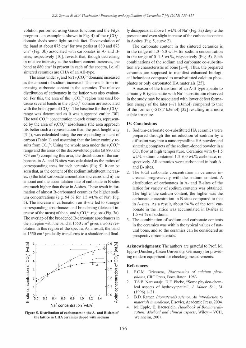
Figure 4. Deconvolution of the band at ~875 cm-1 (sample CHA-3) performed using Gauss functions and
the Fityk program
a) b)
156
Z.Z. Zyman & M.V. Tkachenko / Processing and Application of Ceramics 7 [4] (2013) 153–157
volution performed using Gauss functions and the Fityk program - an example is shown in Fig. 4) of the v2CO3
2– domain sheds some light on this fact. Deconvolution of the band at about 875 cm-1 for two peaks at 880 and 873 cm-1 (Fig. 3b) associated with carbonates in A- and B-sites, respectively [10], reveals that, though decreasing in relative intensity as the sodium content increases, the band at 880 cm-1 is present in each of the spectra, i.e. all sintered ceramics are CHA of an AB-type.
The areas under v3 and (or) v2CO32– domains increased
as the amount of sodium increased. This results from in-creasing carbonate content in the ceramics. The relative distribution of carbonates in the lattice was also evaluat-ed. For this, the area of the v2CO3
2– region was used be-cause several bands in the v3CO3
2– domain are associated with the both types of CO3
2–. The baseline for the v2CO32–
range was determined as it was suggested earlier [30]. The total CO3
2– concentration in each ceramics, represent-ed by the area of v2CO3
2– absorbance (the area approach fits better such a representation than the peak height way [31]), was calculated using the corresponding content of carbon (Table 1) and assuming that the latter entirely re-sults from CO3
2–. Using the whole area under the v2CO32–
range and the areas of the deconvoluted peaks (at 880 and 873 cm-1) compiling this area, the distribution of the car-bonates in A- and B-sites was calculated as the ratios of corresponding areas for each ceramics (Fig. 5). It can be seen that, as the content of the sodium substituent increas-es: i) the total carbonate amount also increases and ii) the amount and the accumulation rate of carbonate in B-sites are much higher than those in A-sites. These result in for-mation of almost B-carbonated ceramics for higher sodi-um concentrations (e.g. 94 % for 1.5 wt.% of Na+, Fig. 5). The increase in carbonation on B-site led to stronger corresponding absorbances and broadening (detected in-crease of the areas) of the v2 and v3CO3
2–-regions (Fig. 3a). The overlap of the broadened B-carbonate absorbances in the v3 region with the band at 1550 cm-1 gives a worse res-olution in this region of the spectra. As a result, the band at 1550 cm-1 gradually transforms to a shoulder and final-
ly disappears at above 1 wt.% of Na+ (Fig. 3a) despite the presence and even slight increase of the carbonate content in A-sites (Fig. 5, curve 2).
The carbonate content in the sintered ceramics is in the range of 1.3–6.0 wt.% for sodium concentration in the range of 0–1.5 wt.%, respectively (Fig. 5). Such combinations of the sodium and carbonate co-substitu-tion are characteristic of bone [2–4]. Thus, the prepared ceramics are supposed to manifest enhanced biologi-cal behaviour compared to unsubstituted calcium phos-phates or only carbonated HA materials [25].
A reason of the transition of an A-B type apatite to a mainly B-type apatite with Na+ -substitution observed in the study may be associated with lower defect forma-tion energy of the later (–71 kJ/mol) compared to that of the former (–518.7 kJ/mol) [32] resulting in a more stable structure.
IV. Conclusions1. Sodium-carbonate co-substituted HA ceramics were
prepared through the introduction of sodium by a diffusion way into a nanocrystalline HA powder and sintering compacts of the sodium-doped powder in a CO2 flow at high temperature. Ceramics with 0–1.5 wt.% sodium contained 1.3–6.0 wt.% carbonate, re-spectively. All ceramics were carbonated in both A- and B- sites.
2. The total carbonate concentration in ceramics in-creased progressively with the sodium content. A distribution of carbonates in A- and B-sites of the lattice for variety of sodium contents was obtained. The higher the sodium content, the higher was the carbonate concentration in B-sites compared to that in A-sites. As a result, about 94 % of the total car-bonate in the lattice was accumulated in B-sites at 1.5 wt.% of sodium.
3. The combination of sodium and carbonate contents in the ceramics was within the typical values of nat-ural bone, and so the ceramics can be considered as prospective biomaterials.
Acknowledgments: The authors are grateful to Prof. M. Epple (Duisburg-Essen University, Germany) for provid-ing modern equipment for checking measurements.
References1. F.C.M. Driessens, Bioceramics of calcium phos-
phates, CRC Press, Boca Raton, 1983.2. T.S.B. Narasaraju, D.E. Phebe, “Some physico-chem-
ical aspects of hydroxyapatite”, J. Mater. Sci., 31 (1996) 1–21.
3. B.D. Ratner, Biomaterials science: An introduction to materials in medicine, Elsevier, Academic Press, 2004.
4. M. Epple, E. Baeuerlein, Handbook of Biominerali-sation: Medical and clinical aspects, Wiley – VCH, Weinheim, 2007.
Figure 5. Distribution of carbonates in the A- and B-sites of the lattice in CHA ceramics doped with sodium

157
Z.Z. Zyman & M.V. Tkachenko / Processing and Application of Ceramics 7 [4] (2013) 153–157
5. R.Z. LeGeros, O.R. Ttrauts, E. Klein, J.P. LeGeros, “Two types of carbonate substitution in the apatite structure”, Experientia, 25 (1969) 5–7.
6. D.G.A. Nelson, “The influence of carbonate on the atomic structure and reactivity of hydroxyapatite”, J. Dent. Res., 60 (1981) 1621–1629.
7. L.G. Ellies, D.G.A. Nelson, J.D.B. Featherstone, “Crystallographic structure and surface morphology of sintered carbonated apatites”, J. Biomed. Mater. Res., 22 (1988) 541–553.
8. Y. Doi, T. Koda, N. Wakamatsu, T. Goto, H. Kame-mizu, Y. Moriwaki, M. Adachi, Y. Suwa, “Influence of carbonate on sintering of apatites”, J. Dent. Res., 72 (1993) 1279–1284.
9. J.C. Merry, I.R. Gibson, S.M. Best, W. Bonfield, “Syn-thesis and characterization of carbonate hydroxyapa-tite”, J. Mater. Sci.: Mater. Med., 9 (1998) 779–783.
10. I.R. Gibson, W. Bonfield, “Novel synthesis and charac-terization of an AB-type carbonate-substituted hydroxy-apatite”, J. Biomed. Mater. Res., 59 (2002) 697–708.
11. J.P. Lafon, E. Champion, D. Bernache-Assollant, R. Gibert, A.M. Danna, “Thermal decomposition of car-bonated calcium phosphate apatites”, J. Therm. Anal. Calorim., 72 (2003) 1127–1134.
12. E. Landi, A. Tampiery, G. Celotti, L. Vichi, M. San-dri, “Influence of synthesis and sintering parameters on the characteristics of carbonate apatite”, Biomate-rials, 25 (2004) 1763–1770.
13. Z. Zyman, M. Tkachenko, “CO2 gas-activated sinter-ing of carbonated hydroxyapatites”, J. Eur. Ceram. Soc., 31 (2011) 241–248.
14. I. Lopez-Valero, C. Gome-Lorente, R. Boistelle, “Ef-fects of sodium and ammonium ions on occurrence, evolution and crystallinity of calcium phosphates”, J. Cryst. Growth., 121 (1992) 297–304.
15. J.C. Elliot. Structure and chemistry of the apatites and other calcium orthophosphates, Elsevier, Amsterdam, 1994.
16. S. Kannan, J.H.G. Rocha, J.M.F. Ferreira, “Synthe-sis and thermal stability of sodium, magnesium co-substituted hydroxyapatites”, J. Mater. Sci., 16 (2006) 286–291.
17. J. Ue, R.M. Pilliar, R.A. Kandel, “The effect of sodi-um dopants on calcium polyphosphate biomaterials”, Key Eng. Mater., 361-363 (2008) 281–284.
18. K. Matsunaga, H. Murata, “Formation energies of substitutional sodium and potassium in hydroxyapa-tite”, Mater. Trans., 50 (2009) 1041–1045.
19. H. El Feki, J.M. Savariault, A. Ben Salah, “Structure refinements by the Rietveld method of partially substi-tuted hydroxyapatite: Ca9Na0.5(PO4)4.5(CO3)1.5(OH)2”, J. Alloys Compd., 287 (1999) 114–120.
20. H. El Feki, J.M. Savariault, A. Ben Salah, M. Jemal, “Sodium and carbonate distribution in substituted cal-cium hydroxyapatite”, Solid State Sci., 2 (2000) 577–586.
21. R.M. Wilson, J.C. Elliot, S.E.P. Dowker, R.I. Smith, “Rietveld structure refinement of precipitated carbon-ate apatite using neutron diffraction data”, Biomateri-als, 25 (2004) 2205–2213.
22. M.E. Fleet, X. Liu, “Coupled substitution of type A and B carbonate in sodium-bearing apatite”, Biomate-rials, 28 (2007) 916–926.
23. J.A. Stephen, C. Pace, J.M.S. Skakle, I.R. Gibson, “Comparison of carbonate hydroxyapatite with and without sodium cosubstitution”, Key Eng. Mater., 330-332 (2007) 19–22.
24. S.M. Barinov, I.V. Fadeeva, D. Ferro, J.V. Rau, S. Nunziante Cesaro, V.S. Komlev, A.S. Fomin, “Stabi-lization of carbonate hydroxyapatite by isomorphic substitutions of sodium for calcium”, Russ. J. Inorg. Chem., 53 (2008) 164–168.
25. N. Patel, I.R. Gibson, K.A. Hing, S.M. Best, E. Damien, P.A. Revell, W. Bonfield, “The in vivo re-sponse of phase pure hydroxyapatite and carbonate substituted hydroxyapatite granules of varying size ranges”, Key Eng. Mater., 218-220 (2002) 383–386.
26. K.A. Hing, J.C. Merry, I.R. Gibson, L. Di Silvio, S.M. Best, W. Bonfield, “Effect of carbonate content on the response of human osteoblast-like cells to carbonate substitute hydroxyapatite”, Bioceramics, 12 (1999) 185–188.
27. Z.Z. Zyman, M.V. Tkachenko, D.V. Polevodin, “Prep-aration and characterization of biphasic calcium phos-phate ceramics of desired composition”, J. Mater. Sci.: Mater. Med., 19 (2008) 2819–2825.
28. J.E. Barralet, G.J.P. Fleming, C. Campion, J.J. Har-ris, A.J. Wright, “Formation of translucent hydroxy-apatite ceramics by sintering in carbon dioxide atmo-spheres”, J. Mater. Sci., 38 (2003) 3979–3993.
29. R.D. Shannon, “Revised effective ionic radii and sys-tematic studies of interatomic distances in halides and chalcogenides”, Acta Cryst., A32 (1976) 751–767.
30. A.B.S. Clasen, I.E. Ruyter, “Quantitative determina-tion of type A and B carbonate in human deciduous and permanent enamel by means of Fourier transform infrared spectrometry”, Adv. Dent. Res., 11 (1997) 523–527.
31. B.C. Smith, Fundamentals of Fourier Transform In-frared Spectroscopy, CRC Press, Boca Raton, 2011.
32. S. Peroos, Z. Du, N.H. de Leeuw, “A computer mod-elling study of the uptake, structure and distribution of carbonate defects in hydroxyapatite”, Biomaterials, 27 (2006) 2150–2161.
