soft-tissue calcifications and ossifications in the
TRANSCRIPT

Soft-tissue calcifications and ossifications in the pediatric population: a pattern-based imaging approach with a review of common etiologies
Eugen Lungu, Dang-Long Truong, Ramy El-Jalbout, Julie Barsalou, Nathalie Alos, Jean-Jacques de Bruycker,
Marie-Claude Miron*Authors have no conflicts of interest.

Je n’ai pas établi de relation avec uneorganisation à but lucratif ou sans but lucratif
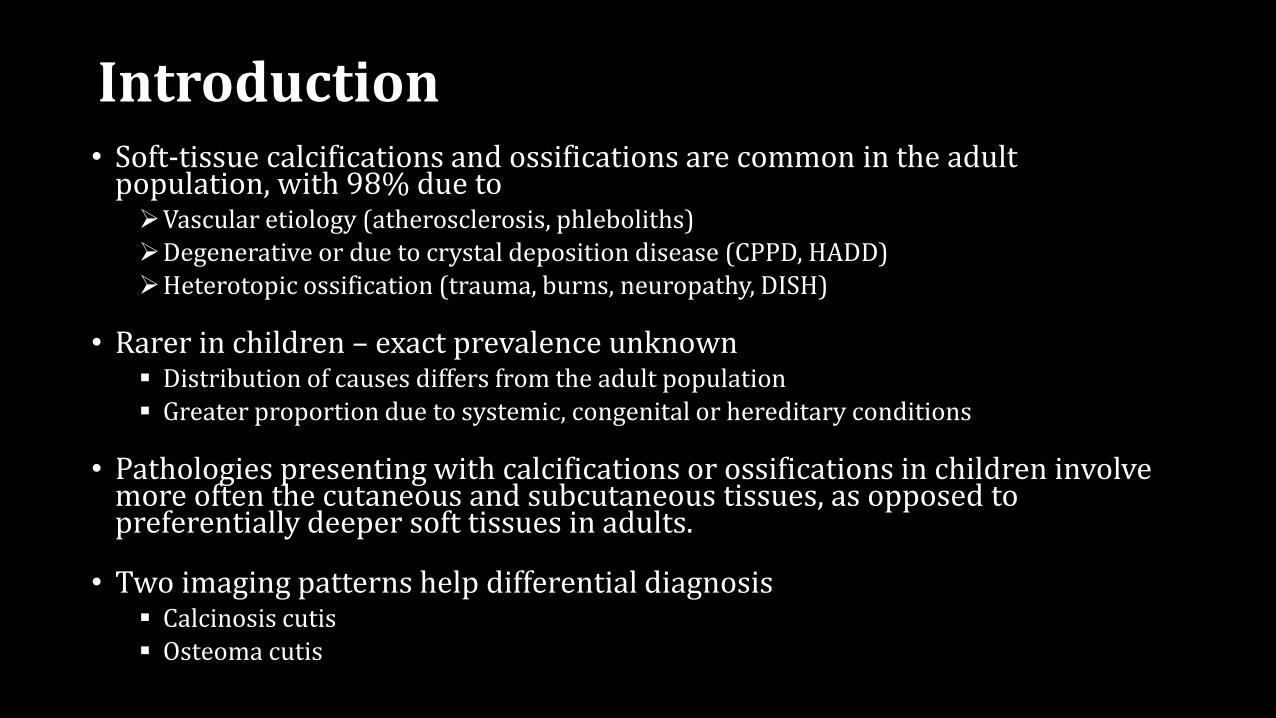
Introduction• Soft-tissue calcifications and ossifications are common in the adult
population, with 98% due to➢Vascular etiology (atherosclerosis, phleboliths)➢Degenerative or due to crystal deposition disease (CPPD, HADD)➢Heterotopic ossification (trauma, burns, neuropathy, DISH)
• Rarer in children – exact prevalence unknown▪ Distribution of causes differs from the adult population▪ Greater proportion due to systemic, congenital or hereditary conditions
• Pathologies presenting with calcifications or ossifications in children involve more often the cutaneous and subcutaneous tissues, as opposed to preferentially deeper soft tissues in adults.
• Two imaging patterns help differential diagnosis▪ Calcinosis cutis▪ Osteoma cutis
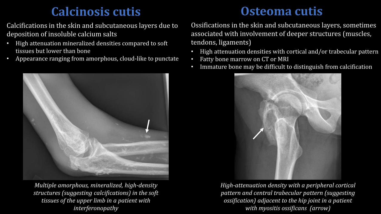
Calcifications in the skin and subcutaneous layers due to deposition of insoluble calcium salts
• High attenuation mineralized densities compared to soft tissues but lower than bone
• Appearance ranging from amorphous, cloud-like to punctate
Calcinosis cutis Osteoma cutisOssifications in the skin and subcutaneous layers, sometimes associated with involvement of deeper structures (muscles, tendons, ligaments)
• High attenuation densities with cortical and/or trabecular pattern• Fatty bone marrow on CT or MRI• Immature bone may be difficult to distinguish from calcification
Multiple amorphous, mineralized, high-density structures (suggesting calcifications) in the soft
tissues of the upper limb in a patient with interferonopathy
High-attenuation density with a peripheral cortical pattern and central trabecular pattern (suggesting
ossification) adjacent to the hip joint in a patient with myositis ossificans (arrow)
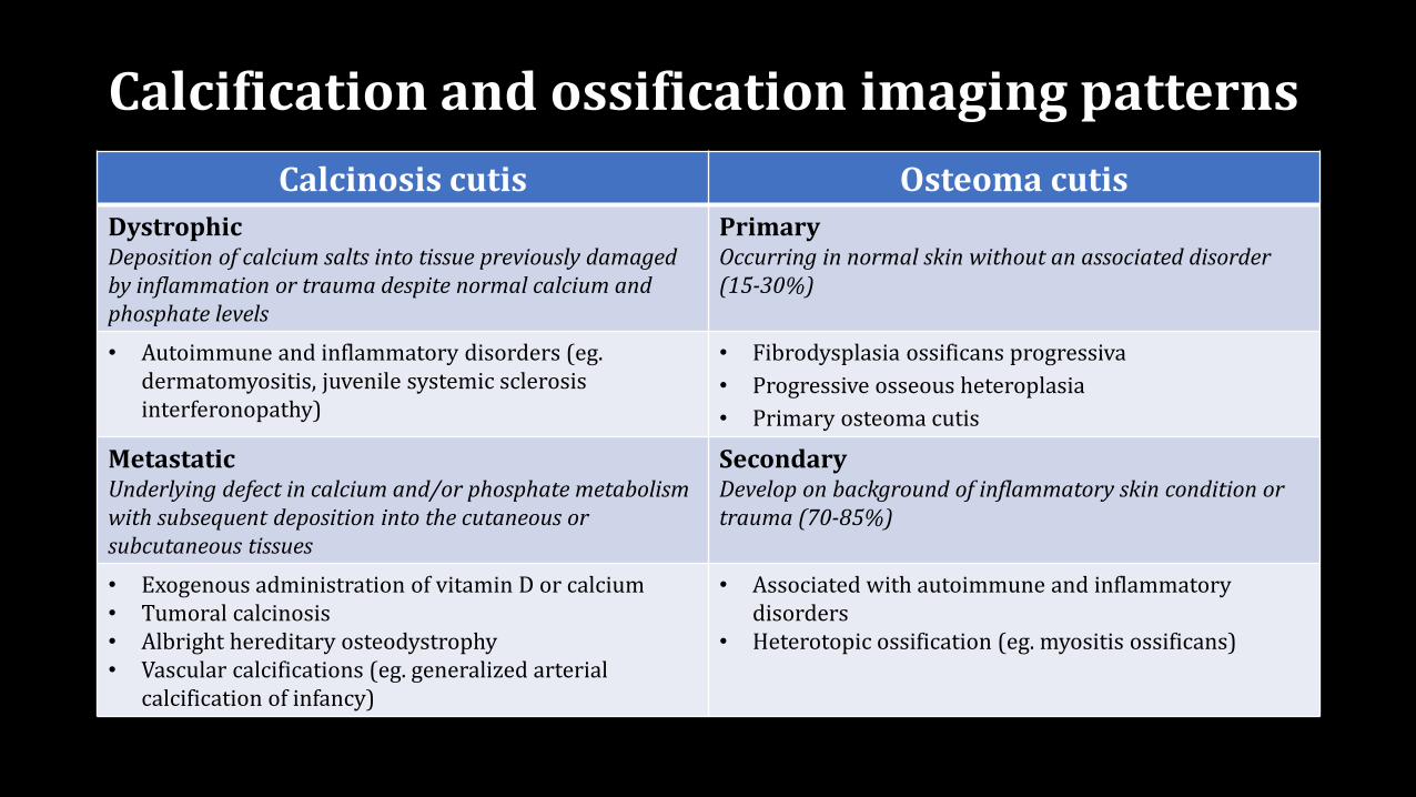
Calcification and ossification imaging patterns
Calcinosis cutis Osteoma cutis
DystrophicDeposition of calcium salts into tissue previously damaged by inflammation or trauma despite normal calcium and phosphate levels
PrimaryOccurring in normal skin without an associated disorder (15-30%)
• Autoimmune and inflammatory disorders (eg.dermatomyositis, juvenile systemic sclerosis interferonopathy)
• Fibrodysplasia ossificans progressiva
• Progressive osseous heteroplasia
• Primary osteoma cutis
MetastaticUnderlying defect in calcium and/or phosphate metabolism with subsequent deposition into the cutaneous or subcutaneous tissues
SecondaryDevelop on background of inflammatory skin condition or trauma (70-85%)
• Exogenous administration of vitamin D or calcium• Tumoral calcinosis• Albright hereditary osteodystrophy• Vascular calcifications (eg. generalized arterial
calcification of infancy)
• Associated with autoimmune and inflammatory disorders
• Heterotopic ossification (eg. myositis ossificans)
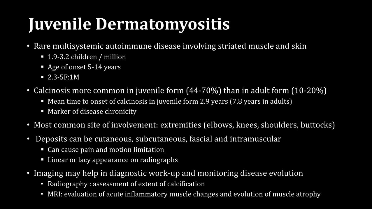
Juvenile Dermatomyositis• Rare multisystemic autoimmune disease involving striated muscle and skin
▪ 1.9-3.2 children / million
▪ Age of onset 5-14 years
▪ 2.3-5F:1M
• Calcinosis more common in juvenile form (44-70%) than in adult form (10-20%)▪ Mean time to onset of calcinosis in juvenile form 2.9 years (7.8 years in adults)
▪ Marker of disease chronicity
• Most common site of involvement: extremities (elbows, knees, shoulders, buttocks)
• Deposits can be cutaneous, subcutaneous, fascial and intramuscular▪ Can cause pain and motion limitation
▪ Linear or lacy appearance on radiographs
• Imaging may help in diagnostic work-up and monitoring disease evolution• Radiography : assessment of extent of calcification
• MRI: evaluation of acute inflammatory muscle changes and evolution of muscle atrophy
Juvenile DermatomyositisA B C D
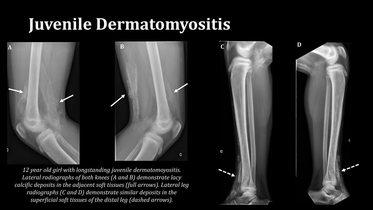
12 year old girl with longstanding juvenile dermatomoyositis. Lateral radiographs of both knees (A and B) demonstrate lacy
calcific deposits in the adjacent soft tissues (full arrows). Lateral leg radiographs (C and D) demonstrate similar deposits in the
superficial soft tissues of the distal leg (dashed arrows).
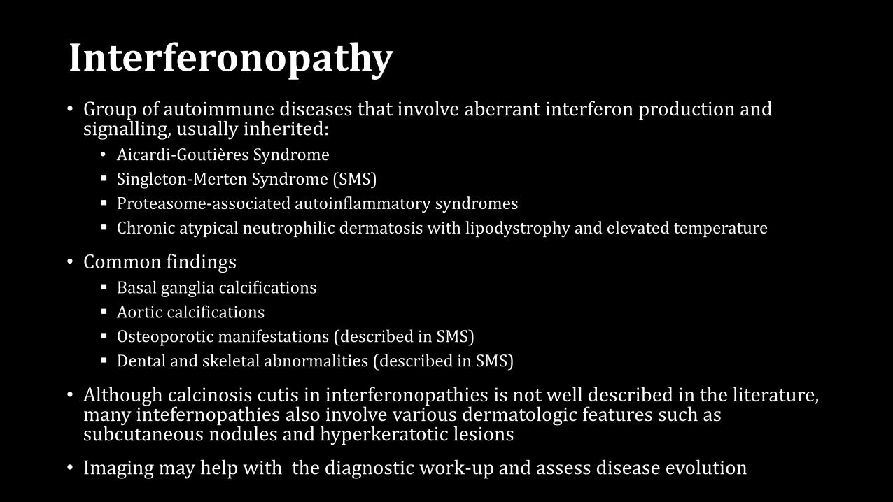
Interferonopathy• Group of autoimmune diseases that involve aberrant interferon production and
signalling, usually inherited:• Aicardi-Goutières Syndrome
▪ Singleton-Merten Syndrome (SMS)
▪ Proteasome-associated autoinflammatory syndromes
▪ Chronic atypical neutrophilic dermatosis with lipodystrophy and elevated temperature
• Common findings▪ Basal ganglia calcifications
▪ Aortic calcifications
▪ Osteoporotic manifestations (described in SMS)
▪ Dental and skeletal abnormalities (described in SMS)
• Although calcinosis cutis in interferonopathies is not well described in the literature, many intefernopathies also involve various dermatologic features such as subcutaneous nodules and hyperkeratotic lesions
• Imaging may help with the diagnostic work-up and assess disease evolution
InterferonopathyA B C
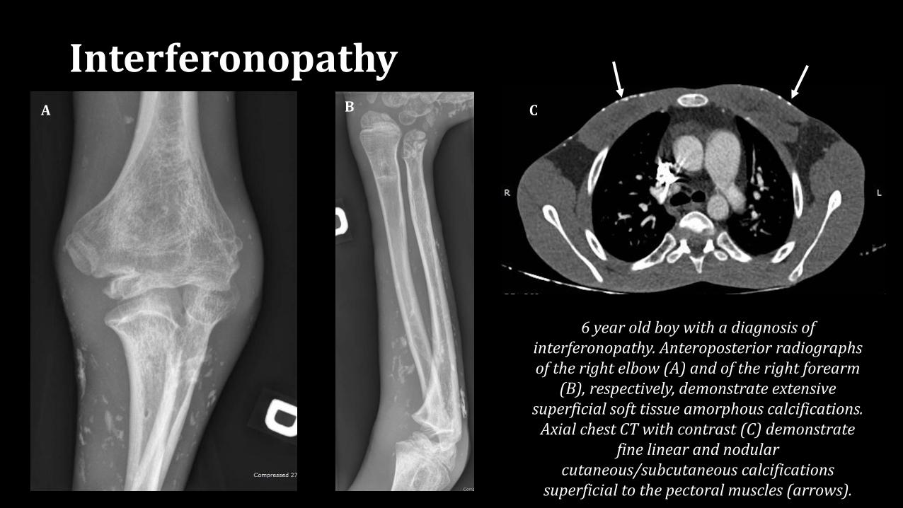
6 year old boy with a diagnosis of interferonopathy. Anteroposterior radiographs of the right elbow (A) and of the right forearm
(B), respectively, demonstrate extensive superficial soft tissue amorphous calcifications.
Axial chest CT with contrast (C) demonstrate fine linear and nodular
cutaneous/subcutaneous calcifications superficial to the pectoral muscles (arrows).
Exogenous administration of vitamin D or calcium
• Common treatment for various conditions▪ Inadequate intake: exclusive breastfeeding without Vitamin D supplementation, insufficient
light exposure
▪ Medication induced: anti-epileptic drugs, HAART, corticosteroids, antifungals
▪ Chronic disease: chronic kidney disease (most common cause), cystic fibrosis, sickle cell disease
• Radiological presentation similar regardless of underlying condition ▪ Metastatic calcification (calcium phosphate product >70 mg / dL)
▪ Deposits usually located in periarticular regions
▪ Metaphyseal bands of sclerosis (heavy calcification of the proliferating cartilage)
• Usually results from chronic and large dose supplementation of vitamin D
• Imaging helps in the diagnostic work-up and assessment of response to treatment
High dose calcium therapy
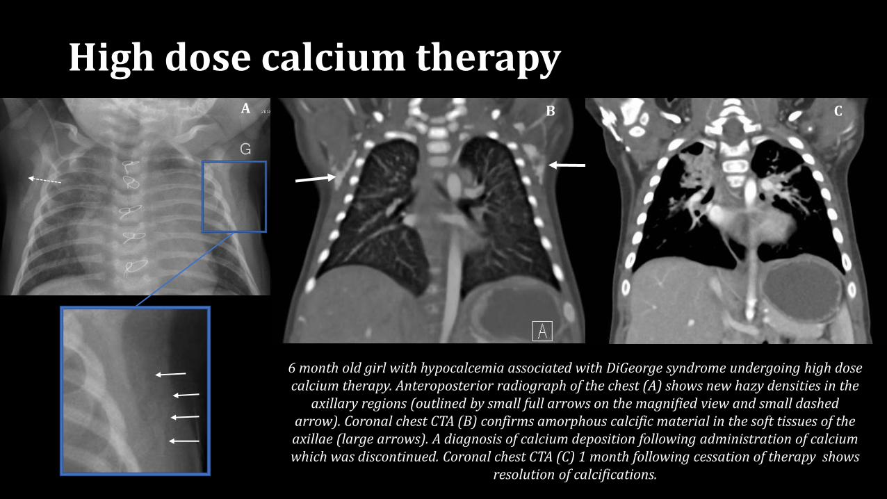
6 month old girl with hypocalcemia associated with DiGeorge syndrome undergoing high dose calcium therapy. Anteroposterior radiograph of the chest (A) shows new hazy densities in the
axillary regions (outlined by small full arrows on the magnified view and small dashed arrow). Coronal chest CTA (B) confirms amorphous calcific material in the soft tissues of the axillae (large arrows). A diagnosis of calcium deposition following administration of calcium which was discontinued. Coronal chest CTA (C) 1 month following cessation of therapy shows
resolution of calcifications.
A B C
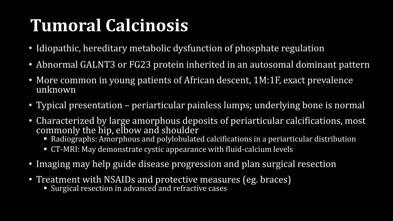
Tumoral Calcinosis• Idiopathic, hereditary metabolic dysfunction of phosphate regulation
• Abnormal GALNT3 or FG23 protein inherited in an autosomal dominant pattern
• More common in young patients of African descent, 1M:1F, exact prevalence unknown
• Typical presentation – periarticular painless lumps; underlying bone is normal
• Characterized by large amorphous deposits of periarticular calcifications, most commonly the hip, elbow and shoulder▪ Radiographs: Amorphous and polylobulated calcifications in a periarticular distribution
▪ CT-MRI: May demonstrate cystic appearance with fluid-calcium levels
• Imaging may help guide disease progression and plan surgical resection
• Treatment with NSAIDs and protective measures (eg. braces)▪ Surgical resection in advanced and refractive cases
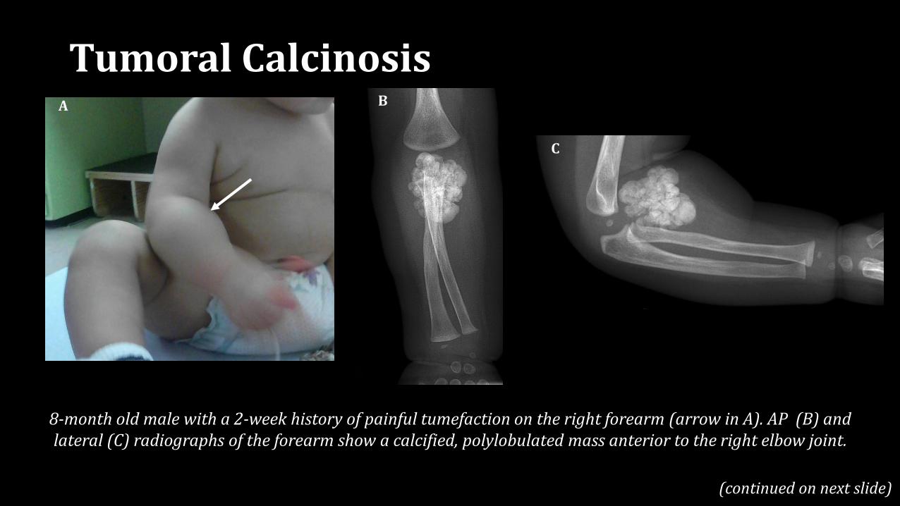
8-month old male with a 2-week history of painful tumefaction on the right forearm (arrow in A). AP (B) and lateral (C) radiographs of the forearm show a calcified, polylobulated mass anterior to the right elbow joint.
A
(continued on next slide)
Tumoral Calcinosis
C
B
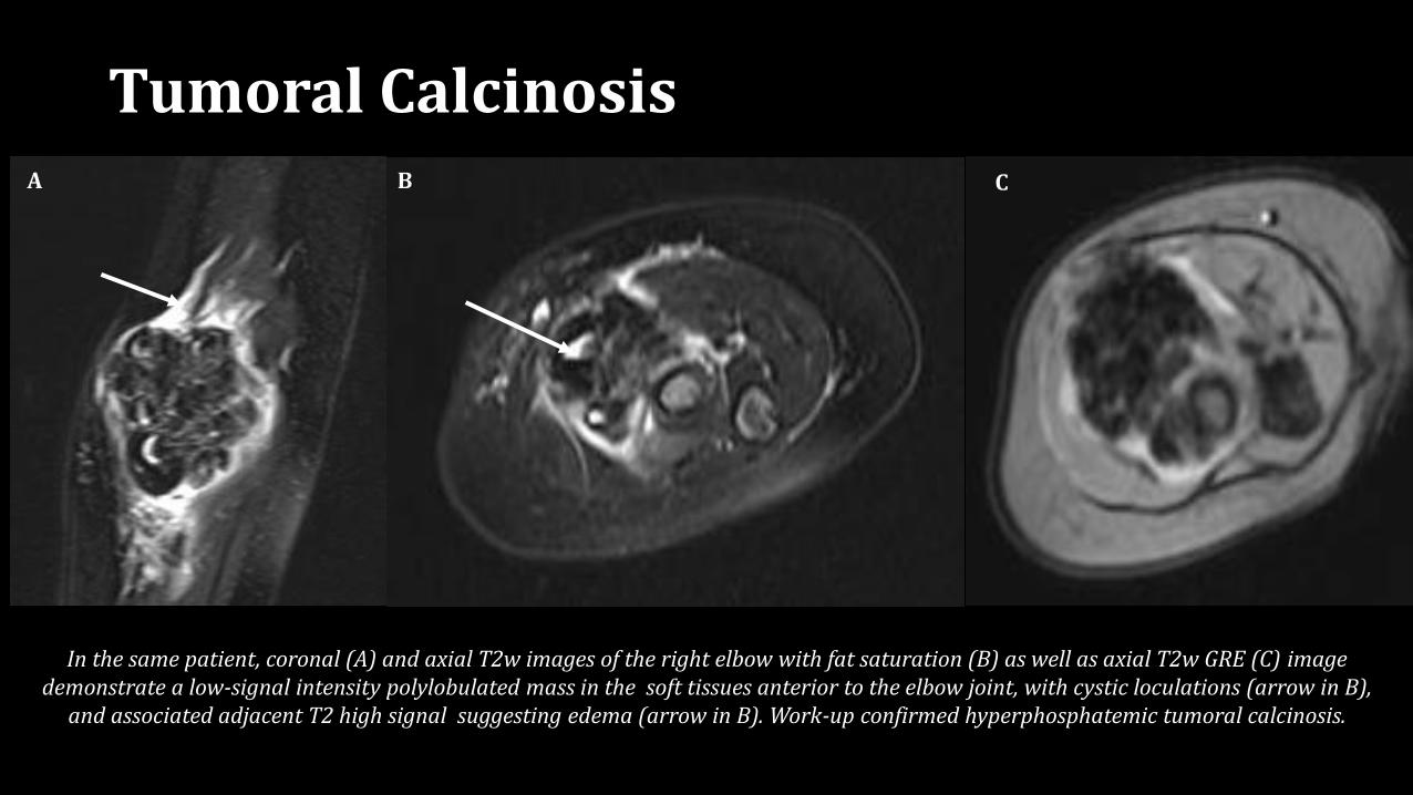
In the same patient, coronal (A) and axial T2w images of the right elbow with fat saturation (B) as well as axial T2w GRE (C) image demonstrate a low-signal intensity polylobulated mass in the soft tissues anterior to the elbow joint, with cystic loculations (arrow in B),
and associated adjacent T2 high signal suggesting edema (arrow in B). Work-up confirmed hyperphosphatemic tumoral calcinosis.
CA B
Tumoral Calcinosis
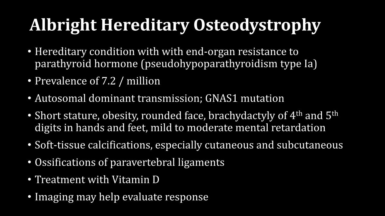
• Hereditary condition with with end-organ resistance to parathyroid hormone (pseudohypoparathyroidism type Ia)
• Prevalence of 7.2 / million
• Autosomal dominant transmission; GNAS1 mutation
• Short stature, obesity, rounded face, brachydactyly of 4th and 5th
digits in hands and feet, mild to moderate mental retardation
• Soft-tissue calcifications, especially cutaneous and subcutaneous
• Ossifications of paravertebral ligaments
• Treatment with Vitamin D
• Imaging may help evaluate response
Albright Hereditary Osteodystrophy
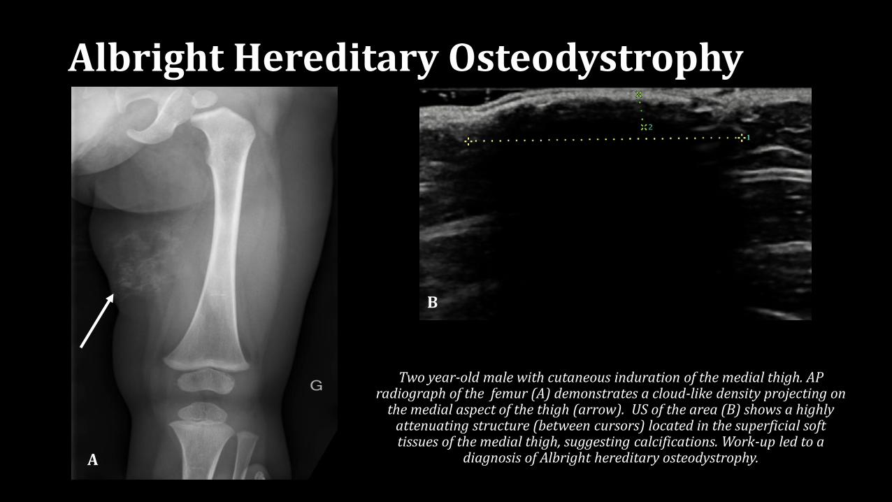
Two year-old male with cutaneous induration of the medial thigh. AP radiograph of the femur (A) demonstrates a cloud-like density projecting on
the medial aspect of the thigh (arrow). US of the area (B) shows a highly attenuating structure (between cursors) located in the superficial soft tissues of the medial thigh, suggesting calcifications. Work-up led to a
diagnosis of Albright hereditary osteodystrophy.A
B
Albright Hereditary Osteodystrophy
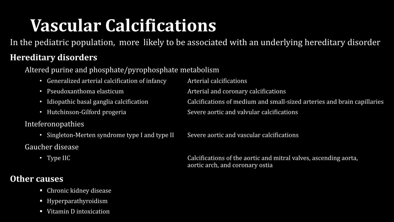
In the pediatric population, more likely to be associated with an underlying hereditary disorder
Hereditary disorders
Altered purine and phosphate/pyrophosphate metabolism
• Generalized arterial calcification of infancy Arterial calcifications
• Pseudoxanthoma elasticum Arterial and coronary calcifications
• Idiopathic basal ganglia calcification Calcifications of medium and small-sized arteries and brain capillaries
• Hutchinson-Gilford progeria Severe aortic and valvular calcifications
Inteferonopathies
• Singleton-Merten syndrome type I and type II Severe aortic and vascular calcifications
Gaucher disease
• Type IIC Calcifications of the aortic and mitral valves, ascending aorta, aortic arch, and coronary ostia
Other causes▪ Chronic kidney disease
▪ Hyperparathyroidism
▪ Vitamin D intoxication
Vascular Calcifications
A B C D
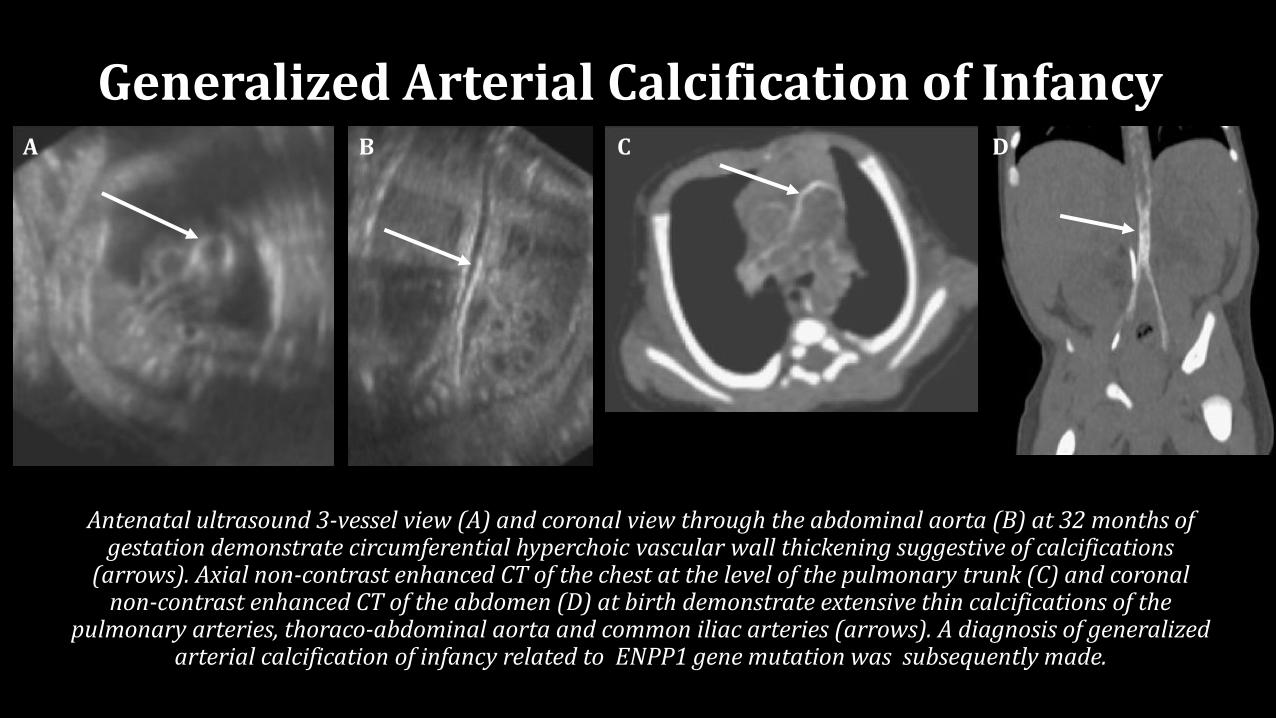
Antenatal ultrasound 3-vessel view (A) and coronal view through the abdominal aorta (B) at 32 months of gestation demonstrate circumferential hyperchoic vascular wall thickening suggestive of calcifications
(arrows). Axial non-contrast enhanced CT of the chest at the level of the pulmonary trunk (C) and coronal non-contrast enhanced CT of the abdomen (D) at birth demonstrate extensive thin calcifications of the
pulmonary arteries, thoraco-abdominal aorta and common iliac arteries (arrows). A diagnosis of generalized arterial calcification of infancy related to ENPP1 gene mutation was subsequently made.
Generalized Arterial Calcification of Infancy
A
B
C
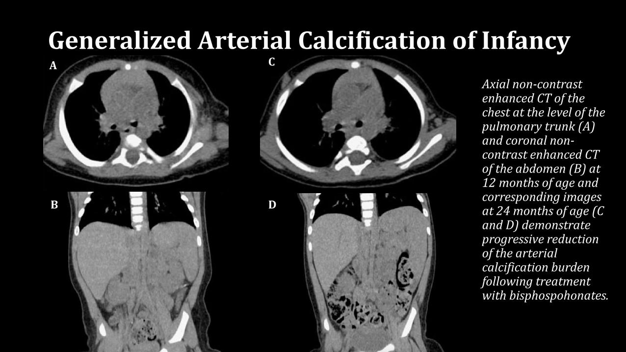
Axial non-contrast enhanced CT of the chest at the level of the pulmonary trunk (A) and coronal non-contrast enhanced CT of the abdomen (B) at 12 months of age and corresponding images at 24 months of age (C and D) demonstrate progressive reduction of the arterial calcification burden following treatment with bisphospohonates.
D
Generalized Arterial Calcification of Infancy
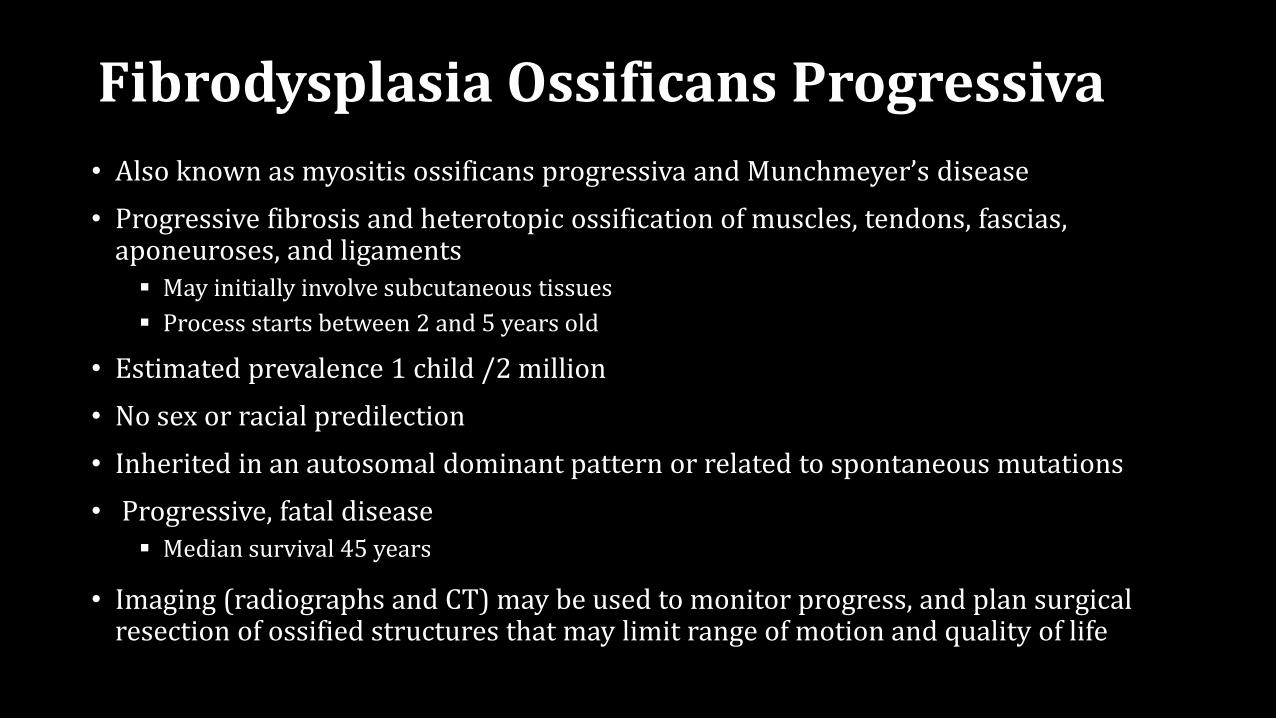
Fibrodysplasia Ossificans Progressiva
• Also known as myositis ossificans progressiva and Munchmeyer’s disease
• Progressive fibrosis and heterotopic ossification of muscles, tendons, fascias, aponeuroses, and ligaments▪ May initially involve subcutaneous tissues
▪ Process starts between 2 and 5 years old
• Estimated prevalence 1 child /2 million
• No sex or racial predilection
• Inherited in an autosomal dominant pattern or related to spontaneous mutations
• Progressive, fatal disease▪ Median survival 45 years
• Imaging (radiographs and CT) may be used to monitor progress, and plan surgical resection of ossified structures that may limit range of motion and quality of life
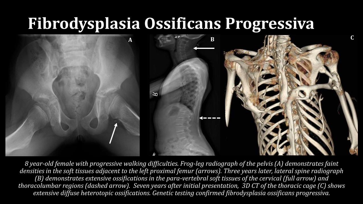
8 year-old female with progressive walking difficulties. Frog-leg radiograph of the pelvis (A) demonstrates faint densities in the soft tissues adjacent to the left proximal femur (arrows). Three years later, lateral spine radiograph
(B) demonstrates extensive ossifications in the para-vertebral soft tissues of the cervical (full arrow) and thoracolumbar regions (dashed arrow). Seven years after initial presentation, 3D CT of the thoracic cage (C) shows
extensive diffuse heterotopic ossifications. Genetic testing confirmed fibrodysplasia ossificans progressiva.
B CA
Fibrodysplasia Ossificans Progressiva
Progressive Osseous Heteroplasia
• Extremely rare, autosomal dominant disorder characterized by dermal ossification (osteoma cutis) beginning in infancy▪ Progresses to extensive bone formation in deep muscle and fascia
• GNAS mutation - similar to Albright hereditary osteodystrophy, part of a spectrum of conditions
• Average age at onset: 1 year old
• Growth retardation due to joint ankylosis, osteopenia
• Surgical removal of heterotopic bone may help, but high rate of recurrence
• Imaging may help guide disease progression and plan surgical intervention
Myositis Ossificans
*
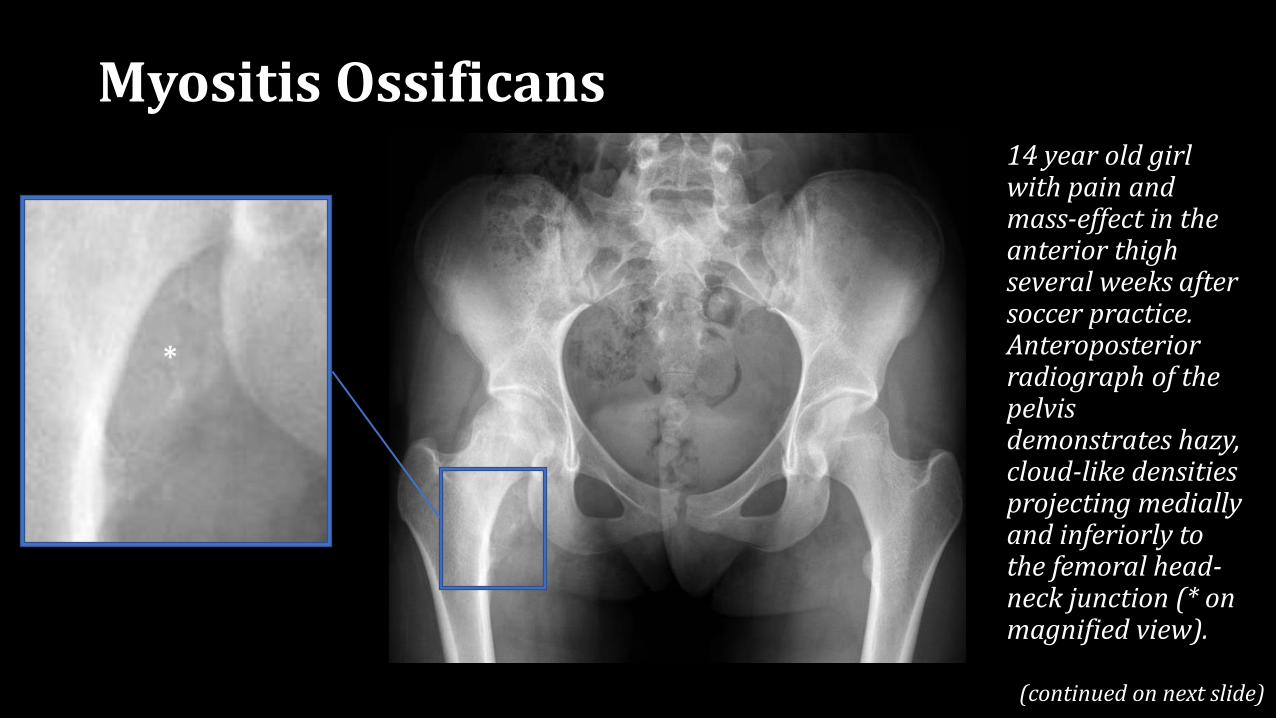
14 year old girl with pain and mass-effect in the anterior thigh several weeks after soccer practice. Anteroposterior radiograph of the pelvis demonstrates hazy, cloud-like densities projecting medially and inferiorly to the femoral head-neck junction (* on magnified view).
(continued on next slide)
Myositis Ossificans
A B
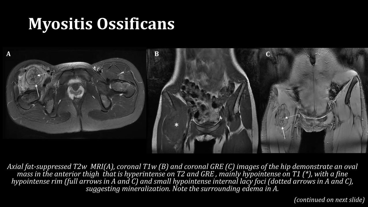
Axial fat-suppressed T2w MRI(A), coronal T1w (B) and coronal GRE (C) images of the hip demonstrate an oval mass in the anterior thigh that is hyperintense on T2 and GRE , mainly hypointense on T1 (*), with a fine
hypointense rim (full arrows in A and C) and small hypointense internal lacy foci (dotted arrows in A and C), suggesting mineralization. Note the surrounding edema in A.
CB
*
(continued on next slide)
Myositis Ossificans
*
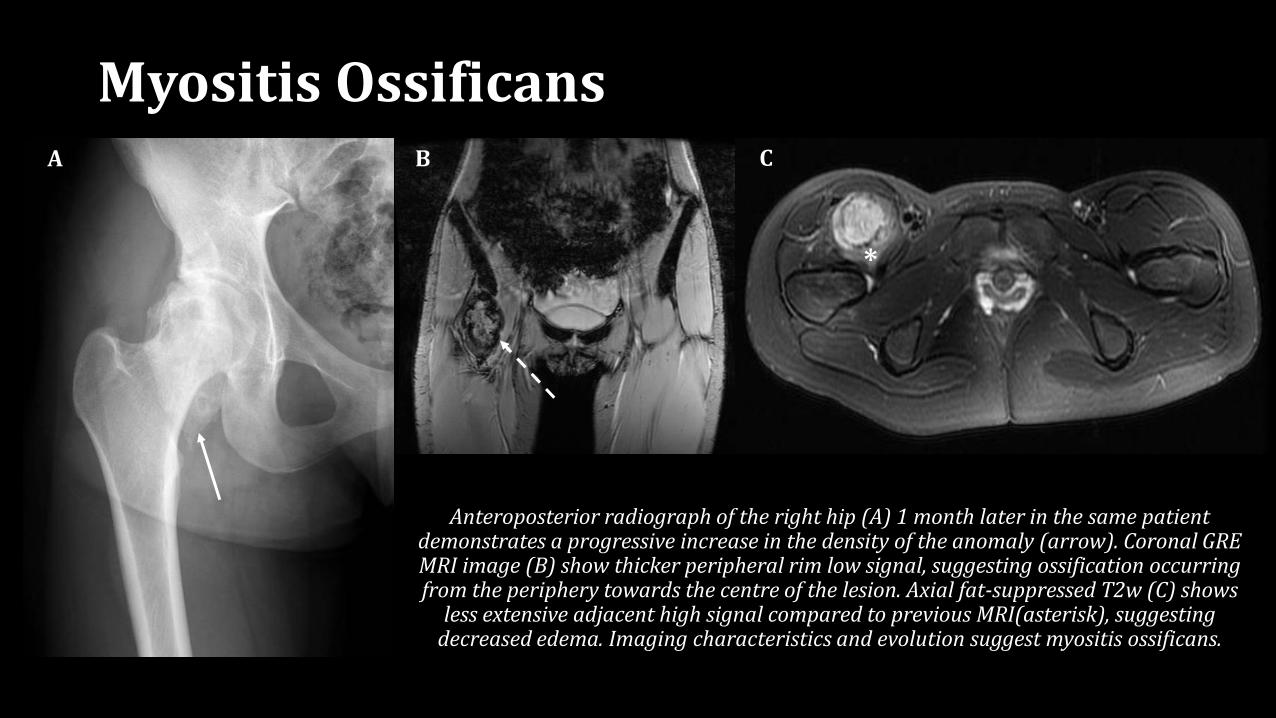
Anteroposterior radiograph of the right hip (A) 1 month later in the same patient demonstrates a progressive increase in the density of the anomaly (arrow). Coronal GRE MRI image (B) show thicker peripheral rim low signal, suggesting ossification occurring from the periphery towards the centre of the lesion. Axial fat-suppressed T2w (C) shows
less extensive adjacent high signal compared to previous MRI(asterisk), suggesting decreased edema. Imaging characteristics and evolution suggest myositis ossificans.
A B C
Imaging caveats
• Differentiating between calcifications and ossifications may be difficult on imaging, especially when superficially-located and small
• Some pathologies may present with either calcifications or ossifications, or with a succession of calcifications becoming ossified
• Imaging may help establish diagnosis in selected etiologies, however it may have a greater role in evaluating disease progression and planning treatment