somatom force 8,57 5,0 1,3 1,7 - zol
TRANSCRIPT

Computed Tomography
Page 1 | © Siemens Healthcare GmbH, 2017
8,57
5,0
1,7
1,3
8,0
SOMATOM Force

© Siemens Healthcare GmbH, 2017
Computed Tomography
SOMATOM Force
Menno Fokke, Clinical education specialist CTForchheim, Germany

Computed Tomography
Page 3 | © Siemens Healthcare GmbH, 2017
8,57
5,0
1,7
1,3
8,0
SOMATOM Force
Is CT nog het stralingskanonvan de radiologie?
Computed Tomography
Page 4 | © Siemens Healthcare GmbH, 2017
8,57
5,0
1,7
1,3
8,0
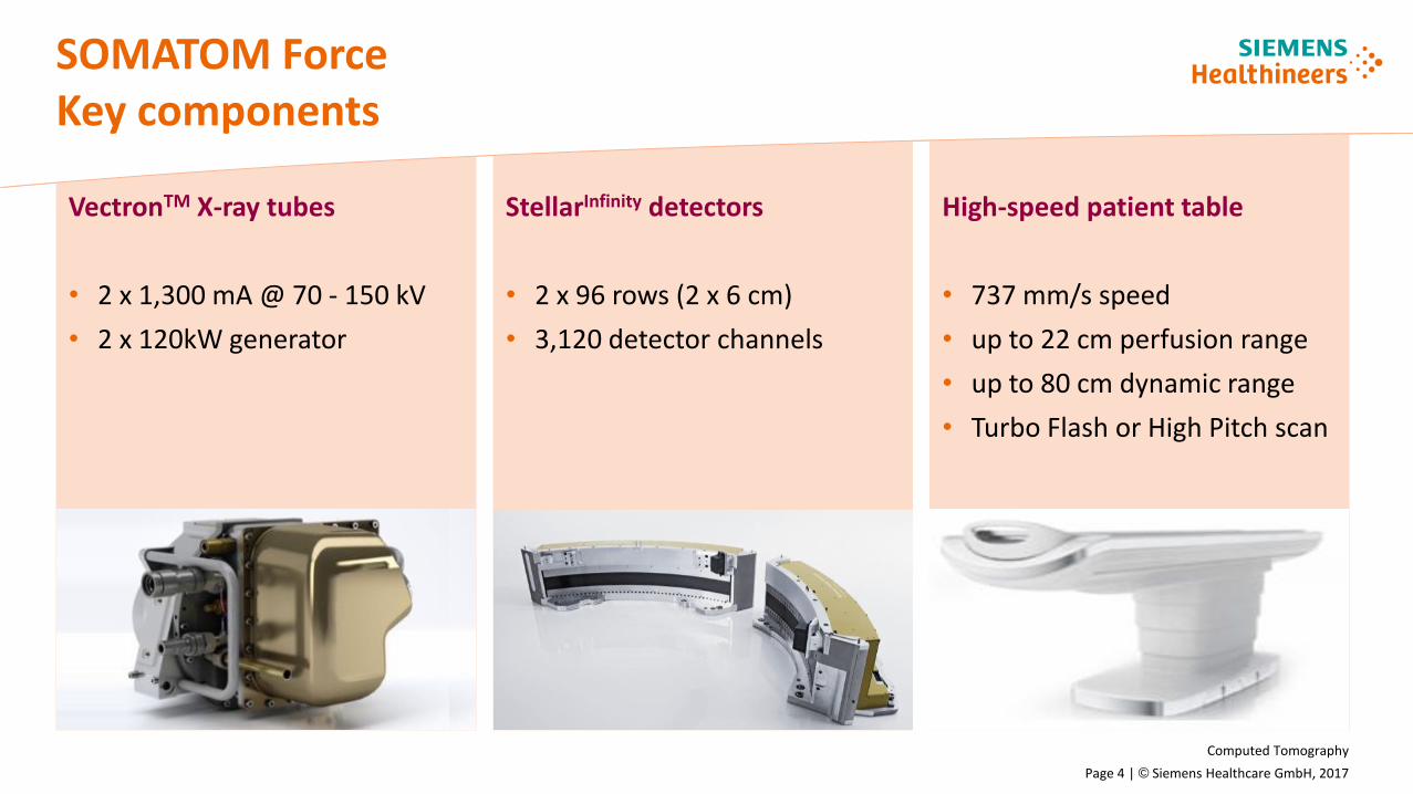
VectronTM X-ray tubes
• 2 x 1,300 mA @ 70 - 150 kV
• 2 x 120kW generator
StellarInfinity detectors
• 2 x 96 rows (2 x 6 cm)
• 3,120 detector channels
SOMATOM ForceKey components
High-speed patient table
• 737 mm/s speed
• up to 22 cm perfusion range
• up to 80 cm dynamic range
• Turbo Flash or High Pitch scan

Computed Tomography
Page 5 | © Siemens Healthcare GmbH, 2017
SOMATOM Force
Collimation: 192 x 0.6 mmScan time: 0.4 sScan length: 289 mmRotation time: 0.25 sSn100 kV, 68 mAsCTDIvol: 0.23 mGyDLP: 8 mGy cm Eff. dose: 0.1 mSv
Lung examinations with significantlyimproved air-to-soft-tissue contrast allow doses of conventional X-ray1)
Low-dose early detection with Tin Filter CT imaging at the dose of conventional X-ray1)
Radiology 2008;248:254-63
Courtesy of UMM, Mannheim, Germany1) Invest Radiol. 2014 Jul;49(7):465-73.
ADMIRE Strength Level 5

Computed Tomography
Page 6 | © Siemens Healthcare GmbH, 2017
Early detection with low-dose lung scansOutstanding image quality even in obese patients
Courtesy of Hospital Saint Joseph, Marsaille, France
SOMATOM Force
Collimation: 192 x 0.6 mmScan time: 2.0 sScan length: 274 mmRotation time: 0.5 s110 kV, 79 mAsCTDIvol: 4.13 mGyDLP: 134 mGy cm Eff. dose: 0.28 mSv
Early detection low-dose lung scan in an obese patient with a very low dose of 0.28 mSv with outstanding image quality.

Computed Tomography
Page 7 | © Siemens Healthcare GmbH, 2017
Cardiac imaging with high heart rates – ultra-low-dose Turbo Flash Spiral at a heart rate of 117 bpm
Courtesy of Baotou Central Hospital, Baotou, Inner Mongolia
SOMATOM Force
Collimation: 2 x 192 x 0.6 mmScan time: 0.12 sScan length: 93 mmRotation time: 0.25 s80 kV, 289 mAsCTDIvol: 1.33 mGyDLP: 19.7 mGy cm Eff. dose: 0.28 mSvHR: 117 bpmCM: 30 mL
Rule out of coronary disease within 0.12 s at average heart rate of 117 bpm with Turbo Flash mode.

Computed Tomography
Page 8 | © Siemens Healthcare GmbH, 2017
8,57
5,0
1,7
1,3
8,0
SOMATOM Force
Is CT nog het stralingskanonvan de radiologie?
Dosis reducerendetechnologie in de Siemens SOMATOM Force

Computed Tomography
Page 9 | © Siemens Healthcare GmbH, 2017
8,57
5,0
1,7
1,3
8,0
CARE technologyCombine Applications Reduce Exposure
Scan preparation
Parameter adjustment
ScanImage recon
Reading
Dose
Reduction
SOMATOM Scanner syngo.via
CARE Dose4D
CARE kV
Iterative Reconstruction
CARE Child
CARE Dashboard
CARE Profile
CARE Dose Configurator
CARE Contrast
Dose
Management
Dose
Visualization

Computed Tomography
Page 10 | © Siemens Healthcare GmbH, 2017
8,57
5,0
1,7
1,3
8,0
More precise configuration
of dose modulation
• Configuration for different body habitus and organs individually
• New and more specific modulation curves
• Definition of threshold values for Dose Alert according to IEC regulations
CARE Dose4D
January 2016Page 10 HC DI CT
Unrestricted © Siemens Healthcare GmbH 2016 All rights reserved.

Computed Tomography
Page 11 | © Siemens Healthcare GmbH, 2017
8,57
5,0
1,7
1,3
8,0
Voltage setting with CARE kV
CARE Dose4D and CARE kV
Dose relevant scan parameters
• Patient's body habitus
• Effective current (mAs)
• Voltage dep. on examination (kV)
Current modulation w/ CARE Dose4D• Automatic patient and organ
specific protocol adaption• Automatic modulation in X,Y
& Z direction in real time

Computed Tomography
Page 12 | © Siemens Healthcare GmbH, 2017
8,57
5,0
1,7
1,3
8,0
CARE kV
0
10
20
30
40
50
60
70
80
90
100
70 80 100 120 140
0
10
20
30
40
50
60
70
80
90
100
70 80 100 120 140
Without CARE kV
With CARE kV
kV
usage
[%]
Tube voltage [kV]
kV
usage
[%]
Tube voltage [kV]
Example: Abdomen @ 100 kV
Dose (CTDIvol): 9.7 mGy
Dose reduction compared to:
• Siemens 120 kV stand. prot.: 14 mGy
• Competitor‘s 120 kV protocol*: 25 mGy
*Source: Internal data evaluation based on anonymous assessment on SRS connected scanners. Clinical image courtesy C. McCullough, Mayo Clinic,
Rochester, MN, USA * Sagara Y et al. Abdominal CT: comparison of low-dose CT with adaptive statistical iterative reconstruction and routine-dose CT
with filtered back projection in 53 patients. AJR Am J Roentgenol. 2010 Sep;195(3):713-9.

Computed Tomography
Page 13 | © Siemens Healthcare GmbH, 2017
8,57
5,0
1,7
1,3
8,0
ADMIRE The newest generation in iterative reconstruction
ADMIRE in three steps
• Raw data statistical modeling: Statistical weighting of all projections in raw data space
• Model based noise cancelation: Noise cancelation based on an intelligent approach in image space
• Advanced system modeling: Advanced modeling is the base for the forward projection and eliminates artifacts
Non ADMIRE ADMIRE

Computed Tomography
Page 14 | © Siemens Healthcare GmbH, 2017
8,57
5,0
1,7
1,3
8,0
ADMIREExcellent image quality
Courtesy of UMM, Mannheim, Germany
WFBP* ADMIRE
*) Weighted Filtered Back Projection

Computed Tomography
Page 15 | © Siemens Healthcare GmbH, 2017
Patient with dissectionLow kV imaging with reduced contrast media (20 mL)
Courtesy of UMM, Mannheim, Germany 1) 400 mg/mL
SOMATOM Force
Collimation: 2 x 192 x 0.6 mmScan time: 1.1 sScan length: 740 mmRotation time: 0.25 s80 kV, 140 mAsCTDIvol: 2.09 mGyDLP: 155 mGy cm Eff. dose: 2.32 mSv
Gated, examination with 20 mL1) of contrast media without breath-holdin patient with limited function in right kidney.

Computed Tomography
Page 16 | © Siemens Healthcare GmbH, 2017
8,57
5,0
1,7
1,3
8,0
Two steps ahead in Freezing Motion“Free-breathing” CT imaging
Ultra-high-speed imaging with the uniqueSOMATOM Force in combination with ADMIRE
1) Image quality as defined by low contrast detectability using a model observer method for evaluation. Equivalent low contrast detectability can be achieved with 80% to 85% less dose using ADMIRE at highest strength level for thin (0.6 mm) reconstruction slices in measured and simulated body and head phantoms for low contrast objects with different contrasts. See ADMIRE date sheet for further information. In clinical practice, the use of ADMIRE may reduce CT patient dose depending on the clinical task, patient size, anatomical location, and clinical practice. A consultation with a radiologist and a physicist should be made to determine the appropriate dose to obtain diagnostic image quality for the particular clinical task.
Next generation Dual Source CT (DSCT) with
• Two VectronTM tubes
• Two StellarInfinity detectors with25% more detector channels (1,840)
• Up to 22 lp/cm (0.24 mm) spatial resolution
• Advanced Modeled Iterative Reconstruction (ADMIRE)1) for lowest possible dose
Up to 66 ms native temporal resolution utilizing DSCT at 250 ms rotation speed

Computed Tomography
Page 17 | © Siemens Healthcare GmbH, 2017
“Free-breathing” CT imagingNo anesthesia, no sedation1) for babies/kids
Courtesy of UMM, Mannheim, Germany1) The inherent temporal resolution – the ‘native’ temporal resolution acquired by the scanner – is highly important to freeze patient motion, e.g. in lung exams or in patients who cannot hold their breath long enough. This is also important, in pediatric CT where it also can help reducing the need for potentially harmful sedation.
Follow-up (SOMATOM Force)
Turbo Flash ultra-low-dose thorax CT
0.4 mSv effective dose; 0.3 s scan time
Pediatric imaging without breath-hold and sedation!1)
Baseline (previous high-end CT)
Standard low-dose thorax CT
3 mSv effective dose
11 s scan time

Computed Tomography
Page 18 | © Siemens Healthcare GmbH, 2017
Preventive care in cardiac imagingUltra-low-dose Turbo Flash Spiral with 0.09 mSv
Courtesy of Adventist Hospital Sydney, Australia
SOMATOM Force
Collimation: 2 x 192 x 0.6 mmScan time: 0.2 sScan length: 142 mmRotation time: 0.25 s70 kV, 128 mAsCTDIvol: 0.36 mGyDLP: 6.6 mGy cm Eff. dose: 0.09 mSvHR: 56 bpmBMI: 24
Rule out of coronary disease within 0.2 s and with ultra-low-dose with only 0.09 mSv.

Computed Tomography
Page 19 | © Siemens Healthcare GmbH, 2017
8,57
5,0
1,7
1,3
8,0
Two steps ahead in Decision MakingPrecise Dual Energy quantification
Dual Energy (DE) imaging with improvedenergy separation
VectronTM tube plus StellarInfinity detectors
• New energy pairings, e.g. 90 and 150 kV Sn for imaging of obese patients
• Up to 35 cm DE field of view (FoV)
• Up to 258 mm/s DE scan speed
• Tin Filter (Selective Photon Shield II) utilization
Up to 30% increased energy separation for better DE imaging outcomes

Computed Tomography
Page 20 | © Siemens Healthcare GmbH, 2017
Precise Dual Energy quantification – pancreatic head tumor illustrated with different DE reconstructions
Courtesy of MUSC Medical Center, Charleston, USA1) Frellesen et al. (2015): Dual Energy CT of the pancreas: improved carcinoma-to-pancreas contrast with a noise-optimized monoenergetic reconstruction algorithm.
SOMATOM Force
Collimation: 2 x 128 x 0.6 mmScan time: 4.35 sScan length: 207mmRotation time: 0.5 s100/Sn150 kV, 117/60 mAsCTDIvol: 6.88 mGyDLP: 162.9 mGy cm Eff. dose: 2.4 mSv
Advanced diagnostic information: Monoenergetic Plus allows energy spectrum shift to lower levels which significantly improves image quality and pancreas-to-lesion constrast1).
Mono+ 55 keV VNC
Mixed

Computed Tomography
Page 21 | © Siemens Healthcare GmbH, 2017
Precise DE quantification – tissue differentiation and therapy response assessment of liver cancer
Courtesy of University Hospital Zuerich, Switzerland
SOMATOM Force
Collimation: 2 x 192 x 0.6 mmScan time: 4 sScan length: 182 mmRotation time: 0.5 s100/Sn150 kV, 202/190 mAsCTDIvol: 11.99 mGyDLP: 254 mGy cm Eff. dose: 3.81 mSv
Dose neutral Dual Energy enables advanced diagnosis of liver metastases and cyst tissue.
Suspected
cyst
Tumor tissue

Computed Tomography
Page 22 | © Siemens Healthcare GmbH, 2017
Kidney-friendly scanning – change treatment decisions with more comprehensive 4D imaging
Courtesy of UMM, Mannheim, Germany1) 400 mgl/mL
SOMATOM Force
Collimation: 48 x 1.2 mmScan time: 36 sScan length: 222 mmRotation time: 0.25 s70 kV, 200 mAsCTDIvol: 43.46 mGyDLP: 905 mGy cm Eff. dose: 13.6 mSvCM: 12 mL
Kidney-friendly dynamic angiography of EVAR, Type II endoleak scanned with only 12 mL1) of contrast media in 72 year old patient.

Computed Tomography
Page 23 | © Siemens Healthcare GmbH, 2017
SOMATOM ForceTwo steps ahead
Slices2 x 192
(2 x 576 recon)mA
1,300 mA@70 kV1,300 mA@80 kV
Rotational coverage 184 mm/rot kV70 - 150 kV10 kV Steps
Max. scan speed
737 mm/s Temp. res. 66 ms Power 240 kW (2 x 120 kW) Channels 1,840
Freezing motion – avoiding artifactsDual Source CT with Turbo Flash Spiral
Kidney-friendly scanningVectronTM X-ray tubes with maximum power at low kV
Perfusion imaging in routineDynamic imaging with low kV and unique Adaptive Dose Shield
Outstanding quantitative certaintyFast Dual Energy acquisition with improved spectral separation
More channels. More coverage. Excellent image quality. StellarInfinity detectors – with anti-scatter 3D collimator grid
Low-dose early detectionTin Filters with ADMIRE

Computed Tomography
Page 24 | © Siemens Healthcare GmbH, 2017
8,57
5,0
1,7
1,3
8,0
SOMATOM Force
Is CT nog het stralingskanonvan de radiologie?

Computed Tomography
Page 25 | © Siemens Healthcare GmbH, 2017
8,57
5,0
1,7
1,3
8,0
Dank voor uw aandacht
Computed TomographyListen.Care.Improve.