somatom go.sim simulation reinvented
TRANSCRIPT
SOMATOM go.Sim
Simulation reinventedsiemens-healthineers.com/somatom-go-sim

•2020•2040
•Cancer patients •Cancer patients in need of RT
+50% up to 60%
Patients that need radiotherapy2
Total of new cases worldwide1
19.3 million
28.9 million
External RTP lasers
Simulationsoftware
Acquisitionworkstation
Laser and QA
Injectortrolley
Contouringworkstation
Staying competitive in a growing marketToday’s healthcare providers are under increasing pressure to deliver radiotherapy to more patients than ever before. This demands innovative solutions that will allow you to work more efficiently and lay the foundations for the best possible treatments and optimal patient outcomes.
A growing problem An imperfect workflow
With cancer cases expected to surge by 50% between 2020 and 2040,1 RT departments will see a huge rise in the number of patients requiring their support.
The rise in RT patient numbers will potentially add further pressure to the already complex and challenging RT workflow. Patients go through a multi-step process that involves multiple data exchanges. At Siemens Healthineers, treatment preparation is our area of expertise. That’s why we want to optimize this part of the process by addressing the lack of integration in existing systems.
SOMATOM go.Sim | Preface
2

of patients feel distressed and anxious5
The challenges in CT Simulation
Introduction | SOMATOM go.Sim
Welcome to a new world of CT simulation.
Precise CT simulation requires fail-safe, reproducible, and streamlined workflows.
SOMATOM go.Sim is a single, integrated software and hardware solution that covers the entire CT simulation process. It was created for one reason – to reduce errors in a complex workflow to potentially reduce time to treatment. Driven by intelligence and automation, SOMATOM go.Sim simplifies your tasks and reduces the likelihood of errors. It helps you to shorten your workflow and save time so that you can focus on what matters most: your patient.
Our understanding of integration extends to every aspect of CT simulation. SOMATOM go.Sim delivers image optimization for target delineation, target margins, and even autocontouring – and by integrating the power of AI, this CT simulator removes the problem of variability in your starting point for treatment planning.
SOMATOM go.Sim creates a calming environment for patients, and its simple operating concept allows staff to spend more time at their side. A single vendor service contract relieves the burden on administrators.
SOMATOM go.Sim is a dedicated CT simulator that can optimize clinical operations. It helps you get the full picture faster so that you can spend less time managing CT simulation and more time focusing on patients.
of RT incidents are caused by manual operation and data exchange3
Contouring is a major source of variability in RT planning4
60%
49%
3

85 cm
SOMATOM go.Sim
Simulation reinvented
SOMATOM go.Sim | Simulation reinvented
sFoV Acquired slices / reconstructed slices Z-axis coverage Rotation time Power Max. table load
60 cm 32 / 64 1.92 cm 0.356, 0.5, 1.0 s 75 kW 227 / 3076 kg (TG-66 compliant tables)
Be certain in simulationIntegrated components are the key to error-free CT simulation. SOMATOM go.Sim aims to give you certainty with a streamlined workflow that is smooth, fast, and able to delivern reproducible and user-independent results.
Drive precision for contouring
To be confident that you are working from a consistent starting point, you need reliable information about tumors and surrounding tissue for every patient. SOMATOM go.Sim provides precise contouring and generates the patient modeling data you need.
Care for patients and usersReducing pressure on operators gives you time to focus on patients and high-quality results. SOMATOM go.Sim is built on a concept that cares for the needs of both patients and users.
Key technical data
4

Co-creation
+ Contouring station
AI
Take integration further | SOMATOM go.Sim
Optimize images specifically for consistent OAR contours DirectORGANS6 offers the world’s first contours generated by a CT simulator using an optimized reconstruction, and deep learning.
Take integration further
Simplify the current practice for particle therapy Dual energy acquisition with DirectSPR6,9 reconstruction makes stopping power images directly available to reduce the systematic errors from HU to stopping power conversion.
Other features:
• 4D CT scanning and Respiratory Motion Management6 with FAST 4D provides automated and reproducible results independent of the operator.
• iMAR6 is our proven metal artifact reduction algorithm that gives you confidence in tumor visualization.
• TwinSpiral Dual Energy6 delivers images with the goal of even sharper contrast for excellent soft-tissue visualization.
+ Acquisition console
Personalize images for target contouringDirectDensity6,7 allows you to tailor kV settings for each patient, and eliminates the need for tube voltage-dependent calibration in the TPS.
+ RTP laser
Minimize sources of errors in QADirect Laser6 provides an automated laser QA procedure with no need to switch workstations or interfaces with integrated patient-marking lasers.
+ Simulation software
Reduce complexity and errors in laser steeringDirect Laser Steering, combined with the Mobile Workflow enables a fast, seamless, and less error-prone workflow for patient marking.
To explore what really matters to you, we spoke to over 300 RT specialists: radiation oncologists, medical physicists, dosimetrists, RTTs, and financial decision makers. We learned about your biggest challenges and created a CT simulator to address them.
Powered by co-creation
Courtesy of Leopoldina Krankenhaus Schweinfurt, Germany 8
5

Remote control
Table movement, mobile scan
Patient observation camera
Keep an eye on the patient at all times
Mobile tablet10
Operate the system from wherever you are
Injector arm6
Orderly environment with gantry-mounted injector arm
Integrated workflow
Simulation, contouring, laser QA software
Direct Laser6
Patient marking without coordinate transfer
GO
SOMATOM go.Sim | Mobile workflow
Mobile Workflow
Go for a trendsetting Mobile Workflow
The new Mobile Workflow is an integrated solution that aims to make CT simulation smoother and less error-prone. The system contains everything you need, and you operate it using a single mobile tablet. This highly innovative setup gives you more time with patients, unparalleled flexibility for your simulation tasks, and greater cost transparency.
In short, the new Mobile Workflow supports certainty in simulation and cares for patients and users.
Be certain in simulation• Straightforward patient marking
with a single system • Correct laser positioning with the
integrated Direct Laser• Intuitive user interface and guidance
Care for patients and users• Focus on patient comfort with a co-created,
patient-centric design• Improved environment for users and greater
cost transparency with an all-in-one solution
6
Input for RT professional (images)
Input for algorithm (dedicated recon)
Metal artifact reduction
Optimized Z-resolution
kV standardization
Streak artifact reduction
Optimized reconstruction
AI-powered Deep Learning contours trained by GANS
Output: consistent OAR contours
AI
DirectORGANS | SOMATOM go.Sim
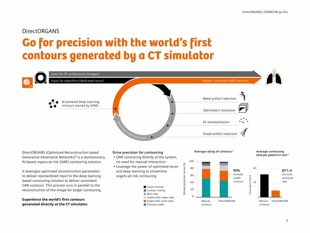
DirectORGANS
Go for precision with the world’s first contours generated by a CT simulator
DirectORGANS (Optimized Reconstruction based Generative Adversarial Networks)8 is a revolutionary, AI-based organs-at-risk (OAR) contouring solution.
It leverages optimized reconstruction parameters to deliver standardized input to the deep learning based contouring solution to deliver consistent OAR contours. This process runs in parallel to the reconstruction of the image for target contouring.
Experience the world’s first contours generated directly at the CT simulator.
Drive precision for contouring• OAR contouring directly at the system,
no need for manual interaction• Leverage the power of optimized recon
and deep learning to streamline organs-at-risk contouring
Average rating of contours11 Average contouring time per patient in min11
100
80
60
40
20
0
20
10
90% clinically usable contours
Ø11.4 min time saving per case
Organ missing Contour missing Must redo Usable after major edits Usable after minor edits Clinicaly usable
Manual contours
Manual contours
DirectORGANS
Blin
ded
phys
icia
n ra
ting
(%)
Tim
e sp
ent (
min
)
DirectORGANS
7

Siemens Healthineers Headquarters Siemens Healthcare GmbH Henkestr. 127 91052 Erlangen, Germany Phone: +49 9131 84-0 siemens.com/healthineers
On account of certain regional limitations of sales rights and service availability, we cannot guarantee that all products included in this brochure are available through the Siemens sales organization worldwide.
Availability and packaging may vary by country and is subject to change without prior notice. Some or all of the features and products described herein may not be available in the United States.
The information in this document contains general technical descriptions of specifications and options as well as standard and optional features that do not always have to be present in individual cases.
Siemens reserves the right to modify the design, packaging, specifications, and options described herein without prior notice. Please contact your local Siemens sales representative for the most current information.
Note: Any technical data contained in this document may vary within defined tolerances. Original images always lose a certain amount of detail when reproduced.
1 New cases - International Agency for Research on Cancer, (IARC) World Health Organization
2 Atun R et al. Expanding global access to radiotherapy. The lancet oncology. 2015; 16(10): 1153-1186
3 Greenwalt J et al. Reducing errors in radiation therapy through electronic safety checklists. Applied Radiation Oncology. 2014: 5–9
4 Jameson MG et al. A review of methods of analysis in contouring studies for radiation oncology. J Med Imaging Radiation Oncol. 2010; 54(5): 401–10
5 Kelly E et al, Reduced patient anxiety as a result of radiation therapist- led psychosocial support: a systematic review, J Med Radiat Sci Sep; 64(3): 220–231
6 Optional7 As shown by measurements with a Gammex 467 Tissue
Characterization Phantom comparing standard reconstruction and DirectDensity reconstruction. Image value to relative electron/mass density conversion for the standard reconstruction was based on a two-linear-equations approach with individual calibration for each tube voltage. For DirectDensity images, a single tube-voltage-independent linear conversion was used. DirectDensity reconstruction is designed for use in Radiation Therapy Planning (RTP) only. DirectDensity reconstruction is not intended to be used for diagnostic imaging
8 Volume rendered image is for illustration purposes only and not part of DirectORGANS.
9 Optional. syngo.via and syngo.via CT Dual Energy DirectSPR is required.
10 Up to 3 additional tablets are optional11 Study results from University Hospital Erlangen, Germany. Published in
Whitepaper DirectORGANS 2.0, Siemens Healthineers, 2021.
Published by Siemens Healthcare GmbH · 10807 0921 online · © Siemens Healthcare GmbH, 2021