some notes on the hawaiian monk seal - scholarspace
TRANSCRIPT

Some Notes on the Hawaiian Monk Seal
JUDITH E. KING1 and R. J. HARRISON2
UP TO 1958 the collections of the British Museum ( N atural History ) possessed nei ther skeletal material nor skin of the Hawaiian or Laysanmonk seal, Monachus schauinslalldi Matschie1905 . Indeed the only remains of this animal inEurope until now have been those brought backby Dr. Schauinsland, amongst which was theskull of the seal later named after him by Matschie (Marschie, 1905) . The stuffed skin ofthis animal is in the Bremen Mus eum and thetype skull is in the Z oological Museum in Berlin , no. 32795 (Wahlert, 1956 ). So it was withgreat pleasure that we received, in the summerof 1958, through the kindness of Mr. Vernon E.Brock, then director of the Territorial Divisionof Fish and Game , Honolulu, H awaii, a youngmale monk seal from Laysan Island that hadbeen shipped complete and frozen to this country. The animal was thawed, inj ected with coloured gelatin in the vascular system, and dissected after fixation, but unfortunately the tissues were too poorly preserved for any accur atehistol ogy.
DESCRIPTION
The seal is young, has a nose-to-rail length of163.5 cm. (5 ft . 6 in .) and weighs 74 .4 kg. (16 4lb. ) complete. Kenyon and Rice (1959 ) give 'the estimated weights of recently weaned pupsas from 95-160 lb . The present animal wascaught June 4, 1958, and as it must have beenvery near to being weaned, its probable date ofbirth must have been ab out April 30. Althoughthe weight is a little high for a recently weanedpup, the animal is in very good condition andis unlikely to have been a yearling, as yearlingsare relatively thin and do not reach the weightand condition of pups that have been feedingfrom their mothers. Similarly the state of theepiphyses and the obvious youth of the skeletonmake it unlikely that it is from a 2-year-oldanimal. The age of the present animal is therefore estimated to be about 5 weeks (the thymusis large and weighs 32.5 g.) . The coat is dark
1 Briti sh Museum (Na tu ral History).2 London Hospital Medical College. Manuscript re
ceived January 15, 1960.
silvery grey dorsally, on the top of the head, onboth sides of the fore and hind flippers, andon the dorsal surface of the tail. Laterally thegrey shades to silvery white ventrally. The hindflippers are a little lighter grey on the innerside towards the ir insertion. There are alsolighter patches round the eyes and sur roundingthe insertion of the supraorbital vibrissae, andalong the upper lip. The lower jaw is light incolour. On the back the hairs are dark greywith a white tip. This white tip becomes longertowards the belly so that the ventral hairs arewhite with a short dark base. The moustachialwhiskers are in five rows on each side of thenose,the upper row having four whiskers andthe other rows approximatel y nine in each. Thewhiskers are dark brown at the base, shading to
straw colour at the tip; they are oval in crosssection and are not beaded as in Phoca (Fig.1) .
The tongue has a notch in its anterior end .The length of the small intestine is approximately 57 ft. The only food remains in thestomach are fish bones and skin and these havebeen identified as being most probably fromthe puffer fish, Arothron m eleagris (Lac.),which is a poor swimmer usually found nearcoral formations . N ematodes, a small cephalopodbeak, and an isop od are also present in the stomach. The nematodes have been identified asContracaecum turgidem, a species previouslydescribed from this seal, and the isopod asLiooneca sp., usually found as an ectoparasiteon fish and probably ingested attached to a fish.In the small intestine there are remains of .atapeworm. It is not in a sufficiently good condition to be identified exactly , but is probablyDiph yllobothrium sp.
The skin and skeleton of this seal are in thecollections of the British Museum (Natural His tory ), the registered number of the skin being58.521 , and that of the skeleton 1958 .11.26.1.
OSTEOLOGY
The skeleton of M. schauimlandi has not previously been described and although it is attempted here to fill this gap the description and
282

Hawaiian Monk Seal-KING and HARRISON 283
FIG. 1. a, Anterior view of the face of M. schauinslandi. b, Lateral view of the face, showing dorsal position of nostrils.
284 PACIFIC SCIENCE, Vol. XV, April 1961
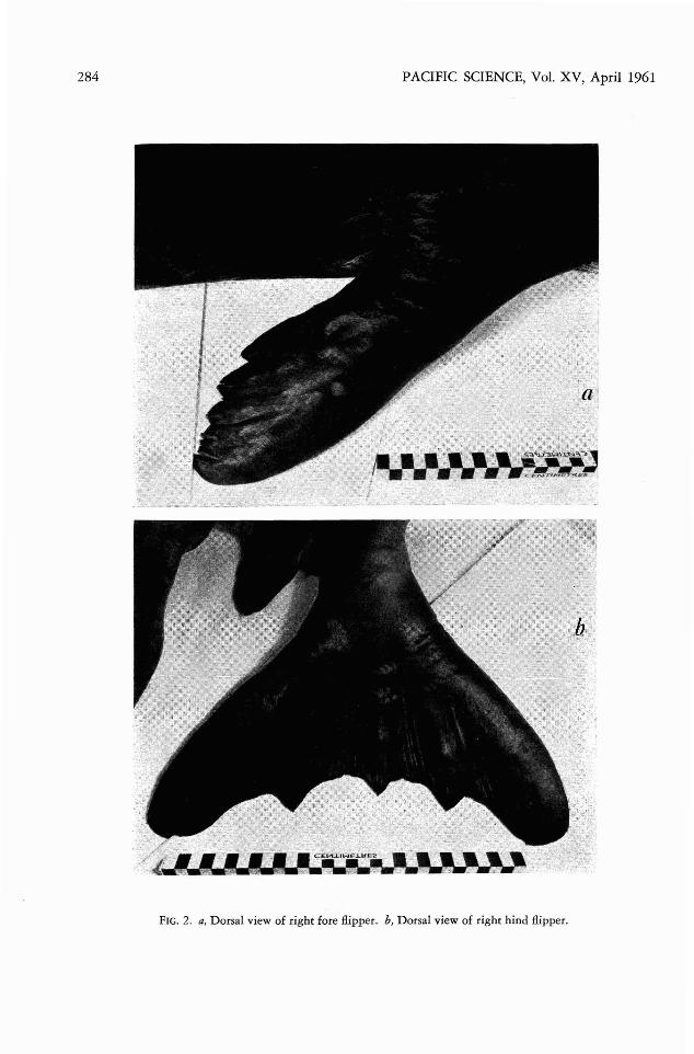
FIG . 2. a, Dorsal view of right fore /lipper. b, Dorsal view of right hind /lipp er.

Hawaiian Monk Seal-KING and HARRISON 285
M easurem ents of Skull of M . sch a u in slandi1958.11.26.1
6. It was noted previously that the posterioredge of the palate of M . schauin'slandi formeda wide V. It is now felt that it is better describ edas U in shape and rather more like that of M .monachus, though there is probably a certai namount of variation in this character.
7. The pterygoid bones curve outwards as inM . tro picalis and in this young skull of M .schauinslandi they are just visible when it isviewed dorsally.
8. Th e coronoid process of the lower jaw IS
narrow and like that of M . tropicalis.9. Examinati on and comparison of the young
skull of M. schauinslandi now available confirms the previous report that it is more likeM. tropicalis than M. m onachus ( Fig. 3) . Thereare no supern umerary bones in the skull.
Kenyon and Rice ( 1959) note that, in thefew skulls of M. schauinslandi they examined,the shape of the palate and the shape of thezygomati c branch of the squamosal do not appear to be constant and are thus not good distinguishing characters. It is certainly agreedthat the shape of the palate is variable and thereis a need for the examination of as many adultskulls as possible to determi ne the amount ofvariation.
comparison are considerably limited by theyouth of the specimen. Th e form of this descrip tion is based on that of the skeletons of M .m onachus and M. tro picalis (King, 1956) andfrequent reference will be made to the detailsin that paper. N o skeleton of a young M . tropicalis was available so the M . schauinslandi skeleton was compared with that of an adult M . tropicalis (1887.8.5.1.) and of a young M . m onachus(1894.7.27.3 ), the skull of which showed by itscondylobasal length (22 4 mm .) and suture age( 13) that it was from an animal of approximately the same size as the Laysan monk seal.
Comparison of M. sc ha u i n sla n di Skull withT hose of M. monachus and M. tropicalis
1. The additional measureme nts now available of the skull of M . schauinslandi confirm theprevious statement ( King, 1956) that skullsof both M . schauinslandi and M . tropicalis areslightly narrower in pr oporti on to their widththan those of M . monachus, though the accuracyof this conclusion is limited by the small num ber of skulls available.
M. M . trap i - M. schau-mo nachus calis insland«
% % %Zygomatic wid th... 59.9-70.3 61.7-62.1 60.9-61.5Snout width at
canines....... ....... 20.9-26.0 20.6-20.9 20.3-20.5Width at externa l
auditory meatus 53.1-5 8.5 49.8- 50.5 50.5- 55.0Width at petrous
bones.... __ ....... ..._ 60.3-64.9 56.3- 59.2 59.2- 64.1
2. The nasal bones of this young animal areas previously described from the type skull. Theanterior ends are in the form of an inverted Vwith the point directed posteriorly; the posteriorends do not taper as much as in M . tropicalis.
3. Th e lower edge of the infraorb ital foramenis, when seen from the front , narrower than theupper edge. This confirms Marschie ( 1905) andis similar to M. tropic-dis.
4. The absence of a maxillary tubercle at theanterior edge of the orbi t also confirms Matschie.
5. As previously noted, the shape of the zygomatic arch is more like that of M . monachusthan M . tropicalis .
mm.
Condylobasal length 220Condylobasilar length 211Basal length 201Basilar length.................................. 192Snout width at canines.................... 45Width of skull at front end of
last upper molars........................ 59Zygomatic width 134Width at upp er edge auditory
meatus 121Width at perrous bones
(mastoid width) 141Palatal length 101Palatilar length................................ 92Width of occipital condyles........ .. 62Length of nasal sutu re.. .................. 50Length of upper molar row.. .......... 55Suture age 13
%
10095.991.487.3 .20.5
26.860.9
55.0
64.145.941.828.222.725.0

286 PACIFIC SCIENCE, Vol. XV, April 1961
FIG . 3. a, Dorsal, b, ventral, and, c, lateral views of the skull of M. schauinslandi. d, Lateral view of lowerjaw.

H awaiian Monk Seal-KING and HARRISON 287
Th e height of the crown of the postcanineteeth in both M. tropicalis and M. schauimlandiis lower than in M. monachus and the maincusp is more rounded, In th is respect M. schauinslandi is more like M. tropicalis, but in thepossession of a single anterior and posteriorcusp it resembles M. monachus. The fourth postcanine has, however, two small anterior cusps
M, tropicalis also has small canines, Themeasurements given below of the upper caninesof an adult male M. tropicalis ( 1889,11.5.1;condylobasal leng th 267 rnrn . suture age 25) arecompared with those of an adult male M, mo nachus of approximately similar size ( 1863.4. 1.1;condylobasal length 273 mm., suture age 26 ) .
M. mona- M , trop -chus icalis
The term "pos t canines" is used here insteadof "molars" and "premolars," as these lattercannot be used pr ecisely when referr ing to pinniped teeth,
UPPER J AW TEETH: As previously menti oned,the upper incisors are set in a straight lineacross the anterior end of the pr emaxillae andin general the setting and shape of the teeth arelike M , tropicalis. The incisors of thi s younganimaldo not have such a pron oun ced "waist"as those of the adult M, tropicalis, but it is moreevident than in the young M , monacbus.
The canines are very small for a male animal.Those of the young M, monacbus, also a male,whose sku ll is only 4 mrn . longer than that ofthe young M, schauinslandi , are much larger.
M, m ona- M , schau-cbus inslandi
l l mrn.est. 17 mm.
(v . worn)
Sk eleton
and two small posterior cusps. The surface ofthe teeth is more rugose than in M . m onachusand the anterior and posterior cusps are verymu ch less distinct, as though it is in process oflosing the second posterior cusps of M . tropicalis. The postcanine teeth are not set obliquely.
LOWER JAW TEETH: The lower incisors aresimilar to those of M. tropi calis, the caninesare small , and the postcan ines are similar tothose in the upper jaw.
N o disease or irregularity in number is present in the teeth of either upper or lower jaws.
SCAPULA: The scapula of M. schauimlandi is.very like that of M . tropicalis. It is similar inthe way that the ante rior edge is directed almosthorizontally forwards from the neck beforesweepi ng round to the dorsal surface. In M.mo nachus the anterior edge is directed forwardsand upwards at an angle of approximately 450
•
The spi ne in the young Laysan seal is represented by a low ridge wi th a well-developedacromion process ( Fig. 4b) .
HUMERUS, RADIUS, ULNA: N o real comparison can be made because of th e youth of M.schauim landi, though the humerus appears tobe sligh tly more robust than that of M,monacbus of similar size.
MANUS: Except that the term inal phalangesare not so extensively grooved for claws as inthe young At monachus, although the clawsthemselves are of approxima tely similar size, themanus is not noticeably different in the thr eespecies (Fig. 4c ) .
PELVIS: The pelvis is very similar in shapeto that of the young M. monacbus although itis more mature as there is no sign of the symphysis between ischium and pubis, and theacetabul um is deep and well formed, while inthe young M. monachus the symp hysis is abouthalf fused and the acetabulum is shallow andmore obviously immature. The shape of theischium and pubis is like that of M. monachusexcep t that the pubis is possibly slightly moreslender. There is no sign of the stout pubis andvery nar row ischium posterior to the ischiaticspine as in M. tropicalis. The ilium is slightlynarrower than in M. m onachus. There is a largeforamen for the obturator nerve just posterior
14mm,
12mm,
D ' I f I i 2, c 1, pc 5,enta orm u a - - _2 1 5
Anterop osterior length atcrow n-roo t juncti on .c.x.c. 15 rnm.
Ante rior height ofcanrne 23 mrn.
(in straight line fromcrown- root junction to tip)
Anterop osteriorlength ....__ 16.5 mm.
Ante rio r heigh t . 27 mm .
T eeth

288 PACIFIC SCIENCE, Vol. XV, April 1961
FIG. 4. a, Right ilium . b, Right scapula. c, Right manus. d, Right pes.

Hawaiian Monk Seal-KING and HARRISON
to the cotyloid notch that is not present inM. monachus or M. tropicalis (F ig. 4a) .
FEMUR, TIBIA, FIBULA: These bones are notsufficiently well formed to be used for anycomparison.
PES : The previous description of the pes ofM . m onachus appl ies also to the pes of thepresent animal. Th ere is practically no indication of the insertio n of the small claws on theterm inal phalanges (Fi g. 4d) .
VERTEBRAL COLUMN: Vertebral formu la:cervical 7, thoracic 15, lumbar 5, sacral 3, caudal13.
Cervical vertebrae. These are more like M.tropicalis than M. monachus in that the transverse processes of vertebrae 4-6 are divided intotwo branches, though the division is less pr onounced than in the adult M. tropicalis. Thereis "a general similarity to M. monachus, thoughthis is probably more because of the simila rityin age. T he neural arches have completely fused.
T horacic vertebrae. These are similar to thoseof the young M. monachus except that the neuralarches are narrower and do not lean so farposteriorly.
Lum bar and caudal vertebrae. These are aspreviously described (Ki ng, 1956).
RIBS: The articulation of the ribs is similarto that in the other monk seals. In order to inject the vascular system and remove the soft partsthe cartil aginous parts of the ribs were cut away.
ABDOMINAL VEINS
The abdominal veins are thin and easily distensible. The posterior vena cava is duplic atedas in Phoca but displays a complicated arrangement of large anastomotic channels ( Fig. 5) nothitherto described in other Pinnipedia. Theright limb of the posterior vena cava is thelarger, is almost straight, and lies a little to therigh t of the midlin e. The smaller left limb extends from the pelvis on the left , passing somewhat obliquely cranialwards to the right to jointhe right limb near the upper pole of the leftkidn ey. A large anastomotic channel passes fromthe right limb at the level of the lower pole ofthe right kidn ey obliquely across the midlineto join the left limb at the level of the middl eof the left kidney. A smaller channel arises fromthe right cranial end of the anastomosis just
289
described, passes dorsal to the right limb , andenters it on the right at the level of the upperpole of the right kidney. Each limb and the twoanastomoti c channels receive numerous tributaries draining the renal stellate plexus as wellas many vessels from the lumbar and pelvicvenous plexuses. The righ t limb of the posteriorvena cava is 2.5 em. in diameter where it isformed by union of the iliac, lumba r, and mostcaudal renal tributaries. It is 3.0 em. in diameterwhere it is joined by the left limb. The commontrunk is 3.0 em. in diame ter throughout the8.0 em. of its extent to the point where it isenclosed by hepatic tissue.
The common trunk of the posterior vena cavais enlarged considerably where it enters the substance of one lobe of the multilobed liver. It hasthe form of a dilated tub e, 15 em. in length and6 em. in diameter in its cranial porti on, lyingon the ventral surface of the liver and surrounded on thr ee sides by a narrow strip ofhepatic tissue. Several large orifices of hepaticveins are present on the lateral walls of th isdilated part of the posterior vena cava. Cran iallythis part of the posterior vena cava enters ahepatic sinus, nearly spherical in shape and approximately 10 em. in diame ter. The sinus ispar tially surrounded by hepatic tissue, but inregions only a thin translucent wall covered byperitoneum separates it from the diaphragm.The sinus is divided by two narrow septa arisingfrom its right wall. Six large hepatic veins openinto the sinus. The capacity of the sinus isestimated to be 450 cc. Figure 6 shows the appearances of the dilated vena cava and the hepat ic sinus.
The intrathoracic part of the posterior venacava is 5 em. in length and 3.5 em. in diameter.No pericardial plexuses of vessels, such as arefound in Phoca, were present and no veinsdrained into this part of the vena cava. An incompl ete sphincter of striated muscle encirclesthe vena cava just cranial to the diaphragm. Thedorsal part of the sph incter was 3.5 em. highand was closely adherent to the vena cava; it wasseparated from the diaphragmatic muscle by anarrow strip of connective tissue. The fibres ofthe sphincter only partially encircle the venacava so that on its ventr al aspect the sphincteris narrowed to a bundle of closely packed fibres

290 PACIFIC SCIENCE, Vol. XV , April 1961
FIG. 5. A drawing from the ventral aspect to show the duplicated posterior vena cava, its anastomoticbranches, and the stellate renal plexus on the surface of the multilobular kidney.
0.5 em. high. The sphincter is shaped like asignet ring. It is supplied by branches of theright phrenic nerve.
KIDNEYS
The right kidney lies 2 em. more cranial thanthe left. The right is 18 em. long, 8 em. broad,and 3.5 em. thick; the left is 16 em. long, 7 em.broad, and 3.5 em. thick. Each kidney is composed of large numbers of renules, each about2.0 em. in diameter. The papilla of each renuleprojects into a single calyx with a ductule thatunites with others to drain eventually into thepelvis of the ureter as in Phoca (Harrison andTomlinson, 1956 ) .
Each kidney is surrounded by a stellate venousplexus, the communicating vessels of which liein the sulci between the renules on the surfaceof the kidney. The plexus is more marked on theventral aspect of the organ. There are numerousanastomoses with lumbar and lateral wall veins,with the inrraverrebral vein, and with the azygosvein. The plexus is drained mainly by a seriesof tortuous tributaries that extend round theupper and lower poles and transversely acrossthe ventral and dorsal surfaces of the body ofthe kidney. These tributaries unite at the medialborder of each kidney to form three or four shorrtrunks that enter the two main limbs of theposterior vena cava or its large a n as t om o t i c

Hawaiian Monk Seal-KING and HARRISON
channels (Fig. 5). The major portion of venousblood is conveyed by interlobular veins reaching the surface between renules to enter thestellate plexus . Some interlobular veins, however, pass towards the hilum of the kidney to
drain into small channels that extend mediallyto the limbs of the posterior vena cava.
HEART AND GREAT VESSELS
The aorta is markedly constricted at a pointimmediately to the left of the origin of the leftsubclavian artery and below the ductus arteriosus. This condition is known as coarctation(coarctus = pressed together) and is the result
FIG. 6. A drawing from the right side of a sagittalsection through the dilated intrahepatic part of theposterior vena cava, the hepatic sinus, and the sphincter about the intrathoracic part of the vena cava. Thehepatic sinus is divided by thin, falciform septa; themain openings of the hepatic veins ate shown as blackcircular areas.
291
of partial obliteration of the dorsal aorta eitherbetween the 4th and 6th arch (above the ductusarteriosus) or below the 6th arch and the dorsalaorta (below the ductus arteriosus). It occursrarely in man: Wood (1956) found coarctationof the aorta in 9' out of 900 cases of congenitalheart disease. It appears to be very rare indeedin mammals and has not been reported in anyanimals dying at the Zoological Gardens, Regent's Park, London (R. W. Fiennes, personalcommunication ). Cordy and Ribelin (1950)describe its occurrence in a bullock associatedwith dextraposition of the heart. It occurs, inman, more frequently in males (4.5: 1 ) , is mostoften found in young adults and 1 per cent ofthe cases have hereditary links (Wood, 1956) .
The transverse diameter of the monk sealaorta at the point of coarctation is 1.0 cm., thatof the first part of the descending aorta is 1.4cm. There does not appear, therefore, to be anypost-stenotic dilatation of the aorta as is oftenfound in man. The ascending aorta and its archare dilated with marked thickening of the wall.The most dilated part is 4.5 em, in diameter;the thickened wall is 3.0 mm . thick as opposedto the wall of the descending aorta, which is1.0 mm. thick. At the point of coarctation thewall of the aorta is thickened by fibrous tissueto 4.0 mm . cranially and to 3.0 mm . caudally;the other parts of the aortic wall are less thick.Three aortic valves are present (only two arepresent in 23 per cent of human subjects withcoarctation, Hamilton and Abbott, 1928). Theaortic ring appears of normal size (aortic stenosis is present in 7.5 per cent of cases in man).
The left ventricular musculature appears hypertrophied, but otherwise the heart is normal.There is no patent foramen ovale and the ductusarteriosus is closed (7 per cent of human casesshow a patent ductus) . The right and left atriaappear to be of normal size and have walls thatdo not look hypertrophied. There is no evidenceof enlargement of vessels that provide collateralcirculations above and below the constriction(internal mammary arteries). No notching ofthe posterior margins of the ribs (which canbe caused by raised blood pressure in the intercostal vessels) is present. The lack of any suchfindings could well be due to the immaturity ofthe animal.

292
OTHER VEINS
A large azygos vein is present just to the rightof the midline; it is 1.25 cm. in diameter andterminates in the anterior vena cava. No left sided azygos vein is present.
An extradural inrravertebral vein is present;at the level of the 12th thoracic vertebra it liesto the left of the cauda equina. At this level itmeasures 1.75 cm. by 1.25 cm. It is somewhatlarger at the level of the 3rd thoracic vertebra.The poor preservation of the specimen prevented detailed examination of the connexionsof the vein . It definitely has connexions with theazygos vein and with the lumbar veins relatedto the stellate renal veins at the lower poles ofthe kidneys as in Phoca. These are clearly shownafter the venous system had been injected withcoloured gelatin.
LUNGS
Superficial examination ( no casts were made)suggests that the arrangement of the bronchialtree is symmetrical as has been described in otherseals (Brown, 1958). Histological examinationreveals the presence of numerous featuresdescribed by Pizey (1954) in the lung of Phoca.The cartilages of the tertiary bronchi are continued far to the periphery of the lung , andbronchial mucous glands are frequent. The lungis divided into numerous lobules by well-markedsepta of loose connective tissue. The bronchiolespossess a series of myo-elastic valves which aresimilar to those of Phoca but not as marked asthose in T ursiops (W islocki, 1929).
TESTIS
Each testis weighs 1.9 g. and measures 25 X15 X 10 mm. in its greatest diameters. The seminiferous tubules are inactive, immature, devoidof lumina, and average 50/ p. in cross-sectioneddiameter. The interstitial tissue is relativelyabundant. Some of its cells are large, polyhedral,and heavily vacuolated , but the majority aresmall, fusiform , and have densely stained nuclei.The connective tissue between the seminiferoustubules is loosely arranged and oedematous.These appearances suggest that the gonad maywell have been precociously enlarged at birthwith hypertrophy of the interstitial tissue as hasbeen described in Halichoerus , Phoca, and Mirounga (see Harrison, 1960, for references ) .
PAOFIC SCIENCE, Vol. XV, April 1961
DISCUSSION
It is difficult to come to any definite conclusions from the examination of this skeleton ofM onachus schauimlandi, not only because of thelack of comparative material but also becauseof the extreme youth of the anim al. It is obviousthat it is more closely related to M. tropicalisthan to M. m onacbus bur this is to be expectedbecause of its geographical position. Kenyonand Rice ( 1959 ) suggest that further study mayindicate a closer relationship between the Laysan and the West Indian monk seals and thatM. schauimlandi may possibly. be a race of M.tropicalis.
The Laysan monk seal possesses certain vascular (venous) modifications seen in other Pinnipedia. These are a duplicated posterior venacava, a stellate renal plexus , a caval sphincter,a hepatic sinus, and an extradural intravertebralvein (Ha rrison and Tomlinson, 1956 ). Thereare, however, certain differences. The pattern ofduplication of the posterior vena cava is morecomplicated than in any seal so far described .There is evidence of persistence of severalanastomotic channels between the two limbs ofthe posterior vena cava. This could be interpreted as persistence of an embryonic state inwhich primitive anastomoses have become enlarged rather than suppressed. The hepatic sinusis not as large as in Phoca or Mirounga, whereasthe curious dilatation of the intrahepatic part ofthe vena cava is undescribed in seals. The sphincter is not as large or as complete as in otherPinnipedia. It could be argued that the monkseal shows less vascular (venous) specializationthan Pbo ca, Mirounga, Leptonycbotes, Lobodon, Halicho erus, and Hydrurga, but more thanZalophus. This could mean that monk seals arenot able to dive for so long a period or as deepas these forms. No observations have been madeon the diving abilities of the Laysan monk seal,but Kenyon and Rice ( 1959) note that theseseals occur regularly only on islands having extensive areas of shallow shoal water and thatthey appear to feed primarily on bottom-livingfishes that they could obtain only in shallowwater . They do travel over deep water, thoughnot necessarily at any great depth. It must , however, be emphasized that the specimen describedhere had coarctation of the aorta. Weare not

Hawaiian Monk Seal-KING and HARRISON
certain that its venous pattern is that prevailing in all Laysan monk seals, and it is frustratingthat the first whole specimen available for examination should be congenitally abnormal.
It is hoped that continuing interest in thisrare and relatively unknown seal will, in duecourse, result in the acquisition of more specimens for study. In the meantime it has been agreat pleasure to see and work on the presentanimal and we are much indebted to Mr. VernonE. Brock and his fellow workers for their kindness. We are also grateful to Dr. J. D. W. Tomlinson for injecting the specimen and for makingsome initial observations.
SUMMARY
A young male monk seal Monachus schauinslandi of an estimated age of 5 weeks has beenreceived by the British Museum (Natural History ). A brief description is given of the external features, stomach contents, and parasites.The skull and skeleton are described and a general similarity to that of M. tropicalis is noted.Certain modifications in the venous system aredescribed; coarctation of the aorta is present.
REFERENCES
ABBOTf, M. E. 1928. Coarctation of the adulttype. Amer. Heart J. 3: 574-618.
BROWN, D. 1958. The bronchial tree in aquaticmammals. J. Anat. Lond. (Proc.) 92: 656.
CORDY, D . R., and W. E. RIBELIN. 1950. Sixcongenital cardiac anomalies in animals. Cornell Vet. 40: 249'-256.
293
HAMILTON, W . F., and M. E. ABBOTf. 1928.Coarctation of adult type. Amer . Heart J. 3:381-492 .
HARRISON, R. J., and J. D . W. TOMLINSON.1956. Observations on the venous systems incertain Pinnipedia and Cetacea. Proc. Zool.Soc. Lond. 126: 205-233.
HARRISON, R. J. 1960. Reproduction and reproductive organs in common seals (Phocavituina ) in the Wash, East Anglia. Mammalia (in press) .
KENYON, K. W ., and D. W . RICE. 1959. Lifehistory of the Hawaiian monk seal. Pacif . Sci.13(3) : 215-252.
KING, J. E. 1956. The monk seals ( GenusMonachus). Bull. Brit. Mus. (Nat. Hist.)Zool. 3 (5) : 203-256.
MATSCHIE, P. 1905. Eine Robbe von Laysan.S. B. Ges. Naturf. Fr. Bed. 1905: 254-262.
PIZEY, N. C. D. 1954. The structure of the pinniped lung. J. Anat. Lond. (Proc.) 88: 552.
SVIHLA, A. 1959. Notes on the Hawaiian monkseal. J. Mamm. 40: 226-229.
WAHLERT, G. VON. 1956. Die Typen und Typoide Des Dberseemuseums Bremen, 4. DieLaysan-Robbe, Monachus schauinslandi Matschie 1905.
--- [?}. Veroff Uberseemuseum Bremen,A.2. Heft 6: 365-366.
WISLOCKI, G. B. 1929. On the structure of thelungs of the porpoise (Tursiops truncatus).Amer. J. Anat. 44: 47-77. .
WOOD, P. 1956. Diseases of the Heart and Circulation. Eyre and Spottiswoode, London.