sonographic evaluation of the axillary artery during...
TRANSCRIPT

Sonographic evaluation of the axillary artery during simulated overhead throwing
positionStapleton, C, Herrington, LC and George, K
http://dx.doi.org/10.1016/j.ptsp.2008.06.009
Title Sonographic evaluation of the axillary artery during simulated overhead throwing position
Authors Stapleton, C, Herrington, LC and George, K
Type Article
URL This version is available at: http://usir.salford.ac.uk/2306/
Published Date 2008
USIR is a digital collection of the research output of the University of Salford. Where copyright permits, full text material held in the repository is made freely available online and can be read, downloaded and copied for noncommercial private study or research purposes. Please check the manuscript for any further copyright restrictions.
For more information, including our policy and submission procedure, pleasecontact the Repository Team at: [email protected].

ARTICLE IN PRESS
1466-853X/$ - s
doi:10.1016/j.pt
�Correspondfax: +44 151 23
E-mail addr
Physical Therapy in Sport 9 (2008) 126–135
www.elsevier.com/ptsp
Original research
Sonographic evaluation of the axillary artery during simulatedoverhead throwing arm positions
Claire Stapletona,�, Lee Herringtonb, Keith Georgea
aResearch Institute for Sport and Exercise Sciences, Liverpool John Moores University, Liverpool, UKbCentre for Rehabilitation and Human Performance Research, University of Salford, Salford, UK
Received 19 February 2008; received in revised form 5 June 2008; accepted 12 June 2008
Abstract
Objectives: The aim of this study was to determine changes in axillary artery diameter and peak systolic velocity in asymptomatic
individuals during upper limb positioning commonly used to assess vascular pathology in athletes.
Design: Repeated measures observational study.
Setting: Physiology laboratory.
Participants: Subjective and objective screening excluded individuals with past, or present, conditions related to neurovascular
compression syndromes. Thirty-one subjects (21 females, 10 males; mean age: 2574 years) were included in the final analysis.
Main outcome measures: Sonographically determined axillary artery diameter and peak systolic velocity, as well as symptom
production, were recorded for a series of 12 randomised arm positions, incorporating varying degrees of abduction, external
rotation, and horizontal flexion/extension.
Results: The majority of arm positions revealed no change in artery diameter and peak systolic velocity. However, at the extreme of
abduction, and arm positions incorporating 1201 abduction, significant (po0.0005) reductions in axillary artery diameter were
noted. All mean results masked wide heterogeneity: 13% demonstrating a greater than 50% reduction in diameter, 10%, a doubling
of peak systolic velocity, and 42%, reporting symptoms.
Conclusions: The number of individual clinically ‘‘positive’’ responses questions the specificity of individual diagnostic tests, such as
the hyperabduction manoeuvre, and highlights the need to interpret test results in conjunction with the subjective assessment and
other physical findings from the objective assessment.
r 2008 Elsevier Ltd. All rights reserved.
Keywords: Ultrasonography; Diagnosis; Physical examination; Axillary artery
1. Introduction
The majority of vascular compression syndromesaffecting the upper limb come under the ‘umbrella term’of thoracic outlet syndrome with most published reportsfocusing on the more commonly compressed subclavianartery, vein or surrounding neural structures (Demondion,Herbinet, Gautier, Duquesnoy, & Cotton, 2006; Gillard
ee front matter r 2008 Elsevier Ltd. All rights reserved.
sp.2008.06.009
ing author. Tel.: +44 151 231 4323;
1 4353.
ess: [email protected] (C. Stapleton).
et al., 2001). Descriptions of the syndrome inconsistentlyinclude compression of the first and second portions of theaxillary artery in the costoclavicular and retro-pectoralisminor space, respectively. Less often mentioned is thethird portion of the axillary artery that lies anterior andinferior to the head of the humerus. This investigationfocuses on this latter portion, and aims to raise awarenessof its potential for injury and highlight the dilemmas ofdiagnosis.
Compressive injury of the axillary artery mostcommonly occurs in athletes performing repetitiveoverhead arm motion (Jackson, 2003). Published reports
ARTICLE IN PRESSC. Stapleton et al. / Physical Therapy in Sport 9 (2008) 126–135 127
of such injury include baseball pitchers, handball,tennis, and volleyball players (Arko, Harris, Zarins, &Olcott, 2001; Fields, Lemak, & Benmenachem, 1986;Ishitobi et al., 2001; Rohrer, Cardullo, Pappas, Phillips,& Wheeler, 1990; Todd, Benvenisty, Hershon, &Bigliani, 1998; Vlychou, Spanomichos, Chatziioannou,Georganas, & Zavras, 2001). The proposed mechanismsfor compressive trauma of the second and third portionsof the axillary artery are a tight or hypertrophiedpectoralis minor muscle (Dijkstra & Westra, 1978;Finkelstein & Johnston, 1993) and anterior translationof the humeral head (Dijkstra & Westra, 1978; Durham,Yao, Pearce, Nuber, & McCarthy, 1995; Vlychou et al.,2001) combined with repetitive overhead activity.
The hyperaduction manoeuvre (Wright, 1945) is usedin conjunction with the clinical presentation andsubjective history to aid the diagnosis of compressiveinjury of the axillary artery (Demondion et al., 2006). Itis thought to compressively stress the vessels in thesubcoracoid region, especially the second portion of theaxillary artery under the pectoralis minor muscle (Baker& Liu, 1993). Clinically, it is postulated that thismanoeuvre stresses the vasculature to accurately repro-duce the signs (e.g., radial pulse disappearance) andsymptoms (e.g., ischaemic pain, paraesthesia, anaesthe-sia, heaviness) of vascular compromise. Published casestudies have used advanced imaging techniques(i.e., arteriograms, angiograms, ultrasound) to confirmsuspected vascular compromise of the axillary arteryand its branches, with patency of the arteries in a neutralupper limb position compared to a stenosed or occludedvessel in the hyperabducted position (Arko et al., 2001;Cormier, Matalon, & Wolin, 1988; Fields et al., 1986;Ishitobi et al., 2001; Kee et al., 1995; Nijhuis & Muller-Wiefel, 1991; Redler, Ruland, & McCue, 1986; Reekers,den Hartog, Kuyper, Kromhout, & Peeters, 1993;Rohrer et al., 1990; Schneider, Kasparyan, Altchek,Fantini, & Weiland, 1999; Todd et al., 1998; Vlychouet al., 2001). Imaging results influence the managementof the condition which, in many cases, results in surgery.It is, therefore, important for clinicians to be aware thatvessel occlusion has been demonstrated not only insymptomatic but in asymptomatic subjects as well. Forexample, Mochizuki et al. (1994) reported posteriorhumeral circumflex artery occlusion in the hyperab-ducted position in 80% of asymptomatic subjects usingmagnetic resonance angiography. Rohrer et al. (1990)reported axillary artery compression in 83% of the 92arms tested (baseball pitchers, athletes and non-athletes)but results were limited by incomplete blood flowassessments and poorly defined testing positions andsubject groups. The hyperabduction manoeuvre isconfused by varying descriptions in the literature;consequently, results are incomparable. Further qualityinvestigations are still required to aid clinical diagnosisand interpretation.
Uniquely, the present study seeks to extend theknowledge of the diagnosis of compressive injury of,specifically, the third portion of the axillary artery bydescribing a number of arm positions rather than aspecific single test. Therefore, the aims of this study areto: establish the arterial diameter (D) and peak systolicvelocity (PSV) at the third portion of the axillary artery,record symptom production at various arm positions,and thus to identify the rate of responses that would,clinically, be classified as positive according toStrandness’s (2002) clinical criteria.
2. Methods
2.1. Subjects
Participants were 41 healthy individuals (age range:18–40 years) recruited via local advertising in theUniversity. Following verbal and written briefing,subjects provided written consent to participate.Local ethical approval was granted by LiverpoolJohn Moores University Ethics Committee. Subjectiveand objective screening was performed by a charteredphysiotherapist to ensure conditions or injuriesthought to contribute to, or predispose, a neurovascularcompression syndrome and/or conditions likely to beexacerbated by the experimental procedure, wereexcluded. Specifically, subjects were excluded if pastmedical history included: previous shouldersurgery, glenohumeral joint dislocations and/or sub-luxations, fractures to the humerus, clavicle and 1st–3rdribs, inflammatory joint conditions, vascular orneurological disorders, or past or present injury to theneck, shoulder, and upper limb. Objective screeningconsisted of active range of motion with overpressurefor cervical, thoracic, scapulo-thoracic, and glenohum-eral range of movements. Two subjects were excludedthrough screening and a further eight were excluded dueto poor clarity of the ultrasound image. The finalanalysis included 21 female and 10 male subjects with acohort age range of 19–35 years (mean7S.D.: 2574years). Subjects were regarded as ‘‘non-overhead throw-ing active individuals’’ based on questions related toparticipation levels in general activity and specificoverhead activities such as, racquet sports, swimming,and rock climbing. All subjects reported right handdominance.
2.2. Subject position and standardisation
With the subject seated and the right arm supportedon a purpose built adaptable arm-rest, anatomicallandmarks were identified to aid consistent reproductionof abduction. At approximately 451 abduction, the axisof a goniometer was placed over the posterior corner of
ARTICLE IN PRESSC. Stapleton et al. / Physical Therapy in Sport 9 (2008) 126–135128
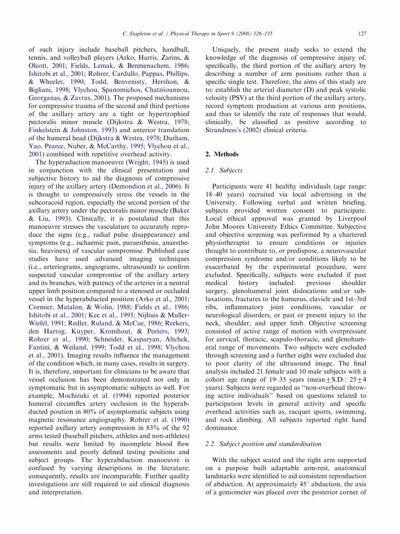
the acromion with the static arm parallel to the spineand the moving arm placed over a line joining the axisand the mid-point between medial and lateral epicon-dyles. For each subject, the height of the arm-rest wasadjusted and marked to indicate 901 abduction and 1201abduction. To determine 1801 abduction, the upper limbwas secured to the back board of the apparatus with thehumerus as close to the ear as possible. Throughouttesting, blocks were secured to the arm-rest to maintainthe desired degree of shoulder girdle horizontal exten-sion/flexion and glenohumeral external rotation. Tocontrol for subclavian artery compression at the scalenetriangle subjects were instructed to maintain an uprightposture with a neutral head position. A large belt wassecured around the subjects’ thorax and the apparatus,and a visual marker at eye level was provided to aidmaintenance of these positions (see Fig. 1). Subjectswere assessed in 12 arm positions (see Table 1) presented
Fig. 1. Subject positioning for baseline position (a) and position 12 (b). Posi
Table 1
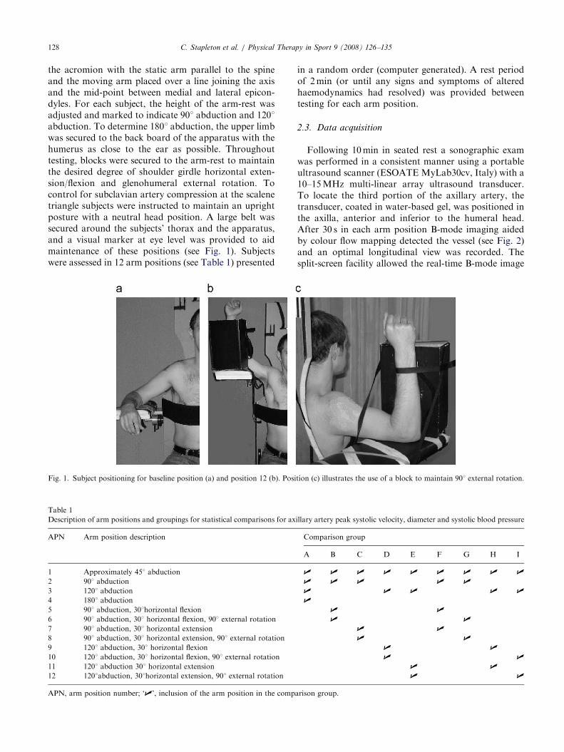
Description of arm positions and groupings for statistical comparisons for ax
APN Arm position description
1 Approximately 451 abduction
2 901 abduction
3 1201 abduction
4 1801 abduction
5 901 abduction, 301horizontal flexion
6 901 abduction, 301 horizontal flexion, 901 external rotation
7 901 abduction, 301 horizontal extension
8 901 abduction, 301 horizontal extension, 901 external rotation
9 1201 abduction, 301 horizontal flexion
10 1201 abduction, 301 horizontal flexion, 901 external rotation
11 1201 abduction 301 horizontal extension
12 1201abduction, 301horizontal extension, 901 external rotation
APN, arm position number; ‘|’, inclusion of the arm position in the comp
in a random order (computer generated). A rest periodof 2min (or until any signs and symptoms of alteredhaemodynamics had resolved) was provided betweentesting for each arm position.
2.3. Data acquisition
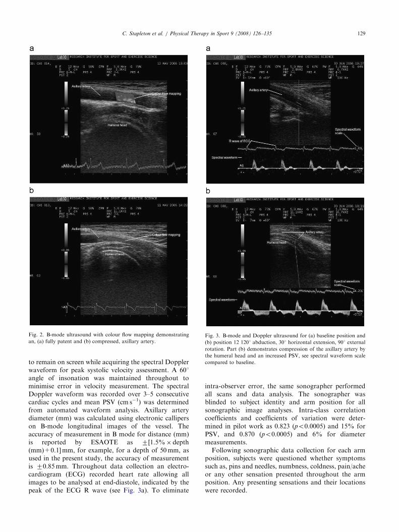
Following 10min in seated rest a sonographic examwas performed in a consistent manner using a portableultrasound scanner (ESOATE MyLab30cv, Italy) with a10–15MHz multi-linear array ultrasound transducer.To locate the third portion of the axillary artery, thetransducer, coated in water-based gel, was positioned inthe axilla, anterior and inferior to the humeral head.After 30 s in each arm position B-mode imaging aidedby colour flow mapping detected the vessel (see Fig. 2)and an optimal longitudinal view was recorded. Thesplit-screen facility allowed the real-time B-mode image
tion (c) illustrates the use of a block to maintain 901 external rotation.
illary artery peak systolic velocity, diameter and systolic blood pressure
Comparison group
A B C D E F G H I
| | | | | | | | || | | | || | | | ||
| || |
| || |
| || |
| || |
arison group.
ARTICLE IN PRESS
Fig. 2. B-mode ultrasound with colour flow mapping demonstrating
an, (a) fully patent and (b) compressed, axillary artery.Fig. 3. B-mode and Doppler ultrasound for (a) baseline position and
(b) position 12 1201 abduction, 301 horizontal extension, 901 external
rotation. Part (b) demonstrates compression of the axillary artery by
the humeral head and an increased PSV, see spectral waveform scale
compared to baseline.
C. Stapleton et al. / Physical Therapy in Sport 9 (2008) 126–135 129
to remain on screen while acquiring the spectral Dopplerwaveform for peak systolic velocity assessment. A 601angle of insonation was maintained throughout tominimise error in velocity measurement. The spectralDoppler waveform was recorded over 3–5 consecutivecardiac cycles and mean PSV (cm s�1) was determinedfrom automated waveform analysis. Axillary arterydiameter (mm) was calculated using electronic calliperson B-mode longitudinal images of the vessel. Theaccuracy of measurement in B mode for distance (mm)is reported by ESAOTE as 7[1.5%� depth(mm)+0.1]mm, for example, for a depth of 50mm, asused in the present study, the accuracy of measurementis 70.85mm. Throughout data collection an electro-cardiogram (ECG) recorded heart rate allowing allimages to be analysed at end-diastole, indicated by thepeak of the ECG R wave (see Fig. 3a). To eliminate
intra-observer error, the same sonographer performedall scans and data analysis. The sonographer wasblinded to subject identity and arm position for allsonographic image analyses. Intra-class correlationcoefficients and coefficients of variation were deter-mined in pilot work as 0.823 (po0.0005) and 15% forPSV, and 0.870 (po0.0005) and 6% for diametermeasurements.
Following sonographic data collection for each armposition, subjects were questioned whether symptomssuch as, pins and needles, numbness, coldness, pain/acheor any other sensation presented throughout the armposition. Any presenting sensations and their locationswere recorded.
ARTICLE IN PRESSC. Stapleton et al. / Physical Therapy in Sport 9 (2008) 126–135130
2.4. Statistical analysis
In order to simulate diagnostic manoeuvres and themanner of their use in clinical practice, the data weregrouped to allow comparison of arm positions either,progressively increasing in range or, progressivelyincreasing in the combination of movements incorpo-rated (see Table 1). Comparison group A determined theeffects of abduction alone. The contribution of externalrotation and horizontal flexion/extension were analysedwithin comparison groups B–E and F–I, respectively.Where data met parametric assumptions one wayrepeated measures ANOVA with pair-wise comparisonsand Bonferroni correction was used. In addition, wheresphericity was violated, F values were adjusted with theGreenhouse–Geisser correction. Data groups not meet-ing the criteria for normality of distribution were subjectto transformation—the type (e.g., log and square root)indicated by the shape of the distribution curve(Tabachnick & Fidell, 1996). Data sets failing transfor-mation employed the non-parametric Friedman’s test.The alpha level was set at 0.01 to reduce the risk of typeI error prevalent with multiple analyses on the samesample.
To ascertain the incidence of individuals withresponses that would, clinically, be defined as positive,Strandness’s (2002) criteria was adopted. Reductions indiameter by at least 50%, and a doubling of PSV,compared to baseline measures would be indicative ofclinically significant arterial narrowing (Strandness,2002).
3. Results
3.1. Diameter
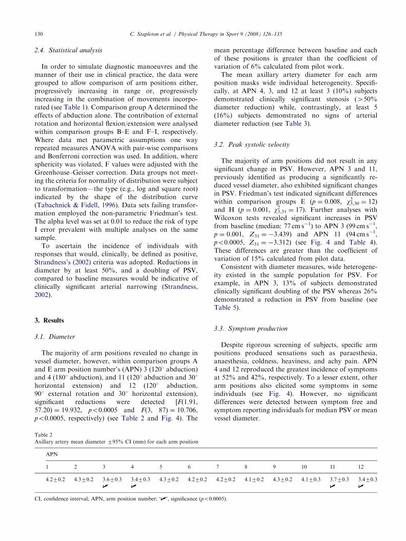
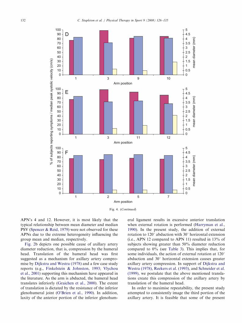
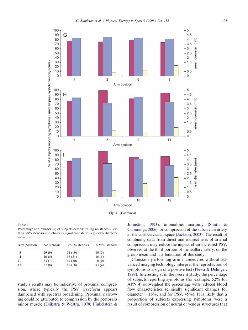
The majority of arm positions revealed no change invessel diameter, however, within comparison groups Aand E arm position number’s (APN) 3 (1201 abduction)and 4 (1801 abduction), and 11 (1201 abduction and 301horizontal extension) and 12 (1201 abduction,901 external rotation and 301 horizontal extension),significant reductions were detected [F(1.91,57.20) ¼ 19.932, po0.0005 and F(3, 87) ¼ 10.706,po0.0005, respectively) (see Table 2 and Fig. 4). The
Table 2
Axillary artery mean diameter 795% CI (mm) for each arm position
APN
1 2 3 4 5 6
4.270.2 4.370.2 3.670.3 3.470.3 4.370.2 4.270.2
| |
CI, confidence interval; APN, arm position number; ‘|’, significance (po0.
mean percentage difference between baseline and eachof these positions is greater than the coefficient ofvariation of 6% calculated from pilot work.
The mean axillary artery diameter for each armposition masks wide individual heterogeneity. Specifi-cally, at APN 4, 3, and 12 at least 3 (10%) subjectsdemonstrated clinically significant stenosis (450%diameter reduction) while, contrastingly, at least 5(16%) subjects demonstrated no signs of arterialdiameter reduction (see Table 3).
3.2. Peak systolic velocity
The majority of arm positions did not result in anysignificant change in PSV. However, APN 3 and 11,previously identified as producing a significantly re-duced vessel diameter, also exhibited significant changesin PSV. Friedman’s test indicated significant differenceswithin comparison groups E (p ¼ 0.008, w23,30 ¼ 12)and H (p ¼ 0.001, w23,31 ¼ 17). Further analyses withWilcoxon tests revealed significant increases in PSVfrom baseline (median: 77 cm s�1) to APN 3 (99 cm s�1,p ¼ 0.001, Z31 ¼ �3.439) and APN 11 (94 cm s�1,po0.0005, Z31 ¼ �3.312) (see Fig. 4 and Table 4).These differences are greater than the coefficient ofvariation of 15% calculated from pilot data.
Consistent with diameter measures, wide heterogene-ity existed in the sample population for PSV. Forexample, in APN 3, 13% of subjects demonstratedclinically significant doubling of the PSV whereas 26%demonstrated a reduction in PSV from baseline (seeTable 5).
3.3. Symptom production
Despite rigorous screening of subjects, specific armpositions produced sensations such as paraesthesia,anaesthesia, coldness, heaviness, and achy pain. APN4 and 12 reproduced the greatest incidence of symptomsat 52% and 42%, respectively. To a lesser extent, otherarm positions also elicited some symptoms in someindividuals (see Fig. 4). However, no significantdifferences were detected between symptom free andsymptom reporting individuals for median PSV or meanvessel diameter.
7 8 9 10 11 12
4.270.2 4.170.2 4.370.2 4.170.3 3.770.3 3.470.3
| |
0005).

ARTICLE IN PRESSC. Stapleton et al. / Physical Therapy in Sport 9 (2008) 126–135 131
4. Discussion
Consistent with the findings of Mochizuki et al. (1994)and Rohrer et al. (1990), the present study’s datareinforces the risk of false positive outcomes withshoulder manoeuvres at the extreme of range. Uniquely,however, the present study’s data extends this risk toinclude diagnostic tests performed at 1201 abduction.
The current data demonstrated significant reductionsin axillary artery diameter for APN’s 3 (1201 abduction),4 (1801 abduction), 11 (1201 abduction with 301horizontal extension) and 12 (1201 abduction, 301horizontal extension, and 901 external rotation). Thecharacteristic responses of arterial diameter, PSV and
0102030405060708090
100
0102030405060708090
100
0102030405060708090
100
% o
f sub
ject
s re
porti
ng s
ympt
oms
/ med
ian
peak
sys
tolic
vel
ocity
(cm
/s)
Arm posi
Arm posi
Arm posi
1 2
1 3
1 2
Symptoms reported
Fig. 4. Bar charts representing the relationship between the percentage of su
arm position in their respective comparison group (A–I).
blood flow to a narrowed arterial segment have beendescribed by Spencer and Reid (1979). According totheir theoretical model, reductions in vessel diameterresult in corresponding increases in peak systolicvelocity. This inverse relationship predicts that diameterreductions less than 20% will not greatly affect PSV.Theoretically, diameter reductions have to be greaterthan 50% before PSV changes will occur. In the presentstudy, no rise in the group median PSV was seen forAPN’s 4 (1801 abduction) and 12 (1201 abduction, 301horizontal extension and 901 external rotation) eventhough statistically significant reductions in vesseldiameter were identified. This is due to the relativelysmall (19%) reduction in mean diameter identified for
00.511.522.533.544.55
00.511.522.533.544.55
00.511.522.533.544.55
mea
n di
amet
er (m
m)
mea
n di
amet
er (m
m)
mea
n di
amet
er (m
m)
tion
tion
tion
3 4
7 8
5 6
PSV Diameter
bjects reporting symptoms, median PSV and mean diameter for each
ARTICLE IN PRESS
0102030405060708090
100
00.511.522.533.544.55
0102030405060708090
100
00.511.522.533.544.55
% o
f sub
ject
s re
porti
ng s
ympt
oms
/ med
ian
peak
sys
tolic
vel
ocity
(cm
/s)
mea
n di
amet
er (m
m)
mea
n di
amet
er (m
m)
mea
n di
amet
er (m
m)
0102030405060708090
100
00.511.522.533.544.55
1Arm position
2 5 7
Arm position1 3 11 12
Arm position1 3 9 10
Fig. 4. (Continued)
C. Stapleton et al. / Physical Therapy in Sport 9 (2008) 126–135132
APN’s 4 and 12. However, it is most likely that thetypical relationship between mean diameter and medianPSV (Spencer & Reid, 1979) were not observed for theseAPNs due to the extreme heterogeneity influencing thegroup mean and median, respectively.
Fig. 2b depicts one possible cause of axillary arterydiameter reduction, that is, compression by the humeralhead. Translation of the humeral head was firstsuggested as a mechanism for axillary artery compro-mise by Dijkstra and Westra (1978) and a few case studyreports (e.g., Finkelstein & Johnston, 1993; Vlychouet al., 2001) supporting this mechanism have appeared inthe literature. As the arm is abducted, the humeral headtranslates inferiorly (Graichen et al., 2000). The extentof translation is dictated by the resistance of the inferiorglenohumeral joint (O’Brien et al., 1990). In addition,laxity of the anterior portion of the inferior glenohum-
eral ligament results in excessive anterior translationwhen external rotation is performed (Harryman et al.,1990). In the present study, the addition of externalrotation to 1201 abduction with 301 horizontal extension(i.e., APN 12 compared to APN 11) resulted in 13% ofsubjects showing greater than 50% diameter reductioncompared to 0% (see Table 3). This implies that, forsome individuals, the action of external rotation at 1201abduction and 301 horizontal extension causes greateraxillary artery compression. In support of Dijkstra andWestra (1978), Reekers et al. (1993), and Schneider et al.(1999), we postulate that the above mentioned transla-tions create this compression of the axillary artery bytranslation of the humeral head.
In order to maximise repeatability, the present studyattempted to consistently image the third portion of theaxillary artery. It is feasible that some of the present
ARTICLE IN PRESS
% o
f sub
ject
s re
porti
ng s
ympt
oms
/ med
ian
peak
sys
tolic
vel
ocity
(cm
/s)
mea
n di
amet
er (m
m)
mea
n di
amet
er (m
m)
mea
n di
amet
er (m
m)
0102030405060708090
100
00.511.522.533.544.55
0102030405060708090
100
00.511.522.533.544.55
0102030405060708090
100
00.511.522.533.544.55
Arm position1 3 10 12
1 3 9 11
1 2 6 8
Arm position
Arm position
Fig. 4. (Continued)
Table 3
Percentage and number (n) of subjects demonstrating no stenosis, less
than 50% stenosis and clinically significant stenosis (450% diameter
reduction)
Arm position No stenosis o50% stenosis 450% stenosis
3 29 (9) 61 (19) 10 (3)
4 16 (5) 68 (21) 16 (5)
11 33 (10) 67 (20) 0 (0)
12 27 (8) 60 (18) 13 (4)
C. Stapleton et al. / Physical Therapy in Sport 9 (2008) 126–135 133
study’s results may be indicative of proximal compres-sion, where typically the PSV waveform appearsdampened with spectral broadening. Proximal narrow-ing could be attributed to compression by the pectoralisminor muscle (Dijkstra & Westra, 1978; Finkelstein &
Johnston, 1993), anomalous anatomy (Smith &Cummings, 2006), or compression of the subclavian arteryat the costoclavicular space (Jackson, 2003). The result ofcombining data from direct and indirect sites of arterialcompression may reduce the impact of an increased PSV,observed at the third portion of the axillary artery, on thegroup mean and is a limitation of this study.
Clinicians performing arm manoeuvres without ad-vanced imaging technology interpret the reproduction ofsymptoms as a sign of a positive test (Plewa & Delinger,1998). Interestingly, in the present study, the percentageof subjects reporting symptoms (for example, 52% forAPN 4) outweighed the percentage with reduced bloodflow characteristics (clinically significant changes fordiameter ¼ 16%, and for PSV, 45%). It is likely that aproportion of subjects expressing symptoms were aresult of compression of neural or venous structures that

ARTICLE IN PRESS
Table 4
Axillary artery median peak systolic velocity and IQR (cm s�1) for each arm position
Arm position
1 2 3 4 5 6 7 8 9 10 11 12
77 (64–92) 75 (64–86) 99 (84–118) 89 (50–110) 78 (67–95) 80 (71–98) 76 (67–99) 74 (59–89) 82 (69–98) 86 (65–98) 94 (81–121) 87 (61–111)
| |
IQR, inter-quartile range; ‘|’, significance (pp0.001).
Table 5
Percentage and number (n) of subjects demonstrating a doubling of
PSV and a drop in PSV compared to the baseline value
Arm position Doubling of PSV Drop in PSV
3 13 (4) 26 (8)
4 10 (3) 35 (11)
11 6 (2) 23 (7)
12 10 (3) 37 (11)
C. Stapleton et al. / Physical Therapy in Sport 9 (2008) 126–135134
accompany the arterial vasculature throughout itscourse. These results infer the possibility that diagnosticupper limb manoeuvres are not specific to arterialcauses. Although clinically, obliteration of the radialpulse or, undetectable blood pressure, aids the diagnosisof arterial involvement they do not exclude coexistingnerve or venous compression. Clinically, comparisonwith the contralateral limb aids the conclusion of apositive test response. In this study, it was notconsidered necessary to compare results with thecontralateral limb due to the asymptomatic and non-throwing status of the subjects. In addition, the presentstudy demonstrated no significant interaction betweenarm position and sex. These results were in keeping withthose of Demondion et al.’s (2006) investigation onsimilar asymptomatic subjects where no significantdifference was reported between males and females, orright and left sides.
The mean and median data presented masked wideheterogeneity for diameter and PSV in a sample ofsubjects rigorously screened for an absence of upperlimb pathology. Specifically, at 1201 abduction, 301horizontal extension, and 901 external rotation, 13% ofsubjects demonstrated a greater than 50% reduction invessel diameter, 37% showed a drop in PSV, and 10%showed a doubling in PSV. The cause of suchheterogeneity within the sample is unknown. We suggestthat the following factors may contribute to the testresponse: hyper/hypomobility of the shoulder complex;forward shoulder posture/tight pectoralis minor; exces-sive anterior translation at the glenohumeral joint; andirregular, or anomalous, anatomy of the axillary arteryand its subsidiaries. Further investigation of therelationship between such factors and test responsesmay aid identification of subjects predisposed to arterial
compression syndromes and their subsequent manage-ment. A predisposition to arterial compression would beespecially relevant in the overhead athlete populationwhere repetitive compressive stress and altered haemo-dynamics may lead to arterial damage and the sequelaedescribed in published case studies (Arko et al., 2001;Fields et al., 1986; Ishitobi et al., 2001; Rohrer et al.,1990; Todd et al., 1998; Vlychou et al., 2001).
5. Conclusions
From this study, we can conclude that vascularparameters were largely unaffected by arm movementsin normal healthy adults. Only at end of rangeabduction and arm positions incorporating 1201 abduc-tion were significant alterations recorded in vesseldiameter, PSV, systolic BP, and symptom production.However, the mean values for each outcome maskswidely heterogenous individual responses. At least 10%of subjects demonstrated diameter and PSV changesthat would, clinically, be defined as positive. The causeand clinical relevance of these heterogenous responsesfrom normal, healthy subjects warrants further investi-gation. Is it ‘normal’ for healthy, asymptomaticindividuals to demonstrate axillary artery compressionin specific overhead arm positions? Or, does it indicate apre-clinical predisposition for a neurovascular compres-sion syndrome?
The present study’s results highlight the lack ofspecificity and differential diagnostic power of thehyperabduction manoeuvre using current clinical criter-ia (Strandness, 2002). It is postulated that combining theresults of more than one test may increase specificity.This was investigated by Gillard et al. (2001) in theassessment of subclavian artery compression. Gillardet al. (2001) recommended a minimum of two provoca-tive arm manoeuvres to increase specificity. The resultsof the present study reinforce the lack of specificity ofone individual test and the necessity to integrate allinformation from a clinical assessment (i.e., clinicalpresentation, subjective history, physical examination,special tests, imaging results) to reduce the occurrence ofmisdiagnosis.

ARTICLE IN PRESSC. Stapleton et al. / Physical Therapy in Sport 9 (2008) 126–135 135
Conflict of Interest Statements
None declared.
Ethical Approval
The organisation providing ethical approval andethics protocol reference number where appropriate.
References
Arko, F. R., Harris, E. J., Zarins, C. K., & Olcott, C. (2001). Vascular
complications in high-performance athletes. Journal of Vascular
Surgery, 33, 935–942.
Baker, C. L., & Liu, S. H. (1993). Neurovascular injuries to the
shoulder. Journal of Orthopaedic and Sports Physical Therapy, 18,
360–364.
Cormier, P. J., Matalon, T. A., & Wolin, P. M. (1988). Quadrilateral
space syndrome: A rare cause of shoulder pain. Radiology, 167,
797–798.
Demondion, X., Vidal, C., Herbinet, P., Gautier, C., Duquesnoy, B., &
Cotten, A. (2006). Ultrasonographic assessment of arterial cross-
sectional area in the thoracic outlet on postural maneuvers
measured with power Doppler ultrasonography in both asympto-
matic and symptomatic populations. Journal of Ultrasound in
Medicine, 25, 217–224.
Dijkstra, P. F., & Westra, D. (1978). Angiographic features of
compression of the axillary artery by the musculus pectoralis minor
and the head of the humerus in the thoracic outlet compression
syndrome. Case report. Radiologia Clinica, 47, 423–427.
Durham, J. R., Yao, J. S., Pearce, W. H., Nuber, G. M., & McCarthy,
W. J., 3rd (1995). Arterial injuries in the thoracic outlet syndrome.
Journal of Vascular Surgery, 21, 57–69.
Fields, W. S., Lemak, N. A., & Benmenachem, Y. (1986). Thoracic
outlet syndrome—Review and reference to stroke in a major-league
pitcher. American Journal of Neuroradiology, 7, 73–78.
Finkelstein, J. A., & Johnston, K. W. (1993). Thrombosis of the
axillary artery secondary to compression by the pectoralis minor
muscle. Annals of Vascular Surgery, 7, 287–290.
Gillard, J., Perez-Cousin, M., Hachulla, E., Remy, J., Hurtevent, J. F.,
Vinckier, L., et al. (2001). Diagnosing thoracic outlet syndrome:
Contribution of provocative tests, ultrasonography, electrophysiol-
ogy, and helical computed tomography in 48 patients. Joint, Bone,
Spine: Revue du Rhumatisme, 68, 416–424.
Graichen, H., Stammberger, T., Bonel, H., Karl-Hans, E., Reiser, M.,
& Eckstein, F. (2000). Glenohumeral translation during active and
passive elevation of the shoulder—A 3d open-MRI study. Journal
of Biomechanics, 33, 609–613.
Harryman, D. T., 2nd, Sidles, J. A., Clark, J. M., McQuade, K. J.,
Gibb, T. D., & Matsen, F. A., 3rd (1990). Translation of the
humeral head on the glenoid with passive glenohumeral motion.
Journal of Bone and Joint Surgery (Am), 72A, 1334–1343.
Ishitobi, K., Moteki, K., Nara, S., Akiyama, Y., Kodera, K., &
Kaneda, S. (2001). Extra-anatomic bypass graft for management of
axillary artery occlusion in pitchers. Journal of Vascular Surgery,
33, 797–801.
Jackson, M. R. (2003). Upper extremity arterial injuries in athletes.
Seminars in Vascular Surgery, 16, 232–239.
Kee, S. T., Dake, M. D., Wolfe-Johnson, B., Semba, C. P., Zarins, C.
K., & Olcott, C. (1995). Ischemia of the throwing hand in major
league baseball pitchers: Embolic occlusion from aneurysms of
axillary artery branches. Journal of Vascular and Interventional
Radiology, 6, 979–982.
Mochizuki, T., Isoda, H., Masui, T., Ohkawa, Y., Takahashi, M.,
Takehara, Y., et al. (1994). Occlusion of the posterior humeral
circumflex artery—Detection with MR angiography in healthy-
volunteers and in a patient with quadrilateral space syndrome.
American Journal of Roentgenology, 163, 625–627.
Nijhuis, H. H., & Muller-Wiefel, H. (1991). Occlusion of the brachial
artery by thrombus dislodged from a traumatic aneurysm of the
anterior humeral circumflex artery. Journal of Vascular Surgery, 13,
408–411.
O’Brien, S. J., Neves, M. C., Arnoczky, S. P., Rozbruck, S. R.,
Dicarlo, E. F., Warren, R. F., et al. (1990). The anatomy and
histology of the inferior glenohumeral ligament complex of the
shoulder. American Journal of Sports Medicine, 18, 449–456.
Plewa, M. C., & Delinger, M. (1998). The false-positive rate of thoracic
outlet syndrome shoulder maneuvers in healthy subjects. Academic
Emergency Medicine, 5, 337–342.
Redler, M. R., Ruland, L. J., 3rd, & McCue, F. C., 3rd (1986).
Quadrilateral space syndrome in a throwing athlete. American
Journal of Sports Medicine, 14, 511–513.
Reekers, J. A., den Hartog, B. M., Kuyper, C. F., Kromhout, J. G., &
Peeters, F. L. (1993). Traumatic aneurysm of the posterior
circumflex humeral artery: A volleyball player’s disease? Journal
of Vascular and Interventional Radiology, 4, 405–408.
Rohrer, M. J., Cardullo, P. A., Pappas, A. M., Phillips, D. A., &
Wheeler, H. B. (1990). Axillary artery compression and thrombosis
in throwing athletes. Journal of Vascular Surgery, 11, 761–768.
Schneider, K., Kasparyan, N. G., Altchek, D. W., Fantini, G. A., &
Weiland, A. J. (1999). An aneurysm involving the axillary artery
and its branch vessels in a major league baseball pitcher: A case
report and review of the literature. American Journal of Sports
Medicine, 27, 370–375.
Smith, R. A., Jr., & Cummings, J. P. (2006). The axillary arch:
Anatomy and suggested clinical manifestations. Journal of Ortho-
paedic and Sports Physical Therapy, 36, 425–429.
Spencer, M. P., & Reid, J. M. (1979). Quantitation of carotid stenosis
with continuous-wave (C-W) Doppler ultrasound. Stroke, 10,
326–330.
Strandness, D. (2002). Hemodynamics of arterial stenosis and
occlusion. In D. Strandness (Ed.), Duplex scanning in vascular
disorders (3rd ed., p. 68). Lippincott Williams and Wilkins.
Tabachnick, B., & Fidell, L. (1996). Normality, linearity, and
homoscedasticity. In B. Tabachnick, & L. Fidell (Eds.), Using
multivariate statistics (4th ed., p. 82). New York: Harper Collins.
Todd, G. J., Benvenisty, A. I., Hershon, S., & Bigliani, L. U. (1998).
Aneurysms of the mid axillary artery in major league baseball
pitchers—A report of two cases. Journal of Vascular Surgery, 28,
702–707.
Vlychou, M., Spanomichos, G., Chatziioannou, A., Georganas, M., &
Zavras, G. M. (2001). Embolisation of a traumatic aneurysm of the
posterior circumflex humeral artery in a volleyball player. British
Journal of Sports Medicine, 35, 136–137.
Wright, I. (1945). The neurovascular syndrome produced by hyper-
abduction of the arm. American Heart Journal, 29, 1–19.