sonography of the neonatal spine: part 1, normal anatomy...
TRANSCRIPT

AJR:188, March 2007 733
AJR 2007; 188:733–738
0361–803X/07/1883–733
© American Roentgen Ray Society
Lowe et al.Sonography of Neonatal Spine
Pe d i a t r i c I m ag i n g • P i c t o r i a l E s s ay
Sonography of the Neonatal Spine: Part 1, Normal Anatomy, Imaging Pitfalls, and Variations That May Simulate Disorders
Lisa H. Lowe1,2
Andrew J. Johanek1,3
Charlotte W. Moore1,2
Lowe LH, Johanek AJ, Moore CW
Keywords: neonatal imaging, neuroradiology, pediatric radiology, sonography, spine
DOI:10.2214/AJR.05.2159
Received December 16, 2005; accepted after revision February 28, 2006.
Awarded a Bronze Medal poster exhibit at the 2005 annual meeting of the American Roentgen Ray Society, New Orleans, LA.
1Department of Radiology, The University of Missouri–Kansas City, Kansas City, MO.
2Department of Radiology, Children’s Mercy Hospital and Clinics, 2401 Gillham Rd., Kansas City, MO 64108. Address correspondence to L. H. Howe ([email protected]).
3Department of Radiology, St. Luke’s Hospital, Kansas City, MO.
CMEThis article is available for CME credit. See www.arrs.org for more information.
OBJECTIVE. Our objective is to discuss neonatal spine sonography with emphasis on im-aging pitfalls and normal variants that may simulate disease and to distinguish them from truespinal disorders.
CONCLUSION. Sonography of the neonatal spine is now accepted as a highly sensitive,readily available screening study that can be used to evaluate various anomalies of the lumbarspine in most infants younger than 4 months.
lthough MRI has been consideredthe imaging gold standard, recentadvances in sonography have al-lowed its image quality to im-
prove significantly enough that its diagnosticvalue is equal to that of MRI [1]. Sonographycan now characterize nearly all spinal anom-alies sufficiently in the first days of life. Thisallows the clinical determination of whetherthe lesion requires urgent intervention orwhether further radiologic evaluation withstudies such as MRI can be delayed until ther-apeutic intervention is more imminent. In part1 of this pictorial essay, we discuss lumbarspine embryology, sonography techniquesand indications, normal anatomy, and devel-opmental variations and pitfalls that may sim-ulate disease. Part 2 covers abnormal entities.
EmbryologyTo understand spine anomalies, knowledge
of embryonic development is necessary. TheCNS starts to form during the third gestationalweek, beginning with the process known as neu-rulation (Fig. 1A). Next, canalization occurs atthe distal end of the neural tube in the caudal cellmass, resulting in an ependyma-lined neuraltube that unites with the rest of the spinal cord toform the conus medullaris and ventriculus ter-minalis (Fig. 1B). Finally, at 38 days of gesta-tion, retrogressive differentiation occurs(Fig. 1C), forming the filum terminale [2–5].
Technique and IndicationsImages are obtained in the longitudinal and
transverse planes using a linear 5–12-MHztransducer (Fig. 2). The vertebral level is deter-
mined by counting down from the 12th rib andconfirmed by counting up from the L5–S1junction or the tip of coccyx. If the vertebrallevel is unclear, correlation with radiographs(possibly with a marker) may help. Color orpower Doppler sonography may be used as anadjunct to better characterize soft-tissuemasses found on the skin or in the spinal canal.
Indications for lumbar spine sonography in-clude multiple congenital anomalies placingan infant at increased risk, complicated sacraldimple (location above the gluteal crease, bot-tom of pit not seen, possible drainage fromdimple, and presence of skin stigmata), soft-tissue mass suspected of being spina bifida oc-culta, determination of reason for failed lum-bar puncture, and location of CSF that may betapped [1, 6]. Low-risk lesions include simplemidline dimples (< 5 mm in diameter, within2.5 cm of the anus, no other cutaneous stig-mata). High-risk lesions include atypical dim-ples (> 5 mm in diameter, > 2.5 cm above theanus), hemangiomas, cutis aplasia, hairypatches, and skin tags [7]. MRI is the study ofchoice when surgical therapy is required, suchas with open spinal dysraphism or obviousCSF drainage from a skin dimple or sinus tract.
Normal Variants That May Simulate Disorders
Several common normal variants that maybe confused with disorders on lumbar spinesonography will be discussed, includingventriculus terminalis, filar cyst, prominentfilum terminale, cauda equina pseudomass,pseudosinus tract, and dysmorphic coccyx.Familiarity with these variations can prevent
A

Lowe et al.
734 AJR:188, March 2007
misinterpretation and referrals for unneededadditional clinical or imaging evaluation.
Ventriculus TerminalisThe ventriculus terminalis, often seen on
sonography and MRI in children youngerthan 5 years, is due to incomplete fetal regres-sion of the embryonic terminal ventricle inthe conus medullaris [6, 8] (Fig. 3).
Filar CystThe so-called filar cyst is an interesting in-
cidental finding that has only recently beenstudied, perhaps at least partly because of be-ing detected more often with improved sono-graphic equipment. Although this lesion hasbeen scantly described in the radiology liter-ature, it is often visible on sonography. Thereis no autopsy description of a filar cyst, whichbegs the questions of its origin and its validityas an entity [3, 4]. In addition, the nomencla-ture for this lesion is confusing in that it hasbeen termed both “ventriculus terminalis”and “filar cyst” by various authors [6–8]. Weprefer the latter term, filar cyst, to specify thefilar location from the conus medullaris loca-tion of the ventriculus terminalis.
Possible explanations for the origin of thefilar cyst include that perhaps the normalarachnoid reflections form a pseudocystlikestructure or that it is a true ependyma-linedcystic embryonic remnant (possibly indis-tinguishable from the ventriculus termina-lis) that is disrupted by the act of openingthe dura during autopsy. Regardless of itsorigin, it is a normal variant that alone hasno known clinical significance and that doesnot require additional imaging [4]. If MRI isperformed, in our experience, the filar cystis less reliably visible than on sonography.
Strict imaging criteria for filar cysts shouldbe applied (location midline, within filum,just below conus; fusiform shape, well-de-fined, hypoechoic appearance of a simplecyst) to avoid the potential for underdiag-nosing a true disorder (Figs. 4 and 5).
Prominent Filum TerminaleA prominent filum terminale may cause
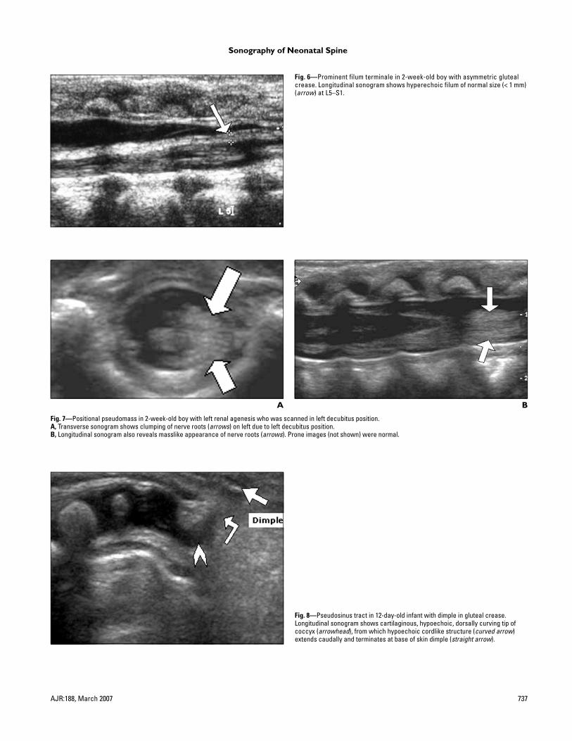
concern when it stands out as particularlyechogenic in comparison with other nerveroots. It is distinguished as normal by its thick-ness and typical midline course [1] (Fig. 6).
“Pseudomass” due to Positional Nerve Root Clumping
Positional clumping of the nerve roots oc-curs when an infant is scanned in the decubi-tus position. Rescanning the child prone willcause the “mass” to disappear as the nerveroots return to their normal position (Fig. 7).
Pseudosinus TractAnother common normal variant is a pseu-
dosinus tract, which is seen on sonography asa residual cordlike region composed of fi-brous tissue extending from a skin dimple tothe coccyx (Fig. 8). True dermal sinus tractsrarely occur at the tip of the coccyx and aretypically found in a more cranial location.However, a careful search should be made forany mass or fluid along the course of the fi-brous tract. If CSF is draining via a dimple,then a true sinus tract is likely, and MRI is theimaging technique of choice.
Dysmorphic CoccyxThe tip of the coccyx can vary widely in
shape, and in some cases may mimic a masswhen palpated on physical examination (Fig. 9).
Conclusion Neonatal spinal sonography is a useful
screening technique for occult spinal anoma-lies; it can characterize normal anatomy andnormal variants that may simulate disorders.Familiarity with these findings will preventmisinterpretation and inappropriate referrals.
References1. Dick EA, Patel K, Owens CM, De Bruyn R. Spi-
nal ultrasound in infants. Br J Radiol 2002;
75:384–392
2. Barkovich AJ. Normal development of the neonatal
and infant brain, skull, and spine. In: Barkovich AJ.
Pediatric neuroimaging, 3rd ed. Philadelphia, PA:
Lippincott Williams & Wilkins, 2000:50–54
3. Gamble HJ. Electron microscope observations
upon the conus medullaris and filum terminale of
human fetuses. J Anat 1971; 110:173–179
4. Malas MA, Salbacak A, Buyukmumcu M, Seker M,
Koyluoglu B, Karabulut AK. An investigation of the
conus medullaris termination level during the pe-
riod of fetal development to adulthood. Kaibogaku
Zasshi 2001; 76:453–459
5. Sadler T. Langman’s medical embryology, 5th ed.
Baltimore, MD: Lippincott Williams & Wilkins,
1985:334–337, 343–345
6. Unsinn KM, Geley T, Freund MC, Gassner I. US
of the spinal cord in newborns: spectrum of nor-
mal findings, variants, congenital anomalies,
and acquired diseases. RadioGraphics 2000;
20:923–938
7. Kriss VM, Desai NS. Occult spinal dysraphism in
neonates: assessment of high-risk cutaneous stig-
mata on sonography. AJR 1998; 171:1687–1692
8. Coleman LT, Zimmerman RA, Rorke LB. Ven-
triculus terminalis of the conus medullaris: MR
findings in children. Am J Neuroradiol 1995;
16:1421–1426

Sonography of Neonatal Spine
AJR:188, March 2007 735
A
CB
Fig. 1—Schematics illustrate three stages of spinal cord development.A, Neurulation (closure of neural tube) is process of progression from neural plate to neural groove to neural tube. (Reprinted with permission from Sadler T. Langman’s medical embryology, 5th ed. Baltimore, MD: Lippincott Williams & Wilkins, 1985:335 [5]) B, Canalization occurs when multiple microcysts form and coalesce in caudal cell mass (arrows), which fuses to distal neural tube (arrowheads), forming primitive spinal cord. (Reprinted with permission from Barkovich AJ. Normal development of the neonatal and infant brain, skull and spine. In: Barkovich AJ. Pediatric neuroimaging, 3rd ed. Philadelphia, PA: Lippincott Williams & Wilkins, 2000:624 [2]) C, Retrogressive differentiation (programmed cell death) is process whereby caudal cell mass and neural tube regress in size to form fetal conus medullaris, ventriculus terminalis, and filum terminale. Note labeled structures. (Reprinted with permission from Barkovich AJ. Normal development of the neonatal and infant brain, skull and spine. In: Barkovich AJ. Pediatric neuroimaging, 3rd ed. Philadelphia, PA: Lippincott Williams & Wilkins, 2000:624 [2])
A
B
Fig. 2—1-week-old boy with normal lumbar spine sonogram and history of unilateral renal agenesis. A, Transverse lumbar sonogram shows normal anatomy as labeled. V = vertebra, transverse process (arrowhead).B, Longitudinal lumbar sonogram shows normal anatomy as labeled. Note central echoic complex (arrowheads), a normal finding that results from interface of central end of anterior median fissure and not central spinal canal.
Neural crest
Notocord
Surfaceectoderm
Neural crest
Neural tube
Neural groove
Spiral ganglion
Neural plate
Conusmedullaris
Ventriculusterminalis
Filum terminale

Lowe et al.
736 AJR:188, March 2007
A B
Fig. 3—1-month-old boy with ventriculus terminalis who was referred for deep sacral dimple and who is developmentally normal at 18 months.A, Longitudinal sonogram of spine reveals distention of distal lumbar spinal canal just above conus medullaris (arrowhead). Size smaller than 5 mm and stability over time distinguish this normal variant from small syrinx.B, Sagittal T2-weighted MR image at age 7 months shows stable distention of distal spinal canal (arrowhead), excluding syrinx.
A BFig. 4—Filar cyst in 14-day-old girl with deep sacral dimple and normal motor development.A, Transverse sonogram of proximal cauda equina shows well-defined, midline, cystic collection (arrow). Note normal ventral and dorsal nerve root bundles (arrowheads).B, Longitudinal sonogram reveals well-defined fusiform “cyst” in midline (arrow) just below conus medullaris. Also note prominent echogenic central spinal canal (arrowhead), a normal variant seen in some children.
A BFig. 5—Filar cyst in 5-week-old boy with multiple anomalies who had been followed up with MRI at age 2 months. A, Longitudinal sonogram of filum and cauda equina (arrowhead) shows unusually long filar cyst (calipers). Despite its length, it meets criteria for filar cyst: location just below conus medullaris, fusiform shape, well defined, thin walled, and hypoechoic.B, Longitudinal T2-weighted MR image shows ill-defined filar cyst (arrows) that is better seen on sonography.
Sonography of Neonatal Spine
AJR:188, March 2007 737
Fig. 6—Prominent filum terminale in 2-week-old boy with asymmetric gluteal crease. Longitudinal sonogram shows hyperechoic filum of normal size (< 1 mm) (arrow) at L5–S1.
A B
Fig. 7—Positional pseudomass in 2-week-old boy with left renal agenesis who was scanned in left decubitus position.A, Transverse sonogram shows clumping of nerve roots (arrows) on left due to left decubitus position.B, Longitudinal sonogram also reveals masslike appearance of nerve roots (arrows). Prone images (not shown) were normal.
Fig. 8—Pseudosinus tract in 12-day-old infant with dimple in gluteal crease. Longitudinal sonogram shows cartilaginous, hypoechoic, dorsally curving tip of coccyx (arrowhead), from which hypoechoic cordlike structure (curved arrow) extends caudally and terminates at base of skin dimple (straight arrow).

Lowe et al.
738 AJR:188, March 2007
A B
Fig. 9—Misshapen coccyx in two neonatal girls, each with palpable “lump” beneath sacral dimple in gluteal crease.A, Longitudinal sonogram of coccyx in 2-week-old girl shows hypoechoic cartilaginous tip (arrowheads), which is acutely angulated dorsally as it extends toward skin surface. Palpated “lump” was tip of coccyx.B, Longitudinal sonogram of coccyx in 2-week-old girl reveals it is straightened, with loss of its normal ventral curve. Hypoechoic cartilaginous tip (arrowhead) extends dorsally toward skin surface, causing clinically palpable “lump.”
F O R Y O U R I N F O R M A T I O N
The reader’s attention is directed to part 2 accompanying this article, titled “Sonography of the Neonatal Spine: Part 2, Spinal Disorders,” which begins on page 739.
F O R Y O U R I N F O R M A T I O N
This article is available for CME credit. See www.arrs.org for more information.