sox17is essential for the specification of cardiac ... · sox17is essential for the specification...
TRANSCRIPT
Sox17 is essential for the specification of cardiacmesoderm in embryonic stem cellsYu Liu*, Masanori Asakura*†, Hironori Inoue*‡, Teruya Nakamura*§, Motoaki Sano*¶, Zhiyv Niu*�, Michelle Chen**,Robert J. Schwartz*�, and Michael D. Schneider*††
*Center for Cardiovascular Development, Baylor College of Medicine, Houston, TX 77030; and **Agilent Technologies, Santa Clara, CA 95051
Edited by Eric N. Olson, University of Texas Southwestern Medical Center, Dallas, TX, and approved January 17, 2007 (received for review October 14, 2006)
Early steps for cardiac specification are problematic for the studyof mammalian embryos, which has favored using pluripotent cellsthat recapitulate cardiac myogenesis. Furthermore, circuits gov-erning cardiac specification have relevance to the application of EScells and other cells for heart repair. In mouse teratocarcinomacells, canonical Wnts that inhibit heart formation in avian oramphibian embryos and explants activate cardiogenesis, paradox-ically. Here, we show that the Wnt/�-catenin pathway also isessential for cardiac myogenesis to occur in ES cells, acting at agastrulation-like stage, mediating mesoderm formation and pat-terning (two prerequisites for cardiac myogenesis itself). Amonggenes associated temporally with this step was Sox17, encoding anendodermal HMG-box transcription factor. Using lentiviral vectorsfor RNA interference in differentiating ES cells, an essential role forSox17 was proven in cardiac muscle cell formation. Sox17 short-hairpin RNA suppresses cardiac myogenesis selectively, actingsubsequent to mesoderm formation yet before induction of Mesp1and Mesp2, a pair of related basic helix–loop–helix transcriptionfactors that together are indispensable for creating heart meso-derm. Sox17 short-hairpin RNA blocks cardiac myogenesis non-cellautonomously and impairs the induction of Hex, a homeodomaintranscription factor that is known to be required for the productionof endoderm-derived heart-inducing factors.
cardiac myogenesis � differentiation � �-catenin � heart � Wnt
S tem cells elicit intense interest on dual, complementarygrounds. As fundamental science, pluripotent cells with a
capacity to enter many or all lineages provide access intopathways governing cell fate, including transcription factor net-works and extracellular cues that activate them. As appliedscience, the use of adult and ES cells in potential therapiesarouses the imagination of clinicians, basic researchers, and thelay public alike. Although barriers to successful implementationare many, a cell-based approach to myocyte regeneration ishighly cogent for cardiac disease (1, 2). Muscle cell death fromacute ischemic injury is the most prevalent adult heart disorder;heart failure entails ongoing apoptosis; adult ventricular musclecells characteristically do not reenter the cell cycle; and newlyfound progenitor cells in adult hearts are insufficient, on theirown, to execute adequate self-repair. Although effects of graftedcells apart from myocyte creation might also be beneficial, theneed for myocyte replacement once cell death has ensuedreinforces the logic of dissecting cardiogenesis in pluripotentcells more systematically. Genome-wide expression profilingprovides one route to such information.
Peptide growth factors instrumental to cardiac myogenesisinclude members of the bone morphogenetic protein (BMP) andWnt families (3, 4). BMPs are the most commonly substantiatedinductive signal, corresponding to an instruction supplied byendoderm during embryogenesis. In gastrulating frog and chickembryos and postgastrulation mesodermal explants, the ‘‘canon-ical’’ glycogen synthase kinase-3�/�-catenin/T cell factor-dependent Wnt pathway antagonizes cardiac myogenesis (3, 4).Conversely, a ‘‘noncanonical’’ Wnt, Wnt11, promotes cardiomy-ocyte formation in these systems and mouse embryonal carci-
noma cells (5). Surprisingly, the canonical Wnt pathway was alsoessential in the latter, for cardiomyocyte formation (6). Threeexplanations might reconcile this seeming dichotomy. Embryo-nal carcinoma cells, although an informative model for cardio-genesis (7–9), are tumor-derived and potentially anomalous;phylogenetic differences could matter; or, canonical Wnts mightdrive cardiomyocyte formation by an early developmental eventin pluripotent cells, preceding the stage at which they inhibitheart formation in chick and frog embryos.
Here, we tested whether the canonical Wnt/�-catenin pathwaywas essential for cardiac muscle specification in mouse ES cells,which are of openly greater relevance than embryonal carcino-mas in fidelity to normal development and utility for heartrepair. Cardiac myogenesis depended stringently on canonicalWnt signals, acting in an early interval controlling mesodermformation, a presumptive requirement for cardiogenesis per se.Associated with this gastrulation-like intermediary state andcontingent on canonical Wnts was the endodermal transcriptionfactor Sox17 (10–12). Sox17 was indispensable for cardiac myo-genesis in this system, shown by RNA interference. Sox17short-hairpin RNA (shRNA) did not impair mesendodermformation yet suppressed the induction of Mesp1 and Mesp2,transcription factors that together are pivotal to cardiac speci-fication in primitive mesoderm (13).
ResultsThe Canonical Wnt Pathway Drives an Early Step Toward CardiacMyogenesis in ES Cells. First, we examined mouse ES cells for theoperation of Wnt- and BMP-dependent cardiogenic pathways[supporting information (SI) Fig. 5]. Noggin and soluble Frizzled8 (sFz8) prevented the expression of genes for cardiogenictranscription factors (Nkx2–5, GATA-4) and cardiac structuralproteins [� myosin heavy chains (�MyHC), Ryr2], induction ofsarcomeric MyHC protein, and formation of beating colonies.
Author contributions: Y.L., M.A., and H.I. contributed equally to this work; Y.L., M.A., H.I.,T.N., R.J.S., and M.D.S. designed research; Y.L., M.A., H.I., T.N., M.S., Z.N., and M.C.performed research; Y.L., M.A., H.I., T.N., and M.S. contributed new reagents/analytic tools;Y.L., M.A., H.I., T.N., M.S., M.C., R.J.S., and M.D.S. analyzed data; and Y.L., M.A., H.I., andM.D.S. wrote the paper.
Conflict of interest: M.C. is an employee of Agilent Technologies. All other authors declareno conflict of interest.
This article is a PNAS direct submission.
Abbreviations: BMP, bone morphogenetic protein; Dkk, dickkopf; EB, embryoid body;MyHC, myosin heavy chain; QRT-PCR, quantitative RT-PCR; shRNA, short-hairpin RNA; sFz8,soluble Frizzled 8; BIO, 6-bromoindirubin-3�-oxime.
†Present address: National Cardiovascular Center, Osaka 565-8565, Japan.
‡Present address: Kagoshima University, Kagoshima 899-8520, Japan.
§Present address: University of Utah, Salt Lake City, UT 84132.
¶Present address: Keio University School of Medicine, Tokyo 160-8582, Japan.
�Present address: Institute for Biosciences and Technology, Texas A&M University HealthScience Center, Houston, TX 77030.
††To whom correspondence should be addressed. E-mail: [email protected].
This article contains supporting information online at www.pnas.org/cgi/content/full/0609100104/DC1.
© 2007 by The National Academy of Sciences of the USA
www.pnas.org�cgi�doi�10.1073�pnas.0609100104 PNAS � March 6, 2007 � vol. 104 � no. 10 � 3859–3864
DEV
ELO
PMEN
TAL
BIO
LOG
Y

Next, to perturb the Wnt/glycogen synthase kinase-3� (GSK-3�)/�-catenin pathway selectively, we used dickkopf 1 (Dkk1) [ahigh-affinity ligand for the coreceptor LRP6 (14)] and 6-bromoindirubin-3�-oxime [BIO, an inhibitor of GSK-3� (15)].Dkk1 suppressed Nkx2–5 and �-MyHC in both cell types. Con-versely, BIO bypassed the inhibition by sFz8 (SI Figs. 6 and 7).BIO had no effect on cardiac myogenesis in the absence of sFz8,when given on days 0–4 (not shown).
In pluripotent systems, canonical Wnts might promote cardio-genesis itself or a prerequisite prior step like mesoderm formation.Indeed, transient treatment of ES cells with Dkk1 during days 0–3was especially effective at blocking cardiac myogenesis (SI Fig. 6 Gand H). Thus, canonical Wnts impinge on cardiac myogenesisthrough an early step in the development of ES cells from aninitially undifferentiated ground state.
Expression Profiling of Wnt- and BMP-Dependent Genes IdentifiesMesoderm Transcriptional Factors Among the Early Targets. To can-vas potential Wnt- and BMP-dependent mechanisms for cardiac
myogenesis in pluripotent cells, we obtained genome-wide expres-sion profiles (Fig. 1; SI Materials and Methods). Key findings weresubstantiated by quantitative RT-PCR (QRT-PCR) or Westernblotting (SI Fig. 5E; data not shown) and were retested by usingDkk1 (Fig. 1B). In ES cells at 7–9 d, the impact of sFz8 and Nogginon cardiac transcription factors and structural genes was readilyevident (Fig. 1Ac). Fewer than one gene in 40 fulfilled the criteriaused. Significantly repressed GeneOntology clusters (fulfilling thecriteria of developmental regulation, Wnt dependence, and BMPdependence) included muscle development, heart development,striated muscle thick filament, Z disk, and angiogenesis (P �0.0008). Others bearing on cardiac cell fate and function includedtranscription factor activity and cytoskeleton (P � 0.0001). Anal-ogously, the only significant GenMAPP clusters at 7–9 d werestriated muscle contraction and smooth muscle contraction (P �0.0002). Affected cardiogenic genes included Nkx2.5, Mef2c, Mef2d,Gata4, Gata5, Gata6, Tbx20, Hand1, Hod/Hop, Irx3, Irx5, and Isl1.Affected cardiac structural genes included those for sarcomericproteins (Actc1, Mybpc3, Myh6, Myl2, Myl3, Myl4, Myl7, Myom1,
Fig. 1. Microarray analysis of Wnt- and BMP-dependent genes in differentiating ES cells identifies multiple early targets. (A) Partial cluster analysis of genesfulfilling the criteria of developmental regulation plus modulation by both sFz8 and Noggin at �1 days. Genes shown were chosen from the significantly inhibitedGeneOntology and GenMAPP clusters, Entrez Gene entries, PubMed, and Mouse Genome Informatics database. The neurogenesis cluster shown for comparisonin c, below, is taken from the developmentally up-regulated genes whose induction was enhanced by sFz8 and Noggin. n � 4 for 0–3 d; n � 2 for 5–9 d. (B) QRT-PCRconfirmation of selected day three findings as contingent on canonical Wnts.
3860 � www.pnas.org�cgi�doi�10.1073�pnas.0609100104 Liu et al.

Tmod1, Tnnc1, Tnni1, Tnni3, Tnnt2, and Ttn), cytoskeletal proteins(Des, Smpx), junctional proteins (Dsp, Jph1), and calcium ho-meostasis (Atp2a2, Pln, Ryr2, and Srl). Genes for vasculogenesis(Bves, Cdh5, Flt1, Hey1, Kdr, Myocd, Pdgfra, Pdgfrb, Tie1, Tie2, andVegfa) and hematopoiesis (Gata2, Hba-a1, Hba-x, Hbb-bh1, Hbb-y,and Lmo2) also were inhibited. Conversely, neurogenesis and braindevelopment GeneOntology clusters were up-regulated by sFz8and Noggin (P � 0.0003; Fig. 1Ac). Thus, inhibition was selectivefor cardiomyocytes and closely related mesoderm derivatives.
At 3 and 5 d, TGF-� signaling was the only GenMAPP clusterimpaired by sFz8 and Noggin (P � 0.001 and 0.00002, respectively),including Bambi, Bmp5, Bmper, Fst, Smad1, Tgfb1, and Foxh1, anuclear target of Nodal required for the anterior heart field (16)(Fig. 1A a and b). Repressed GeneOntology clusters were devel-opment (P � 0.00004 and 0.001), transcription factor activity (P �0.0004 and 0.00006), other synonymous groups, and heart devel-opment (P � 0.0009 at 5 d).
Notably, on day 3, sFz8 and Noggin disrupted the induction ofmany transcription factors that are required for mesoderm forma-tion or mesoderm patterning (T/Brachyury, Eomes, Evx1, Lhx1,
Mesp1, Mixl1, and Snail1) (17–20) (Fig. 1Aa and SI Fig. 5E). Mesp1functions subsequent to T as the earliest molecular marker ofcardiac precursor cells that migrate through the primitive streak andis essential along with Mesp2 for cardiac myogenesis in committedmesoderm cells (13). Other affected genes with known roles incardiac myogenesis included Fgf8 (21) and Tdgf1/Cripto (22). Sim-ilar results were obtained in P19Cl6 cells (SI Fig. 7).
An Essential Role for Sox17 in Cardiac Mesoderm Specification. Inprioritizing among early targets whose importance in cardiac myo-cyte creation is unknown or ambiguous, we were attracted to Sox17.Sox17 has established roles in endoderm development (10–12) yetalso is enriched in the cardiac crescent (23), cardiac side populationcells (24), and differentiating Sca-1� cardiac progenitor cells (notshown). In differentiating ES cells, transient induction of Sox17,greatest on day 5, contrasts with the late onset of Sox18 and smalldevelopmental fluctuations in Sox7, the other F group Sox genes(Fig. 2A; not shown). Confirming our microarray findings byQRT-PCR, Sox17 was suppressed by Noggin and sFz8. Implicatingcanonical Wnts, the block by sFz8 was overcome by BIO, andWnt3a augmented Sox17 induction (Fig. 2 B and C).
Fig. 2. Regulation of Sox17 in differentiating ES cells by the canonical Wnt pathway. (A–C) QRT-PCR. n � 3; *, P � 0.05 vs. control cells. (A) sFz8 and Nogginsuppress Sox17 in differentiating ES cells. (B) Wnt3a up-regulates Sox17. (C) BIO rescues Sox17 from repression by sFz8. (D) Immunoblotting for epitope-taggedSox17 and Sox18, showing efficacy and specificity of Sox17 shRNAs.
Liu et al. PNAS � March 6, 2007 � vol. 104 � no. 10 � 3861
DEV
ELO
PMEN
TAL
BIO
LOG
Y
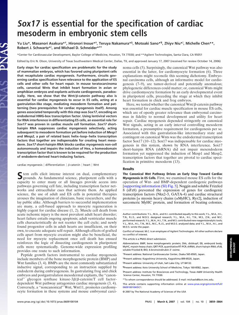
We created two independent lentiviral vectors that suppressSox17 selectively (Fig. 2D). shRNAs were coexpressed along withenhanced GFP, infected ES cells were flow-sorted (SI Fig. 8 A andB), and the upper 50% of eGFP� cells were collected for functionalstudies (Fig. 3). Control cells were transduced with virus expressingenhanced GFP plus shRNA against firefly luciferase. Suppressionof endogenous Sox17 was confirmed, along with the direct Sox17targets Foxa1 and Foxa2 (11), and four of the five tested cardiogenic
transcription factor genes (Nkx2.5, Tbx5, Mef2c, and Myocd). Theexception was Gata4 (not shown), which may signify a differinghierarchical relationship in the cardiomyocyte lineage, or Sox17-independent induction in a different cell type like endoderm.Cardiac structural genes (�-MyHC, Ryr2), sarcomeric protein stain-ing, and the prevalence of beating embryoid bodies (EBs) were allsuppressed, to nearly the same degree as by sFz8 or Noggin (Fig. 3A–C; compare with SI Fig. 5). Identical results were obtained with
Fig. 3. An essential role for Sox17 in cardiac specification. (A–C) Suppression of cardiac myogenesis by Sox17 shRNA, shown by using QRT-PCR (A),immunostaining (B), and the prevalence of beating EBs (C). (D) QRT-PCR showing Sox17 acts downstream of mesendoderm formation but upstream from Mesp1and Mesp2. n � 3; *, P � 0.05 vs. control cells.
3862 � www.pnas.org�cgi�doi�10.1073�pnas.0609100104 Liu et al.

the second Sox17 shRNA (SI Fig. 8C; not shown). Cardiac myo-genesis was not inhibited by shRNAs against two other endoderm-expressed genes, Pdx1 and Afp (SI Fig. 8D).
Next, we examined at which step Sox17 functions (Fig. 3D). Sox17shRNA did not impair down-regulation of Oct4 (a master regulatorof pluripotency), induction of T and Gsc (indicators of mesend-oderm formation), or the hematopoietic lineage marker Runx1. Bycontrast, Sox17 shRNA suppressed both Mesp1 and Mesp2 (Fig.3D). Thus, under these conditions, a Sox17-dependent pathwayplays a selective role in mesoderm patterning. By contrast to Runx1,Sox17 shRNA did suppress the hematopoietic/endothelial lineagemarker Flk1 at 4–5 d; interestingly, Flk1�/CXCR4�/VE-Cadherin�
ES cells generate cardiac myocytes in culture (25), and fate-mapping has shown Flk1� cells to be cardiac muscle progenitors inmice (26).
Sox17 Controls a Non-Cell-Autonomous Pathway for Cardiac Myogen-esis in ES Cells. Consistent with Foxa1 and Foxa2 being direct targetsof Sox17 in Xenopus endoderm (11), Sox17 shRNA significantlysuppressed both, as mentioned (Fig. 3A). By contrast, Sox17 shRNAdid not alter several other endodermal markers including Afp (Fig.3D), Gata4, and Sox7 (not shown). Together, this suggests thatSox17 mediates a specific pathway or subset of endoderm, ratherthan panendoderm. An endodermal gene of known importance toheart formation is Hex, which regulates paracrine signals for cardiacmyogenesis, depends on the canonical Wnt pathway, and mediatescardiac development in Xenopus and mice (27, 28). Notably, Hexinduction at 4–5 d was markedly attenuated by Sox17 shRNA, to thelevels in undifferentiated cells (Fig. 3D).
To determine whether Sox17 functions in ES cells primarilythrough cell-autonomous or non-cell-autonomous effects, wild-type cells were aggregated at various ratios (6:1 to 1:6) with cellsharboring Sox17 shRNA (SI Fig. 8E). At 6 d, the two cell popula-tions were flow-sorted by using the GFP reporter and assayedseparately for cardiogenic transcription factors (Fig. 4A). At eachconcentration, cocultured ES cells expressed Nkx2.5 and Tbx5 atlevels distinct from those predicted for a cell-autonomous mecha-nism. An excess of wild-type cells completely rescued suppressionof Nkx2.5 and Tbx5 in cells deficient for Sox17. Conversely, evenequal numbers of cells containing Sox17 shRNA were sufficient tosuppress Nkx2.5 and Tbx5 in wild-type cells. Thus, the impact ofSox17 on cardiac myogenesis in ES cells is best explained bynon-cell-autonomous mechanisms.
Sox17 Mediates Inactivation of the Canonical Wnt Pathway in Differ-entiating ES Cells. Of the HMG box proteins, the Sox family relatesmost closely to T cell factor/Lef, the nuclear targets for �-catenin(29). Mouse Sox17 bound to in vitro-translated �-catenin, by meansof the latter’s armadillo domains, as shown for Xenopus Xsox17�(30) (Fig. 4B). Several Sox proteins interfere with canonical Wntsignaling (31–33), but it is unknown whether this is true formammalian Sox17. Indeed, mouse Sox17 interfered with the tran-scriptional function of constitutively active �-catenin in 293T cells(Fig. 4C).
Consistent with this inhibitory effect, in ES cells Sox17 shRNAaugmented the transcription of a T cell factor reporter gene �5-foldat 4.5–5 d, normally the time of Sox17 induction, with no effectbeforehand (SI Fig. 8 F–I). This concurs with the observed down-regulation of Hex, a �-catenin-inhibited gene (27) by Sox17 shRNAand suggests Sox17 might be instrumental to the normal inactiva-tion of canonical Wnt signals in EBs after mesendoderm formation.
DiscussionExtracellular signals for cardiomyocyte specification, along withtheir intracellular effectors, provide fundamental insights into thecardiac fate and translational clues to augment cardiomyocytecreation. An obligatory role for canonical Wnts was substantiatedin mouse ES cells, restricted to the first days of differentiation, with
genes for gastrulation, mesoderm formation, and mesoderm pat-terning among the most obvious early responses. Our findingsconcur with other evidence for canonical Wnts and endodermalsignals as mediating mesoderm formation and patterning in differ-entiating ES cells (19, 20, 34, 35). More importantly for the presentreport, early targets were identified by microarray profiling, and onthis basis we investigated Sox17. Twenty Sox transcription factorsexist in mice, with diverse functions in development (36). Sox17 isknown best as an endoderm marker and is required for definitivegut endoderm in mice and other species (10, 12, 37, 38). Here, twoindependent shRNAs against Sox17 suppressed cardiac myogen-esis, a previously unseen function of the gene.
The action of Sox17 was preferential for mesodermal patterningnot mesendoderm formation, blocking both Mesp1 and Mesp2 but
Fig. 4. Sox17 controls a non-cell-autonomous pathway for cardiac myogen-esis in differentiating ES cells. (A) Wild-type and knockdown cells were cocul-tured as EBs, flow-separated, and analyzed by QRT-PCR. (B) Autoradiogramshowing immobilized Sox17 binds [35S]�-catenin, by means of the armadillorepeats. (C) Sox17 inhibits �-catenin-dependent transcription, shown by tran-sient cotransfection of constitutive active �-catenin, Sox17, and TOPFLASH in293T cells. n � 3; *, P � 0.05 vs. TOPFLASH alone; †, P � 0.05 vs. TOPFLASH plus�-catenin S37A. (D) Provisional model.
Liu et al. PNAS � March 6, 2007 � vol. 104 � no. 10 � 3863
DEV
ELO
PMEN
TAL
BIO
LOG
Y

neither T nor Gsc. Impaired expression of Hex and Wnt11 inSox17-deficient cells, plus the cell mixing study, all support theinference that Sox17 functions here in a circuit for endoderm-derived signals driving cardiac myogenesis by primitive mesoderm(Fig. 4D). The direct target of Sox17 could be the signal itself, thenumber of signal-emitting endoderm-specified cells, or a regulatorof the signal [as suggested by effects on Dab2 (Fig. 1Ab) and itstarget Hex (27, 39)]. The possibility that Sox17 functions cell-autonomously in mesoderm-specified cells is not supported by ourdata thus far.
Curiously, Sox17-null mice have no reported cardiac phenotype(38). Possibly, cardiac differentiation was insufficiently examined:only Gata4 was tested, and we found Gata4 to be independent ofSox17. Also, factors contributing to early cardiac development arehighly redundant. In the embryo, cardiac myogenesis might draw ona more complete ensemble of signals and mediators than in areductionist model: if so, embryonic development might be lessvulnerable than EBs to loss of Sox17. Beyond its impact onfundamental knowledge of ES cell differentiation, dissecting theSox17-dependent pathway for cardiac mesoderm specification mayhave applied significance, if used to help drive ES cells to a cardiacfate. Translational implications may hold importance, even if EScells depart from in utero development in one or more ways.
MethodsCell Culture. AB2.2 cells were differentiated by EB formation (40).EBs were collected on day 5, except where otherwise specified, andplated on 0.1% gelatin-coated dishes. P19Cl6 cells were differen-tiated by using 1% dimethyl sulfoxide (6).
Microarray Analyses. Over the 2-year course of the studies, differingchipsets and platforms were available, but for internal consistencya single technology was used in each set of comparisons. Samples
were compared by using Affymetrix (Santa Clara, CA) MG 430 2.0arrays for ES cells and Affymetrix MG U74Av2 arrays for P19Cl6cells � sFz8. Fluorescence intensities were captured with anAffymetrix GeneArray 2500 Scanner. Samples from P19Cl6 cells �Noggin were compared by two-color hybridization by using Agilent(Palo Alto, CA) 22K mouse 60-mer arrays and an Agilent dual laserscanner. Expression data were analyzed by using dChip2004 (41)and GeneSpring 7 (Agilent). Differences were defined as develop-mental regulation (�2 vs. day 0), regulation by both sFz8 andNoggin (�1.2 vs. control cells), and absolute change �100.
Lentiviral Vectors. pLL3.7 was from L. Van Parijs (MassachusettsInstitute of Technology, Boston, MA). Sox17 shRNA and Sox17shRNA-2 target the sequences 5�-gcaggtgaagcgcatgaag-3� (nt 1513–31) and 5�-gcacggaattcgaacagta-3� (nt 2178–96), respectively, whichlie 3� to the conserved F group domain and have no significantsimilarity to other Sox family transcripts. For transduction, freshlytrypsin-dissociated AB2.2 cells were mixed with lentivirus at amultiplicity of infection of 100, by using 8 �g/ml polybrene (Sigma,St. Louis, MO). Three days later, the upper 50% of EGFP� cellswere isolated (Beckman–Coulter Altra, Fullerton, CA) and sub-jected to EB culture as above.
We thank H. Akiyama (M. D. Anderson Cancer Center, Houston, TX), A.Bradley (Wellcome Trust Sanger Institute, Cambridge, U.K.), S. Byers(Georgetown University School of Medicine, Washington, DC), L. VanParijs (Massachusetts Institute of Technology, Cambridge, MA), and D.Trono (Ecole Polytechnique Federale de Lausanne, Lausanne, Switzer-land) for reagents and M. Ramirez, L. Shirley, the Baylor Microarray Core,the Baylor Flow Cytometry Core, and Agilent for technical assistance. Thiswork was supported by the National Institutes of Health, M. D. AndersonFoundation Professorship, and Fondation Leducq Transatlantic Network ofExcellence for Cardiovascular Research (M.D.S.).
1. Oh H, Bradfute SB, Gallardo TD, Nakamura T, Gaussin V, Mishina Y, PociusJ, Michael LH, Behringer RR, Garry DJ, et al. (2003) Proc Natl Acad Sci USA100:12313–12318.
2. Dimmeler S, Zeiher AM, Schneider MD (2005) J Clin Invest 115:572–583.3. Olson EN, Schneider MD (2003) Genes Dev 17:1937–1956.4. Foley A, Mercola M (2004) Trends Cardiovasc Med 14:121–125.5. Pandur P, Lasche M, Eisenberg LM, Kuhl M (2002) Nature 418:636–641.6. Nakamura T, Sano M, Songyang Z, Schneider MD (2003) Proc Natl Acad Sci
USA 100:5834–5839.7. Monzen K, Shiojima I, Hiroi Y, Kudoh S, Oka T, Takimoto E, Hayashi D,
Hosoda T, Habara-Ohkubo A, Nakaoka T, et al. (1999) Mol Cell Biol19:7096–7105.
8. Naito AT, Akazawa H, Takano H, Minamino T, Nagai T, Aburatani H,Komuro I (2005) Circ Res 97:144–151.
9. Peng CF, Wei Y, Levsky JM, McDonald TV, Childs G, Kitsis RN (2002) PhysiolGenom 9:145–155.
10. Alexander J, Stainier DY (1999) Curr Biol 9:1147–1157.11. Sinner D, Rankin S, Lee M, Zorn AM (2004) Development (Cambridge, UK)
131:3069–3080.12. Yasunaga M, Tada S, Torikai-Nishikawa S, Nakano Y, Okada M, Jakt LM,
Nishikawa S, Chiba T, Era T, Nishikawa SI (2005) Nat Biotechnol 23:1542–1550.
13. Kitajima S, Takagi A, Inoue T, Saga Y (2000) Development (Cambridge, UK)127:3215–3226.
14. Semenov MV, Tamai K, Brott BK, Kuhl M, Sokol S, He X (2001) Curr Biol11:951–961.
15. Sato N, Meijer L, Skaltsounis L, Greengard P, Brivanlou AH (2004) Nat Med10:55–63.
16. von Both I, Silvestri C, Erdemir T, Lickert H, Walls JR, Henkelman RM,Rossant J, Harvey RP, Attisano L, Wrana JL (2004) Dev Cell 7:331–345.
17. Ng ES, Azzola L, Sourris K, Robb L, Stanley EG, Elefanty AG (2005)Development (Cambridge, UK) 132:873–884.
18. Kouskoff V, Lacaud G, Schwantz S, Fehling HJ, Keller G (2005) Proc Natl AcadSci USA 102:13170–13175.
19. Tada S, Era T, Furusawa C, Sakurai H, Nishikawa S, Kinoshita M, Nakao K,Chiba T, Nishikawa S (2005) Development (Cambridge, UK) 132:4363–4374.
20. Gadue P, Huber TL, Paddison PJ, Keller GM (2006) Proc Natl Acad Sci USA103:16806–16811.
21. Sun X, Meyers EN, Lewandoski M, Martin GR (1999) Gene Dev 13:1834–1846.22. Parisi S, D’Andrea D, Lago CT, Adamson ED, Persico MG, Minchiotti G
(2003) J Cell Biol 163:303–314.23. Masino AM, Gallardo TD, Wilcox CA, Olson EN, Williams RS, Garry DJ
(2004) Circ Res 95:389–397.24. Martin CM, Meeson AP, Robertson SM, Hawke TJ, Richardson JA, Bates S,
Goetsch SC, Gallardo TD, Garry DJ (2004) Dev Biol 265:262–275.25. Yamashita JK, Takano M, Hiraoka-Kanie M, Shimazu C, Peishi Y, Yanagi K,
Nakano A, Inoue E, Kita F, Nishikawa S (2005) FASEB J 19:1534–1536.26. Motoike T, Markham DW, Rossant J, Sato TN (2003) Genesis 35:153–159.27. Foley AC, Mercola M (2005) Genes Dev 19:387–396.28. Hallaq H, Pinter E, Enciso J, McGrath J, Zeiss C, Brueckner M, Madri J,
Jacobs HC, Wilson CM, Vasavada H, et al. (2004) Development (Cambridge,UK) 131:5197–5209.
29. Laudet V, Stehelin D, Clevers H (1993) Nucleic Acids Res 21:2493–2501.30. Logan CY, Nusse R (2004) Annu Rev Cell Dev Biol 20:781–810.31. Zhang C, Basta T, Jensen ED, Klymkowsky MW (2003) Development (Cam-
bridge, UK) 130:5609–5624.32. Akiyama H, Lyons JP, Mori-Akiyama Y, Yang X, Zhang R, Zhang Z, Deng JM,
Taketo MM, Nakamura T, Behringer RR, et al. (2004) Genes Dev 18:1072–1087.
33. Zorn AM, Barish GD, Williams BO, Lavender P, Klymkowsky MW, VarmusHE (1999) Mol Cell 4:487–498.
34. Lindsley RC, Gill JG, Kyba M, Murphy TL, Murphy KM (2006) Development(Cambridge, UK) 133:3787–3796.
35. Naito AT, Shiojima I, Akazawa H, Hidaka K, Morisaki T, Kikuchi A, KomuroI (2006) Proc Natl Acad Sci USA 103:19812–19817.
36. Schepers GE, Teasdale RD, Koopman P (2002) Dev Cell 3:167–170.37. Clements D, Woodland HR (2000) Mech Dev 99:65–70.38. Kanai-Azuma M, Kanai Y, Gad JM, Tajima Y, Taya C, Kurohmaru M, Sanai
Y, Yonekawa H, Yazaki K, Tam PP, et al. (2002) Development (Cambridge, UK)129:2367–2379.
39. Morris SM, Tallquist MD, Rock CO, Cooper JA (2002) EMBO J 21:1555–1564.40. Wobus AM, Guan K, Yang HT, Boheler KR (2002) Methods Mol Biol
185:127–156.41. Li C, Wong WH (2001) Proc Natl Acad Sci USA 98:31–36.
3864 � www.pnas.org�cgi�doi�10.1073�pnas.0609100104 Liu et al.