soybean root suberin: anatomical distribution, chemical
TRANSCRIPT
Soybean Root Suberin: Anatomical Distribution,Chemical Composition, and Relationship toPartial Resistance to Phytophthora sojae 1[W][OA]
Raymond Thomas, Xingxiao Fang, Kosala Ranathunge, Terry R. Anderson,Carol A. Peterson, and Mark A. Bernards*
Environmental Stress Biology Group, Department of Biology, University of Western Ontario,London, Ontario, Canada N6A 5B7 (R.T., M.A.B.); Department of Biology, University ofWaterloo, Waterloo, Ontario, Canada N2L 3G1 (X.F., K.R., C.A.P.); and Agriculture and Agri-Food Canada,Greenhouse and Processing Crops Research Centre, Harrow, Ontario, Canada N0R 1G0 (T.R.A.)
Soybean (Glycine max L. Merr.) is a versatile and important agronomic crop grown worldwide. Each year millions of dollars ofpotential yield revenues are lost due to a root rot disease caused by the oomycete Phytophthora sojae (Kaufmann & Gerdemann).Since the root is the primary site of infection by this organism, we undertook an examination of the physicochemical barriers insoybean root, namely, the suberized walls of the epidermis and endodermis, to establish whether or not preformed suberin (i.e.naturally present in noninfected plants) could have a role in partial resistance to P. sojae. Herein we describe the anatomicaldistribution and chemical composition of soybean root suberin as well as its relationship to partial resistance to P. sojae.Soybean roots contain a state I endodermis (Casparian bands only) within the first 80 mm of the root tip, and a state IIendodermis (Casparian bands and some cells with suberin lamellae) in more proximal regions. A state III endodermis (withthick, cellulosic, tertiary walls) was not present within the 200-mm-long roots examined. An exodermis was also absent, butsome walls of the epidermal and neighboring cortical cells were suberized. Chemically, soybean root suberin resembles atypical suberin, and consists of waxes, fatty acids, v-hydroxy acids, a,v-diacids, primary alcohols, and guaiacyl- and syringyl-substituted phenolics. Total suberin analysis of isolated soybean epidermis/outer cortex and endodermis tissues demonstrated(1) significantly higher amounts in the endodermis compared to the epidermis/outer cortex, (2) increased amounts in theendodermis as the root matured from state I to state II, (3) increased amounts in the epidermis/outer cortex along the axis ofthe root, and (4) significantly higher amounts in tissues isolated from a cultivar (‘Conrad’) with a high degree of partialresistance to P. sojae compared with a susceptible line (OX760-6). This latter correlation was extended by an analysis of nineindependent and 32 recombinant inbred lines (derived from a ‘Conrad’ 3 OX760-6 cross) ranging in partial resistance to P.sojae: Strong negative correlations (20.89 and 20.72, respectively) were observed between the amount of the aliphaticcomponent of root suberin and plant mortality in P. sojae-infested fields.
Suberin is a complex biopolymer with a poly(phe-nolic) component associated with the cell wall and apoly(aliphatic) component between the cell wall andplasma membrane (for review, see Kolattukudy, 1980,1984; Bernards, 2002). Suberin is deposited along withassociated waxes in the cell walls of root epidermal,exodermal, and endodermal cells, as well as the corkcells of the bark of plants that undergo secondary
thickening (Esau, 1977; Wilson and Peterson, 1983).The chemical composition of suberin is distinct fromthat of lignins and cutins. The suberin poly(aliphatic)domain is described as a glycerol-bridged, three-dimensional, polyester network of a,v-dioic acids,v-hydroxy acids, long-chain fatty acids, mid-chain-oxidized fatty acids, and esterified hydroxycinnamicacids. The suberin poly(phenolic) domain, on theother hand, consists of a covalently cross-linked hy-droxycinnamic acid/hydroxycinnamyl alcohol-derivedmatrix (Kolattukudy, 1980, 1984; Zeier and Schreiber,1997; Bernards and Lewis, 1998; Bernards, 2002).
In roots, suberization occurs in specific locations,where its pattern of deposition and composition varieswith plant species and developmental stage (Wilsonand Peterson, 1983; Schreiber et al., 1999). For example,up to three distinct morphological stages have beendescribed for the endodermis, based on the patternand extent of suberization (Van Fleet, 1961). In the firststage (state I), Casparian bands (deposits of poly[phe-nolic] and poly[aliphatic] components in the interstices
1 This work was supported by a Strategic Grant from the NaturalSciences and Engineering Research Council of Canada.
* Corresponding author; e-mail [email protected]; fax 519–661–3935.
The author responsible for distribution of materials integral to thefindings presented in this article in accordance with the policydescribed in the Instructions for Authors (www.plantphysiol.org) is:Mark A. Bernards ([email protected]).
[W] The online version of this article contains Web-only data.[OA] Open Access articles can be viewed online without a sub-
scription.www.plantphysiol.org/cgi/doi/10.1104/pp.106.091090
Plant Physiology, May 2007, Vol. 144, pp. 299–311, www.plantphysiol.org � 2007 American Society of Plant Biologists 299
of the primary wall) are found in the anticlinal walls ofthe cells. These modifications can be detected histo-chemically with stains for lipids or phenolics in rootcross sections. As it is the first to develop, this stage isusually initiated close to the root tip. In many species,the endodermis undergoes further development to asecond stage (state II) characterized by the additionaldeposition of thin suberin lamellae on the inner sur-faces of all walls of at least some endodermal cells. Athird and final stage (state III) of endodermal devel-opment may occur. Here, additional cellulosic wallsthat may contain additional poly(phenolics) and/ormultiple suberin lamellae are deposited on the innerface of the cell walls, often forming U-shaped thick-enings. These differences in endodermal developmentare accompanied by qualitative (types of suberinmonomers) and quantitative (amounts of monomers)differences in the chemical composition of the suberindeposited in the walls (Schreiber et al., 1999; Zeieret al., 1999b; Enstone et al., 2003). By contrast, epider-mal cells of onion (Allium cepa), for example, containsuberin distributed in faint bands within all the walls(Peterson et al., 1978); this was termed ‘‘diffuse su-berin.’’ Many angiosperm species also form an exo-dermis (Perumalla et al., 1990; Peterson and Perumalla,1990) that has Casparian bands and other features incommon with the endodermis. (For reviews of thistopic, see Enstone et al. [2003] and Ma and Peterson[2003].)
The unique chemical composition of suberin, itsdensity of deposition, and location determine its phys-iological roles, one of which is to act as a barrier topenetration by pathogens (Kolattukudy and Espelie,1989; Schreiber et al., 1994; Lulai and Corsini, 1998).Suberin can serve as a nearly impermeable solutebarrier and a physical barrier to fungal penetration.There are also indications that soluble compoundsassociated with the suberin polymer, such as phenolicsor wax components, may themselves act as antifungalagents (Kolattukudy, 1984; Biggs and Miles, 1988;Lulai and Corsini, 1998). The epidermis, which con-tains a nonlamellar or diffuse suberin, is in directcontact with the environment and is often the site ofinitial penetration by soil-borne pathogens; as such, itoffers the first line of defense against pathogenic attack.The suberized endodermis, on the other hand, servesas the last line of defense before pathogens invade thevascular cylinder and spread throughout the plant(Kolattukudy and Espelie, 1989; Enkerli et al., 1997;Enstone et al., 2003; Huitema et al., 2004). It is not onlypreexisting suberin that acts as a barrier; there areseveral reports of fungal or viral attack eliciting thedeposition of suberin in the walls of cells in and aroundthe site of penetration, serving to limit the spread ofinfection (Tripplett, 1984; Kolattukudy and Espelie,1989). It can be argued that suberin and suberizationplay a major role in plant defense against pathogens,and could be a useful target for the developmentof increased resistance in specific plant-pathogeninteractions.
Soybean (Glycine max L. Merr.) is a versatile and im-portant agronomic crop grown worldwide. Each yearsubstantial losses are sustained because of a root rotdisease caused by the oomycete Phytophthora sojae(Kaufmann & Gerdemann) (Wilcox, 1987). In this plant-pathogen interaction, the root is the primary site ofinfection and the disease is managed by developingcultivars with qualitative resistant (e.g. gene-for-geneor race-specific resistance) or quantitative resistance(also referred to as partial or field resistance) to path-ogenic strains of P. sojae isolated from the field(Schmitthenner, 1985; Kamoun et al., 1999; Burnhamet al., 2003; Dorrance et al., 2003). Pathogen strains withnovel virulence characteristics, arising from gene-for-gene selective pressure, reinitiate the cycle of resistantvariety development. While several studies have indi-cated that suberization might be part of the generalresponse of plants to controlling disease infection, theextent to which this occurs in the soybean root systemis unknown. Even more fundamentally, little is knownabout the pattern(s) of suberin deposition in soybeanroot tissues and whether there is any correlation be-tween the extent or nature of preformed suberin inroots of different cultivars and their degree of resis-tance to P. sojae. This study addresses these issues.
RESULTS
Suberization Patterns in Soybean Roots
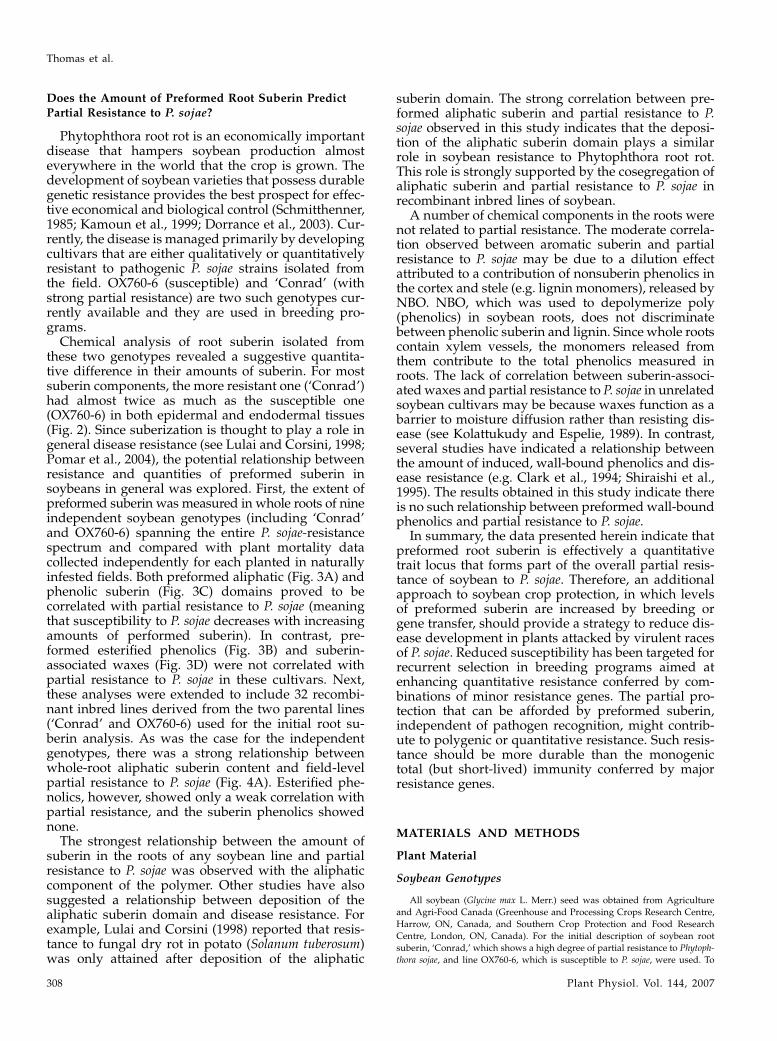
The results of all histochemical tests were the samefor the two genotypes studied in detail (‘Conrad,’which shows a high degree of partial resistance to P.sojae, and line OX760-6, which is susceptible to thispathogen). As indicated by autofluorescence, phenoliccompounds were located in the epidermal walls asclose to the root tip as 10 mm (Fig. 1A). By 50 mm,these compounds were also found in the walls of theadjacent cortical parenchyma (Fig. 1B). As the rootaged, more phenolics appeared to have accumulatedin walls of cells throughout the cortex. After stainingsections taken near the root tip with the lipophilicfluorochrome Fluorol yellow 088 (FY), yellowish greenfluorescence could be seen in all walls of the epidermis(Fig. 1C). In control (unstained) sections viewed withUV light, epidermal and cortical walls were blue. Thecombination of this color with yellow from the stainproduces a greenish hue. In sections farther from thetip, e.g. 90 mm, lipids were also located in all walls ofthe epidermis and the adjacent cortical layer (Fig. 1D).In sections at a distance of 50 mm (and farther) fromthe root tip, most walls of the epidermis and a few ofthose of the adjacent cortical layer resisted acid digestion(Fig. 1E). Taken together, the histochemical evidenceindicates that the walls of the epidermis containedsuberin. Although some of the cortical walls becamesuberized within 50 mm of the tip, they did not have aCasparian band and, thus, an exodermis was absent.
In the endodermis, state I development had occurredin the distal 80 mm of the root (Fig. 1F). Proximal to
Thomas et al.
300 Plant Physiol. Vol. 144, 2007
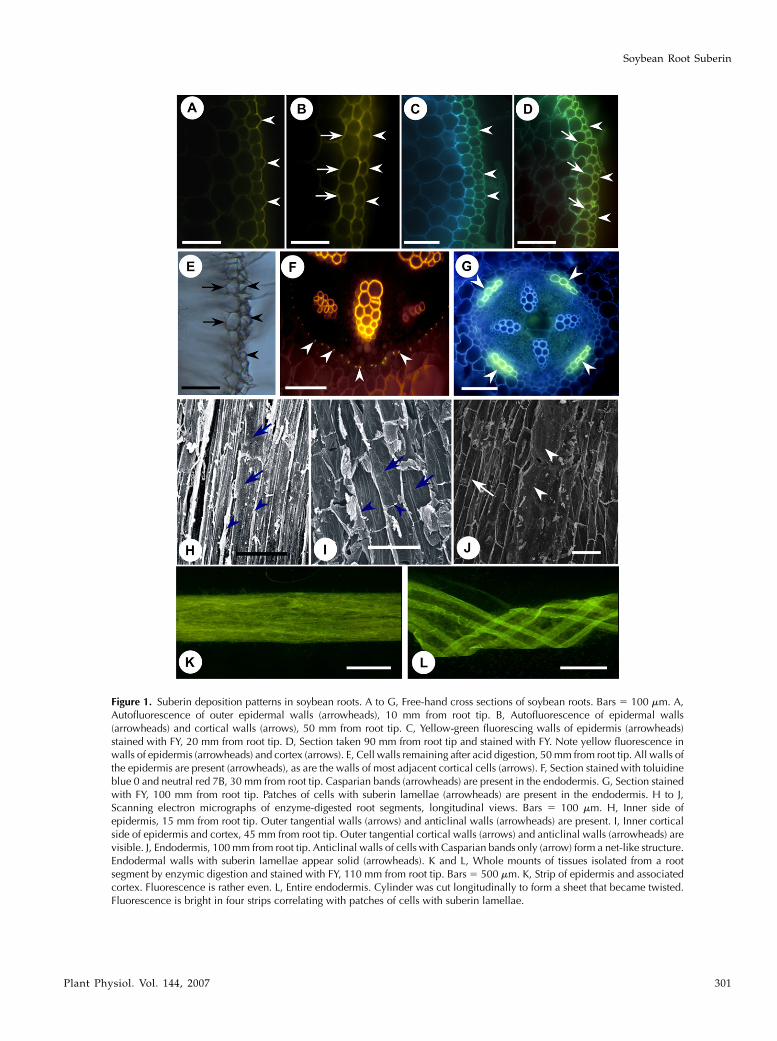
Figure 1. Suberin deposition patterns in soybean roots. A to G, Free-hand cross sections of soybean roots. Bars 5 100 mm. A,Autofluorescence of outer epidermal walls (arrowheads), 10 mm from root tip. B, Autofluorescence of epidermal walls(arrowheads) and cortical walls (arrows), 50 mm from root tip. C, Yellow-green fluorescing walls of epidermis (arrowheads)stained with FY, 20 mm from root tip. D, Section taken 90 mm from root tip and stained with FY. Note yellow fluorescence inwalls of epidermis (arrowheads) and cortex (arrows). E, Cell walls remaining after acid digestion, 50 mm from root tip. All walls ofthe epidermis are present (arrowheads), as are the walls of most adjacent cortical cells (arrows). F, Section stained with toluidineblue 0 and neutral red 7B, 30 mm from root tip. Casparian bands (arrowheads) are present in the endodermis. G, Section stainedwith FY, 100 mm from root tip. Patches of cells with suberin lamellae (arrowheads) are present in the endodermis. H to J,Scanning electron micrographs of enzyme-digested root segments, longitudinal views. Bars 5 100 mm. H, Inner side ofepidermis, 15 mm from root tip. Outer tangential walls (arrows) and anticlinal walls (arrowheads) are present. I, Inner corticalside of epidermis and cortex, 45 mm from root tip. Outer tangential cortical walls (arrows) and anticlinal walls (arrowheads) arevisible. J, Endodermis, 100 mm from root tip. Anticlinal walls of cells with Casparian bands only (arrow) form a net-like structure.Endodermal walls with suberin lamellae appear solid (arrowheads). K and L, Whole mounts of tissues isolated from a rootsegment by enzymic digestion and stained with FY, 110 mm from root tip. Bars 5 500 mm. K, Strip of epidermis and associatedcortex. Fluorescence is rather even. L, Entire endodermis. Cylinder was cut longitudinally to form a sheet that became twisted.Fluorescence is bright in four strips correlating with patches of cells with suberin lamellae.
Soybean Root Suberin
Plant Physiol. Vol. 144, 2007 301
this, a state II endodermis was evident in which somecells had developed suberin lamellae (Fig. 1G). Thelamella-containing cells in the state II endodermisoccurred near the phloem, while passage cells withoutlamellae were found near the xylem poles. With thisarrangement, the tetrarch pattern of the xylem led tothe development of four patches of cells with suberinlamellae in the endodermal cylinder (Fig. 1G). At 160mm from the root tip, about half of the endodermalcells had formed lamellae. No state III endodermis wasobserved in any of the samples analyzed (i.e. up to200 mm from the root tip).
Isolation of Intact Endodermal and Epidermal Tissue
Exhaustive treatment of soybean root segments withpectinase and cellulase allowed isolation of epider-mal/adjacent cortical and endodermal walls. Scanningelectron microscopy of the inner face of a strip ofisolated epidermis, originally 10 to 20 mm from theroot tip, localized the suberin to the outer tangentialand anticlinal walls of the epidermis (Fig. 1H). How-ever, in tissue isolated 40 to 50 mm from the root tip, theouter tangential and anticlinal walls of the adjacentcortical cells were also present (Fig. 1I). Henceforth, theterm ‘‘epidermis’’ will be used to designate the epider-mis and its associated cortical cell walls. Isolated stateII endodermal tissue showed both a fine web of anti-clinal endodermal cell walls corresponding to files ofcells containing only Casparian bands (Fig. 1J, arrows)and an opaque, dense area corresponding to files ofcells containing suberin lamellae as well as Casparianbands (Fig. 1J, arrowheads).
After staining with FY, the cylinder of isolated epi-dermal walls fluoresced a uniform, light yellow-green(Fig. 1K). The walls of the similarly stained, isolated
state II endodermal cylinders, on the other hand, dis-played four bands of bright yellow-green fluorescingwalls (consistent with the development of suberinlamella in longitudinal files of cells along the root axis)alternating with bands of weakly fluorescing walls(consistent with the presence of a Casparian bandonly; Fig. 1L). These isolated epidermal and endoder-mal tissues, along with the corresponding state Iendodermal tissues in younger areas of the root,were used to obtain a detailed chemical descriptionof soybean root suberin.
Chemical Analysis of Soybean Root Suberin
Total Suberin in Soybean Root Epidermis and Endodermis
The first 70 mm of endodermal root tissue (mea-sured from the root tip) was used to analyze thechemical composition of the Casparian band, while thenext 90 to 160 mm from the root tip was used toanalyze the chemical composition of the suberin la-mella (with contributions from the Casparian band).Epidermal tissues from the same segments were ana-lyzed to determine the chemical composition of thesecells along the axis of the root. In the case of theepidermis, the amounts of suberin components wereexpressed on the basis of root surface area (assuming adiameter of 0.76–0.90 mm); for the endodermis, theamounts were expressed on the basis of endodermalsurface area (assuming a diameter of 0.27–0.32 mm).
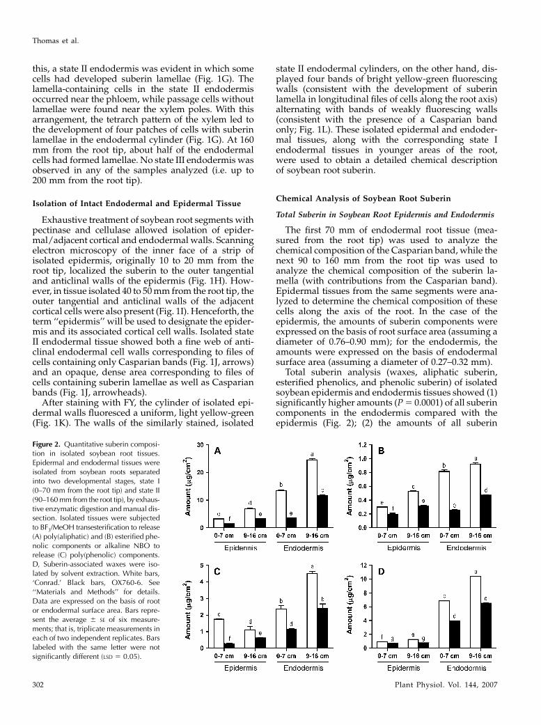
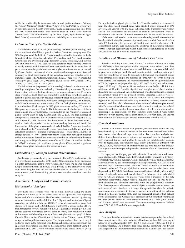
Total suberin analysis (waxes, aliphatic suberin,esterified phenolics, and phenolic suberin) of isolatedsoybean epidermis and endodermis tissues showed (1)significantly higher amounts (P 5 0.0001) of all suberincomponents in the endodermis compared with theepidermis (Fig. 2); (2) the amounts of all suberin
Figure 2. Quantitative suberin composi-tion in isolated soybean root tissues.Epidermal and endodermal tissues wereisolated from soybean roots separatedinto two developmental stages, state I(0–70 mm from the root tip) and state II(90–160 mm from the root tip), by exhaus-tive enzymatic digestion and manual dis-section. Isolated tissues were subjectedto BF3/MeOH transesterification to release(A) poly(aliphatic) and (B) esterified phe-nolic components or alkaline NBO torelease (C) poly(phenolic) components.D, Suberin-associated waxes were iso-lated by solvent extraction. White bars,‘Conrad.’ Black bars, OX760-6. See‘‘Materials and Methods’’ for details.Data are expressed on the basis of rootor endodermal surface area. Bars repre-sent the average 6 SE of six measure-ments; that is, triplicate measurements ineach of two independent replicates. Barslabeled with the same letter were notsignificantly different (LSD 5 0.05).
Thomas et al.
302 Plant Physiol. Vol. 144, 2007
components of the endodermis increased as the rootmatured from state I to state II (Fig. 2); (3) in general,the amount of all suberin components of the epidermisincreased along the axis of the root (Fig. 2); and (4) allsuberin components were found in significantly higheramounts (P 5 0.05) in tissues isolated from the P. sojae-resistant ‘Conrad’ versus the P. sojae-susceptible lineOX760-6.
Transesterification with BF3/MeOH released ali-phatic suberin monomers. The average total aliphaticsuberin was highest in the state II endodermis of‘Conrad’ (24.6 mg cm22) and lowest (1.55 mg cm22) inthe youngest epidermis (0–70 mm) of OX760-6 (Fig.2A). Treatment with BF3/MeOH also released vanillic,syringic, ferulic, p-coumaric, and p-hydroxybenzoicacids (esterified phenolics), in total amounts rangingbetween 0.29 mg cm22 in the 0- to 70-mm epidermalsegments and 0.95 mg cm22 in the state II (90–160 mm)endodermis (Fig. 2B). Vanillin and syringin were themain aromatic suberin monomers released by nitro-benzene oxidation (NBO) from all tissues studied. Thetotal amounts ranged from 0.2 mg cm22 in young epi-dermal tissues (0–70 mm) to 8.0 mg cm22 in the mature(state II) endodermis (Fig. 2C). Isolated epidermal andendodermal tissues yielded small amounts of suberin-associated waxes, ranging from 0.71 mg cm22 in theepidermis (0–70 mm) to 10.4 mg cm22 in the moremature (90–160 mm) endodermis (Fig. 2D).
Genotype Variation in Soybean Root Suberin
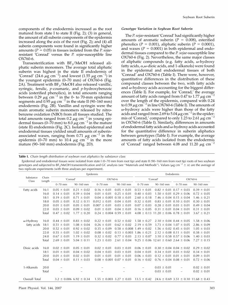
The P. sojae-resistant ‘Conrad’ had significantly higheramounts of aromatic suberin (P 5 0.008), esterifiedphenolics (P 5 0.001), aliphatic suberin (P 5 0.0001),and waxes (P 5 0.0001) in both epidermal and endo-dermal tissues compared to the P. sojae-susceptible lineOX760-6 (Fig. 2). Nevertheless, the same major classesof aliphatic compounds (e.g. fatty acids, v-hydroxyfatty acids, a,v-dioic acids, and 1-alkanols) were foundin the epidermal and endodermal tissues of both‘Conrad’ and OX760-6 (Table I). There were, however,quantitative differences in the distribution of thesecompound classes between the two, with fatty acidsand v-hydroxy acids accounting for the biggest differ-ences (Table I). For example, for ‘Conrad,’ the averageamount of fatty acids ranged from 0.47 to 1.77 mg cm22
over the length of the epidermis, compared with 0.24to 0.59 mg cm22 in line OX760-6 (Table I). The amounts ofv-hydroxy acids were higher than those of the fattyacids and ranged from 2.69 to 5.04 mg cm22 in the epider-mis of ‘Conrad,’ compared to only 1.23 to 2.61 mg cm22
in OX760-6 (Table I). Similarly, differences in amountsof endodermal fatty acids and v-hydroxy acids accountedfor the quantitative difference in suberin aliphaticsbetween genotypes (Table I). For example, the averageamounts of fatty acids isolated from the endodermisof ‘Conrad’ ranged between 4.08 and 11.20 mg cm22
Table I. Chain length distribution of soybean root aliphatics by substance class
Epidermal and endodermal tissues were isolated from state I (0–70 mm from root tip) and state II (90–160 mm from root tip) roots of two soybeangenotypes and subjected to BF3/MeOH transesterification and GC analysis (see ‘‘Materials and Methods’’). Values (mg cm22 6 SE) are the average oftwo replicate experiments (with three analyses per experiment).
Substance
Class
Chain
Length
Epidermis Endodermis
‘Conrad’ OX760-6 ‘Conrad’ OX760-6
0–70 mm 90–160 mm 0–70 mm 90–160 mm 0–70 mm 90–160 mm 0–70 mm 90–160 mm
Fatty acids 16:1 0.05 6 0.01 0.21 6 0.02 0.16 6 0.01 0.05 6 0.01 0.53 6 0.01 0.82 6 0.01 0.17 6 0.03 0.39 6 0.0116:0 0.14 6 0.01 0.39 6 0.04 0.01 6 0.01 0.25 6 0.01 0.40 6 0.03 1.50 6 0.01 0.29 6 0.06 0.72 6 0.0518:1 0.19 6 0.01 0.71 6 0.01 0.04 6 0.001 0.18 6 0.01 2.60 6 0.18 7.46 6 0.04 0.15 6 0.04 1.46 6 0.2118:0 0.05 6 0.01 0.32 6 0.11 0.012 6 0.01 0.04 6 0.01 0.32 6 0.01 0.83 6 0.01 0.10 6 0.01 0.30 6 0.0120:0 0.01 6 0.01 0.05 6 0.01 0.007 6 0.01 0.03 6 0.01 0.07 6 0.03 0.28 6 0.01 0.03 6 0.01 0.49 6 0.0422:0 0.03 6 0.01 0.09 6 0.02 0.01 6 0.01 0.04 6 0.01 0.16 6 0.05 0.31 6 0.01 0.04 6 0.01 0.31 6 0.01Total 0.47 6 0.02 1.77 6 0.20 0.24 6 0.004 0.59 6 0.01 4.08 6 0.13 11.20 6 0.06 0.78 6 0.01 3.67 6 0.21
v-Hydroxyfatty acids
16:0 0.44 6 0.01 0.83 6 0.02 0.22 6 0.01 0.52 6 0.02 1.58 6 0.27 2.10 6 0.04 0.44 6 0.05 1.58 6 0.0618:1 0.51 6 0.01 1.08 6 0.04 0.26 6 0.01 0.62 6 0.02 2.19 6 0.59 3.73 6 0.04 1.07 6 0.05 2.86 6 0.0920:0 0.52 6 0.01 0.92 6 0.02 0.35 6 0.09 0.58 6 0.008 1.49 6 0.02 1.56 6 0.02 0.45 6 0.01 1.05 6 0.0322:0 0.55 6 0.01 1.02 6 0.02 0.08 6 0.02 0.13 6 0.003 1.86 6 0.21 2.12 6 0.08 0.11 6 0.01 0.38 6 0.0124:0 0.67 6 0.01 1.19 6 0.01 0.32 6 0.02 0.77 6 0.03 2.13 6 0.07 3.10 6 0.58 0.57 6 0.06 1.40 6 0.03Total 2.69 6 0.01 5.04 6 0.11 1.23 6 0.03 2.61 6 0.04 9.25 6 0.06 12.61 6 0.60 2.64 6 0.06 7.27 6 0.15
Dioic acids 16:0 0.02 6 0.01 0.05 6 0.01 0.02 6 0.01 0.03 6 0.01 0.06 6 0.01 0.30 6 0.04 0.04 6 0.02 0.29 6 0.0218:1 0.01 6 0.01 0.04 6 0.02 0.04 6 0.03 0.03 6 0.01 0.04 6 0.01 0.34 6 0.01 0.03 6 0.02 0.34 6 0.0120:0 0.01 6 0.01 0.02 6 0.01 0.01 6 0.01 0.01 6 0.01 0.06 6 0.03 0.12 6 0.01 0.01 6 0.01 0.09 6 0.01Total 0.04 6 0.01 0.11 6 0.03 0.08 6 0.001 0.07 6 0.01 0.16 6 0.02 0.76 6 0.04 0.08 6 0.01 0.72 6 0.06
1-Alkanols 20:0 – – – – – 0.03 6 0.01 – 0.02 6 0.01Total – – – – – 0.03 6 0.01 – 0.02 6 0.01
Overall Total 3.2 6 0.006 6.92 6 0.34 1.55 6 0.003 3.27 6 0.03 13.5 6 0.42 24.6 6 0.69 3.51 6 0.10 11.68 6 0.43
Soybean Root Suberin
Plant Physiol. Vol. 144, 2007 303
compared to 0.78 and 3.67 mg cm22 in OX760-6,while the v-hydroxy acids varied from 9.25 to 12.61mg cm22 in ‘Conrad’ compared to 2.64 to 7.27 mg cm22
in OX760-6.The chain length distribution of aliphatic suberin
monomers ranged from C16 to C24 regardless of thetissue or root section investigated, with C18:1 acidderivatives usually in greatest abundance (Table I).Closer inspection, however, revealed that while thesame distribution of monomeric chain length existedin the epidermis and endodermis of both genotypes,there were quantitative differences in the amounts ofsome key monomers. Specifically, ‘Conrad’ had moreC18:1 carboxylic acid (especially in the endodermis)and C16, C18:1, C22, and C24 v-hydroxy acids than didOX760-6 (Table I).
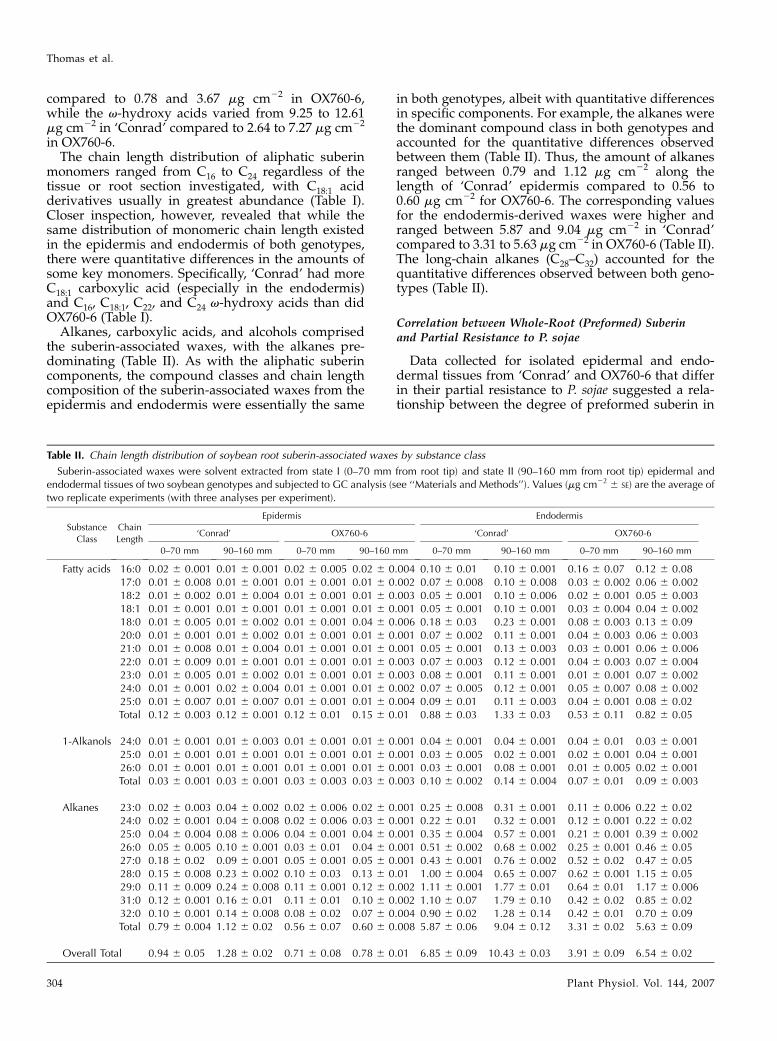
Alkanes, carboxylic acids, and alcohols comprisedthe suberin-associated waxes, with the alkanes pre-dominating (Table II). As with the aliphatic suberincomponents, the compound classes and chain lengthcomposition of the suberin-associated waxes from theepidermis and endodermis were essentially the same
in both genotypes, albeit with quantitative differencesin specific components. For example, the alkanes werethe dominant compound class in both genotypes andaccounted for the quantitative differences observedbetween them (Table II). Thus, the amount of alkanesranged between 0.79 and 1.12 mg cm22 along thelength of ‘Conrad’ epidermis compared to 0.56 to0.60 mg cm22 for OX760-6. The corresponding valuesfor the endodermis-derived waxes were higher andranged between 5.87 and 9.04 mg cm22 in ‘Conrad’compared to 3.31 to 5.63 mg cm22 in OX760-6 (Table II).The long-chain alkanes (C28–C32) accounted for thequantitative differences observed between both geno-types (Table II).
Correlation between Whole-Root (Preformed) Suberinand Partial Resistance to P. sojae
Data collected for isolated epidermal and endo-dermal tissues from ‘Conrad’ and OX760-6 that differin their partial resistance to P. sojae suggested a rela-tionship between the degree of preformed suberin in
Table II. Chain length distribution of soybean root suberin-associated waxes by substance class
Suberin-associated waxes were solvent extracted from state I (0–70 mm from root tip) and state II (90–160 mm from root tip) epidermal andendodermal tissues of two soybean genotypes and subjected to GC analysis (see ‘‘Materials and Methods’’). Values (mg cm22 6 SE) are the average oftwo replicate experiments (with three analyses per experiment).
Substance
Class
Chain
Length
Epidermis Endodermis
‘Conrad’ OX760-6 ‘Conrad’ OX760-6
0–70 mm 90–160 mm 0–70 mm 90–160 mm 0–70 mm 90–160 mm 0–70 mm 90–160 mm
Fatty acids 16:0 0.02 6 0.001 0.01 6 0.001 0.02 6 0.005 0.02 6 0.004 0.10 6 0.01 0.10 6 0.001 0.16 6 0.07 0.12 6 0.0817:0 0.01 6 0.008 0.01 6 0.001 0.01 6 0.001 0.01 6 0.002 0.07 6 0.008 0.10 6 0.008 0.03 6 0.002 0.06 6 0.00218:2 0.01 6 0.002 0.01 6 0.004 0.01 6 0.001 0.01 6 0.003 0.05 6 0.001 0.10 6 0.006 0.02 6 0.001 0.05 6 0.00318:1 0.01 6 0.001 0.01 6 0.001 0.01 6 0.001 0.01 6 0.001 0.05 6 0.001 0.10 6 0.001 0.03 6 0.004 0.04 6 0.00218:0 0.01 6 0.005 0.01 6 0.002 0.01 6 0.001 0.04 6 0.006 0.18 6 0.03 0.23 6 0.001 0.08 6 0.003 0.13 6 0.0920:0 0.01 6 0.001 0.01 6 0.002 0.01 6 0.001 0.01 6 0.001 0.07 6 0.002 0.11 6 0.001 0.04 6 0.003 0.06 6 0.00321:0 0.01 6 0.008 0.01 6 0.004 0.01 6 0.001 0.01 6 0.001 0.05 6 0.001 0.13 6 0.003 0.03 6 0.001 0.06 6 0.00622:0 0.01 6 0.009 0.01 6 0.001 0.01 6 0.001 0.01 6 0.003 0.07 6 0.003 0.12 6 0.001 0.04 6 0.003 0.07 6 0.00423:0 0.01 6 0.005 0.01 6 0.002 0.01 6 0.001 0.01 6 0.003 0.08 6 0.001 0.11 6 0.001 0.01 6 0.001 0.07 6 0.00224:0 0.01 6 0.001 0.02 6 0.004 0.01 6 0.001 0.01 6 0.002 0.07 6 0.005 0.12 6 0.001 0.05 6 0.007 0.08 6 0.00225:0 0.01 6 0.007 0.01 6 0.007 0.01 6 0.001 0.01 6 0.004 0.09 6 0.01 0.11 6 0.003 0.04 6 0.001 0.08 6 0.02Total 0.12 6 0.003 0.12 6 0.001 0.12 6 0.01 0.15 6 0.01 0.88 6 0.03 1.33 6 0.03 0.53 6 0.11 0.82 6 0.05
1-Alkanols 24:0 0.01 6 0.001 0.01 6 0.003 0.01 6 0.001 0.01 6 0.001 0.04 6 0.001 0.04 6 0.001 0.04 6 0.01 0.03 6 0.00125:0 0.01 6 0.001 0.01 6 0.001 0.01 6 0.001 0.01 6 0.001 0.03 6 0.005 0.02 6 0.001 0.02 6 0.001 0.04 6 0.00126:0 0.01 6 0.001 0.01 6 0.001 0.01 6 0.001 0.01 6 0.001 0.03 6 0.001 0.08 6 0.001 0.01 6 0.005 0.02 6 0.001Total 0.03 6 0.001 0.03 6 0.001 0.03 6 0.003 0.03 6 0.003 0.10 6 0.002 0.14 6 0.004 0.07 6 0.01 0.09 6 0.003
Alkanes 23:0 0.02 6 0.003 0.04 6 0.002 0.02 6 0.006 0.02 6 0.001 0.25 6 0.008 0.31 6 0.001 0.11 6 0.006 0.22 6 0.0224:0 0.02 6 0.001 0.04 6 0.008 0.02 6 0.006 0.03 6 0.001 0.22 6 0.01 0.32 6 0.001 0.12 6 0.001 0.22 6 0.0225:0 0.04 6 0.004 0.08 6 0.006 0.04 6 0.001 0.04 6 0.001 0.35 6 0.004 0.57 6 0.001 0.21 6 0.001 0.39 6 0.00226:0 0.05 6 0.005 0.10 6 0.001 0.03 6 0.01 0.04 6 0.001 0.51 6 0.002 0.68 6 0.002 0.25 6 0.001 0.46 6 0.0527:0 0.18 6 0.02 0.09 6 0.001 0.05 6 0.001 0.05 6 0.001 0.43 6 0.001 0.76 6 0.002 0.52 6 0.02 0.47 6 0.0528:0 0.15 6 0.008 0.23 6 0.002 0.10 6 0.03 0.13 6 0.01 1.00 6 0.004 0.65 6 0.007 0.62 6 0.001 1.15 6 0.0529:0 0.11 6 0.009 0.24 6 0.008 0.11 6 0.001 0.12 6 0.002 1.11 6 0.001 1.77 6 0.01 0.64 6 0.01 1.17 6 0.00631:0 0.12 6 0.001 0.16 6 0.01 0.11 6 0.01 0.10 6 0.002 1.10 6 0.07 1.79 6 0.10 0.42 6 0.02 0.85 6 0.0232:0 0.10 6 0.001 0.14 6 0.008 0.08 6 0.02 0.07 6 0.004 0.90 6 0.02 1.28 6 0.14 0.42 6 0.01 0.70 6 0.09Total 0.79 6 0.004 1.12 6 0.02 0.56 6 0.07 0.60 6 0.008 5.87 6 0.06 9.04 6 0.12 3.31 6 0.02 5.63 6 0.09
Overall Total 0.94 6 0.05 1.28 6 0.02 0.71 6 0.08 0.78 6 0.01 6.85 6 0.09 10.43 6 0.03 3.91 6 0.09 6.54 6 0.02
Thomas et al.
304 Plant Physiol. Vol. 144, 2007
soybean roots and resistance to P. sojae. That is to saythat a greater degree of suberization was found in theroot tissues of ‘Conrad,’ which shows a higher degreeof partial resistance compared to that of the moresusceptible OX760-6. To further explore this relation-ship, the extent of preformed suberin in the roots ofnine independent soybean lines differing in theirpartial resistance to P. sojae was measured. Due tothe labor-intensive nature of epidermis and endoder-mis tissue isolation, whole roots were used for thisanalysis.
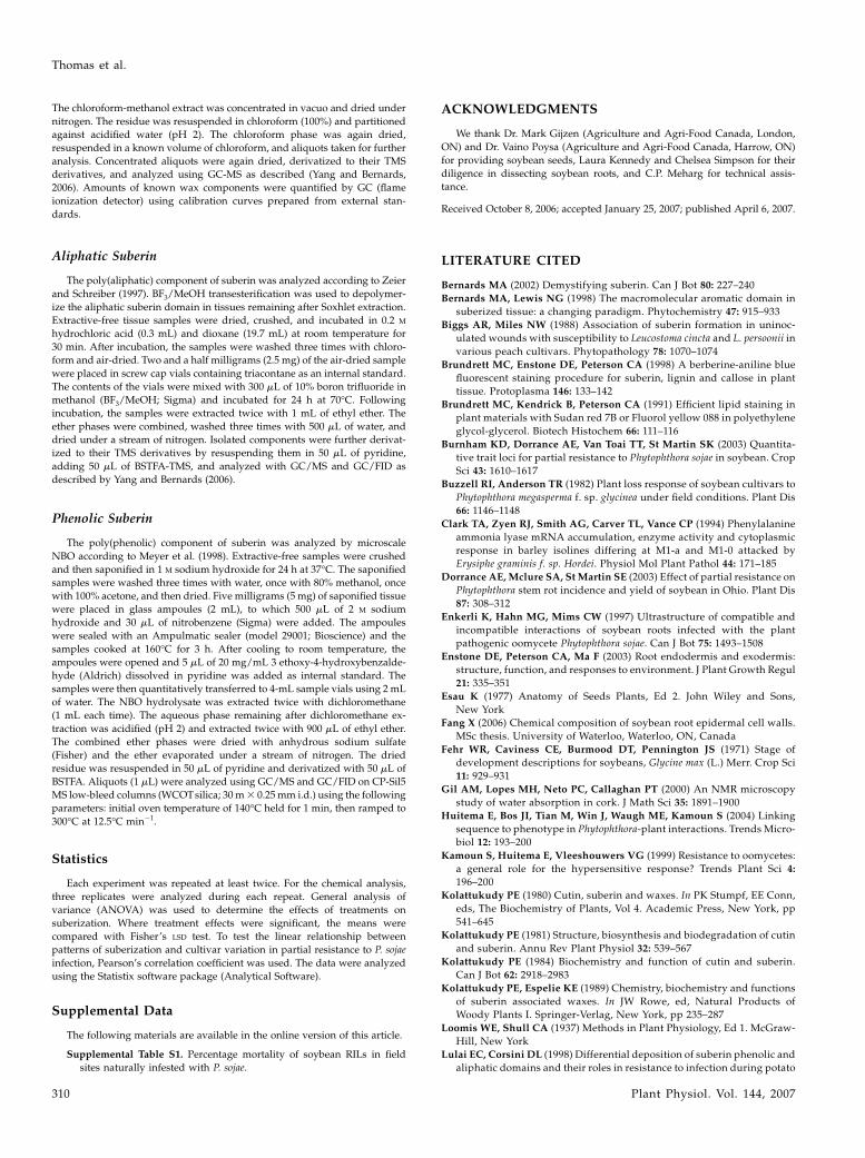
Plant mortality data collected for plants grown underfield conditions in plots naturally infested with P. sojaewere plotted against the amount of aliphatic suberin,esterified phenolics, phenolic suberin, and suberin-associated waxes measured from whole roots of green-house-grown plants (Fig. 3). According to these data,(1) the same differences in amount of suberin compo-nents observed for isolated tissues between ‘Conrad’and OX760-6 were observed at the whole-root level,and (2) the Pearson’s correlation coefficient (r) wasstrongly negative (r 5 20.89) between plant mortalityand aliphatic suberin (Fig. 3A). By contrast, r was low(r 5 20.22) for the relationship between plant mortalityand esterified phenolics (Fig. 3B), only moderatelynegative (r 5 20.55) for the relationship between plantmortality and phenolic suberin (Fig. 3C), and evenpositive for the relationships between plant mortalityand suberin-associated waxes (r 5 0.16; Fig. 3D).
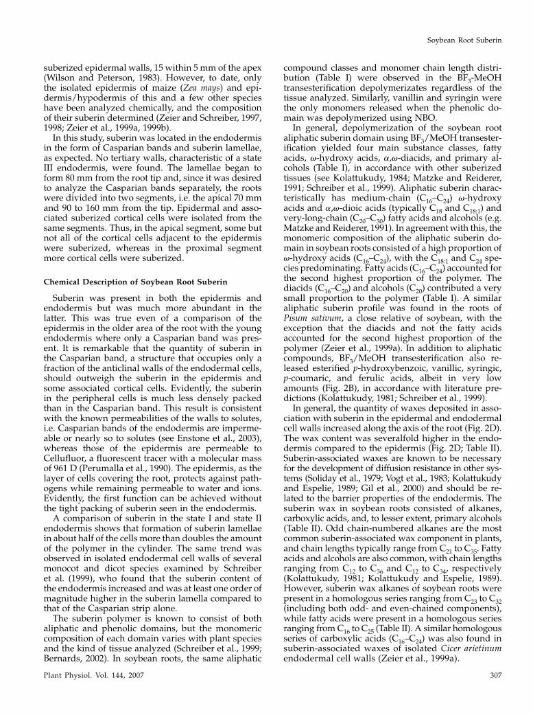
Inheritance of Whole-Root (Preformed) Suberin andPartial Resistance to P. sojae
To test whether whole-root (preformed) suberin isgenetically linked with partial resistance to P. sojae,seed was obtained from 32 recombinant inbred lines
developed from an initial cross between ‘Conrad’ andOX760-6 performed by Dr. Vaino Poysa (Agricultureand Agri-Food Canada, Greenhouse and ProcessingCrops Research Centre, Harrow, ON, Canada), forwhich plant mortality data were also available. Eachline was grown under identical greenhouse conditionsand their whole-root suberin content measured.
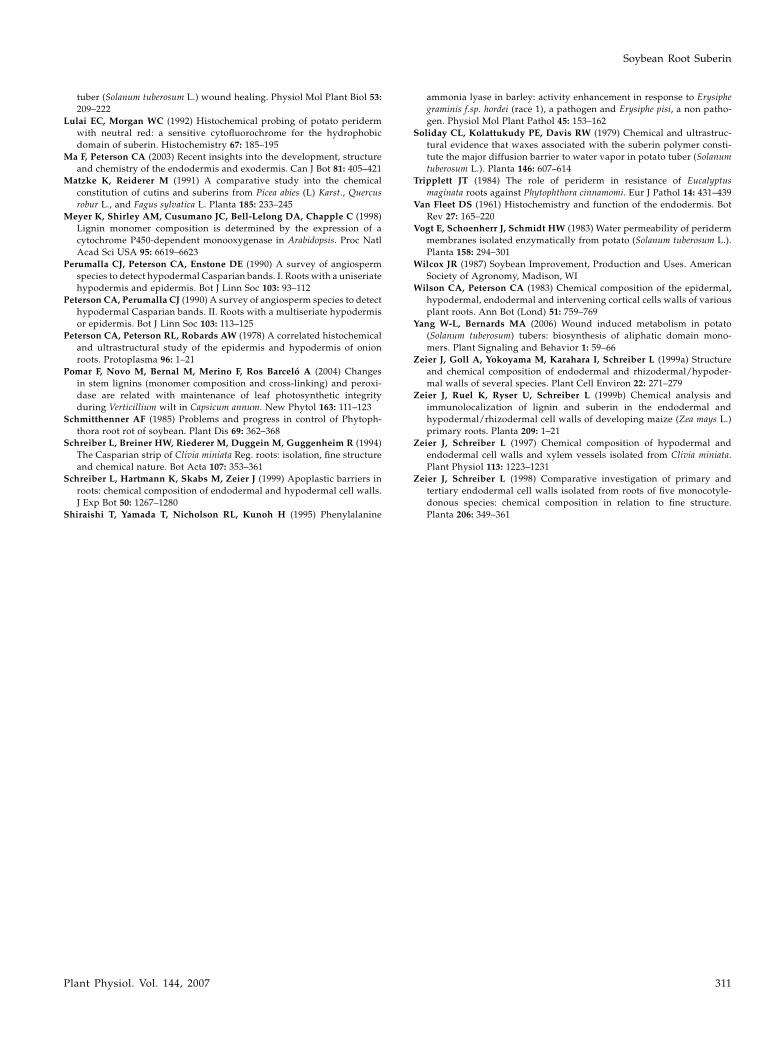
Plant mortality data from two individual field plotsnaturally infested with P. sojae, but varying in inoculumdensity, collected over two growing seasons (n 5 4),were plotted against quantitative measures of whole-root suberin components. For the recombinant inbredlines, the Pearson’s correlation coefficient (r) for therelationship between aliphatic suberin and partial re-sistance to P. sojae were consistently high in all fourplots (20.61 to 20.92; Table III), suggesting that rootsuberin content cosegregates with partial resistance toP. sojae. Weaker correlations were observed for esteri-fied phenolics (20.27 to 20.63), phenolic suberin(0.02 to 20.23), and suberin-associated waxes (20.04to 20.30; Table III).
When data for plant mortality from all field plotsand both experimental replicates of suberin measure-ments were combined, a strong negative (20.72)Pearson’s correlation coefficient was still observedbetween aliphatic suberin and partial resistance to P.sojae in the 32 recombinant inbred lines (Fig. 4A).Other suberin components showed either only weak(r 5 20.34; esterified phenolics) or no (r 5 0.02; suberinphenolics) correlation with partial resistance when allfield loss data were combined (data not shown). Whenexamined more finely, it could be shown that the maincomponents contributing to the correlation betweenaliphatic suberin and partial resistance to P. sojaewere v-hydroxy fatty acids (r 5 20.60 to 20.90, TableIII; r 5 20.72 [combined], Fig. 4B), with smaller
Figure 3. Relationship between preformedwhole-root suberin and soybean partial re-sistance to P. sojae. Ten-day-old roots fromnine independent soybean genotypes weresubjected to solvent extraction, BF3/MeOHtransesterification, and alkaline NBO (see‘‘Materials and Methods’’ for details). Dataobtained for aliphatic suberin (A), esteri-fied phenolic (B), phenolic suberin (C), andsuberin-associated (D) wax components wereplotted against plant mortality data obtainedfrom plantings of the same genotypes infields naturally infested with P. sojae. Geno-types used (% mortality) were ‘Westag 97’(1%), ‘Conrad’ (4%), ‘Elgin’ (5%), ‘Williams’(40%), ‘Steele’ (60%), ‘Sloan’ (70%),‘Haro(1-7)1’ (80%), ‘OX20-8’ (80%), andOX760-6 (80%). The data for ‘Conrad’(diamonds)andOX760-6 (squares) arehigh-lighted. Each data point represents an inde-pendent estimate of the suberin componentmeasured. Regression lines are shown toindicate trends. The r values are Pearson’scorrelation coefficients.
Soybean Root Suberin
Plant Physiol. Vol. 144, 2007 305

contributions by free fatty acids (r 5 20.31 to 20.65,Table III; r 5 20.50 [combined], Fig. 4C) and a,v-dioicacids (r 5 20.03 to 20.17, Table III; r 5 20.27 [com-bined], Fig. 4D).
DISCUSSION
Patterns of Suberization in Soybean Roots
An anatomical analysis of roots of the two soybeangenotypes (‘Conrad’ and OX760-6), from which epi-dermal and endodermal tissues were isolated andseparated prior to chemical analysis, indicated thepresence of suberin in the epidermal walls. This poly-mer appeared to be confined to the epidermis near the
root tip, but 50 mm and farther from the tip was alsodetected in the adjacent cortical cells. Failure of theepidermal cells to separate from each other, and alsofrom the adjacent cortical cells in older root regions,during digestion with acid or enzymes, indicates thatthe suberin polymer was continuous across the middlelamellae joining these cells together. When viewedwith a transmission electron microscope, the suberinin the epidermal walls appeared as very faint, elec-tron-dense bands (Fang, 2006), similar to the pattern ofsuberin seen earlier in onion and termed ‘‘diffusesuberin’’ (Brundrett et al., 1988). The occurrence ofsuberin in the root epidermis is not unusual. Forexample, based on results of histochemical tests andacid digestions, the roots of 21 of 27 species tested have
Table III. Pearson’s correlation coefficients for the relationship between preformed suberin components and partial resistance to P. sojae
Whole-root suberin components measured for 32 recombinant inbred lines derived from ‘Conrad’ and OX760-6 were plotted against plantmortality data collected in 2003 and 2004 from field plots naturally infested with P. sojae. Values are Pearson’s correlation coefficients (r) with P values(to two significant figures) in parentheses. Suberin data are from two independently replicated experiments, each with triplicate measurements ofsuberin components. Data from each experimental replicate were plotted against plant mortality data from field plot. n 5 102 per plot, per replicate.Overall P 5 0.05.
Suberin
Components
Pearson’s Correlation Coefficient (r) and P Values
2003 2004
Plot A Plot B Plot A Plot B
Replicate 1 Replicate 2 Replicate 1 Replicate 2 Replicate 1 Replicate 2 Replicate 1 Replicate 2
Aliphatic suberin 20.61 (0.01) 20.69 (0.01) 20.62 (0.01) 20.70 (0.01) 20.74 (0.01) 20.92 (0.01) 20.63 (0.01) 20.81 (0.01)v-Hydroxy acids 20.62 (0.01) 20.75 (0.01) 20.60 (0.01) 20.74 (0.01) 20.80 (0.01) 20.90 (0.01) 20.70 (0.01) 20.82 (0.01)Fatty acids 20.50 (0.01) 20.34 (0.08) 20.60 (0.01) 20.31 (0.02) 20.44 (0.01) 20.65 (0.01) 20.43 (0.01) 20.46 (0.01)Diacids 20.03 (0.81) 20.14 (0.18) 20.08 (0.44) 20.16 (0.12) 20.17 (0.11) 20.13 (0.22) 20.07 (0.47) 20.10 (0.51)Esterified phenols 20.50 (0.01) 20.27 (0.02) 20.48 (0.01) 20.30 (0.01) 20.50 (0.01) 20.63 (0.01) 20.61 (0.01) 20.52 (0.01)Phenolic 20.02 (0.88) 20.12 (0.27) 0.02 (0.89) 20.06 (0.31) 20.12 (0.27) 20.22 (0.32) 20.11 (0.30) 20.23 (0.30)Suberin-associated
waxes20.04 (0.97) 20.10 (0.31) 20.21 (0.04) 20.12 (0.17) 20.15 (0.17) 20.20 (0.02) 20.30 (0.02) 20.18 (0.12)
Figure 4. Cosegregation of preformedwhole-root aliphatic suberin and soybeanpartial resistance to P. sojae. Ten-day-oldroots from 32 recombinant inbred lines gen-erated from a cross between soybean ‘Con-rad’ (resistant) and OX760-6 (susceptible)were subjected to BF3/MeOH transesterifica-tion (see ‘‘Materials and Methods’’ for de-tails). A, Data obtained for aliphatic suberincomponents were plotted against plant mor-tality data obtained from four independentplantings of the same recombinant inbredlines at two different field sites naturallyinfested with P. sojae. The data for the paren-tal lines ‘Conrad’ (diamonds) and OX760-6(squares) are highlighted. B to D, Individualsubstance class data extracted from plot (A)are plotted separately for v-hydroxy fattyacids (B), fatty acids (C), and a,v-dioic acids(D). Each data point represents an indepen-dent estimate of the aliphatic suberin com-ponent measured. Regression lines areshown to indicate trends. The r values arePearson’s correlation coefficients.
Thomas et al.
306 Plant Physiol. Vol. 144, 2007
suberized epidermal walls, 15 within 5 mm of the apex(Wilson and Peterson, 1983). However, to date, onlythe isolated epidermis of maize (Zea mays) and epi-dermis/hypodermis of this and a few other specieshave been analyzed chemically, and the compositionof their suberin determined (Zeier and Schreiber, 1997,1998; Zeier et al., 1999a, 1999b).
In this study, suberin was located in the endodermisin the form of Casparian bands and suberin lamellae,as expected. No tertiary walls, characteristic of a stateIII endodermis, were found. The lamellae began toform 80 mm from the root tip and, since it was desiredto analyze the Casparian bands separately, the rootswere divided into two segments, i.e. the apical 70 mmand 90 to 160 mm from the tip. Epidermal and asso-ciated suberized cortical cells were isolated from thesame segments. Thus, in the apical segment, some butnot all of the cortical cells adjacent to the epidermiswere suberized, whereas in the proximal segmentmore cortical cells were suberized.
Chemical Description of Soybean Root Suberin
Suberin was present in both the epidermis andendodermis but was much more abundant in thelatter. This was true even of a comparison of theepidermis in the older area of the root with the youngendodermis where only a Casparian band was pres-ent. It is remarkable that the quantity of suberin inthe Casparian band, a structure that occupies only afraction of the anticlinal walls of the endodermal cells,should outweigh the suberin in the epidermis andsome associated cortical cells. Evidently, the suberinin the peripheral cells is much less densely packedthan in the Casparian band. This result is consistentwith the known permeabilities of the walls to solutes,i.e. Casparian bands of the endodermis are imperme-able or nearly so to solutes (see Enstone et al., 2003),whereas those of the epidermis are permeable toCellufluor, a fluorescent tracer with a molecular massof 961 D (Perumalla et al., 1990). The epidermis, as thelayer of cells covering the root, protects against path-ogens while remaining permeable to water and ions.Evidently, the first function can be achieved withoutthe tight packing of suberin seen in the endodermis.
A comparison of suberin in the state I and state IIendodermis shows that formation of suberin lamellaein about half of the cells more than doubles the amountof the polymer in the cylinder. The same trend wasobserved in isolated endodermal cell walls of severalmonocot and dicot species examined by Schreiberet al. (1999), who found that the suberin content ofthe endodermis increased and was at least one order ofmagnitude higher in the suberin lamella compared tothat of the Casparian strip alone.
The suberin polymer is known to consist of bothaliphatic and phenolic domains, but the monomericcomposition of each domain varies with plant speciesand the kind of tissue analyzed (Schreiber et al., 1999;Bernards, 2002). In soybean roots, the same aliphatic
compound classes and monomer chain length distri-bution (Table I) were observed in the BF3-MeOHtransesterification depolymerizates regardless of thetissue analyzed. Similarly, vanillin and syringin werethe only monomers released when the phenolic do-main was depolymerized using NBO.
In general, depolymerization of the soybean rootaliphatic suberin domain using BF3/MeOH transester-ification yielded four main substance classes, fattyacids, v-hydroxy acids, a,v-diacids, and primary al-cohols (Table I), in accordance with other suberizedtissues (see Kolattukudy, 1984; Matzke and Reiderer,1991; Schreiber et al., 1999). Aliphatic suberin charac-teristically has medium-chain (C16–C24) v-hydroxyacids and a,v-dioic acids (typically C18 and C18:1) andvery-long-chain (C20–C30) fatty acids and alcohols (e.g.Matzke and Reiderer, 1991). In agreement with this, themonomeric composition of the aliphatic suberin do-main in soybean roots consisted of a high proportion ofv-hydroxy acids (C16–C24), with the C18:1 and C24 spe-cies predominating. Fatty acids (C16–C24) accounted forthe second highest proportion of the polymer. Thediacids (C16–C20) and alcohols (C20) contributed a verysmall proportion to the polymer (Table I). A similaraliphatic suberin profile was found in the roots ofPisum sativum, a close relative of soybean, with theexception that the diacids and not the fatty acidsaccounted for the second highest proportion of thepolymer (Zeier et al., 1999a). In addition to aliphaticcompounds, BF3/MeOH transesterification also re-leased esterified p-hydroxybenzoic, vanillic, syringic,p-coumaric, and ferulic acids, albeit in very lowamounts (Fig. 2B), in accordance with literature pre-dictions (Kolattukudy, 1981; Schreiber et al., 1999).
In general, the quantity of waxes deposited in asso-ciation with suberin in the epidermal and endodermalcell walls increased along the axis of the root (Fig. 2D).The wax content was severalfold higher in the endo-dermis compared to the epidermis (Fig. 2D; Table II).Suberin-associated waxes are known to be necessaryfor the development of diffusion resistance in other sys-tems (Soliday et al., 1979; Vogt et al., 1983; Kolattukudyand Espelie, 1989; Gil et al., 2000) and should be re-lated to the barrier properties of the endodermis. Thesuberin wax in soybean roots consisted of alkanes,carboxylic acids, and, to lesser extent, primary alcohols(Table II). Odd chain-numbered alkanes are the mostcommon suberin-associated wax component in plants,and chain lengths typically range from C21 to C35. Fattyacids and alcohols are also common, with chain lengthsranging from C12 to C36 and C12 to C34, respectively(Kolattukudy, 1981; Kolattukudy and Espelie, 1989).However, suberin wax alkanes of soybean roots werepresent in a homologous series ranging from C23 to C32(including both odd- and even-chained components),while fatty acids were present in a homologous seriesranging from C16 to C25 (Table II). A similar homologousseries of carboxylic acids (C16–C24) was also found insuberin-associated waxes of isolated Cicer arietinumendodermal cell walls (Zeier et al., 1999a).
Soybean Root Suberin
Plant Physiol. Vol. 144, 2007 307
Does the Amount of Preformed Root Suberin Predict
Partial Resistance to P. sojae?
Phytophthora root rot is an economically importantdisease that hampers soybean production almosteverywhere in the world that the crop is grown. Thedevelopment of soybean varieties that possess durablegenetic resistance provides the best prospect for effec-tive economical and biological control (Schmitthenner,1985; Kamoun et al., 1999; Dorrance et al., 2003). Cur-rently, the disease is managed primarily by developingcultivars that are either qualitatively or quantitativelyresistant to pathogenic P. sojae strains isolated fromthe field. OX760-6 (susceptible) and ‘Conrad’ (withstrong partial resistance) are two such genotypes cur-rently available and they are used in breeding pro-grams.
Chemical analysis of root suberin isolated fromthese two genotypes revealed a suggestive quantita-tive difference in their amounts of suberin. For mostsuberin components, the more resistant one (‘Conrad’)had almost twice as much as the susceptible one(OX760-6) in both epidermal and endodermal tissues(Fig. 2). Since suberization is thought to play a role ingeneral disease resistance (see Lulai and Corsini, 1998;Pomar et al., 2004), the potential relationship betweenresistance and quantities of preformed suberin insoybeans in general was explored. First, the extent ofpreformed suberin was measured in whole roots of nineindependent soybean genotypes (including ‘Conrad’and OX760-6) spanning the entire P. sojae-resistancespectrum and compared with plant mortality datacollected independently for each planted in naturallyinfested fields. Both preformed aliphatic (Fig. 3A) andphenolic suberin (Fig. 3C) domains proved to becorrelated with partial resistance to P. sojae (meaningthat susceptibility to P. sojae decreases with increasingamounts of performed suberin). In contrast, pre-formed esterified phenolics (Fig. 3B) and suberin-associated waxes (Fig. 3D) were not correlated withpartial resistance to P. sojae in these cultivars. Next,these analyses were extended to include 32 recombi-nant inbred lines derived from the two parental lines(‘Conrad’ and OX760-6) used for the initial root su-berin analysis. As was the case for the independentgenotypes, there was a strong relationship betweenwhole-root aliphatic suberin content and field-levelpartial resistance to P. sojae (Fig. 4A). Esterified phe-nolics, however, showed only a weak correlation withpartial resistance, and the suberin phenolics showednone.
The strongest relationship between the amount ofsuberin in the roots of any soybean line and partialresistance to P. sojae was observed with the aliphaticcomponent of the polymer. Other studies have alsosuggested a relationship between deposition of thealiphatic suberin domain and disease resistance. Forexample, Lulai and Corsini (1998) reported that resis-tance to fungal dry rot in potato (Solanum tuberosum)was only attained after deposition of the aliphatic
suberin domain. The strong correlation between pre-formed aliphatic suberin and partial resistance to P.sojae observed in this study indicates that the deposi-tion of the aliphatic suberin domain plays a similarrole in soybean resistance to Phytophthora root rot.This role is strongly supported by the cosegregation ofaliphatic suberin and partial resistance to P. sojae inrecombinant inbred lines of soybean.
A number of chemical components in the roots werenot related to partial resistance. The moderate correla-tion observed between aromatic suberin and partialresistance to P. sojae may be due to a dilution effectattributed to a contribution of nonsuberin phenolics inthe cortex and stele (e.g. lignin monomers), released byNBO. NBO, which was used to depolymerize poly(phenolics) in soybean roots, does not discriminatebetween phenolic suberin and lignin. Since whole rootscontain xylem vessels, the monomers released fromthem contribute to the total phenolics measured inroots. The lack of correlation between suberin-associ-ated waxes and partial resistance to P. sojae in unrelatedsoybean cultivars may be because waxes function as abarrier to moisture diffusion rather than resisting dis-ease (see Kolattukudy and Espelie, 1989). In contrast,several studies have indicated a relationship betweenthe amount of induced, wall-bound phenolics and dis-ease resistance (e.g. Clark et al., 1994; Shiraishi et al.,1995). The results obtained in this study indicate thereis no such relationship between preformed wall-boundphenolics and partial resistance to P. sojae.
In summary, the data presented herein indicate thatpreformed root suberin is effectively a quantitativetrait locus that forms part of the overall partial resis-tance of soybean to P. sojae. Therefore, an additionalapproach to soybean crop protection, in which levelsof preformed suberin are increased by breeding orgene transfer, should provide a strategy to reduce dis-ease development in plants attacked by virulent racesof P. sojae. Reduced susceptibility has been targeted forrecurrent selection in breeding programs aimed atenhancing quantitative resistance conferred by com-binations of minor resistance genes. The partial pro-tection that can be afforded by preformed suberin,independent of pathogen recognition, might contrib-ute to polygenic or quantitative resistance. Such resis-tance should be more durable than the monogenictotal (but short-lived) immunity conferred by majorresistance genes.
MATERIALS AND METHODS
Plant Material
Soybean Genotypes
All soybean (Glycine max L. Merr.) seed was obtained from Agriculture
and Agri-Food Canada (Greenhouse and Processing Crops Research Centre,
Harrow, ON, Canada, and Southern Crop Protection and Food Research
Centre, London, ON, Canada). For the initial description of soybean root
suberin, ‘Conrad,’ which shows a high degree of partial resistance to Phytoph-
thora sojae, and line OX760-6, which is susceptible to P. sojae, were used. To
Thomas et al.
308 Plant Physiol. Vol. 144, 2007
verify the relationship between root suberin and partial resistance, ‘Westag
97,’ ‘Elgin,’ ‘Williams,’ ‘Steele,’ ‘Sloan,’ ‘Haro(1-7)1,’ and ‘OX20-8,’ which vary
in their resistance to P. sojae, were used. Finally, 32 representative lines from
the .60 recombinant inbred lines derived from an initial cross between
‘Conrad’ and OX760-6 (maintained by Dr. Vaino Poysa, Agriculture and Agri-
Food Canada) were used to examine the heritability of root suberin.
Determination of Partial Resistance
Partial resistance of ‘Conrad’ (4% mortality), OX760-6 (80% mortality), and
the recombinant inbred line population derived from them (ranging from 1%–
90%; Supplemental Table S1) to P. sojae was determined in two trials in fields
6a and 6b at the Woodslee substation (Agriculture and Agri-Food Canada,
Greenhouse and Processing Crops Research Centre, Woodslee, ON) in both
2003 and 2004 (n 5 4). The Woodslee sites consist of Brookston clay-loam soil
naturally infested with P. sojae and have been used annually for more than 25
years to evaluate the partial resistance of soybean cultivars to root rot caused
by P. sojae. Partial resistance of all other cultivars/lines represents a historical
summary of field performance at the Woodslee nurseries, collected over a
number of years (T.R. Anderson, unpublished data). These were (% mortality)
‘Westag 97’ (1%), ‘Elgin’ (5%), ‘Williams’ (40%), ‘Steele’ (60%), ‘Sloan’ (70%),
‘Haro(1-7)1’ (80%), and ‘OX20-8’ (80%).
Partial resistance (expressed as % mortality) is based on the number of
seedlings and plants that die or develop characteristic symptoms of Phytoph-
thora root rot between the time of emergence to approximately the R3 growth
stage (Fehr et al., 1971). Plant loss or mortality under these conditions has been
related to partial resistance in previous studies (Buzzell and Anderson, 1982).
The cultivars used in this study were sown in single-row plots, 3 m in length,
with 50 seeds per row and a row spacing of 50 cm. Each plot was replicated 43
in a randomized block design. In 2003, plots were sown on May 27, while in
2004 plots were sown on June 7. The total number of plants emerged, both
symptomatic and asymptomatic, was included in the ‘‘number of emerged
plants’’ count taken on July 4, 2003, and June 7, 2004. The total number of
asymptomatic plants (i.e. the ‘‘plant stand’’) was counted on August 8, 2003,
and August 10, 2004. For each plot the number of plants that were wilted,
dead, or had a characteristic brown lesion extending from the soil line in the
lower nodes of the plant was considered to be killed by P. sojae and therefore
not included in the ‘‘plant stand’’ count. Percentage mortality per plot was
calculated as follows: [(number of emerged plants 2 plant stand)/number of
emerged plants] 3 100%. Plants with brown lesions that developed at lower
nodes but not from the soil line were considered to be infected with the stem
canker organism Diaporthe phaseolorum (Cke. & Ell.) Sacc. var caulivora (Athow
& Caldwell) and were not considered as lost plants. Other root rot organisms
seldom cause plant mortality at the Woodslee sites.
Cultivation of Plants for Suberin Determination
Seeds were germinated and grown in vermiculite in 15.5-cm-diameter pots
in a greenhouse maintained at 25�C, under 24 h continuous light. Beginning
4 d after germination, plants were watered daily with one-quarter-strength
Knop’s solution (Loomis and Shull, 1937). By day 10, the primary roots were
over 20 cm long but had not reached the bottoms of the pots. Lateral roots
were removed, and the remaining primary roots were harvested and used for
analysis.
Anatomical Analysis and Tissue Isolation
Histochemical Analysis
Free-hand cross sections were cut at 5-mm intervals along the entire
lengths of the roots to follow suberization of the epidermis and adjoining
layers, and of the endodermis. The Casparian band was detected by viewing
cross sections stained with toluidine blue 0 (Sigma) and neutral red (Sigma)
according to Lulai and Morgan (1992). Free-hand cross sections were first
stained for 1 min in fresh 0.05% toluidine blue 0 in 0.1 M acetate buffer, pH 4.4,
then transferred to 0.1% neutral red in 0.01 M phosphate buffer, pH 6.5. The
stained sections were rinsed several times in water, then mounted in water
and observed with blue light using a Zeiss Axiophot microscope (Carl Zeiss
Canada; filters: exciter 450–490 nm, dichroitic mirror 510 nm, barrier LP520)
equipped with epifluorescence optics. The Casparian band fluoresced bright
yellow in spots in the anticlinal endodermal cell walls. Diffuse suberin in the
epidermis and suberin lamellae in the endodermis were detected using FY
(Brundrett et al., 1991). Fresh root cross sections were placed in 0.01% (w/v)
FY in polyethylene glycol-glycerol for 1 h. Then the sections were removed
from the dye, rinsed several times with distilled water, mounted in 75%
glycerol, and viewed with UV light. Suberized walls fluoresce bright yellow,
and in the endodermis are indicative of state II development. Walls of
endodermal cells in state III would also stain with FY but would be thicker.
Walls were considered to contain suberin when they autofluoresced under
blue light (indicating the presence of phenolic compounds), fluoresced yellow
with FY (indicating the presence of lipids), and resisted digestion with
concentrated sulfuric acid (indicating the existence of the suberin polymer).
For the latter test, sections were placed in concentrated sulfuric acid on a slide
and incubated for 48 h prior to observation.
Isolation and Observation of Suberized Cell Walls
Suberin-containing tissues from ‘Conrad,’ a cultivar tolerant to P. sojae,
and OX760-6, a line susceptible to this oomycete, were isolated, and their
suberin and associated waxes characterized. Primary roots were subdivided
to generate a distal part with the endodermis in state I and a proximal part
with the endodermis in state II. Isolated epidermal and endodermal tissues
were obtained according to the methods of Schreiber et al. (1994). Root parts
were cut into 1-cm segments and vacuum-infiltrated with an aqueous solution
of 5% (w/v) pectinase (Aspergillus niger, 25 U mg21; Sigma) and cellulase (A.
niger, 1.92 U mg21; Sigma) in 50 mM sodium acetate buffer, pH 4.0, for a
minimum of 20 min. Partially digested root samples were placed under a
dissecting microscope, and the epidermal and endodermal tissues separated
mechanically using two fine-pointed forceps. Separated samples were re-
turned to the pectinase and cellulase solution for 2 weeks for complete
digestion of any unsuberized cells. Any remaining tracheary elements were
removed and discarded. Microscopic observation of whole samples stained
with FY (as described above) was used to determine the purity of the isolated
tissues. In addition, isolated tissues were observed with a scanning electron
microscope. In preparation for this, samples were washed with water,
dehydrated with acetone, critical point dried, coated with gold, and viewed
with a Hitachi S57 microscope. Isolated tissues were stored at 220�C.
Chemical Analyses
Suberin is an intractable cell wall polymer, the amount of which can only
be estimated by quantitative analysis of the monomers released from suber-
ized tissues after chemical depolymerization. For complete analysis, two
different depolymerization techniques are required: one to degrade the
poly(phenolic) domain and another to degrade the poly(aliphatic) domain.
Prior to degradation, the suberized tissue is first exhaustively extracted with
CHCl3-MeOH, which yields an extractive-free cell wall residue for analysis.
The organic-soluble compounds provide a measure of the wax associated with
suberin.
To depolymerize the poly(phenolic) domain of suberin, we used micro-
scale alkaline NBO (Meyer et al., 1998), which yields (primarily) p-hydroxy-
benzaldehyde, vanillin, syringin, vanillic acid, and syringic acid moieties that
can be analyzed by gas chromatography-mass spectrometry (GC-MS) as their
trimethylsilyl (TMS) derivatives. The NBO products provide an estimate of
‘‘phenolic suberin.’’ The poly(aliphatic) domain, on the other hand, was
degraded by BF3/MeOH-catalyzed transesterification, which yields methyl
esters of carboxylic acids and free alcohols. The latter are trimethylsilylated
prior to GC-MS analysis. The process yields both a measure of the poly
(aliphatic) components (herein termed ‘‘aliphatic suberin’’) as well as ester-
ified phenolics (i.e. phenolics associated with the poly[aliphatic] domain).
With the exception of whole-root tissue analysis, where data are expressed per
unit mass of extractive-free root tissue, the quantitative data for suberin
components are expressed in terms of surface area, either of the cylinder
defined by the outer surface of the root (for epidermal tissue) or that defined
by the endodermis. For ‘Conrad,’ root diameters of 0.78 mm (first 70 mm) and
0.87 mm (90–160 mm) and endodermis diameters of 0.27 mm (first 70 mm)
and 0.32 mm (90–160 mm) were used. The corresponding values for OX760-6
were 0.76, 0.90, 0.27, and 0.30 mm, respectively.
Wax Analysis
To obtain the suberin-associated waxes (soluble compounds), the isolated
tissue samples were first extracted using chloroform:methanol (2:1) overnight,
followed by methanol:water (80%) for 3 h and acetone (100%) for 1 h in a
Soxhlet apparatus. The extractive-free tissue residue remaining was divided
and subjected to BF3-MeOH transesterification and alkaline NBO (see below).
Soybean Root Suberin
Plant Physiol. Vol. 144, 2007 309
The chloroform-methanol extract was concentrated in vacuo and dried under
nitrogen. The residue was resuspended in chloroform (100%) and partitioned
against acidified water (pH 2). The chloroform phase was again dried,
resuspended in a known volume of chloroform, and aliquots taken for further
analysis. Concentrated aliquots were again dried, derivatized to their TMS
derivatives, and analyzed using GC-MS as described (Yang and Bernards,
2006). Amounts of known wax components were quantified by GC (flame
ionization detector) using calibration curves prepared from external stan-
dards.
Aliphatic Suberin
The poly(aliphatic) component of suberin was analyzed according to Zeier
and Schreiber (1997). BF3/MeOH transesterification was used to depolymer-
ize the aliphatic suberin domain in tissues remaining after Soxhlet extraction.
Extractive-free tissue samples were dried, crushed, and incubated in 0.2 M
hydrochloric acid (0.3 mL) and dioxane (19.7 mL) at room temperature for
30 min. After incubation, the samples were washed three times with chloro-
form and air-dried. Two and a half milligrams (2.5 mg) of the air-dried sample
were placed in screw cap vials containing triacontane as an internal standard.
The contents of the vials were mixed with 300 mL of 10% boron trifluoride in
methanol (BF3/MeOH; Sigma) and incubated for 24 h at 70�C. Following
incubation, the samples were extracted twice with 1 mL of ethyl ether. The
ether phases were combined, washed three times with 500 mL of water, and
dried under a stream of nitrogen. Isolated components were further derivat-
ized to their TMS derivatives by resuspending them in 50 mL of pyridine,
adding 50 mL of BSTFA-TMS, and analyzed with GC/MS and GC/FID as
described by Yang and Bernards (2006).
Phenolic Suberin
The poly(phenolic) component of suberin was analyzed by microscale
NBO according to Meyer et al. (1998). Extractive-free samples were crushed
and then saponified in 1 M sodium hydroxide for 24 h at 37�C. The saponified
samples were washed three times with water, once with 80% methanol, once
with 100% acetone, and then dried. Five milligrams (5 mg) of saponified tissue
were placed in glass ampoules (2 mL), to which 500 mL of 2 M sodium
hydroxide and 30 mL of nitrobenzene (Sigma) were added. The ampoules
were sealed with an Ampulmatic sealer (model 29001; Bioscience) and the
samples cooked at 160�C for 3 h. After cooling to room temperature, the
ampoules were opened and 5 mL of 20 mg/mL 3 ethoxy-4-hydroxybenzalde-
hyde (Aldrich) dissolved in pyridine was added as internal standard. The
samples were then quantitatively transferred to 4-mL sample vials using 2 mL
of water. The NBO hydrolysate was extracted twice with dichloromethane
(1 mL each time). The aqueous phase remaining after dichloromethane ex-
traction was acidified (pH 2) and extracted twice with 900 mL of ethyl ether.
The combined ether phases were dried with anhydrous sodium sulfate
(Fisher) and the ether evaporated under a stream of nitrogen. The dried
residue was resuspended in 50 mL of pyridine and derivatized with 50 mL of
BSTFA. Aliquots (1 mL) were analyzed using GC/MS and GC/FID on CP-Sil5
MS low-bleed columns (WCOTsilica; 30 m 3 0.25 mm i.d.) using the following
parameters: initial oven temperature of 140�C held for 1 min, then ramped to
300�C at 12.5�C min21.
Statistics
Each experiment was repeated at least twice. For the chemical analysis,
three replicates were analyzed during each repeat. General analysis of
variance (ANOVA) was used to determine the effects of treatments on
suberization. Where treatment effects were significant, the means were
compared with Fisher’s LSD test. To test the linear relationship between
patterns of suberization and cultivar variation in partial resistance to P. sojae
infection, Pearson’s correlation coefficient was used. The data were analyzed
using the Statistix software package (Analytical Software).
Supplemental Data
The following materials are available in the online version of this article.
Supplemental Table S1. Percentage mortality of soybean RILs in field
sites naturally infested with P. sojae.
ACKNOWLEDGMENTS
We thank Dr. Mark Gijzen (Agriculture and Agri-Food Canada, London,
ON) and Dr. Vaino Poysa (Agriculture and Agri-Food Canada, Harrow, ON)
for providing soybean seeds, Laura Kennedy and Chelsea Simpson for their
diligence in dissecting soybean roots, and C.P. Meharg for technical assis-
tance.
Received October 8, 2006; accepted January 25, 2007; published April 6, 2007.
LITERATURE CITED
Bernards MA (2002) Demystifying suberin. Can J Bot 80: 227–240
Bernards MA, Lewis NG (1998) The macromolecular aromatic domain in
suberized tissue: a changing paradigm. Phytochemistry 47: 915–933
Biggs AR, Miles NW (1988) Association of suberin formation in uninoc-
ulated wounds with susceptibility to Leucostoma cincta and L. persoonii in
various peach cultivars. Phytopathology 78: 1070–1074
Brundrett MC, Enstone DE, Peterson CA (1998) A berberine-aniline blue
fluorescent staining procedure for suberin, lignin and callose in plant
tissue. Protoplasma 146: 133–142
Brundrett MC, Kendrick B, Peterson CA (1991) Efficient lipid staining in
plant materials with Sudan red 7B or Fluorol yellow 088 in polyethylene
glycol-glycerol. Biotech Histochem 66: 111–116
Burnham KD, Dorrance AE, Van Toai TT, St Martin SK (2003) Quantita-
tive trait loci for partial resistance to Phytophthora sojae in soybean. Crop
Sci 43: 1610–1617
Buzzell RI, Anderson TR (1982) Plant loss response of soybean cultivars to
Phytophthora megasperma f. sp. glycinea under field conditions. Plant Dis
66: 1146–1148
Clark TA, Zyen RJ, Smith AG, Carver TL, Vance CP (1994) Phenylalanine
ammonia lyase mRNA accumulation, enzyme activity and cytoplasmic
response in barley isolines differing at M1-a and M1-0 attacked by
Erysiphe graminis f. sp. Hordei. Physiol Mol Plant Pathol 44: 171–185
Dorrance AE, Mclure SA, St Martin SE (2003) Effect of partial resistance on
Phytophthora stem rot incidence and yield of soybean in Ohio. Plant Dis
87: 308–312
Enkerli K, Hahn MG, Mims CW (1997) Ultrastructure of compatible and
incompatible interactions of soybean roots infected with the plant
pathogenic oomycete Phytophthora sojae. Can J Bot 75: 1493–1508
Enstone DE, Peterson CA, Ma F (2003) Root endodermis and exodermis:
structure, function, and responses to environment. J Plant Growth Regul
21: 335–351
Esau K (1977) Anatomy of Seeds Plants, Ed 2. John Wiley and Sons,
New York
Fang X (2006) Chemical composition of soybean root epidermal cell walls.
MSc thesis. University of Waterloo, Waterloo, ON, Canada
Fehr WR, Caviness CE, Burmood DT, Pennington JS (1971) Stage of
development descriptions for soybeans, Glycine max (L.) Merr. Crop Sci
11: 929–931
Gil AM, Lopes MH, Neto PC, Callaghan PT (2000) An NMR microscopy
study of water absorption in cork. J Math Sci 35: 1891–1900
Huitema E, Bos JI, Tian M, Win J, Waugh ME, Kamoun S (2004) Linking
sequence to phenotype in Phytophthora-plant interactions. Trends Micro-
biol 12: 193–200
Kamoun S, Huitema E, Vleeshouwers VG (1999) Resistance to oomycetes:
a general role for the hypersensitive response? Trends Plant Sci 4:
196–200
Kolattukudy PE (1980) Cutin, suberin and waxes. In PK Stumpf, EE Conn,
eds, The Biochemistry of Plants, Vol 4. Academic Press, New York, pp
541–645
Kolattukudy PE (1981) Structure, biosynthesis and biodegradation of cutin
and suberin. Annu Rev Plant Physiol 32: 539–567
Kolattukudy PE (1984) Biochemistry and function of cutin and suberin.
Can J Bot 62: 2918–2983
Kolattukudy PE, Espelie KE (1989) Chemistry, biochemistry and functions
of suberin associated waxes. In JW Rowe, ed, Natural Products of
Woody Plants I. Springer-Verlag, New York, pp 235–287
Loomis WE, Shull CA (1937) Methods in Plant Physiology, Ed 1. McGraw-
Hill, New York
Lulai EC, Corsini DL (1998) Differential deposition of suberin phenolic and
aliphatic domains and their roles in resistance to infection during potato
Thomas et al.
310 Plant Physiol. Vol. 144, 2007
tuber (Solanum tuberosum L.) wound healing. Physiol Mol Plant Biol 53:
209–222
Lulai EC, Morgan WC (1992) Histochemical probing of potato periderm
with neutral red: a sensitive cytofluorochrome for the hydrophobic
domain of suberin. Histochemistry 67: 185–195
Ma F, Peterson CA (2003) Recent insights into the development, structure
and chemistry of the endodermis and exodermis. Can J Bot 81: 405–421
Matzke K, Reiderer M (1991) A comparative study into the chemical
constitution of cutins and suberins from Picea abies (L) Karst., Quercus
robur L., and Fagus sylvatica L. Planta 185: 233–245
Meyer K, Shirley AM, Cusumano JC, Bell-Lelong DA, Chapple C (1998)
Lignin monomer composition is determined by the expression of a
cytochrome P450-dependent monooxygenase in Arabidopsis. Proc Natl
Acad Sci USA 95: 6619–6623
Perumalla CJ, Peterson CA, Enstone DE (1990) A survey of angiosperm
species to detect hypodermal Casparian bands. I. Roots with a uniseriate
hypodermis and epidermis. Bot J Linn Soc 103: 93–112
Peterson CA, Perumalla CJ (1990) A survey of angiosperm species to detect
hypodermal Casparian bands. II. Roots with a multiseriate hypodermis
or epidermis. Bot J Linn Soc 103: 113–125
Peterson CA, Peterson RL, Robards AW (1978) A correlated histochemical
and ultrastructural study of the epidermis and hypodermis of onion
roots. Protoplasma 96: 1–21
Pomar F, Novo M, Bernal M, Merino F, Ros Barcelo A (2004) Changes
in stem lignins (monomer composition and cross-linking) and peroxi-
dase are related with maintenance of leaf photosynthetic integrity
during Verticillium wilt in Capsicum annum. New Phytol 163: 111–123
Schmitthenner AF (1985) Problems and progress in control of Phytoph-
thora root rot of soybean. Plant Dis 69: 362–368
Schreiber L, Breiner HW, Riederer M, Duggein M, Guggenheim R (1994)
The Casparian strip of Clivia miniata Reg. roots: isolation, fine structure
and chemical nature. Bot Acta 107: 353–361
Schreiber L, Hartmann K, Skabs M, Zeier J (1999) Apoplastic barriers in
roots: chemical composition of endodermal and hypodermal cell walls.
J Exp Bot 50: 1267–1280
Shiraishi T, Yamada T, Nicholson RL, Kunoh H (1995) Phenylalanine
ammonia lyase in barley: activity enhancement in response to Erysiphe
graminis f.sp. hordei (race 1), a pathogen and Erysiphe pisi, a non patho-
gen. Physiol Mol Plant Pathol 45: 153–162
Soliday CL, Kolattukudy PE, Davis RW (1979) Chemical and ultrastruc-
tural evidence that waxes associated with the suberin polymer consti-
tute the major diffusion barrier to water vapor in potato tuber (Solanum
tuberosum L.). Planta 146: 607–614
Tripplett JT (1984) The role of periderm in resistance of Eucalyptus
maginata roots against Phytophthora cinnamomi. Eur J Pathol 14: 431–439
Van Fleet DS (1961) Histochemistry and function of the endodermis. Bot
Rev 27: 165–220
Vogt E, Schoenherr J, Schmidt HW (1983) Water permeability of periderm
membranes isolated enzymatically from potato (Solanum tuberosum L.).
Planta 158: 294–301
Wilcox JR (1987) Soybean Improvement, Production and Uses. American
Society of Agronomy, Madison, WI
Wilson CA, Peterson CA (1983) Chemical composition of the epidermal,
hypodermal, endodermal and intervening cortical cells walls of various
plant roots. Ann Bot (Lond) 51: 759–769
Yang W-L, Bernards MA (2006) Wound induced metabolism in potato
(Solanum tuberosum) tubers: biosynthesis of aliphatic domain mono-
mers. Plant Signaling and Behavior 1: 59–66
Zeier J, Goll A, Yokoyama M, Karahara I, Schreiber L (1999a) Structure
and chemical composition of endodermal and rhizodermal/hypoder-
mal walls of several species. Plant Cell Environ 22: 271–279
Zeier J, Ruel K, Ryser U, Schreiber L (1999b) Chemical analysis and
immunolocalization of lignin and suberin in the endodermal and
hypodermal/rhizodermal cell walls of developing maize (Zea mays L.)
primary roots. Planta 209: 1–21
Zeier J, Schreiber L (1997) Chemical composition of hypodermal and
endodermal cell walls and xylem vessels isolated from Clivia miniata.
Plant Physiol 113: 1223–1231
Zeier J, Schreiber L (1998) Comparative investigation of primary and
tertiary endodermal cell walls isolated from roots of five monocotyle-
donous species: chemical composition in relation to fine structure.
Planta 206: 349–361
Soybean Root Suberin
Plant Physiol. Vol. 144, 2007 311