spatial attention: normal processes and their breakdownrizzo... · spatial attention: normal...
TRANSCRIPT
Spatial attention: normal processesand their breakdown
Shaun P. Vecera, PhDa,*, Matthew Rizzo, MDb
aDepartment of Psychology, University of Iowa, Iowa City, IA 52242, USAbDepartment of Neurology, University of Iowa, Iowa City, IA 52242, USA
High-level vision involves processes such as object recognition, selectiveattention, and visuomotor action, at the interface of perception andcognition. This article focuses on selective attention, a set of processes thatallows selection of some stimuli over others and performance ofmultiple tasksin a coordinated manner [1,2]. This discussion is restricted to visuospatialattention—those attentional processes that select visual stimuli based on theirspatial location. Spatial attention is perhaps the most widely studied varietyof attention in normal populations and neurologic populations.
Before discussing spatial attention, this article addresses what attentionis, and what it is useful for. A common view is that attentional processes arerequired because the environment contains more information than can beprocessed and comprehended at any given time. Attentional processes canbe viewed as protecting an organism from information overload and areselective in that they allow processing of some stimuli while disregardingothers. The stimuli that are selected may become attended for severalreasons. For example, when faced with much sensory input, an optimalstrategy is to attend to those that are relevant to current behavior. Forexample, a driver’s distance from a lead vehicle is relevant for the task ofsafe driving, but cell phone conversation generally is not. The processes thatpermit an organism to choose some environmental inputs over others arereferred to collectively as attention.
Neurol Clin N Am 21 (2003) 575–607
This article was prepared with partial support from grants from the National Science
Foundation (BCS 99-10727), the National Institute of Mental Health (MH60636), and the
National Institute of Neurological Disease and Stroke (P01 NS19632). The contents of this
article are solely the responsibility of the authors and do not necessarily represent the official
views of the funding agencies.
* Corresponding author: Department of Psychology, E11 Seashore Hall, University of
Iowa, Iowa City, IA 52242-1407.
E-mail address: [email protected] (S.P. Vecera).
0733-8619/03/$ - see front matter � 2003 Elsevier Inc. All rights reserved.
doi:10.1016/S0733-8619(02)00103-2
The term ‘‘attention’’ appears in everyday language, but this intuitive,folk psychology use does not provide a solid definition of attention. The useof ‘‘attention’’ in the psychologic literature also is problematic, because itis often used to refer to tasks that require attention, as opposed to theprocesses of attention [2]. Task-defined attention does not explain themechanisms or processes that permit the selection to occur. For example,consider a task in which an observer views two spatially adjacent letters, onered and one green, and is asked to report the red letter. Although this taskrequires the observer to ‘‘pay attention’’ to the red letter, it does notelucidate the mechanisms of attention, such as whether or not the attendedletter is enhanced relative to the unattended letter, whether or not theunattended letter is inhibited relative to the attended letter, or both.
A more precise mechanistic, or ‘‘process-oriented,’’ definition of attentionis useful for understanding when attention is necessary and how attentionmight operate. In the simple task described previously, a process-orienteddefinition of attention proposes how attention allows the red letter to beattended and reported and how the green letter is unattended. Perhaps themost well known process-oriented definition of attention comes fromWilliam James [3], who defined attention as involving ‘‘withdrawal fromsome things in order to deal effectively with others.’’ For James, attentionrestricts processing to some items over others and allows the attended itemto become more salient, or enhanced, relative to unattended items. Theauthors rely on a process-oriented definition of attention and considermechanisms that allow observers to select one spatial location over otherlocations.
Processes of normal spatial attention: review and framework
An understanding of the operation of spatial attention in neurologicallynormal observers can help guide assessments in brain-damaged patients.Knowing which process or subprocesses of spatial attention are disruptedmay be useful for developing assessment techniques or care-giving strategiesand rehabilitation.
Spatial attention involves selecting a stimulus on the basis of its spatiallocation. The region occupied by the item is selected and then receivesfurther cognitive processing (eg, the item might enter visual workingmemory or become the target of a movement). Visuospatial attention alsointersects with many other attentional processes and, for example, can selectgroups of items, based on how they cohere or group together. This latterform of selection has been referred to as object-based attention. Thus,spatial selection is central to many forms of object-based attention [4–6].
Spatial attention is closely associated with early processing, beforestimulus identity is known. Attention is directed to a location in visual spaceand an item there is identified, possibly by enhancing the perception or
576 S.P. Vecera, M. Rizzo / Neurol Clin N Am 21 (2003) 575–607
binding the features of that item. This ‘‘early selection’’ account was one ofthe first theoretic views of attention [7], but does not imply that attentiononly operates at an early level, before stimulus identification. There aremany varieties of attention and attentional selection [2,8,9]. Some atten-tional mechanisms operate earlier and others operate later in this frame-work [1].
Two experimental paradigms have contributed to the understanding ofspatial attention: the spatial cuing paradigm and the visual search paradigm.In a typical spatial cuing paradigm, a stimulus or instruction precedesa target stimulus. This stimulus or instruction is referred to as the ‘‘precue’’(or ‘‘cue’’), and the cue either predicts the target’s location or does notpredict the target’s location. In Posner’s [10] widely used task, depicted inFig. 1, each trial begins with a cue intended to orient an observer’s attentionto one of several locations. The cue can be a peripheral flicker (Fig. 1A) atthe location where a target may appear or a centrally presented symbol, suchas an arrow (Fig. 1B), that points to the location where a target may appear.After a delay, a target is presented and the observer indicates that he or shedetects the target (eg, by pressing a button as soon as the target appears) ordiscriminates among several targets (eg, reporting if the target was a ‘‘T’’ or
Fig. 1. The order of events in Posner’s spatial cuing task. Observers are asked to detect
the appearance of a target that has been validly or invalidly precued. (A) Peripheral precue
that automatically summons spatial attention to the cued region. (B) Central, symbolic precue
that can be used to voluntarily shift spatial attention to the cued region. (C ) Typical results
from spatial cuing studies. The graph plots the difference between response times to invalid and
valid trials. Nonpredictive peripheral precues, in which valid and invalid trials are equally likely,
result in attentional benefits initially, followed by a period of inhibition termed ‘‘inhibition of
return’’ (IoR). Predictive central precues require more time to produce an attentional benefit,
and these cues may not produce IoR in some circumstances.
577S.P. Vecera, M. Rizzo / Neurol Clin N Am 21 (2003) 575–607
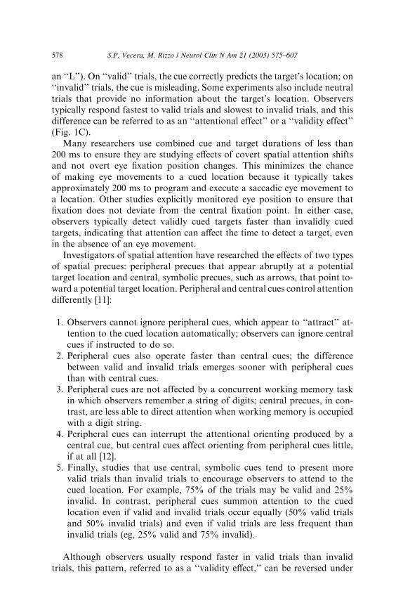
an ‘‘L’’). On ‘‘valid’’ trials, the cue correctly predicts the target’s location; on‘‘invalid’’ trials, the cue is misleading. Some experiments also include neutraltrials that provide no information about the target’s location. Observerstypically respond fastest to valid trials and slowest to invalid trials, and thisdifference can be referred to as an ‘‘attentional effect’’ or a ‘‘validity effect’’(Fig. 1C).
Many researchers use combined cue and target durations of less than200 ms to ensure they are studying effects of covert spatial attention shiftsand not overt eye fixation position changes. This minimizes the chanceof making eye movements to a cued location because it typically takesapproximately 200 ms to program and execute a saccadic eye movement toa location. Other studies explicitly monitored eye position to ensure thatfixation does not deviate from the central fixation point. In either case,observers typically detect validly cued targets faster than invalidly cuedtargets, indicating that attention can affect the time to detect a target, evenin the absence of an eye movement.
Investigators of spatial attention have researched the effects of two typesof spatial precues: peripheral precues that appear abruptly at a potentialtarget location and central, symbolic precues, such as arrows, that point to-ward a potential target location. Peripheral and central cues control attentiondifferently [11]:
1. Observers cannot ignore peripheral cues, which appear to ‘‘attract’’ at-tention to the cued location automatically; observers can ignore centralcues if instructed to do so.
2. Peripheral cues also operate faster than central cues; the differencebetween valid and invalid trials emerges sooner with peripheral cuesthan with central cues.
3. Peripheral cues are not affected by a concurrent working memory taskin which observers remember a string of digits; central precues, in con-trast, are less able to direct attention when working memory is occupiedwith a digit string.
4. Peripheral cues can interrupt the attentional orienting produced by acentral cue, but central cues affect orienting from peripheral cues little,if at all [12].
5. Finally, studies that use central, symbolic cues tend to present morevalid trials than invalid trials to encourage observers to attend to thecued location. For example, 75% of the trials may be valid and 25%invalid. In contrast, peripheral cues summon attention to the cuedlocation even if valid and invalid trials occur equally (50% valid trialsand 50% invalid trials) and even if valid trials are less frequent thaninvalid trials (eg, 25% valid and 75% invalid).
Although observers usually respond faster in valid trials than invalidtrials, this pattern, referred to as a ‘‘validity effect,’’ can be reversed under
578 S.P. Vecera, M. Rizzo / Neurol Clin N Am 21 (2003) 575–607
some situations. That is, invalidly cued targets can be detected faster thanvalidly cued targets. In studies using unpredictive peripheral cues (50% validand 50% invalid), observers respond faster to valid trials than invalid trialswhen the interval between the cue and target is less than 200 ms. As theinterval exceeds 200 ms, observers respond faster to targets at the uncuedlocation (invalid trials). This effect is called inhibition of return (IoR) [13];IoR has been hypothesized to facilitate visual search, or visual foraging, byreducing the probability that a previously attended location be re-attendedshortly after attention is withdrawn from that region [14,15]. Furtherresearch is needed, however, to test this hypothesis because the findings ofinhibitory tagging in visual search have not always been replicated [16].
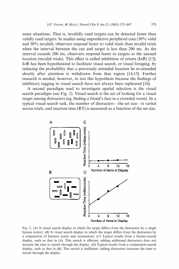
A second paradigm used to investigate spatial selection is the visualsearch paradigm (see Fig. 2). Visual search is the act of looking for a visualtarget among distracters (eg, finding a friend’s face in a crowded room). In atypical visual search task, the number of distracters—the set size—is variedacross trials, and reaction time (RT) is measured as a function of the set size.
Fig. 2. (A) A visual search display in which the target differs from the distracters by a single
feature (color). (B) A visual search display in which the target differs from the distracters by
a conjunction of features (color and orientation). (C) Typical results from a feature-search
display, such as that in (A). This search is efficient; adding additional distracters does not
increase the time to search through the display. (D) Typical results from a conjunction-search
display, such as that in (B). This search is inefficient; adding distracters increases the time to
search through the display.
579S.P. Vecera, M. Rizzo / Neurol Clin N Am 21 (2003) 575–607
An ‘‘efficient’’ visual search is characterized by search functions withshallow slopes (Fig. 2B) and an ‘‘inefficient’’ visual search is characterizedby search functions with steep slopes (Fig. 2D). Although early studies onvisual search equated shallow search functions with parallel processing andsteep search functions with serial, attentive processing [17], this parallel–serial distinction has fallen out of favor because some serial-lookingprocesses can arise from parallel processing mechanisms [18,19]. Forexample, serial-looking search, in which responses become slower as moreitems are added to a display, can be produced from a limited-capacityparallel search mechanism. In a limited capacity parallel search, multipleitems can be processed in parallel, but because this search has a limitedcapacity, searching through multiple items takes longer than searchingthrough fewer items. As a consequence, most theorists discuss efficiency ofvisual search, with efficient searches characterized by shallow search func-tions (slopes <10 ms per item) and inefficient searches characterized by steepsearch functions (slopes >20 ms per item [20]).
Several studies suggest that visual search involves attending to thelocations occupied by the items in a search array. Prinzmetal et al [21]demonstrated that adding cues to a visual search task could improve targetidentification on valid trials compared with invalid trials. Thus, knowingthe spatial region of a target helps observers correctly identify a target. Kimand Cave [22] showed that a visual search display can influence how quicklyan observer responds to a spatial probe. In this task, observers performed avisual search task and determined if a target was present or absent from adisplay. After some of the visual search displays, a small dot appeared, andobservers had to press a button as soon as they detected this dot (similar tothe task in Posner’s spatial cuing task discussed previously). Observers werefastest to detect the dot when it appeared in the same location as the targetin the visual search task, suggesting that spatial attention had been directedto the target location, which then allowed the subsequent dot to be detectedrapidly.
Control and effects of attention: theoretic issues
Having discussed the major tasks used to study spatial attention, thisarticle now turns to two theoretically important issues for spatial attention:‘‘How is spatial attention controlled?’’ and ‘‘What are the effects of directingspatial attention to an item?’’ Attentional control involves those parametersand processes that determine which items become attended and which donot. Attentional control parameters determine which items attention selects.For example, abruptly appearing stimuli, such as a peripheral cue, controlthe allocation of attention by capturing attention automatically.
There are different parameters that influence attentional control. Twogeneral classes of control are top-down sources that arise from the currentbehavioral goals and bottom-up sources that arise from sensory stimuli
580 S.P. Vecera, M. Rizzo / Neurol Clin N Am 21 (2003) 575–607
present in a scene [20,23]. These two sources can be illustrated by re-considering the visual search task. In a typical visual search task, observersare instructed to search for a particular target, such as a black vertical line,that appears in a field of distracters (Fig. 2). The target description—that is,the target an observer is actively looking for—can be conceptualized as a‘‘template’’ that is temporarily stored in visual memory. This memorytemplate influences visual search in a top-down manner; observers activelyattempt to look at black and vertical items. The actual scene presented in avisual search task provides the bottom-up information that is searchedthrough; this information indicates where objects are located and whichfeatures (eg, color, orientation, shape, and so forth) are present at eachlocation. Visual search requires the observer to find a balance between thetop-down information and the bottom-up information.
An example of an effective search is searching for a single feature, such asa black vertical line among white vertical lines (Fig. 2). When the targetdiffers from the distracters by one feature (eg, color; see Fig. 2A), thebottom-up information is consistent with the top-down information inconstraining where an observer should search. Such a search is efficient; thetarget ‘‘pops out’’ at the viewer and can be identified rapidly, irrespective ofthe number of items in the scene (Fig. 2B). A less efficient search involvessearching for a conjunction of features, such as a black vertical line amongblack horizontal lines and white vertical lines (Fig. 2C). In this search, anysingle piece of bottom-up information is not unique to the target item, so thebottom-up constraints are weaker than in the feature search. Top-downconstraints are required to resolve the competition among the input items.Conjunction searches are inefficient and operate more slowly than featuresearches. When the bottom-up constraints are no longer unique, the searchbecomes progressively slower as items are added to a display (Fig. 2D).
Bottom-up and top-down control of spatial attention also has beenexamined using spatial cuing tasks, which direct attention to a locationbefore a target event occurs. The type of spatial cue used can bias attentionalcontrol to favor bottom-up factors or top-down factors. For example,peripheral cues that involve an abrupt luminance change (eg, a flicker in thevisual periphery) automatically attract attention via bottom-up controlparameters, and this attentional capture occurs irrespective of the observer’sintentions. In contrast, central, symbolic cues orient attention only if theobserver voluntarily interprets the cue and shifts attention accordingly.Because central cues are dependent on task-related goals and observers’expectancies, they involve top-down control processes. As in visual search,the control of attention in spatial cuing tasks involves a balance betweenbottom-up and top-down factors. Although bottom-up peripheral cuescapture attention, they may be influenced by top-down attentional controlsettings (eg, expectations of where the target will appear).
How can bottom-up and top-down attentional control parameters beintegrated into a single framework for control? A popular framework for
581S.P. Vecera, M. Rizzo / Neurol Clin N Am 21 (2003) 575–607
understanding attentional control is Desimone and Duncan’s [23] biasedcompetition account of visual search. In the biased competition account,the visual stimuli that provide bottom-up (stimulus–based) input competewith each other for attention. An observer’s goals—such as the targettemplate—provide the top-down (goal driven) control of attention. The top-down inputs can bias processing to favor one of the bottom-up inputs overthe others. The competition among items is biased by a top-down signal,hence the name ‘‘biased competition.’’
Another important issue in the study of attention is how an attendedstimulus is processed differently from an unattended stimulus. For example,the neural representation of an attended item could be enhanced relative tothe representations of unattended items. Or, an attended item could in-tegrate together the visual attributes of the attended stimulus, allowing thefeatures of attended objects to be bound together [17,24]. There are severaleffects of attention highlighted in recent theories and supported by empiricdata:
1. Attention reduces an observer’s uncertainty in making judgments abouta stimulus. Under this ‘‘decision-noise account’’ or ‘‘noise reductionaccount’’ [25], optimal performance (ie, accuracy) decreases as the num-ber of stimuli increases, because each stimulus contains some uncertainty(or random noise). Attention reduces the random noise associatedwith the attended stimulus.
2. Attention also may reduce noise at a perceptual level of representationby enhancing the signal-to-noise ratio of attended items. This ‘‘sensory-gain account’’ hypothesizes that attention enhances the perceptionof attended items compared with unattended items. This effect of atten-tion is to allow attended items to be of higher fidelity than unattendeditems [26].
3. Attention may be needed to bind together the features of an object [27].Consider a display that contains a red circle and a blue square. Howdoes the visual system bind the features ‘‘red’’ and ‘‘circle’’ together andavoid the incorrect combination of ‘‘red’’ and ‘‘square’’? One solution tothis binding problem is to focus attention on a single stimulus, thereby‘‘gluing’’ or binding the features together. Directing spatial attention toa location reduces the number of incorrect feature combinations at thatattended location [21].
4. Spatial attention also seems to have the effect of increasing the spatialresolution of perception. Multiple stimuli that appear in the lower visualfield are more easily distinguished from one another than multiplestimuli that appear in the upper visual field, possibly as a result ofparietal-lobe attentional processes. A single stimulus in the upper field,however, is perceived as accurately as a single stimulus that appears inthe lower field. Thus, the differences in spatial attention between theupper and lower fields are produced when multiple stimuli are close to
582 S.P. Vecera, M. Rizzo / Neurol Clin N Am 21 (2003) 575–607
one another and require attention to be ‘‘narrowed’’ around a singletarget stimulus.
5. Finally, spatial attention seems to influence the entry of items into visualshort-term memory [28,29]. When the appearance of many visual objectsmust be retained across a delay, visual short-term memory processes arerequired to retain three to four of the objects. If one of the objects ispreceded with a small spatial precue, this item is more easily remem-bered than other, uncued items.
The use of spatial attention in several situations to produce differenteffects suggests that it is unlikely to be a single process. Instead, spatialattention likely is controlled by several sources of information and likelyarises from several different anatomic areas that work in concert (seediscussions of influential theories [30–32]). This review of the behavioralliterature on spatial attention provides the background for interpretingvarious attentional impairments that follow focal brain damage or that arisein degenerative disorders such as Alzheimer’s disease.
Disorders of spatial attention: focal effects of cortical and subcortical areas
As in neurologically normal participants, most studies of attention withfocal brain-damaged patients emphasized spatial attention. Because behav-ioral neurology and neuropsychology benefited from theoretic analysesof attention and vice versa, the authors emphasize what neuropsychol-ogy can contribute to cognitive theory and how cognitive theory can assistclinical practice in diagnosis and rehabilitation. The authors reviewedthe cortical and subcortical contributions to spatial attention in patient pop-ulations elsewhere [33].
The parietal lobes, neglect, and extinction
Of all the multiple cortical and subcortical areas that contribute to theprocess of spatial attention, the posterior parietal region has been studiedthe most extensively. Unilateral damage to the human parietal lobe resultsin a profound syndrome referred to as neglect or hemineglect [34–38].Because neglect and extinction most often follow right parietal damage,clinical symptoms are most evident for the left side of extrapersonal spaceor the left side of the patient (left hemineglect). Of course, neglect can oc-cur after focal lesions to other areas (eg, frontal lobe areas), but mostneuropsychologic studies of spatial attention focused on parietal-damagedpatients.
Patients with parietal-lobe damage typically fail to attend to stimulifalling on the hemispace opposite the lesion (the contralesional side); thesepatients also may fail to acknowledge a person sitting on the left, eat food
583S.P. Vecera, M. Rizzo / Neurol Clin N Am 21 (2003) 575–607
on the left side of the plate, read words on the left side of a page, and makehead or eye movements to the left. These failures to respond to stimuli in thefields opposite the lesion are not the result of sensory deficits (eg, a visualscotoma or hemianopia). Patients with these sensory disturbances typicallyare aware of and orient to a contralesional hemifield stimulus to compensatefor their impairment. Patients with hemineglect, however, generally areunaware of their deficit, and, if confronted with a defect on the impairedside (such as a hemiparesis), even deny the problem, a condition termedanosognosia. It is difficult, however, to accurately map visual sensory def-icits that occur in a neglected hemifield. The authors suspect that thisdetection problem confounded the results of several studies of visual at-tention in parietal-damaged patients; some of the neglect cases reported inthe literature may show subtle sensory deficits that have gone unnoticed.
There are some procedures that can help distinguish sensory difficultiesfrom attentional difficulties. For example, a patient with auditory neglectmay not notice sounds from the contralesional side of space, even thoughlocalized sounds reach both ears and afferent projections in the auditorysystem are bilateral (ie, each ear projects to both hemispheres). Failure tonotice a contralesional sound by a neglect patient can be distinguished froma peripheral sensory impairment: A patient with a peripheral auditorysensory deficit should remain able to detect sounds localized to either side ofspace because the sound could be carried to both hemispheres by the intactear’s projections (see Heilman et al for a summary [35]).
Attentional neglect and extinction also can affect motor performance. Inmotor neglect, the patient might not move a contralesional limb, despitenormal strength in this limb. The lesions that produce motor neglect mayalso extend into primary motor representations (Brodmann’s area 4) in thefrontal lobe. It is difficult to detect motor neglect in a paretic limb. A limbaffected by motor neglect may seem paretic unless it is observed moving‘‘automatically,’’ including moving away from a noxious stimulus or movingto prevent the affected limb from falling in the patient’s face after beingdropped by the examiner. Motor extinction is distinguished more easilyfrom paresis and neglect. In motor extinction, the patient can move acontralesional limb when the ipsilesional limb is stationary, but not whenthe ipsilesional limb is moving [35]. The co-occurrence of attentional neglectand extinction with motor neglect and extinction suggests that attentionaland motoric processes may share neural mechanisms.
Neglect typically occurs immediately after damage to the parietal regionand, as a patient recovers and the neglect becomes less severe, patients canprocess a single stimulus presented in the contralesional visual field. Thepatients may begin to show another disorder, however, extinction, in whichthey extinguish or fail to notice the stimulus in the contralesional field whentwo stimuli are presented simultaneously in both visual fields. That is,extinction patients exhibit neglect of contralesional stimuli only in thepresence of ipsilateral stimuli. Of course, damage to the parietal cortices
584 S.P. Vecera, M. Rizzo / Neurol Clin N Am 21 (2003) 575–607
may produce a variety of other disturbances, but the focus here is only onthe attentional impairments after damage to the parietal region.
Neglect and extinction can be diagnosed easily at the bedside or clinic. Inone standard bedside evaluation, the patient is asked to fixate the examiner’snose and to indicate (verbally or by pointing) which field contains theexaminer’s wiggling fingers. Patients with neglect fail to report on singlestimuli in the aberrant field, whereas those with extinction fail to detecttargets in the aberrant field with double simultaneous stimulation (fingerwiggling) in both hemifields. Even a patient with extinction, however, mayhave difficulty detecting multiple stimuli presented in the impaired shield,resembling the perceptual defect in simultanagnosia, a full-field defect ofsimultaneous perception often associated with bilateral parietal lobe lesions.
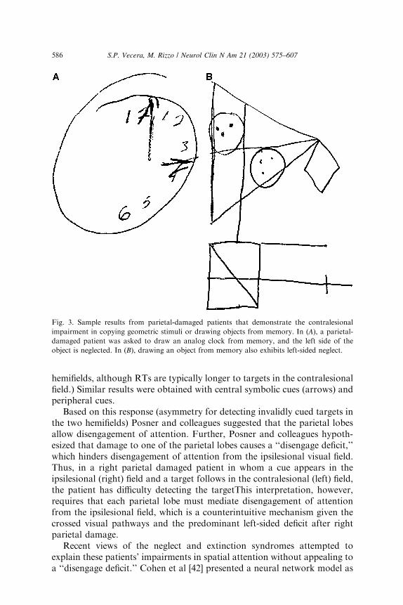
Neglect and extinction also are diagnosed using a variety of widely usedpaper-and-pencil tasks [35,37,38]. These patients neglect features on thecontralesional side of space (and on the contralesional side of objects)during tasks that involve direct copy (eg, complex figure test [CFT]) or recall(eg, Benton visual retention test; CFT recall at 30 minutes) and from mentalimaging of familiar places and objects [39,40]. In many cases, the patientsfail to include features that appear on the left side of the object when askedto reproduce an analog clock from memory. For example, all the numbersmight be placed on the right side of the clock (Fig. 3).
Another widely used paper-and-pencil task is the line bisection task. Inthis task, patients are asked to divide a horizontal line in half. Patients withhemispatial neglect often bisect the line to the right of center, presumablybecause they do not attend to the left-most portion of the line. In anothertask, object cancellation, patients view a cluttered display containing severalobjects (eg., lines, letters, geometric shapes, and so forth) and are asked tocross out (ie, cancel) all the objects in the display. Again, patients withdamage to the right parietal area fail to detect objects in the left visual field,and these objects are not cancelled by the patient. Severely affected patientsmay fail to detect their errors on visual inspection. Patients with visual lossalone (eg, homonymous hemianopia) do not make these types of errors online bisection and cancellation tasks, provided that gaze and head movementare not restricted and viewing time is longer than a few seconds.
Insights into the attentional impairments that follow parietal-lobedamage have come from theoretically motivated studies using the at-tentional paradigms discussed previously. Posner and colleagues [41] wereamong the first to study patients with parietal-lobe damage under theguidance of an explicit cognitive theory of attention. Using Posner’s spatialcuing task, Posner et al [41] found an asymmetry in attentional orienting inparietal-damaged patients, who were slower to detect invalidly cued targetspresented in the contralesional field. That is, a patient with right parietaldamage was slower to detect a target in the left field after a cue in the rightfield than to detect a target in the right field after a cue in the left field.(These patients could detect validly cued targets appearing in each visual
585S.P. Vecera, M. Rizzo / Neurol Clin N Am 21 (2003) 575–607
hemifields, although RTs are typically longer to targets in the contralesionalfield.) Similar results were obtained with central symbolic cues (arrows) andperipheral cues.
Based on this response (asymmetry for detecting invalidly cued targets inthe two hemifields) Posner and colleagues suggested that the parietal lobesallow disengagement of attention. Further, Posner and colleagues hypoth-esized that damage to one of the parietal lobes causes a ‘‘disengage deficit,’’which hinders disengagement of attention from the ipsilesional visual field.Thus, in a right parietal damaged patient in whom a cue appears in theipsilesional (right) field and a target follows in the contralesional (left) field,the patient has difficulty detecting the targetThis interpretation, however,requires that each parietal lobe must mediate disengagement of attentionfrom the ipsilesional field, which is a counterintuitive mechanism given thecrossed visual pathways and the predominant left-sided deficit after rightparietal damage.
Recent views of the neglect and extinction syndromes attempted toexplain these patients’ impairments in spatial attention without appealing toa ‘‘disengage deficit.’’ Cohen et al [42] presented a neural network model as
Fig. 3. Sample results from parietal-damaged patients that demonstrate the contralesional
impairment in copying geometric stimuli or drawing objects from memory. In (A), a parietal-
damaged patient was asked to draw an analog clock from memory, and the left side of the
object is neglected. In (B), drawing an object from memory also exhibits left-sided neglect.
586 S.P. Vecera, M. Rizzo / Neurol Clin N Am 21 (2003) 575–607
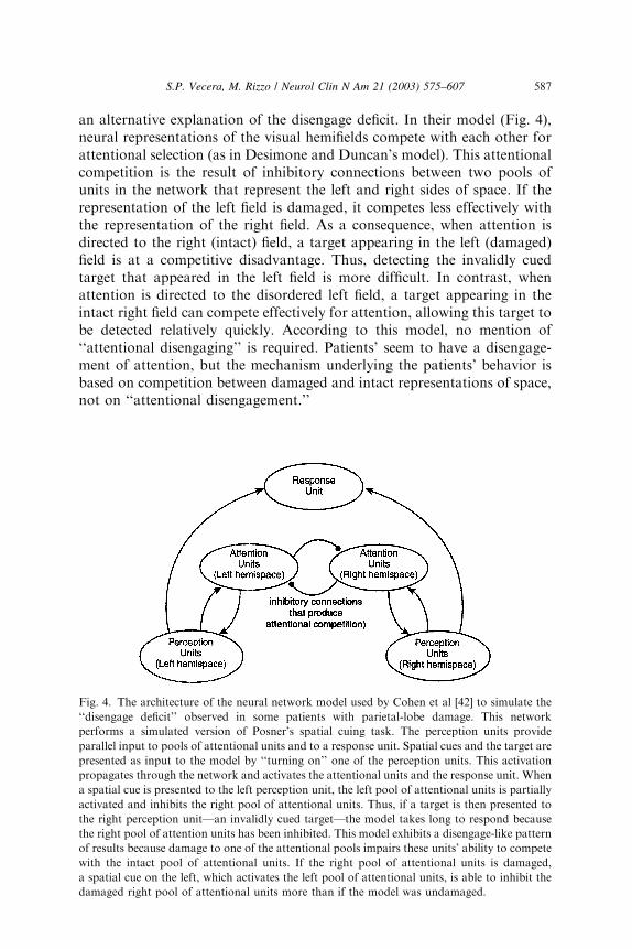
an alternative explanation of the disengage deficit. In their model (Fig. 4),neural representations of the visual hemifields compete with each other forattentional selection (as in Desimone and Duncan’s model). This attentionalcompetition is the result of inhibitory connections between two pools ofunits in the network that represent the left and right sides of space. If therepresentation of the left field is damaged, it competes less effectively withthe representation of the right field. As a consequence, when attention isdirected to the right (intact) field, a target appearing in the left (damaged)field is at a competitive disadvantage. Thus, detecting the invalidly cuedtarget that appeared in the left field is more difficult. In contrast, whenattention is directed to the disordered left field, a target appearing in theintact right field can compete effectively for attention, allowing this target tobe detected relatively quickly. According to this model, no mention of‘‘attentional disengaging’’ is required. Patients’ seem to have a disengage-ment of attention, but the mechanism underlying the patients’ behavior isbased on competition between damaged and intact representations of space,not on ‘‘attentional disengagement.’’
Fig. 4. The architecture of the neural network model used by Cohen et al [42] to simulate the
‘‘disengage deficit’’ observed in some patients with parietal-lobe damage. This network
performs a simulated version of Posner’s spatial cuing task. The perception units provide
parallel input to pools of attentional units and to a response unit. Spatial cues and the target are
presented as input to the model by ‘‘turning on’’ one of the perception units. This activation
propagates through the network and activates the attentional units and the response unit. When
a spatial cue is presented to the left perception unit, the left pool of attentional units is partially
activated and inhibits the right pool of attentional units. Thus, if a target is then presented to
the right perception unit—an invalidly cued target—the model takes long to respond because
the right pool of attention units has been inhibited. This model exhibits a disengage-like pattern
of results because damage to one of the attentional pools impairs these units’ ability to compete
with the intact pool of attentional units. If the right pool of attentional units is damaged,
a spatial cue on the left, which activates the left pool of attentional units, is able to inhibit the
damaged right pool of attentional units more than if the model was undamaged.
587S.P. Vecera, M. Rizzo / Neurol Clin N Am 21 (2003) 575–607
The effects of right parietal damage on visual search are consistent withresults from spatial cuing paradigms. Eglin et al [43] asked patients withparietal damage to search for conjunctions between features (eg, color andshape) amid varying numbers of distractors (see Fig. 2). The patients’responses were slowed dramatically when the distractors appeared in theipsilesional field compared with the contralesional field. The presence ofipsilesional distractors prevented the contralesional representation of thetarget from competing effectively for attention, consistent with Cohen et al’s[42] account of the disengage deficit associated with parietal lobe damage.
Frames of reference for neglectThe phenomena of spatial neglect and extinction can be interpreted with
respect to several coordinate frames. In visual cognition, a coordinate framedetermines the reference point (origin) from which space is measured. Visuallocations can be represented with respect to the environment (eg, the left orright side of a room—an environmental reference frame), the observer (eg,on the observer’s left or right side—a viewer-centered reference frame), oran object (eg, the left or right side of the letter ‘‘B’’—an object-centeredreference frame).
Attempts to define effects of hemineglect in different coordinate systemsmust consider issues of overlap and alignment of the reference frames. Forexample, a ceiling is typically ‘‘up’’ or ‘‘above’’ in environmental coor-dinates and in viewer-centered coordinates (ie, the ceiling appears abovethe viewer’s head or fixation point and above the rest of the room). To studythe effects of reference frames, these reference frames must be disentan-gled from one another, which is typically done by tilting the patient or tiltingan object that the patient is viewing.
Using these types of manipulations to disentangle coordinate systems,Calvanio et al [44] found that neglect can occur in multiple reference frames.Neglect patients with right parietal damage not only failed to report itemsto their left side (viewer-centered coordinates), but also reported few objectsfrom body-centered left and reported few objects from environmental left,suggesting that hemineglect disrupted viewer-centered (or body-centered)and environmental-centered reference frames. Ladavas [45] also reportedresults indicating that neglect can occur in several reference frames (eg,viewer-centered and environment-centered frames).
Other observations indicate that several coordinate or reference systemsmay be damaged in neglect. For example, patients with hemineglect resultingfrom acute or chronic lesions in parietal [46] and parietoinsular regions mayexperience a tilt of visual percepts of the earth-fixed vertical axis away fromthe side of the brain lesion. Such patients might also experience rotation[47,48] or displacement [49,50] of an egocentric visual coordinate systemtoward ipsilesional hemispace. Ipsilesional coordinate shifts have been foundon a kinesthetic ‘‘point straight ahead’’ task [51–53] and on visual tasks inwhich subjects stop a moving spot of light directly in front of the body
588 S.P. Vecera, M. Rizzo / Neurol Clin N Am 21 (2003) 575–607
midline [53–55]. Shifts in subjective body midline position might cause a shiftin the egocentric frame of reference for coding locations in extrapersonalspace, but alone are not sufficient to account for hemineglect [56].
Neglect also may occur with respect to object-centered coordinates, andobjects can reduce the effects of neglect. For example, the eye movementpatterns of parietal-damaged patients demonstrate that object informationthat crosses from the intact field to the neglected field can encourage patientsto search in the neglected field. Patients with hemineglect are more likely tomake eye movements into the neglected field when they view scenes in whicha single object or perceptual group crosses from the intact visual field intothe neglected visual field [57]. If hemineglect patients view a face that falls inboth visual fields, they make fixation transition across the midline betweenthe eye features of face displays even when the features are located in theneglected hemifield. In contrast, when viewing a scene in which objects donot cross from the intact field to the neglected field, fewer fixations occuracross midline.
In addition to objects reducing the effects of neglect, objects encourageneglect to occur in the patient’s intact field, as defined by viewer-centeredcoordinates. Neglect can occur in object-centered coordinates, in which thepatient fails to report information from the left side of an object, even whenthat object falls on the patient’s right side. Some patients with right parietaldamage are less likely to report items from the left side of an object than fromthe right side of an object, even when the left side of the object appears in theright (ipsilesional) visual field [37,58 (review)]. Although there have beenfailures in finding object-centered neglect [59], such failures may occur whenthe objects are secondary to the task that patients are asked to perform.
Neglect also occurs across different sensory modalities. Neglect patientshave difficulty attending to visual and auditory stimuli in contralesionalspace, suggesting that parietal lobe attentional processes operate on arepresentation of space that codes visual and auditory stimuli. Farah et al[60] found evidence for cross-modal neglect in parietal-damaged patientswho were asked to detect a lateralized visual stimulus on a multimodalversion of Posner’s spatial cuing task. This visual target was preceded bya cue presented in either the visual or auditory modality. Patients withparietal damage oriented similarly to both types of cues; they had difficulty‘‘disengaging’’ attention from cues presented on the ipsilesional side ofperipheral space regardless of cue modality. These results suggest thatparietal lobe attention mechanisms are supramodal (see Spence andDriver [61,62] for methodologic concerns with studying cross-modal spatialattention).
Control parameters and spatial attentionAs discussed previously, attention can be deployed—or controlled—by
several factors. Some are environmental (bottom-up, exogenous) factors,such as the appearance of a new object or event. Others are endogenous
589S.P. Vecera, M. Rizzo / Neurol Clin N Am 21 (2003) 575–607
(top-down) factors that arise from within the observer based on goals orexpectations. Which of these control parameters is impaired after parietal-lobe damage? The disorder of attention in parietal-damaged patients seemsto involve bottom-up control parameters. This does not suggest that neglectis a sensory-level impairment. Perceptual processing may be largely intact inneglect patients, but attention is not effectively captured by stimuli on thecontralesional side of space. Although these stimuli have intact perceptualrepresentations, they do not fully capture or drive the damaged attentionalprocesses.
Evidence for poor bottom-up control comes from simulating aspects ofneglect in neurologically normal observers by degrading one side of a visualscene. These observers show the ‘‘disengage’’ deficit if half of a computermonitor is degraded with translucent tracing paper (S. Vecera, unpublisheddata, 2002). Targets on the degraded side of the display are more difficult todetect after a cue on the nondegraded side than targets appearing on thenondegraded side after a cue to the degraded side [63].
Studies using the Posner cuing paradigm suggest that some forms of top-down attentional control are intact after parietal damage. Use of a centralarrow cue (considered an endogenous cue) pointing to the left (contralesional)side can reduce the number of targets missed by right parietal patients;the same reduction in misses does not occur for exogenous peripheral cues[64]. As central arrows and peripheral cues are not alike physically, thesedifferences could be the result of perceptual, not attentional, processes.Bartolomeo et al [65] addressed this issue using peripheral cues only; tomanipulate exogenous and endogenous attention, the cue’s informativenesswasmanipulated bymaking the cue informative. Informative peripheral cues,which indicate the likely location of the target, tap endogenous attentionalprocesses, whereas uninformative cues tap exogenous processes. Bartolomeoet al [65] reported that parietal damaged patients could reduce their responsetimes to contralesional stimuli if most of the invalidly cued targets appeared inthe contralesional field. The patients correctly anticipated the target’sexpected location in the contralesional field and directed attention accord-ingly. Parietal patients can make use of top-down expectancies or task-relevant goals. A contralesional stimulus may not be extinguished if theipsilesional stimulus is task irrelevant and the patient is asked to ignore it[66]. For example, a right parietal patient who is asked to name the color ofeach of two stimuli, one in each visual field, is more likely to extinguish thecontralesional stimulus if the two stimuli are the same color. If the two stimuliare the same shape (eg, both circles) or different shapes (eg, one circle and onesquare), however, only the task-relevant attribute of color influences extinc-tion. The task-irrelevant shape dimension does not influence extinction.
The parietal lobes, neglect, and extinction: attentional effectsHaving addressed the attentional control processes that seem disrupted
after parietal damage, this article now discusses the effects produced by
590 S.P. Vecera, M. Rizzo / Neurol Clin N Am 21 (2003) 575–607
parietal damage. Recall that spatial attention has many hypothesized effects,including (1) providing attended stimuli with greater resources thanunattended stimuli; (2) enhancing sensory-level or decision-level representa-tions for attended items compared with unattended items; (3) bindingtogether the features of an object; and (4) increasing the spatial resolution ofperception. The attentional impairments after parietal damage seem todisrupt some of these attentional effects.
Little is known about attentional resources after parietal damage. Someresults from patients, however, support the sensory enhancement effect ofspatial attention in which attention may ‘‘amplify’’ sensory informationtransmission [26]. The results suggest that bottom-up control is impaired[64,65] and that sensory amplification effects are diminished so that attentionis not captured effectively by events in the contralesional field. More directevidence of the effect of sensory amplification after parietal damage couldbe obtained by using early visual evoked potentials after parietal damage orby applying psychophysical techniques to study amplification effects [67].
Additional evidence also is needed of the role of parietal cortex inreducing decision noise, one of the hypothesized effects of spatial attention.Spatial cues seem to reduce the uncertainty of observer’s decisions [25], andthe disrupted spatial orienting observed suggests that parietal patients mayhave difficulties making decisions about targets in the contralesional field.The authors have preliminary data from a study that investigates decisionnoise effects after right parietal lobe damage (S.P. Vecera and M. Rizzo,unpublished data, 2002). Consistent with a decision-noise deficit, theauthors’ patient showed a larger difference between validly and invalidlycued targets in her contralesional field than in her ipsilesional field. Theseobservations need to be replicated in this patient and others, but the resultssuggest that parietal damage may influence a host of attentional effects,including the reduction of decision noise.
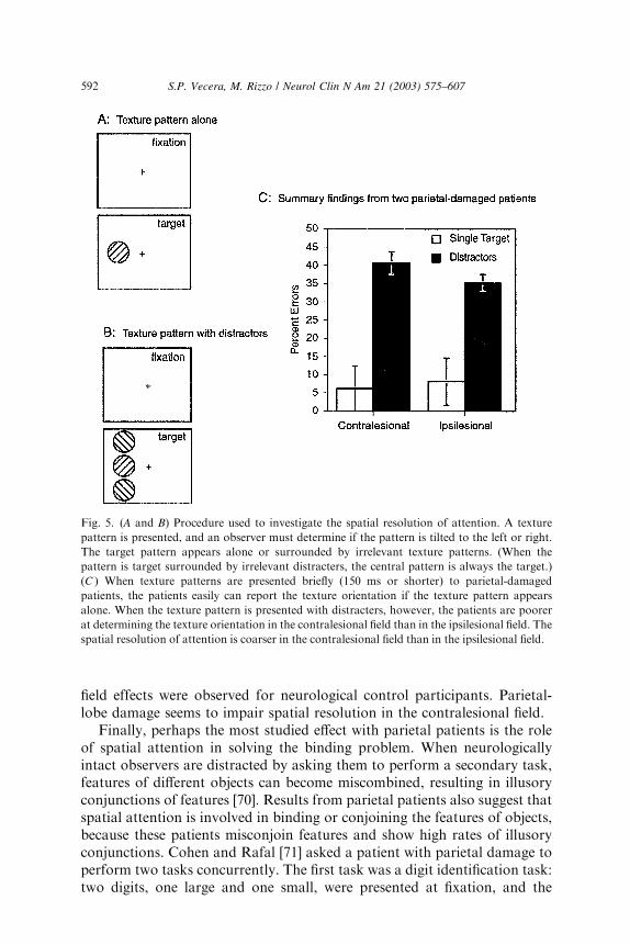
Spatial attention seems to increase the spatial resolution of perception[68,69]. When observers are asked to report the orientation of a texturedpattern, performance differs between the upper and lower visual fields (ie,above and below fixation) when multiple stimuli are present (Fig. 5B) butnot when a single stimulus is present (Fig. 5A). Because there are no upper/lower field differences in primary visual cortex, the visual field differencemay be produced by the resolution of spatial attention, which is greater inthe lower field than in the upper field [69]. The authors have unpublishedresults that support this hypothesis. Patients with parietal-lobe damage wereasked to determine the orientation of a texture grating that appeared aloneor flanked by irrelevant gratings. The stimuli were presented in either thecontralesional or ipsilesional field. In two patients, the authors found novisual field difference for the single texture gratings, but did find a statisticallysignificant field difference when multiple gratings appeared (Fig. 5C).Specifically, the patients’ performance was poorer when multiple items ap-peared in the contralesional field than in the ipsilesional field. No visual
591S.P. Vecera, M. Rizzo / Neurol Clin N Am 21 (2003) 575–607
field effects were observed for neurological control participants. Parietal-lobe damage seems to impair spatial resolution in the contralesional field.
Finally, perhaps the most studied effect with parietal patients is the roleof spatial attention in solving the binding problem. When neurologicallyintact observers are distracted by asking them to perform a secondary task,features of different objects can become miscombined, resulting in illusoryconjunctions of features [70]. Results from parietal patients also suggest thatspatial attention is involved in binding or conjoining the features of objects,because these patients misconjoin features and show high rates of illusoryconjunctions. Cohen and Rafal [71] asked a patient with parietal damage toperform two tasks concurrently. The first task was a digit identification task:two digits, one large and one small, were presented at fixation, and the
Fig. 5. (A and B) Procedure used to investigate the spatial resolution of attention. A texture
pattern is presented, and an observer must determine if the pattern is tilted to the left or right.
The target pattern appears alone or surrounded by irrelevant texture patterns. (When the
pattern is target surrounded by irrelevant distracters, the central pattern is always the target.)
(C ) When texture patterns are presented briefly (150 ms or shorter) to parietal-damaged
patients, the patients easily can report the texture orientation if the texture pattern appears
alone. When the texture pattern is presented with distracters, however, the patients are poorer
at determining the texture orientation in the contralesional field than in the ipsilesional field. The
spatial resolution of attention is coarser in the contralesional field than in the ipsilesional field.
592 S.P. Vecera, M. Rizzo / Neurol Clin N Am 21 (2003) 575–607
patient was instructed to identify the large digit. The second task was a letteridentification task: one of the letters was a target (‘‘F’’ or ‘‘X’’) and the otherwas a distracter (‘‘O’’). The letters were colored, and the patient wasinstructed to name the color and the identity of the target letter (‘‘Was it �F�or �X,� and what color was it?’’). In the second task, there are two types oferrors. The first is a feature error, in which either letter name or color isreported incorrectly. For example, if the patient is presented with a blue ‘‘F’’and a red ‘‘O,’’ reporting a yellow ‘‘F’’ is a feature error. The second type oferror is a conjunction error, in which a feature of the distracter letter ‘‘O’’‘‘migrates’’ to the target letter, forming an illusory conjunction. Forexample, if the patient is presented with a blue ‘‘F’’ and a red ‘‘O’’ butreported a red ‘‘F,’’ the color of the red ‘‘O’’ was misconjoined with thetarget letter ‘‘F.’’ Cohen and Rafal’s patient showed a larger number ofconjunction errors in the contralesional field than in the ipsilesional field.Similar numbers of feature errors were made in the contralesional andipsilesional fields, indicating that feature perception was similar in bothfields. Presumably, the damaged parietal-based spatial attention systemimpaired feature integration, although the individual features are repre-sented, allowing for accurate perception of individual features.
Even more direct evidence for the role of parietal attention areas insolving the binding problem comes from Robertson and colleagues [72],who investigated illusory conjunctions in a patient with bilateral parietaland cerebellar damage. The patient showed features of a syndromeoriginally described by Balint and later called Balint’s syndrome (also seeRizzo and Vecera for a recent review [73]). Patients with Balint’s syndrometypically have deficits in visually guided reaching (optic ataxia) and eyemovements (ocular apraxia). The hallmark of the syndrome, however, isa set of visuospatial impairments including visual disorientation manifestedby a difficulty judging the depth between objects and an extreme inability toperceive more than one object or shape at a time, known as simulta-nagnosia. Robertson et al’s patient could search effectively for a targetdefined by a single feature (eg, searching for a red target or searching for an‘‘X’’).The patient was impaired dramatically at searching for conjunctions(eg, finding a red ‘‘X’’) and for integrating the features of objects, however.Robertson et al’s patient showed illusory conjunction errors even when thevisual display was present for 10 seconds, an exposure duration at whichneurologically intact observers should make no illusory conjunctions.Presumably, this patient’s spatial confusion and simultanagnosia preventedhim from having an accurate representation of spatial location, which isnecessary for conjoining the features of an object.
In summary, patients with damage to a network of structures thatmediate attention, especially parietal lobe areas, have a variety of differentattentional impairments. These impairments extend across different visualreference frames and across different modalities, and many of the typicaleffects of attention, such as the ability to bind features of an object, seem to
593S.P. Vecera, M. Rizzo / Neurol Clin N Am 21 (2003) 575–607
be defective. The bottom-up capture of attention seems to be disrupted inassociation with a failure of sensory amplification. The lack of capture alsomay prevent the damaged hemisphere from competing with the intacthemisphere for attention, resulting in an attentional imbalance that seems tofavor the ipsilesional field (any may appear as a ‘‘disengage deficit’’).
Understanding the control and effects of spatial attention after parietaldamage may offer insights for rehabilitation. If parietal damage disruptsbottom-up capture of spatial attention and involves an attentional im-balance between the cerebral hemispheres, then rehabilitation that increasesthe input to the disrupted hemisphere may reduce the attentional imbalanceand the associated neglect or extinction. Some reports suggest that neglectsymptoms may be reduced by patching the ipsilesional eye, which (1)reduces some of the ipsilesional visual input and (2) reduces the activationof the ipsilateral colliculus (see Heilman et al for a brief discussion of treat-ments for neglect [35]). Unfortunately, most treatments that have been in-vestigated only provide temporary relief from neglect [35].
The frontal lobes and spatial attention
Although deficits in spatial attention have been studied extensively inpatients with parietal lobe damage, it has long been known that spatialneglect and other spatial deficits can follow lesions to frontal cortices (seeBisiach and Vallar for a recent review [74]). Because the frontal lobesparticipate in the operation of multiple cognitive processes, however, in-cluding language, motor control, working memory, and attention, less isknown about the particular attentional processes associated with frontalareas. For example, little is known about the neuropsychology of visualsearch in patients with frontal lobe damage. This lack of knowledge mayoccur because studies of neglect sometimes include patients with frontaldamage and patients with parietal damage, thereby obscuring possibledifferences between these groups; the attentional processes of the medialfrontal lobe seem to differ from those associated with the parietal lobe (seeSwick and Knight for an overview [75]).
One area of spatial attention studied in patients with frontal lobe damageis the control of overt attention—the production of eye movements.Typically, directing the eyes to a region of space is preceded by directingcovert spatial attention to the target region [76]. Lesions of superior frontallobe areas that include the frontal eye fields (FEF) seem to disrupt sometypes of overt eye movements. Guitton et al [77] demonstrated that eyemovements to an abruptly appearing visual target (a ‘‘prosaccade’’ in whichthe eyes move to the target) do not differ between frontal patients with FEFdamage and control patients with temporal lobe damage. Eye movementsin a direction opposite an abruptly appearing target (‘‘antisaccades’’),however, are dramatically impaired in frontal patients. In the antisaccadetask, the FEF patients often made reflexive eye movements to the target
594 S.P. Vecera, M. Rizzo / Neurol Clin N Am 21 (2003) 575–607
location instead of moving in the opposite direction. When the FEF patientsdid make antisaccades, the latency of their eye movements was longer thanthose of control patients. FEF seems to play an inhibitory role in overtattention by preventing unwanted reflexive eye movements.
Guitton et al’s study is important for demonstrating the role of frontallobe areas in overt attention, but does not address the different types ofspatial cues that can control covert and overt attention. To address the roleof different spatial cues (peripheral cues versus central symbolic cues), Henikand colleagues [78] studied patients with frontal lobe lesions that includedsuperior dorsolateral prefrontal cortex, which included the FEF andpatients with frontal damage that excluded FEF. The patients performedtwo tasks. One task was a saccade task, in which eye movements were madeto a peripheral location that was cued with either a central arrow cue ora peripheral cue. The other task was a detection task in which the patientspressed a key that corresponded to a target signal; this target signal waspreceded by either a central arrow cue or a peripheral cue. In both tasks,half of the cues (central and peripheral) were valid and half were neutral;there were no invalid cues.
Henik et al [78] found that FEF lesions disrupted eye movements toperipheral locations, but not all eye movements were disrupted equally. TheFEF patients were slower to make eye movements into the contralesionalfield than into the ipsilesional field after central cues. Also, after peripheralcues, the FEF patients made faster eye movements into the contralesionalfield than into the ipsilesional field. Patients with intact FEF made eyemovements into the contralesional and ipsilesional field approximatelyequally after central and peripheral cues. The results from the FEF patientsindicate that voluntary overt orienting is impaired in this group—thesepatients are slowed only in directing eye movements to the contralesionalfield after the symbolic arrow cue. The authors hypothesize that the FEFpatients may be speeded in directing overt attention into contralesional spaceafter a peripheral cue because the FEF lesion may disinhibit ipsilesionalmidbrain areas; this disinhibition has the effect of inhibiting the oppositecolliculus and delaying reflexive eye movements into the ipsilesional field.Finally, the FEF group did not show any impairments in the detection task,suggesting that the attentional impairments were confined to overt attention(eye movements); covert attention could be directed to cued locations andfacilitate responses that did not involve an eye movement.
Finally, patients with frontal lobe lesions also exhibit an attentionalimpairment related to covert attention (ie, attentional orienting that doesnot require eye movements). Voluntary attentional orienting in frontalpatients seems to be impaired. Using Posner’s spatial cuing task withsymbolic precues, Alivisatos and Milner [79] presented patients with wordcues that either signaled the upcoming target’s location (valid trials) orprovided no information about the target’s location (neutral trials). Frontallobe patients showed a smaller attentional benefit (the difference in
595S.P. Vecera, M. Rizzo / Neurol Clin N Am 21 (2003) 575–607
performance between valid and neutral trials) than control participants ortemporal lobe patients. Koski et al [80] reported similar results using cen-trally presented arrow cues that either validly predicted or did not predictthe upcoming target’s location. Again, frontal patients showed smaller at-tentional benefits than either control participants or temporal lobe patients.The frontal patients in these studies had varied lesion locations that includeddorsolateral frontal areas and ventromedial frontal areas. The authors’results suggest that at least one frontal patient, EVR [81], cannot usesymbolic precues (words or eye gaze direction) to direct spatial attention,although he can use peripheral precues to orient spatial attention.
Superior colliculus and pulvinar: subcortical influences on spatial attention
In addition to the cortical control of spatial attention, there are at leasttwo subcortical regions that seem critical for spatial selection: the superiorcolliculus and the pulvinar nucleus of the thalamus. Most studies inves-tigating the role of these subcortical regions used Posner’s spatial cuing taskto investigate covert spatial attention.
Progressive supranuclear palsy (PSP) is a neurodegenerative disorder thatresembles Parkinson’s disease and affects the basal ganglia. One differencebetween these two degenerative disorders is that PSP produces a markedimpairment of voluntary gaze control, likely the result of damage todegeneration of dorsal midbrain structures, especially the superior collicu-lus [82]. PSP patients have an inability to make voluntary saccadic eyemovements, especially vertical eye movements. For example, PSP patientsoften do not look up at a person who is speaking to them and cannot makesaccades to move their eyes upward if given a command to do so. If the PSPpatient, however, is asked to continue to fixate a target while the head istilted downward, the eyes are able to maintain fixation by moving upwardslowly using vestibulo-ocular mechanisms more closely related to smoothpursuit movements.
In a series of studies, Posner and colleagues and Rafal et al investigatedspatial orienting in PSP patients [83,84]. PSP patients were cued to locationsthat appeared in one of four locations: above, below, left, or right offixation. After the cue, a target appeared at the validly cued location or at aninvalidly cued location, and patients were instructed to press a button assoon as they detected the target. There were two cue conditions in thesestudies. In the exogenous condition, the cue was a peripheral flicker valid on50% of trials and invalid on 50% of invalid trials; in the endogenouscondition, the cue was a central arrow that was predictive (valid on 80% ofthe trials and invalid on 20% of the trials). The patients were faster atdetecting validly cued targets than invalidly cued targets in the horizontaldirection but not in the vertical direction; control patients with Parkinson’sdisease showed no differences between orienting horizontally or vertically[84]. Also, the movement of attention in PSP patients was slower in the
596 S.P. Vecera, M. Rizzo / Neurol Clin N Am 21 (2003) 575–607
vertical direction than in the horizontal direction. Finally, there was a slight,but nonsignificant, trend for a greater impairment in the exogenous cuecondition than in the endogenous cue condition, possibly because endo-genous cues involve more cortical processing than exogenous cues.
Based on their results, Posner and colleagues and Rafal et al suggestedthat the superior colliculus might be involved with one specific component ofspatial attention. In general, spatial attention may involve several componentoperations for normal performance, including disengaging from an attendedlocation, moving attention to a new location, and engaging attention at thenew location. PSP patients seem to have difficulty with the ‘‘move operator’’that permits attention to be moved from one location to another: PSPpatients orient spatial attention more slowly in the vertical direction than inthe horizontal direction. This finding suggests that the superior colliculusmay be involved with the movement of spatial attention. Of course, the samedifficulties with the disengagement hypothesis of parietal damage also applyhere. Specifically, a disorder that seems to be produced by a damagedattentional ‘‘mover’’ or ‘‘move process’’ might be the result of damage toa system that does not contain an explicit move process.
PSP also seems to affect IoR, the decreased tendency to re-orientattention to a previously attended location described above in studies ofneurologically normal subjects performing Posner’s task. PSP patients alsoshow reduced IoR effects in the vertical direction than in the horizontaldirection. Control patients with Parkinson’s disease exhibited IoR in thehorizontal and vertical directions, suggesting that IoR may be linked to theeye movement system controlled by the superior colliculus [85].
The pulvinar nucleus of the thalamus seems involved in the subcorticalcontrol of spatial attention. Pulvinar lesions can cause neglect andattentional impairments. Rafal and Posner [86] had acute pulvinar patientsperform a spatial cuing task with highly predictive (80% valid and 20%invalid) peripheral cue. The patients were slower to detect targets in thecontralesional field than in the ipsilesional field across long cue-targetintervals (almost a second). Rafal and Posner inferred that the pulvinarmight be responsible for engaging spatial attention at a cued location.Across both hemifields, RT to validly cued targets decreased as the timebetween the cue and target increased, which suggests that patients withpulvinar lesions can deploy, or move, spatial attention similarly in bothvisual fields. The pulvinar patients showed a ‘‘disengage deficit’’ similarto that of parietal patients, but only for short cue-target intervals, unlikeparietal patients, suggesting that pulvinar lesions produce a differentdisengage deficit from parietal lesions. The results from pulvinar patientssuggest that attention can be disengaged (eventually) from an attendedlocation and moved to a new location, but that attention a ineffectivelyengaged at the new location. CT scans revealed posterior thalamic damagein these patients, however, which could have involved areas other than thepulvinar. Converging evidence from neuroimaging studies and single-unit
597S.P. Vecera, M. Rizzo / Neurol Clin N Am 21 (2003) 575–607
recordings suggests that the pulvinar plays a role in visuospatial attention[87,88], although these studies use different attentional paradigms makingit difficult to completely understand the attentional functions of thepulvinar.
Disorders of spatial attention in Alzheimer’s disease
The diagnosis Alzheimer’s disease (AD) involves progressive memoryimpairment [89,90]; recent reviews underscore the progressive impairment ofseveral attentional processes [91–93]. Spatial attention impairments in ADare important because most higher-level cognitive processes require someform of attention and because patients with AD might present with at-tentional impairments before presenting with other cognitive impairments,such as memory impairments [92].
As with studies of control participants and focal brain-damaged in-dividuals, research on spatial attention in patients with AD has reliedon the spatial cuing and the visual search paradigms discussed previously.The findings from these information-processing paradigms and the theoriesdeveloped from these paradigms generally are consistent with results fromstandardized tests used in clinical practice. For example, tasks such as theuseful field of view (UFOV) and the ‘‘Starry Night’’ target detection taskdemonstrate attentional impairments in patients with AD [94].
Spatial selective attention, spatial cuing, and AD
Patients with AD show difficulties using spatial attention to orient toa location to detect an upcoming event on Posner’s spatial cuing paradigm(see Fig. 1). In one of the earliest studies, Parasuraman et al [95] presentedpatients with AD with a symbolic central precue—a small arrow thatpointed to the left or right (an endogenous precue). After this precue, a targetletter appeared, which the patients categorized as a consonant or vowel.Patients with AD categorized validly cued targets faster than neutrally cuedtargets, and this benefit was similar in magnitude to the control participants.Patients with AD, however, showed a larger attentional cost than thecontrol participants and were disproportionately slower in categorizing aninvalidly cued target than categorizing a neutrally cued target. Similarresults were obtained when peripheral precues (exogenous precues) precededthe letter to be categorized.
What is the implication of increased attentional costs in AD? Recall thatthe parietal-damaged patients studied by Posner et al [41] showed a largerattentional cost in the ipsilesional visual field compared with the contrale-sional visual field and proposed that this pattern reflected impairment indisengaging spatial attention from its current focus. Using this analysis,Parasuraman and colleagues [95,96] argued that patients with AD have animpairment in disengaging spatial attention. As discussed previously, how-ever, there are plausible alternatives to the ‘‘disengage deficit’’ hypothesized
598 S.P. Vecera, M. Rizzo / Neurol Clin N Am 21 (2003) 575–607
in parietal-damaged patients (eg, attentional competition across differenthemifields). Whether or not these alternatives explain the increasedattentional costs observed in patients with AD could be a direction forfruitful research.
In addition to investigating the benefits and costs of spatial precuing,studies in AD investigated the IoR of spatial attention. As discussedpreviously, when the time delay between a spatial precue and target is suf-ficiently long (200 ms or slightly more), neurologically normal participantsare faster to detect invalidly cued targets than validly cued targets [13],and this pattern of response times is thought to reflect the difficulty inreturning spatial attention to a previously attended location. Under someconditions, patients with AD do not exhibit IoR [97]. When a spatial precueappears in a peripheral location, spatial attention can be directed awayfrom this precued region spontaneously by the observer or by anotherspatial precue presented at a new location [13,98]. Patients with AD showIoR when a second spatial precue directs attention away from a currentlyattended location but not when attention is directed away from the currentlyattended region spontaneously by the individual with AD [97]. This failureto find IoR without a second spatial precue suggests that attentional re-orienting may be sluggish in AD. Patients with AD may re-orient spatialattention or sample visual space using spatial attention more slowly thanneurologically healthy individuals.
Visual search and AD
Patients with AD differ from healthy control participants in using spatialattention to search through a cluttered visual scene. Foster et al [99] askedpatients with AD and controls to search for a target (a filled circle) amongdifferent numbers of distracters so that the search task was either a featuresearch or a conjunction search (see Fig. 2). In the feature search, a shadedcircle appeared among filled circles; in the conjunction search, a shadedcircle appeared among shaded squared and unfilled circles. Recall thatneurologically healthy younger observers perform feature searches efficient-ly; adding additional distracters slows target identification little, if at all. Incontrast, feature searches are performed inefficiently; as distracters areadded to the display, the time to identify the target increases.
Foster and colleagues found that patients with AD seemed to respondslower in the feature search than did control patients, yet appeared to searchthe displays efficiently. Specifically, as the number of distracters increased,patients with AD and controls did not show dramatic increases in baselineresponse time. With the addition of a distracter, moderately dementedpatients were slowed by 4.25 ms, mildly demented patients were slowed by0.71 ms, and controls were slowed by 0.53 ms. All these estimates ofsearch efficiency fall within the ‘‘efficient’’ range described in the cognitiveliterature on search [20].
599S.P. Vecera, M. Rizzo / Neurol Clin N Am 21 (2003) 575–607
When individuals with AD performed a conjunction search task, theydiffered from controls in two respects. First, as with the feature searchtask, patients with AD responded more slowly than control participants.Second, and more theoretically interesting, the individuals with ADshowed a more dramatic increase in response times than control partic-ipants as distracters were added to the display. That is, the slope of thesearch function was steeper for the patients with AD than for the controls.Moderately demented patients were slowed by an average of 50.25 msfor every distracter added to a scene; mildly demented patients wereslowed by an average of 34.9 ms per distracter; and controls were slowedon average by 24.5 ms per distracter. Although all these slope estimatesare in the ‘‘inefficient’’ range [20], the differences among the slopesindicates an impairment in spatial attention in the group with AD, espe-cially for the moderately demented subset of patients. One straightfor-ward explanation of the increase in slopes for the patients with ADperforming the conjunction search is that they have an impairment indisengaging spatial attention from the currently attended item. As thenumber of items to search increases, this disengage difficulty dispropor-tionately slows response times compared with control participants. Thisdisengage account is consistent with the interpretation of other attentionalimpairments exhibited by individuals with AD [95,96]. As with the dis-engage deficit hypothesis for parietal-damaged patients, however, there arelikely to be alternative explanations for a disengage deficit hypothesis inpatients with AD. Again, it is possible that attentional competition acrossthe two hemifields could produce a disengage-like pattern of response timeresults. Other attentional effects, including decision noise reduction, mightbe impaired in patients with AD, as discussed later.
Finally, visual search in patients with AD also has been used toinvestigate the spatial scale, or spatial window, of spatial attention. Thevisual search paradigm used to study spatial scale differs from the standardsearch paradigm developed by Treisman and used by Foster et al [99]. Inthis adapted visual search task, patients with AD view a rectangular array ofletters that was preceded by a spatial precue [100,101]. The size of theprecued region is manipulated so that it encompasses a single letter in thesearch display (high precision of localization), one column of two or threeletters (medium precision), or the entire set of letters (low precision). Fur-ther, in this paradigm, the target of visual search can appear in the precuedregion (valid trial) or outside the precued region (invalid trial). Using thisprocedure, Greenwood et al [101] found that performing a conjunctionsearch task was facilitated by valid precues, but that this facilitation effectwas smaller for patients with AD [100]. Performance in a feature search taskwas affected little by the size of the precued region, supporting the hy-pothesis that feature searches are performed in parallel across the visual fieldand require little or no attention. These results indicate that patients withAD may be impaired in controlling the size of the window of spatial
600 S.P. Vecera, M. Rizzo / Neurol Clin N Am 21 (2003) 575–607
attention, which might be narrower in patients with AD than in normalindividuals [100,102,103].
Unanswered questions regarding spatial attention and AD
There seem to be observable impairments in spatial attention in AD.These impairments are studied because spatial attention plays a central rolein high-level cognitive processes, such as visual short-term memory [28] andspatial memory [104,105]. General attentional impairments may be the causeof other cognitive deficits observed in AD [106] and the cause of the declineof everyday behaviors. It has become increasingly clear to those who workwith patients with AD that understanding how attention can produce thecognitive decline in AD depends on cognitive theories of attention [107]. Forthis reason, future studies of attention in AD must rely on mature theoriesof attention developed in cognitive psychology.
Earlier in this article, some of the major theoretic issues regarding spatialattention developed by cognitive theories were reviewed. Although many ofthese issues have been explored in patients with AD (eg, endogenous versusexogenous precuing), most of these issues remain unexplored. For example,many cognitive theorists point out the difficulties in studying simpledetection tasks using Posner’s precuing task [25,108]. Also, there are manyeffects of spatial attention, and spatial precuing results and visual searchresults can be explained by hypothesizing that spatial attention reduces anobserver’s uncertainty in making judgments about a target item [25,108–111]. One hypothesis regarding AD is that declines in these patients’cognitive and attentional abilities might be produced by less reliable (ie,noisy) information processing. Such a hypothesis could be addressed usingeither a spatial precuing task or a visual search task that does not relyexclusively on response time measures.
Summary
Although ‘‘attention’’ is a general term in everyday folk and psychologicuse, using research from cognitive psychology allows a focus on theprocesses associated with attention. A process-oriented definition of at-tention [2] makes ‘‘attention’’ a concept that can be studied rigorously.Attention is necessary for eliminating unwanted sensory inputs or irrelevantbehavioral tasks and is useful when some cognitive system or processreceives too many inputs. Attention acts to restrict the number of inputs andallow processing to continue in an effective manner. Although there aremany forms of attentional selection, spatial attention is the most studied andperhaps the best understood form of selection.
Spatial attention is the variety of attention most widely studied inneuropsychologic populations. As was evident from the authors’ review,different neuropsychologic syndromes can be characterized as involving
601S.P. Vecera, M. Rizzo / Neurol Clin N Am 21 (2003) 575–607
different difficulties with the component processes of spatial attention.Different neural structures work in concert to produce normal spatial selec-tion, and damage to any of these neural structures, including the parietaland frontal lobes, pulvinar, and superior colliculus, produce an attentionalimpairment. Attentional impairments often have been studied in cases offocal brain damage resulting from stroke, traumatic brain injury, tumor,and surgical resection. Focal or multifocal lesions caused by neurodegener-ative impairments, such as PSP and AD, however, also have marked effectson neural processes of attention. Patients with AD exhibit a range ofimpairments in spatial attention, and these impairments have been studiedrelatively little.
Despite the understanding of attention provided by various patientpopulations, gaps remain in knowledge of the mechanisms involved inattending to space. For example, most studies of neurologic patients haveused a simple spatial cuing paradigm with highly predictive cues. Althoughthis task yields robust results and can be adapted for use with many differentpatient groups, the spatial cuing effects produced by such tasks havemultiple interpretations (eg, allocation of resources versus reduction ofdecision noise).
This review highlights the general point that different neural sites seem tobe responsible for different forms of attentional control, such as object-based attention, attentional task switching, and the executive control ofattention [8,33]. Understanding the integration of these neural sites and theirrelationship to cognitive processes and, ultimately, behavior, will increaseunderstanding of normal and disordered attentional selection.
References
[1] Allport A. Attention and control. Have we been asking the wrong questions? A critical
review of twenty-five years. In: Meyer DE, Kornblum S, editors. Attention and
performance. Cambridge (MA): MIT Press; 1993.
[2] Luck SJ, Vecera SP. Attention: from tasks to mechanisms. In: Yantis S, editor. Stevens’
handbook of experimental psychology, vol. 1. Sensation and perception. New York:
Wiley; 2002. p. 235–86.
[3] James W. Principles of psychology. New York: Holt; 1890, p. 381–2.
[4] Vecera SP, Farah MJ. Does visual attention select objects or locations? J Exp Psychol
[Gen] 1994;123:146–60.
[5] Vecera SP. Grouped locations and object-based attention: comment on Egly, Driver, and
Rafal (1994). J Exp Psychol [Gen] 1994;123(3):316–20.
[6] Vecera SP. Toward a biased competition account of object-based segregation and
attention. Brain and Mind 2000;1:353–84.
[7] Broadbent DE. Perception and communication. New York: Pergamon; 1958.
[8] Vecera SP, Luck SJ. Attention. In: Ramachandran VS, editor. Encyclopedia of the human
brain. San Diego: Academic Press; 2002. p. 269–84.
[9] Luck SJ, Hillyard SA. The operation of selective attention at multiple stages of
processing: evidence from human and monkey electrophysiology. In: Gazzaniga MS,
editor. The cognitive neurosciences. 2nd edition. Cambridge (MA): MIT Press; 2000.
602 S.P. Vecera, M. Rizzo / Neurol Clin N Am 21 (2003) 575–607
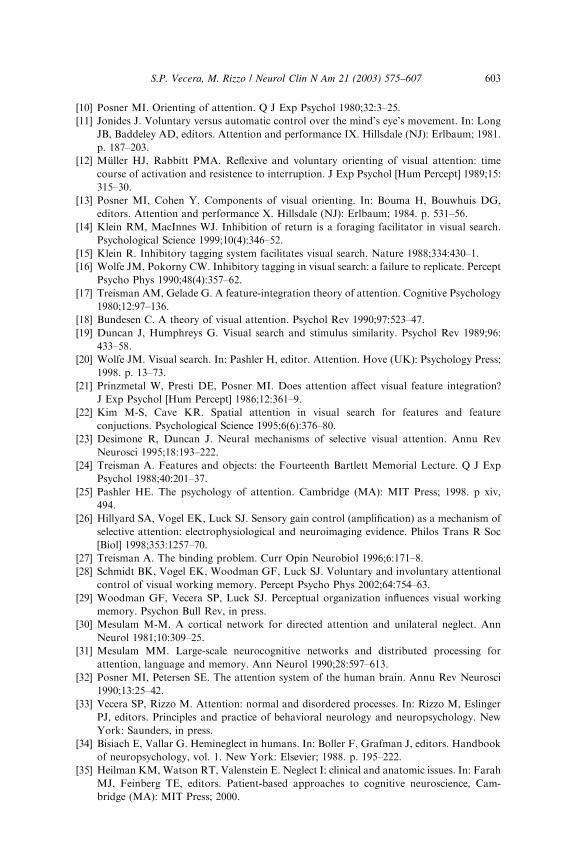
[10] Posner MI. Orienting of attention. Q J Exp Psychol 1980;32:3–25.
[11] Jonides J. Voluntary versus automatic control over the mind’s eye’s movement. In: Long
JB, Baddeley AD, editors. Attention and performance IX. Hillsdale (NJ): Erlbaum; 1981.
p. 187–203.
[12] Muller HJ, Rabbitt PMA. Reflexive and voluntary orienting of visual attention: time
course of activation and resistence to interruption. J Exp Psychol [Hum Percept] 1989;15:
315–30.
[13] Posner MI, Cohen Y. Components of visual orienting. In: Bouma H, Bouwhuis DG,
editors. Attention and performance X. Hillsdale (NJ): Erlbaum; 1984. p. 531–56.
[14] Klein RM, MacInnes WJ. Inhibition of return is a foraging facilitator in visual search.
Psychological Science 1999;10(4):346–52.
[15] Klein R. Inhibitory tagging system facilitates visual search. Nature 1988;334:430–1.
[16] Wolfe JM, Pokorny CW. Inhibitory tagging in visual search: a failure to replicate. Percept
Psycho Phys 1990;48(4):357–62.
[17] Treisman AM, Gelade G. A feature-integration theory of attention. Cognitive Psychology
1980;12:97–136.
[18] Bundesen C. A theory of visual attention. Psychol Rev 1990;97:523–47.
[19] Duncan J, Humphreys G. Visual search and stimulus similarity. Psychol Rev 1989;96:
433–58.
[20] Wolfe JM. Visual search. In: Pashler H, editor. Attention. Hove (UK): Psychology Press;
1998. p. 13–73.
[21] Prinzmetal W, Presti DE, Posner MI. Does attention affect visual feature integration?
J Exp Psychol [Hum Percept] 1986;12:361–9.
[22] Kim M-S, Cave KR. Spatial attention in visual search for features and feature
conjuctions. Psychological Science 1995;6(6):376–80.
[23] Desimone R, Duncan J. Neural mechanisms of selective visual attention. Annu Rev
Neurosci 1995;18:193–222.
[24] Treisman A. Features and objects: the Fourteenth Bartlett Memorial Lecture. Q J Exp
Psychol 1988;40:201–37.
[25] Pashler HE. The psychology of attention. Cambridge (MA): MIT Press; 1998. p xiv,
494.
[26] Hillyard SA, Vogel EK, Luck SJ. Sensory gain control (amplification) as a mechanism of
selective attention: electrophysiological and neuroimaging evidence. Philos Trans R Soc
[Biol] 1998;353:1257–70.
[27] Treisman A. The binding problem. Curr Opin Neurobiol 1996;6:171–8.
[28] Schmidt BK, Vogel EK, Woodman GF, Luck SJ. Voluntary and involuntary attentional
control of visual working memory. Percept Psycho Phys 2002;64:754–63.
[29] Woodman GF, Vecera SP, Luck SJ. Perceptual organization influences visual working
memory. Psychon Bull Rev, in press.
[30] Mesulam M-M. A cortical network for directed attention and unilateral neglect. Ann
Neurol 1981;10:309–25.
[31] Mesulam MM. Large-scale neurocognitive networks and distributed processing for
attention, language and memory. Ann Neurol 1990;28:597–613.
[32] Posner MI, Petersen SE. The attention system of the human brain. Annu Rev Neurosci
1990;13:25–42.
[33] Vecera SP, Rizzo M. Attention: normal and disordered processes. In: Rizzo M, Eslinger
PJ, editors. Principles and practice of behavioral neurology and neuropsychology. New
York: Saunders, in press.
[34] Bisiach E, Vallar G. Hemineglect in humans. In: Boller F, Grafman J, editors. Handbook
of neuropsychology, vol. 1. New York: Elsevier; 1988. p. 195–222.
[35] Heilman KM,Watson RT, Valenstein E. Neglect I: clinical and anatomic issues. In: Farah
MJ, Feinberg TE, editors. Patient-based approaches to cognitive neuroscience, Cam-
bridge (MA): MIT Press; 2000.
603S.P. Vecera, M. Rizzo / Neurol Clin N Am 21 (2003) 575–607
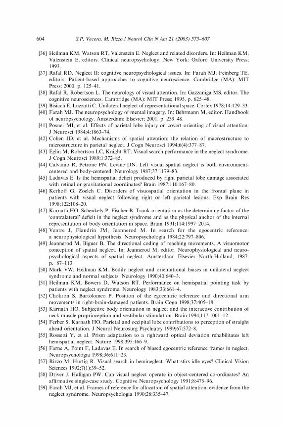
[36] Heilman KM, Watson RT, Valenstein E. Neglect and related disorders. In: Heilman KM,
Valenstein E, editors. Clinical neuropsychology. New York: Oxford University Press;
1993.
[37] Rafal RD. Neglect II: cognitive neuropsychological issues. In: Farah MJ, Feinberg TE,
editors. Patient-based approaches to cognitive neuroscience. Cambridge (MA): MIT
Press; 2000. p. 125–41.
[38] Rafal R, Robertson L. The neurology of visual attention. In: Gazzaniga MS, editor. The
cognitive neurosciences. Cambridge (MA): MIT Press; 1995. p. 625–48.
[39] Bisiach E, Luzzatti C. Unilateral neglect of representational space. Cortex 1978;14:129–33.
[40] Farah MJ. The neuropsychology of mental imagery. In: Behrmann M, editor. Handbook
of neuropsychology. Amsterdam: Elsevier; 2001. p. 239–48.
[41] Posner MI, et al. Effects of parietal lobe injury on covert orienting of visual attention.
J Neurosci 1984;4:1863–74.
[42] Cohen JD, et al. Mechanisms of spatial attention: the relation of macrostructure to
microstructure in parietal neglect. J Cogn Neurosci 1994;6(4):377–87.
[43] Eglin M, Robertson LC, Knight RT. Visual search performance in the neglect syndrome.
J Cogn Neurosci 1989;1:372–85.
[44] Calvanio R, Petrone PN, Levine DN. Left visual spatial neglect is both environment-
centered and body-centered. Neurology 1987;37:1179–83.
[45] Ladavas E. Is the hemispatial deficit produced by right parietal lobe damage associated
with retinal or gravitational coordinates? Brain 1987;110:167–80.
[46] Kerhoff G, Zoelch C. Disorders of visuospatial orientation in the frontal plane in
patients with visual neglect following right or left parietal lesions. Exp Brain Res
1998;122:108–20.
[47] Karnath HO, Schenkely P, Fischer B. Trunk orientation as the determining factor of the
�contralateral� deficit in the neglect syndrome and as the physical anchor of the internal
representation of body orientation in space. Brain 1991;114:1997–2014.
[48] Ventre J, Flandrin JM, Jeannerod M. In search for the egocentric reference:
a neurophysiological hypothesis. Neuropsychologia 1984;22:797–806.
[49] Jeannerod M, Biguer B. The directional coding of reaching movements. A visuomotor
conception of spatial neglect. In: Jeannerod M, editor. Neurophysiological and neuro-
psychological aspects of spatial neglect. Amsterdam: Elsevier North-Holland; 1987.
p. 87–113.
[50] Mark VW, Heilman KM. Bodily neglect and orientational biases in unilateral neglect
syndrome and normal subjects. Neurology 1990;40:640–3.
[51] Heilman KM, Bowers D, Watson RT. Performance on hemispatial pointing task by
patients with neglect syndrome. Neurology 1983;33:661–4.
[52] Chokron S, Bartolomeo P. Position of the egocentric reference and directional arm
movements in right-brain-damaged patients. Brain Cogn 1998;37:405–18.
[53] Karnath HO. Subjective body orientation in neglect and the interactive contribution of
neck muscle proprioception and vestibular stimulation. Brain 1994;117:1001–12.
[54] Ferber S, Karnath HO. Parietal and occipital lobe contributions to perception of straight
ahead orientation. J Neurol Neurosurg Psychiatry 1999;67:572–8.
[55] Rossetti Y, et al. Prism adaptation to a rightward optical deviation rehabilitates left
hemispatial neglect. Nature 1998;395:166–9.
[56] Farne A, Point F, Ladavas E. In search of biased egocentric reference frames in neglect.
Neuropsychologia 1998;36:611–23.
[57] Rizzo M, Hurtig R. Visual search in hemineglect: What stirs idle eyes? Clinical Vision
Sciences 1992;7(1):39–52.
[58] Driver J, Halligan PW. Can visual neglect operate in object-centered co-ordinates? An
affirmative single-case study. Cognitive Neuropsychology 1991;8:475–96.
[59] Farah MJ, et al. Frames of reference for allocation of spatial attention: evidence from the
neglect syndrome. Neuropsychologia 1990;28:335–47.
604 S.P. Vecera, M. Rizzo / Neurol Clin N Am 21 (2003) 575–607
[60] Farah MJ, et al. Parietal lobe mechanisms of spatial attention: modality-specific or
supramodal? Neuropsychologia 1989;27:461–70.
[61] Spence C, Driver J. Audiovisual links in endogenous covert spatial attention. J Exp
Psychol [Hum Percept] 1996;22(4):1005–30.
[62] Spence C, Driver J. Audiovisual links in exogenous covert spatial orienting. Percept
Psycho Phys 1997;59(1):1–22.
[63] Farah MJ, Monheit MA, Wallace MA. Unconscious perception of ‘‘extinguished’’ visual
stimuli: Reassessing the evidence. Neuropsychologia 1991;29(10):949–58.
[64] Ladavas E, Carletti M, Gori G. Automatic and voluntary orienting of attention in
patients with visual neglect: horizontal and vertical dimensions. Neuropsychologia
1994;32:1195–208.
[65] Bartolomeo P, et al. Modulating the attentional bias in unilateral neglect: The effects of
the strategic set. Exp Brain Res 2001;137:432–44.
[66] Baylis GC, Driver J, Rafal RD. Visual extinction and stimulus repetition. J Cogn
Neurosci 1993;5(4):453–66.
[67] Lu Z, Dosher BA. External noise distinguishes attention mechanisms. Vision Res 1998;38:
1183–98.
[68] Intriligator J, Cavanagh P. The spatial resolution of visual attention. Cognitive
Psychology 2001;43(3):171–216.
[69] He S, Cavanagh P, Intriligator J. Attentional resolution and the locus of visual awareness.
Nature 1996;383:334–7.
[70] Treisman A, Schmidt H. Illusory conjunctions in the perception of objects. Cognitive
Psychology 1982;14:107–41.
[71] Cohen A, Rafal RD. Attention and feature integration: illusory conjunctions in a patient
with a parietal lobe lesion. Psychological Science 1991;2:106–10.
[72] Friedman-Hill SR, Robertson LC, Treisman A. Parietal contributions to visual
feature binding: evidence from a patient with bilateral lesions. Science 1995;269(5225):
853–85.
[73] Rizzo M, Vecera SP. Psychoanatomical substrates of Balint’s syndrome. J Neurol
Neurosurg Psychiatry 2002;72(2):162–78.
[74] Bisiach E, Vallar G. Unilateral neglect in humans. In: Boller F, Grafman J, Rizzolatti G,
editors. Handbook of neuropsychology. Amsterdam: Elsevier; 2001. p. 459–502.
[75] Swick D, Knight RT. Cortical lesions and attention. In: Parasuraman R, editor. The
attentive brain. Cambridge (MA): MIT Press; 1998. p. 143–62.
[76] Colby CL, Goldberg ME. Space and attention in parietal cortex. Annu Rev Neurosci
1999;22:319–49.
[77] Guitton D, Buchtel HA, Douglas RM. Frontal lobe lesions in man cause difficulties in
suppressing reflexive glances and in generating goal-directed saccades. Exp Brain Res
1985;58:455–72.
[78] Henik A, Rafal R, Rhodes D. Endogenously generated and visually guided saccades after
lesions of the human frontal eye fields. J Cogn Neurosci 1994;6(4):400–11.
[79] Alivisatos B, Milner B. Effects of frontal or temporal lobectomy on the use of advance
information in a choice reaction time task. Neuropsychologia 1989;27:495–503.
[80] Koski LM, Paus T, Petrides M. Directed attention after unilaterial frontal excisions in
humans. Neuropsychologia 1998;36(2):1363–71.
[81] Eslinger PJ, Damasio AR. Severe disturbance of higher cognition after bilateral frontal
lobe ablation. Patient EVR. Neurology 1985;25:1731–41.
[82] Rafal R, Grimm RJ. Progressive supranuclear palsy: functional analysis of the response to
methysergide and antiparkinsonian agents. Neurology 1981;31:1507–18.
[83] Posner MI, Cohen Y, Rafal RD. Neural systems control of spatial orienting. Philadelphia
Transactions of the Royal Society of London 1982;B298:187–98.
[84] Rafal RD, et al. Orienting of attention in progressive supranuclear palsy. Brain 1988;111:
267–80.
605S.P. Vecera, M. Rizzo / Neurol Clin N Am 21 (2003) 575–607
[85] Posner MI, et al. Inhibition of return: neural basis and function. Cognitive Neuro-
psychology 1985;2(3):211–28.
[86] Rafal RD, Posner MI. Deficits in human visual spatial attention following thalamic
lesions. Proc Natl Acad Sci USA 1987;84:7349–53.
[87] Petersen SE, Robinson DL, Keys W. Pulvinar nuclei of the behaving Rhesus Monkey:
visual responses and their modulation. J Neurophysiol 1985;54:867–86.
[88] Petersen SE, Robinson DL, Morris JD. Contributions of the pulvinar to visual spatial
attention. Neuropsychologia 1987;25:97–105.
[89] Petersen RC, et al. Practice parameter: early detection of dementia. Neurology 2001;
56(9):1133–42.
[90] McKhannG, et al. Clinical diagnosis of Alzheimer’s disease. Neurology 1984;34(7):939–44.
[91] Parasuraman R, Haxby JV. Attention and brain function in Alzheimer’s disease: a review.
Neuropsychology 1993;7(3):242–72.
[92] Balota DA, Faust ME. Attention in dementia of the Alzheimer’s type. In: Boller F,
Cappa SF, editors. Handbook of neuropsychology 2nd edition. Amsterdam: Elsevier;
2001. p. 51–80.
[93] Perry RJ, Hodges JR. Attention and executive deficits in Alzheimer’s disease: a critical
review. Brain 1999;122:383–404.
[94] Rizzo M, et al. Visual attention impairments in Alzheimer’s disease. Neurology 2000;
54:1954–9.
[95] Parasuraman R, et al. Visuospatial attention in dementia of the Alzheimer type. Brain
1992;115:711–33.
[96] Greenwood PM, et al. Genetics and visual attention: selective deficits in healthy adult
carriers of the e4 allele of the apolipoprotein E gene. Proc Natl Acad Sci USA
2000;97:11661–6.
[97] Faust ME, Balota DA. Inhibition of return and visuospatial attention in healthy older
adults and individuals with dementia of the Alzheimer’s type. Neuropsychology
1997;11:13–29.
[98] Maylor EA, Hockey R. Inhibitory components of externally controlled covert orienting in
visual space. J Exp Psychol [Hum Percept] 1985;11:777–87.
[99] Foster JK, BehrmannM, Stuss DT. Visual attention deficits in Alzheimer’s disease: simple
versus conjoined feature search. Neuropsychology 1999;13(2):223–45.
[100] Parasuraman R, Greenwood PM, Alexander GE. Alzheimer disease constricts the
dynamic range of spatial attention in visual search. Neuropsychologia 2000;38:
1126–35.
[101] Greenwood PM, Parasuraman R, Alexander GE. Controlling the focus of spatial
attention during visual search: effects of advanced aging and Alzheimer’s disease.
Neuropsychology 1997;11(1):3–12.
[102] Coslett HB, et al. Narrowing the spotlight: a visual attentional disorder in presumed
Alzheimer’s disease. Neurocase 1995;1(4):305–18.
[103] Thaiss L, de Bleser R. Visual agnosia: a case of reduced attentional ‘‘spotlight’’? Cortex
1992;28(4):601–21.
[104] Awh ES. Attention-based rehearsal in spatial working memory. Dissertation Abstracts
International: Section B: the Sciences & Engineering 1996;57(6-B):4048.
[105] Awh E, Jonides J. Spatial working memory and spatial selective attention. In: Para-
suraman R, editor. The attentive brain. Cambridge (MA): MIT Press; 1998. p. 353–80.
[106] Nebes RD, Brady CB. Focused and divided attention in Alzheimer’s disease. Cortex
1989;25:305–15.
[107] Nebes RD. The cognitive neuropsychology of Alzheimer’s disease. In: Farah MJ,
Feinberg TE, editors. Patient-based approaches to cognitive neuroscience. Cambridge
(MA): MIT Press; 2000. p. 369–74.
[108] Shiu L, Pashler H. Negligible effect of spatial precuing on identification of single digits.
J Exp Psychol [Hum Percept] 1994;20:1037–54.
606 S.P. Vecera, M. Rizzo / Neurol Clin N Am 21 (2003) 575–607

[109] Palmer J. Attention in visual search: distinguishing four causes of a set-size effect. Current
Directions in Psychological Science 1995;4(4):118–23.
[110] Eckstein MP. The lower visual search efficiency for conjunctions is due to noise and not
serial attentional processing. Psychological Science 1998;9(2):111–8.
[111] Eckstein MP, et al. A signal detection model predicts the effects of set size on visual search
accuracy for feature, conjunction, triple conjunction, and disjunction displays. Percept
Psychophysiol 2000;62:425–51.
607S.P. Vecera, M. Rizzo / Neurol Clin N Am 21 (2003) 575–607