special edition - mastocytosis society...2 tmsforacure.org | special edition 2017 the mastocytosis...
TRANSCRIPT

© 2017 The Mastocytosis Society, Inc. All rights reserved
SPECIAL EDITION HEALTH CARE PROFESSIONALS EDITION
The Mastocytosis ChroniclesThe Mastocytosis Society, Inc. | 2017

2 tmsforacure.org | Special Edition 2017
The Mastocytosis Society, Inc. (TMS) was founded in 1995 by Bill Abbottsmith, Linda Buchheit, Olive Clayson, Iris Dissinger, Bill Hingst, and Joe Palk. At that time very little was known about Mastocytosis, so these pioneering individuals sought to fill a massive void with some answers to their multitude of questions about this rare disease. They found one another through NORD, with sheer determination and extensive research.
The first support group meeting was held in Baltimore at the Inner Harbor in 1994 and was attended by Linda Buchheit and Bill Hingst. The second meeting was held the following year at Linda Buchheit’s home in Ohio. Fourteen members attended that year. Little did they know how fruitful their efforts would be and what a lifeline they would become as more and more patients joined each year!
Until 1990 many patients diagnosed with Mastocytosis were given a very grim prognosis. Up until that time, Mastocytosis was not often considered when physicians were making a differential diagnosis, and many cases were completely missed, resulting in patient death. At that point, signs of the disease were then discovered on autopsy; however, because so little was known about Mastocytosis, it was
presumed that Mastocytosis was one of the causes of death, when in fact the patient had often died of other causes, and the Mastocytosis was an incidental finding! On the other hand, more advanced cases of aggressive Mastocytosis were also recognized during post-mortem exams, leading pathologists to identify all forms of Mastocytosis as having a high associated mortality rate. Fortunately, that prognosis has improved as more patients are diagnosed and treated sooner, and more physicians research and treat this disease. Today, we know that pediatric patients have greater than a 75% chance of outgrowing their disease at or before puberty, and adults with Indolent Systemic Mastocytosis can have a near normal life expectancy if they avoid triggers and take their medication!
Founding Members: Today’s accomplishments are built on the foundations laid by the early volunteers, and we are grateful for their efforts. TMS is where it is today because of the seeds that they planted in 1994 and in the early years. Below are some of the earliest members, but there have been many more champions who have served their fellow patients and families affected by Mastocytosis and Mast Cell Activation Disorders by volunteering for TMS. We salute you!
Past Board Members: THANK YOU to all of our past board members as they are our strong foundation for all the wonderful and exciting things happening now and in the future for TMS!
Our History

3tmsforacure.org | Special Edition 2017
In this issue
5 Overview, Definitions, Diagnosis and Classification
9 Cytology of Mast Cells
10 Cutaneous Mastocytosis Variants
12 Systemic Mastocytosis Variants
16 Mast Cell Activation Syndrome Variants
18 Signs, Symptoms And Triggers
21 Tests
25 Treatments For Mast Cell Disorders
27 Medications To Treat Mast Cell Disorders
29 Pediatric Mast Cell Disorders: Facts in Brief
36 Visual Guide to Diagnosing Mastocytosis
40 Medical & Research Centers that Treat Patients with Mast Cell Diseases
43 Medical Advisory Board
45 The Mastocytosis Society Printed Materials
46 Medical Reference Highlights
49 Mast Cell Connect Patient Registry brochure
51 Support Group Contacts
The Mastocytosis ChroniclesThe Mastocytosis Society, Inc. | Spring 2017 - Volume 23 Issue 1
Mast Cell Disorder Challenges Meetings and US Network Update
By Susan Jennings, PhD, and Valerie Slee, RN, BSN - February 2017
Since 2014, The Mastocytosis Society, Inc. (TMS) has hosted small ancillary Mast Cell Disorder Challenges meetings during the annual gatherings of several physician specialty associations. The objectives of these meetings have been to bring together specialist physicians, drug company representatives and members of the TMS Research Committee to identify the primary challenges facing the mast cell disorder community in the United States and to explore possible actions that would address those challenges. A key conclusion from our initial Challenges meetings was that the establishment of a US Network for Mast Cell Disorders would be extremely helpful in overcoming many of the challenges faced by our disease community. During these meetings, our US physicians have received significant support from a number of international mast cell disorder specialists, who have shared their experiences of forming networks in their own countries and more broadly in Europe. TMS is committed to supporting activities that will lead to the formation of a US network under the leadership of Cem Akin, MD, PhD, and Jason Gotlib, MD, MS, as Co-Chairs. The American Academy of Allergy, Asthma and Immunology (AAAAI) Mast Cell Disorder Committee has also agreed to participate in this effort. Challenges meetings have been held while specialists have been gathered for American Society of Hematology and AAAAI Annual Meetings and immediately prior to the 2015 European Competence Network on Mastocytosis (ECNM) Annual Meeting.
Please see www.tmsforacure.org for more information and updates on our Mast Cell Disorder Challenges Meetings and progress on formation of a US Network for Mast Cell Disorders.

4 tmsforacure.org | Special Edition 2017
Committees
Aggressive Systemic Mastocytosis and Mast Cell Leukemia/Sarcoma Committee:Valerie M. Slee, Co-ChairMishele Cunningham, Co-ChairTMS Conference and Walkathon Committees([email protected])([email protected])Drug Shortage Committee([email protected]) Valerie M. Slee, Co-Chair ([email protected] or [email protected])Emily A. Menard, Co-Chair ([email protected])Fundraising Committee([email protected]) Patricia BeggiatoStephen ReyGrant Committee([email protected])Valerie M. Slee, RN, BSN, ChairMishele Cunningham, RN, BSN, PHN Rita BarlowPatricia BeggiatoMastocytosis Chronicles Committee([email protected]) Sandy Johnson, Editor/ChairJohn Gilligan, Graphic Designer Judy Thompson, Copy EditorMedia Relations Committee([email protected])Ariella Cohen, ChairMedical Conference Planning Committee([email protected]) Mishele Cunningham, ChairWanda HermanMedical Device CommitteePeg Kleeve, ChairMembership CommitteeNikki Martinez- Co-ChairNicole Rogne0 Co-Chair([email protected])MPN Advocacy Committee([email protected]) Jennifer Dratch, ChairValerie M. Slee Pediatric Committee([email protected]) Stacy Rawson Sheldon, Co-Chair Kelli Foster, Co-ChairKolleen BarlowPolitical and Patient Advocacy CommitteePatricia Beggiato, Co-Chair (Board Liaison) Kelli Foster, Co-ChairResearch Committee([email protected])Susan Jennings, PhD, ChairValerie M. Slee, RN, BSNCeleste Finnerty, PhDSmoldering Systemic Mastocytosis CommitteeMichele Q. Kress, ChairValerie M. SleeSpecial Edition Chronicles Committee([email protected])Mishele Cunningham, RN, BSN, PHN, ChairValerie M. Slee, RN, BSNSusan Jennings, PhDDoris Hoopes, Copy EditorSupport Groups Committee([email protected]) Rita Barlow, Co-ChairCheri Smith Co-ChairWebsite/Technology Committee([email protected])Russell Hirshon, WebmasterCasey Cunningham, Technology Committee Chair
TMS Medical Advisory Board
TMS is proud to be a Lay Organization member of The AmericanAcademy of Allergy Asthma and Immunology (AAAAI)
TMS is a long-standing National Organization Member of the National Organization for Rare Disorders (NORD)
We thank each of these doctors for their time, caring, and expertise.
Ivan Alvarez-Twose, MDK. Frank Austen, MD (Honorary)Patrizia Bonadonna, MDJoseph Butterfield, MDMariana Castells, MD, PhD Madeleine Duvic, MD
Luis Escribano, MD, PhD, Jason Gotlib, MD, MSNorton J. Greenberger, MDMatthew J. Hamilton, MDOlivier Hermine, MD, PhDNicholas Kounis, MD, PhD
Anne Maitland, MD, PhDLarry Schwartz, MD, PhD Theoharis Theoarides, MD, PhDCelalettin Ustun, M.D.Peter Valent, MDSrdan Verstovsek, MD, PhD
Special Edition For Health Care Professionals
The special edition of The Mastocytosis Chronicles has been published specifically for physicians and health care professionals since 2007. This edtion contains diagnostic and treatment protocols for mastocytosis and mast cell activation disorders, locations of mast cell disorder treatment centers, physician contact information, documentation of research articles, and other pertinent information. For additional information visit www.tmsforacure.org.
Board of DirectorsValerie M. Slee RN, BSN: Chair Medical Advisory Board Liaison Patient Referral Coordinator [email protected]
Rita Barlow: Vice Chair Patient Support and Advocacy [email protected]
Gail Barbera: Secretary [email protected]
Stephen Rey: Treasurer [email protected]
Mishele Cunningham, RN, BSN, PHN: Education and Medical Conference Chair [email protected]
Patricia Beggiato: Fundraising and Political Advocacy Chair [email protected]
Stacy Sheldon: Pediatric Chair [email protected]
Our MissionThe Mastocytosis Society, Inc. is a nonprofit organization dedicated to supporting patients affected by Mast Cell Disease, as well as their families, caregivers, and physicians through research, education, and advocacy.

5tmsforacure.org | Special Edition 2017
What are Mast Cells?
Mast cells (MC) are immune system cells that live in the bone marrow and in body tissues, internal and external, such as the gastrointestinal tract, the lining of the airway, and the skin. Everyone has mast cells in their body, and they play many complex and critical roles in keeping us healthy. The positive roles that they play include protecting us from infection, and helping our body by participating in the inflammatory process. However, mast cells are also involved in allergic reactions, from the tiny swelling that appears after a mosquito bite to a life threatening, full-blown anaphylaxis.
Mast cells have within them small sacs, or granules, surrounded by membranes. The sacs contain many different kinds of substances called mediators, which participate in all of the roles above, including allergic response and anaphylaxis. The mediators are selectively released when there is an allergic or mast cell based reaction.1
There is a difference between someone who is healthy, with mast cells that are functioning normally, and someone with a mast cell disorder, whose mast cells may be activating inappropriately in response to triggers, or may also be proliferating and accumulating in organ tissues.
What are Mast Cell Disorders?
Mast cell disorders are caused by the proliferation and accumulation of genetically altered mast cells and/or the inappropriate release of mast cell mediators,
creating symptoms in multiple organ systems.2 The two major forms of mast cell disorders
are mastocytosis and mast cell activation syndromes (MCAS). Mast cell disorders
can cause tremendous suffering and disability due to symptomatology from
daily mast cell mediator release, and/or symptoms arising from infiltration and accumulation of mast cells in major organ systems. Although systemic mastocytosis is a rare disease,3 those suffering with MCAS have recently
been increasingly recognized and diagnosed. As a result, patients with
MCAS appear to represent a growing
Mast cell granule (sac) which contains mediators
Figure 1. Mast cell (electron micrograph)
MAST CELLS AND MAST CELL DISORDERS
Overview, Definitions, Diagnosis and Classification
Continued on page 6

6 tmsforacure.org | Special Edition 2017
Overview, Definitions, Diagnosis and ClassificationContinued from page 5
proportion of the mast cell disorder patient population.4,
5 It is important to note that the process of mast cell activation can occur in anyone, even without a mast cell disorder, as well as in patients with both mastocytosis and MCAS.6
MASTOCYTOSIS
Definition
Mastocytosis has been defined in the literature as an abnormal accumulation of mast cells in one or more organ systems. Previously classified by the World Health Organization (WHO) as a myeloproliferative
neoplasm, mastocytosis is now classified in its own category under myeloid neoplasms.7 Broadly
separated into three categories, cutaneous mastocytosis (CM), systemic mastocytosis
(SM) and mast cell sarcoma these diseases occur in both children and
adults. CM is considered a benign skin disease representing the majority of pediatric cases. In 67-80% of pediatric cases seen, resolution will occur before or in early adulthood.8-10 In pediatric mastocytosis, symptoms of mast
cell mediator release may occur systemically as a result of mast cell
mediators released from skin lesions.10 This, however, does not necessarily
indicate systemic disease. The incidence of systemic pediatric disease was previously
unknown, but systemic forms have now been proven to exist in some children.8-10 The majority
of adult patients are diagnosed with systemic disease. Skin involvement, typically maculopapular cutaneous mastocytosis/urticaria pigmentosa, is common in adult patients and can provide an important clue to accurate diagnosis.11, 12
Diagnosis and Classification13-17
CM is diagnosed by the presence of typical skin lesions and a positive skin biopsy demonstrating characteristic clusters of mast cells. The preferred method of diagnosing SM is via bone marrow (BM) biopsy. The WHO has established criteria for diagnosing SM, summarized18 as follows:
Major ª: Multifocal dense infiltrates of mast cells (MCs) (> 15 MCs in aggregate) in tryptase stained biopsy sections of the bone marrow or other extracutaneous organ
Minorª: • More than 25% of MCs in bone marrow or
other extracutaneous organ(s) show abnormal morphology (i.e. are atypical MC type 1 or are spindle–shaped MCs) in multifocal lesions in histologic examination
• KIT mutation at codon 816b in extracutaneous organ(s) (in most cases bone marrow cells are examined)
• KIT+MCs in bone marrow show aberrant expression of CD2 and/or CD25
• Serum total tryptase > 20 ng/mL (does not count in patients who have ANHMD-type disease.)
Abbreviation Key:
KIT: KIT tyrosine kinase receptor; MC(s): Mast cells; AHNMD: associated (clonal) hematologic non-mast cell lineage disease.
ª If at least one major criterion and one minor criterion OR at least three minor criteria are fulfilled, the diagnosis of systemic mastocytosis can be established. b Activating mutations at codon 816, in most cases, KIT D816V.

7tmsforacure.org | Special Edition 2017
MAST CELL ACTIVATION SYNDROMES
Definition
Existence of a subset of mast cell disorder patients who experience episodes of mast cell activation without detectable evidence of a proliferative mast cell disorder was postulated over 20 years ago.19, 20 Over the last two decades, with development of improved methodology for identification of abnormal mast cells,21-24 it became apparent that there were patients who exhibited symptoms of mast cell mediator release who did not fulfill the criteria for SM.25, 26 Thus began the evolution of discussions about other forms of mast cell disorders, both clonal and nonclonal, which became known as Mast Cell Activation Syndromes (MCAS).6, 27, 28
Diagnosis and Proposed Classification
Recognition by specialist physicians of the importance of mast cell activation in disease led to an international Mast Cell Disorders Working Conference emphasizing this topic in September of 2010. Consensus statements were published regarding classification of and diagnostic criteria for mast cell disorders,6 where mast cell activation plays a prominent role.
Mediators produced by mast cells have a considerable effect on specific symptomatology. Symptoms, including, but not limited to flushing, pruritis (itching), urticaria (hives), headache, gastrointestinal symptoms (including diarrhea, nausea, vomiting, abdominal pain, bloating, gastroesophageal reflux), and hypotension (low blood pressure), allow a patient to meet the first of three required co-criterion for systemic mast cell activation when the patient exhibits symptoms involving two or more organ systems in parallel, which recur, or are chronic, are found not to be caused by any other condition or disorder other than mast cell activation, and require treatment or therapy.6, 28
The second required co-criterion for systemic mast cell activation depends on documentation that mast cells are directly involved in the symptomatology. An increase in the serum level of tryptase, above baseline and within a narrow (generally accepted as one to two hour) window of time after a symptomatic episode, is proposed as the preferred method for providing evidence of mast cell involvement according to these criteria.6, 28-30 The consensus article provides a method for calculating the required minimum rise in serum tryptase.6 After a reaction, a level of serum tryptase that is a minimum of 20% above the basal serum tryptase level, plus 2 ng/ml, will meet the second criterion listed above for a mast cell activation event. Consensus members also agreed that when serum tryptase evaluation is not available or when the tryptase level does not rise sufficiently to meet the required increase for the co-criterion, other mediator tests could suffice. A rise in urinary n-methyl histamine, prostaglandin-D2, or its metabolite, 11β-prostaglandin-F2α (24-hour urine test for any of the three), is considered an alternative for the co-criterion related to a requirement for a mast cell mediator level rise during a systemic mast cell activation event.6
Finally, the third co-criterion requires a response (based on response criteria15) to medications that inhibit the action of histamine.6 In addition, in those with typical mast cell activation symptoms, a “complete or major” response to drugs that inhibit other mediators produced by mast cells or block mast cell mediator release can be regarded as fulfillment of the third co-criterion for MCAS.6, 28
Continued on page 8

8 tmsforacure.org | Special Edition 2017
Mast cell types Morphology Types of disease
Normal/reactive Round, well-granulated, with granules that fill the cytoplasm and obscure the nucleus; round to oval nucleus
Normal marrow, mast cell hyperplasia, well differentiated SM
Atypical type I
(spindle shaped)
Hypogranular, enlarged, with cytoplasmic projections
Indolent SM, ASM, SM-AHN
Atypical type II
(promastocyte)
Enlarged and round, hypogranular; indented bilobed nuclei
Mast cell leukemia, myelomastocytic leukemia
Metachromatic blast
(immature)
Hypogranular with a few large metachromatic granules; high nuclear-to-cytoplasm ratio; smooth chromatin in nuclei
Mast cell leukemia, myelomastocytic leukemia
References
1. Gilfillan AM, Austin SJ, Metcalfe DD. Mast cell biology: introduction and overview. Adv Exp Med Biol. 2011;716:2-12.
2. Theoharides TC, Valent P, Akin C. Mast Cells, Mastocytosis, and Related Disorders. N Engl J Med. 2015 Jul 9;373(2):163-72.
3. Horny HP, Sotlar K, Valent P, Hartmann K. Mastocytosis: a disease of the hematopoietic stem cell. Dtsch Arztebl Int. 2008 Oct;105(40):686-92.
4. Akin C, Valent P, Metcalfe DD. Mast cell activation syndrome: proposed diagnostic criteria. J Allergy Clin Immunol. 2010 Dec;126(6):1099-104 e4.
5. Afrin LB. Presentation, diagnosis and management of mast cell activation syndrome. In: Murray DB, editor. Mast cells: phenotypic features, biological functions and role in immunity. Hauppauge: Nova Science Publishers, Inc.; 2013. p. 155-232.
6. Valent P, Akin C, Arock M, Brockow K, Butterfield JH, Carter MC, et al. Definitions, criteria and global classification of mast cell disorders with special reference to mast cell activation syndromes: a consensus proposal. Int Arch Allergy Immunol. 2012;157(3):215-25.
7. Arber DA, Orazi A, Hasserjian R, Thiele J, Borowitz MJ, Le Beau MM, et al. The 2016 revision to the World Health Organization classification of myeloid neoplasms and acute leukemia. Blood. 2016 May 19;127(20):2391-405.
8. Torrelo A, Alvarez-Twose I, Escribano L. Childhood mastocytosis. Curr Opin Pediatr. 2012 Aug;24(4):480-6.
9. Fried AJ, Akin C. Primary mast cell disorders in children. Curr Allergy Asthma Rep. 2013 Dec;13(6):693-701.
10. Meni C, Bruneau J, Georgin-Lavialle S, Le Sache de Peufeilhoux L, Damaj G, Hadj-Rabia S, et al. Paediatric mastocytosis: a systematic review of 1747 cases. Br J Dermatol. 2015 Mar;172(3):642-51.
11. Berezowska S, Flaig MJ, Rueff F, Walz C, Haferlach T, Krokowski M, et al. Adult-onset mastocytosis in the skin is highly suggestive of systemic mastocytosis. Mod Pathol. 2014 Jan;27(1):19-29.
12. Hartmann K, Escribano L, Grattan C, Brockow K, Carter MC, Alvarez-Twose I, et al. Cutaneous manifestations in patients with mastocytosis: Consensus report of the European Competence Network on Mastocytosis; the American Academy of Allergy, Asthma & Immunology; and the European Academy of Allergology and Clinical Immunology. J Allergy Clin Immunol. 2016 Jan;137(1):35-45.
13. Valent P, Horny HP, Escribano L, Longley BJ, Li CY, Schwartz LB, et al. Diagnostic criteria and classification of mastocytosis: a consensus proposal. Leuk Res. 2001 Jul;25(7):603-25.
14. Valent P, Horny H-P, Li CY, Longley JB, Metcalfe DD, Parwaresch RM, et al. Mastocytosis. Jaffe ES, Harris NL, Stein H, Vardiman JW, editors. World Health Organization (WHO) Classification of Tumours. Pathology and Genetics. Tumours of Haematopoietic and Lymphoid Tissues. Lyon: IARC Press; 2001.
15. Valent P, Akin C, Escribano L, Fodinger M, Hartmann K, Brockow K, et al. Standards and standardization in mastocytosis: consensus statements on diagnostics, treatment recommendations and response criteria. Eur J Clin Invest. 2007 Jun;37(6):435-53.
16. Horny HP, Akin C, Metcalfe DD, Escribano L, Bennett JM, Valent P, et al. Mastocytosis (mast cell disease) Swerdlow SH, Campo
E, Harris NL, Jaffe ES, Pileri SA, Stein H, et al., editors. World Health Organization (WHO) Classification of Tumours. Pathology and Genetics. Tumours of Haematopoietic and Lymphoid Tissues. Lyon: IARC Press; 2008.
17. Valent P, Escribano L, Broesby-Olsen S, Hartmann K, Grattan C,
Brockow K, et al. Proposed diagnostic algorithm for patients with suspected mastocytosis: a proposal of the European Competence Network on Mastocytosis. Allergy. 2014 Oct;69(10):1267-74.
18. Valent P. Diagnostic evaluation and classification of mastocytosis. Immunol Allergy Clin North Am. 2006 Aug;26(3):515-34.
19. Roberts LJ, 2nd, Oates JA. Biochemical diagnosis of systemic mast cell disorders. J Invest Dermatol. 1991 Mar;96(3):19S-24S; discussion S-5S.
20. Metcalfe DD. Classification and diagnosis of mastocytosis: current status. J Invest Dermatol. 1991 Mar;96(3):2S-4S.
21. Nagata H, Worobec AS, Oh CK, Chowdhury BA, Tannenbaum S, Suzuki Y, et al. Identification of a point mutation in the catalytic domain of the protooncogene c-kit in peripheral blood mononuclear cells of patients who have mastocytosis with an associated hematologic disorder. Proc Natl Acad Sci U S A. 1995 Nov 7;92(23):10560-4.
22. Longley BJ, Tyrrell L, Lu SZ, Ma YS, Langley K, Ding TG, et al. Somatic c-KIT activating mutation in urticaria pigmentosa and aggressive mastocytosis: establishment of clonality in a human mast cell neoplasm. Nat Genet. 1996 Mar;12(3):312-4.
23. Escribano L, Orfao A, Diaz-Agustin B, Villarrubia J, Cervero C, Lopez A, et al. Indolent systemic mast cell disease in adults: immunophenotypic characterization of bone marrow mast cells and its diagnostic implications. Blood. 1998 Apr 15;91(8):2731-6.
24. Horny HP. Mastocytosis: an unusual clonal disorder of bone marrow-derived hematopoietic progenitor cells. Am J Clin Pathol. 2009 Sep;132(3):438-47.
25. Sonneck K, Florian S, Mullauer L, Wimazal F, Fodinger M, Sperr WR, et al. Diagnostic and subdiagnostic accumulation of mast cells in the bone marrow of patients with anaphylaxis: monoclonal mast cell activation syndrome. Int Arch Allergy Immunol. 2007;142(2):158-64.
26. Akin C, Scott LM, Kocabas CN, Kushnir-Sukhov N, Brittain E, Noel P, et al. Demonstration of an aberrant mast-cell population with clonal markers in a subset of patients with “idiopathic” anaphylaxis. Blood. 2007 Oct 1;110(7):2331-3.
27. Horny HP, Sotlar K, Valent P. Evaluation of mast cell activation syndromes: impact of pathology and immunohistology. Int Arch Allergy Immunol. 2012;159(1):1-5.
28. Valent P. Mast cell activation syndromes: definition and classification. Allergy. 2013 Apr;68(4):417-24.
29. Schwartz LB, Sakai K, Bradford TR, Ren S, Zweiman B, Worobec AS, et al. The alpha form of human tryptase is the predominant type present in blood at baseline in normal subjects and is elevated in those with systemic mastocytosis. J Clin Invest. 1995 Dec;96(6):2702-10.
30. Schwartz LB, Irani AM. Serum tryptase and the laboratory diagnosis of systemic mastocytosis. Hematol Oncol Clin North Am. 2000 Jun;14(3):641-57.
Overview, Definitions, Diagnosis and ClassificationContinued from page 7

Mast cell types Morphology Types of disease
Normal/reactive Round, well-granulated, with granules that fill the cytoplasm and obscure the nucleus; round to oval nucleus
Normal marrow, mast cell hyperplasia, well differentiated SM
Atypical type I
(spindle shaped)
Hypogranular, enlarged, with cytoplasmic projections
Indolent SM, ASM, SM-AHN
Atypical type II
(promastocyte)
Enlarged and round, hypogranular; indented bilobed nuclei
Mast cell leukemia, myelomastocytic leukemia
Metachromatic blast
(immature)
Hypogranular with a few large metachromatic granules; high nuclear-to-cytoplasm ratio; smooth chromatin in nuclei
Mast cell leukemia, myelomastocytic leukemia
SM: Systemic mastocytosis
ASM: Aggressive systemic mastocytosis
SM-AHN: Systemic mastocytosis with an associated hematologic neoplasm [previously referred to as SM-AHNMD (systemic mastocytosis with an associated (clonal) hematologic non-mast cell lineage disease]
Reference
1. George TI, Horny HP. Systemic mastocytosis. Hematol Oncol Clin North Am. 2011 Oct;25(5):1067-83, vii.
Cytology of Mast Cells1 By Tracy I. George, MD
tmsforacure.org | Special Edition 2017 9

10 tmsforacure.org | Special Edition 2017
An international consensus task force of mast cell disorder specialists has recently proposed updates to the diagnostic criteria and classification for cutaneous disease.1 Typical skin lesions found in mastocytosis, along with a positive Darier’s sign (see below), is the major criterion for diagnosing skin involvement in patients with mastocytosis. The two minor criteria are identified via skin lesion biopsy: increased mast cell numbers and the presence of an (activating) KIT mutation.1, 2 Cutaneous mastocytosis (CM) includes maculopapular cutaneous mastocytosis (MPCM), also known as urticaria pigmentosa (UP), diffuse cutaneous mastocytosis (DCM), and cutaneous mastocytoma.1 The taskforce recommends that telangiectasia macularis eruptiva perstans (TMEP) be removed as a separate category because, although some adult patients may have telangiectatic lesions on their chest, shoulders, neck and back, they may also demonstrate maculopapular lesions in other places, therefore fulfilling criteria for MPCM.
Most cases of pediatric mastocytosis fall into one of the above categories and may or may not include symptoms of systemic mast cell activation, including anaphylaxis, as a result of mediators released from the skin.3, 4 Pediatric CM encompasses a variety of clinical manifestations. In children, some forms of CM will spontaneously resolve, some will go on to be diagnosed as indolent systemic mastocytosis (ISM), with a smaller percentage identified as well-differentiated systemic mastocytosis (WDSM).5
In most adults with skin lesions typical for mastocytosis (in particular, the maculopapular type), systemic disease will ultimately be found, leading to a diagnosis of systemic mastocytosis, usually in an indolent form (indolent systemic mastocytosis).1, 6
Definitions1, 7
Darier’s sign is an important diagnostic finding of patients with mastocytosis. It can be elicited by stroking
an existing CM lesion with a wooden tongue depressor, approximately 5 times with moderate pressure. Within a few minutes, a wheal and flare reaction of the lesion will be seen. A positive Darier’s sign is usually seen in pediatric patients, but not always in adults. It may be decreased by treatment with antihistamines. If the testing procedure for Darier’s sign is not done properly, false positives or false negatives may result. Darier’s sign is to be applied to the evaluation of fixed cutaneous lesions except in the case of a pediatric patient with cutaneous mastocytoma or nodular lesions. Testing for Darier’s sign may provoke a systemic reaction and should either be performed with the greatest of caution or avoided.
Dermatographism is a skin reaction characterized by a wheal and flare response when normal skin, not affected by skin lesions, is stroked with a tongue depressor, finger nails or other instrument. The nick-name for dermatographism is skin writing disease.
A macule is a lesion that is flat and even with the surrounding skin, identified by a change in color compared to the surrounding skin.
A papule is a small bump or elevated lesion, up to 1 cm in diameter, containing no visible fluid.
A nodule is a growth of abnormal tissue just below the skin.
A bulla is a large blister filled with fluid.
Telangiectasia is a vascular lesion formed by dilatation of a group of small blood vessels.
VARIANTS OF CUTANEOUS MASTOCYTOSIS
Maculopapular Cutaneous Mastocytosis (MPCM)/Urticaria Pigmentosa (UP)1
• May be seen in infants, children or adults• Adults presenting with maculopapular lesions have
a very high likelihood of systemic disease, most frequently indolent systemic mastocytosis (ISM)
• Rarely, an adult presents with maculopapular lesions who does not have systemic disease, and has a diagnosis of MPCM
• Red maculopapular lesions tend to wheal when scratched (positive Darier’s sign)
• Blister formation can occur with rubbing or stroking
Cutaneous Mastocytosis Variants

11tmsforacure.org | Special Edition 2017
of lesion and is associated with pruritis8
• Encompasses several clinical entities with different outcomes, including: pitted melanotic macules, reddish brown telangiectatic macules, lightly pigmented papules, brownish papules, and small nodules
• This group is divided into two sub-variants
° Monomorphic variant - Mostly seen in adults and in a small subgroup
of children - Small maculopapular lesions, similar in shape,
size and color - Adults most typically express the KIT D816V
mutation in exon 17 of the KIT gene - In adults, thigh, axilla, trunk, extremities and
neck may be involved - 95% of adults diagnosed with ISM, 50% with
advanced systemic mastocytosis [systemic mastocytosis with an associated clonal hematologic non-mast cell lineage disease (SM-AHNMD or SM-AHN) or aggressive systemic mastocytosis (ASM)] and less than 50 % of mast cell leukemia patients exhibit this variant
- Children presenting with this form may have increased serum tryptase and a tendency toward systemic disease that persists into adulthood
- The type of lesions can vary during the course of the disease, i.e., nodules during infancy may turn into plaques at age 6
° Polymorphic variant - Mostly seen in children - Can be macular, plaque or nodular, with lesions
of variable shape, color and size - Although, children typically express mutations
in exon 8, 9, 11 or 17 of the KIT gene, KIT mutations may be negative
- Usually involving head, neck and extremities - May involve blistering upon irritation until 3
years of age - Prognosis is favorable with regression of
disease in adolescence or young adulthood
Diffuse Cutaneous Mastocytosis (DCM)1
• Skin thickened, hyperpigmented and diffusely infiltrated
• Can involve up to 100% of the skin with the trunk, head and scalp heavily affected
• Can appear at birth or early infancy; may persist into adulthood, possibly as well differentiated systemic mastocytosis (WDSM)5
• Blisters, some of which are hemorrhagic, and bullae may be present and dermatographism may be prominent
• Flushing is a common symptom• Tryptase may be elevated due to increased mast cell
burden in the skin and can be indicative of WDSM5
Cutaneous Mastocytoma1
• Usually present at birth• Elevated lesion(s) (up to a total of three lesions)
which usually resolves during childhood• Four cutaneous mastocytomas or more become a
diagnosis of MPCM• Multiple mastocytomas may evolve into adult WDSM5
References
1. Hartmann K, Escribano L, Grattan C, Brockow K, Carter MC, Alvarez-Twose I, et al. Cutaneous manifestations in patients with mastocytosis: Consensus report of the European Competence Network on Mastocytosis; the American Academy of Allergy, Asthma & Immunology; and the European Academy of Allergology and Clinical Immunology. J Allergy Clin Immunol. 2016 Jan;137(1): 35-45.
2. Valent P, Akin C, Escribano L, Fodinger M, Hartmann K, Brockow K, et al. Standards and standardization in mastocytosis: consensus statements on diagnostics, treatment recommendations and response criteria. Eur J Clin Invest. 2007 Jun;37(6):435-53.
3. Matito A, Carter M. Cutaneous and systemic mastocytosis in children: a risk factor for anaphylaxis? Curr Allergy Asthma Rep. 2015 May;15(5):22.
4. Meni C, Bruneau J, Georgin-Lavialle S, Le Sache de Peufeilhoux L, Damaj G, Hadj-Rabia S, et al. Paediatric mastocytosis: a systematic review of 1747 cases. Br J Dermatol. 2015 Mar;172(3):642-51.
5. Torrelo A, Alvarez-Twose I, Escribano L. Childhood mastocytosis. Curr Opin Pediatr. 2012 Aug;24(4):480-6.
6. Berezowska S, Flaig MJ, Rueff F, Walz C, Haferlach T, Krokowski M, et al. Adult-onset mastocytosis in the skin is highly suggestive of systemic mastocytosis. Mod Pathol. 2014 Jan;27(1):19-29.
7. Venes D, Thomas CL. Taber’s cyclopedic medical dictionary. 19 ed. Philadelphia: F.A. Davis Co.; 2001.
8. Castells M, Metcalfe DD, Escribano L. Diagnosis and treatment of cutaneous mastocytosis in children: practical recommendations. Am J Clin Dermatol. 2011 Aug 1;12(4):259-70.

12 tmsforacure.org | Special Edition 2017
Systemic mastocytosis (SM) consists of a group of rare, heterogeneous disorders involving growth and accumulation of abnormal mast cells (MC) in one or multiple extracutaneous (non-skin) organ systems (Table 1). Standard technique can be used to obtain an iliac crest bone marrow (BM) biopsy and aspirate smear for diagnosis. Aspirated BM should be allocated for flow cytometry to assess for the presence of mast cells with aberrant phenotype (i.e., co-expression of CD25). Immunohistochemistry for KIT, mast cell
tryptase, and CD25 should be performed on sections of the biopsy.1-5
Recent Updates in Diagnosis
A new diagnostic algorithm has been proposed by the European Competence Network on Mastocytosis for evaluating patients with suspected mastocytosis.6 Recommendations for KIT mutation analysis, including in peripheral blood, have also been recently published.7
Systemic Mastocytosis Variants
Table 1. Major Variants of Systemic Mastocytosis8
SYSTEMIC MASTOCYTOSISISM (Indolent systemic mastocytosis) WHO criteria for SM met, MC burden low, +/- skin lesions, no C findings, no evidence of AHNMD• Bone marrow mastocytosis: ISM with BM involvement, but no skin lesionsSSM (Smoldering systemic mastocytosis)WHO criteria for SM met, typically with skin lesions, with 2 or more B findings, but no C findings.
Advanced Disease VariantsSM-AHNMD (SM with associated clonal hematologic non mast cell lineage disease)/ SM-AHN (SM with an associated hematologic neoplasm)*Meets criteria for SM and also criteria for an AHNMD (MDS, MPN, MDS/MPN, AML), or other WHO-defined myeloid hematologic neoplasm, +/- skin lesions.ASM (Aggressive systemic mastocytosis)Meets criteria for SM with one or more C findings. No evidence of MCL, +/- skin lesions.MCL (Mast cell leukemia)Meets criteria for SM. BM biopsy shows a diffuse infiltration, usually compact, by atypical, immature MCs. BM aspirate smears show 20% or more MCs. Typical MCL: MCs comprise 10% or more of peripheral blood white cells. Aleukemic MCL: < 10% of peripheral blood white cells are MCs. Usually without skin lesions.
*SM-AHN is the recently updated term from the 2016 WHO classification of mastocytosis;9 a lymphoproliferative disorder or plasma cell dyscrasia may rarely be diagnosed with SM.
WHO: World Health Organization; BM: bone marrow; MC: mast cell; MDS: myelodysplastic syndrome; MPN: myeloproliferative neoplasm; MDS/MPN: myelodysplastic syndrome/ myeloproliferative neoplasm overlap disorders; AML: acute myeloid leukemia.

13tmsforacure.org | Special Edition 2017
INDOLENT SYSTEMIC MASTOCYTOSIS
The majority of adult patients fit into this category, fulfilling the criteria for indolent systemic mastocytosis (ISM).2, 10-12 The bone marrow, gastrointestinal tract, skeletal system, nervous system and skin may be affected. Some patients may have enlarged livers and spleens and lymphadenopathy. Mediator-related symptoms are common, but the grade of bone marrow infiltration is low (usually less than 5 percent) with the bone marrow fulfilling the criteria for SM and 80-90% of the patients exhibiting a positive D816V KIT mutation. In most patients the serum tryptase concentration exceeds 20 ng/mL, but a normal level of tryptase does not rule out either mastocytosis or another mast cell activation disorder. Treatment usually includes mediator-targeting drugs, including antihistamines, but does not usually require cytoreductive agents, although there are exceptions.
Isolated bone marrow mastocytosis (BMM) is a variant of indolent SM.12 BMM is characterized by the absence of skin lesions, lack of multi-organ involvement, and an increased incidence of anaphylaxis.13
Well differentiated SM (WDSM) first described in 200414, is reported in the literature as a rare variant that fulfills the major criterion for SM and continues to be studied by researchers.15-17 WDSM is distinguished from pediatric cutaneous mastocytosis by its inclusion in
the systemic category, despite that 91% of patients with WDSM have childhood onset of disease, with familial involvement in 39%. There is a heterogeneous presentation of lesions, maculopapular, nodular and diffuse cutaneous, that may involve a large percentage of the skin.17 Severe mast cell symptoms can occur and the variant may persist into adulthood in a low percentage of cases. The mast cells often do not express CD25 or CD2 that are part of the minor World Health Organization (WHO) criterion for SM, but may have CD30. Also, roughly 90% of WDSM patients don’t have the KIT D816V or other exon 17 KIT mutations.17 Bone marrow analysis identifies mast cells in WDSM patients as notably large, round, mature-appearing mast cells with the absence of the spindle-shaped mast cells typically seen in SM.15 Baseline serum tryptase levels
Table 2. B and C Findings8
B FindingsBM biopsy showing > 30% infiltration by MCs (focal, dense aggregates) and serum total tryptase level > 200 ng/mLMyeloproliferation or signs of dysplasia in non–MC lineage(s), no prominent cytopenias; criteria for AHNMD not metHepatomegaly and/or splenomegaly on palpation without impairment of organ function and/or lymphadenopathy on palpation/imaging (> 2 cm)
C Findings*Cytopenia(s): ANC < 1 x 10⁹/L, Hb < 10 g/dL, or platelets < 100 x 109/LHepatomegaly on palpation with impairment of liver function, ascites, and/or portal hypertensionSkeletal lesions: osteolyses and/or pathologic fracturesPalpable splenomegaly with hypersplenismMalabsorption with weight loss from gastrointestinal tract MC infiltrates
* Must be attributable to the MC infiltrate.
91% of patients with WDSM have childhood onset of disease, with familial involvement in 39%
Continued on page 14

14 tmsforacure.org | Special Edition 2017
Systemic Mastocytosis VariantsContinued from page 13
in these patients are usually lower than what is frequently detected in SM, except in a variable percentage of children at onset. Imatinib mesylate has been used in some patients with severe cases of WDSM, since these patients do not usually carry the KIT D816V mutation, which causes resistance to imatinib.18
SMOLDERING SYSTEMIC MASTOCYTOSIS
Smoldering systemic mastocytosis (SSM) was recently moved out of the WHO ISM category and into its own category under SM.9 In SSM, two or more B findings, but no C findings (Table 2) are found and there is a greater possibility that the disease will progress to a more aggressive variant.
ADVANCED SYSTEMIC MASTOCYTOSIS VARIANTS8
Systemic Mastocytosis with Associated Clonal Hematologic Non-Mast Cell Lineage Disease (AHNMD)/SM with an Associated Hematologic Neoplasm (SM-AHN)
SM-AHN is the recently updated term for SM-AHNMD from the 2016 WHO classification of mastocytosis.9 These patients fit the criteria for SM and they fit the WHO criteria for myelodysplastic syndrome (MDS), myeloproliferative neoplasm (MPN), MDS/MPN overlap disorder, or acute myeloid leukemia (AML), with or without skin lesions.8, 19, 20 Patients are treated for both the SM component and for the associated hematologic neoplasm.
Aggressive Systemic Mastocytosis
In this rare variant, aggressive systemic mastocytosis (ASM) patients fit the criteria for SM, with or without skin lesions, and also meet criteria for one or more C findings (Table 2).8 Patients with ASM often require chemotherapy.
Mast Cell Leukemia21
In this rare variant, mast cell leukemia (MCL) patients fit the criteria for SM, and a bone marrow aspirate smear shows that 20% or more of the cells are mast cells, or 10% or more mast cells are seen in circulating blood.8,
21, 22 The mast cells have malignant features. A 2014 international consensus proposal recommends that MCL be separated into acute and chronic23 subvariants based on whether or not C findings (Table 2) are present.21 In addition, it recommends a distinction between a primary form of MCL and a secondary form that evolves from an existing mast cell neoplasm, such as ASM or mast cell sarcoma. There is a prognostic pre-phase identified in patients with ASM with 5-19% mast cells in bone marrow smears, associated with rapid progression. It has been proposed that this condition be called “ASM in transformation to MCL” (ASM-t). Prognosis can be variable based on the form of disease; life expectancy has been extended, in some cases, due to advances in cytoreductive therapy.24 It is important to note that myelomastocytic leukemia (MML), which is a differential diagnosis, is not regarded by mast cell disorder specialists as a subvariant of MCL or SM and should be considered a secondary condition.21
References
1. Valent P, Akin C, Escribano L, Fodinger M, Hartmann K, Brockow K, et al. Standards and standardization in mastocytosis: consensus statements on diagnostics, treatment recommendations and response criteria. Eur J Clin Invest. 2007 Jun;37(6):435-53.
2. Alvarez-Twose I, Morgado JM, Sanchez-Munoz L, Garcia-Montero A, Mollejo M, Orfao A, et al. Current state of biology and diagnosis of clonal mast cell diseases in adults. Int J Lab Hematol. 2012 Oct;34(5):445-60.
3. Horny HP, Sotlar K, Valent P. Mastocytosis: state of the art. Pathobiology. 2007;74(2):121-32.
4. Horny HP, Valent P. Diagnosis of mastocytosis: general histopathological aspects, morphological criteria, and immunohistochemical findings. Leuk Res. 2001 Jul;25(7):543-51.
5. Escribano L, Garcia Montero AC, Nunez R, Orfao A. Flow cytometric analysis of normal and neoplastic mast cells: role in diagnosis and follow-up of mast cell disease. Immunol Allergy Clin North Am. 2006 Aug;26(3):535-47.

15tmsforacure.org | Special Edition 2017
6. Valent P, Escribano L, Broesby-Olsen S, Hartmann K, Grattan C, Brockow K, et al. Proposed diagnostic algorithm for patients with suspected mastocytosis: a proposal of the European Competence Network on Mastocytosis. Allergy. 2014 Oct;69(10):1267-74.
7. Arock M, Sotlar K, Akin C, Broesby-Olsen S, Hoermann G, Escribano L, et al. KIT mutation analysis in mast cell neoplasms: recommendations of the European Competence Network on Mastocytosis. Leukemia. 2015 Jun;29(6):1223-32.
8. Gotlib J, Pardanani A, Akin C, Reiter A, George T, Hermine O, et al. International Working Group-Myeloproliferative Neoplasms Research and Treatment (IWG-MRT) & European Competence Network on Mastocytosis (ECNM) consensus response criteria in advanced systemic mastocytosis. Blood. 2013 Mar 28;121(13):2393-401.
9. Arber DA, Orazi A, Hasserjian R, Thiele J, Borowitz MJ, Le Beau MM, et al. The 2016 revision to the World Health Organization classification of myeloid neoplasms and acute leukemia. Blood. 2016 May 19;127(20):2391-405.
10. Carter MC, Metcalfe DD, Komarow HD. Mastocytosis. Immunol Allergy Clin North Am. 2014 Feb;34(1):181-96.
11. Valent P. Mastocytosis: a paradigmatic example of a rare disease with complex biology and pathology. Am J Cancer Res. 2013;3(2):159-72.
12. Pardanani A. Systemic mastocytosis in adults: 2013 update on diagnosis, risk stratification, and management. Am J Hematol. 2013 May 30.
13. Zanotti R, Bonadonna P, Bonifacio M, Artuso A, Schena D, Rossini M, et al. Isolated bone marrow mastocytosis: an underestimated subvariant of indolent systemic mastocytosis. Haematologica. 2011 Mar;96(3):482-4.
14. Akin C, Fumo G, Yavuz AS, Lipsky PE, Neckers L, Metcalfe DD. A novel form of mastocytosis associated with a transmembrane c-kit mutation and response to imatinib. Blood. 2004 Apr 15;103(8):3222-5.
15. Torrelo A, Alvarez-Twose I, Escribano L. Childhood mastocytosis. Curr Opin Pediatr. 2012 Aug;24(4):480-6.
16. Fried AJ, Akin C. Primary mast cell disorders in children. Curr Allergy Asthma Rep. 2013 Dec;13(6):693-701.
17. Alvarez-Twose I, Jara-Acevedo M, Morgado JM, Garcia-Montero A, Sanchez-Munoz L, Teodosio C, et al. Clinical, immunophenotypic, and molecular characteristics of well-differentiated systemic mastocytosis. J Allergy Clin Immunol. 2016 Jan;137(1):168-78 e1.
18. Alvarez-Twose I, Gonzalez P, Morgado JM, Jara-Acevedo M, Sanchez-Munoz L, Matito A, et al. Complete response after imatinib mesylate therapy in a patient with well-differentiated systemic mastocytosis. J Clin Oncol. 2012 Apr 20;30(12):e126-9.
19. Stoecker MM, Wang E. Systemic mastocytosis with associated clonal hematologic nonmast cell lineage disease: a clinicopathologic review. Arch Pathol Lab Med. 2012 Jul;136(7):832-8.
20. Wang SA, Hutchinson L, Tang G, Chen SS, Miron PM, Huh YO, et al. Systemic mastocytosis with associated clonal hematological non-mast cell lineage disease: clinical significance and comparison of chomosomal abnormalities in SM and AHNMD components. Am J Hematol. 2013 Mar;88(3):219-24.
21. Valent P, Sotlar K, Sperr WR, Escribano L, Yavuz S, Reiter A, et al. Refined diagnostic criteria and classification of mast cell leukemia (MCL) and myelomastocytic leukemia (MML): a consensus proposal. Ann Oncol. 2014 Sep;25(9):1691-700.
22. Georgin-Lavialle S, Lhermitte L, Dubreuil P, Chandesris MO, Hermine O, Damaj G. Mast cell leukemia. Blood. 2013 Feb 21;121(8):1285-95.
23. Valent P, Sotlar K, Sperr WR, Reiter A, Arock M, Horny HP. Chronic mast cell leukemia: a novel leukemia-variant with distinct morphological and clinical features. Leuk Res. 2015 Jan;39(1):1-5.
24. Gotlib J, Kluin-Nelemans HC, George TI, Akin C, Sotlar K, Hermine O, et al. Efficacy and Safety of Midostaurin in Advanced Systemic Mastocytosis. N Engl J Med. 2016 Jun 30;374(26):2530-41.
Mast cell sarcoma is a rare tumor that may present in many different anatomic locations and age groups, and prognosis is generally poor. Mast cell sarcoma is often misdiagnosed because the presenting cells bear little resemblance to normal mast cells and spindle-shaped mast cells frequently seen in systemic mastocytosis.3 The cells of mast cell sarcoma more closely resemble “atypical type II mast cells” or “promastocytes” that are associated with some cases of aggressive systemic mastocytosis.1, 3 Pathological examination of the tumor has shown it to be highly malignant with an aggressive growth pattern.3, 4 Patients with this tumor do not fulfill the criteria for SM.1 The imatinib mesylate-resistant KIT D816V mutation has not been found in reported mast cell sarcomas, such that use of imatinib has been attempted in some patients.3
References
1. Valent P, Horny HP, Escribano L, Longley BJ, Li CY, Schwartz LB, et al. Diagnostic criteria and classification of mastocytosis: a consensus proposal. Leuk Res. 2001 Jul;25(7):603-25.
2. Horny HP, Akin C, Metcalfe DD, Escribano L, Bennett JM, Valent P, et al. Mastocytosis (mast cell disease) Swerdlow SH, Campo E, Harris NL, Jaffe ES, Pileri SA, Stein H, et al., editors. World Health Organization (WHO) Classification of Tumours. Pathology and Genetics. Tumours of Haematopoietic and Lymphoid Tissues. Lyon: IARC Press; 2008.
3. Ryan RJ, Akin C, Castells M, Wills M, Selig MK, Nielsen GP, et al. Mast cell sarcoma: a rare and potentially under-recognized diagnostic entity with specific therapeutic implications. Mod Pathol. 2013 Apr;26(4):533-43.
4. Georgin-Lavialle S, Aguilar C, Guieze R, Lhermitte L, Bruneau J, Fraitag S, et al. Mast cell sarcoma: a rare and aggressive entity--report of two cases and review of the literature. J Clin Oncol. 2013 Feb 20;31(6):e90-7.
Mast Cell Sarcoma1, 2

16 tmsforacure.org | Special Edition 2017
PRIMARY MCAS
Primary MCAS results from a clonal population of mast cells, where a genetic alteration in the cells exists, and may be due to mastocytosis or to monoclonal Mast Cell Activation Syndrome (MMAS). Primary MCAS with mastocytosis can be diagnosed if the patient fulfils criteria for MCAS and fulfills the WHO criteria for mastocytosis. MMAS is a distinct disease characterized by the presence of abnormal mast cells and fulfillment of criteria for MCAS, but where sufficient criteria for a diagnosis of mastocytosis are not identified.1-10
SECONDARY MCAS
Secondary MCAS is diagnosed when mast cell activation occurs as an indirect result of another disease or condition.1-3, 9, 11 Physician awareness of the presence of secondary MCAS will allow for more appropriate mast cell activation-targeted treatments, in addition to primary disease-related medications, to be provided. In addition to the widespread example of IgE-dependent allergy as a cause of secondary MCAS, other diseases that can cause secondary MCAS have been reviewed in the literature.1-3, 11
IDIOPATHIC MCAS
Idiopathic MCAS is proposed as a final diagnosis after proposed MCAS criteria have been fulfilled and a thorough evaluation has excluded the possibility of another known underlying cause for this activation.2,
12 Idiopathic MCAS is therefore nonclonal, with regard to current diagnostic capabilities related to mast cell analyses, and has been presented and discussed in the literature by a variety of mast cell disorder specialists.1-3,
9-13 Review of other causes of MCAS to aid physicians in evaluation for the exclusionary diagnosis of idiopathic MCAS have also been provided.1-3, 10
Additional Considerations for MCAS
It is recognized by researchers that current diagnostic methods for capturing a rise in mast cell mediators after a symptomatic episode are not ideal.12, 14, 15 Some patients who present with typical and recurrent signs and symptoms of mast cell activation do not present with elevated levels of mediators for which we are currently able to test. Non-specialist physicians may most commonly use serum tryptase levels to exclude a mast cell disorder. However, some MCAS specialists have indicated that tryptase rises are not seen as often in patients with certain forms of MCAS, and that other changes in bloodwork and urine tests can sometimes be more reliable.13, 14 Additionally, there is a very narrow window of time (1-2 hours after symptoms begin) during which to obtain a serum tryptase test to indicate mast cell activation,2 such that obtaining laboratory evidence of the event can prove difficult in many circumstances. Some specialists suggest that despite lack of proof of elevated mast cell mediators, a response to mast cell or mast cell mediator blockers should be determined in such patients.12 If a patient responds well to anti-mediator treatment and fulfills the other proposed criteria,2 with the exception of
Mast Cell Activation Syndrome Variants1-3
Sometimes multiple mast cell (or mast cell mediator) blocking therapies must be tried before successful symptom resolution is attained.

17tmsforacure.org | Special Edition 2017
displaying a rise in mediators, then a diagnosis of idiopathic MCAS remains open for consideration, as long as other diagnoses continue to be considered (please see Valent article noted below for more information on differential diagnoses). The patient should be periodically monitored to try to capture a rise in any of the mediators for which commercial testing is both available and recognized as a widely accepted diagnostic standard.12
Even the co-criterion requiring a response to mast cell targeted therapy can be difficult to obtain in some patients. Sometimes multiple mast cell (or mast cell mediator) blocking therapies must be tried before successful symptom resolution is attained.3, 16 Also, it is reported in another study, that only one third of MCAS patients experience a complete resolution with treatment; one third have a major response and another third have a minor response, and a combination of drugs is usually required to achieve control of symptoms.10
Please see the following article for more information on mast cell activation syndromes, including potential causes, symptoms, variants, effects of comorbidities and other possible diagnoses to exclude:
Valent P. Mast cell activation syndromes: definition and classification. Allergy. 2013 Apr;68(4):417-24.
References
1. Akin C, Valent P, Metcalfe DD. Mast cell activation syndrome: proposed diagnostic criteria. J Allergy Clin Immunol. 2010 Dec;126(6):1099-104 e4.
2. Valent P, Akin C, Arock M, Brockow K, Butterfield JH, Carter MC, et al. Definitions, criteria and global classification of mast cell disorders with special reference to mast cell activation syndromes: a consensus proposal. Int Arch Allergy Immunol. 2012;157(3):215-25.
3. Valent P. Mast cell activation syndromes: definition and classification. Allergy. 2013 Apr;68(4):417-24.
4. Akin C, Scott LM, Kocabas CN, Kushnir-Sukhov N, Brittain E, Noel P, et al. Demonstration of an aberrant mast-cell population with clonal markers in a subset of patients with “idiopathic” anaphylaxis. Blood. 2007 Oct 1;110(7):2331-3.
5. Valent P, Akin C, Escribano L, Fodinger M, Hartmann K, Brockow K, et al. Standards and standardization in mastocytosis: consensus statements on diagnostics, treatment recommendations and response criteria. Eur J Clin Invest. 2007 Jun;37(6):435-53.
6. Sonneck K, Florian S, Mullauer L, Wimazal F, Fodinger M, Sperr WR, et al. Diagnostic and subdiagnostic accumulation of mast cells in the bone marrow of patients with anaphylaxis: monoclonal mast cell activation syndrome. Int Arch Allergy Immunol. 2007;142(2):158-64.
7. Bonadonna P, Perbellini O, Passalacqua G, Caruso B, Colarossi S, Dal Fior D, et al. Clonal mast cell disorders in patients with systemic reactions to hymenoptera stings and increased serum tryptase levels. J Allergy Clin Immunol Pract. 2009 Mar;123(3):680-6.
8. Alvarez-Twose I, Gonzalez de Olano D, Sanchez-Munoz L, Matito A, Esteban-Lopez MI, Vega A, et al. Clinical, biological, and molecular characteristics of clonal mast cell disorders presenting with systemic mast cell activation symptoms. J Allergy Clin Immunol. 2010 Jun;125(6):1269-78 e2.
9. Akin C, Metcalfe DD. Mastocytosis and mast cell activation syndromes presenting as anaphylaxis. In: Castells MC, editor. Anaphylaxis and hypersensitivity reactions. New York: Humana Press; 2011. p. 245-56.
10. Picard M, Giavina-Bianchi P, Mezzano V, Castells M. Expanding spectrum of mast cell activation disorders: monoclonal and idiopathic mast cell activation syndromes. Clin Ther. 2013 May;35(5):548-62.
11. Valent P, Horny HP, Triggiani M, Arock M. Clinical and laboratory parameters of mast cell activation as basis for the formulation of diagnostic criteria. Int Arch Allergy Immunol. 2011;156(2):119-27.
12. Cardet JC, Castells MC, Hamilton MJ. Immunology and clinical manifestations of non-clonal mast cell activation syndrome. Curr Allergy Asthma Rep. 2013 Feb;13(1):10-8.
13. Hamilton MJ, Hornick JL, Akin C, Castells MC, Greenberger NJ. Mast cell activation syndrome: a newly recognized disorder with systemic clinical manifestations. J Allergy Clin Immunol. 2011 Jul;128(1):147-52 e2.
14. Molderings GJ, Brettner S, Homann J, Afrin LB. Mast cell activation disease: a concise practical guide for diagnostic workup and therapeutic options. J Hematol Oncol. 2011;4:10.
15. Afrin LB. Polycythemia from mast cell activation syndrome: lessons learned. Am J Med Sci. 2011 Jul;342(1):44-9.
16. Afrin LB. Presentation, diagnosis and management of mast cell activation syndrome. In: Murray DB, editor. Mast cells: phenotypic features, biological functions and role in immunity. Hauppauge: Nova Science Publishers, Inc.; 2013. p. 155-232.

18 tmsforacure.org | Special Edition 2017
Mast cells can be activated through both IgE and non-IgE-related mechanisms, resulting in the release of mediators, such as tryptase, histamine, heparin, leukotrienes and prostaglandins.1 This activation can occur in a healthy person, for example in response to a mosquito bite, and in patients with both mastocytosis and mast cell activation syndrome (MCAS). Patients with mastocytosis have extra mast cells that can activate and release their mediators, in addition to the possibility of mast cells that may more readily release mediators, resulting in increased mediator-induced symptoms. Patients with MCAS may also have mast cells that are signaled to release their mediators more easily; this may depend on genetics, tissue location of the reacting mast cells, the trigger that initiates the response, or even coexisting conditions.2, 3 Symptomatology can arise from both mediator release and/or from mast cell proliferation, accumulation and infiltration in tissues, depending on the form of mast cell disease. Triggers can be common to both patients with mastocytosis and MCAS, but may be different for each patient.
SYMPTOMS AND TRIGGERS OF MAST CELL ACTIVATION (MASTOCYTOSIS AND MCAS)
Mast Cell Activation and Triggers
Mast cells can be activated to release mediators by multiple triggers. Possible triggers of mediator release are shown below in Figure 1. Please note that any patient with a mast cell disorder can potentially react to any trigger, and triggers can change over the course of the disease. In addition, patients may experience reactions to virtually any medications, including medications that they have tolerated previously. Common medication reactions in mast cell disorder patients include, but are not limited to: opioids, antibiotics, NSAIDs, alcohol-containing medicines and intravenous vancomycin. Use with caution. More information related to drug hypersensitivity in mast cell disorders is available in a position paper by European specialists (http://onlinelibrary.wiley.com/doi/10.1111/all.12617/full).4
Signs, Symptoms And Triggers
Figure 1. Some Potential Mast Cell Triggers5-8
Heat, cold or sudden temperature changes,
Sun/sunlight
Stress: emotional, physical, including pain,
or environmental (i.e., weather changes, pollution, pollen, pet
dander, etc.)
Drugs (opioids, NSAIDs, antibiotics and some
local anesthetics) and contrast dyes
Natural odors, chemical odors,
perfumes and scents
Venoms (bee, wasp, mixed vespids, spiders, fire ants,
jelly fish, snakes, biting insects, such as flies,
mosquitos and fleas, etc.)
Exercise Fatigue Food or beverages, including alcohol
Mechanical irritation, friction, vibration
Infections (viral, bacterial or fungal)

19tmsforacure.org | Special Edition 2017
Mast Cell Mediator Symptoms
The myriad symptoms patients with mast cell disorders experience during mast cell activation can wreak havoc on patients on a daily basis, and multiple organ systems, including pulmonary, cardiovascular, dermatologic, gastrointestinal, musculoskeletal, and neurologic can be involved. Table 1 lists some potential effects linked to specific mediators.1, 8-15 Symptoms (Table 2) may include, but are not limited to: flushing of the face, neck, and chest; headache; tachycardia and chest pain; abdominal pain, bloating, gastroesophageal reflux disease (GERD), diarrhea, vomiting; uterine cramps or bleeding; rashes, including maculopapular cutaneous mastocytosis (MPCM)/urticaria pigmentosa (UP), telangiectatic lesions; bone/muscle pain, osteosclerosis, osteopenia, osteoporosis; itching, +/- rash; blood pressure instability; brain fog, cognitive dysfunction; anxiety/depression; lightheadedness, syncope; and anaphylaxis. These symptoms may appear as acute (as in anaphylaxis, see Table 3) or as chronic conditions. It should be noted that the manifestation of anaphylaxis or similar symptoms among infants and preschoolers may be more difficult to identify.
Table 1. Possible Effects of Some Mast Cell Mediators15, 16
MEDIATOR POSSIBLE EFFECTSHistamine Flushing, itching, diarrhea, hypotension
Leukotrienes Shortness of breath
Prostaglandins Flushing, bone pain, brain fog, cramping
Tryptase Osteoporosis, skin lesions
Interleukins Fatigue, weight loss, enlarged lymph nodes
Heparin Osteoporosis, problems with clotting/bleeding
Tumor Necrosis Factor-α
Fatigue, headaches, body aches
This list is by no means complete and serves as an example. Mast cells secrete many mediators responsible for numerous symptoms within different organ systems.
Table 2. Mast Cell Mediator Symptoms14, 15
MAST CELL MEDIATOR SYMPTOMSANAPHYLAXISFlushing of the face, neck, and chest
Itching, +/- rash
Hives, skin rashes
Angioedema (swelling)
Nasal itching and congestion
Wheezing and shortness of breath
Throat itching and swelling
Headache and/or brain fog, cognitive dysfunction, anxiety, depression
Diarrhea, nausea, vomiting, abdominal pain, bloating, gastroesophageal reflux disease (GERD)
Bone/muscle pain, osteosclerosis, osteopenia, osteoporosis
Light-headedness, syncope/fainting
Rapid heart rate, chest pain
Low blood pressure, high blood pressure at the start of a reaction, blood pressure instability
Uterine cramps or bleeding
Table 3. When Does this Become Anaphylaxis?
[Taken from the American Academy of Allergy, Asthma and Immunology (AAAAI) Anaphylaxis Emergency Action Plan17 and the Anaphylaxis Guidelines Pocketcard (http://eguideline.guidelinecentral.com/i/55265-anaphylaxis)18]
Anaphylaxis is an acute life-threatening systemic reaction that results from the sudden, rapid, systemic release of mediators.
MOUTH Itching, swelling of lips and/or tongue
THROAT* Itching, tightness/closure, hoarseness
SKIN Itching, hives, redness, swelling
GUT Vomiting, diarrhea, cramps
LUNG* Shortness of breath, cough, wheeze
HEART* Weak pulse, dizziness, passing out
Only a few symptoms may be present. Severity of symptoms can change quickly. *Some symptoms can be life-threatening. ACT FAST! Use your anaphylaxis action plan!17 An AAAAI
Continued on page 20

20 tmsforacure.org | Special Edition 2017
Signs, Symptoms And Triggers
Continued from page 19
Anaphylaxis Card (http://www.aaaai.org/Aaaai/media/MediaLibrary/PDF%20Documents/Libraries/Anaphylaxis-Card.pdf) in English and Spanish is also available.
SIGNS AND SYMPTOMS OF MAST CELL PROLIFERATION, ACCUMULATION AND INFILTRATION (MASTOCYTOSIS)
Advanced disease symptoms may include the following signs of mast cell proliferation, accumulation and infiltration: anemia, thrombocytopenia, ascites, bone fractures, gastrointestinal abnormalities, and enlargement of the liver, spleen, and lymph nodes.19, 20 Mast cell proliferation, accumulation and infiltration can occur in systemic mastocytosis (SM), smoldering SM (SSM), aggressive SM (ASM), SM with an associated hematologic neoplasm (SM-AHN) [previously called “SM with associated clonal hematologic non mast cell lineage disease” (SM-AHNMD)],21 or mast cell leukemia (MCL). B and C findings (see Systemic Mastocytosis Variants section), in addition to meeting the criteria for SM (see Overview section), clearly define these signs and assist physicians with the diagnosis.
References
1. Castells M. Mast cell mediators in allergic inflammation and mastocytosis. Immunol Allergy Clin North Am. 2006 Aug;26(3):465-85.
2. Akin C. Mast cell activation disorders. J Allergy Clin Immunol Pract. 2014 May-Jun;2(3):252-7 e1; quiz 8.
3. Valent P. Mast cell activation syndromes: definition and classification. Allergy. 2013 Apr;68(4):417-24.
4. Bonadonna P, Pagani M, Aberer W, Bilo MB, Brockow K, Oude Elberink H, et al. Drug hypersensitivity in clonal mast cell disorders: ENDA/EAACI position paper. Allergy. 2015 Jul;70(7):755-63.
5. Silva I, Carvalho S, Pinto PL, Machado S, Rosado Pinto J. Mastocytosis: a rare case of anaphylaxis in paediatric age and literature review. Allergol Immunopathol (Madr). 2008 May-Jun;36(3):154-63.
6. Jennings S, Russell N, Jennings B, Slee V, Sterling L, Castells M, et al. The Mastocytosis Society survey on mast cell disorders: patient experiences and perceptions. J Allergy Clin Immunol Pract. 2014 Jan-Feb;2(1):70-6.
7. Boyden SE, Desai A, Cruse G, Young ML, Bolan HC, Scott LM, et al. Vibratory Urticaria Associated with a Missense Variant in ADGRE2. N Engl J Med. 2016 Feb 18;374(7):656-63.
8. Akin C, Metcalfe DD. Mastocytosis and mast cell activation syndromes presenting as anaphylaxis. In: Castells MC, editor. Anaphylaxis and hypersensitivity reactions. New York: Humana Press; 2011. p. 245-56.
9. Escribano L, Akin C, Castells M, Orfao A, Metcalfe DD. Mastocytosis: current concepts in diagnosis and treatment. Ann Hematol. 2002 Dec;81(12):677-90.
10. Castells M, Austen KF. Mastocytosis: mediator-related signs and symptoms. Int Arch Allergy Immunol. 2002 Feb;127(2):147-52.
11. Butterfield JH, Weiler CR. Prevention of mast cell activation disorder-associated clinical sequelae of excessive prostaglandin D(2) production. Int Arch Allergy Immunol. 2008;147(4):338-43.
12. Akin C, Valent P, Metcalfe DD. Mast cell activation syndrome: proposed diagnostic criteria. J Allergy Clin Immunol. 2010 Dec;126(6):1099-104 e4.
13. Afrin LB. Presentation, diagnosis and management of mast cell activation syndrome. In: Murray DB, editor. Mast cells: phenotypic features, biological functions and role in immunity. Hauppauge: Nova Science Publishers, Inc.; 2013. p. 155-232.
14. Valent P, Akin C, Arock M, Brockow K, Butterfield JH, Carter MC, et al. Definitions, criteria and global classification of mast cell disorders with special reference to mast cell activation syndromes: a consensus proposal. Int Arch Allergy Immunol. 2012;157(3):215-25.
15. Theoharides TC, Valent P, Akin C. Mast Cells, Mastocytosis, and Related Disorders. N Engl J Med. 2015 Jul 9;373(2):163-72.
16. Carter MC, Metcalfe DD, Komarow HD. Mastocytosis. Immunol Allergy Clin North Am. 2014 Feb;34(1):181-96.
17. American Academy of Allergy Asthma and Immunology Anaphylaxis Emergency Action Plan. 2016 [9/9/16]; Available from: https://www.aaaai.org/aaaai/media/medialibrary/pdf%20documents/libraries/anaphylaxis-emergency-action-plan.pdf.
18. Lieberman P, American College of Allergy, Asthma and Immunology and American Academy of Allergy, Asthma and Immunology. Anaphylaxis Guidelines Pocketcard. Baltimore, MD: International Guidelines Center; 2011.
19. Gotlib J, Pardanani A, Akin C, Reiter A, George T, Hermine O, et al. International Working Group-Myeloproliferative Neoplasms Research and Treatment (IWG-MRT) & European Competence Network on Mastocytosis (ECNM) consensus response criteria in advanced systemic mastocytosis. Blood. 2013 Mar 28;121(13):2393-401.
20. Lim KH, Tefferi A, Lasho TL, Finke C, Patnaik M, Butterfield JH, et al. Systemic mastocytosis in 342 consecutive adults: survival studies and prognostic factors. Blood. 2009 Jun 4;113(23):5727-36.
21. Arber DA, Orazi A, Hasserjian R, Thiele J, Borowitz MJ, Le Beau MM, et al. The 2016 revision to the World Health Organization classification of myeloid neoplasms and acute leukemia. Blood. 2016 May 19;127(20):2391-405.

21tmsforacure.org | Special Edition 2017
Tests
First and foremost, a careful examination of the skin should be undertaken, looking for characteristic lesions of mastocytosis. If lesions are found, the physician should stroke the lesion firmly with a tongue depressor 5 or 6 times to see if it urticates (Darier’s sign). However, flushing and systemic low blood pressure can result from attempts to identify Darier’s sign in young children who have cutaneous mastocytoma or a polymorphic variant of maculopapular cutaneous mastocytosis with nodular lesions, such that this test should be avoided in these patients.1, 2 Darier’s sign is positive in almost all children and most of the adults who have skin involvement in mastocytosis. An international consensus task force of mast cell disorder specialists has recently proposed that Darier’s sign be included as part of the major criterion for diagnosing skin involvement in mastocytosis patients.2 Clear areas of skin can be stroked in the same way noted above to check for dermatographism, or skin writing, in which the area stroked becomes inflamed. Darier’s sign and dermatographism are characteristic cutaneous symptoms in mast cell disorders.
Tests for Mast Cell Activation and Mast Cell Activation Syndrome (MCAS)
An increase in the serum level of tryptase, above baseline and within a narrow (generally accepted as one to two hour) window of time after a symptomatic episode, is proposed as the preferred method for providing evidence of mast cell involvement.3-5 An international consensus article provides a method for calculating the required minimum rise in serum tryptase:5
After a reaction, a level of serum tryptase that is a minimum of 20% above the basal serum tryptase level, plus 2 ng/ml, will meet the second criterion for a mast cell activation event. For example, if a patient had a basal (baseline level, at least 24 hours after a reaction) serum tryptase level of 8 ng/ml, a 20% rise, plus 2 ng/ml, would be 11.6 ng/ml. To meet the above criterion for serum tryptase, the patient would need a post-reaction serum tryptase level above 11.6 ng/ml. The calculation would be conducted as follows:
(basal level, plus 20%) + additional 2 ng/ml = the serum tryptase level, after a reaction, that must be exceeded in order to meet a rise in serum tryptase considered a mast cell activation event
Consensus members also agreed that when serum tryptase evaluation is not available or when the tryptase level does not rise sufficiently to meet the required increase for the co-criterion, other mediator tests could suffice. A rise in urinary n-methyl histamine, prostaglandin-D2, or its metabolite, 11β-prostaglandin-F2α (24-hour urine test for any of the three), is considered an alternative for the co-criterion related to a requirement for a mast cell mediator level rise during a systemic mast cell activation event.5, 6 Some practitioners currently utilize other tests to make a diagnosis of mast cell activation. While we strongly recognize that we are limited in that there are many mast cell mediators, and yet we have commercial tests available for less than five of them here in the US, The Mastocytosis Society, Inc. (TMS) cannot endorse the use of other mediator markers as diagnostic tools until they have been adequately evaluated and proven as valid for mast cell disorders in sound, scientific research. TMS strongly supports and currently funds research to identify better markers for mast cell activation.
TMS does recognize, however, that capturing a mediator rise is not always easy, and depends on many factors, internal and environmental. We have seen 24-hour urine samples test negative simply because the lab technician did not refrigerate the sample in a timely manner (when the test was repeated and handled properly, the result was positive). Therefore, we support the use of a clinical diagnosis and advise that the patient continues to be treated when the following criteria have been met:7
Continued on page 22

22 tmsforacure.org | Special Edition 2017
• An exhaustive work-up has ruled out other medical conditions with similar symptoms and presentations
• The patient has exhibited consistent symptoms of mast cell activation in 2 or more organ systems during the same period of time, such as skin, gastrointestinal tract, central nervous system, etc.
• The patient responds to antimediator therapy• The patient is monitored on a regular basis, with
testing for mediator rises performed periodically, by a mast cell or other specialist and/or in conjunction with an established local allergist or other physician
• The patient is evaluated for other disease processes on an ongoing basis in order to be inclusive of any new changes in the patient’s condition
Routine and Follow-up Testing for MCAS
In patients who demonstrate a mediator rise, mediator testing should be repeated periodically. In addition, a complete blood count (CBC) with differential, blood chemistries, immunoglobulin levels, liver function tests, DEXA scans for bone density, and other testing may all be done as part of the routine exam, depending on the patient’s age, presenting symptoms, coexisting conditions and medication profile.8
Tests for Clonal Mast Cell Disorders Such as Systemic Mastocytosis or Monoclonal MCAS
Bone Marrow Biopsy
Standard technique can be used to obtain an iliac crest bone marrow biopsy and aspirate smear for diagnosis. Aspirated bone marrow should be allocated for flow cytometry to assess for the presence of mast cells with aberrant phenotype (i.e., co-expression of CD25). KIT mutation testing (see below) can also be performed on bone marrow aspirate. Immunohistochemistry for KIT, mast cell tryptase, and CD25 should be performed on sections of the biopsy.1, 9-12
KIT Mutation Testing13
To understand why KIT testing is necessary, one must first understand the difference between clonal and non-clonal mast cell disorders. Clonal means that there is a defect in a person’s mast cell DNA, which results in their mast cells having abnormal characteristics. Although the most common defect seen in mast cell disease is KIT D816V, it is not the only one that can result in an abnormal disease process. Numerous other mutations in KIT have been associated with mastocytosis, and in the absence of a KIT D816V mutation, other testing can be performed to identify them, including KIT sequencing. If you have no change (no mutation, such as a KIT mutation) identified in your mast cell DNA, but experience mast cell activation, then you may have non-clonal disease, such as idiopathic mast cell activation syndrome.
There has been a peptide nucleic acid mediated PCR based test available for years that can identify the KIT D816V mutation in peripheral blood, and it has been able to detect the mutation in 44% of systemic mastocytosis patients tested.14 A newer test, an allele-specific oligonucleotide qPCR test, has proven to be much more sensitive and reliable. Patients with indolent systemic mastocytosis with skin involvement, for example, were
TestsContinued from page 21
In patients who demonstrate a mediator rise, mediator testing should be repeated periodically.

23tmsforacure.org | Special Edition 2017
found to have the KIT D816V mutation 92% of the time using the newer allele-specific qPCR blood test.14
Although the more sensitive test for the KIT D816V mutation (the allele-specific qPCR, with a sensitivity of 0.01%) that can be performed in peripheral blood samples has been developed, is not yet widely available here in the US. However, Mayo Clinic in Rochester, MN can perform the allele-specific oligonucleotide PCR (ASO-PCR) test for KIT D816V and the test may be available through several other labs in the US. Currently in the US, the result is often reported as either positive or negative, although in a research setting, results can be presented in more detail as an “allelic frequency”, which is essentially a measure of the extent to which the mutation is present versus KIT without that mutation (the allelic frequency can help in determining disease prognosis). It is important to note that receiving a negative test does not rule out a mast cell disorder.13, 15 If an adult with systemic mastocytosis does not test positive for the KIT D816V mutation using sensitive testing methods, then sequencing of KIT might be considered.
Knowing the KIT mutation status can be very important when considering therapeutic options such as new medications and chemotherapy. The development of the allele-specific qPCR test will make peripheral blood KIT testing more widely available in the near future. More information on the use of KIT mutation testing in mast cell disorders (including potential use in prognosis and is available in published recommendations from the European Competence Network on Mastocytosis.
Routine and Follow-up Testing for Systemic Mastocytosis (SM) and Smoldering SM
Examinations should occur periodically and include:13
• Dermatological exam (with stroking for Darier’s sign)• Careful palpation of the liver, spleen and lymph nodes• A full neuropsychological evaluation• CBC with differential
• Serum tryptase and 24-hour urines for N-methyl histamine, prostaglandin D2 (PGD2), 11β-prostaglandin F2α
• Liver function tests, serum albumin, serum LDH, and serum alkaline phosphatase
• Blood chemistries• Total immunoglobulins or total IgE, if indicated by
previous testing• Serum β2-microglobulin• DEXA scans for bone density; nuclear medicine bone
scan, if indicated• Bone marrow biopsy with flow cytometry and
cytology, when indicated• Allele-specific qPCR for KIT D816V mutation in
peripheral blood/bone marrow, if not already performed; KIT sequencing, if indicated13
• CT scan/ultrasound, if indicated• Other tests may be performed, as indicated, if there
is a suspected hematologic disorder or to evaluate the individual patient’s symptoms.
Diagnostic Workup for Advanced Systemic Mastocytosis Variants or Associated Hematological Disorders1, 13, 16, 17
When advanced disease or an associated hematological disorder is suspected, further evaluation of the patient beyond a bone marrow biopsy and aspirate with flow cytometry may include:
• Comprehensive bloodwork• X-ray or CT scan of the chest, looking for evidence
of significantly enlarged lymph nodes (greater than 2 cm in diameter)
• X-ray, nuclear medicine bone scan of the skeletal system, or bone density scan looking for osteoporosis, osteosclerosis, or areas where calcium has been completely lost from bone
• CT scan or ultrasound of the abdomen, looking for enlarged liver or spleen, enlarged lymph nodes, or the collection of fluid Continued on page 24

24 tmsforacure.org | Special Edition 2017
TestsContinued from page 23
• Endoscopy/colonoscopy and biopsy of the gastrointestinal tract, looking for evidence of mast cell infiltration, ulcers, or areas of bleeding. Mast cell infiltration can be identified by aggregates of 15 or more abnormal mast cells, or sheets of mast cells. Abnormal mast cells can be identified by the presence of CD25 on these cells.18
• Other tests may include next-generation sequencing and myeloid gene panels for additional genetic lesions.
References
1. Valent P, Akin C, Escribano L, Fodinger M, Hartmann K, Brockow K, et al. Standards and standardization in mastocytosis: consensus statements on diagnostics, treatment recommendations and response criteria. Eur J Clin Invest. 2007 Jun;37(6):435-53.
2. Hartmann K, Escribano L, Grattan C, Brockow K, Carter MC, Alvarez-Twose I, et al. Cutaneous manifestations in patients with mastocytosis: Consensus report of the European Competence Network on Mastocytosis; the American Academy of Allergy, Asthma & Immunology; and the European Academy of Allergology and Clinical Immunology. J Allergy Clin Immunol. 2016 Jan;137(1):35-45.
3. Schwartz LB, Sakai K, Bradford TR, Ren S, Zweiman B, Worobec AS, et al. The alpha form of human tryptase is the predominant type present in blood at baseline in normal subjects and is elevated in those with systemic mastocytosis. J Clin Invest. 1995 Dec;96(6):2702-10.
4. Schwartz LB, Irani AM. Serum tryptase and the laboratory diagnosis of systemic mastocytosis. Hematol Oncol Clin North Am. 2000 Jun;14(3):641-57.
5. Valent P, Akin C, Arock M, Brockow K, Butterfield JH, Carter MC, et al. Definitions, criteria and global classification of mast cell disorders with special reference to mast cell activation syndromes: a consensus proposal. Int Arch Allergy Immunol. 2012;157(3):215-25.
6. Hamilton MJ, Hornick JL, Akin C, Castells MC, Greenberger NJ. Mast cell activation syndrome: a newly recognized disorder with systemic clinical manifestations. J Allergy Clin Immunol. 2011 Jul;128(1):147-52 e2.
7. Cardet JC, Castells MC, Hamilton MJ. Immunology and clinical manifestations of non-clonal mast cell activation syndrome. Curr Allergy Asthma Rep. 2013 Feb;13(1):10-8.
8. Picard M, Giavina-Bianchi P, Mezzano V, Castells M. Expanding spectrum of mast cell activation disorders: monoclonal and idiopathic mast cell activation syndromes. Clin Ther. 2013 May;35(5):548-62.
9. Alvarez-Twose I, Morgado JM, Sanchez-Munoz L, Garcia-Montero A, Mollejo M, Orfao A, et al. Current state of biology and diagnosis of clonal mast cell diseases in adults. Int J Lab Hematol. 2012 Oct;34(5):445-60.
10. Horny HP, Sotlar K, Valent P. Mastocytosis: state of the art. Pathobiology. 2007;74(2):121-32.
11. Horny HP, Valent P. Diagnosis of mastocytosis: general histopathological aspects, morphological criteria, and immunohistochemical findings. Leuk Res. 2001 Jul;25(7):543-51.
12. Escribano L, Garcia Montero AC, Nunez R, Orfao A. Flow cytometric analysis of normal and neoplastic mast cells: role in diagnosis and follow-up of mast cell disease. Immunol Allergy Clin North Am. 2006 Aug;26(3):535-47.
13. Arock M, Sotlar K, Akin C, Broesby-Olsen S, Hoermann G, Escribano L, et al. KIT mutation analysis in mast cell neoplasms: recommendations of the European Competence Network on Mastocytosis. Leukemia. 2015 Jun;29(6):1223-32.
14. Jara-Acevedo M, Teodosio C, Sanchez-Munoz L, Alvarez-Twose I, Mayado A, Caldas C, et al. Detection of the KIT D816V mutation in peripheral blood of systemic mastocytosis: diagnostic implications. Mod Pathol. 2015 Aug;28(8):1138-49.
15. Kristensen T, Vestergaard H, Bindslev-Jensen C, Moller MB, Broesby-Olsen S, Mastocytosis Centre OUH. Sensitive KIT D816V mutation analysis of blood as a diagnostic test in mastocytosis. Am J Hematol. 2014 May;89(5):493-8.
16. Gotlib J, Pardanani A, Akin C, Reiter A, George T, Hermine O, et al. International Working Group-Myeloproliferative Neoplasms Research and Treatment (IWG-MRT) & European Competence Network on Mastocytosis (ECNM) consensus response criteria in advanced systemic mastocytosis. Blood. 2013 Mar 28;121(13):2393-401.
17. Lim KH, Tefferi A, Lasho TL, Finke C, Patnaik M, Butterfield JH, et al. Systemic mastocytosis in 342 consecutive adults: survival studies and prognostic factors. Blood. 2009 Jun 4;113(23):5727-36.
18. Hahn HP, Hornick JL. Immunoreactivity for CD25 in gastrointestinal mucosal mast cells is specific for systemic mastocytosis. Am J Surg Pathol. 2007 Nov;31(11):1669-76.

25tmsforacure.org | Special Edition 2017
Mast Cell Activation/Mediator Release Symptoms
Controlling symptoms of mast cell activation/mediator release starts with avoiding the very triggers which we know will initiate mast cell activation in us, and the triggers can be very individual. Avoiding heat, cold, abrupt changes in temperature, sunlight, strong odors/perfumes and chemical smells can help many patients. Caution must be taken around venomous creatures such as bees, wasps, hornets, spiders, jellyfish and snakes, etc. Stress and fatigue can be major triggers for many patients, as can viruses, bacterial and fungal infections. Sometimes a simple change in routine can trigger us!
Many foods can trigger mast cells to activate and release their mediators; shellfish, peanuts, nuts, citrus, and high histamine foods are high on the list of potential triggers known to bother some people, but not others. Medications to be taken with caution include NSAIDs such as ibuprofen, toradol, aspirin (this can be confusing, because aspirin can also be used as a treatment for those with high prostaglandin levels; when used as a treatment it must be started under the supervision of a physician!), opioid narcotics, alcohol, the intravenous form of vancomycin (the oral form is usually fine), some anesthetics, some antibiotics, and topical agents, like benzocaine. However, everyone is different, and anyone can react to anything, and you can even react to something that you have never reacted to before, so always proceed with caution. Always have someone with you when taking a new medication, starting a new treatment, or traveling to a new place.
The most irritating thing can be that sometimes we do not even know what the trigger was that sent us into a mast cell frenzy! In that case, treat the symptoms, get some rest, and then review the last few days to see if you can spot a culprit. Also, remember that your response may be delayed. You may have an alcoholic drink on Saturday, and then be symptomatic several days later. Keeping a food, medicine and symptom diary can help you connect the dots!
Treatment of mastocytosis depends on the symptoms and the classification of disease.1-3 Symptoms of mast cell activation/mediator release are treated with H1 and H2 antihistamines, mast cell stabilizers, leukotriene inhibitors, and possibly aspirin (under direct supervision of a physician). All mast cell disease patients should carry two doses of self-injectable epinephrine, unless otherwise contraindicated (glucagon may need to be administered for patients on beta-blockers). Patients should also be instructed on how to self-administer the epinephrine while lying down, to maximize rapid absorption of the drug. Every patient should carry a physician-signed American Academy of Allergy, Asthma and Immunology Anaphylaxis Action Plan at all times.
Treatment of MCAS is similar to that listed above for mastocytosis symptoms related to mast cell activation and mediator release.4-6
Treatments For Mast Cell Disorders
Keeping a food, medicine and symptom diary can help you connect the dots!
Continued on page 26

26 tmsforacure.org | Special Edition 2017
Treatments For Mast Cell DisordersContinued from page 25
There has been growing recognition of the detrimental effects on cognition (mental clouding and other cognitive impairments) caused by long term use of antihistamines.7 A high risk group of patients 65 years and older (defined as patients taking 50 mg per day for 3 years diphenhydramine or doxepin or 25 mg for 6 years), were found to have a significant association between diphenhydramine use and cognitive impairment.8 Similarly, high doses of sedating antihistamines such as diphenhydramine can cause increased seizure activity, seen mostly in children. In addition, a tolerance to or a dependence upon diphenhydramine may result in a need for even higher doses.7 Caution and restraint must be used when taking antihistamines long term in order to help preserve neurological function. While these drugs are critical to us for their antimediator effects, we must work with our physicians to titrate them to the lowest dose necessary to achieve control of mast cell activation symptoms.
Additional Symptoms of Indolent Systemic Mastocytosis
A suggested order of treatment options for adult patients with indolent systemic mastocytosis, aimed at symptom control, and including suggested therapies for osteoporosis, can be found in table 3 of this article: http://onlinelibrary.wiley.com/doi/10.1002/ajh.23931/full from the American Journal of Hematology.9
Advanced Disease
Therapies exist for smoldering systemic mastocytosis (SSM) and advanced systemic mastocytosis, and promising new treatments are being developed. Prominent among these newer treatments are tyrosine kinase inhibitors (TKIs) targeting the KIT kinase10, 11 (e.g., midostaurin10, 12). Imatinib is approved therapy for adult aggressive systemic mastocytosis (ASM) patients lacking the KIT D816V mutation or if mutation status is unknown. Additional standard therapies for advanced variants are interferon, the chemotherapeutic agent cladribine, and
tyrosine kinase inhibitors such as midostaurin.9, 12 These chemotherapeutic agents are used in combination with antimediator therapy to control symptoms and reduce the overall mast cell burden. In patients with systemic mastocytosis with associated clonal hematologic non-mast cell lineage disease (SM-AHNMD)/systemic mastocytosis with an associated hematologic neoplasm (SM-AHN), therapy selection usually depends on the associated disease, which is commonly more aggressive than the SM part. Mast cell leukemia and sarcoma require a polychemotherapy approach.
References
1. Carter MC, Metcalfe DD, Komarow HD. Mastocytosis. Immunol Allergy Clin North Am. 2014 Feb;34(1):181-96.
2. Fried AJ, Akin C. Primary mast cell disorders in children. Curr Allergy Asthma Rep. 2013 Dec;13(6):693-701.
3. Cardet JC, Akin C, Lee MJ. Mastocytosis: update on pharmacotherapy and future directions. Expert Opin Pharmacother. 2013 Oct;14(15):2033-45.
4. Lee MJ, Akin C. Mast cell activation syndromes. Ann Allergy Asthma Immunol. 2013 Jul;111(1):5-8.
5. Cardet JC, Castells MC, Hamilton MJ. Immunology and clinical manifestations of non-clonal mast cell activation syndrome. Curr Allergy Asthma Rep. 2013 Feb;13(1):10-8.
6. Picard M, Giavina-Bianchi P, Mezzano V, Castells M. Expanding spectrum of mast cell activation disorders: monoclonal and idiopathic mast cell activation syndromes. Clin Ther. 2013 May;35(5):548-62.
7. Theoharides TC, Stewart JM. Antihistamines and Mental Status. J Clin Psychopharmacol. 2016 Jun;36(3):195-7.
8. Gray SL, Anderson ML, Dublin S, Hanlon JT, Hubbard R, Walker R, et al. Cumulative use of strong anticholinergics and incident dementia: a prospective cohort study. JAMA Intern Med. 2015 Mar;175(3):401-7.
9. Pardanani A. Systemic mastocytosis in adults: 2015 update on diagnosis, risk stratification, and management. Am J Hematol. 2015 Mar;90(3):250-62.
10. Verstovsek S. Advanced systemic mastocytosis: the impact of KIT mutations in diagnosis, treatment, and progression. Eur J Haematol. 2013 Feb;90(2):89-98.
11. Ustun C, DeRemer DL, Akin C. Tyrosine kinase inhibitors in the treatment of systemic mastocytosis. Leuk Res. 2011 Sep;35(9):1143-52.
12. Gotlib J, Kluin-Nelemans HC, George TI, Akin C, Sotlar K, Hermine O, et al. Efficacy and Safety of Midostaurin in Advanced Systemic Mastocytosis. N Engl J Med. 2016 Jun 30;374(26):2530-41.mastocytosis. Am J Surg Pathol. 2007 Nov;31(11):1669-76.

27tmsforacure.org | Special Edition 2017
ALL PATIENTS:
Self-Injectable Epinephrine (two doses; e.g., EpiPen®/EpiPen Jr®) should be carried by all patients with a mast cell disorder at all times, even if previous anaphylaxis has not occurred. Both the patient and family members/caregivers should be trained on administering the epinephrine!
Please visit the American Academy of Allergy, Asthma and Immunology (AAAAI) website for more information on anaphylaxis.
http://www.aaaai.org/conditions-and-treatments/allergies/anaphylaxis
Basic Medications for Symptomatic Patients with Mast Cell Disorders1-4
• H1 antihistamines: help with itching, abdominal pain, flushing, headaches, brain fog
• H2 antihistamines: help with gastrointestinal symptoms and overall mast cell stability (all mast cell activation symptoms)
• Mast cell stabilizers: help with stomach and intestinal symptoms and brain fog
• Leukotriene inhibitors: help with respiratory symptoms and overall mast cell stability (all mast cell activation symptoms)
• Aspirin therapy (under direct supervision of a physician): if tolerated and if prostaglandins are elevated, helps with flushing, brain fog and bone pain
Note: The H1 and H2 antihistamines are necessary to stabilize receptors on the mast cell. Therefore, if additional medication is required for control of gastroesophageal reflux (GERD), a proton pump inhibitor may be added to this protocol, but it cannot replace the H2 antihistamine.
Please see Tables 1-6 for lists of some specific drugs in these different categories.
Please see Table 7 for a list of some specific drugs for advanced systemic mastocytosis.
Table 1. Some First Generation H1 Antihistamines
Brand Name Generic Name
Atarax® Hydroxyzine hydrochlorideBenadryl® DiphenhydramineChlortrimeton® ChlorpheniramineDoxepin®, Sinequan® Doxepin hydrochlorideTavist® Clemastine
Table 2. Some Second Generation H1 Antihistamines (may tend to cause less drowsiness)
Brand Name Generic NameAllegra® FexofenadineClaritin® LoratidineClarinex® DesloratidineZaditor®/Zaditen® (in Europe)*
Ketotifen
Xyzal® LevocetirizineZyrtec® Cetirizine
*Zaditor® is only available in the US as eye drops; Zaditen® is available by prescription, but it must be obtained from a compounding pharmacy or from abroad.
Table 3. Some H2 Antihistamines
Brand Name Generic NameAxid® NizatidinePepcid® FamotidineTagament® CimetidineZantac® Ranitidine
Medications To Treat Mast Cell Disorders
Continued on page 28

28 tmsforacure.org | Special Edition 2017
Medications To Treat Mast Cell DisordersContinued from page 27
Table 4. Some Leukotriene Inhibitors
Brand Name Generic Name
Singulair® MontelukastAccolate® ZafirlukastZyflo®/Zyflo CR® Zileuton
Table 5. Mast Cell Stabilizers
Brand Name Generic NameGastrocrom® Oral cromolyn sodiumZaditor®/Zaditen® (in Europe)*
Ketotifen
Algonot, Neuroprotect, etc.
Food supplements containing bioflavonoids such as quercetin and luteolin
Bayer aspirin; Aspirin; ASA
Aspirin, acetylsalicylic acid (for those with high prostaglandin levels; aspirin therapy must be initiated under the direct supervision of a physician!)
* Zaditor® is only available in the US as eye drops; Zaditen® is available by prescription, but it must be obtained from a compounding pharmacy or from abroad.
Table 6. Proton Pump Inhibitors to Help with GERD (Gastroesophageal Reflux)
Brand Name Generic Name
Aciphex® RabeprazoleDexilant® DexlansoprazoleNexium® EsomeprazolePrevacid® LansoprazolePrilosec® OmeprasoleProtonix® Pantoprazole
Table 7. Some Chemotherapy Drugs for Selected Patients with Smoldering and Advanced Variants of Systemic Mastocytosis1, 5
Brand Name Generic Name
Gleevec® ImatinibMasivet® MasitinibSprycel® DasatinibTasigna® NilotinibMidostaurin® PKC 412Hydrea® HydroxyureaLeustatin®, Leustat®, Litak®
Cladribine, 2-CDA
Intron® Interferon Alfa-2b
There are several more therapies in the pipeline, including additional tyrosine kinase inhibitors and other targeted therapies.
Sometimes symptoms change, and it becomes necessary to increase or decrease doses of medications, or to add additional medications to a patient’s prescribed protocol. The simplest change made in conjunction with your mast cell specialist can make such a difference in your symptoms! Although it is tempting to change dosing regimens on your own, please always work with your physician to achieve the safest, most effective outcome!
References
1. Pardanani A. Systemic mastocytosis in adults: 2015 update on diagnosis, risk stratification, and management. Am J Hematol. 2015 Mar;90(3):250-62.
2. Theoharides TC, Valent P, Akin C. Mast Cells, Mastocytosis, and Related Disorders. N Engl J Med. 2015 Jul 9;373(2):163-72.
3. Akin C. Mast cell activation disorders. J Allergy Clin Immunol Pract. 2014 May-Jun;2(3):252-7 e1; quiz 8.
4. Picard M, Giavina-Bianchi P, Mezzano V, Castells M. Expanding spectrum of mast cell activation disorders: monoclonal and idiopathic mast cell activation syndromes. Clin Ther. 2013 May;35(5):548-62.
5. C, DeRemer DL, Akin C. Tyrosine kinase inhibitors in the treatment of systemic mastocytosis. Leuk Res. 2011 Sep;35(9):1143-52.

29tmsforacure.org | Special Edition 2017
THE MASTOCYTOSIS SOCIETY, INC.
Pediatric Mast Cell Disorders: Facts in BriefBy Valerie M. Slee, RN, BSN, Mishele Cunningham, RN, BSN, PHN, and Susan Jennings, PhD
Continued on page 30
Pediatric mast cell disorders, a group of rare diseases, are characterized by either the presence of too many mast cells in the skin or other tissues (pediatric mastocytosis),1 or recurrent symptoms arising from release of mast cell mediators in two or more organ systems, in parallel (mast cell activation syndrome, MCAS). Mast cells are instrumental in mediating anaphylaxis, and children with mast cell disorders are at higher risk to develop both provoked and unprovoked episodes of anaphylaxis. A child whose disease appears to be confined to the skin may still exhibit systemic symptoms due to mast cell activation and mediator release.2
Symptoms common to pediatric mastocytosis and MCAS include flushing of the face and neck, dermatographism, gastrointestinal complaints [such as diarrhea, abdominal pain, nausea, gastroesophageal reflux (GERD)], pruritis, dyspnea, headache, lethargy, fatigue, and neuropsychiatric symptoms. Many children with these disorders may complain of generally feeling unwell, may have difficulty identifying or localizing specific symptoms, or may seem to present with several symptoms of mast cell activation, while others may seem to have very few or none.
Pediatric cutaneous mastocytosis (CM) encompasses a variety of clinical manifestations. In children, some of these varieties will spontaneously resolve, some will go on to be diagnosed as indolent systemic mastocytosis (ISM) and some will evolve into well-differentiated systemic mastocytosis (WDSM).2
DEFINITIONS1, 3
Darier’s sign is an important diagnostic finding of patients with mastocytosis. It can be elicited by stroking an existing CM lesion with a wooden tongue depressor, approximately 5 times with moderate pressure. Within a few minutes, a wheal and flare reaction of the lesion will be seen.
A positive Darier’s sign is usually seen in pediatric patients, but not always in adults. It may be decreased by treatment with antihistamines. If the testing procedure for Darier’s sign is not done properly, false positives or false negatives may result. Darier’s sign is to be applied to the evaluation of fixed cutaneous lesions except in the case of a pediatric patient with cutaneous mastocytoma or nodular lesions. Testing for Darier’s sign may provoke a systemic reaction and should either be performed with the greatest of caution or avoided.
Dermatographism is a skin reaction characterized by a wheal and flare response when normal skin, not affected by skin lesions, is stroked with a tongue depressor, finger nails or other instrument. The nick-name for dermatographism is skin writing disease.
A macule is a lesion that is flat and even with the surrounding skin, identified by a change in color compared to the surrounding skin.
A papule is a small bump or elevated lesion, up to 1 cm in diameter, containing no visible fluid.
A nodule is a growth of abnormal tissue just below the skin.
A bulla is a large blister filled with fluid.
Telangiectasia is a vascular lesion formed by dilatation of a group of small blood vessels.
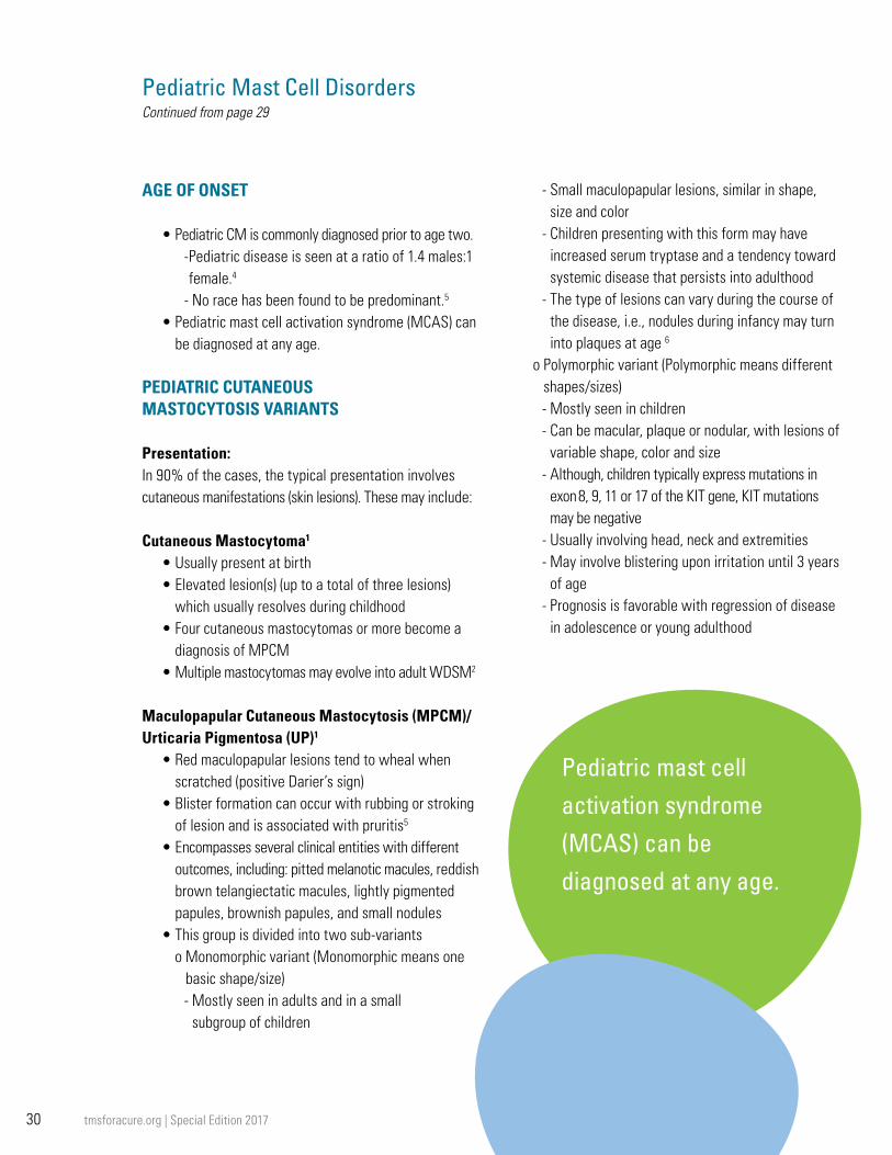
30 tmsforacure.org | Special Edition 2017
Pediatric Mast Cell DisordersContinued from page 29
AGE OF ONSET
• Pediatric CM is commonly diagnosed prior to age two.- Pediatric disease is seen at a ratio of 1.4 males:1 female.4
- No race has been found to be predominant.5
• Pediatric mast cell activation syndrome (MCAS) can be diagnosed at any age.
PEDIATRIC CUTANEOUS MASTOCYTOSIS VARIANTS
Presentation: In 90% of the cases, the typical presentation involves cutaneous manifestations (skin lesions). These may include: Cutaneous Mastocytoma1
• Usually present at birth• Elevated lesion(s) (up to a total of three lesions)
which usually resolves during childhood• Four cutaneous mastocytomas or more become a
diagnosis of MPCM• Multiple mastocytomas may evolve into adult WDSM2
Maculopapular Cutaneous Mastocytosis (MPCM)/Urticaria Pigmentosa (UP)1
• Red maculopapular lesions tend to wheal when scratched (positive Darier’s sign)
• Blister formation can occur with rubbing or stroking of lesion and is associated with pruritis5
• Encompasses several clinical entities with different outcomes, including: pitted melanotic macules, reddish brown telangiectatic macules, lightly pigmented papules, brownish papules, and small nodules
• This group is divided into two sub-variantso Monomorphic variant (Monomorphic means one
basic shape/size)- Mostly seen in adults and in a small
subgroup of children
- Small maculopapular lesions, similar in shape, size and color
- Children presenting with this form may have increased serum tryptase and a tendency toward systemic disease that persists into adulthood
- The type of lesions can vary during the course of the disease, i.e., nodules during infancy may turn into plaques at age 6
o Polymorphic variant (Polymorphic means different shapes/sizes)- Mostly seen in children- Can be macular, plaque or nodular, with lesions of
variable shape, color and size- Although, children typically express mutations in
exon 8, 9, 11 or 17 of the KIT gene, KIT mutations may be negative
- Usually involving head, neck and extremities- May involve blistering upon irritation until 3 years
of age- Prognosis is favorable with regression of disease
in adolescence or young adulthood

31tmsforacure.org | Special Edition 2017
Continued on page 32
Diffuse Cutaneous Mastocytosis (DCM)1
• Skin thickened, hyperpigmented and diffusely infiltrated
• Can involve up to 100% of the skin with the trunk, head and scalp heavily affected• Can appear at birth or early infancy; may persist into
adulthood, possibly as well differentiated systemic mastocytosis (WDSM)2
• Blisters, some of which are hemorrhagic; bullae may be present and dermatographism may be prominent
• Flushing is a common symptom• Tryptase may be elevated due to increased mast
cell burden in the skin, as most patients do not have systemic organ involvement, and can be indicative of WDSM2
SYMPTOMS OF MAST CELL ACTIVATION Which May be Seen in Both Pediatric CM and MCAS6
• Itching• Flushing• Darier’s sign and dermatographism• Abdominal pain, nausea, diarrhea, bloating, colic in
infants, GERD• Bone and muscle pain• Headache• Fatigue• Neuropsychiatric symptoms, such as: brain fog,
ADD/ADHD, irritability, behavioral issues, seizures• Anaphylaxis
GUIDELINES FOR DIAGNOSIS
Pediatric CM• Completion of a thorough patient history• Careful skin examination and biopsy of lesions with
mast cell stains (hematoxylin, eosin, giemsa stains) and immunohistochemistry for tryptase and KIT (CD117)
• Acquisition of labs, including complete blood count, peripheral smear, serum chemistry, serum tryptase and liver function tests
• Exam of liver and spleen for hepatosplenomegaly by ultrasound or scan
• Any other exam relevant to individual symptoms (endoscopy, colonoscopy, bone scan, etc.)
• Bone marrow biopsy and aspirate with flow cytometry only if clinical suspicion of systemic or progressive disease:- abnormal peripheral blood counts, organomegaly,
significant lymphadenopathy, severe recurrent systemic mast cell mediator-related symptoms, persistent high tryptase, persistence of disease into adulthood5, 7
Pediatric MCAS
• Although specific guidelines do not exist for diagnosing pediatric MCAS, proposed consensus criteria for diagnosing MCAS have been utilized by specialists.8
• Three criteria must be met:o The patient exhibits symptoms of mast cell
activation involving two or more organ systems at the same time, which recur or are always present, cannot be attributed to any other disease or condition and require treatment.8
o The patient demonstrates a rise in either total serum tryptase (above baseline and within one to two hours of a symptomatic episode; see below for calculation method to determine if the rise indicates mast cell activation has occurred) or one of the three urinary mediators, n-methyl histamine, prostaglandin-D2, or its metabolite, 11β-prostaglandin-F2α (24-hour urine test for any of the three, also best captured after a symptomatic episode).8 Additionally, Mayo Clinic (Rochester) has a test available to measure urinary levels of leukotrienes that is not yet incorporated

32 tmsforacure.org | Special Edition 2017
Pediatric Mast Cell DisordersContinued from page 31
into this criteria, as it is not yet widely available. “It requires further study to determine if all patients with MCAS will demonstrate a rise in one of the known mast cell mediators for which tests are available.”6
- The consensus article provides a method for calculating the required minimum rise in serum tryptase.8 After a reaction, a level of serum tryptase that is a minimum of 20% above the basal serum tryptase level, plus 2 ng/ml, will meet the second criterion listed above for a mast cell activation event. For example, if a patient had a basal (baseline level, at least 24 hours after a reaction) serum tryptase level of 8 ng/ml, a 20% rise, plus 2 ng/ml, would be 11.6 ng/ml. To meet the above criterion for serum tryptase, the patient would need a post-reaction serum tryptase level above 11.6 ng/ml. The calculation would be conducted as follows:
(8 ng/ml x 1.2) + 2 ng/ml = 11.6 ng/ml (basal level plus 20%) + additional 2 ng/ml = the serum tryptase level, after a reaction, that must be exceeded in order to meet a rise in serum tryptase considered a mast cell activation event
o The patient must display a response (based on response criteria9) to antimediator therapy.8
SOME POTENTIAL TRIGGERS TO AVOID (VARIES BY PATIENT)
• Heat and/or cold; abrupt changes in temperature; sun/sunlight
• Friction or pressure on the skin; vibration• Specific foods: very individualized but may include
shellfish, high histamine foods such as left-overs, salicylate-containing foods, nuts, peanuts and other potential allergens
• Contrast dyes and medications, including: opioid narcotics, alcohol as an additive or in any form, IV
vancomycin, neomycin, benzocaine, and certain anesthetics.10 See TMS Emergency Protocol.
• Venoms (bee, wasp, mixed vespids, spiders, fire ants, jelly fish, snakes, biting insects, such as flies, mosquitos and fleas, etc.)Bacterial, viral and fungal infections
• Stress: physical, including pain, emotional or environmental
• Fatigue• Exercise• Perfumes, odors, natural odors and chemical
exposures TREATMENT GUIDELINES FOR PEDIATRIC CM AND MCAS
• Identification and avoidance of triggers• H1 and H2 antihistamines
- H1: loratadine, cetirizine, desloratadine, diphenhydramine, hydroxyzine, fexofenadine, chlorpheniramine maleate, doxepin
- H2: ranitidine, cimetidine, famotidine• Leukotriene inhibitors
- Montelukast, zileuton, zafirlukast• UVA/UVB Photolight therapy (treatment option for pediatric CM only)• Mast cell stabilizers
- Oral cromolyn sodium- Ketotifen
• Injectable epinephrine- EpiPen®/EpiPen Jr® auto injector
• Topical treatments- Steroid creams- Cromolyn sodium cream 1%-5%
• No chemotherapy is indicated in cutaneous or indolent systemic mastocytosis in children, unless evidence of progression to aggressive disease is identified

33tmsforacure.org | Special Edition 2017
Continued on page 34
PROGNOSIS
Pediatric CM • Benign course will be seen in approximately 70% of
patients.2
• Approximately 30% of pediatric mastocytosis cases persist into adulthood.2
• Children with extensive bullous lesions appear to be at increased risk of shock or sudden death from anaphylaxis.11
• Children with widespread skin lesions (MPCM/UP & DCM) are at increased risk for severe systemic reaction due to potential mast cell mediator release from affected skin.11
Pediatric MCAS• There is no data on prognosis for pediatric patients
with MCAS; however all patients with MCAS are at increased risk for anaphylaxis and a potentially poor outcome. Therefore, these children need to be followed by an allergist familiar with pediatric MCAS and be treated with antimediator therapy, when indicated and always carry two doses of injectable epinephrine.
SUPPORT SERVICES
• The Mastocytosis Society, Inc. is a 501(c)3, nonprofit organization dedicated to supporting patients affected by Mastocytosis or Mast Cell Activation Disorders, as well as their families, caregivers, and physicians through research, education and advocacy.
• The Mastocytosis Society, Inc. coordinates support groups in nearly every state.
• Mastokids.org is a site where parents and caregivers of children with mastocytosis or mast cell disease can come to learn, find support, and discover a safe environment to interact with other families.
References:
1. Hartmann K, Escribano L, Grattan C, Brockow K, Carter MC, Alvarez-Twose I, et al. Cutaneous manifestations in patients with mastocytosis: Consensus report of the European Competence Network on Mastocytosis; the American Academy of Allergy, Asthma & Immunology; and the European Academy of Allergology and Clinical Immunology. J Allergy Clin Immunol. 2016 Jan;137(1):35-45.
2. Torrelo A, Alvarez-Twose I, Escribano L. Childhood mastocytosis. Curr Opin Pediatr. 2012 Aug;24(4):480-6.
3. Venes D, Thomas CL. Taber’s cyclopedic medical dictionary. 19 ed. Philadelphia: F.A. Davis Co.; 2001.
4. Meni C, Bruneau J, Georgin-Lavialle S, Le Sache de Peufeilhoux L, Damaj G, Hadj-Rabia S, et al. Paediatric mastocytosis: a systematic review of 1747 cases. Br J Dermatol. 2015 Mar;172(3):642-51.
5. Castells M, Metcalfe DD, Escribano L. Diagnosis and treatment of cutaneous mastocytosis in children: practical recommendations. Am J Clin Dermatol. 2011 Aug 1;12(4):259-70.
6. Butterfield J, et al., American Academy of Allergy, Asthma and Immunology Mast Cell Disorder Committee and The Mastocytosis Society, Inc. Mastocytosis and Mast Cell Activation Syndrome (MCAS). Laminated sheet, 2012.
7. Fried AJ, Akin C. Primary mast cell disorders in children. Curr Allergy Asthma Rep. 2013 Dec;13(6):693-701.
8. Valent P, Akin C, Arock M, Brockow K, Butterfield JH, Carter MC, et al. Definitions, criteria and global classification of mast cell disorders with special reference to mast cell activation syndromes: a consensus proposal. Int Arch Allergy Immunol. 2012;157(3):215-25.
9. Valent P, Akin C, Escribano L, Fodinger M, Hartmann K, Brockow K, et al. Standards and standardization in mastocytosis: consensus statements on diagnostics, treatment recommendations and response criteria. Eur J Clin Invest. 2007 Jun;37(6):435-53.
10. Bonadonna P, Pagani M, Aberer W, Bilo MB, Brockow K, Oude Elberink H, et al. Drug hypersensitivity in clonal mast cell disorders: ENDA/EAACI position paper. Allergy. 2015 Jul;70(7):755-63.
11. Alvarez-Twose I, Vano-Galvan S, Sanchez-Munoz L, Morgado JM, Matito A, Torrelo A, et al. Increased serum baseline tryptase levels and extensive skin involvement are predictors for the severity of mast cell activation episodes in children with mastocytosis. Allergy. 2012 Jun;67(6):813-21.
Pediatric Mast Cell Disorders: Facts in Brief Copyright © 2016 The Mastocytosis Society, Inc. All rights reserved.

34 tmsforacure.org | Special Edition 2017
Visual Guide to Diagnosing MastocytosisThe following pages are a photo journal of examples of how mast cell disorders can present. A majority of the pictures are of skin manifestations of mastocytosis. While cutaneous mastocytosis can include maculopapular cutaneous mastocytosis (MPCM), formerly known as urticaria pigmentosa (UP), telangiectasia macularis eruptiva perstans (TMEP), diffuse cutaneous mastocytosis (DCM), and cutaneous mastocytoma, skin manifestations can also occur in systemic mastocytosis (SM), mast cell activation syndrome (MCAS) and idiopathic anaphylaxis patients as well.
Most cases of childhood-onset mastocytosis fall into one of the cutaneous mastocytosis categories listed above and may or may not include symptoms of systemic mast cell activation as a result of mediators released from the skin. It should be noted that the formerly used term “UP” encompasses a variety of clinical manifestations. In children, some of these varieties will fade away, some will develop into indolent systemic mastocytosis and some will evolve into a newly described entity called well-differentiated systemic mastocytosis.
Pic. 1- Female adult with smoldering systemic mastocytosis and maculopapular cutaneous lesions, monomorphic type associated with this disease (formerly known as urticaria pigmentosa or UP)
Pic. 3- Female child with cutaneous mastocytosis and characteristic maculopapular, polymorphic skin lesions (formerly known as urticaria pigmentosa or UP)
Pic. 2- Female adult athlete with maculopapular cutaneous lesions, monomorphic type (formerly known as urticaria pigmentosa or UP), during a flare when the lesions are swelling
Pic. 4- Female child with cutaneous mastocytoma on shoulder, which can present with an elevated lesion which is red or tannish brown

35tmsforacure.org | Special Edition 2017
Pic. 5- Female adult with indolent systemic mastocytosis and confluent maculopapular, cutaneous lesions, monomorphic types (formerly known as urticaria pigmentosa or UP)
Pic. 6- Male child with cutaneous mastocytosis with polymorphic lesions and other rashes
Pic. 7- Female adult with smoldering systemic mastocytosis (SSM), and typical maculopapular, cutaneous lesions, monomorphic type (formerly called urticaria pigmentosa or UP) during a flare
Pic. 8- Male child with cutaneous mastocytosis, characterized by maculopapular cutaneous lesions, polymorphic type (formerly known as urticaria pigmentosa or UP). Note that in some children, during a flare in response to a trigger, lesions may become bullous or blistered.

36 tmsforacure.org | Special Edition 2017
Pic. 9- Male child with cutaneous mastocytosis during flare causing blisters in his maculopapular cutaneous lesions
Pic. 10- Male child with mast cell activation syndrome, during flushing episode
Pic. 11- Male child with the maculopapular cutaneous lesions, polymorphic type, consistent with cutaneous mastocytosis (formerly called urticaria pigmentosa or UP)

37tmsforacure.org | Special Edition 2017
Pic. 13- Cutaneous mastocytoma, normal and inflamed
Pic. 15- Female with idiopathic anaphylaxis, hives (urticaria) and dermatographism
Pic. 12- Adult female with maculopapular, cutaneous lesions, monomorphic type during a flare
Pic. 14- Female child with maculopapular, polymorphic lesions of cutaneous mastocytosis
For more information on skin manifestations in mastocytosis (including a large selection of photos) and to review the source of our publication’s descriptions of cutaneous mastocytosis variants, please see the following FULL TEXT article, which is freely available online:
Hartmann K, Escribano L, Grattan C, Brockow K, Carter MC, Alvarez-Twose I, et al. Cutaneous manifestations in patients with mastocytosis: Consensus report of the European Competence Network on Mastocytosis; the American Academy of Allergy, Asthma & Immunology; and the European Academy of Allergology and Clinical Immunology. J Allergy Clin Immunol. 2016 Jan;137(1):35-45

38 tmsforacure.org | Special Edition 2017
Please note carefully any clarification of what each center specializes in. For example, some centers only treat patients with biopsy confirmed systemic mastocytosis, some centers only treat aggressive or malignant disease, some treat only adults or children, and many also treat mast cell activation syndromes/mast cell activation disorders (MCAS/MCAD). All centers listed can do the entire work-up including evaluation, physical exam, mediator testing and bone marrow biopsy with flow cytometry and appropriate stains for c-kit D816V mutation, tryptase, and expression of CD2 and CD25 antigen markers. Please be very clear when making your appointment to ask what you can expect to occur during your visit.
United States of America
CaliforniaStanford Cancer Center 875 Blake Wilbur Drive, Room 2327B Stanford, CA 94305-5821
Contact: Jason Gotlib, MD, MS Associate Professor of Medicine (Hematology) Director, Stanford Hematology Fellowship Program Director, MPN Center
Stanford Cancer Institute 875 Blake Wilbur Drive, Room 2324
Stanford, CA 94305-5821 Phone: 650-498-6000 Fax: 650-724-5203
Email: [email protected] Specialization: Biopsy proven only; including systemic mastocytosis (SM) only, aggressive SM and mast cell leukemia. Adults. Diagnostic, treatment, and research.
ColoradoUniversity of Colorado Hospital Blood Cancer/Bone Marrow Transplant Program 1665 Aurora Ct, Rm 2257 Aurora, CO 80045 Contact: William A. Robinson, MD, PhD Professor, Division of Medical Oncology/Rella and Monroe Rifkin Endowed Chair Email: [email protected] Phone: 720-848-2869 Fax: 720-848-0704 Specialization: All mast cell related diseases including systemic mastocytosis (SM), aggressive systemic mastocytosis (ASM) and mast cell leukemia (MCL).
Adults. Diagnostic (bone marrow biopsy can be arranged), treatment, and research.
MarylandNational Institutes of Health: National Institute of Allergy and Infectious Diseases
NIH, NIAID Building 10, Room 11C207 10 Center Drive - MSC1881 Bethesda, MD 20892-1881
Contact: Dean D. Metcalfe, MD, Chief, Laboratory of Allergic Diseases
Email: [email protected] Phone: 301-496-2165 Fax: 301-480-8384
Contact: Melody Carter, MD, Pediatrics
Email: [email protected]
Specialization: Referrals only. Biopsy proven only; including systemic mastocytosis (SM) only, aggressive SM and mast cell leukemia. Adults and pediatric.
Diagnostic, treatment, and research. Bone marrow biopsies. Also adult idiopathic anaphylaxis. MassachusettsBrigham and Women’s Hospital and Dana Farber Cancer Institute Center of Excellence for Mastocytosis (and Mast Cell Activation Disorders)
Brigham and Women’s Hospital 850 Boylston St., Suite 450 Chestnut Hill, MA 02467
Director: Mariana Castells, MD, PhD
Email: [email protected] Phone: 617-732-9850
Fax: 617-731-2748
Contact: Norton J. Greenberger, MD, GI
Email: [email protected] Phone: 617-732-6389 Fax: 617-264-5277
Contact: Richard Horan, MD Email: [email protected] Phone: 617-732-9850 Fax: 617-731-2748
Contact: Daniel DeAngelo, MD, PhD Email: [email protected] Phone: 617-632-6028
Address: DFCI, 450 Brookline Ave., Dana D1B30 Boston, MA 02215
Specialization: All mast cell related diseases including MCAS/MCAD. Adults and pediatric. Diagnostic, treatment, and research. Can arrange bone marrow biopsies.
Tufts University School of Medicine 136 Harrison Avenue Boston, MA 02111
Contact: Theoharis Theoharides, MD, PhD, Professor of Pharm. and Internal Medicine
Email: [email protected] Phone: 617-636-6866 Fax: 617-636-2456
Does not see patients in clinic
MichiganUniversity of Michigan Comprehensive Cancer Center Myeloproliferative Neoplasms and Systemic Mastocytosis Clinic 1500 East Medical Center Drive
Medical & Research Centers that Treat Patients with Mast Cell Diseases

39tmsforacure.org | Special Edition 2017
Ann Arbor, MI 48109
Cem Akin, M.D., Ph.D.
Professor of Medicine
University of Michigan
Department of Internal Medicine
Division of Allergy and Clinical Immunology
24 Frank Lloyd Wright Drive
PO Box 442, Suite H-2100
Ann Arbor, MI 48106-0422
Phone: 734-936-5634
Fax: 734-647-6263 Phone (new patient coordinator): 734-232-2071
Specialization: Biopsy-proven only -- systemic mastocytosis (indolent, smoldering, aggressive SM), SM-AHNMD, and mast cell leukemia. Will perform diagnostic marrows for patients with elevated tryptase or biopsy-proven cutaneous disease.
Adults. Diagnostic, treatment, and research.
MinnesotaMayo Clinic – Allergy Department
Mayo Clinic Center of Excellence for Mast Cell and Eosinophil Disorders W15-B Mayo Clinic 200 SW 1st St. Rochester, MN 55905
Co-Director: Joseph Butterfield, MD Email: [email protected]
Co-Director: Catherine Weiler, MD, PhD Email: [email protected]
Phone: 507-284-9077 Fax: 507-284-0902
Mayo Clinic – Hematology Department
Contact: Ayalew Tefferi, MD and
Animesh Pardanani M.B.B.S., PhD
Phone: (507) 284-5363
Specialization: All mast cell related diseases including MCAS/MCAD. Adults and pediatric. Diagnostic, bone marrow biopsy, treatment, and research. University of Minnesota Program for Mast Cell Diseases Website: http://mastcell.umn.edu Contact: Lawrence B. Afrin, MD (program director; sees adult mast cell disease patients) Email: [email protected] Contact: Celalettin Ustun, MD (sees adult advanced/aggressive mastocytosis patients) Email: [email protected]
University of Minnesota Division of Hematology, Oncology & Transplantation 420 Delaware St. SE, MMC 480 Minneapolis, MN 55455 Phone: 612-624-0123 Fax: 612-625-6919
Specialization: All mast cell-related diseases. Adult and pediatric patients. Diagnostic and treatment services and basic and clinical research. OhioUniversity of Cincinnati
Jonathan A. Bernstein, M.D.
Professor of Clinical Medicine
Department of Internal Medicine
Division of Immunology/Allergy Section
Editor-in-Chief Journal of Asthma
University of Cincinnati College of Medicine
231 Albert Sabin Way, ML#563
Cincinnati, Ohio 45267-0563
Email: [email protected]
Office phone: 513-558-5533
Office fax: 513-558-3799
Specialization: All mast cell related diseases including MCAS/MCAD. Adults and pediatric. Diagnostic, treatment, and research. Can arrange bone marrow biopsies. Private family practice.
OklahomaUniversity of Oklahoma, College of Medicine 535 NW 9th St, Ste 325 Oklahoma City, OK 73102
Contact: Philip B. Miner Jr., MD Clinical Professor of Medicine, President and Medical Director, Oklahoma Foundation for Digestive Research
Email: [email protected]
Phone: 405-601-6620
Fax: 405-601-6635
Continued on page 40

40 tmsforacure.org | Special Edition 2017
Medical & Research Centers that Treat Patients with Mast Cell DiseasesContinued from page 39
TexasMD Anderson Cancer Center 1515 Holcombe Blvd, Unit 428 Houston, TX 77030
Contact: Srdan Verstovsek, MD, PhD, Associate Professor, Leukemia Department
Email: [email protected] Phone: 713-792-7305 Fax: 713-794-4297
Specialization: Systemic mastocytosis (SM) only, aggressive SM and mast cell leukemia. Adults. Diagnostic, treatment, and research.
UtahThe University of Utah School of Medicine Department of Internal Medicine, Hematology Division
30 N 1900 E, Room 5C402 Salt Lake City, UT 84132
Contact: Michael Deininger, MD, PhD
Phone: 801-585-3229
Email: [email protected]
Specialization: Bone marrow biopsy confirmed mastocytosis, aggressive disease and mast cell leukemia.
VirginiaVirginia Commonwealth University
P.O. Box 980263
1250 East Marshall St. Richmond, VA 23298
Contact: Dr. Larry Schwartz, MD, PhD Internal Medicine: Rheumatology, Allergy, and Immunology
Email: [email protected]
Phone: 804-828-9685 Fax: 804-828-0283
Specialization: All mast cell related diseases including MCAS/MCAD. Adults and pediatric. Diagnostic, treatment, and research. Can arrange bone marrow biopsies
Please note that the names of these centers and specialists are listed for informational purposes only. The Mastocytosis Society, Inc. is not responsible for any diagnostic evaluations, treatment or information provided as a result of visits or interactions with these medical professionals.
International (Active Centers)
AustriaMedical University of Vienna
BrazilUniversity of Sao Paulo, Sao Paulo
DenmarkOdense University Hospital
FranceAssociation Française pour les Initiatives de Recherches sur le Mastocyte et les Mastocytoses (AFIRMM)
GermanyUniversity of Berlin University of Cologne Technical University Munich Ludwig-Maximilians-University Munich
GreeceUniversity Hospital of Athens - Attikon
ItalyUniversity of Naples
IsraelTechnion- Israel Institute of Technology, Haifa
The NetherlandsUniversity Hospital Groningen
PolandUniversity of Gdansk
PortugalUniversity of Porto
SpainCentro de Estudios de Mastocitosis de Castilla a Mancha (CLMast)
SwedenKarolinska University Hospital, Stockholm
SwitzerlandKantonsspital Aarau, Aarau
TurkeyUniversity of Istanbul
United KingdomGuy’s and St. Thomas’ Trust - London
Note: For additional current information on specialties and contacts within each European center visit: www.ecnm.net

41tmsforacure.org | Special Edition 2017
Ivan Alvarez-Twose, MDStaff Physician and Clinical Coordinator, Instituto de Estudios de Mastocitosis de Castilla La Mancha (CLMast) Toledo, Spain Phone: 0034-615-653-157 Email: [email protected]
K. Frank Austen, MD (Honorary)Astra Zeneca Professor of Respiratory and Inflammatory Diseases
Department of Medicine Brigham and Women’s Hospital
Smith Building, Room 638 One Jimmy Fund Way Boston, MA 02115
Email: [email protected] Phone: 617-525-1300 Fax: 617-525-1310
Patrizia Bonadonna, MD
Allergy and Immunology Clinic
Responsible for Hymenoptera venom allergy, drug allergy and allergy diseases
Multidisciplinar Outpatient Clinic of Mastocytosis
Allergy Department
Verona General and University Hospital
Piazzale Stefani 1
Verona, Italy
Email: [email protected] Phone: +390458122556 Fax: +390458122048
Joseph Butterfield, MD
Co-Director, Mayo Clinic Center of Excellence for Mast Cell and Eosinophil Disorders
W15-B Mayo Clinic 200 SW 1st Street Rochester, MN 55905
Email: [email protected] Phone: 507-284-9077 Fax: 507-284-0902
Mariana Castells, MD, PhD
Associate Director, Mastocytosis Center of Excellence Brigham and Women’s Hospital Allergy and Clinical Immunology
850 Boylston Street, Suite 540 Chestnut Hill, MA 02467
Email: [email protected] Phone: 617-732-9850 Fax: 617-731-2748
Madeleine Duvic, MD
Professor and Deputy Chair, Dermatology, UT MD Anderson Cancer Center
1515 Holcombe Blvd, Unit 1452
Email: [email protected] Phone: 713 745-4615 Fax: 713 745-3597
Luis Escribano, MD, PhD
Coordinator, Spanish Network on Mastocytosis (REMA) Associated Research, Servicio de Citometría, Centro de Investigación del Cáncer, Universidad de Salamanca
Salamanca, Spain E-mail: [email protected]
Jason Gotlib, MD, MS
Associate Professor of Medicine (Hematology), Director, Stanford Hematology Fellowship Program Director, MPN Center
Stanford Cancer Institute 875 Blake Wilbur Drive, Room 2324 Stanford, CA 94305-5821
Email: [email protected] Phone: 650-725-0744 Fax: 650-724-5203
Norton J. Greenberger, MD
Clinical Professor of Medicine/ Gastroenterology
Harvard Medical School Senior Physician
Brigham and Women’s Hospital 75 Francis Street Boston, MA 02115
Email: [email protected] Phone: 617-732-6389 Fax: 617-264-5277
Matthew J. Hamilton, MD
Assistant Professor of Medicine
Harvard Medical School Division of Gastroenterology
Brigham and Women's Hospital
75 Francis St. Boston, MA 02115
Email: [email protected] Phone: 617-732-6389 Fax: 617-566-0338
Medical Advisory BoardContact Information: The Mastocytosis Society, Inc. is a nonprofit volunteer organization guided by a board of medical advisors who donate their time and expertise in support of the TMS mission. They have graciously agreed to act as a point of contact for other physicians and health care providers needing additional information about mastocytosis and mast cell activation disorders.
Continued on page 42

42 tmsforacure.org | Special Edition 2017
Olivier Hermine, MD, PhD
Université Sorbonne Paris Cité
Department of Clinical Hematology
National Center of Mastocytosis
Hôpital Necker
149-161 Rue de Sèvres 75015 Paris, France
Laboratory of Cellular and Molecular Mechanisms of Hematological Disorders and Therapeutic Implications
INSERM U 1163 / CNRS ERL 8254
Labex on Red Cell and Iron Metabolism
Institut IMAGINE (Lab 215-217)
24 Boulevard du Montparnasse
75015 Paris, France
Email: [email protected] Tel: 33-1-44-49-52-82 (office)
Nicholas Kounis, MD, PhD
Patras Highest Institute of Education and Technology
Professor of Medicine in Cardiology Department of Medical Sciences
7 Aratou St. Queen Olgas Square Patras 26221, Greece
Email: [email protected] Phone: +302610279579 Fax: +302610279579
Anne Maitland, MD, PhD
Asst. Professor, Dept. of Medicine and Dept. of Otolaryngology
Icahn School of Medicine at Mount Sinai
New York, NY 10029
Medical Director,
Comprehensive Allergy & Asthma Care, PLLC
Department of Otolaryngology 5 East 98th Street, 8th Floor New York, NY 10029
55 South Broadway, 2nd floor Tarrytown, NY 10591
Email: [email protected] Phone: 914-631-3283 Fax: 914-631-3284
Larry Schwartz, MD, PhD
Internal Medicine: Rheumatology, Allergy, and Immunology
Virginia Commonwealth University P.O. Box 980263 1250 East Marshall Street Richmond, VA 23298
Email: [email protected] Phone: 804-828-9685 Fax: 804-828-0283
Theoharis Theoarides, MD, PhD
Professor of Pharmacology and Internal Medicine
Tufts University School of Medicine 136 Harrison Avenue Boston, MA 02111
Phone: 617-636-6866 Fax: 617-636-2456 Email: [email protected]
Megha M Tollefson, MD
Assistant Professor of Dermatology and Pediatrics
Mayo Clinic
200 First Street SW
Rochester, MN 55905
Email: [email protected] Phone: (507) 284-3579 Fax: (507) 284-2072
Celalettin Ustun, MD
Associate Professor of Medicine
Division of Hematology, Oncology & Transplantation
University of Minnesota 420 Delaware St. SE, MMC 480 Minneapolis, MN 55455
Email: [email protected] Phone: 612-624-0123 Fax: 612-625-6919
Peter Valent, MD
Department of Internal Medicine I Division of Hematology and Hemostaseology
University of Vienna Währinger Gürtel 18-20 A-1090 Vienna, Austria
Email: [email protected] Phone:+43-1 40400-5488 or -6086 Fax:+43 1 40400 4030
Srdan Verstovsek, MD, PhD
Associate Professor Leukemia Department
MD Anderson Cancer Center 1515 Holcombe Blvd, Unit 428 Houston, TX 77030
Email: [email protected] Phone: 713-792-7305 Fax: 713-794-4297
Medical & Research Centers that Treat Patients with Mast Cell DiseasesContinued from page 41

43tmsforacure.org | Special Edition 2017
Mastocytosis and mast cell activation disorders are complicated and not well-known diseases. To help educate and spread awareness, The Mastocytosis Society, Inc. (TMS) is pleased to offer informational material to physicians and patients.
Card and Brochure Dimensions: Spot Card, Generic Business 2" x 3.5" Informational Brochure, Tri-fold 8.5" x 11"
The Mastocytosis Society Printed Materials
Infant Card
Tri-fold Informational Brochures Symptoms, diagnosis and treatment of mast cell disorders.
Name ____________________________________________
Address ___________________________________________
City _____________________________________________
State __________________Zip _______________________
Phone ____________________________________________
Email _____________________________________________
Please indicate a quantity next to each item Tri-fold Informational Brochures
____ Emergency Care For Mast Cell Disorder Patients____ Systemic Mastocytosis Including Indolent &
Aggressive Variants____ Mastocytosis and Mast Cell Activation Disorders Cards____ Infant Card
Ordering Information
TMS printed material is available for free on our website. If your medical office would like printed copies, please fill out the form or email us at [email protected]
The Mastocytosis Society, Inc., P.O. Box 416 Sterling, MA 01564 | [email protected]
Images not to scale

44 tmsforacure.org | Special Edition 2017
January 2017
FILE DESCRIPTION: This file contains selected references, listed by topic, that might be of interest to mast cell disorder patients, their caregivers, physicians or others. Although efforts were made to compile a list of references containing information on a variety of topics related to mast cell disorders, this is NOT a complete list of all articles available on this subject. All references were obtained through searches of the PubMed database (http://www.ncbi.nlm.nih.gov/pubmed). Additional individually relevant references can be obtained by searching the PubMed database.
DISCLAIMER: Listing of an article in this file does not imply TMS support of its authors or contents and an article that is not listed in this file does not imply a lack of support of its authors or contents. Patients should consult with their doctors, or, if necessary, mast cell specialists, regarding any questions or concerns related to applicability, accuracy and individual usefulness of information presented in these articles.
TOPICS INCLUDED IN THIS FILE:
• International Consensus Statements, Position Papers and WHO Criteria 1-13
• Reviews and Expert Opinions 14-44
• Laboratory Tests, Pathology, Immunohistology and Flow Cytometry 5, 15, 38, 40, 42, 43, 45-51
• Perioperative Care/Pre-Medication for Dental Work, Diagnostic Testing or Surgical Procedures 4, 37, 52-54
• Therapy 15, 19, 20, 36, 37, 40, 55-59
• The Mastocytosis Society Survey on Mast Cell Disorders 60
1. Arber DA, Orazi A, Hasserjian R, Thiele J, Borowitz MJ, Le Beau MM, et al. The 2016 revision to the World Health Organization classification of myeloid neoplasms and acute leukemia. Blood. 2016 May 19;127(20):2391-405. http://www.ncbi.nlm.nih.gov/pubmed/27069254
2. Hartmann K, Escribano L, Grattan C, Brockow K, Carter MC, Alvarez-Twose I, et al. Cutaneous manifestations in patients with mastocytosis: Consensus report of the European Competence Network on Mastocytosis; the American Academy of Allergy, Asthma & Immunology; and the European Academy of Allergology and Clinical Immunology. J Allergy Clin Immunol. 2016 Jan;137(1):35-45. http://www.ncbi.nlm.nih.gov/pubmed/26476479
3. Ustun C, Gotlib J, Popat U, Artz A, Litzow M, Reiter A, et al. Consensus Opinion on Allogeneic Hematopoietic Cell Transplantation in Advanced Systemic Mastocytosis. Biol Blood Marrow Transplant. 2016 Aug;22(8):1348-56. http://www.ncbi.nlm.nih.gov/pubmed/27131865
4. Bonadonna P, Pagani M, Aberer W, Bilo MB, Brockow K, Oude Elberink H, et al. Drug hypersensitivity in clonal mast cell disorders: ENDA/EAACI position paper. Allergy. 2015 Jul;70(7):755-63. http://www.ncbi.nlm.nih.gov/pubmed/25824492
5. Arock M, Sotlar K, Akin C, Broesby-Olsen S, Hoermann G, Escribano L, et al. KIT mutation analysis in mast cell neoplasms: recommendations of the European Competence Network on Mastocytosis. Leukemia. 2015 Jun;29(6):1223-32. http://www.ncbi.nlm.nih.gov/pubmed/25650093
6. Valent P, Escribano L, Broesby-Olsen S, Hartmann K, Grattan C, Brockow K, et al. Proposed diagnostic algorithm for patients with suspected mastocytosis: a proposal of the European Competence Network on Mastocytosis. Allergy.
MASTOCYTOSIS AND MAST CELL ACTIVATION SYNDROMES
Medical Reference Highlights

45tmsforacure.org | Special Edition 2017
2014 Oct;69(10):1267-74. http://www.ncbi.nlm.nih.gov/pubmed/24836395
7. Valent P, Sotlar K, Sperr WR, Escribano L, Yavuz S, Reiter A, et al. Refined diagnostic criteria and classification of mast cell leukemia (MCL) and myelomastocytic leukemia (MML): a consensus proposal. Ann Oncol. 2014 Sep;25(9):1691-700. http://www.ncbi.nlm.nih.gov/pubmed/24675021
8. Gotlib J, Pardanani A, Akin C, Reiter A, George T, Hermine O, et al. International Working Group-Myeloproliferative Neoplasms Research and Treatment (IWG-MRT) & European Competence Network on Mastocytosis (ECNM) consensus response criteria in advanced systemic mastocytosis. Blood. 2013 Mar 28;121(13):2393-401. http://www.ncbi.nlm.nih.gov/pubmed/23325841
9. Valent P, Akin C, Arock M, Brockow K, Butterfield JH, Carter MC, et al. Definitions, criteria and global classification of mast cell disorders with special reference to mast cell activation syndromes: a consensus proposal. Int Arch Allergy Immunol. 2012;157(3):215-25. http://www.ncbi.nlm.nih.gov/pubmed/22041891
10. Horny HP, Akin C, Metcalfe DD, Escribano L, Bennett JM, Valent P, et al. Mastocytosis (mast cell disease) Swerdlow SH, Campo E, Harris NL, Jaffe ES, Pileri SA, Stein H, et al., editors. World Health Organization (WHO) Classification of Tumours. Pathology and Genetics. Tumours of Haematopoietic and Lymphoid Tissues. Lyon: IARC Press; 2008.
11. Valent P, Akin C, Escribano L, Fodinger M, Hartmann K, Brockow K, et al. Standards and standardization in mastocytosis: consensus statements on diagnostics, treatment recommendations and response criteria. Eur J Clin Invest. 2007 Jun;37(6):435-53. http://www.ncbi.nlm.nih.gov/pubmed/17537151
12. Valent P, Horny H-P, Li CY, Longley JB, Metcalfe DD, Parwaresch RM, et al. Mastocytosis. Jaffe ES, Harris NL, Stein H, Vardiman JW, editors. World Health Organization (WHO) Classification of Tumours. Pathology and Genetics. Tumours of Haematopoietic and Lymphoid Tissues. Lyon: IARC Press; 2001.
13. Valent P, Horny HP, Escribano L, Longley BJ, Li CY, Schwartz LB, et al. Diagnostic criteria and classification of mastocytosis: a consensus proposal. Leuk Res. 2001 Jul;25(7):603-25. http://www.ncbi.nlm.nih.gov/pubmed/11377686
14. Monnier J, Georgin-Lavialle S, Canioni D, Lhermitte L, Soussan M, Arock M, et al. Mast cell sarcoma: new cases and literature review. Oncotarget. 2016 Oct 04;7(40):66299-309. http://www.ncbi.nlm.nih.gov/pubmed/27602777
15. Azana JM, Torrelo A, Matito A. Update on Mastocytosis (Part 2): Categories, Prognosis, and Treatment. Actas Dermosifiliogr. 2016 Jan-Feb;107(1):15-22. http://www.ncbi.nlm.nih.gov/pubmed/26525106
16. Azana JM, Torrelo A, Matito A. Update on Mastocytosis (Part 1): Pathophysiology, Clinical Features, and Diagnosis. Actas Dermosifiliogr. 2016 Jan-Feb;107(1):5-14. http://www.ncbi.nlm.nih.gov/pubmed/26546030
17. Bonadonna P, Bonifacio M, Lombardo C, Zanotti R. Hymenoptera Allergy and Mast Cell Activation Syndromes. Curr Allergy Asthma Rep. 2016 Jan;16(1):5. http://www.ncbi.nlm.nih.gov/pubmed/26714690
18. Pieri L, Bonadonna P, Elena C, Papayannidis C, Grifoni FI, Rondoni M, et al. Clinical presentation and management practice of systemic mastocytosis. A survey on 460 Italian patients. Am J Hematol. 2016 Jul;91(7):692-9. http://www.ncbi.nlm.nih.gov/pubmed/27060898
Continued on page 46

46 tmsforacure.org | Special Edition 2017
19. Valent P, Akin C, Metcalfe DD. Mastocytosis 2016: Updated WHO Classification and Novel Emerging Treatment Concepts. Blood. 2016 Dec 28. http://www.ncbi.nlm.nih.gov/pubmed/28031180
20. Gonzalez de Olano D, Matito A, Orfao A, Escribano L. Advances in the understanding and clinical management of mastocytosis and clonal mast cell activation syndromes. F1000Res. 2016 Nov 14;5:2666. https://www.ncbi.nlm.nih.gov/pubmed/27909577
21. Alvarez-Twose I, Jara-Acevedo M, Morgado JM, Garcia-Montero A, Sanchez-Munoz L, Teodosio C, et al. Clinical, immunophenotypic, and molecular characteristics of well-differentiated systemic mastocytosis. J Allergy Clin Immunol. 2016 Jan;137(1):168-78 e1. http://www.ncbi.nlm.nih.gov/pubmed/26100086
22. Carter MC, Clayton ST, Komarow HD, Brittain EH, Scott LM, Cantave D, et al. Assessment of clinical findings, tryptase levels, and bone marrow histopathology in the management of pediatric mastocytosis. J Allergy Clin Immunol. 2015 Dec;136(6):1673-9 e3. http://www.ncbi.nlm.nih.gov/pubmed/26044856
23. Valent P. Diagnosis and management of mastocytosis: an emerging challenge in applied hematology. Hematology Am Soc Hematol Educ Program. 2015 Dec 5;2015(1):98-105. http://www.ncbi.nlm.nih.gov/pubmed/26637707
24. Bonadonna P, Bonifacio M, Lombardo C, Zanotti R. Hymenoptera Anaphylaxis and C-kit Mutations: An Unexpected Association. Curr Allergy Asthma Rep. 2015 Aug;15(8):49. http://www.ncbi.nlm.nih.gov/pubmed/26149588
25. Theoharides TC, Valent P, Akin C. Mast Cells, Mastocytosis, and Related Disorders. N Engl J Med. 2015 Jul 9;373(2):163-72. http://www.ncbi.nlm.nih.gov/pubmed/26154789
26. Matito A, Carter M. Cutaneous and systemic mastocytosis in children: a risk factor for anaphylaxis? Curr Allergy Asthma Rep. 2015 May;15(5):22. http://www.ncbi.nlm.nih.gov/pubmed/26139333
27. Meni C, Bruneau J, Georgin-Lavialle S, Le Sache de Peufeilhoux L, Damaj G, Hadj-Rabia S, et al. Paediatric mastocytosis: a systematic review of 1747 cases. Br J Dermatol. 2015 Mar;172(3):642-51. http://www.ncbi.nlm.nih.gov/pubmed/25662299
28. Valent P, Sotlar K, Sperr WR, Reiter A, Arock M, Horny HP. Chronic mast cell leukemia: a novel leukemia-variant with distinct morphological and clinical features. Leuk Res. 2015 Jan;39(1):1-5. http://www.ncbi.nlm.nih.gov/pubmed/25443885
29. Akin C. Mast cell activation disorders. J Allergy Clin Immunol Pract. 2014 May-Jun;2(3):252-7 e1; quiz 8. http://www.ncbi.nlm.nih.gov/pubmed/24811013
30. Akin C, Valent P. Diagnostic criteria and classification of mastocytosis in 2014. Immunol Allergy Clin North Am. 2014 May;34(2):207-18. http://www.ncbi.nlm.nih.gov/pubmed/24745670
31. Niedoszytko M, Bonadonna P, Oude Elberink JN, Golden DB. Epidemiology, diagnosis, and treatment of Hymenoptera venom allergy in mastocytosis patients. Immunol Allergy Clin North Am. 2014 May;34(2):365-81. http://www.ncbi.nlm.nih.gov/pubmed/24745680
Medical Reference HighlightsContinued from page 45

47tmsforacure.org | Special Edition 2017
32. Weiler CR, Butterfield J. Mast cell sarcoma: clinical management. Immunol Allergy Clin North Am. 2014 May;34(2):423-32. http://www.ncbi.nlm.nih.gov/pubmed/24745684
33. Rossini M, Zanotti R, Viapiana O, Tripi G, Orsolini G, Idolazzi L, et al. Bone involvement and osteoporosis in mastocytosis. Immunol Allergy Clin North Am. 2014 May;34(2):383-96. http://www.ncbi.nlm.nih.gov/pubmed/24745681
34. Brockow K. Epidemiology, prognosis, and risk factors in mastocytosis. Immunol Allergy Clin North Am. 2014 May;34(2):283-95. http://www.ncbi.nlm.nih.gov/pubmed/24745674
35. Moura DS, Georgin-Lavialle S, Gaillard R, Hermine O. Neuropsychological features of adult mastocytosis. Immunol Allergy Clin North Am. 2014 May;34(2):407-22. http://www.ncbi.nlm.nih.gov/pubmed/24745683
36. Carter MC, Metcalfe DD, Komarow HD. Mastocytosis. Immunol Allergy Clin North Am. 2014 Feb;34(1):181-96. http://www.ncbi.nlm.nih.gov/pubmed/24262698
37. Fried AJ, Akin C. Primary mast cell disorders in children. Curr Allergy Asthma Rep. 2013 Dec;13(6):693-701. http://www.ncbi.nlm.nih.gov/pubmed/24150753
38. Valent P. Mast cell activation syndromes: definition and classification. Allergy. 2013 Apr;68(4):417-24. http://www.ncbi.nlm.nih.gov/pubmed/23409940
39. Georgin-Lavialle S, Lhermitte L, Dubreuil P, Chandesris MO, Hermine O, Damaj G. Mast cell leukemia. Blood. 2013 Feb 21;121(8):1285-95. http://www.ncbi.nlm.nih.gov/pubmed/23243287
40. Cardet JC, Castells MC, Hamilton MJ. Immunology and clinical manifestations of non-clonal mast
cell activation syndrome. Curr Allergy Asthma Rep. 2013 Feb;13(1):10-8. http://www.ncbi.nlm.nih.gov/pubmed/23212667
41. Afrin LB. Presentation, diagnosis and management of mast cell activation syndrome. In: Murray DB, editor. Mast cells: phenotypic features, biological functions and role in immunity. Hauppauge: Nova Science Publishers, Inc.; 2013. p. 155-232.
42. Alvarez-Twose I, Morgado JM, Sanchez-Munoz L, Garcia-Montero A, Mollejo M, Orfao A, et al. Current state of biology and diagnosis of clonal mast cell diseases in adults. Int J Lab Hematol. 2012 Oct;34(5):445-60. http://www.ncbi.nlm.nih.gov/pubmed/22551157
43. Torrelo A, Alvarez-Twose I, Escribano L. Childhood mastocytosis. Curr Opin Pediatr. 2012 Aug;24(4):480-6. http://www.ncbi.nlm.nih.gov/pubmed/22790101
44. Akin C, Valent P, Metcalfe DD. Mast cell activation syndrome: proposed diagnostic criteria. J Allergy Clin Immunol. 2010 Dec;126(6):1099-104 e4. http://www.ncbi.nlm.nih.gov/pubmed/21035176
45. Teodosio C, Mayado A, Sanchez-Munoz L, Morgado JM, Jara-Acevedo M, Alvarez-Twose I, et al. The immunophenotype of mast cells and its utility in the diagnostic work-up of systemic mastocytosis. J Leukoc Biol. 2015 Jan;97(1):49-59. http://www.ncbi.nlm.nih.gov/pubmed/25381388
46. Horny HP, Sotlar K, Valent P. Mastocytosis: immunophenotypical features of the transformed mast cells are unique among hematopoietic cells. Immunol Allergy Clin North Am. 2014 May;34(2):315-21. http://www.ncbi.nlm.nih.gov/pubmed/24745676
47. Doyle LA, Hornick JL. Pathology of extramedullary mastocytosis. Immunol Allergy Clin North Am. 2014 May;34(2):323-39. http://www.ncbi.nlm.nih.gov/pubmed/24745677

48 tmsforacure.org | Special Edition 2017
48. Horny HP, Sotlar K, Valent P. Evaluation of mast cell activation syndromes: impact of pathology and immunohistology. Int Arch Allergy Immunol. 2012;159(1):1-5. http://www.ncbi.nlm.nih.gov/pubmed/22555026
49. Horny HP, Sotlar K, Valent P. Mastocytosis: state of the art. Pathobiology. 2007;74(2):121-32. http://www.ncbi.nlm.nih.gov/pubmed/17587883
50. Horny HP, Valent P. Diagnosis of mastocytosis: general histopathological aspects, morphological criteria, and immunohistochemical findings. Leuk Res. 2001 Jul;25(7):543-51. http://www.ncbi.nlm.nih.gov/pubmed/11377679
51. Jawhar M, Schwaab J, Hausmann D, Clemens J, Naumann N, Henzler T, et al. Splenomegaly, elevated alkaline phosphatase and mutations in the SRSF2/ASXL1/RUNX1 gene panel are strong adverse prognostic markers in patients with systemic mastocytosis. Leukemia. 2016 Dec;30(12):2342-50. http://www.ncbi.nlm.nih.gov/pubmed/27416984
52. Matito A, Morgado JM, Sanchez-Lopez P, Alvarez-Twose I, Sanchez-Munoz L, Orfao A, et al. Management of Anesthesia in Adult and Pediatric Mastocytosis: A Study of the Spanish Network on Mastocytosis (REMA) Based on 726 Anesthetic Procedures. Int Arch Allergy Immunol. 2015;167(1):47-56. http://www.ncbi.nlm.nih.gov/pubmed/26160029
53. Dewachter P, Castells MC, Hepner DL, Mouton-Faivre C. Perioperative Management of Patients with Mastocytosis. Anesthesiology. 2013 Oct 16. http://www.ncbi.nlm.nih.gov/pubmed/24135579
54. Carter MC, Uzzaman A, Scott LM, Metcalfe DD, Quezado Z. Pediatric mastocytosis: routine anesthetic management for a complex disease. Anesth Analg. 2008 Aug;107(2):422-7. http://www.
ncbi.nlm.nih.gov/pubmed/18633019
55. Arock M, Akin C, Hermine O, Valent P. Current treatment options in patients with mastocytosis: status in 2015 and future perspectives. Eur J Haematol. 2015 Jun;94(6):474-90. http://www.ncbi.nlm.nih.gov/pubmed/25753531
56. Rossini M, Zanotti R, Orsolini G, Tripi G, Viapiana O, Idolazzi L, et al. Prevalence, pathogenesis, and treatment options for mastocytosis-related osteoporosis. Osteoporos Int. 2016 Feb 18. http://www.ncbi.nlm.nih.gov/pubmed/26892042
57. Gotlib J, Kluin-Nelemans HC, George TI, Akin C, Sotlar K, Hermine O, et al. Efficacy and Safety of Midostaurin in Advanced Systemic Mastocytosis. N Engl J Med. 2016 Jun 30;374(26):2530-41. http://www.ncbi.nlm.nih.gov/pubmed/27355533
58. Ustun C, Arock M, Kluin-Nelemans HC, Reiter A, Sperr WR, George T, et al. Advanced systemic mastocytosis: from molecular and genetic progress to clinical practice. Haematologica. 2016 Oct;101(10):1133-43. http://www.ncbi.nlm.nih.gov/pubmed/27694501
59. Gotlib J. Tyrosine Kinase Inhibitors and Therapeutic Antibodies in Advanced Eosinophilic Disorders and Systemic Mastocytosis. Curr Hematol Malig Rep. 2015 Dec;10(4):351-61. http://www.ncbi.nlm.nih.gov/pubmed/26404639
60. Jennings S, Russell N, Jennings B, Slee V, Sterling L, Castells M, et al. The Mastocytosis Society survey on mast cell disorders: patient experiences and perceptions. J Allergy Clin Immunol Pract. 2014 Jan-Feb;2(1):70-6. http://www.ncbi.nlm.nih.gov/pubmed/24565772
Medical Reference HighlightsContinued from page 47

49tmsforacure.org | Special Edition 2017
What Is M
ast Cell C
onnect?
Mast Cell Connect is an electronic patient registry
created to advance the understanding of mastocytosis
as a disease. It is a voluntary online registry to collect inform
ation from patients w
ith mastocytosis on their
experiences with the disease. The ultim
ate goal of M
ast Cell Connect is to help speed the development of
new treatm
ents for people with m
astocytosis.
The registry allows m
astocytosis patients and caregivers to enter inform
ation about their experience living w
ith the disease directly into a secure, web-based
data collection tool. Those who participate in M
ast Cell Connect w
ill be able to view the de-identified sum
mary
of responses from other patients, and can choose to
receive information about ongoing clinical trials and
other related research studies.
You are invited to learn more and to consider
participating in this important effort. By providing
researchers with a database of detailed m
edical inform
ation about people with a specific disease,
registries have proven to be a valuable tool in better understanding rare diseases like m
astocytosis. Q
uestions?
For questions about the goals of the Mast Cell
Connect registry, contact the study doctor at m
astcellregistry@blueprintm
edicines.com or at
617-714-6678.
For all other questions, contact the Mast Cell
Connect registry coordinator at coordinator@
mastcellconnect.org.
MastC
ellConnect.org
MastC
ellConnect.org
Together, we can advance
mastocytosis research
Mast Cell Connect
Patient Registry
About M
astocytosis
Mastocytosis is a rare disease in w
hich imm
une cells know
n as mast cells abnorm
ally build up in the skin, bone m
arrow and other parts of the body. In healthy
people, mast cells produce so-called m
ediators, like histam
ine, that help activate the imm
une system and
appropriately direct disease-fighting blood cells. In m
astocytosis, however, the abnorm
al build-up of mast
cells leads to high levels of these mediators, and can
cause symptom
s that resemble allergies, including hives,
flushing, shortness of breath and anaphylactic shock. The signs, sym
ptoms and severity of m
astocytosis vary w
idely, but in more severe cases, m
ast-cell accumulation
in the organs results in organ function impairm
ent.
In patients with m
astocytosis, mast cells can accum
ulate in the skin (know
n as cutaneous mastocytosis, or CM
) and/or in tissues including bone, bone m
arrow, liver,
spleen and the gastrointestinal tract (known as system
ic m
astocytosis, or SM).
In almost all patients w
ith SM, a genetic m
utation known
as KIT D816V is believed to be the root cause of the
disease. The genetic mutation is not hereditary, and it is
highly unusual for SM to run in fam
ilies. Today, most
treatments for SM
are used to provide symptom
relief due to lim
ited treatment options, and there is no cure
for the disease.
What Is Mast Cell Connect?
Mast Cell Connect is an electronic patient registry created to advance the understanding of mastocytosis as a disease. It is a voluntary online registry to collect information from patients with mastocytosis on their experiences with the disease. The ultimate goal of Mast Cell Connect is to help speed the development of new treatments for people with mastocytosis.
The registry allows mastocytosis patients and caregivers to enter information about their experience living with the disease directly into a secure, web-based data collection tool. Those who participate in Mast Cell Connect will be able to view the de-identified summary of responses from other patients, and can choose to receive information about ongoing clinical trials and other related research studies.
You are invited to learn more and to consider participating in this important effort. By providing researchers with a database of detailed medical information about people with a specific disease, registries have proven to be a valuable tool in better understanding rare diseases like mastocytosis. Questions?
For questions about the goals of the Mast Cell Connect registry, contact the study doctor at [email protected] or at 617-714-6678.
For all other questions, contact the Mast Cell Connect registry coordinator at [email protected].
MastCellConnect.orgMastCellConnect.org
Together, we can advance mastocytosis research
Mast Cell Connect Patient Registry
About Mastocytosis
Mastocytosis is a rare disease in which immune cells known as mast cells abnormally build up in the skin, bone marrow and other parts of the body. In healthy people, mast cells produce so-called mediators, like histamine, that help activate the immune system and appropriately direct disease-fighting blood cells. In mastocytosis, however, the abnormal build-up of mast cells leads to high levels of these mediators, and can cause symptoms that resemble allergies, including hives, flushing, shortness of breath and anaphylactic shock. The signs, symptoms and severity of mastocytosis vary widely, but in more severe cases, mast-cell accumulation in the organs results in organ function impairment.
In patients with mastocytosis, mast cells can accumulate in the skin (known as cutaneous mastocytosis, or CM) and/or in tissues including bone, bone marrow, liver, spleen and the gastrointestinal tract (known as systemic mastocytosis, or SM).
In almost all patients with SM, a genetic mutation known as KIT D816V is believed to be the root cause of the disease. The genetic mutation is not hereditary, and it is highly unusual for SM to run in families. Today, most treatments for SM are used to provide symptom relief due to limited treatment options, and there is no cure for the disease.
What Is M
ast Cell C
onnect?
Mast Cell Connect is an electronic patient registry
created to advance the understanding of mastocytosis
as a disease. It is a voluntary online registry to collect inform
ation from patients w
ith mastocytosis on their
experiences with the disease. The ultim
ate goal of M
ast Cell Connect is to help speed the development of
new treatm
ents for people with m
astocytosis.
The registry allows m
astocytosis patients and caregivers to enter inform
ation about their experience living w
ith the disease directly into a secure, web-based
data collection tool. Those who participate in M
ast Cell Connect w
ill be able to view the de-identified sum
mary
of responses from other patients, and can choose to
receive information about ongoing clinical trials and
other related research studies.
You are invited to learn more and to consider
participating in this important effort. By providing
researchers with a database of detailed m
edical inform
ation about people with a specific disease,
registries have proven to be a valuable tool in better understanding rare diseases like m
astocytosis. Q
uestions?
For questions about the goals of the Mast Cell
Connect registry, contact the study doctor at m
astcellregistry@blueprintm
edicines.com or at
617-714-6678.
For all other questions, contact the Mast Cell
Connect registry coordinator at coordinator@
mastcellconnect.org.
MastC
ellConnect.org
MastC
ellConnect.org
Together, we can advance
mastocytosis research
Mast Cell Connect
Patient Registry
About M
astocytosis
Mastocytosis is a rare disease in w
hich imm
une cells know
n as mast cells abnorm
ally build up in the skin, bone m
arrow and other parts of the body. In healthy
people, mast cells produce so-called m
ediators, like histam
ine, that help activate the imm
une system and
appropriately direct disease-fighting blood cells. In m
astocytosis, however, the abnorm
al build-up of mast
cells leads to high levels of these mediators, and can
cause symptom
s that resemble allergies, including hives,
flushing, shortness of breath and anaphylactic shock. The signs, sym
ptoms and severity of m
astocytosis vary w
idely, but in more severe cases, m
ast-cell accumulation
in the organs results in organ function impairm
ent.
In patients with m
astocytosis, mast cells can accum
ulate in the skin (know
n as cutaneous mastocytosis, or CM
) and/or in tissues including bone, bone m
arrow, liver,
spleen and the gastrointestinal tract (known as system
ic m
astocytosis, or SM).
In almost all patients w
ith SM, a genetic m
utation known
as KIT D816V is believed to be the root cause of the
disease. The genetic mutation is not hereditary, and it is
highly unusual for SM to run in fam
ilies. Today, most
treatments for SM
are used to provide symptom
relief due to lim
ited treatment options, and there is no cure
for the disease.

Get
ting
Invo
lved
Who
can
join
?Pe
ople
with
a d
iagn
osis
of m
asto
cyto
sis,
incl
udin
g sy
stem
ic m
asto
cyto
sis
(SM
), cu
tane
ous
mas
tocy
tosi
s (C
M) a
nd th
eir v
aria
nts,
are
invi
ted
to jo
in M
ast C
ell
Conn
ect.
To jo
in, y
ou m
ust b
e ab
le to
pro
vide
info
rmed
co
nsen
t. An
yone
und
er 1
8, o
r adu
lts w
ho c
anno
t mak
e th
eir o
wn
med
ical
dec
isio
ns o
r wou
ld p
refe
r to
have
so
meo
ne e
lse
ente
r the
ir in
form
atio
n, m
ust h
ave
a fa
mily
mem
ber,
med
ical
car
egiv
er, l
egal
gua
rdia
n or
ot
her d
esig
nee
to re
gist
er o
n th
eir b
ehal
f.
Wha
t do
es p
artic
ipat
ing
in t
he r
egis
try
invo
lve?
If yo
u jo
in M
ast C
ell C
onne
ct, y
ou w
ill be
ask
ed to
co
mpl
ete
a qu
estio
nnai
re a
bout
you
r exp
erie
nce
livin
g w
ith m
asto
cyto
sis,
as
wel
l as
to s
hare
med
ical
reco
rds
that
des
crib
e yo
ur d
iagn
osis
, tre
atm
ents
, sym
ptom
s an
d ch
ange
s in
the
dise
ase
over
tim
e. Y
ou m
ay o
ccas
iona
lly
be a
sked
add
ition
al s
urve
y qu
estio
ns, a
nd to
ens
ure
the
regi
stry
’s ac
cura
cy, y
ou w
ill be
ask
ed to
upd
ate
your
in
form
atio
n a
few
tim
es a
yea
r.
Who
has
acc
ess
to M
ast
Cel
l Con
nect
? Th
e br
oade
r med
ical
com
mun
ity, i
nclu
ding
rese
arch
ers,
phys
icia
ns, p
atie
nt a
dvoc
acy
grou
ps a
nd c
ompa
nies
en
gage
d in
mas
tocy
tosi
s re
sear
ch, c
an re
ques
t acc
ess
to
the
regi
stry
. All
info
rmat
ion
in th
e re
gist
ry is
de-
iden
tified
, m
eani
ng it
has
bee
n st
rippe
d of
info
rmat
ion
that
co
uld
be u
sed
to id
entif
y yo
u. A
s a
part
icip
ant,
you
have
imm
edia
te a
cces
s to
the
pool
of d
e-id
entifi
ed
surv
ey a
nsw
ers.
Add
ition
al R
esou
rces
Her
e ar
e m
ore
reso
urce
s th
at y
ou m
ay fi
nd u
sefu
l if y
ou
have
mas
tocy
tosis
, car
e fo
r som
eone
with
mas
tocy
tosis
, or
wou
ld lik
e to
lear
n m
ore
abou
t par
ticip
atin
g in
clin
ical t
rials:
ww
w.s
yste
mic
mas
tocy
tosi
s.co
m
The
Mas
tocy
tosi
s So
ciet
yw
ww
.tmsf
orac
ure.
org
Nat
iona
l Org
aniz
atio
n fo
r R
are
Dis
ease
s (N
OR
D):
M
asto
cyto
sis
ww
w.ra
redi
seas
es.o
rg/r
are-
dise
ases
/mas
tocy
tosi
s
Euro
pean
Com
pete
nce
Net
wor
k on
Mas
tocy
tosi
sw
ww
.mas
tocy
tosi
s.eu
Abo
ut t
he S
pons
or
Abo
ut B
luep
rint
Med
icin
es
Blue
prin
t Med
icin
es is
a b
iote
chno
logy
com
pany
dev
elop
ing
a ne
w
inve
stig
atio
nal t
reat
men
t for
sys
tem
ic m
asto
cyto
sis
(SM
). At
Blu
eprin
t M
edic
ines
, we
are
mot
ivat
ed b
y on
e go
al: t
o dr
amat
ical
ly im
prov
e th
e liv
es o
f peo
ple
with
deb
ilita
ting
dise
ases
. Our
inve
stig
atio
nal t
hera
pies
ar
e cu
rren
tly in
clin
ical
stu
dies
for S
M, g
astr
oint
estin
al s
trom
al tu
mor
s an
d he
pato
cellu
lar c
arci
nom
a. F
or m
ore
info
rmat
ion,
ple
ase
visi
t w
ww
.blu
epri
ntm
edic
ines
.com
.
Abo
ut P
atie
ntC
ross
road
s Pa
tient
Cros
sroa
ds is
a le
ader
in b
uild
ing
web
-bas
ed p
atie
nt re
gist
ries
desi
gned
to a
dvan
ce re
sear
ch a
nd c
onne
ct p
atie
nts
with
rese
arch
ers,
ad
voca
tes
and
indu
stry
org
aniz
atio
ns w
orki
ng to
und
erst
and
or tr
eat
spec
ific
dise
ases
and
con
ditio
ns. F
or m
ore
info
rmat
ion,
vis
it w
ww
.pat
ient
cros
sroa
ds.c
om.
Spon
sore
d by
P
ower
ed b
y
The
mor
e w
e un
ders
tand
abo
ut
mas
tocy
tosi
s an
d th
e m
ore
peop
le
part
icip
ate
in re
sear
ch a
nd c
linic
al
tria
ls, t
he m
ore
we
can
help
adv
ance
re
sear
ch a
nd s
peed
dev
elop
men
t of
new
trea
tmen
ts fo
r mas
tocy
tosi
s.
Lear
n fr
om o
ther
pat
ient
s’ e
xper
ienc
es.
By p
artic
ipat
ing,
you
will
gain
acc
ess
to d
ata
and
insi
ghts
gl
eane
d fro
m o
ther
pat
ient
s’ re
spon
ses
that
may
be
usef
ul in
bet
ter u
nder
stan
ding
you
r ow
n di
seas
e.
Find
out
abo
ut c
linic
al t
rial
s an
d ot
her
rese
arch
stu
dies
. You
can
sig
n up
to b
e no
tified
ab
out c
linic
al tr
ials
and
oth
er re
sear
ch s
tudi
es th
at y
ou
may
be
elig
ible
for b
ased
on
the
info
rmat
ion
you
ente
r in
to th
e re
gist
ry.
Adv
ance
res
earc
h an
d sp
eed
deve
lopm
ent
of
new
tre
atm
ents
. By
impr
ovin
g ou
r und
erst
andi
ng o
f m
asto
cyto
sis a
nd it
s im
pact
on
patie
nts
over
tim
e, y
ou c
an
help
spu
r the
dev
elop
men
t of n
ew p
oten
tial t
reat
men
ts.
Why
Join
?
Patie
nt C
ross
road
s™

51tmsforacure.org | Special Edition 2017
Support Group ContactsUnited StatesARIZONA -Phoenix Rachael Nathan Sean-Michael Gettys [email protected] CALIFORNIA Northern California Michelle Lamanna [email protected] San Diego [email protected] San Francisco Cay or Ginny [email protected] [email protected] Southern California Davita Greenwald and Janet Bender [email protected] COLORADO [email protected] FLORIDA Michele Kress/Sandy Johnson [email protected] ILLINOIS Chicago Patti DalBello [email protected] [email protected] Southern Illinois Cheri Smith 618-444-6000 [email protected] [email protected]
MICHIGAN Julia Stewart [email protected] MINNESOTA / NORTH CENTRAL STATES Mishele Cunningham [email protected] [email protected] MISSOURI Kansas City Ann Kingman [email protected] St. Louis Cheri Smith 618-444-6000 [email protected] [email protected] NEBRASKA Jim and Betty McKee [email protected] NEVADA Las Vegas Jill Rosen-Zamir [email protected] NORTH CAROLINA Emily Bolden/Sharon Renfroe [email protected] NORTHEAST / NEW ENGLAND STATES Rita Barlow 413-862-4556 [email protected]
OHIO Linda Buchheit [email protected] OREGON / WASHINGTON- Pacific Northwest Jan Groh [email protected] [email protected] PENNSYLVANIA Kathie Murphy [email protected] TENNESSEE/SOUTHEAST STATES Patty Smith [email protected] VIRGINIA / MARYLAND / DELAWARE Maria Dastur [email protected] [email protected] WASHINGTON DC Patricia Beggiato [email protected] [email protected]
AUSTRALIA David Mayne [email protected] Brazil Lisa Morrison Thuler Mastocitose Brasil: [email protected] CANADA Shawna Lechner-Rumpel, President [email protected] [email protected] 306-789-9800
GERMANY Andrea Koenig Mastozytose Initiative - Selbsthilfenetzwerk e.V. Andrea Köenig M.A.-Chairwoman Marshallstraße 114 89231 Neu-Ulm, Germany [email protected] www.mastozytose.com www.mastocytosis.eu UNITED KINGDOM Dawn Brogden, Co-Chair [email protected] Jess Hobart, Co-Chair [email protected]
International

The Mastocytosis ChroniclesP.O. Box 416 Sterling, MA 01564
Return Service Requested
The Mastocytosis Society, Inc. would like to invite you to stop by our exhibitor booth at any of the following Medical Conferences:
American Academy of Pediatrics
American Society of Hematology
American Academy of Dermatology
American Academy of Allergy Asthma
and Immunology
TMS Needs Your Help!
If you find the information and support provided by TMS helpful for you or your patients, please consider making a monetary contribution to our organization. Donations, easily made through our website, www.tmsforacure.org, help us fulfill our mission of Research, Education, Advocacy and Support for Mast Cell Disorders. TMS is an all-volunteer organization that receives funding directly from people affected by mast cell disorders. Any donation is appreciated!
TMS Launches New Website!
In February 2017, TMS launched our new and vastly improved website for patients, families, caregivers, physicians and others with an interest in mast cell disorders, including mastocytosis and mast cell activation syndromes. The site contains an expanded version of information displayed in this publication, with reference hyperlinks, resource materials, research grant information, articles written by our specialist physicians, stories about our community, and much more, updated regularly.