special report - tennessee
TRANSCRIPT

Special Report
Recommendations for Comprehensive Stroke Centers A Consensus Statement From the Brain Attack Coalition
Mark J. Alberts, MD; Richard E. Latchaw, MD; Warren R. Selman, MD; Timothy Shephard, RN;Mark N. Hadley, MD; Lawrence M. Brass, MD; Walter Koroshetz, MD; John R. Marler, MD;
John Booss, MD; Richard D. Zorowitz, MD; Janet B. Croft, PhD; Ellen Magnis, MBA;Diane Mulligan; Andrew Jagoda, MD; Robert O’Connor, MD; C. Michael Cawley, MD;
J.J. Connors, MD; Jean A. Rose-DeRenzy, CN, RN; Marian Emr; Margo Warren;Michael D. Walker, MD; for the Brain Attack Coalition
Background and Purpose—To develop recommendations for the establishment of comprehensive stroke centers capable of delivering the full spectrum of care to seriously ill patients with stroke and cerebrovascular disease. Recommendations were developed by members of the Brain Attack Coalition (BAC), which is a multidisciplinary group of members from major professional organizations involved with the care of patients with stroke and cerebrovascular disease.
Summary of Review—A comprehensive literature search was conducted from 1966 through December 2004 using Medline and Pub Med. Articles with information about clinical trials, meta-analyses, care guidelines, scientific guidelines, and other relevant clinical and research reports were examined and graded using established evidence-based medicine approaches for therapeutic and diagnostic modalities. Evidence was also obtained from a questionnaire survey sent to leaders in cerebrovascular disease. Members of BAC reviewed literature related to their field and graded the scientific evidence on the various diagnostic and treatment modalities for stroke. Input was obtained from the organizations represented by BAC. BAC met on several occasions to review each specific recommendation and reach a consensus about its importance in light of other medical, logistical, and financial factors.
Conclusions—There are a number of key areas supported by evidence-based medicine that are important for a comprehensive stroke center and its ability to deliver the wide variety of specialized care needed by patients with serious cerebrovascular disease. These areas include: (1) health care personnel with specific expertise in a number of disciplines, including neurosurgery and vascular neurology; (2) advanced neuroimaging capabilities such as MRI and various types of cerebral angiography; (3) surgical and endovascular techniques, including clipping and coiling of intracranial aneurysms, carotid endarterectomy, and intra-arterial thrombolytic therapy; and (4) other specific infrastructure and programmatic elements such as an intensive care unit and a stroke registry. Integration of these elements into a coordinated hospital-based program or system is likely to improve outcomes of patients with strokes and complex cerebrovascular disease who require the services of a comprehensive stroke center. (Stroke. 2005;36:1597-1618.)
Key Words: cerebrovascular disorders � cerebral hemorrhage � healthcare systems � patient care � university medical centers
Stroke is a common and serious disorder. Each year, See Editorial Comment, pg 1616 �750 000 individuals have a new or recurrent stroke in
the United States.1 Hospitalizations attributable to stroke appear to be increasing, with 822 000 per year in 1997 compared with 593 000 per year in 1988.2 These figures
suggest that the care of patients hospitalized because of a stroke will continue to be a significant health care issue into the foreseeable future.
Received January 5, 2005; accepted February 8, 2005. From the Department of Radiology (R.E.L.), University of California at Davis, Sacramento; Department of Neurosurgery (W.R.S.), University
Hospitals of Cleveland, Ohio; Neuroscience Consultants (T.S.), Richmond, Va; Department of Neurosurgery (M.N.H.), University of Alabama at Birmingham; Neurology Service (L.B.), VA Connecticut Healthcare System, New Haven; Neurology Service (W.K.), Massachusetts General Hospital, Boston; National Institute of Neurological Diseases and Stroke (J.R.M., M.E., M.W., M.D.W.), Bethesda, Md; The Office of the National Director of Neurology of the Department of Veterans Affairs (J.B.), West Haven, Conn; Department of Physical Medicine and Rehabilitation (R.Z.), University of Pennsylvania, Philadelphia; Miami Cardiac and Vascular Institute (J.J.C.III), Florida; Centers for Disease Control and Prevention (J.C.), Atlanta, Ga; American Stroke Association (E.M.), Dallas, Tex; National Stroke Association (D.M.), Englewood, Colo; Department of Emergency Medicine (A.J.), Mt. Sinai School of Medicine, New York, NY; Christiana Care Health System (R.O.), Newark, Del; Department of Neurosurgery (M.C.), Emory University, Atlanta, Ga; and OSF Saint Francis Medical Center (J.A.R.-D.), Peoria, Ill.
Correspondence to Mark J. Alberts, MD, Director, Stroke Program, Northwestern University Medical School, 710 N Lake Shore Dr, Room 1420, Chicago, IL 60611. E-mail [email protected]
© 2005 American Heart Association, Inc.
Stroke is available at http://www.strokeaha.org DOI: 10.1161/01.STR.0000170622.07210.b4
1597

1598 Stroke July 2005
In 2000, the Brain Attack Coalition (BAC) discussed the concept of stroke centers and proposed 2 types of centers: primary and comprehensive.3 A primary stroke center (PSC) has the necessary staffing, infrastructure, and programs to stabilize and treat most acute stroke patients. Details about specific elements of a PSC have been published previously.3
Efforts are now under way to credential facilities as PSCs.4
Several dozen hospitals have either been certified as PSCs or are going through a certification process. Although PSCs provide stroke patients with high-quality care, some patients with complex stroke types, severe deficits, or multiorgan disease may require and benefit from specialized care and technological resources not available in a typical PSC. Such patients often require advanced diagnostic and treatment procedures directed by specially trained physicians and other health care professionals.
A comprehensive stroke center (CSC) is defined as a facility or system with the necessary personnel, infrastructure, expertise, and programs to diagnose and treat stroke patients who require a high intensity of medical and surgical care, specialized tests, or interventional therapies. The types of patients who might use and benefit from a CSC include (but are not limited to) patients with large ischemic strokes or hemorrhagic strokes, those with strokes from unusual etiologies or requiring specialized testing or therapies, or those requiring multispecialty management. Additional functions of a CSC would be to act as a resource center for other facilities in their region, such as PSCs. This might include providing expertise about managing particular cases, offering guidance for triage of patients, making diagnostic tests or treatments available to patients treated initially at a PSC, and being an educational resource for other hospitals and health care professionals in a city or region.
In an effort to provide guidance to health care professionals, hospitals, and administrators, BAC has established recommendations for the development of a CSC or system. The purposes of this article are to present the key components of a CSC or system and outline how each element of a CSC can be met and documented. These recommendations should be viewed with some flexibility so that individual facilities and health care systems may develop their own CSC criteria on the basis of these recommendations, yet modified to address and meet local practices and preferences. The designation of a PSC versus a CSC does not imply a difference in the quality of care, which is expected to be high at both types of centers. As this document is circulated and discussed, BAC anticipates further refinements and improvements that will meet the needs of patients, health care providers, hospitals, and health care systems.
Methods Five processes were used for the development of the CSC recommendations: (1) a comprehensive literature review, (2) a questionnaire survey of stroke thought leaders, (3) input from the professional organizations represented by BAC, (4) grading of published medical evidence for treatments and diagnostic tests, and (5) group consensus of the BAC executive committee. The literature review was conducted using the Medline database and Pub Med from 1966 to December 2004. English language articles that focused on various tests, techniques, expertise, or programs related to the care of stroke patients were
reviewed and evaluated. Meta-analyses, consensus statements, practice guidelines, and position articles were also reviewed. Some components such as personnel and staffing are not easily graded using evidence-based medicine protocols. In such cases, the other methods listed above were used to formulate recommendations.
Members of the executive committee of BAC were asked to query their parent organizations for guidance about the essential elements for a CSC. Their specific recommendations were considered for inclusion into this document. The questionnaire survey consisted of 40 questions dealing with various potential elements of a CSC. It was mailed to 160 stroke program directors and other physicians with interest and expertise in stroke care (ie, vascular neurosurgeons, neurologists, emergency department [ED] physicians). Respondents were asked to rank each element on a scale of 1 to 5 (least important to most important) in terms of importance for the care of stroke patients in a CSC and to indicate whether their hospital had each element.
Where appropriate, standard evidence-based medicine assessment criteria were used to grade recommendations for various therapies used at a CSC (Table 1).5 For diagnostic testing, we evaluated the evidence using criteria developed recently for assessing the utility of cerebral perfusion techniques (with some modifications).6 Responses from the questionnaire cited above were used when appropriate. Finally, BAC executive committee members met on several occasions in person and via teleconference to review and refine the list of elements for a CSC and to develop suggestions for how each component could be documented.
Results The key components of a CSC or system can be defined in 4 major areas: (1) personnel with specific areas of expertise, (2) specialized diagnostic and treatment techniques, (3) facility infrastructure, and (4) other programmatic areas (Table 2). These elements are best illustrated by the types of patients cared for in a CSC and the medical needs of those patients (Table 3). Results of the national survey are included in the appendix.
Personnel and Clinical Expertise A CSC should have the following personnel: (1) a center director, (2) neurologists and neurosurgeons, (3) surgeons with expertise performing carotid endarterectomy (CEA), (4) diagnostic radiologists, (5) physicians with expertise in interventional endovascular neuroradiology procedures and techniques (6) ED personnel and links to emergency medical services (EMS), (7) radiology technologists, (8) nursing staff who are trained in the care of stroke patients, (9) advanced practice nurses (APNs), (10) physicians with expertise in critical care or neurointensive care, echocardiography, carotid ultrasound (U/S), and transcranial Doppler (TCD), (11) physicians and therapists with training in rehabilitation, and (11) case managers and social workers.
Strong leadership is an important element for the successful formation and operation of a CSC. The need for the CSC director to have a significant amount of training and expertise in vascular neurology or neurosurgery is supported by the questionnaire survey and BAC. The CSC director might be a neurologist or neurosurgeon, although other medical professionals could fulfill this role. Examples of qualifications for a CSC director include �2 of the following: (1) a board-certified neurologist or neurosurgeon who has completed a stroke fellowship or vascular neurosurgery fellowship or has equivalent experience, (2) board certified in vascular neurology, (3) a fellow of the Stroke Council of the American Heart

� � �
Alberts et al Recommendations for Comprehensive Stroke 1599
TABLE 1. Approach to Grading Recommendations*
Level of Evidence Therapy/Treatment Diagnostic Test
I Data from RCT with sufficient statistical power to make false Evidence from prospective study(s) in a broad spectrum of positive/negative findings unlikely; treatment may be patients; gold standard comparisons when appropriate; high
FDA-approved accuracy rate
II Data from RCT, but may have false positives or negatives; may Evidence from prospective study of a narrow patient population or not be FDA-approved, but Rx is widely or commonly used in well-designed retrospective studies of a broad population;
many medical centers comparison with gold standard or other reasonable validated alternative test
III Data from nonrandomized cohort studies; Rx is used in some Evidence from retrospective studies in a narrow patient population settings but not widely adopted
IV Data from nonrandomized studies using historical controls Most evidence from case series or expert opinion panels
V Data from anecdotal case series or several case reports
Strength of Recommendation
Grade A Supported by level I evidence
Grade B Supported by level II evidence
Grade C Supported by level III, IV, or V evidence
Grade D � � �
Established as useful/predictive for condition in specific population
Probably useful/predictive for condition in specific population
Possible useful/predictive for condition in specific population
Data inadequate or conflicting; value of test unclear or controversial
*In cases in which this type of grading is not directly applicable to a specific recommendation, the authors considered the body of available evidence and practice standards to determine the appropriate grading.
RCT indicates randomized controlled trial; Rx, therapy.
Association (AHA) (4) a clinician who diagnoses and treats �50 patients with cerebrovascular disease annually; (5) a clinician with �10 peer-reviewed publications dealing with cerebrovascular disease, (6) a clinician with �12 continuing medical education (CME) credits each year in areas directly related to cerebrovascular disease, and (7) other criteria as determined by the local health care system.
The center director or his/her designee should be available 24 hours per day, 7 days per week (24/7) to provide leadership and deal with difficult medical, logistical, and administrative issues. It is expected that in most cases, the center director would be involved in the assessment of patients and provide consultative advice to other treating physicians. It is recommended that �1 other physicians with expertise in cerebrovascular disease also be on staff so that continuous 24/7 coverage can be assured. A CSC should have �1 neurologists (preferably with fellowship training in vascular neurology). Published observational studies have shown that stroke patients cared for by neurologists have improved outcomes compared with care by other physicians (level IIIC).7,8 Such physicians should be available within 20 minutes to answer emergency calls by phone and be available in-house within 45 minutes if needed. The need for a neurosurgeon is discussed below.
Many patients cared for in a CSC will have hemorrhagic strokes and require care in an intensive care unit (ICU). Physicians with training in critical care medicine or neurocritical care should be part of the CSC to manage these patients in the ICU or neuroscience ICU.9,10 Such personnel would typically be a board-eligible or board-certified neurologist, neurosurgeon, anesthesiologist, or internist who has completed either a critical care fellowship or neurocritical care fellowship. It is recommended that these clinicians care for �20 patients with acute strokes per year and attend �4
hours per year of CME activities (or similar educational programs) related to or focused on cerebrovascular disease.
Although it is difficult to quantify the quality of nursing care, the consensus of BAC and other practitioners is that high-quality nursing care is a key factor in determining patient outcomes after a stroke. The majority of nurses caring for stroke patients in an ICU, stroke unit, and ward should be registered nurses. All nurses in a CSC should be familiar with standard neurologic assessments and scales, stroke protocols, care maps, ongoing research projects, and new patient care techniques related to stroke. Nurses caring primarily for stroke patients should attend training sessions sponsored by the CSC (ie, in services, seminars, specialized lectures) �3 times per year. Such nurses should participate in �10 hours of continuing education units (CEUs) activities (or other educational programs) annually that are related to or focused on cerebrovascular disease. Each nurse should have a file that documents his/her participation in the above activities. It is suggested that each CSC nurse (stroke unit or ICU) attend �1 national or regional meeting every other year that focuses on some aspect of cerebrovascular disease.
An APN is a vital team member involved in several important aspects of a CSC such as patient care, care maps, research activities, stroke registries, educational programs, and quality assurance.11 The designation of APN could include a nurse practitioner, master’s-prepared clinical nurse specialist, or American Board of Neuroscience Nurses– certified nurse. It is recommended that a CSC have �1 APN (or similar personnel) to implement and coordinate the programs outlined below. This recommendation is supported by BAC as well as the survey results.
It is vital that the CSC staff be fully integrated with EMS personnel and ED staff. EMS and ED personnel should be very familiar with the diagnosis and treatment of patients with cerebrovascular disease. Several studies have docu

1600 Stroke July 2005
TABLE 2. Components of a CSC
Recommendation (grade) Optional
Personnel with expertise in the following areas
Vascular neurology
Vascular neurosurgery
APN
Neuroscience intensive care
Nursing director for stroke program
Vascular surgery
Diagnostic radiology/neuroradiology
Interventional/endovascular physician(s)
Critical care medicine
Physical medicine and rehabilitation
Rehabilitation therapy (physical, occupational, speech therapy)
Staff stroke nurse(s)
RT
Swallowing assessment
Diagnostic techniques
MRI with diffusion (IA)
MRA/MRV (IA)
CTA (IA)
Digital cerebral angiography (IA)
TCD (IA)
Carotid duplex U/S (IA)
Transesophageal echo (IA)
Surgical and interventional therapies
CEA (IA)
Clipping of intracranial aneurysm (IA)
Placement of ventriculostomy (IA)
Hematoma removal/draining (IIB–VC)
Placement of intracranial pressure transducer (VC)
Endovascular ablation of IAs/AVMs (IA)
IA reperfusion therapy (IIB)
Endovascular Rx of vasospasm (IIIC)
Infrastructure
MR perfusion (IIB)
CT perfusion (IIIC)
Xenon CT (IIIC)
SPECT (IIIC)
PET (IIB)
Stenting/angioplasty of extracranial vessels (IIB)*
Stenting/angioplasty of intracranial vessels (IIIC)*
Stroke unit† (IA)
ICU Stroke clinic
Operating room staffed 24/7
Interventional services coverage 24/7
Stroke registry (IIIC)
Educational/research programs
Community education (IA)
Community prevention (IA)
Professional education
Patient education
Air ambulance
Neuroscience ICU
Clinical research
Laboratory research
Fellowship program
Presentations at national meetings
*Although these therapies are currently not supported by grade IA evidence, they may be useful for selected patients in some clinical settings. Therefore, a CSC that does not offer these therapies should have an established referral mechanism and protocol to send appropriate patients to another facility that does offer these therapies; †stroke unit may be part of an ICU.
Rx indicates therapy.
mented the importance of the EMS system and ED personnel focus on cerebrovascular disease. Ideally, the ED physicians for the rapid identification and transportation of stroke and should be board certified. They should meet with the CSC patients and the initiation of therapy.12–16 EMS and ED director at least semiannually and review care issues. Other personnel should attend initial and ongoing educational pro- aspects of the integration of the ED/EMS personnel with a grams (ie, in services, CME programs, grand rounds) that stroke center are reviewed in the PSC recommendations.3

Alberts et al
TABLE 3. Use of CSC Components in Various Patient Populations
Ischemic Stroke ICH SAH
Personnel
Vascular neurologist X X X
Neurosurgeon X X X
Intensivist As needed X X
Vascular surgeon X
Endovascular specialist X X X
Care setting
Stroke unit X X X
ICU X X X
Neuroimaging
MRI/MRA, DWI X X X
MRV X X X
Digital angiography X X X
Carotid ultrasound X
TCD X X
TEE X
Endovascular therapy
Aneurysm ablation X
AVM embolization X X
Angioplasty for vasospasm X
Stent/angioplasty for atherothrombosis X
Reperfusion techniques X
Surgery
Ventriculostomy X X X
Intracranial pressure transducer X X X
Hemicraniectomy X X
Hematoma removal X X
Aneurysm clipping X
CEA X
Brain biopsy X X
Rehabilitation assessments and treatments (physical therapy, occupational therapy, and speech therapy) are an important component of acute care and long-term recovery and should begin soon after the patient is admitted and stabi-lized.17,18 Below is a more complete discussion of the personnel recommendations for rehabilitation. Expertise in assessing swallowing function is an important element of a CSC because of high rates of dysphagia in stroke patients (up to 50%) and a risk of aspiration pneumonia.19,20 These assessments are often performed by a specially trained speech therapist or otolaryngologist, although nurses and others can perform some swallowing evaluations.21 Case managers and social workers who have experience dealing with stroke patients and their families/caregivers are an invaluable resource. It is recommended that a CSC have �1 case manager or social worker on staff to provide coverage for patients in need of his/her services.
Much of what distinguishes a CSC from other facilities is expertise and infrastructure in 3 key areas: diagnostic radiology, endovascular therapy, and surgery. These areas are vital
Recommendations for Comprehensive Stroke 1601
in the management of patients with large ischemic strokes and hemorrhagic strokes, and they are discussed below in detail. There is a separate section that reviews recommendations for rehabilitation.
Diagnostic Imaging: Techniques and Personnel Patients in a CSC need accurate imaging of the brain and related vasculature and physiological evaluation regarding the effects of cerebral ischemia and hemorrhage. Appropriate computed tomographic resources are a prerequisite for being a PSC, the recommendations for which are not repeated.3
This section details the recommendations for other imaging and related techniques.
MRI and Related Techniques The contrast resolution of MRI is significantly higher than computed tomography (CT), making it far more sensitive than CT for detecting the often subtle abnormalities seen in early cerebral ischemia and other conditions.22,23,24 Numerous studies have clearly demonstrated the superiority of MRI for detecting acute ischemia (especially in the posterior fossa) as well as other processes that can present with stroke-like symptoms (grade IA).5,25–28 Basic MRI at a CSC must be available on a 24/7 basis, even if personnel are called in from home. An MRI should be completed within 2 hours of the test being ordered at a CSC.
Diffusion-weighted MRI (DWI) is very sensitive for detecting cerebral ischemia within minutes after its onset, far exceeding any other imaging method available today.29,30
Calculation of the apparent diffusion coefficient is important to confirm that a diffusion abnormality is attributable to ischemia.31 DWI detects �90% of acute ischemic lesions in the brain.27,32–34 Patterns of stroke seen with DWI may also provide important information about stroke mechanism.35
Although results of DWI may not affect outcome, it is a valuable diagnostic tool and should be part of the evaluation of patients with an acute ischemic stroke (grade IA).36,37,38 It should be performed as part of a standard MRI, with the same time requirements.
Magnetic resonance (MR) perfusion provides valuable information about blood flow in specific brain regions and vascular territories. It can be useful in determining the size of a perfusion deficit and identifying brain tissue that may be ischemic but not infarcted.39 MR perfusion defects may correlate with clinical outcomes.40 However, the clinical utility of MR perfusion for guiding therapy or affecting outcome has not been documented by large prospective trials; therefore, it is considered an optional element of a CSC (grade IIB).6,37,38
MR angiography (MRA) is an effective and noninvasive technique for visualizing abnormalities of the extracranial and the intracranial cerebral circulation. The overall sensitivity and specificity of MRA for extracranial carotid disease is 82% to 86% and 98%, respectively.24,41 MRA is more sensitive than U/S alone for diagnosing high-grade extracranial carotid stenosis.42 Its accuracy for detecting significant high-grade extracranial vascular disease in some cases (particularly elliptic centric contrast MRA) approaches that of catheter-based digital angiography (CA), considered to be the

1602 Stroke July 2005
“gold standard,” with a sensitivity of 97% and a specificity of 95% (grade IA).43 The use of intravenously injected contrast material has further increased the accuracy, spatial resolution, and reproducibility of MRA.43– 47 The accuracy and reproducibility of MRA for detecting intracranial stenoses is less.48
The time frame for doing MRA is similar to that for a brain MRI.
MRA can also be useful for detecting intracranial aneurysms. The accuracy of this technique depends on the size of the aneurysm, the field strength of the magnet, and the type of MRA sequence used.24 For intracranial aneurysms �5 mm, nonenhanced 3D time-of-flight MRA performed on a 1.5-T system has an accuracy of �85% relative to CA, although accuracy approaches 100% with increasing aneurysm size (grade IIB).49 –51
MR venography (MRV) is a safe, rapid, and noninvasive technique to diagnose cerebral venous thrombosis (CVT).52 A positive MRV can eliminate the need for invasive cerebral angiography in many cases of CVT, although false positives can occur.53–55 Software for performing MRV is available on all current scanner systems. Because of the ability of MRV to noninvasively diagnose CVT and the wide availability of MRV, it is a recommended technique for a CSC (grade IIB).
Catheter Angiography (Grade IA) Digital subtraction angiography (DSA) represents the gold standard for the detection and characterization of cerebral aneurysms, arteriovenous malformations (AVMs), and arteriovenous fistulae (AVFs), and for measuring the exact degree of stenosis in extracranial and intracranial arteries (grade IA).56 –59 It is the procedure of choice for evaluating the third- and fourth-order intracranial branches to make a diagnosis of a central nervous system (CNS) vasculitis.24
Single-plane systems suffice for diagnostic uses, although biplane systems provide a shorter examination time and fewer injections. Because of the emergent nature of some of the stroke types discussed above, cerebral angiography must be available at a CSC on a 24/7 basis, with support personnel available to come in from home for a procedure within 60 minutes of being called. A CSC must demonstrate a periprocedure stroke and death rate of �1% and an overall serious complication rate of �2% for CA.60
CT Angiography (Grade IA) CT angiography (CTA) is a noninvasive technique that is very useful for rapidly imaging the large vessels in the neck and many first- and second-order arteries in the brain. CTA can detect vascular stenoses, acute emboli, and cerebral aneurysms with a high degree of sensitivity and specificity.61– 63 The spatial resolution of CTA is superior to MRA, and a “string sign” may be detected more accurately than even DSA because of its cross-sectional image acquisition and ability to detect minute amounts of contrast material.64 In general, CTA has sensitivities and specificities of 80% to 100% for detecting high-grade extracranial lesions.64–66 CTA has a sensitivity of 53% to 100% and a specificity of 87% to 100% for detecting intracranial aneurysms. For aneurysms �7 mm, CTA has a sensitivity of 95% and a specificity of 98.9%.67 Most recent studies have reported sensitivities and specificities for CTA of �90% to 95% when compared with
digital angiography for the detection of aneurysms.68 –71 In some cases, a CTA can detect an aneurysm missed by CA.62
CTA cannot provide the same detailed cerebral hemodynamic data provided by CA, nor can it accurately image small cerebral vessels. However, because of the significant flexibility and accuracy of CTA, particularly for patients who cannot undergo an MRA or a conventional cerebral angiogram, and its noninvasive nature, it is a recommended element for a CSC (grade IA). It is possible that in the near future, CTA might replace CA for many indications.72
Extracranial Ultrasonography (Grade IA) Carotid U/S is relatively inexpensive, very safe, and can be used to noninvasively screen for disease and follow known disease in the extracranial carotid and vertebral arteries. It can be used in patients unable to receive contrast dyes or in whom an MRA is contraindicated (pacemaker, metal implants, etc). The sensitivity and specificity of carotid U/S can be as high as 85% to 90% for hemodynamically significant lesions at the carotid bifurcation, although it is less sensitive for disease in the vertebral arteries.24,41,73 Because of its ease of use and accuracy, it is recommended that a CSC have extracranial U/S and demonstrate acceptable proficiency using guidelines established by the Intersocietal Committee for the Accreditation of Vascular Laboratories (ICAVL) or a similar credentialing organization.
Transcranial Doppler (Grade IA) TCD is a safe, noninvasive, and low-cost technique for imaging the large intracranial vessels at the skull base. It is used in patients with acute cerebral ischemia for the detection of intracranial stenosis and occlusions and for the detection of vasospasm in patients with neurological deterioration after subarachnoid hemorrhage (SAH).74 –77 For the detection of vasospasm, TCD has a sensitivity of 80% and a specificity of 95% compared with CA.78,79 Other studies have shown TCD to be useful for monitoring recanalization after thrombolytic therapy.80 Based on its accuracy and importance in monitoring patients with SAH, TCD is a recommended element of a CSC.81 As with carotid U/S, the TCD laboratory should track their results and seek certification from ICAVL or a similar organization.
Transthoracic and Transesophageal Echocardiography (Grade IA) Because a significant percentage of strokes are of cardioembolic origin, cardiac imaging is an important test in most stroke patients.5 Practice guidelines support cardiac imaging in cases of transient ischemic attack and stroke.82 Transthoracic echocardiography (TTE) is a routine test used to image the heart for the presence of clots, valvular abnormalities, and the determination of left ventricular function and wall motion abnormalities.83 Transesophageal echocardiography (TEE) is a highly sensitive test for detecting several cardiac and aortic lesions that may cause ischemic strokes, including thrombi in the left atrium, masses on the mitral and aortic valves, a patent foramen ovale, intra-atrial septal aneurysm, and atherothromobotic lesions in the aortic arch.84–89 Numerous studies have proven the increased sensitivity of TEE compared with TTE in patients with ischemic strokes.90,91 TTE

Alberts et al Recommendations for Comprehensive Stroke 1603
and TEE must be performed and interpreted by technicians and cardiologists with training in these techniques.92
Tests of Cerebral Blood Flow and Metabolism There are a variety of methodologies currently available that assess cerebral blood flow, including MR perfusion, CT perfusion, single-photon emission CT, positron emission tomography (PET), and xenon CT.6 PET provides data about cerebral blood flow, brain metabolism, and degree of ischemia. It may be useful in some cases for guiding acute therapy (grade IIIC).93,94 However, sophisticated hardware is required to detect and measure these isotopes, and their production requires expensive infrastructure. All of these tests are noninvasive. There is no compelling data that these tests alter management or outcomes in most patients. Some cannot be done on an emergent basis. They may be most useful at the present time as part of research protocols.
Diagnostic Radiology Personnel A CSC must have physicians available to evaluate imaging studies 24/7. Although it is preferable that the attending physician be a fellowship-trained neuroradiologist, very few institutions have an in-house fellowship-trained neuroradiologist on a 24/7 basis, although many have a general diagnostic radiologist available in-house 24/7. For urgent neuroimaging studies, physicians experienced in interpreting head CT and brain MRI studies must be available to read these scans within 20 minutes of their completion. The proliferation of telecommunications systems for the rapid assessment of diagnostic images makes quite feasible the requirement that an emergency CT scan or MRI is evaluated by a neuroradiologist, general diagnostic radiologist, or other suitably trained physician in a variety of care settings within 20 minutes of scan acquisition.95,96
Because of the need for the performance of a CT scan within 25 minutes, there must be an in-house technologist capable of performing a CT scan and any CT-based studies. The American Society of Radiological Technologists, American Registry of Radiological Technologists, and the Joint Review Committee on Education in Radiologic Technology all have requirements for the training, testing, and certification of technologists performing all types of imaging studies. A CSC must have �1 certified radiology technologist trained in CT techniques in-house on a 24/7 basis.
The requirement that a CSC will perform MRI studies on a 24/7 basis means that a qualified MR technologist must be available (but not necessarily in-house) on a 24/7 basis. The technologist may take calls from home as long as he/she can be at the hospital within 1 hour of being paged. A similar requirement applies to technologists and technicians needed to perform a cerebral angiogram. U/S and various cerebral perfusion studies are commonly elective, and the availability of technologists to perform them will vary among institutions.
Endovascular Therapy: Procedures and Personnel Endovascular techniques and devices are being used with increasing frequency for the treatment of a variety of cerebrovascular diseases. These include ablation of cerebral aneurysms, angioplasty and stenting of occlusive lesions, intracranial angioplasty for vasospasm, intra-arterial (IA)
thrombolysis for acute stroke, and embolization of AVMs and AVFs.
The endovascular ablation of aneurysms is a safe and effective alternative to surgical clipping in selected patients (grade IA). Published multicenter trial results and guidelines support the use of endovascular therapy in such patients.97,98
A multinational trial of endovascular treatment using the Guglielmi detachable coil (GDC) versus surgical clipping of ruptured intracranial aneurysms found a 7% absolute risk reduction of death or dependency in patients treated with the GDC compared with surgery.99 These results may not be extrapolated to all patients with all types of aneurysms. Complete aneurysm ablation may be less common with endovascular coiling than with clipping, and there may be a higher rate of early rebleeding.100 The long-term durability of endovascular ablation versus surgical clipping remains un-clear.100 Some aneurysms appear to be better treated with an endovascular approach and others with surgical therapy. Therefore, a CSC is required to have the capability to perform microsurgical neurovascular clipping and neuroendovascular coiling.
Vasospasm is a frequent and deadly complication of an SAH.101 Medical management such as hemodynamic therapy often fails to reverse the clinical effects of the vaso-spasm.102,103 Catheter-directed intracerebral IA infusion of vasodilators is an important therapeutic option used routinely in some cases of vasospasm with mixed results (grade IIIC).104 –108
spasm.
Intracranial angioplasty for vasospasm has a success rate of �90% in correcting the angiographically visible vasospasm, with clinical improvement in 60% to 80% of patients (grade IIIC)106,109,110 and a complication rate of 2% to 4%.111 Although angioplasty for vasospasm has not been subjected to rigorous clinical study, it is considered very effective and is a standard therapy for severe vaso
110 –112 Because the other therapeutic options for symptomatic vasospasm are limited and often ineffective, the ability to perform intracranial angioplasty or IA infusions of vasodilators is recommended for a CSC. If a CSC is temporarily unable to offer this therapy, it is recommended that protocols be developed for the rapid transfer of patients needing these treatments to a nearby facility that does offer this therapy.
IA thrombolysis involves the use of advanced angiographic techniques for the placement of a microcatheter into a cerebral vessel for the infusion of a thrombolytic drug. IA thrombolytics have increased efficacy compared with intravenous lytics for dissolving thrombi within the large arteries at the skull base, although it carries a 10% to 18% risk of symptomatic intracerebral hemorrhage (ICH) in some cas-es.113–116 The use of IA lytic agents might extend the time window for therapy beyond the 3-hour requirement for intravenous thrombolysis.114 One prospective, randomized trial of IA pro-urokinase showed a 15% absolute increase in good neurologic outcomes and a 10% rate of symptomatic ICH.115 Other smaller case series have also found significant benefits for IA thrombolysis in stroke patients with large artery occlusions.117–119
There is currently no fibrinolytic agent with a Food and Drug Administration (FDA) label indication for IA adminis

1604 Stroke July 2005
tration for the treatment of acute ischemic stroke. However, there has been extensive experience with this technique, it is commonly used at many medical centers, and it is recommended in the current AHA Advanced Cardiac Life Support handbook.120 –122 Based on all of these factors and the consensus of BAC, IA lytics are considered a recommended component of a CSC (grade IIB). Complication rates should be monitored closely. Mechanical thrombectomy techniques for the cerebral circulation are also being developed that use a variety of devices such as microcatheters, snares, clot retrievers, and balloons123–126(grade VC). A clot retrieval device recently received FDA approval, although clinical experience is limited.127 Intrasinus lytic agents may also be efficacious in treating selected cases of CVT, although this therapy has not been studied in a rigorous manner128 –132
(grade VC). Carotid angioplasty and stenting (CAS) may be an option
for the treatment of selected patients with symptomatic or asymptomatic carotid artery stenosis. Over the past 10 years, the technical success rate for CAS has risen to �97%, and the complication rates have fallen.133,134 However, there is a paucity of data from prospective, randomized studies comparing the efficacy and safety of CAS to CEA or to best current medical therapy One randomized study of 220 patients with symptomatic carotid artery stenosis found the 1-year stroke and vascular death rate to be higher in the stent group versus CEA group (10.4% versus 4.6%), although these differences were not statistically significant.135 The Carotid and Vertebral Artery Transluminal Angioplasty Study (CAVATAS) compared endarterectomy with angioplasty (25% of patients also received a stent) in patients with carotid or vertebral artery stenosis.136 Overall, there were no significant differences in major outcomes such as stroke and death. Data from a large unrandomized registry found the 1-year stroke and death rate to be 11% in the stent group, about half of whom had asymptomatic lesions.137 A study of high surgical–risk patients found the 30-day complication rate of carotid stenting to be half that of CEA when using a distal protection device (6% versus 12%).138
There is general agreement that CAS may be an acceptable treatment option in patients thought to be at high risk for a CEA (ie, restenosis after CEA, radiation fibrosis, fibromuscular dysplasia, surgically inaccessible stenosis, contralateral carotid disease, and significant cardiac or pulmonary dis-ease).139,140 Stenting may also be considered in patients with arterial dissection that is unresponsive to medical therapy or in whom treatment with anticoagulation is contraindicat-ed.141,142 Based on all of this information, stenting of extracranial carotid arteries for atherothrombotic disease is grade IIB and is considered an optional element of a CSC (see below). The National Institutes of Health (NIH)–sponsored multicenter CREST (Carotid Revascularization: Endarterectomy versus Stenting) trial that is now under way will hopefully determine the relative safety and efficacy of CAS compared with CEA in patients with average surgical risk and symptomatic extracranial carotid stenosis.143
BAC recommends that for patients with average surgical risk, such as those who would have qualified for enrollment in the North American Symptomatic Carotid Endarterectomy
Trial (NASCET) and the Asymptomatic Carotid Atherosclerosis Study (ACAS), CAS should be performed as part of a randomized clinical trial such as CREST or under a local institutional review board–approved investigational program. CAS placement should only be performed by an individual or team with training and expertise in cerebral angiography, cerebrovascular pathophysiology, hemodynamics, and neurovascular interventions.144,145
Angioplasty and stenting of stenotic lesions in the intracranial circulation (including vertebral– basilar territory) is another area of great interest because of the poor outcome in patients who fail medical therapy.146 However, there is a paucity of data from randomized controlled trials to properly evaluate this treatment approach.147 Some studies have found 30-day complications rates of 5% to 30% and 12-month stroke and death rates of 28% to 40% for intracranial angioplasty/stenting.148 –151 In comparison, the Warfarin–As-pirin Symptomatic Intracranial Disease (WASID) study found an annual stroke rate of 15% for patients with intracranial symptomatic lesions with �50% stenosis.152
The Stenting of Symptomatic Lesions of the Vertebral and Intracranial Arteries (SSYLVIA) trial reported a stroke and death rate of 6.6% in 30 days and a 12-month stroke and death rate of 13.2%.153 The SSYLVIA trial resulted in FDA approval for a specific angioplasty balloon and stent for intracranial atherosclerotic stenosis. The 2 largest reported series of long-term stroke prevention for intracranial angioplasty or stenting demonstrated very low long-term stroke/ death rates of �3.5%, with �300 patient years of follow-up in 1 series and 70 patient years in the other.151,154 Because of the lack of data from large, prospective randomized trials, extracranial and intracranial angioplasty/stenting for cerebrovascular disease is considered an optional component for a CSC, although there are selected cases in which such techniques may be of value (grade IVC). If a center does offer this procedure, it is recommended that cases be entered into a registry to track outcomes. It is recommended that if a CSC does not offer extracranial and intracranial angioplasty/stent-ing, it has available a referral arrangement to send selected patients to another facility that does offer these interventions.
Some AVMs cannot be easily treated with conventional surgery alone. Preoperative embolization may decrease the flow sufficiently so that surgical resection is possible.155,156 In other cases, embolization may reduce the size of the AVM sufficiently to allow focused irradiation.157,158 Rarely, embolization alone may be curative.159 Some AVFs are treated solely with endovascular ablation techniques, whereas others might be treated with surgery alone. Occasionally, a combination of methods must be used.160,161 These techniques have not been studied in rigorous, randomized clinical trials (grade IIIC).162,163
A neuroendovascular specialist (eg, endovascular surgical neuroradiologist) is recommended as a necessary component of a CSC. An individual with such expertise is capable of performing extracranial and intracranial angioplasty and stenting for atherosclerosis or vasospasm as well as performing emergency catheter-directed IA stroke therapy. The endovascular treatment of patients with cerebral aneurysms, AVMs, and AVFs requires these specialized skills.98 These

Alberts et al
neuroendovascular procedures are technically and cognitively demanding and should only be performed by physicians with formal and specific training (or equivalent experience) in neurointerventional therapy, working in coordination with a
team.144,145multidisciplinary Specific pathways and guidelines for the training and credentialing of a neuroendovascular specialist and for carotid stenting have been published.164 –167 In all cases, BAC recommends that the neuroendovascular specialists receive specific formal training and accrue significant experience in a procedure because past studies have shown that more experience and an increasing number of procedures reduce complication rates.168
It is recommended that a multidisciplinary team evaluate patients before and after some of the endovascular procedures outlined above to discuss treatment options and assess for complications during and after the intervention.169 This is important because of the emerging and changing role of endovascular approaches to disease treatment but should also be applied to some vascular surgical procedures. Other vital team members include nurses and technologists with training in endovascular procedures. A registry should be established to track treatments, outcomes, and complications. Yearly comparisons should be made between the CSC complication rate and rates reported from national surveys and guidelines.
For all of the endovascular and surgical procedures performed at a CSC, the number, indications, and outcomes should be recorded and available for review. A quality assurance process should confirm that procedures and therapies are performed for appropriate indications, with rates of success and complications that meet acceptable standards. The committee should define a list of appropriate indicators that would trigger automatic chart review. When case reviews find significant deviations in the standards of care, the committee should recommend corrective action through appropriate methods.
Neurosurgery and Vascular Surgery Some patients with an ICH, SAH, large ischemic stroke, or significant carotid disease will require surgical interventions (Table 3). Operative procedures may include evacuation of intracerebral hematoma, clipping of an intracranial aneurysm, excision of an AVM, placement of a ventriculostomy for drainage of cerebrospinal fluid, a CEA, decompressive craniotomy, and other procedures.97,156,170 –172 The medical evidence in support of these recommendations varies between levels IIB and VC depending on the specific clinical scenario, especially in the case of ICH.170,173,174 Nonetheless, there are several surgical procedures important for the management of stroke patients that only a neurosurgeon can perform such as insertion of a ventriculostomy, clipping of an intracranial aneurysm, excision of an AVM, or removal of an intracerebral hematoma. Therefore, neurosurgical expertise must be available in a CSC on a 24/7 basis. The attending neurosurgeons at a CSC should have expertise and experience in cerebrovascular surgery. At all times, there must be personnel in-house (or able to be at the hospital within 30 minutes) who are capable of performing emergent neurosurgical procedures and treating life-threatening intracranial conditions such as increased intracranial pressure, mass effect from a hemor-
Recommendations for Comprehensive Stroke 1605
rhage, etc. Such personnel may include residents or fellows in an approved neurosurgery residency training program. Such trainees must have attending-level back-up, available on a 24/7 basis, by a board-eligible or board-certified neurosurgeon. Written neurosurgical call schedules must be available in the ED and hospital wards of a CSC.
A CSC must have individuals with expertise and experience in microsurgery for aneurysm clipping and surgical excision of AVMs. A study of in-hospital deaths after craniotomies performed for unruptured aneurysms between 1987 and 1993 in New York state hospitals revealed a 53% decrease in mortality rate in the 21 hospitals in which �10 craniotomies per year were performed, compared with the 89 hospitals in which �10 craniotomies per year were performed (5.3% versus 11.2% mortality rate, respectively.)175
The range for mortality and morbidity rates for surgical clipping of unruptured intracranial aneurysms is quite broad, varying from 0% to 7% for death, and 4% to 15.3% for complications.176 A meta-analysis of 2460 patients reported a mortality rate of 2.6% and a morbidity rate of 10.9%.177 An international study of clipping of unruptured aneurysms reports a 1-year mortality of 3.2%, a 5.8% rate of moderate or severe disability, and a 6% rate of isolated cognitive impairment.178
For patients with SAH attributable to ruptured intracranial aneurysms, hospital volume is also strongly associated with outcomes. A study in New York state found a 43% reduction in mortality in hospitals that operated on �30 SAH/IA patients per year.175 Another study found that hospitals that cared for �21 patients per year with aneurysmal SAH had a significantly lower mortality rate (32% versus 49%) and reduced rate of adverse outcomes (56% versus 76%) compared with hospitals with lower volumes.179 A recent study of 16 399 hospitalizations for SAH in 18 states found that hospitals caring for �19 patients per year with SAH had reduced mortality compared with lower volume centers.180
Another recent study based on data from 1995 to 1999 found that hospitals with very low patient volumes for cerebral aneurysm clipping had higher mortality rates than very high-volume hospitals for emergency and elective surgeries.181
Based on these data, it is recommended that for an institution to be considered a CSC, the institution should care for �20 SAH patients per year and should accomplish �10 craniotomies per year for aneurysm clipping (grade IA). This does not preclude lower-volume centers from having excellent outcomes, nor does it guarantee that high-volume centers will achieve excellent results.182,183 Each center should monitor its perioperative complication rates and overall outcomes for comparison with national benchmarks after correcting for various comorbidities. Each neurosurgeon should participate in �10 such cases per year. The perioperative mortality rate for aneurysm clipping at a CSC should be documented, reviewed, and compared with published outcomes. For the treatment of AVMs, individual expertise is of paramount importance in treating these complex lesions. The CSC should have the capability and expertise to provide the full spectrum of treatment options required for the treatment of

1606 Stroke July 2005
TABLE 4. Complication Rates for CEA
Asymptomatic Symptomatic
Perioperative mortality* �0.2% �0.7%
Perioperative stroke and death* �3% �6%
*Perioperative refers to within 30 days of surgery (see text for references).
AVMs, including microsurgical excision, endovascular embolization, and stereotactic radiosurgery.156
CEA can reduce the risk of stroke in patients with symptomatic and asymptomatic carotid artery stenosis (grade IA).184 –186 A CEA is typically performed by a vascular surgeon or a neurosurgeon. Documentation of expertise in this operation is critical to ensuring its efficacy. Published recommendations for perioperative complication rates for CEA should be used as benchmarks (Table 4).171,187 Results of CSC neurosurgeons and vascular surgeons should be audited on a yearly basis, and the results of a rolling average �3 years should be compared with published outcome and complication rates.
Revascularization procedures and microvascular techniques are important options for the treatment of some ischemic disorders, and in the management of complex intracranial aneurysms. A large, prospective, randomized study of EC/IC by-pass found that this surgery was not of benefit for the management of carotid occlusion/stenosis, or middle cerebral artery occlusion/stenosis (grade I A).188
Other neurosurgical procedures may have benefit in the management of select patients. For example, microsurgical bypass procedures are an important treatment option for a select but diverse group of other disorders, including moyamoya disease, aneurysms requiring sacrifice of the parent vessel, and tumors needing vessel occlusion (grade IV C).189 –194,199,200 –202 There has been renewed interest in these procedures for patients with symptomatic carotid occlusion.191
Recent studies have indicated that a select group of patients with carotid occlusion in whom there is a documented alteration of cerebrovascular reserve, such as shown by an increase in the oxygen extraction fraction, have an increased risk of subsequent stroke.196 –198 An NIH-sponsored multicenter study is currently underway to determine if superficial temporal to middle cerebral artery bypass is effective in reducing the risk of stroke in this select group of patients (http://www.clinicaltrials.gov/ct/show/ NCT00029146).195 Based on all of these considerations, and the limited treatment options for some of these patients, it is recommended that a CSC have the ability and equipment to perform revascularization procedures and microvascular surgery (grade III C–IV C). Perioperative complications and graft patency rates should be tracked prospectively.
Neurosurgeons are needed for procedures to diagnose and treat increased intracranial pressure, including placement of an intracranial pressure transducer, placement of a ventriculostomy, and performance of a decompressive craniectomy (grade IIB).170,202–207 At a CSC, an attending neurosurgeon (or senior-level resident) must be available within 30 minutes for such procedures on a 24/7 basis. Periprocedure complication rates should be tracked and reviewed, and any significant deviations should be addressed at regular quality im-provement/quality assessment (QI/QA) meetings.
Infrastructure
EMS, ED, Referral, and Triage Integration of acute stroke care begins with the EMS system and extends to the ED. Many of these components were reviewed in the PSC publication.3 Written care protocols for acute stroke patients should be available to EMS and ED personnel. Such protocols should be reviewed and revised at least annually. EMS areas that should be included in such protocols include (1) rapid, efficient patient assessment and triage; (2) prehospital EMS communication with hospital staff; and (3) medical stabilization en route. Systems should be in place to allow for rapid communication between EMS and ED personnel during the transportation of acute stroke patients.208,209 The ED should have well-defined and -documented procedures for calling the acute stroke team, including a call schedule. The ED should have a door-to-needle time of �60 minutes for the administration of intravenous tissue plasminogen activator (tPA) to stroke pa-tients.3,210 The EMS and ED staff should meet and review patient care issues with the CSC staff at least twice per year. At least 2 specific assessment criteria and benchmarks related to acute stroke care should be defined, measured, and reviewed annually. It is recommended that the EMS and ED physicians, nurses, and paramedics attend �2 in-service programs (or equivalent educational program) annually that focus on acute stroke care.
The CSC should be viewed as a community and regional resource in the management of stroke patients. EMS and ED personnel, along with members of the stroke team, should play an active role in the triage of acute stroke patients. This might include advice about diagnostic procedures, acute therapies, and referral to an appropriate facility if transfer is required or requested. For example, personnel at a CSC should be available to health care personnel at other hospitals and provide them with guidance and recommendations about the diagnosis and treatment of specific patients. In an emergency setting, such guidance might also include the review of radiologic studies via teleradiology techniques as well as advice about the use of acute therapies such as tPA. A CSC might also coordinate acute care within a geographic region to ensure that patients are transferred appropriately in a timely manner to the facility best suited to care for them. Several examples exist of successful regional acute triage systems for stroke patients.211–214 Whenever such communications occur, CSC personnel should not be legally liable, assuming that they provide prudent advice that is consistent with community medical standards.
Stroke Unit and ICU The PSC recommendations include a discussion about the importance of a stroke unit if patients are going to be admitted to the hospital (grade IA).3 More recent studies have generated additional data supporting the efficacy and cost-effectiveness of stroke units.215,216 A recent study found that stroke patients cared for in a stroke unit with continuous cardiac telemetry monitoring were more likely to have significant cardiac arrhythmias detected compared with stroke unit patients who did not have such monitoring.217 This observation is shared by many vascular neurologists. There

Alberts et al Recommendations for Comprehensive Stroke 1607
fore, we recommend that a stroke unit include continuous cardiac and respiratory monitoring when deemed clinically appropriate.
3).
A CSC must have a full ICU because some patients at a CSC (ie, those with large ICHs or SAHs) will require the services of a typical ICU (ie, intubation, ventilator support, peripheral artery and pulmonary artery catheters, ventriculostomies, and use of parenteral vasoactive medications; Table
218 –221 A dedicated neurosciences ICU, although desirable, is not required. The stroke unit may be part of the ICU, although this may not be an efficient use of ICU beds and nursing resources in some hospitals because many ischemic stroke patients do not require the costly infrastructure of an ICU.
The ICU in a CSC should be staffed by physicians with training in cerebrovascular disease and critical care, although they may be different persons. Training in cerebrovascular disease has been defined previously. The critical care physicians must have completed an accredited critical care fellowship program. The ICU director should also have �8 hours per year of CME training (or equivalent educational activities) related to cerebrovascular disease. Formal training as a neurointensivist, although preferred, is not a requirement. Coverage by attending physicians or residents with expertise in cerebrovascular disease and critical care must be available 24/7 with a written call schedule. Such coverage may consist of a team approach, with some members from critical care medicine or anesthesiology and others from neurosurgery or neurology.
The nurse:patient ratio in an ICU caring for critically ill stroke patients should be 1:1 or 1:2. It is preferred (but not required) that nurses caring for neuroscience patients in the ICU be board certified in neuroscience nursing. The ICU nursing staff must be trained to assess neurologic function and to deal with several aspects of neurocritical care, including (1) function of ventriculostomy and external ventricular drainage apparatus, (2) treatment of increased intracranial pressure, (3) care of patients with ICH and SAH, (4) care of patients after thrombolytic therapy, (5) treatment of blood pressure abnormalities with parenteral vasoactive agents, (6) management of intubated/ventilated patients, and (7) detailed neurologic assessments and scales (ie, NIH Stroke Scale and Glascow Coma Scale). Such training can be documented by attendance at in-service sessions, participation in regional or national courses, and other modalities as established by the CSC staff and hospital administration. It is recommended that nurses in the ICU caring for stroke patients receive �10 hours per year of CEU credit (or other educational programs) in areas related to cerebrovascular disease.
All stroke patients should be fully evaluated to determine the etiology of their stroke, which is vital for planning their treatment and deciding on approaches for secondary preven-tion.222 While the stroke patient is hospitalized, steps should be taken to reduce the risk of peristroke complications such as cerebral edema, aspiration pneumonia, infection, myocardial
(DVT).21,223–225infarction, and deep venous thrombosis A multidisciplinary care pathway is very useful to ensure proper diagnostic and preventive measures are taken in all patients.226 –229 Medical and surgical therapies should be initi
events.ated to prevent secondary strokes and other vascular
230 –233 Frequent communications between and among the care team, the patient, and their family will alleviate anxiety and improve planning for poststroke care.
Rehabilitation and Poststroke Care
(grade IA).
Rehabilitation after a stroke is a key element for patients. There are 6 areas of focus for poststroke rehabilitation: (1) training for maximum recovery, (2) prevent and treat comorbid conditions, (3) enhance psychosocial coping, (4) promote integration into the community, (5) prevent recurrent strokes and other vascular events, and (6) enhance quality of life.18
Rehabilitation of stroke survivors should begin as soon as possible. Published studies have demonstrated that organized multidisciplinary stroke rehabilitation reduces death, death or disability, and death or institutionalization (grade IA).17,18,229,234 –239
Mobilization of the stroke survivor and resumption of self-care activities should occur as soon as medically feasible. Rehabilitation may increase the stroke patient’s quality of life and reduce the financial and physical burden on society
236,240,241 In addition to inpatient rehabilitation, outpatient rehabilitation programs can improve outcomes and prevent deterioration (grade IIB).242
Rehabilitation services should be directed by a physician with board certification in physical medicine and rehabilitation (ie, physiatrist) or by other properly trained individuals (ie, neurologist experienced in stroke rehabilitation or other physicians or PhDs with fellowship training in rehabilitation). All therapists, social workers, and nurse case managers must meet requirements for state licensure, and have �1 year of experience in the treatment of stroke survivors. The physical therapists, speech therapists, and social workers must complete a master’s degree, whereas the occupational therapists and nurse case managers must complete at least a bachelor’s degree. The nurse case managers and social workers must have adequate knowledge of inpatient rehabilitation facilities and community resources in their geographic regions.
A CSC should have physical, occupational, and speech therapists on staff or readily available by consultation for patient assessment and therapy during the acute hospitalization. Consults for physical medicine and rehabilitation, physical therapy, occupational therapy, and speech therapy should be requested and completed (when deemed medically necessary) usually within 24 hours of admission so that medical and therapeutic evaluations may begin as soon as the stroke survivor is medically stable. If the CSC does not have inpatient rehabilitation facilities on site, they should have documented referral protocols and a working knowledge of nearby facilities.
Other important measures include steps to prevent and treat medical complications of stroke such as aspiration pneumonia and other infections, cerebral edema/herniation, DVT, pressure sores, and contractures.172,223,224 Poststroke care should include assessment and treatment (when possible) of cognitive decline, depression, and social implications of stroke.225 These various therapies have been reviewed in other recent publications and are not reiterated here.18
Education Educational programs are deemed a very important component of a CSC. Such programs can be divided into profes
1608 Stroke July 2005
sional and public efforts. For professional programs, it is recommended that the CSC staff prepare and present �2 educational courses per year aimed at health care professionals within or outside of the CSC.
IA).
Public education is a vital component that can improve acute care by decreasing delay times for presentation (grade
243,244 Public education about stroke risk factors may also facilitate improved therapy (grade IIB).245–247 Other public education programs related to vascular disease risk factors may also be useful, although results of randomized trials have been mixed.248 –251 It is recommended that a CSC sponsor at least 2 public educational activities each year that focus on some aspect of stroke. These could include lectures, screening for stroke risk factors, health fairs, and similar events. Such events could be advertised and marketed to high-risk groups in such areas as those with a high minority population, areas with poor access to health care, and other underserved populations.
A fellowship in cerebrovascular disease accomplishes several important goals, such as providing additional training and experience and preparing physicians for a career that focuses on cerebrovascular disease. The Accreditation Council for Graduate Medical Education (ACGME) has recently approved the formation of an officially recognized fellowship program in vascular neurology. A CSC does not need to have a fellowship program to provide excellent patient care. It is recommended that a CSC that chooses to offer a fellowship in cerebrovascular disease follow the guidelines of the ACGME and work closely with the American Academy of Neurology in developing a training curriculum.
Research Research programs are an important component of academic medical centers, and some community hospitals are also becoming involved in clinical research trials.252,253 Past studies have documented that patients involved in clinical research have better outcomes than patients not involved in such research (grade IIIC).254 –257 However, a hospital can clearly provide excellent care as a CSC and not be involved in any research. Therefore, research is considered an optional component of a CSC.
Other A stroke registry is an important element of a CSC. A stroke registry is a systematic collection of data that deals with stroke care, risk factors, outcomes, and related issues.258 –262
Such a registry is important for tracking outcomes and defining areas in need of improvement and is included in the recommendations for a PSC. It is recommended that a CSC have a stroke registry or another similar data collection tool (grade IIIC). A pilot project for developing a national acute stroke registry is currently under way.263
A stroke clinic could have a multidisciplinary faculty that would address several components of stroke care, including prevention, rehabilitation, medical complications, and social issues. It is recognized that some CSC facilities may not have the space for a multidisciplinary stroke clinic or that other logistical factors could limit the formation of such a clinic. In addition, such care could be provided in another clinic setting
such as a multidisciplinary vascular clinic. For this reason, the stroke clinic is an optional component of a CSC.
An air ambulance is an important transportation option for some hospitals because of specific geographic considerations. In large cities, such a service may reduce transportation times because of high traffic volume on congested highways. In rural locations, an air ambulance may be a vital service offered by a CSC to transport acute stroke patients from outlying facilities.264 Studies have shown that such a service can reduce treatment times for thrombolytic therapy in acute stroke (grade IIB).212,265 However, a CSC in some urban settings can certainly function well without such a service. Therefore, an air ambulance is an important but optional feature of a CSC, depending largely on local transportation, referral, and geographic factors.
Quality assurance and improvement are important processes for maintaining and enhancing the quality of health care at any type of stroke center.3 This is particularly true in a CSC, in which very ill patients with complex diseases are sometimes treated with potentially dangerous medications and procedures. Because of the serious consequences of stroke and its related complications, as well as the complexity of surgical and endovascular procedures performed at a CSC, it is essential that outcomes be monitored. A multidisciplinary institutional quality assurance committee should meet on a regular basis to monitor quality benchmarks and review complications. This committee should include members who participate in the care of stroke patients as well as others who are not participating directly in such care but could provide insight into overall patient care. The goal of this quality assurance program is quality improvement, correction of errors, and systems improvement.
In addition to monitoring the outcomes of procedures performed at a CSC, the quality assurance committee should also monitor the overall care of patients. A database or registry should be established that allows for tracking of parameters such as length of stay, treatments received, discharge destination and status, incidence of complications (such as aspiration pneumonia, urinary tract infection, and DVT), and discharge medications. Specific benchmarks and indicators should be set and reviewed on a regular basis (at least annually). There are several multi-institutional or national databases that can be used to establish appropriate benchmarks.
Discussion The diagnosis and treatment of some patients with complex types of stroke or with severe deficits and multiorgan problems may require more resources and a higher intensity of care than is offered in many hospitals and in most PSCs. Such patients often require and may benefit from advanced diagnostic and treatment procedures directed by specialty-trained physicians and other health care professionals. A CSC would have the staffing, expertise, infrastructure, and programs to meet the needs of these patients and to serve as a resource to the PSCs in the region.
Many of the recommendations outlined above are meant to benefit the types of patients likely to be cared for in a CSC, namely those with hemorrhagic strokes, large ischemic

Alberts et al
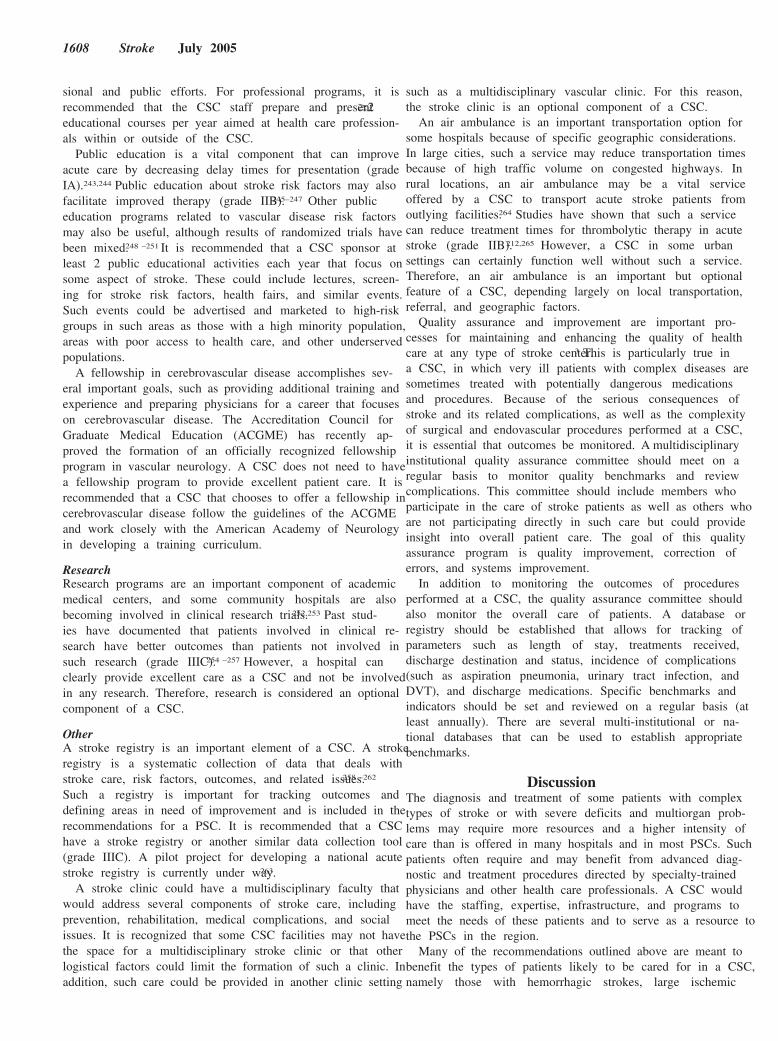
Representation of how various facilities caring for stroke patients could be organized based on a hospital network or a defined geographic area. Patients can arrive at the various facilities via direct admission or transfer between facilities. N indicates nonstroke center facility.
strokes, strokes of unknown or unusual etiology, and patients with multisystem involvement. Unlike most of the recomvcmendations for a PSC, there is a relative paucity of data proving that some of the components of a CSC change outcomes. However, there are data to support the utility and advantages of most of our recommendations, particularly in the areas of neuroimaging and certain specialized therapeutic procedures. There are emerging data that support the efficacy of other interventions related to neurosurgery and endovascular therapies. As these fields advance, we anticipate the publication of further studies that provide outcomes data for many of these interventions.
The costs for establishing and maintaining a CSC will vary depending on the existing staffing, infrastructure, and programs at a particular hospital. Several of the components of a CSC are quite costly, including an MRI scanner and an angiography suite, each of which can cost in excess of $1 million. Many hospitals that strive to be a CSC already have some of the equipment and infrastructure in place, which would reduce the costs considerably. The hiring of additional personnel (ie, endovascular interventionists and neurointensivists) and having staff members on call would also increase the costs, perhaps by $200 000 to $300 000 or more each year. Some of these costs could be offset by hospital billing for some procedures and physician billing for professional services. At this time, data do not exist to project a meaningful or accurate cost-benefit analysis for a CSC. Individual hospitals will have to develop business plans that consider the volume of stroke patients, payor mix, average costs and revenue per case, and future growth plans.
In the current health care environment, there continues to be growth of hospital networks and systems throughout the country. Within such a network or system, one approach to acute stroke care might be to designate some hospitals as PSCs and others as CSCs (Figure). This approach would allow for patients, equipment, and expertise to be concentrated at specific hospitals rather than spread throughout the entire network. This is quite similar to the paradigm used for other complex diseases that require a multidisciplinary team approach such as trauma, cancer, severe burns, and organ transplantation. A recent review documented the effective-
Recommendations for Comprehensive Stroke 1609
APPENDIX Results of National Survey for Components ofComprehensive Stroke Center Elements
Priority % of Respondents Score* With Component
Personnel
Stroke neurologist 4.8 92
CEA surgeon 4.7 95
24/7 radiologists (IH) 4.7 92
Stroke medical director 4.6 85
Interdisciplinary team 4.5 74
24/7 neurosurgeon (IH) 4.4 95
Staff CME 4.4 85
Vascular neurosurgeon 4.4 82
Stroke faculty 4.1 80
Nursing stroke director 4.1 64
Stroke nurse staff 4.1 64
Neurointensivist 3.9 62
Stroke APN 3.7 46
Diagnostic techniques
MRA 4.7 100
Carotid ultrasound 4.6 100
MRI with diffusion 4.6 92
MR perfusion 4 90
Transcranial Doppler 3.7 82
CT angiography 3.5 80
Surgical and endovascular therapies
IA lytics 4.4 90
IR coverage 24/7 4.4 80
IR-coil/stents 4.3 82
Endovascular IA ablation 4.3 77
Stent/angioplasty 3.9 87
Infrastructure
Stroke OP clinic 4.4 77
Stroke unit 4.4 72
Registry 4.3 82
Neuroscience ICU 4.3 69
Pathways 4.2 95
Air ambulance 4.2 92
Interdisciplinary rounds 4.2 67
Educational research programs
Community educational program 4.6 89
Prevention program 4.6 82
Hospital CME 4.5 87
QI program 4.5 71
Clinical research 4.4 95
National faculty meetings 4 90
Community screening 4 77
Research grants 3.8 80
Drug research 3.7 90
Fellowship 3.7 70
*Priority score is based on the respondent’s opinion of how important the component is for a CSC. The range for each element was 1 to 5, with 1 being the least important and 5 being the most important.
IH indicates in-hospital.

1610 Stroke July 2005
ness of the trauma center network for enhancing treatment and improving outcomes for patients with acute traumatic injuries.266
Is there a specific number of hospitalized stroke patients needed to support the formation of a CSC? Without specific cost-benefit data for a CSC, it is difficult to define specific patient volumes. It might be possible to extrapolate from some of the studies cited above that correlate volumes with outcomes. For example, if a CSC must care for �20 patients per year with SAH, and SAH represents �10% of all stroke cases admitted to a CSC hospital (this number may be increased because of referral bias of very ill patients), then the number of annual stroke admissions should approximate 200. However, there will be significant heterogeneity based on the surrounding population, catchment area, regional resources, referral patterns, local competition, etc.
Medical knowledge and technology are advancing at a rapid rate in many areas. BAC appreciates the fact that new techniques will become available for diagnostic studies, and new approaches will be developed for systemic and endovascular therapies of stroke. These recommendations are meant to be flexible and modifiable as new diagnostic and therapeutic options become available.
In summary, BAC has developed recommendations for CSCs and systems that will serve to guide the development of such centers and ensure that patients with cerebrovascular disease receive timely and effective care. We are hopeful that these recommendations will also assist hospitals and referral networks of PSCs and CSCs so that stroke patients receive care and resources that are most appropriate for their clinical condition. As new data become available, it will be important to modify these recommendations to reflect best practices and current guidelines.
Acknowledgments The authors wish to thank Nancy Hart, MA, and Sara Fassino for their skillful and dedicated administrative support in producing this manuscript. BAC consists of the following organizations and representatives who have approved publication of this document: American Academy of Neurology (Dr Koroshetz); American Association of Neurological Surgeons (Dr Selman); American Association of Neuroscience Nurses (Mr Shepherd, Ms Rose-DeRenzy); American College of Emergency Physicians (Dr Jagoda); American Stroke Association, a Division of the American Heart Association (Ms Magnis); American Society of Interventional and Therapeutic Neuroradiology (Dr Connors), American Society of Neuroradiology (Dr Latchaw); Centers for Disease Control and Prevention (Dr Croft); Congress of Neurological Surgeons (Drs Hadley and Cawley); National Association of EMS Physicians (Dr O’Connor); National Stroke Association (Ms Mulligan); National Institute of Neurological Disorders and Stroke (Drs Marler and Walker, Ms Emr, Ms Warren); Physical Medicine and Rehabilitation (Dr Zorowitz); Stroke Belt Consortium (Dr Alberts); and Veterans Health Administration (Drs Brass and Booss).
References 1. Broderick J, Brott T, Kothari R, Miller R, Khoury J, Pancioli A, Gebel
J, Mills D, Minneci L, Shukla R. The Greater Cincinnati/Northern Kentucky Stroke Study: preliminary first-ever and total incidence rates of stroke among blacks. Stroke. 1998;29:415–521.
2. Fang J, Alderman M. Trend of stroke hospitalization, United States, 1988 –1997. Stroke. 2001;32:2221–2226.
3. Alberts MA, Hademenos G, Latchaw RE, Jagoda A, Marler JR, Mayberg MR, Starke RD, Todd HW, Viste KM, Girgus M, Shephard T,
Emr M, Shwayder P, Walker MD; for the Brain Attack Coalition. Recommendations for the establishment of primary stroke centers. J Am Med Assoc. 2000;283:3102–3109.
4. Adams R, Acker J, Alberts M, Andrews L, Atkinson R, Fenelon K, Furlan A, Girgus M, Horton K, Hughes R, Koroshetz W, Latchaw R, Magnis E, Mayberg M, Pancioli A, Robertson RM, Shephard T, Smith R, Smith SC Jr, Smith S, Stranne SK, Kenton EJ III, Bashe G, Chavez A, Goldstein L, Hodosh R, Keitel C, Kelly-Hayes M, Leonard A, Morgenstern L, Wood JO. Recommendations for improving the quality of care through stroke centers and systems: an examination of stroke center identification options: multidisciplinary consensus recommendations from the Advisory Working Group on Stroke Center Identification Options of the American Stroke Association. Stroke. 2002;33: e1– e7.
5. Adams H, Adams R, Brott T, del Zoppo G, Furlan A, Goldstein L, Grubb R, Higashida R, Kidwell C, Kwiatkowski T, Marler J, Hademenos G. Guidelines for the early management of patients with ischemic stroke. Stroke. 2003;34:1056 –1083.
6. Latchaw RE, Yonas H, Hunter GJ, Yuh WT, Ueda T, Sorensen AG, Sunshine JL, Biller J, Wechsler L, Higashida R, Hademenos G; Council on Cardiovascular Radiology of the American Heart Association. Guidelines and recommendations for perfusion imaging in cerebral ischemia: a scientific statement for healthcare professionals by the writing group on perfusion imaging, from the Council on Cardiovascular Radiology of the American Heart Association. Stroke. 2003;34: 1084 –1104.
7. Mitchell JB, Ballard DJ, Whisnant JP, Ammering CJ, Samsa GP, Matchar DB. What role do neurologists play in determining the costs and outcomes of stroke patients? Stroke. 1996;27:1937–1943.
8. Goldstein L, Matchar D, Hoff-Lindquist J, Samsa G, Horner R. VA Stroke Study: neurologist care is associated with increased testing but improved outcomes. Neurology. 2003;61:792–796.
9. Fanshawe M, Venkatesh B, Boots RJ. Outcome of stroke patients admitted to intensive care: experience from an Australian teaching hospital. Anaesth Intensive Care. 2002;30:628 – 632.
10. Becker K. Intensive care unit management of the stroke patient. Neurol Clin. 2000;18:439 – 454.
11. Russell D, Vorder-Bruegge M, Burns S. Effect of an outcomes-managed approach to care of neuroscience patients by acute care nurse practitioners. Am J Crit Care. 2002;4:353–362.
12. Suyama J, Crocco T. Prehospital care of the stroke patient. Emerg Med Clin North Am. 2002;20:537–552.
13. Schroeder EB, Rosamond WD, Morris DL, Evenson KR, Hinn AR. Determinants of use of emergency medical services in a population with stroke symptoms: the Second Delay in Accessing Stroke Healthcare (DASH II) Study. Stroke. 2000;31:2591–2596.
14. Wein TH, Staub L, Felberg R, Hickenbottom SL, Chan W, Grotta JC, Demchuk AM, Groff J, Bartholomew LK, Morgenstern LB. Activation of emergency medical services for acute stroke in a nonurban population: the T.L.L. Temple Foundation Stroke Project. Stroke. 2000;31: 1925–1928.
15. Kothari R, Jauch E, Broderick J, Brott T, Sauerbeck L, Khoury J, Liu T. Acute stroke: delays to presentation and emergency department evaluation. Ann Emerg Med. 1999;33:3– 8.
16. Rosamond WD, Gorton RA, Hinn AR, Hohenhaus SM, Morris DL. Rapid response to stroke symptoms: the Delay in Accessing Stroke Healthcare (DASH) study. Acad Emerg Med. 1998;5:45–51.
17. Sacco RL, Benjamin EJ, Broderick JP, Dyken M, Easton DJ, Feinberg WM, Goldstein LB, Gorelick PB, Howard G, Kittner SJ, Manolio TA, Whisnant JP, Wolf PA. American Heart Association Prevention Conference, IV: Prevention and Rehabilitation of Stroke: Risk Factors. Stroke. 1997;28:1507–1517.
18. Greshman GE, Alexander D, Bishop DS, Giuliani C, Goldberg G, Holland A, Kelly-Hayes M, Linn RT, Roth EJ, Stason WB, Trombly CA. Rehabilitation. Stroke. 1997;28:1522–1526.
19. Alberts MJ, Horner J, Gray L, Brazer SR. Aspiration after stroke: lesion analysis by brain MRI. Dysphagia. 1992;7:170 –173.
20. Horner J, Massey EW, Rishi JE, Lathrop DL, Chase KN. Aspiration following stroke: clinical correlates and outcome. Neurology. 1988;38: 1359 –1362.
21. O’Neill PA. Swallowing and prevention of complications. Br Med Bul. 2000;56:457– 465.
22. Bushberg J, Seibert J, Leidholdt E, Boone J. The Essential Physics of Medical Imaging. 2nd ed. Philadelphia, Pa: Lippincott, Williams & Wilkins; 2002.

Alberts et al
23. Yuh WTC, Crain MR, Lees DJ, Greene GM, Ryals TJ, Sato Y. MR imaging of cerebral ischemia: findings in the first 24 hours. AJNR. 1991;12:621– 629.
24. Culebras A, Kase CS, Masdeu JC, Fox AJ, Bryan RN, Grossman CB, Lee DH, Adams HP, Thies W. Practice guidelines for the use of imaging in transient ischemic attacks and acute stroke. A report of the Stroke Council, American Heart Association. Stroke. 1997;28:1480 –1497.
25. Sorensen AG, Buonanno FS, Gonzalez RG, Schwamm LH, Lev MH, Huang-Hellinger FR, Reese TG, Weisskoff RM, Davis TL, Suwanwela N, Can U, Moreira JA, Copen WA, Look RB, Finklestein SP, Rosen BR, Koroshetz WJ. Hyperacute stroke: evaluation with combined multi-section diffusion-weighted and hemodynamically weighted echo-planar MR imaging. Radiology. 1996;199:391– 401.
26. Anonymous. Emergency interventional stroke therapy: a statement from the American Society of Interventional and Therapeutic Neuroradiology Stroke Task Force of the American Society of Neuroradiology and the Society of Cardiovascular and Interventional Radiology. Am J Neuroradiol. 2001;22:54.
27. Schellinger PD, Jansen O, Fiebach JB, Pohlers O, Ryssel H, Heiland S, Steiner T, Hacke W, Sartor K. Feasibility and practicality of MR imaging of stroke in the management of hyperacute cerebral ischemia. Am J Neuroradiol. 2000;21:1184 –1189.
28. Barber D, Darby D, Desmond P, Gerraty RP, Yang Q, Li T, Jolley D, Donnan GA, Tress BM, Davis SM. Identification of major ischemic change: diffusion-weighted imaging versus computed tomography. Stroke. 1999;30:2059 –2065.
29. Fiebach JB, Schellinger PD, Jansen O, Meyer M, Wilde P, Bender J, Schramm P, Juttler E, Oehler J, Hartmann M, Hahnel S, Knauth M, Hacke W, Sartor K. CT and diffusion-weighted MR imaging in randomized order: diffusion-weighted imaging results in higher accuracy and lower interrater variability in the diagnosis of hyperacute ischemic stroke. Stroke. 2002;33:2206 –2210.
30. Oppenheim C, Stanescu R, Dormont D, Crozier S, Marro B, Samson Y, Rancurel G, Marsault C. False-negative diffusion-weighted MR findings in acute ischemic stroke. Am J Neuroradiol. 2000;21:1434 –1440.
31. Hasegawa Y, Fisher M, Latour LL, Dardzinski BJ, Sotak CH. MRI diffusion mapping of reversible and irreversible ischemic injury in focal brain ischemia. Neurology. 1994;44:1484 –1490.
32. Albers GW. Diffusion-weighted MRI for evaluation of acute stroke. Neurology. 1998;51:S47–S49.
33. Albers GW, Lansberg MG, Norbash AM, Tong DC, O’Brien MW, Woolfenden AR, Marks MP, Moseley ME. Yield of diffusion-weighted MRI for detection of potentially relevant findings in stroke patients. Neurology. 2000;54:1562–1567.
34. Gonzalez RG, Schaefer PW, Buonanno FS, Schwamm LH, Budzik RF, Rordorf G, Wang B, Sorensen AG, Koroshetz WJ. Diffusion-weighted MR imaging: diagnostic accuracy in patients imaged within 6 hours of stroke symptom onset. Radiology. 1999;210:155–162.
35. Roh J-K, Kang D-W, Lee S-H, Yoon B-W, Chang K-H. Significance of acute multiple brain infarction on diffusion-weighted imaging. Stroke. 2000;31:688 – 694.
36. Lansberg MG, Albers GW, Beaulieu C, Marks MP. Comparison of diffusion-weighted MRI and CT in acute stroke. Neurology. 2000;54: 1557–1561.
37. Latchaw RE. The roles of diffusion and perfusion MR imaging in acute stroke management. Am J Neuroradiol. 1999;20:957–959.
38. Keir S, Wardlaw J. Systemic review of diffusion and perfusion imaging in acute ischemic stroke. Stroke. 2000;31:2723–2731.
39. Sunshine JL, Bambakidis N, Tarr RW, Lanzieri CF, Zaidat OO, Suarez JI, Landis DM, Selman WR. Benefits of perfusion MRI relative to diffusion in the diagnosis and treatment of hyperacute stroke. Radiology. 2001;22:915–921.
40. Schaefer PW, Hunter GJ, He J, Hamberg LM, Sorensen AG, Schwamm LH, Koroshetz WJ, Gonzalez RG. Predicting cerebral ischemic infarct volume with diffusion and perfusion MR imaging. Am J Neuroradiol. 2002;23:1785–1794.
41. Blakeley D, Oddone E, Hasselbald V, Simel D, Matchar D. Noninvasive carotid artery testing: a meta-analytic review. Ann Intern Med. 1995;122:360 –367.
42. Nederkoorn PJ, van der Graaf Y, Hunink MG. Duplex ultrasound and magnetic resonance angiography compared with digital subtraction angiography in carotid artery stenosis: a systematic review. Stroke. 2003;34:1324 –1332.
43. Alvarez-Linera J, Benito-Leon J, Escribano J, Campollo J, Gesto R. Prospective evaluation of carotid artery stenosis: elliptic centric
Recommendations for Comprehensive Stroke 1611
contrast-enhanced MR angiography and spiral CT angiography compared with digital subtraction angiography. Am J Neuroradiol. 2003;24:1012–1019.
44. Huston J III, Fain SB, Wald JT, Luetmer PH, Rydberg CH, Covarrubias DJ, Riederer SJ, Bernstein MA, Brown RD, Meyer FB, Bower TC, Schleck CD. Carotid artery: elliptic centric contrast-enhanced MR angiography compared with conventional angiography. Radiology. 2001;218:138 –143.
45. Johnston DC, Eastwood JD, Nguyen T, Goldstein LB. Contrast-enhanced magnetic resonance angiography of carotid arteries: utility in routine clinical practice. Stroke. 2002;33:2834 –2838.
46. Goyal M, Nicol J, Gandhi D. Evaluation of carotid artery stenosis: contrast-enhanced magnetic resonance angiography compared with conventional digital subtraction angiography. Can Assoc Radiol J. 2004;55: 111–119.
47. Cosottini M, Pingitore A, Puglioli M, Michelassi MC, Lupi G, Abbruzzese A, Calabrese R, Lombardi M, Parenti G, Bartolozzi C. Contrast-enhanced three-dimensional magnetic resonance angiography of atherosclerotic internal carotid stenosis as the noninvasive imaging modality in revascularization decision making. Stroke. 2003;34: 660 – 664.
48. Hirai T, Korogi Y, Ono K, Nagano M, Maruoka K, Uemura S, Takahashi M. Prospective evaluation of suspected stenoocclusive disease of the intracranial artery: combined MR angiography and CT angiography compared with digital subtraction angiography. Am J Neuroradiol. 2002;23:93–101.
49. Huston J III, Nichols DA, Luetmer PH, Goodwin JT, Meyer FB, Wiebers DO, Weaver AL. Blinded prospective evaluation of sensitivity of MR angiography to known intracranial aneurysms: importance of aneurysm size. Am J Neuroradiol. 1994;15:1607–1614.
50. Suzuki IM, Matsui, Ueda F, Hattori Y, Minami T, Kobayashi K, Tawara M, Kawashima H, Takanaka T, Yamashita J. Contrast-enhanced MR angiography (enhanced 3-D fast gradient echo) for diagnosis of cerebral aneurysms. Neuroradiology. 2002;44:17–20.
51. Jager HR, Mansmann U, Hausmann O, Partzsch U, Moseley IF, Taylor WJ. MRA versus digital subtraction angiography in acute subarachnoid hemorrhage: a blinded multireader study of prospectively recruited patients. Neuroradiology. 2000;42:313–326.
52. Vogl TJ, Bergman C, Villringer A, Einhaupl K, Lissner J, Felix R. Dural sinus thrombosis: value of venous MR angiography for diagnosis and follow-up. Am J Roentgenol. 1994;162:1191–1198.
53. Raizer JJ, DeAngelis LM. Cerebral sinus thrombosis diagnosed by MRI and MR venography in cancer patients. Neurology. 2000;54:1222–1226.
54. Ayanzen RH, Bird CR, Keller PJ, McCully FJ, Theobald MR, Heiserman JE. Cerebral MR venography: normal anatomy and potential diagnostic pitfalls. AJNR. 2000;21:74 –78.
55. Reichenbach JR, Essig M, Haacke EM, Lee BC, Przetak C, Kaiser WA, Schad LR. High-resolution venography of the brain using magnetic resonance imaging. MAGMA. 1998;6:62– 69.
56. Carmody RF, Seeger JF. Intracranial applications of digital subtraction angiography. Crit Rev Diagn Imaging. 1984. 23:1– 40.
57. Cross DT III. Interventional neuroradiology. Current Opinion in Neurology. 1993;6:891– 899.
58. Dogan A, Dempsey RJ. Diagnostic modalities for carotid artery disease. Neurosurg Clin N Am. 2000;11:205–220.
59. Baxter AB, Cohen WA, Maravilla KR. Imaging of intracranial aneurysms and subarachnoid hemorrhage. Neurosurg Clin N Am. 1998. 9:445– 462.
60. Joint Standards of Practice Task Force of the Society of Cardiovascular and Interventional Radiology tASoITN, and the American Society of Neuroradiology. Quality improvement guidelines for adult diagnostic neuroangiography. Am J Neuroradiol. 2000;21:146 –150.
61. Lev MH, Farkas J, Rodriguez VR, Schwamm LH, Hunter GJ, Putman CM, Rordorf GA, Buonanno FS, Budzik R, Koroshetz WJ, Gonzalez RG. CT angiography in the rapid triage of patients with hyperacute stroke to intraarterial thrombolysis: accuracy in the detection of large vessel thrombus. J Comput Assist Tomogr. 2001;25:520 –528.
62. Villablanca JP, Hooshi P, Martin N, Jahan R, Duckwiler G, Lim S, Frazee J, Gobin YP, Sayre J, Bentson J, Vinuela F. Three-dimensional helical computerized tomography angiography in the diagnosis, characterization, and management of middle cerebral artery aneurysm: comparison with conventional angiography and intraoperative finding. J Neurosurg. 2002;97:1322–1332.

1612 Stroke July 2005
63. Skutta B, Furst G, Eilers J, Ferbert A, Kuhn F. Intracranial stenoocclusive disease: double-detector helical CT angiography versus digital subtraction angiography. AJNR. 1999;20:791–799.
64. Randoux B, Marro B, Koskas F. Carotid artery stenosis: prospective comparison of CT, three-dimensional gadolinium enhanced MR, and conventional angiography. Radiology. 2001;220:179 –185.
65. Long A, Lepoutre A, Corbillon E, Branchereau A. Critical review of non- or minimally invasive methods (duplex ultrasonography, MR- and CT-angiography) for evaluating stenosis of the proximal internal carotid artery. Eur J Vasc Endovasc Surg. 2002;24:43–52.
66. Anderson GB, Ashforth R, Steinke DE, Ferdinandy R, Findlay JM. CT angiography for the detection and characterization of carotid artery bifurcation disease. Stroke. 2000;31:2168 –2174.
67. van Gelder JM. Computed tomographic angiography for detecting cerebral aneurysms: implications of aneurysm size distribution for the sensitivity, specificity, and likelihood ratios. Neurosurgery. 2003;53: 597– 605.
68. Pedersen HK, Bakke SJ, Hald JK, Skalpe IO, Anke IM, Sagsveen R, Langmoen IA, Lindegaard KE, Nakstad PH. CTA in patients with acute subarachnoid haemorrhage. A comparative study with selective, digital angiography and blinded, independent review. Acta Radiol. 2001;42: 43– 49.
69. Young N, Dorsch NW, Kingston RJ, Markson G, McMahon J. Intracranial aneurysms: evaluation in 200 patients with spiral CT angiography. Eur Radiol. 2001;11:123–130.
70. Lai PH, Yang CF, Pan HB, Chen C, Ho JT, Hsu SS. Detection and assessment of circle of Willis aneurysms in acute subarachnoid hemorrhage with three-dimensional computed tomographic angiography: correlation with digital substraction angiography findings. J Formos Med Assoc. 1999;98:672– 677.
71. Preda L, Gaetani P, Rodriguez y Baena R, Di Maggio EM, La Fianza A, Dore R, Fulle I, Solcia M, Cecchini A, Infuso L, Campani R. Spiral CT angiography and surgical correlations in the evaluation of intracranial aneurysms. Eur Rad. 1998;8:739 –745.
72. Dehdashti A, Rufenacht DA, Delavelle J, Reverdin A, de Tribolet N. Therapeutic decision and management of aneurysmal subarachnoid haemorrhage based on computed tomographic angiography. Br J Neurosurg. 2003;17:46 –53.
73. Carroll BA. Duplex sonography in patients with hemispheric symptoms. J Ultrasound Med. 1989;8:535–540.
74. Caplan LR, Brass LM, DeWitt LD, Adams RJ, Gomez C, Otis S, Weschler LR, von Reutern GM. Transcranial Doppler ultrasound: present status. Neurology. 1990;40:696 –700.
75. Gomez CR, Brass LM, Tegler CH, Babikian VL, Sloan MA, EF, Wechsler LR. The transcranial Doppler standardization project. Phase I results. The TCD Study Group, American Society of Neuroimaging. J Neuroimaging. 1993;3:190 –192.
76. Babikian VL, Feldmann E, Wechsler LR, Newell DW, Gomez CR, Bogdahn U, Caplan LR, Spencer MP, Tegeler C, Ringelstein EB, Alexandrov AV. Transcranial Doppler ultrasound: year 2000 update. J Neuroimaging. 2000;10:101–115.
77. Report of the American Academy of Neurology TaTAS. Assessment. Transcranial Doppler. Neurology. 1990;40:680 – 681.
78. Clyde BL, Resnick DK, Yonas H, Smith HA, Kaufmann AM. The relationship of blood velocity as measured by transcranial Doppler ultrasonography to cerebral blood flow as determined by stable xenon computed tomographic studies after aneurismal subarachnoid hemorrhage. Neurosurgery. 1996;38:896 –904.
79. Davis SM, Andrews JT, Lichtenstein M, Rossiter SC, Kaye AH, Hopper J. Correlations between cerebral arterial velocities, blood flow, and delayed ischemia after subarachnoid hemorrhage. Stroke. 2002;23: 492– 497.
80. Christou I, Alexandrov AV, Burgin WS, Wojner AW, Felberg RA, Malkoff M, Grotta JC. Timing of recanalization after tissue plasminogen activator therapy determined by transcranial Doppler correlates with clinical recovery from ischemic stroke. Stroke. 2000;31:1812–1816.
81. Sloan MA, Alexandrov AV, Tegeler CH, Spencer MP, Caplan LR, Feldmann E, Wechsler LR, Newell DW, Gomez CR, Babikian VL, Lefkowitz D, Goldman RS, Armon C, Hsu CY, Goodin DS; Therapeutics and Technology Assessment Subcommittee of the American Academy of Neurology. Assessment: transcranial Doppler ultrasoundography. Report of the Therapeutics and Technology Assessment Subcommittee of the American Academy of Neurology. Neurology. 2004; 62:1468 –1481.
82. Feinberg WM, Albers GW, Barnett HJM, Biller J, Caplan LR, Carter LP, Hart RG, Hobson RW, Kronmal RA, Moore WS, Robertson JT, Adams HP, Mayberg M. Guidelines for the management of transient ischemic attacks. Stroke. 1994;25:1320 –1335.
83. Rem JA, Hachinski VC, Boughner DR, Barnett HJM. Value of cardiac monitoring and echocardiography in TIA and stroke patients. Stroke. 1985;16:950 –956.
84. Rodriguez C, Homma S, Di Tullio M. Transesophageal echocardiography in stroke. Cardiol Rev. 2000;8:140 –147.
85. Fagan SM, Chan KL. Transesophageal echocardiography risk factors for stroke in nonvalvular atrial fibrillation. Echocardiography. 2000;17: 365–372.
86. Jagasia DH, Williams B, Ezekowitz MD. Clinical implication of antiembolic trials in atrial fibrillation and role of transesophageal echocardiography in atrial fibrillation. Curr Opin Cardiol. 2000;15:58 – 63.
87. Manning WJ. Role of transesophageal echocardiography in the management of thromboembolic stroke. Am J Cardiol. 1997;80:19D–28D.
88. Husain AM, Alter M. Transesophageal echocardiography in diagnosing cardioembolic stroke. Clin Cardiol. 1995;18:705–708.
89. Homma S, Sacco R, Di Tullio M, Sciacca R, Mohr J; for the PFO in Cryptogenic Stroke Study (PICSS) Investigators. Effect of medical treatment in stroke patients with patent foramen ovale. Circulation. 2002;105:2625–2631.
90. Pearson AC, Labovitz AJ, Tatineni S, Gomez CR. Superiority of trans-esophageal echocardiography in detecting cardiac source of embolism in patients with cerebral ischemia of uncertain etiology. J Am Coll Cardiol. 1991;17:66 –72.
91. Pop G, Sutherland GR, Koudstaal PJ, Sit TW, de Jong G, Roelandt JR. Transesophageal echocardiography in the detection of intracardiac embolic sources in patients with transient ischemic attacks. Stroke. 1990;21:560 –565.
92. ACC/ASE Committee on Clinical Application of Echocardiography. ACC/AHA Guidelines for the Clinical Application of Echocardiography. A Report of the American College of Cardiology/ American Heart Association Task Force on Practice Guidelines (Committee on Clinical Application of Echocardiography) Developed in Collaboration With the American Society of Echocardiography. Circulation. 1997;95: 1686 –1744.
93. De Reuck J, Leys D, De Keyser J. Is positron emission tomography useful in stroke? Acta Neurol Belg. 1997;97:168 –171.
94. Higano S, Uemura K, Shishido F, Kanno I, Tomura N, Sakamoto K. Evaluation of critically perfused area in acute ischemic stroke for therapeutic reperfusion: a clinical PET study. Ann Nucl Med. 1993;7: 167–171.
95. Szabo T, Goth J, Dominich S, Kozmann G, Szolgay P, Barsony P. Novel neuroradiological image processing and information retrieval in a telestroke system. Stud Health Techno Inform. 2003;95:298 –303.
96. Johnston KC, Worrall BB, Teleradiology Assessment of Computerized Tomographs Online Reliability Study. Teleradiology Assessment of Computerized Tomographs Online Reliability Study (TRACTORS) for acute stroke evaluation. Telemed J E Health. 2003;9:227–233.
97. Johnston SC, Higashida RT, Barrow DL, Caplan LR, Dion JE, Hademenos G, Hopkins LN, Molyneux A, Rosenwasser RH, Vinuela F, Wilson CB; Committee on Cerebrovascular Imaging of the American Heart Association Council on Cardiovascular Radiology. Recommendations for the endovascular treatment of intracranial aneuryms. A statement for healthcare professionals from the committee on cerebrovascular imaging of the American Heart Association Council on Cardiovascular Radiology. Stroke. 2002:2536 –2544.
98. ACGME. Program requirements for residency education in endovascular surgical neuroradiology. www.acgme.org/downloads/ RRC_progReq/422pr403.pdf; 2003.
99. Molyneux A, Kerr R, Stratton I, Sandercock P, Clarke M, Shrimpton J, Holman R; International Subarachnoid Aneurysm Trial (ISAT) Collaborative Group. International subarachnoid aneurysm trial (ISAT) of neurosurgical clipping versus endovascular coiling in 2143 patients with ruptured intracranial aneurysms: a randomized trial. Lancet. 2002;360: 1267–1274.
100. Harbaugh RE, Heros RC, Hadley MN. More on ISAT. Lancet. 2003; 361:783–784
101. Pasqualin A. Epidemiology and pathophysiology of cerebral vasospasm following subarachnoid hemorrhage. J Neurosurg Sci. 1998;42:15–21.
102. Sen J, Belli A, Albon H, Morgan L, Petzold A, Kitchen N. Triple-H therapy in the management of aneurysmal subarachnoid haemorrhage. Lancet Neurol. 2003;2:614 – 621.

Alberts et al
103. Treggiari MM, Walder B, Suter PM, Romand JA. Systematic review of the prevention of delayed ischemic neurological deficits with hypertension, hypervolemia, and hemodilution therapy following subarachnoid hemorrhage. J Neurosurg. 2003;98:978 –984.
104. Firlik KS, Kaufmann AM, Firlik AD, Jungreis CA, Yonas H. Intra-ar-terial papaverine for the treatment of cerebral vasospasm following aneurysmal subarachnoid hemorrhage. Surg Neurol. 1999;51:66 –74.
105. Liu JK, Tenner MS, Oestreich HM, Couldwell WT. Reversal of radiographically impending stroke with multiple intraarterial papaverine infusions in severe diffuse cerebral vasospasm induced by subarachnoid hemorrhage. Acta Neurochir. 2001;143:1249 –1255.
106. Newell DW, Elliott JP, Eskridge JM, Winn HR. Endovascular therapy for aneurysmal vasospasm. Crit Care Clin. 1999;15:685– 699.
107. Numaguchi Y, Zoarski GH, Clouston JE, Zagardo MT, Simard JM, Aldrich EF, Sloan MA, Maurer PK, Okawara SH. Repeat intra-arterial papaverine for recurrent cerebral vasospasm after subarachnoid haemorrhage. Neuroradiology. 1997;39:751–759.
108. Polin RS, Hansen CA, German P, Chadduck JB, Kassell NF. Intra-ar-terially administered papaverine for the treatment of symptomatic cerebral vasospasm. Neurosurgery. 1998;42:1256 –1264.
109. Higashida RT, Halbach VV, Dowd CF, Dormandy B, Bell J, Hieshima GB. Intravascular balloon dilatation therapy for intracranial arterial vasospasm: patient selection, technique, and clinical results. Neurosurg Rev. 1992;15:89 –95.
110. Zwienenberg-Lee M, Hartman J, Rudisill N, Muizelaar J. Endovascular management of cerebral vasospasm. Neurosurgery. In press.
111. Eskridge J. A practical approach to the treatment of vasospasm. Am J Neuroradiol. 1997;18:1653–1660.
112. Dawson R, Barr J, Connors J, et al. Standards of practice: the American Society of Interventional and Therapeutic Neuroradiology. AJNR. 2001;22 (suppl).
113. Pessin M, del Zoppo G, Furlan A. Thrombolytic treatment in acute stroke: review and update of selective topics. In: Moskowitz M, Caplan L, eds. Cerebrovascular Diseases. Nineteenth Princeton Stroke Conference. Boston, Mass: Butterworth-Heinmann; 1995:409 – 418.
114. Lisboa RC, Jovanovic BD, Alberts MJ. Analysis of the safety and efficacy of intra-arterial thrombolytic therapy in ischemic stroke. Stroke. 2002;33:2866 –2871.
115. Furlan A, Higashida R, Wechsler L, Gent M, Rowley H, Kase C, Pessin M, Ahuja A, Callahan F, Clark WM, Silver F, Rivera F. Intra-arterial prourokinase for acute ischemic stroke. The PROACT II study: a randomized controlled trial. Prolyse in Acute Cerebral Thromboembolism. J Am Med Assoc. 1999;282:2003–2011.
116. Kidwell CS, Saver JL, Carneado J, Sayre J, Starkman S, Duckwiler G, Gobin YP, Jahan R, Vespa P, Villablanca JP, Liebeskind DS, Vinuela F. Predictors of hemorrhagic transformation in pateints receiving intra-ar-terial thrombolysis. Stroke. 2002;33:717–724.
117. Qureshi AI, Pande RU, Kim SH, Hanel RA, Kirmani JF, Yahia AM. Third generation thrombolytics for the treatment of ischemic stroke. Curr Opin Investig Drugs. 2002;3:1729 –1732.
118. Arnold M, Schroth G, Nedeltchev K, Loher T, Remonda L, Stepper F, Sturzenegger M, Mattle HP. Intra-arterial thrombolysis in 100 patients with acute stroke due to middle cerebral artery occlusion. Stroke. 2002; 33:1828 –1833.
119. Schellinger PD, Fiebach JB, Mohr A, Ringleb PA, Jansen O, Hacke W. Thrombolytic therapy for ischemic stroke—a review. Part II—intra-ar-terial thrombolysis, vertebrobasilar stroke, phase IV trials, and stroke imaging. Crit Care Med. 2001;29:1819 –1825.
120. Jahan R, Duckwiler GR, Kidwell CS, Sayre JW, Gobin YP, Villablanca JP, Saver J, Starkman S, Martin N, Vinuela F. Intraarterial thrombolysis for treatment of acute stroke: experience in 26 patients with long-term follow-up. Am J Neuroradiol. 1999;20:1291–1299.
121. Ueda T, Hatakeyama T, Kohno K, Kumon Y, Sakaki S. Endovascular treatment for acute thrombotic occlusion of the middle cerebral artery: local intraarterial thrombolysis combined with percutaneous trans-luminal angioplasty. Neuroradiology. 1999;39:99 –104.
122. Advanced Cardiac Life Support Providers Manual. Dallas, Tex: American Heart Association; 2003.
123. Chopko BW, Kerber C, Wong W, Georgy B. Transcatheter snare removal of acute middle cerebral artery thromboembolism: technical case report. Neurosurgery. 2000;46:1529 –1531.
124. Fourie P, Duncan IC. Microsnare-assisted mechanical removal of intra-procedural distal middle cerebral arterial thromboembolism. Am J Neuroradiol. 2003;24:630 – 632.
Recommendations for Comprehensive Stroke 1613
125. Kerber CW, Barr JD, Berger RM, Chopko BW. Snare retrieval of intracranial thrombus in patients with acute stroke. J Vasc Interv Radiol. 2002;13:1269 –1274.
126. Starkman S; for the MERCI Investigators. Results of the combined MERCI I-II trials. Stroke. 2004;35:240 (Abstract).
127. Gobin YP, Starkman S, Duckwiler GR, Grobelny T, Kidwell CS, Jahan R, Pile-Spellman J, Segal A, Vinuela F, Saver JL. MERCI 1: a phase 1 study of mechanical embolus removal in cerebral ischemia. Stroke. 2004;35:2848 –2854.
128. Ciccone A, Canhao P, Falcao F, Ferro J, Sterzi R. Thrombolysis for cerebral vein and dural sinus thrombosis. Stroke. 2004;35:2428.
129. Tsai FY, Wang AM, Matovich VB, Lavin M, Berberian B, Simonson TM, Yuh WT. MR staging of acute dural sinus thrombosis: correlation with venous pressure measurements and implications for treatment and prognosis. Am J Neuroradiol. 1995;16:1021–1029.
130. Lee SK, terBrugge KG. Cerebral venous thrombosis in adults: the role of imaging evaluation and management. Neuroimaging Clin N Am. 2003;13:139 –152.
131. Yamini B, Loch Macdonald R, Rosenblum J. Treatment of deep cerebral venous thrombosis by local infusion of tissue plasminogen activator. Surg Neurol. 2001;55:340 –346.
132. Chow K, Gobin YP, Saver J, Kidwell C, Dong P, Vinuela F. Endovascular treatment of dural sinus thrombosis with rheolytic thrombectomy and intra-arterial thrombolysis. Stroke. 2000;31:1420 –1425.
133. Phatouros CC, Higashida RT, Malek AM, Meyers PM, Lempert TE, Dowd CF, Halbach VV. Carotid artery stent placement for atherosclerotic disease: rationale, technique, and current status. Radiology. 2000;217:26 – 41.
134. Theron JG, Payelle GG, Coskun O, Huet HF, Guimaraens L. Carotid artery stenosis: treatment with protected balloon angioplasty and stent placement. Radiology. 2000;201:627– 636.
135. Abberts MJ; for the Publications Committee of the WALLSTENT Study. Stroke. 2001;32:325. [Abstract.]
136. Spence D, Elisziw M. Endovascular versus surgical treatment in patients with carotid artery and vertebral artery transluminal angioplasty study (CAVATAS): a randomized trial. Lancet. 2001;357:1729 –1737.
137. Roubin GS, New G, Iyer SS, Vitek JJ, Al-Mubarak N, Liu MW, Yadav J, Gomez C, Kuntz RE. Immediate and late clinical outcomes of carotid artery stenting in patients with symptomatic and asymptomatic carotid artery stenosis: a 5-year prospective analysis. Circulation. 2001;103: 532–537.
138. Yadav JS, Wholey MH, Kuntz RE, Fayad P, Katzen BT, Mishkel GJ, Bajwa TK, Whitlow P, Strickman NE, Jaff MR, Popma JJ, Snead DB, Cutlip DE, Firth BG, Ouriel K; Stenting and Angioplasty with Protection in Patients at High Risk for Endarterectomy Investigators. Protected carotid-artery stenting versus endarterectomy in high-risk patients. N Engl J Med. 2004;351:1493–1501.
139. Kirsch EC, Khangure MS, van Schie GP, Lawrence-Brown MM, Stewart-Wynne EG, McAuliffe W. Carotid arterial stent placement: results and follow-up in 53 patients. Radiology. 2001;220:737–744.
140. Hobson RW II, Lal BK, Chakhtoura E, Goldstein J, Haser PB, Kubicka R, Cerveira J, Pappas PJ, Padberg FT, Jamil Z, Chaktoura E. Carotid artery stenting: analysis of data for 105 patients at high risk. J Vasc Surg. 2003;37:1234 –1239.
141. Cohen JE, Leker RR, Gotkine M, Gomori M, Ben-Hur T. Emergent stenting to treat patients with carotid artery dissection: clinically and radiologically directed therapeutic decision making. Stroke. 2003; e254 – e257.
142. Malek AM, Higashida RT, Phatouros CC, Lempert TE, Meyers PM, Smith WS, Dowd CF, Halbach VV. Endovascular management of extracranial carotid artery dissection achieved using stent angioplasty. Am J Neuroradiol. 2000;21:1280 –1292.
143. Hobson RW II, Howard VJ, Brott TG, Howard G, Roubin GS, Ferguson RD; for the CREST Executive Committee. Organizing the Carotid Revascularization Endarterectomy versus Stenting Trial (CREST): National Institutes of Health, Health Care Financing Administration, and industry funding. Curr Control Trials Cardiovasc Med. 2001;2: 160 –164.
144. Connors JJ III, Sacks D, Becker GJ, Barr JD. Carotid artery angioplasty and stent placement: quality improvement guidelines to ensure stroke risk reduction. J Vasc Interv Radiol. 2003;14:1095–1097.
145. Barr JD, Connors JJ III, Sacks D, Wojak JC, Becker GJ, Cardella JF, Chopko B, Dion JE, Fox AJ, Higashida RT, Hurst RW, Lewis CA, Matalon TA, Nesbit GM, Pollock JA, Russell EJ, Seidenwurm DJ, Wallace RC; American Society of Interventional and Therapeutic Neu

1614 Stroke July 2005
roradiology; American Society of Neuroradiology; Society of Interventional Radiology. Quality improvement guidelines for the performance of cervical carotid angioplasty and stent placement. Developed by a collaborative panel of the American Society of Interventional and Therapeutic Neuroradiology, the American Society of Neuroradiology, and the Society of Interventional Radiology. J Vasc Interv Radiol. 2003;14: 1079 –1093.
146. Thijs VN, Albers GW. Symptomatic intracranial atherosclerosis: outcome of patients who fail. Neurology. 2000;55:490 – 497.
147. Lylyk P, Cohen JE, Ceratto R, Ferrario A, Miranda C. Angioplasty and stent placement in intracranial atherosclerotic stenoses and dissections. Am J Neuroradiol. 2002;23:430 – 436.
148. Gress DR, Smith WS, Dowd CF, Van Halbach V, Finley RJ, Higashida RT. Angioplasty for intracranial symptomatic vertebrobasilar ischemia. Neurosurgery. 2002;51:23–27.
149. Connors JJ III, Wojak JC. Percutaneous transluminal angioplasty for intracranial atherosclerotic lesions. J Neurosurg. 1999;91:415– 423.
150. Takis C, Kwan ES, Pessin MS, Jacobs DH, Caplan LR. Intracranial angioplasty: experience and complications. Am J Neuroradiol. 1997;18: 1661–1668.
151. Marks M, Marcellus M, Norbash AM, Steinberg GK, Tong D, Albers GW. Outcome of angioplasty for atherosclerotic intracranial stenosis. Stroke. 1999;30:1065–1069.
152. Chimowitz MI, Lynn MJ, Howlett-Smith H, Stern BJ, Hertzberg VS, Frankel MR, Levine SR, Chaturvedi S, Kasner SE, Benesch CG, Sila CA, Jovin TG, Romano JG; Warfarin-Aspirin Symptomatic Intracranial Disease Trial Investigators. Comparison of warfarin and aspirin for symptomatic intracranial arterial stenosis. N Engl J Med. 2005;352: 1305-16
153. The SSYLVIA Study Investigators. Stenting of symptomatic atherosclerotic lesions in the vertebral or intracranial arteries (SSYLVIA): study results. Stroke. 2004;35:1388 –1392.
154. Wojak J. Intracranial angioplasty/stenting: long-term results from single center. In: American Heart Association 28th International Stroke Conference. Phoenix, Ariz; 2003.
155. Vinuela F, Dion JE, Duckwiler G, Martin NA, Lylyk P, Fox A, Pelz D, Drake CG, Girvin JJ, Debrun G. Combined endovascular embolization and surgery in the management of cerebral arteriovenous malformation: experience with 101 cases. J Neurosurg. 1991;75:856 – 864.
156. Ogilvy CS, Stieg PE, Awad I, Brown RD Jr, Kondziolka D, Rosenwasser R, Young WL, Hademenos G; Special Writing Group of the Stroke Council, American Stroke Association. AHA Scientific Statement: Recommendations for the management of intracranial arteriovenous malformations: a statement for healthcare professionals from a special writing group of the Stroke Council, American Stroke Association. Stroke. 2001; 32:1458 –1471.
157. Gobin YP, Laurent A, Merienne L, Schlienger M, Aymard A, Houdart E, Casasco A, Lefkopoulos D, George B, Merland JJ. Treatment of brain arteriovenous malformations by embolization and radiosurgery. J Neurosurg. 1996;85:19 –28.
158. Caldarelli M, Di Rocco C, Iannelli A, Rollo M, Tamburrini G, Velardi F. Combined management of intracranial vascular malformations in children. J Neurosurg Sci. 1997;41:315–324.
159. Debrun GM, Aletich V, Ausman JI, Charbel F, Dujovny M. Embolization of the nidus of brain arteriovenous malformations with n-butyl cyanoacrylate. Neurosurgery. 1997;40:112–120.
160. Cognard C, Houdart E, Casasco A, et al. Endovascular therapy and long-term results for intracranial dural arteriovenous fistulae. In: Connors J, Wojak J, eds. Interventional Neuroradiology Strategies and Practical Techniques. Philadelphia, Pa: WB Saunders; 1999:198 –214.
161. Awad I. The diagnosis and management of intracranial dural arteriovenous malformations. Contemp Neurosurg. 1991;13:1– 6.
162. Martin NA, Khanna R, Doberstein C, Bentson J. Therapeutic embolization of arteriovenous malformations: the case for and against. Clin Neurosurg. 2000;46:295–318.
163. Deveikis JP. Endovascular therapy of intracranial arteriovenous malformations. Materials and techniques. Neuroimaging Clin N Am. 1998;8: 401– 424.
164. Higashida RT, Hopkins LN, Berenstein A, Halbach VV, Kerber C. Program requirements for residency/fellowship education in neuroendovascular surgery/interventional neuroradiology: a special report on graduate medical education. Am J Neuroradiol. 2000;21:1153–1159.
165. American College of Radiology. Standard for the performance of diagnostic cervicocerebral angiography in adults. American College of
Radiology Standards 2000 –2001. Reston, Va: American College of Radiology; 2000:415– 426.
166. Connors JJ III, Sacks D, Furlan AJ, Selman WR, Russell EJ, Stieg PE, Hadley MN, Neuro Vascular Coalition Writing Group; American Academy of Neurology; American Association of Neurological Surgeons; American Society of Interventional and Therapeutic Neuroradiology; American Society of Neuroradiology; Congress of Neurological Surgeons; AANS/CNS Cerebrovascular Section; Society of Interventional Radiology. Training, competency, and credentialing standards for diagnostic cervicocerebral angiography, carotid stenting, and cerebrovascular intervention: a joint statement from the American Academy of Neurology, the American Association of Neurological Surgeons, the American Society of Interventional and Therapeutic Neuroradiology, the American Society of Neuroradiology, the Congress of Neurological Surgeons, the AANS/CNS Cerebrovascular Section, and the Society of Interventional Radiology. J Vasc Interv Radiol. 2004;15:1347–1356.
167. Sacks D, Connors JJ III. Carotid stenting, stroke prevention, and training. J Vasc Interv Radiol. 2004;15:1381–1384.
168. Vitek JJ, Roubin GS, Al-Mubarek N, New G, Iyer SS. Carotid artery stenting: technical considerations. AJNR. 2000;21:1736 –1743.
169. Ramee SR, Dawson R, McKinley KL, Felberg R, Collins TJ, Jenkins JS, Awaad MI, White CJ. Provisional stenting for symptomatic intracranial stenosis using a multidisciplinary team. Catheter Cardiovasc Interv. 2001;52:457– 467.
170. Broderick JP, Adams HP, Barsan W, Feinberg WM, Feldmann E, Grotta JC, Kase C, Krieger D, Mayberg MR, Tilley BC, Zabramski JM, Zuccarello M. Guidelines for the management of spontaneous intracerebral hemorrhage. A statement for healthcare professionals from a special writing group of the Stroke Council, American Heart Association. Stroke. 1999;30:905–915.
171. Biller J, Feinberg WM, Castaldo JE, Whittemore AD, Harbaugh RE, Dempsy RJ, Caplan LR, Kresowick TF, Matchar DB, Toole JF, Easton JD, Adams HP, Brass LM, Hobson RW, Brott TG, Sternau L. Guidelines for carotid endarterectomy. A statement for healthcare professionals from a special writing group of the Stroke Council, American Heart Association. Stroke. 1998;29:554 –562.
172. Biller J, Patrick JT. Management of medical complications of stroke. J Stroke and Cerebrovasc Dis. 1997;6:217–220.
173. Morley NC, Berge E, Cruz-Flores S, Whittle IR. Surgical decompression for cerebral oedema in acute ischemic stroke. Cochrane Database Syst Rev. 2002;CD003435.
174. Prasad K, Shrivastava A. Surgery for primary supratentorial intracerebral haemorrhage. Cochrane Database Syst Rev. 1999;CD000200.
175. Solomon R, Mayer S, Tarmey J, et al. Relationship between the volume of craniotomies for cerebral aneurysm performed at New York state hospitals and in-hospital moratality. Stroke. 1996;27:13–17.
176. Bederson JB, Awad IA, Wiebers DO, Piepgras D, Haley EC Jr, Brott T, Hademenos G, Chyatte D, Rosenwasser R, Caroselli C. Recommendations for the management of patients with unruptured intracranial aneuryms: a statement for healthcare professionals from the Stroke Council of the American Heart Association. Circulation. 2000;102: 2300 –2308.
177. Raaymakers T, Rinkel G, Limburg M, Algra A. Mortality and morbidity of surgery for unruptured intracranial aneurysms-a meta analysis. Stroke. 1998;29:1531–1538.
178. International Study of Unruptured Intracranial Aneurysms Investigators. Unruptured intracranial aneurysms: risk of rupture and risks of surgical intervention. N Engl J Med. 1998;339:1725–1733.
179. Bardach NS, Zhao S, Gress DR, Lawton MT, Johnston SC. Association between subarachnoid hemorrhage outcomes and number of cases treated in California. Stroke. 2002;33:1851–1856.
180. Cross DT III, Tirschwell DL, Clark MA, Tuden D, Derdeyn CP, Moran CJ, Dacey RG Jr. Mortality rates after subarachnoid hemorrhage: variations according to hospital case volume in 18 states. J Neurosurg. 2003;99:810 – 817.
181. Cowan JA Jr, Dimick JB, Wainess RM, Upchurch GR Jr, Thompson BG. Outcomes after cerebral aneurysm clip occlusion in the United States: the need for evidence-based hospital referral. J Neurosurg. 2003; 99:947–952.
182. Heros R. Editorial: case volume and mortality. J Neurosurg. 2003;99: 805– 806.
183. Heros R. Editorial: case volume and outcome. J Neurousrg. 2003;99: 945–946.
184. Barnett HJM, Taylor DW, Eliasziw M, Fox AJ, Ferguson GG, Haynes RB, Rankin RN, Clagett GP, Hachinski VC, Sackett DL, Thorpe KE,

Alberts et al
Math M, Meldrum HE; for the North American Symptomatic Carotid Endarterectomy Trial (NASCET) Collaborators. Benefit of carotid endarterectomy in patient with symptomatic moderate or severe stenosis. N Engl J Med. 1998;339:1415–1425.
185. European Carotid Surgery Trialists’ Collaborative Group. Endarterectomy for moderate symptomatic carotid stenosis: interim results from the MRC European Carotid Surgery Trial. Lancet. 1996;347:1591–1593.
186. Executive Committee for the Asymptomatic Carotid Atherosclerosis Study. Endarterectomy for asymptomatic carotid artery stenosis. J Am Med Assoc. 1995;273:1421–1428.
187. Moore WS, Barnett HJ, Beebe HG, Bernstein EF, Brener BJ, Brott T, Caplan LR, Day A, Goldstone J, Hobson RW II, et al. Guidelines for carotid endarterectomy. A multidisciplinary consensus statement from the Ad Hoc Committee, American Heart Association. Circulation. 1995; 91:566 –579.
188. EC/IC Bypass Study Group. Failure of extracranial-intracranial arterial bypass to reduce the risk of ischemic stroke: results of an international randomized study. N Engl J Med. 1985;313:1191–1200.
189. Chazono H, Okamoto Y, Matsuzaki Z, Ogino J, Endo S, Matsuoka T, Horikoshi T, Nukui H, Hadeishi H, Yasui N. Extracranial-intracranial bypass for reconstruction of internal carotid artery in the management of head and neck cancer. Ann Vasc Surg. 2003;17:260 –265.
190. Jafar JJ, Russell SM, Woo HH. Treatment of giant intracranial aneurysms with saphenous vein extracranial-to-intracranial bypass grafting: indications, operative technique, and results in 29 patients. Neurosurgery. 2002;51:138 –144.
191. Guthikonda M, Guyot LL, Diaz FG. Future of extracranial-intracranial bypass. Neurol Res. 2002;24:S80 –S83.
192. Nussbaum ES, Erickson DL. Extracranial-intracranial bypass for ischemic cerebrovascular disease refractory to maximal medical therapy. Neurosurgery. 2000;46:37– 42.
193. Hadley M, Spetzler R. Contemporary application of the extracranial-intracranial bypass for cerebral revascularization. Neurosurgery. 1988; 9:1– 6.
194. Spetzler R, Carter L. Revascularization and aneurysm surgery: current status. Neurosurgery. 1985;16:111–116.
195. Adams HP Jr, Powers WJ, Grubb RL Jr, Clarke WR, Woolson RF. Preview of a new trial of extracranial-to-intracranial anastomosis: the carotid occlusion surgery study. Neurosurg Clin N Am. 2001;12: 613– 624.
196. Derdeyn C, Yundt K, Videen T, Carpenter D, Grubb R, Powers W. Increased oxygen extraction fraction is associated with prior ischemic events in patients with carotid occlusion. Stroke. 1998;29:754 –758.
197. Grubb R, Derdeyn C, Fritsch S, Carpenter D, Yundt K, Videen T, Spitznagel E, Powers W. Importance of hemodynamic factors in the prognosis of symptomatic carotid occlusion. J Am Med Assoc. 1998; 280:1055–1060.
198. Powers W, Zazulia A. The use of positron emission tomography in cerebrovascular disease. Neuroimaging Clin N Am. 2003;13:741–758.
199. Ikezaki K. Rational approach to treatment of moyamoya disease in childhood. J Child Neurol. 2000;15:350 –356.
200. Srinivasan J, Britz GW, Newell DW. Cerebral revascularization for moyamoya disease in adults. Neurosurg Clin N Am. 2001;12:585–594.
201. Lawton M, Hamilton M, Morcos J, Spetzler R. Revascularization and aneurysm: current techniques, indications, and outcomes. Neurosurgery. 1996;38:83–94.
202. Anonymous. The Brain Trauma Foundation. The American Association of Neurological Surgeons. The Joint Section on Neurotrauma and Critical Care. Recommendations for intracranial pressure monitoring technology. J Neurotrauma. 2000;17:497–506.
203. Anonymous. The Brain Trauma Foundation. The American Association of Neurological Surgeons. The Joint Section on Neurotrauma and Critical Care. Indications for intracranial pressure monitoring. J Neurotrauma. 2000;17:479 – 491.
204. Lang EW, Chesnut RM. Intracranial pressure. Monitoring and management. Neurosurg Clin N Am. 1994;5:573– 605.
205. Cho DY, Chen TC, Lee HC. Ultra-early decompressive craniectomy for malignant middle cerebral artery infarction. Surg Neurol. 2003;60: 227–232.
206. Kuroki K, Taguchi H, Sumida M, Yukawa O, Murakami T, Onda J, Eguchi K. Decompressive craniectomy for massive infarction of middle cerebral artery territory. No Shinkei Geka. 2001;29:831– 835.
207. Schwab S, Steiner T, Aschoff A, Schwarz S, Steiner HH, Jansen O, Hacke W. Early hemicraniectomy in patients with complete middle cerebral artery infarction. Stroke. 1998;29:1888 –1893.
Recommendations for Comprehensive Stroke 1615
208. Anonymous. A systems approach to immediate evaluation and management of hyperacute stroke. Experience at eight centers and implications for community practice and patient care. The National Institute of Neurological Disorders and Stroke (NINDS) rt-PA Stroke Study Group. Stroke. 1997;28:1530 –1540.
209. Zweifler RM, Drinkard R, Cunningham S, Brody ML, Rothrock JF. Implementation of a stroke code system in Mobile, Alabama. Diagnostic and therapeutic yield. Stroke. 1997;28:981–983.
210. Adams HP Jr, Brott TG, Furlan AJ, Gomez CR, Grotta J, Helgason CM, Kwiatkowski T, Lyden PD, Marler JR, Torner J, Feinberg W, Mayberg M, Thies W. Guidelines for thrombolytic therapy for acute stroke: a supplement to the guidelines for the management of patients with acute ischemic stroke. Circulation. 1996;94:1167–1174.
211. Riopelle RJ, Howse DC, Bolton C, Elson S, Groll DL, Holtom D, Brunet DG, Jackson AC, Melanson M, Weaver DF. Regional access to acute ischemic stroke intervention. Stroke. 2001;32:652– 655.
212. Conroy MB, Rodriguez SU, Kimmel SE, Kasner SE. Helicopter transfer offers a potential benefit to patients with acute stroke. Stroke. 1999;30: 2580 –2584.
213. Rymer MM, Thurtchley D, Summers D; Mid-America Brain Stroke Institute Stroke Team. Expanded modes of tissue plasminogen activator delivery in a comprehensive stroke center increases regional acute stroke interventions. Stroke. 2003;34:e58 – e60.
214. Wang DZ, Rose JA, Honings DS, Garwacki DJ, Milbrandt JC. Treating acute stroke patients with intravenous tPA. The OSF stroke network experience. Stroke. 2000;31:77– 81.
215. Langhorne P, Dennis M. Stroke units: the next 10 years. Lancet. 2004; 363:834 – 835.
216. Launois R, Giroud M, Megnigbeto AC, Le Lay K, Presente G, Mahagne MH, Durand I, Gaudin AF. Estimating the cost-effectiveness of stroke units in France compared with conventional care. Stroke. 2004;35: 770 –775.
217. Sulter G, Elting JW, Langedijk M, Maurits NM, De Keyser J. Admitting acute ischemic stroke patients to a stroke care monitoring unit versus a conventional stroke unit: a randomized pilot study. Stroke. 2003;34: 101–104.
218. Torner JC, Davis P, Leira E. Epidemiology of stroke in requiring intensive care. New Horiz. 1997;5:422– 432.
219. Diringer MN, Edwards DF. Admission to a neurologic/neurosurgical intensive care unit is associated with reduced mortality rate after intracerebral hemorrhage. Crit Care Med. 2001;29:635– 640.
220. Furie K, Feldmann E. Treating intracerebral hemorrhage effectively in the ICU. The key steps: provide supportive care and determine the cause. J Crit Illn. 1995;10:794 –796, 799 – 800, 803– 4.
221. Burtin P, Bollaert PE, Feldmann L, Nace L, Lelarge P, Bauer P, Larcan A. Prognosis of stroke patients undergoing mechanical ventilation. Intensive Care Med. 1994;20:32–36.
222. Mohr JP, Albers GW, Amarenco P, Babikian VL, Biller J, Brey RL, Coull B, Easton JD, Gomez CR, Helgason CM, Kase CS, Pullicino PM, Turpie AGG. Etiology of stroke. Stroke. 1997;28:1501–1506.
223. van der Worp HB, Kappelle LJ. Complications of acute ischemic stroke. Cerebrovasc Dis. 1998;8:124 –132.
224. Alberts MJ, Brass LM. Assessment of early deterioration. J Stroke Cerebrovasc Dis. 1999;8:171–175.
225. McGuire JR, Harvey RL. The prevention and management of complications after stroke. Phys Med Rehabil Clin N Am. 1999;10:857– 874.
226. Pearson SD, Goulart-Fisher D, Lee TH. Critical pathways as a strategy for improving care: problems and potential. Ann Intern Med. 1995;123: 941–948.
227. Ringel SP, Hughes RL. Evidence-based medicine, critical pathways, practice guidelines, and managed care. Arch Neurol. 1996;53:867– 871.
228. Bowen J, Yaste C. Effect of a stroke protocol on hospital costs of stroke patients. Neurology. 1994;44:1961–1964.
229. Jørgensen HS, Nakayama H, Raasschou HO, Larsen K, Hu bbe P, Olsen TS. The effect of a stroke unit: reductions in mortality, discharge rate to nursing home, length of hospital, stay, and cost. Stroke. 1995;26: 1178 –1182.
230. Antithrombotic Trialists’ Collaboration. Collaborative meta-analysis of randomised trials of antiplatelet therapy for prevention of death, myocardial infarction, and stroke in high risk patients. BMJ. 2002;324: 71– 86.
231. Barnett HJM, Eliasziw M, Meldrum HE. Prevention of ischaemic stroke. BMJ. 1999;318:15391543.
232. Straus S, Majumdar S, McAlister F. New evidence for stroke prevention. J Am Med Assoc. 2002;288:1388 –1395.

1616 Stroke July 2005
233. Alberts MJ. Secondary prevention of stroke and the expanding role of the neurologist. Cerebrovasc Dis. 2002;13:12–16.
234. Indredavik B, Bakke F, Solberg R, Rokseth R, Haaheim LL, Holme I. Benefit of a stroke unit: a randomized controlled trial. Stroke. 1991;22:1026 –1031.
235. Indredavik B, Slørdahl SA, Bakke F, Rokseth R, Håheim LL. Stroke unit treatment: long-term effects. Stroke. 1997;28:1861–1866.
236. Indredavik B, Bakke F, Slørdahl SA, Rokseth R, Håheim LL. Stroke unit treatment improves long-term quality of life: a randomized controlled trial. Stroke. 1998;29:895– 899.
237. Langhorne P, for the Stroke Unit Trialists’ Collaboration. Collaborative systematic review of the randomised trials of organised inpatient (stroke unit) care after stroke. BMJ. 1997;314:1151–1159.
238. Kaste M, Skyhoj Olsen T, Orgogozo JM, Bogousslavsky J, Hacke W; for the European Stroke Initiative Executive Committee. Organization of stroke care: education, stroke units, and rehabilitation. Cerebrovasc Dis. 2000;10(suppl 3):1–11.
239. Lorish TR. Stroke rehabilitation. Clin Geriatr Med. 1993;9:705–716. 240. Strand T, Asplund K, Eriksson S, Littner F, Wester PO. A non-intensive
stroke unit reduces functional disability and the need for long-term hospitalization. Stroke. 1985;16:29 –34.
241. Helgason CM, Wolf PA. American Heart Association Prevention Conference IV: Prevention and Rehabilitation of Stroke-Executive Summary. Circulation. 1997;96:701–707.
242. Outpatient Service Trialists. Rehabilitation therapy services for stroke patients living at home: systematic review of randomised trials. Lancet. 2004;363:352–356.
243. Morgenstern LB, Staub L, Chan W, Wein TH, Bartholomew LK, King M, Felberg RA, Burgin WS, Groff J, Hickenbottom SL, Saldin K, Demchuk AM, Kalra A, Dhingra A, Grotta JC. Improving delivery of acute stroke therapy. Stroke. 2002;33:160 –166.
244. Alberts MJ, Perry A, Dawson DV, Bertels C. Effects of public and professional education on reducing the delay in presentation and referral of stroke patients. Stroke. 1992;23:352–356.
245. Levenkron JC, Greenland P, Bowley N. Teaching risk-factor counseling skills: a comparison of two instructional methods. Am J Prev Med. 1990.
246. Leyden D, Cullinane EM, Wincze J, Saritelli A, Thompson PD. Teaching behavioral medicine using individual coronary heart disease risk factors. Prev Med. 1987;16:269 –273.
247. Nilasena DS, Kresowik TF, Piskac AF, Hemann RA, Brenton MA, Wilwert JM, Hendel ME, Nevins LE. Warfarin education and monitoring in Medicare patients with atrial fibrillation: results from the National Stroke Project. Stroke. 2001;32:376 (Abstract).
248. Martin I. Implementation of WHO/ISH Guidelines: role and activities of WHO. Clin Exp Hypertens. 1999;21:659 – 669.
249. Tudor-Smith C, Nutbeam D, Moore L, Catford J. Effects of the Heartbeat Wales programme over five years on behavioural risks for cardiovascular disease: quasi-experimental comparison of results from Wales and a matched reference area. BMJ. 1998;316:818 – 822.
250. Barthold J, Pearson J, Ellsworth A, Mason C, Hohensee T, McLaud B, Lewis C. A cardiovascular health education program for rural schools. J Sch Health. 1993;63:298 –301.
251. Gyarfas I. Lifestyle for preventing cardiovascular disease. J Cardiovasc Pharmacol. 1990;16:S1–S2.
252. Maurer LH, Davis T, Hammond S, Smith E, West P, Doolittle M. Clinical trials in a rural population: professional education aspects. J Cancer Educ. 2001;16:89 –92.
253. Stitt JA. A system of tele-oncology at the University of Wisconsin Hospital and Clinics and regional oncology affiliate institutions. WMJ. 1998;97:38 – 42.
254. Van Dongen JA, van de Velde CJ. The benefits of participation in clinical trials. Eur J Surg Oncol. 1996;22:561–562.
255. Merlino J, Malangoni M, Smith C, Lange R. Prospective randomized trials affect the outcomes of intraabdominal infection. Ann Surg. 2001; 233:859 – 866.
256. Albert SM, Sano M, Marder K, Jacobs DM, Brandt J, Albert M, Stern Y. Participation in clinical trials and long-term outcomes in Alzheimer’s disease. Neurology. 1997;49:38 – 43.
257. Concato J, Shah N, Horwitz RI. Randomized, controlled trials, observational studies, and the hierarchy of clinical designs. N Engl J Med. 2000;342:1887–1892.
258. Steiner MM, Brainin M; Austrian Stroke Registry for Acute Stroke Units. The quality of acute stroke units on a nation-wide level: the Austrian Stroke Registry for acute stroke units. Eur J Neurol. 2003;10: 353–360.
259. Lee BI, Nam HS, Heo JH, Kim DI; Yonsei Stroke Team. Yonsei Stroke Registry. Analysis of 1000 patients with acute cerebral infarctions. Cerebrovasc Dis. 2001;12:145–151.
260. Lin YT, Lo YK, Kuo HC, Chang YT, Chang MH, Li JY. Stroke registry in Kaohsiung Veterans General Hospital. Chung Hua i Hsueh Tsa Chih. 2002;65:307–313.
261. Bronnum-Hansen H, Davidsen M, Thorvaldsen P, Danish MSG. Long-term survival and causes of death after stroke. Stroke. 2001;32: 2131–2136.
262. Vienna Stroke Study Group. The Vienna Stroke Registry— objectives and methodology. Wiener Klinische Wochenschrift. 2001; 113:141–147.
263. Wattigney WA, Croft JB, Mensah GA, Alberts MJ, Shephard TJ, Gorelick PB, Nilasena DS, Hess DC, Walker MD, Hanley DF Jr, Shwayder P, Girgus M, Neff LJ, Williams JE, LaBarthe DR, Collins JL; Paul Coverdell National Acute Stroke Registry. Establishing data elements for the Paul Coverdell National Acute Stroke Registry: part 1: proceedings of an expert panel. Stroke. 2003;34:151–156.
264. Silliman S, Quinn B, Huggett V, Merino J. Use of a field-to-stroke center helicopter transport program to extend thrombolytic therapy to rural residents. Stroke. 2003;34:729 –733.
265. Thomas S, Kociszewski C, Schwamm L, Wedel S. The evolving role of helicopter emergency medical services in the transfer of stroke patients to specialized centers. Prehosp Emerg Care. 2002;6:210 –214.
266. MacKenzie E, Hoyt D, Sacra J, Jurkovich G, Carlini A, Teitelbaum S, Teter H. National inventory of hospital trauma centers. J Am Med Assoc. 2003;289:1515–1522.
Editorial Comment
Organized Stroke Care
The Core of Effective Stroke Care Provision
The past decade has witnessed a dramatic change in treatment of acute stroke, leaving the era of an indifferent approach firmly behind. However, equally important to the development of particular emergency treatments is the recognition that the organization of stroke services per se plays a key role in the provision of effective therapies and in improving the overall outcome after stroke.
In this issue of the journal, the consensus statement from the Brain Attack Coalition (BAC), a group with representatives from major professional and advocacy organizations involved in stroke care, with extensive recommendations for comprehensive stroke centers is published.1 The report is a companion to the recommendations for the establishment of primary stroke centers published by the same group in 2000.2

Alberts et al
Basic Stroke Care: Still Not Fully Established The previous report of the BAC2 detailed 11 key elements of a primary stroke center to improve patient care and outcomes. Recommendations included acute stroke teams, stroke units, written care protocols, and an integrated emergency response system. A major impetus for the recommendation was clearly to establish the infrastructure and logistics to permit broad implementation of intravenous tissue plasminogen activator therapy within the 3-hour window according to the criteria of the NINDS trial. A survey showed that in 2001, emergency services for acute stroke therapy were in place at the majority of hospitals,3 and the situation has further improved later on.
However, and somewhat surprisingly from a non-US perspective, establishment of stroke units was given less emphasis in the BAC recommendations. Stroke units were not considered to be required for primary stroke centers that did not intend to provide care beyond the hyperacute period (ie, longer than the emergency department evaluation and emergency therapy), and stroke units did not need to be distinct hospital wards or units. In contrast, acute stroke teams, which lack scientific support for efficacy, were included among the key elements. The hospital survey3 showed that stroke units (and continuing staff education—an integral component of a stroke unit) were established at only 38% of the hospitals. A recent report provided a much more positive result (stroke units at 85% of hospitals), but only data from 34 academic centers participating in a quality improvement project were included in this study.4
Similar problems in translating knowledge into practice are presents in almost all other regions and countries worldwide. In the registry of the Canadian Stroke Network 31% received care on a stroke unit.5 The proportion was also low (23%) in an Australian stroke audit.6 In Europe, the 1995 Helsingborg declaration, prepared by World Health Organization Europe and the European Stroke Council in collaboration with 4 other professional societies, strongly emphasized the need to establish stroke units with acute admissions and set the target that organized stroke care should be available for all patients in the year 2005.7 However, this development is still in progress and the target has not been met. In most European countries, stroke unit care is available for half of all patients or less and is unevenly distributed.8 Only in Scandinavia has the proportion gradually increased to �75%. In most European countries, the elements of comprehensive stroke unit care outlined by the Stroke Unit Trialists’ collaboration9,10
have been adopted and include assessment and monitoring, physiological management, early mobilization, skilled nursing care, and short-term multidisciplinary team rehabilitation services. At most centers, intravenous tissue plasminogen activator is given in such a unit. Thus, acute thrombolytic therapy is an integral part of the activities of a stroke unit, closely linked to prehospital and emergency services.
Clearly, the emphasis on various aspects of organized stroke care differs between regions, countries, and continents, which may reflect traditions as well as variations in health care systems. Intravenous tissue plasminogen activator therapy and stroke unit care are both top priorities in modern stroke management. However, we should not forget that because stroke unit care is applicable to all patients with
Recommendations for Comprehensive Stroke 1617
stroke, the overall impact of this therapy is several-fold larger than that of intravenous tissue plasminogen activator therapy, for which patient selection criteria apply.11
Comprehensive Stroke Care A proportion of stroke patients need more specialized therapy than what could be provided by a primary stroke center or nonintensive care stroke unit. The BAC defines a comprehensive stroke center (CSC) as a facility for stroke patients who require high-intensity medical and surgical care, specialized tests, and interventional therapies. The report reviews the scientific background and delineates the personnel expertise, advanced neuroimaging capabilities, surgical and endovascular techniques, and infrastructure at a CSC.1
There is clear consensus that intensive care facilities are needed for a proportion of patients with stroke, eg, those patients who require intubation, ventilatory support, and intracranial pressure (ICP) monitoring. There is also strong scientific support (level 1 evidence) for surgical and endovascular therapy of intracranial aneurysms. However, the precise application of several other advanced therapies reviewed still remains to be established because of paucity of data from randomized controlled trials. Endovascular interventions for acute arterial occlusions intracranially (intra-ar-terial thrombolysis, clot removal, angioplasty), decompressive craniectomy for malignant middle cerebral artery infarction, extracranial carotid angioplasty and stenting, and microsurgical procedures for large-vessel occlusive disease belong to this group of treatments for which level I evidence permitting a grade A recommendation are still not available. Such procedures are today performed at many centers on an individual patient basis, but further studies are needed before their widespread use can be endorsed. The BAC recommendations adequately emphasize that, if performed, they should be performed by physicians who have the necessary skill and expertise. CSC also have an important role in contributing to systematic collection of data on such procedures and to provide the framework for performing randomized controlled trials and contribute to high-quality research. Quality control and stroke registers are also rightly emphasized as important elements at a CSC.
Comprehensive stroke centers also have an important role to act as an expertise resource and to provide education for other stroke facilities in the region. Establishment of partnerships between medical centers is necessary to provide best possible care and may include, eg, telemedicine service. Public education is another important activity. Even very recently published data show that more than two-thirds or more of all patients with stroke cannot even be considered for intravenous thrombolytic therapy within a 3-hour window because of patient delays in seeking emergency care.12
Changing the patients’ behavior in the event of acute suspected stroke remains a major challenge.
Most Patients Are Not Cured by Acute Stroke Therapies
For almost all patients with stroke, even those with a mild one,13 the event represents a major change in life. Although much attention has been given to management issues in the

1618 Stroke July 2005
acute phase, the initial hospital period is extremely short viewed in a lifetime perspective. Even with intravenous tissue plasminogen activator and more advanced therapies, the proportion of all patients who are actually cured is very small. The majority will need further rehabilitation for long periods. Rehabilitation should start already in the acute phase, and even at an intensive care unit, as the patient’s condition permit. Appropriately, rehabilitation has been given a separate section in the BAC recommendations, although this part is less detailed than the other sections.
Comprehensive Stroke Centers: Part of the Broad Package of Stroke Services Needed
Despite scientific proof for more than a decade on the effectiveness of organized stroke care, stroke systems are not fully implemented in practice. Opportunities for treatment are missed with serious consequences. All efforts to improve organization of stroke care should therefore be welcomed. Whereas activities at a CSC will certainly be modified as advanced diagnostic and therapeutic options are redefined or new therapies will emerge, it is an advantage that the infrastructure is in place so that organization of care is not lagging behind scientific advances.
The present recommendations should be viewed as an important part of the broad range of facilities that needs be implemented for stroke management in different stages.14
Establishing well-organized systems for stroke care is a major challenge for all regions and continents, and it deserves the full cooperation of professionals and health care providers
Bo Norrving, MD, PhD Department of Neurology
University Hospital Lund, Sweden.
References 1. Recommendations for Comprehensive Stroke Centers: A Consensus
Statement from the Brain Attack Coalition. Stroke. 2005;36:1597–1616.
2. Alberts MA, Hademenos G, Latchaw RE, Jagoda A, Marler JR, Mayberg MR, Starke RD, Todd HW, Viste KM, Girgus M, Shephard T, Emr M, Schwayder P, Walker MD, for the Brain Attack Coalition. Recommendations for the establishment of primary stroke centers. J Am Med Assoc. 2000;283:3102–3109.
3. Kidwell CS, Shephard T, Tonn S, Lawyer B, Murdock M, Koroshetz W, Alberts M, Hademenos GJ, Saver JL. Establishment of primary stroke centers. A survey of physician attitudes and hospital resources. Neurology. 2003;60:1452–1456.
4. Douglas VC, Tong DC, Gillum LA, Zhao S, Brass LM, Dostal J, Johnston SC. Do the Brain Attack Coalition’s criteria for stroke centers improve care for ischemic stroke? Neurology. 2005;64:422– 427.
5. Kapral MK, Laupacis A, Phillips SJ, Silver FL, Hill MD, Fang J, Richards J, Tu JV, for the Investigators of the Registry of the Canadian Stroke Network. Stroke. 2004;35:1756 –1762.
6. Duffy BK, Phillips PA, Davis SM, Donnan GA, Vedadhaghi ME, on behalf of the Stroke in Hospitals: an Australian Review of Treatment (START) Investigators. Evidence-based care and outcomes of acute stroke managed in hospital speciality units. Med J Aust. 2003;178: 318 –323.
7. Aboderin I, Venables G. Stroke management in Europe. Pan European Consensus Meeting on Stroke Management. J Intern Med. 1996;240: 173–180.
8. Bogousslavsky J, Hennerici M. European Stroke Services: from evidence to practice. Cerebrovasc Dis. 2003;15:1–32.
9. Organised inpatient (stroke unit) care for stroke. Cochrane Database Syst Rev. 2002; Issue 2:CD000197.
10. Langhorne P, Pollock A. What are the components of effective stroke unit care? Age Aging. 2002;31:365–371.
11. Warlow C, Sudlow C, Dennis M, Wardlaw J, Sandercock P. Stroke. Lancet. 2003;362:1211–1224.
12. California Acute StrokePilot Registry (CASPR) Investigators. Prioritizing interventions to improve rates of thrombolysis for ischemic stroke. Neurology. 2005;64:654 – 659.
13. Carlsson GE, Mo ller A, Blomstrand C. Consequences of mild stroke in persons �75 years—1 1-year follow-up. Cerebrovasc Dis. 2003;16: 383–388.
14. Schwamm LH, Pancioli A, Acker JE III, Goldstein LB, Zorowitz RD, Shephard TJ, Moyer P, Gorman M, Johnston SC, Duncan PW, Gorelick P, Frank J, Stranne SK, Smith R, Federspiel W, Horton KB, Magnis E, Adams RJ; American Stroke Association’s Task Force on the Development of Stroke Systems. Recommendations for the Establishment of Stroke Systems of Care. Recommendations from the Am Stroke Associ-ation’s Task Force on the Development of Stroke Systems. Stroke. 2005; 36:690 –703.