specificity of the medaka enteropeptidase serine protease ... · specificity of the medaka...
TRANSCRIPT
Specificity of the medaka enteropeptidase serineprotease and its usefulness as a biotechnologicaltool for fusion-protein cleavageKatsueki Ogiwara and Takayuki Takahashi*
Laboratory of Molecular and Cellular Interactions, Faculty of Advanced Life Science, Hokkaido University, Sapporo 060-0810, Japan
Edited by Ryuzo Yanagimachi, University of Hawaii, Honolulu, HI, and approved March 12, 2007 (received for review November 24, 2006)
We cloned two distinct cDNAs for enteropeptidase (EP) from theintestine of the medaka, Oryzias latipes, which is a small freshwa-ter teleost. The mRNAs code for EP-1 (1,036 residues) and EP-2(1,043 residues), both of which have a unique, conserved domainstructure of the N-terminal heavy chain and C-terminal catalyticserine protease light chain. When compared with mammalian EPserine proteases, the medaka enzyme exhibited extremely lowamidolytic activity for small synthetic peptide substrates. Twelvemutated forms of the medaka EP protease were produced bysite-directed mutagenesis. Among them, one mutant protease,E173A, was found to have considerably reduced nonspecific hy-drolytic activities both for synthetic and protein substrates withoutserious reduction of its Asp-Asp-Asp-Asp-Lys (D4K)-cleavage activ-ity. For the cleavage of fusion proteins containing a D4K-cleavagesite, the medaka EP proteases were shown to have advantagesover their mammalian counterparts. Based on our present data, wepropose that the E173A mutant is the most appropriate protease tospecifically cleave proteins containing the D4K cleavage sequence.
cleavage specificity � medaka fish
Enteropeptidase (EP) (enterokinase, EC 3.4.21.9) is a two-chain, membrane-bound serine protease of the duodenal
mucosa that converts trypsinogen to active trypsin (1). Trypsinthus produced then cleaves and activates other zymogens inpancreatic secretions, including chymotrypsinogen, proelastase,procarboxypeptidases, and some prolipases. Therefore, EP hasbeen recognized to play a critical role in regulating proteindigestion within the lumen of the gut. The biological importanceof EP is demonstrated by the malabsorption and malnutrition ofpatients with primary EP deficiency, a genetic disorder resultingin no or little EP activity in the duodenum (2).
EP has been intensively studied in the last decade. To date, EPhas been molecularly cloned from several sources, including cattle(3, 4), humans (5), pigs (6), rats (7), and mice (8). These studiesprovided much information on the structural details and organi-zation of EP, and opened a path to further investigation of themolecular properties of this protease. For example, it was demon-strated that the N-terminal heavy chain is required for efficientactivation of trypsinogen by the serine protease domain of theC-terminal light chain (9, 10). In addition, a recent study by Lu etal. (11) established the tertiary structure of the bovine EP catalyticdomain. The study demonstrated the involvement of Lys-99, whichis situated in a unique exosite on the enzyme surface, in the specificcleavage of trypsinogen and similar peptidyl substrates. Morerecently, a mucin-like domain found in the heavy chain of EP hasbeen shown to be a possible targeting signal for apical sorting of theprotein (12).
EP is also of biotechnological interest because of the uniquesubstrate specificity of the serine protease domain. The highdegree of specificity exhibited by EP makes it a suitable reagentfor cleaving bacterially produced proteins. Indeed, the catalyticdomain of bovine EP is now widely used as a valuable tool forthis purpose (13).
It is generally believed that EP (or EP-like enzyme) is present inall vertebrates. This belief comes from the finding that in almost allvertebrate species a short peptide sequence of Asp-Asp-Asp-Asp-Lys (D4K) is found in the presumed activation site of trypsinogens(14). However, no information on EP in vertebrates other thanmammals has been made available to date. Here, we report on theisolation of cDNAs encoding EP of the medaka (Oryzias latipes), afreshwater teleost, and its expression in several tissues. The presentstudy also describes some enzymatic properties of the catalyticserine protease domain. Surprisingly, the protease domain ofmedaka EP exhibited very limited amidolytic activity for any of thesynthetic peptide substrates tested, indicating that the medakaprotease itself is much more highly specific for the D4K sequencethan its mammalian counterparts. We further generated variousmutant proteases of medaka EP by site-directed mutagenesis. Someof the mutated proteases exhibited cleavage specificity that wasstricter than that of the WT enzyme, and may prove to be moreeffective tools for recombinant protein technology.
ResultscDNA Cloning and Expression of Medaka EP. The present approachgave two distinct medaka EP cDNA clones, EP-1 (3997-bp,AB272104) and EP-2 (4036-bp, AB272105). The full-length EP-1cDNA clone contained an ORF that codes a protein of 1,036 aa,whereas the EP-2 clone codes a protein of 1,043 aa residues[supporting information (SI) Fig. 5]. The deduced amino acidsequence of the medaka EP was homologous to those of itsmammalian counterparts. As in mammalian EPs, unique domainstructures were found in the N-terminal heavy chain of the fishprotein (Fig. 1A). However, the extent of sequence identity betweenthe medaka and mammalian EPs varies considerably from onedomain to another: the identity is 21% in the mucin-like domain,45% in the low-density lipoprotein receptor domain 1, 41% in theCl r/s domain 1, 49% in the MAM domain (named for the motifsfound in Meprin, Xenopus laevis A5 protein, and protein tyrosinephosphatase �), 57% in Cl r/s domain 2, 47% in low-densitylipoprotein receptor domain 2, and 23% in the MSCR domain. TheC-terminal serine protease domain of the fish EP exhibited 53%identity to mammalian EP serine proteases (Fig. 1B).
In RT-PCR analyses, using primer sets specific for the twomedaka EPs, we observed that the band intensities of amplified
Author contributions: K.O. performed research; K.O. and T.T. analyzed data; K.O. and T.T.designed research; and K.O. and T.T. wrote the paper.
The authors declare no conflict of interest.
This article is a PNAS Direct Submission.
Abbreviations: �NA, �-naphthyamide; EP, enteropeptidase; MCA, 4-methylcoumaryl-7-amide;sctPA, single-chain tissue-type plasminogen activator; tPA, tissue-type plasminogen activator.
Data deposition: The sequences reported in this paper have been deposited in the DNA DataBank of Japan (accession nos. AB272104, AB272105, and AB272106).
*To whom correspondence should be addressed. E-mail: [email protected].
This article contains supporting information online at www.pnas.org/cgi/content/full/0610447104/DC1.
© 2007 by The National Academy of Sciences of the USA
www.pnas.org�cgi�doi�10.1073�pnas.0610447104 PNAS � April 24, 2007 � vol. 104 � no. 17 � 7021–7026
BIO
CHEM
ISTR
Y
products were greater for EP-1 than for EP-2 at every PCR cycle(SI Fig. 5B). We also performed RT-PCR, using primers commonto the two EP transcripts. Amplified products (1,235 bp for EP-1and 1,246 bp for EP-2) were gel-purified and cloned into pBlue-script II KS(�), and the recombinant plasmids were transformedinto Escherichia coli strain JM109. Forty-four clones were randomlypicked for nucleotide sequencing; 26 clones were picked for EP-1,and 18 clones were picked for EP-2. The results indicated that EP-1is the dominant EP species expressed in the medaka intestine. Theresults of a Southern blot analysis support the presence of at leasttwo distinct copies of the EP gene in the medaka (SI Fig. 5C).
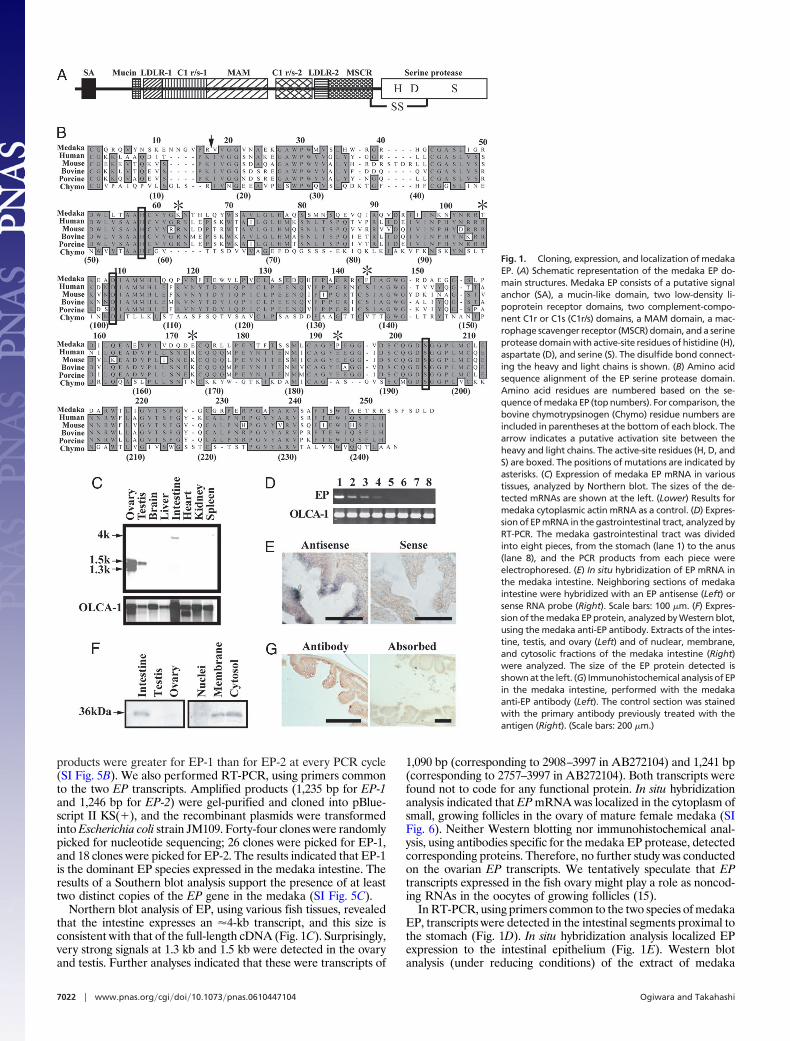
Northern blot analysis of EP, using various fish tissues, revealedthat the intestine expresses an �4-kb transcript, and this size isconsistent with that of the full-length cDNA (Fig. 1C). Surprisingly,very strong signals at 1.3 kb and 1.5 kb were detected in the ovaryand testis. Further analyses indicated that these were transcripts of
1,090 bp (corresponding to 2908–3997 in AB272104) and 1,241 bp(corresponding to 2757–3997 in AB272104). Both transcripts werefound not to code for any functional protein. In situ hybridizationanalysis indicated that EP mRNA was localized in the cytoplasm ofsmall, growing follicles in the ovary of mature female medaka (SIFig. 6). Neither Western blotting nor immunohistochemical anal-ysis, using antibodies specific for the medaka EP protease, detectedcorresponding proteins. Therefore, no further study was conductedon the ovarian EP transcripts. We tentatively speculate that EPtranscripts expressed in the fish ovary might play a role as noncod-ing RNAs in the oocytes of growing follicles (15).
In RT-PCR, using primers common to the two species of medakaEP, transcripts were detected in the intestinal segments proximal tothe stomach (Fig. 1D). In situ hybridization analysis localized EPexpression to the intestinal epithelium (Fig. 1E). Western blotanalysis (under reducing conditions) of the extract of medaka
Fig. 1. Cloning, expression, and localization of medakaEP. (A) Schematic representation of the medaka EP do-main structures. Medaka EP consists of a putative signalanchor (SA), a mucin-like domain, two low-density li-poprotein receptor domains, two complement-compo-nent C1r or C1s (C1r/s) domains, a MAM domain, a mac-rophage scavenger receptor (MSCR) domain, and a serineprotease domain with active-site residues of histidine (H),aspartate (D), and serine (S). The disulfide bond connect-ing the heavy and light chains is shown. (B) Amino acidsequence alignment of the EP serine protease domain.Amino acid residues are numbered based on the se-quence of medaka EP (top numbers). For comparison, thebovine chymotrypsinogen (Chymo) residue numbers areincluded in parentheses at the bottom of each block. Thearrow indicates a putative activation site between theheavy and light chains. The active-site residues (H, D, andS) are boxed. The positions of mutations are indicated byasterisks. (C) Expression of medaka EP mRNA in varioustissues, analyzed by Northern blot. The sizes of the de-tected mRNAs are shown at the left. (Lower) Results formedaka cytoplasmic actin mRNA as a control. (D) Expres-sion of EP mRNA in the gastrointestinal tract, analyzed byRT-PCR. The medaka gastrointestinal tract was dividedinto eight pieces, from the stomach (lane 1) to the anus(lane 8), and the PCR products from each piece wereelectrophoresed. (E) In situ hybridization of EP mRNA inthe medaka intestine. Neighboring sections of medakaintestine were hybridized with an EP antisense (Left) orsense RNA probe (Right). Scale bars: 100 �m. (F) Expres-sion of the medaka EP protein, analyzed by Western blot,using the medaka anti-EP antibody. Extracts of the intes-tine, testis, and ovary (Left) and of nuclear, membrane,and cytosolic fractions of the medaka intestine (Right)were analyzed. The size of the EP protein detected isshown at the left. (G) Immunohistochemical analysis of EPin the medaka intestine, performed with the medakaanti-EP antibody (Left). The control section was stainedwith the primary antibody previously treated with theantigen (Right). (Scale bars: 200 �m.)
7022 � www.pnas.org�cgi�doi�10.1073�pnas.0610447104 Ogiwara and Takahashi
intestine but not ovary and testis extract, using specific anti-EPantibodies against the catalytic domain, detected a 36-kDa immu-noreactive band (Fig. 1F Left). A polypeptide band of the samemolecular mass was detected in both cytosolic and membranefractions of the intestine (Fig. 1F Right). Western blotting of theintestine extract under nonreducing conditions gave no clear band(data not shown). Immunohistochemical analysis, using the anti-body, demonstrated the epithelial localization of EP in the intestine(Fig. 1G). The extract of medaka intestine exhibited enzyme activityfor the synthetic EP substrate Gly-Asp-Asp-Asp-Asp-Lys-�-naphthylamide [GD4K-�-naphthyamide (�NA)]. Using this activityas a marker, the apparent molecular mass of intact EP wasestimated by gel filtration to be 440 kDa (SI Fig. 7A). The fractionhaving GD4K-�NA-hydrolyzing activity showed a 36-kDa polypep-tide in Western blotting under reducing conditions (SI Fig. 7B Left).Again, the same fraction did not show any clear band with thecurrent antibody when analyzed under nonreducing conditions (SIFig. 7B Right). Taken together, these results indicate that the fishintestine contains active, membrane-bound EP. Part of the mole-cule exists in the intestine in a soluble form that is probablydetached from the epithelial cell membrane. The adult medakaintestinal epithelium contains most of the cell types (enterocytes,goblet cells, and enteroendocrine cells) observed in the smallintestine of other vertebrates, but lacks crypts containing Panethcells and intestinal stem cells (16). Our current data suggest thatmedaka EP is localized in the enterocytes in the proximal intestinalepithelium.
The Medaka EP Catalytic Serine Protease Domain Has Distinct Prop-erties as Compared with Its Mammalian Counterparts. The active32-kDa C-terminal serine protease domains of both EP-1 and EP-2were prepared to characterize their enzymatic properties (SI Fig.8A). Both enzymes showed maximal activities for GD4K-�NA atpH 8, but EP-1 was approximately three times more active thanEP-2 (SI Fig. 8B). To examine the effects of EP-1 and EP-2 on thephysiological substrate trypsinogen, a 866-bp medaka trypsinogencDNA (AB272106), which codes for a protein of 242 aa (SI Fig. 9),was obtained from the intestine. Using the sequence, a recombinantfusion protein of medaka trypsinogen was prepared. The trypsino-gen was converted to active trypsin by EP-1 faster than by EP-2 (SIFig. 8C). The behavior of the two proteases in response to variousprotease inhibitors was undistinguishable (SI Table 2). From theseresults, together with the finding that EP-1 is the dominantlyexpressed form in the intestine, we selected EP-1 for the followingexperiments.
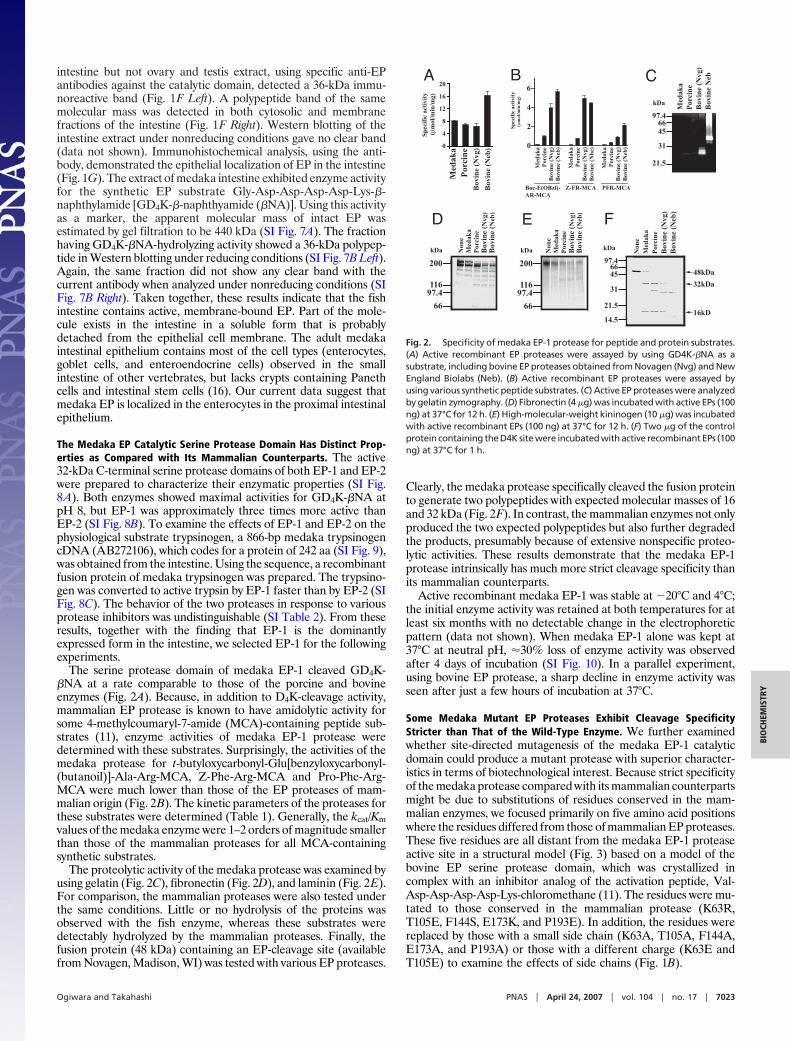
The serine protease domain of medaka EP-1 cleaved GD4K-�NA at a rate comparable to those of the porcine and bovineenzymes (Fig. 2A). Because, in addition to D4K-cleavage activity,mammalian EP protease is known to have amidolytic activity forsome 4-methylcoumaryl-7-amide (MCA)-containing peptide sub-strates (11), enzyme activities of medaka EP-1 protease weredetermined with these substrates. Surprisingly, the activities of themedaka protease for t-butyloxycarbonyl-Glu[benzyloxycarbonyl-(butanoil)]-Ala-Arg-MCA, Z-Phe-Arg-MCA and Pro-Phe-Arg-MCA were much lower than those of the EP proteases of mam-malian origin (Fig. 2B). The kinetic parameters of the proteases forthese substrates were determined (Table 1). Generally, the kcat/Kmvalues of the medaka enzyme were 1–2 orders of magnitude smallerthan those of the mammalian proteases for all MCA-containingsynthetic substrates.
The proteolytic activity of the medaka protease was examined byusing gelatin (Fig. 2C), fibronectin (Fig. 2D), and laminin (Fig. 2E).For comparison, the mammalian proteases were also tested underthe same conditions. Little or no hydrolysis of the proteins wasobserved with the fish enzyme, whereas these substrates weredetectably hydrolyzed by the mammalian proteases. Finally, thefusion protein (48 kDa) containing an EP-cleavage site (availablefrom Novagen, Madison, WI) was tested with various EP proteases.
Clearly, the medaka protease specifically cleaved the fusion proteinto generate two polypeptides with expected molecular masses of 16and 32 kDa (Fig. 2F). In contrast, the mammalian enzymes not onlyproduced the two expected polypeptides but also further degradedthe products, presumably because of extensive nonspecific proteo-lytic activities. These results demonstrate that the medaka EP-1protease intrinsically has much more strict cleavage specificity thanits mammalian counterparts.
Active recombinant medaka EP-1 was stable at �20°C and 4°C;the initial enzyme activity was retained at both temperatures for atleast six months with no detectable change in the electrophoreticpattern (data not shown). When medaka EP-1 alone was kept at37°C at neutral pH, �30% loss of enzyme activity was observedafter 4 days of incubation (SI Fig. 10). In a parallel experiment,using bovine EP protease, a sharp decline in enzyme activity wasseen after just a few hours of incubation at 37°C.
Some Medaka Mutant EP Proteases Exhibit Cleavage SpecificityStricter than That of the Wild-Type Enzyme. We further examinedwhether site-directed mutagenesis of the medaka EP-1 catalyticdomain could produce a mutant protease with superior character-istics in terms of biotechnological interest. Because strict specificityof the medaka protease compared with its mammalian counterpartsmight be due to substitutions of residues conserved in the mam-malian enzymes, we focused primarily on five amino acid positionswhere the residues differed from those of mammalian EP proteases.These five residues are all distant from the medaka EP-1 proteaseactive site in a structural model (Fig. 3) based on a model of thebovine EP serine protease domain, which was crystallized incomplex with an inhibitor analog of the activation peptide, Val-Asp-Asp-Asp-Asp-Lys-chloromethane (11). The residues were mu-tated to those conserved in the mammalian protease (K63R,T105E, F144S, E173K, and P193E). In addition, the residues werereplaced by those with a small side chain (K63A, T105A, F144A,E173A, and P193A) or those with a different charge (K63E andT105E) to examine the effects of side chains (Fig. 1B).
Fig. 2. Specificity of medaka EP-1 protease for peptide and protein substrates.(A) Active recombinant EP proteases were assayed by using GD4K-�NA as asubstrate, including bovine EP proteases obtained from Novagen (Nvg) and NewEngland Biolabs (Neb). (B) Active recombinant EP proteases were assayed byusing various synthetic peptide substrates. (C) Active EP proteases were analyzedby gelatin zymography. (D) Fibronectin (4 �g) was incubated with active EPs (100ng) at 37°C for 12 h. (E) High-molecular-weight kininogen (10 �g) was incubatedwith active recombinant EPs (100 ng) at 37°C for 12 h. (F) Two �g of the controlprotein containing the D4K site were incubated with active recombinant EPs (100ng) at 37°C for 1 h.
Ogiwara and Takahashi PNAS � April 24, 2007 � vol. 104 � no. 17 � 7023
BIO
CHEM
ISTR
Y
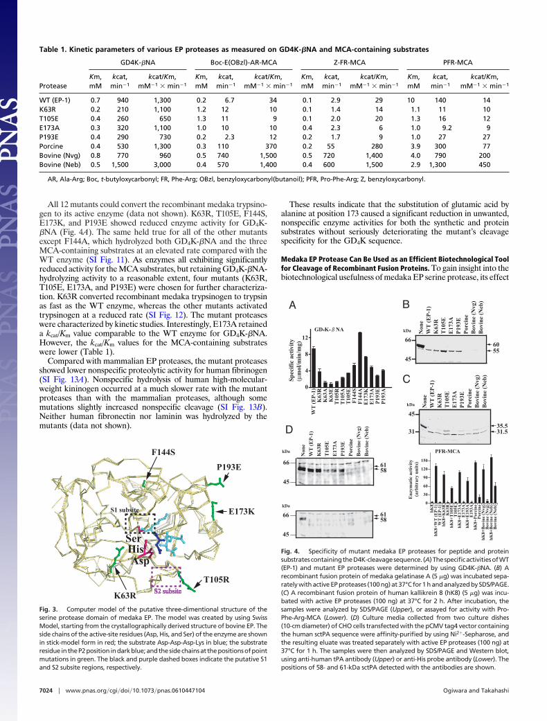
All 12 mutants could convert the recombinant medaka trypsino-gen to its active enzyme (data not shown). K63R, T105E, F144S,E173K, and P193E showed reduced enzyme activity for GD4K-�NA (Fig. 4A). The same held true for all of the other mutantsexcept F144A, which hydrolyzed both GD4K-�NA and the threeMCA-containing substrates at an elevated rate compared with theWT enzyme (SI Fig. 11). As enzymes all exhibiting significantlyreduced activity for the MCA substrates, but retaining GD4K-�NA-hydrolyzing activity to a reasonable extent, four mutants (K63R,T105E, E173A, and P193E) were chosen for further characteriza-tion. K63R converted recombinant medaka trypsinogen to trypsinas fast as the WT enzyme, whereas the other mutants activatedtrypsinogen at a reduced rate (SI Fig. 12). The mutant proteaseswere characterized by kinetic studies. Interestingly, E173A retaineda kcat/Km value comparable to the WT enzyme for GD4K-�NA.However, the kcat/Km values for the MCA-containing substrateswere lower (Table 1).
Compared with mammalian EP proteases, the mutant proteasesshowed lower nonspecific proteolytic activity for human fibrinogen(SI Fig. 13A). Nonspecific hydrolysis of human high-molecular-weight kininogen occurred at a much slower rate with the mutantproteases than with the mammalian proteases, although somemutations slightly increased nonspecific cleavage (SI Fig. 13B).Neither human fibronectin nor laminin was hydrolyzed by themutants (data not shown).
These results indicate that the substitution of glutamic acid byalanine at position 173 caused a significant reduction in unwanted,nonspecific enzyme activities for both the synthetic and proteinsubstrates without seriously deteriorating the mutant’s cleavagespecificity for the GD4K sequence.
Medaka EP Protease Can Be Used as an Efficient Biotechnological Toolfor Cleavage of Recombinant Fusion Proteins. To gain insight into thebiotechnological usefulness of medaka EP serine protease, its effect
Table 1. Kinetic parameters of various EP proteases as measured on GD4K-�NA and MCA-containing substrates
Protease
GD4K-�NA Boc-E(OBzl)-AR-MCA Z-FR-MCA PFR-MCA
Km,mM
kcat,min�1
kcat/Km,mM�1 � min�1
Km,mM
kcat,min�1
kcat/Km,mM�1 � min�1
Km,mM
kcat,min�1
kcat/Km,mM�1 � min�1
Km,mM
kcat,min�1
kcat/Km,mM�1 � min�1
WT (EP-1) 0.7 940 1,300 0.2 6.7 34 0.1 2.9 29 10 140 14K63R 0.2 210 1,100 1.2 12 10 0.1 1.4 14 1.1 11 10T105E 0.4 260 650 1.3 11 9 0.1 2.0 20 1.3 16 12E173A 0.3 320 1,100 1.0 10 10 0.4 2.3 6 1.0 9.2 9P193E 0.4 290 730 0.2 2.3 12 0.2 1.7 9 1.0 27 27Porcine 0.4 530 1,300 0.3 110 370 0.2 55 280 3.9 300 77Bovine (Nvg) 0.8 770 960 0.5 740 1,500 0.5 720 1,400 4.0 790 200Bovine (Neb) 0.5 1,500 3,000 0.4 570 1,400 0.4 600 1,500 2.9 1,300 450
AR, Ala-Arg; Boc, t-butyloxycarbonyl; FR, Phe-Arg; OBzl, benzyloxycarbonyl(butanoil); PFR, Pro-Phe-Arg; Z, benzyloxycarbonyl.
Fig. 3. Computer model of the putative three-dimentional structure of theserine protease domain of medaka EP. The model was created by using SwissModel, starting from the crystallographically derived structure of bovine EP. Theside chains of the active-site residues (Asp, His, and Ser) of the enzyme are shownin stick-model form in red; the substrate Asp-Asp-Asp-Lys in blue; the substrateresidue intheP2position indarkblue;andthesidechainsat thepositionsofpointmutations in green. The black and purple dashed boxes indicate the putative S1and S2 subsite regions, respectively.
Fig. 4. Specificity of mutant medaka EP proteases for peptide and proteinsubstratescontainingtheD4K-cleavagesequence. (A)ThespecificactivitiesofWT(EP-1) and mutant EP proteases were determined by using GD4K-�NA. (B) Arecombinant fusion protein of medaka gelatinase A (5 �g) was incubated sepa-rately with active EP proteases (100 ng) at 37°C for 1 h and analyzed by SDS/PAGE.(C) A recombinant fusion protein of human kallikrein 8 (hK8) (5 �g) was incu-bated with active EP proteases (100 ng) at 37°C for 2 h. After incubation, thesamples were analyzed by SDS/PAGE (Upper), or assayed for activity with Pro-Phe-Arg-MCA (Lower). (D) Culture media collected from two culture dishes(10-cm diameter) of CHO cells transfected with the pCMV tag4 vector containingthe human sctPA sequence were affinity-purified by using Ni2�-Sepharose, andthe resulting eluate was treated separately with active EP proteases (100 ng) at37°C for 1 h. The samples were then analyzed by SDS/PAGE and Western blot,using anti-human tPA antibody (Upper) or anti-His probe antibody (Lower). Thepositions of 58- and 61-kDa sctPA detected with the antibodies are shown.
7024 � www.pnas.org�cgi�doi�10.1073�pnas.0610447104 Ogiwara and Takahashi
on various fusion proteins containing a D4K-cleavage site wasexamined. Medaka gelatinase A (17) was synthesized as a fusionprotein containing a His tag and D4K sequence at the N terminusin the E. coli expression system, using the pET30 expression vector.Both the WT and mutant proteases converted the 60-kDa fusionprotein to a 55-kDa protein (Fig. 4B). At the same substrate/protease ratio, mammalian EP serine proteases extensively digestedthe fusion protein.
Next, we synthesized a 35.5-kDa protein of human kallikrein 8(hK8) in the same E. coli expression system. Digestion with all EPproteases commonly generated 31.5-kDa active hK8 by cleaving theD4K sequence (Fig. 4C Upper). The EP protease-treated sampleswere directly assayed for hK8 activity by using Pro-Phe-Arg-MCA,a good synthetic peptide substrate for hK8 (18). All of the samplestreated with the medaka or mammalian EP proteases exhibitedPro-Phe-Arg-MCA-hydrolyzing activity (Fig. 4C Lower). As ex-pected, none of the medaka EP proteases (WT EP-1, K63R, T105E,E173A, or E193A) showed any significant enzyme activity. Incontrast, considerable enzyme activities were detected with porcineand bovine (Neb) EP proteases. Fusion protein that had beendigested with the bovine (Nvg) protease had very low activity,presumably because of inactivation of the EP protease itself duringincubation. These results demonstrate that the medaka EP proteaseused for cleaving the fusion protein has no serious effect on hK8activity.
Finally, a human single-chain tissue-type plasminogen activator(sctPA) fusion protein containing an 11-residue sequence of aHis-tag/EP-susceptible site at the N terminus of mature tPA wasgenerated by CHO cells and used as a substrate for medaka andmammalian EP proteases. The protein samples treated with themedaka WT or mutant EP proteases, but not with mammalianones, showed two polypeptides (58 and 61 kDa) detectable withanti-human tPA antibodies (Fig. 4D Upper). The specific antibodyfor the His-tag sequence did not recognize the polypeptides (Fig.4D Lower). These results indicate that the medaka proteasesproperly cleave the fusion protein at the EP-cleavage site toproduce sctPA, and also demonstrate that mammalian EP pro-teases extensively degrade the sctPA fusion protein.
Taken together, our results suggest that the medaka proteases aremore effective than their mammalian counterparts as fusion-protein cleavage enzymes for the preparation of desired recombi-nant proteins containing the D4K cleavage sequence.
DiscussionAlmost all trypsinogen sequences known to date contain a highlyconserved tetra-aspartate sequence preceding the lysine-isoleucine scissile peptide bond. Although EP is widely consid-ered to play a role in trypsinogen activation in all vertebratespecies, there has been no report on EP from nonmammalianspecies. The present study is thus the first to report on themolecular and biochemical characterizations of EP from a lowervertebrate, the medaka.
Medaka EP mRNA exists in two distinct forms, EP-1 and EP-2,in the intestine. A comparison of the entire amino acid sequencesof EP-1 (1,036 residues) and EP-2 (1,043 residues) revealed adifference of only 22 aa, including an insertion of seven residues inEP-2. Our data suggest that EP-1 and EP-2 mRNA are expressedat a ratio of �3:2 in the intestine. It remains to be determinedwhether they are indeed translated at this ratio. Moreover, it is notknown at present whether they have a discrete role in vivo.
To our surprise, medaka EP serine protease per se showed verylow activity for three synthetic peptide substrates, t-butyloxycar-bonyl-Glu[benzyloxycarbonyl(butanoil)]-Ala-Arg-MCA, Z-Phe-Arg-MCA, and Pro-Phe-Arg-MCA, that are rapidly hydrolyzed bythe mammalian enzymes. A comparison of the kinetic parametersbetween medaka and mammalian EP proteases revealed that theremarkably low activities of the fish enzyme for these substrateswere due to the reduced kcat value. This finding led us to examine
whether site-directed mutagenesis could produce a mutant enzymewith further reduced amidolytic activity for the synthetic substrates.
Twelve mutant medaka EP proteases were generated by mu-tagenesis at positions Lys-63 [corresponding to Arg60f in bovine EPprotease (11)], Thr-105 (bovine Arg-98), Phe-144 (bovine Ser-137),Glu-173 (bovine Lys-167), and Pro-193 (bovine Glu-185). Thesepositions all occur on the surface of the medaka EP serine proteasemodel (Fig. 3), and, with the exception of medaka EP proteaseposition 105 (bovine 98), the residues are located a considerabledistance from the enzyme active site. A previous crystalline struc-tural study (11) and other biochemical studies (10, 11) on mam-malian EP protease indicate that the minimal recognition sequencefor EP consists of Lys at P1 and Asp at P2 of the D4K sequence.Therefore, the five residues we targeted are probably not essentialfor substrate recognition. As is clear from the medaka EP proteasemodel, the structure around the S1 site highly resembles that ofbovine EP protease, indicating that the catalytic mechanism andcontacts with Lys at P1 are conserved with mammalian EP pro-teases. The interaction of Asp at P2 with the side chain of Lys-106(bovine Lys-99) at S2 is also conserved, because GD4K-�NA ishydrolyzed at a comparable rate by the medaka and bovineenzymes. However, the microenvironment around the S2 site ispresumed to be different between medaka and mammalian EPproteases (Fig. 3). A slight difference in the structure around the S2site might underlie the detectable difference in catalytic activitiesfor MCA-containing peptide substrates. We indeed observed thatthe T105E mutant displayed significantly lower catalytic activity forGD4K-�NA. A plausible explanation for this activity decrease isthat replacement of Thr by Glu at position 105, which is next toLys-106 at the S2 site, could affect interaction of Asp at P2 withLys-106 (bovine Lys-99) at S2.
To examine the effects of side chains of the target residues oncatalytic activity, we replaced these by Ala. Mutagenesis haddifferent effects on each of the enzyme activities. One of themutants, E173A, was interesting in that it showed significantly loweractivities than the WT enzyme toward all synthetic substratestested. In addition, this mutant enzyme retained a low nonspecificproteolytic activity for protein substrates (high-molecular-weightkininogen and fibrinogen), with no serious reduction in D4K-cleaving activity for fusion proteins (gelatinase A, hK8, and tPA).Because none of the targeted residues is essential for the EPprotease catalytic activity, changes in the activity by mutagenesis arepresumably because of conformational change brought about bysubstitution of residues at regions distant from the active site of theenzyme.
We note here that, as demonstrated with bovine EP (9), activityof the enzyme for D4K-containing substrates depends the heavychain. The presence of the heavy chain had almost no effect onrecognition of the small peptide substrate GD4K-�NA, whereas thischain had profound effects on recognition of the macromolecularsubstrate trypsinogen (9). Furthermore, bovine EP is able tohydrolyze specifically several biologically active peptides in vitro(19), and substrate length greatly affects the catalytic efficiency ofthis hydrolysis. Activity dependency on the heavy chain and peptidesubstrate length remain to be determined for medaka EP.
Because EP serine proteases preferably hydrolyze the D4Ksequence, this motif has been intentionally introduced for thespecific cleavage of fusion proteins. Bovine EP serine protease isnow widely used for this purpose. The current system, using thebovine enzyme, works reasonably well in many cases but requireshandling with great care. We often encounter the following diffi-culties. (i) Bovine EP protease primarily cleaves at the EP-cleavagesite of recombinant fusion proteins; however, other peptide bondsare also hydrolyzed to a considerable degree by nonspecific pro-teolytic activity. This results in a low yield of the protein in question.(ii) For preparing active recombinant proteases, the bovine EPprotease used for cleavage of the inactive fusion protein could bean obstacle. This is particularly serious when the proteases to be
Ogiwara and Takahashi PNAS � April 24, 2007 � vol. 104 � no. 17 � 7025
BIO
CHEM
ISTR
Y
examined are ones with very low activity for synthetic and proteinsubstrates. Significant nonspecific activities of bovine EP proteaseoften make it difficult to determine whether the target recombinantproteases have been successfully activated.
To address this latter difficulty, we examined the enzyme activ-ities of medaka EP protease for synthetic MCA-containing sub-strates, which are known to be nonspecifically hydrolyzed by themammalian counterparts, including bovine EP (11), although thesynthetic substrates themselves are not physiologically relevant. Asdemonstrated in the present study, the serine protease domain ofmedaka EP has a stricter specificity for almost all of the substratestested compared with the mammalian EP proteases. The disadvan-tages mentioned above associated with bovine EP could be over-come by using the medaka protease. Medaka WT EP proteasewould be adequate for the preparation of recombinant nonproteo-lytic enzymes. However, in view of the efficient cleavage at the D4Ksite and the minimum nonspecific hydrolysis at peptide and amidebonds, we recommend use of the E173A mutant enzyme. MedakaWT EP protease and its mutant can be prepared in large quantityin the E. coli expression system. We believe that, using the medakaEP serine proteases as cleavage enzymes for fusion proteins con-taining the D4K cleavage sequence, the desired recombinant pro-teins can be easily and effectively produced.
Materials and MethodsPreparation of the Recombinant EP Serine Protease Domain. Twodistinct medaka EP cDNA clones, EP-1 (3,997 bp, AB272104) andEP-2 (4,036 bp, AB272105), were obtained as described in SIMaterials and Methods.
A DNA fragment, including the coding sequence for the medakaEP-1 or EP-2 catalytic domain, was amplified by PCR, using apBluescript II plasmid containing cDNA of the catalytic domain asthe template. The upper and lower primers were 5�-CGCGGATC-CCAAGCTGGTGTGGTGGGTGG-3� and 5�-CCCAAGCTT-TCAGTCTAGATCTGAGAA-3�, respectively, which haveBamHI and HindIII sites at the respective 5� termini. The productwas ligated into the cloning site of a pET30a expression vector(Novagen). Expression of the recombinant medaka EP catalyticdomain in the E. coli expression system was carried out as describedin ref. 17. The medaka EP catalytic domain was produced as afusion protein with an extra amino acid sequence of 50 residues atits N terminus; the vector-derived N-terminal stretch contained aHis tag and an S-protein sequence. Harvested cells were lysed, andthe insoluble materials were dissolved in a solubilization buffercontaining 6 M urea, 50 mM Tris�HCl (pH 7.6), and 0.5 M NaCl.Solubilized proteins were subjected to affinity chromatography onNi2�-Sepharose (GE Healthcare Biosciences, Piscataway, NJ), andeluted with the same buffer, containing 50 mM histidine. Elutedrecombinant proteins were renatured by dialysis against 50 mMTris�HCl (pH 8.0). The fusion protein was then incubated in 50 mMTris�HCl (pH 8.0) containing 0.5 M NaCl with trypsin immobilizedon Sepharose 4B at room temperature for 1 h. The immobilizedtrypsin was then removed by filtration. The resulting sample, whichcontained not only active EP protease but also inactive enzymeprotein, was fractionated on a column of Resource Q in AKTA
Purifier (GE Healthcare Biosciences) to remove inactive enzyme.A trace amount of trypsin often contained in the sample thusprepared was removed by passage through an aprotinin-Sepharose4B column (Sigma, St. Louis, MO).
Active recombinant enzyme of the porcine EP serine proteasedomain (Ile-800 to His-1034) (6) was prepared basically accordingto the method described above. Bovine EP serine protease wasobtained from Novagen and New England Biolabs (Schwalbach,Germany).
Site-Directed Mutagenesis. Site-directed mutagenesis of medakaEP-1 was carried out to produce various mutant proteases. For eachmutant, two PCR products were first amplified with medaka EP-1cDNA as a template, using the following two primer combinations:the upper primer described above with the respective antisenseprimer (SI Table 3) and the lower primer described above with thesense primer (SI Table 3). Using a mixture of these amplified DNAsas the template, a second PCR was performed with the upper andlower primers. The PCR products were digested with BamHI andHindIII, gel-purified, and ligated into the pET30a expressionvector. All mutants were confirmed by DNA sequencing. Thesubsequent procedures for preparation of mutant proteases werethe same as for the WT protein described above.
Hydrolysis of Proteins by the EP Catalytic Serine Protease Domain.Human plasma fibronectin (Chemicon, Temecula, CA), humanfibrinogen (Merk Biosciences, Tokyo, Japan), human high-molecular-weight kininogen (Calbiochem, La Jolla, CA), mouselaminin (Biomedical Technologies, Stoughton, MA), D4K cleavagesite-containing control protein (Novagen), medaka gelatinase A(17), trypsinogen (this study), human kallikrein 8 (hK8) (18), andhuman tPA were incubated at 37°C in 20 mM Tris�HCl buffer (pH7.4) containing 50 mM NaCl and 2 mM CaCl2 with various EPserine proteases at ratios (wt/wt) ranging from 20:1 to 100:1. Afterincubation, samples were subjected to SDS/PAGE, followed byCoomassie brilliant blue staining. Gelatin zymography was con-ducted as described in ref. 17, except that gels were incubated in 20mM Tris�HCl buffer (pH 7.4) containing 50 mM NaCl and 2 mMCaCl2.
Computer Modeling of the Serine Protease Domain of Medaka EP.Three-dimensional structure prediction was carried out by usingSwiss Model. The serine protease domain of medaka EP wasmodeled in the ‘‘optimize’’ mode after manual alignment of thesequence with bovine EP.
The methods used for the cloning of medaka EP-1 and EP-2 andtrypsinogen, preparations of recombinant proteins other than EP,RT-PCR, Northern blotting, Southern blotting, in situ hybridiza-tion, antibody production, Western blotting, immunohistochemis-try, gel-filtration chromatography, and other enzymic methods aredescribed in SI Materials and Methods.
We thank Dr. M. Yao and I. Tanaka (Hokkaido University) for valuablecomments on the structure of EP. This work was supported in part byMinistry of Education, Culture, Sports, Science and Technology of JapanGrant-in-Aid for Scientific Research 17370021 (to T.T.).
1. Light A, Janska H (1989) Trends Biochem Sci 14:110–112.2. Grishan FK, Lee PC, Lebenthal E, Johnson P, Bradley CA, Greene HL (1983) Gastroenterology
85:727–731.3. LaVallie ER, Rehemtulla A, Racie LA, DiBlasio EA, Ferenz C, Grant KL, Light A, McCoy JM
(1993) J Biol Chem 268:23311–23317.4. Kitamoto Y, Yuan X, Wu Q, McCourt DW, Sadler JE (1994) Proc Natl Acad Sci USA
91:7588–7592.5. Kitamoto Y, Veile RA, Donis-Keller H, Sadler JE (1995) Biochemistry 34:4562–4568.6. Matsushima M, Ichinose M, Yahagi N, Kakei K, Tsukada S, Miki K, Kurokawa K, Tashiro K,
Shiokawa K, Shinomiya K, et al. (1994) J Biol Chem 269:19976–19982.7. Yahagi N, Ichinose M, Matsushima M, Matsubara Y, Miki K, Kurokawa K, Fukamachi H,
Tashiro K, Shiokawa K, Kageyama T, et al. (1996) Biochem Biophys Res Commun 219:806–812.8. Yuan X, Zheng X, Lu D, Rubin DC, Pung CY, Sadler JE (1998) Am J Physiol 274:342–349.9. Lu D, Yuan, X., Zheng. X. & Sadler JE (1997) J Biol Chem 272:31293–31300.
10. Mikhailova AG, Rumsh LD (1999) FEBS Lett 442:226–230.11. Lu D, Futterer K, Korolev S, Xinglong Z, Tan K, Waksman G, Sadler JE (1999) J Mol Biol
292:361–373.12. Zheng X, Sadler JE (2002) J Biol Chem 277:6858–6863.13. Collins-Racie LA, McColgan JM, Grant KL, DiBlasio-Smith EA, McCoy JM, LaVallie ER
(1995) Biotechnology 13:982–987.14. Bricteux-Gregoire S, Schyns R, Florkin M (1972) Comp Biochem Physiol 42B:23–39.15. Costa FF (2005) Gene 357:83–94.16. Rombout JH, Stroband HW, Taverne-Thiele JJ (1984) Cell Tissue Res 236:207–216.17. Ogiwara K, Takano N, Shinohara M, Murakami M, Takahashi T (2005) Proc Natl Acad Sci USA
102:8442–8447.18. Rajapakse S, Ogiwara K, Takano N, Moriyama A, Takahashi T (2005) FEBS Lett 579:6879–6884.19. Likhareva VV, Mikhailova AG, Vaskovsky BV, Garanin SK, Rumsh LD (2002) Lett Peptide Sci
9:71–76.
7026 � www.pnas.org�cgi�doi�10.1073�pnas.0610447104 Ogiwara and Takahashi