spinal injuries
TRANSCRIPT

Spinal injuriesSpinal injuries
GEORGE SAPKASGEORGE SAPKAS
11stst Orthopaedic Department Orthopaedic DepartmentAthens UniversityAthens UniversityAtticon HospitalAtticon Hospital

5% -10% of 5% -10% of unconscious patients unconscious patients who present to the who present to the Emergency Dept as Emergency Dept as the result of a M.V.A. the result of a M.V.A. or fall, have a major or fall, have a major injury to the Cervical injury to the Cervical SpineSpine

Spinal cord injury occurs in more Spinal cord injury occurs in more than 11.000/USA pts per year/USA than 11.000/USA pts per year/USA or in 40- 50 persons per millionor in 40- 50 persons per million
Injuries of the Cervical Spine Injuries of the Cervical Spine produce neurological damage in produce neurological damage in approximately 40% of patientsapproximately 40% of patients

1/3 of cervical injuries occur at the 1/3 of cervical injuries occur at the Levels CLevels C11 to C to C22
1/2 of cervical injuries occur at the 1/2 of cervical injuries occur at the levels Clevels C55 to C to C77

Most fatal cervical spine Most fatal cervical spine injuries occur in upper cervical injuries occur in upper cervical levels, either at cranio-cervical levels, either at cranio-cervical junction or at Cjunction or at C11 - C - C2 2 level.level.

Injuries of the sub Injuries of the sub --axial cervical spine axial cervical spine ( C( C33--77) are among ) are among the most common the most common and potentially and potentially most devastating most devastating injuries involving injuries involving the axial skeletonthe axial skeleton

Approximately Approximately 10% of traumatic 10% of traumatic cord injuries have cord injuries have no obvious no obvious roentgenroentgenοοgraphic graphic evidence of evidence of vertebral injuries vertebral injuries

Arterial supplyArterial supply
1.1. Anterior spinal artery (in central sulcus)Anterior spinal artery (in central sulcus)
2.2. Posterior spinal arteries (2, Posterior spinal arteries (2, posterolateral)posterolateral)
3.3. Vertebral arteryVertebral arterya.a. In foramen transversarium CIn foramen transversarium C66 C C22
b.b. Crosses posterolateral arch of CCrosses posterolateral arch of C11 (5cm (5cm from mid-line)from mid-line)


Cord Cord 1.1. Central –grey matter –cellsCentral –grey matter –cells
a.a. Anterior = 1Anterior = 1oo motor motorb.b. Posterior – 1Posterior – 1oo sensory sensory
2.2. Peripheral – tractsPeripheral – tractsa.a. Lateral spinothalamicLateral spinothalamic
i.i. Pain and temperaturePain and temperatureii.ii. Antero-lateral Antero-lateral
b.b. Lateral corticospinalLateral corticospinali.i. MotorMotorii.ii. Mid lateralMid lateral
c.c. Posterior columnsPosterior columnsi.i. Proprioception, deep pressure, Proprioception, deep pressure,
vibrationvibrationii.ii. Posterior 1/3 of cordPosterior 1/3 of cord
d.d. Orientation (medial Orientation (medial peripheral) peripheral)i.i. Arms, thoracic, legs, sarcal Arms, thoracic, legs, sarcal ii.ii. Medial portion of tracts = most Medial portion of tracts = most
proximal function (arms)proximal function (arms)iii.iii. Peripheral portion of tracts = most Peripheral portion of tracts = most
distal functiondistal function

Roots Roots 1.1. CC11- exits above C- exits above C11, body , body
2.2. CC22 a.a. Greater occipitalGreater occipital
b.b. Exits between CExits between C11 and C and C22
3.3. CC33 exits between C exits between C22 and and CC33
4.4. CC88 exits between C exits between C77 and and TT11
5.5. TT11 exits between T exits between T11 and and TT22

Pathophysiology of spinal cord injuryPathophysiology of spinal cord injury
Tator, 1996; Fehlings, 1999; Slucky, 1999; Giraldi, 1999; Ramer 2000

Primary injuryPrimary injury
Initial contusion and compression of cordInitial contusion and compression of cord Damage to:Damage to:
– neuronal cells, neuronal cells,
– axonal membranesaxonal membranes– blood vesselsblood vessels

Secondary injurySecondary injury An auto-destructive cascade of biomechanical An auto-destructive cascade of biomechanical
events lasting hours to days that expands the events lasting hours to days that expands the zone of injuryzone of injury
Independent or synergistic mechanisms Independent or synergistic mechanisms – Vascular Vascular
Reduced blood flowReduced blood flow Loss of autoregulationLoss of autoregulation Neurogenic shockNeurogenic shock HemorrhageHemorrhage Loss of micro-circulationLoss of micro-circulation VasospasmVasospasm thrombosisthrombosis
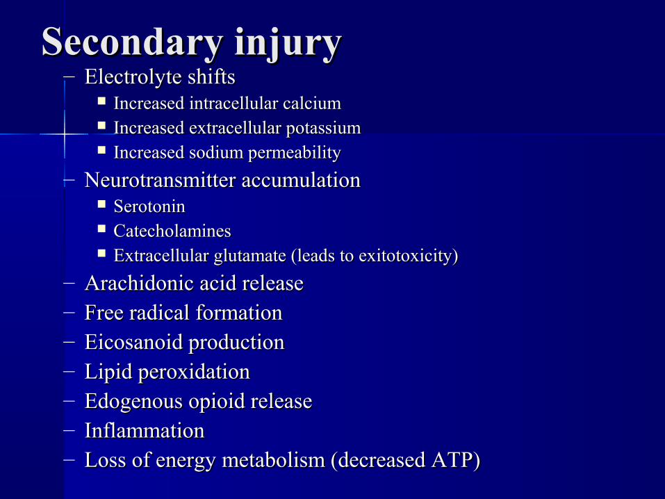
Secondary injurySecondary injury– Electrolyte shiftsElectrolyte shifts
Increased intracellular calciumIncreased intracellular calcium Increased extracellular potassiumIncreased extracellular potassium Increased sodium permeabilityIncreased sodium permeability
– Neurotransmitter accumulationNeurotransmitter accumulation SerotoninSerotonin CatecholaminesCatecholamines Extracellular glutamate (leads to exitotoxicity)Extracellular glutamate (leads to exitotoxicity)
– Arachidonic acid releaseArachidonic acid release– Free radical formationFree radical formation– Eicosanoid productionEicosanoid production– Lipid peroxidationLipid peroxidation– Edogenous opioid releaseEdogenous opioid release– InflammationInflammation– Loss of energy metabolism (decreased ATP)Loss of energy metabolism (decreased ATP)

Cord syndromesCord syndromes

Anterior cordAnterior cord– Anterior 2/3 of cord “not” Anterior 2/3 of cord “not”
functioning (anterior spinal functioning (anterior spinal artery)artery)
– No motorNo motor– No sharp/dull, hot/cold No sharp/dull, hot/cold
discriminationdiscrimination– Posterior columns intact: Posterior columns intact:
deep pain, prop.deep pain, prop.– Mechanism Mechanism
flexion/compression type flexion/compression type injuries, disc or retropulsed injuries, disc or retropulsed bonebone
– Limited recoveryLimited recovery

Central CordCentral Cord Central portion of cord Central portion of cord
(cell bodies)(cell bodies)– Central portion of tracts Central portion of tracts
(upper extremities > lower (upper extremities > lower extremities)extremities)
– < motor in upper < motor in upper extremitiesextremities
– < sensory in upper < sensory in upper extremitiesextremities
– Better lower ext. functionBetter lower ext. function– May have sarcal sparing May have sarcal sparing

– MechanismMechanism Extension injuriesExtension injuries Ussually elderly patients with spondylosisUssually elderly patients with spondylosis Narrow canalsNarrow canals
– CongenitalCongenital– Large osteophytesLarge osteophytes– ““Stiff” spinesStiff” spines
– Some improvement expectedSome improvement expected
Central CordCentral Cord

Brown – Sequard syndromeBrown – Sequard syndrome– Hemi – section of cordHemi – section of cord– Ipsilateral motor lossIpsilateral motor loss
Lateral corticospinal tractLateral corticospinal tract Motor cells from cord at levelMotor cells from cord at level
– Contra - lateral sensoryContra - lateral sensory Lateral spinothalamic tractLateral spinothalamic tract Axons have crossed over 1-2 Axons have crossed over 1-2
segments highersegments higher
– Ipsilateral post column function Ipsilateral post column function lossloss
– Mechanism penetrating injuryMechanism penetrating injury
– May recover significantlyMay recover significantly

– Posterior column loss (vibration, deep Posterior column loss (vibration, deep pressure, proprioception)pressure, proprioception)
– Mechanism – direct traumaMechanism – direct trauma– Rare Rare
Posterior cordPosterior cord

– Sparing (motor and or sensory) below Sparing (motor and or sensory) below level of injurylevel of injury
– Definition level = last normal Definition level = last normal functioning levelfunctioning level
Incomplete SCIIncomplete SCI

Complete SCIComplete SCI– No motor or sensory below lesionNo motor or sensory below lesion– Spinal blockSpinal block
Cessation of reflex functionCessation of reflex function Etiology ?? Etiology ??
– Chemical/ electrophysiologic Chemical/ electrophysiologic dysfunctiondysfunction
Ends with return of B.C. reflex, anal Ends with return of B.C. reflex, anal wink or “48 hours”wink or “48 hours”
– Bulbo-cavernosus (B.C>) reflexBulbo-cavernosus (B.C>) reflex Spinal cord reflex, no central controlSpinal cord reflex, no central control 10% do not have B.C. reflex return 10% do not have B.C. reflex return
(need time period to call complete (need time period to call complete (see F-2 C)(see F-2 C)

Injuries of the upper cervical spineInjuries of the upper cervical spine(C0 – C1 – C2)(C0 – C1 – C2)


PCRSUMMIT

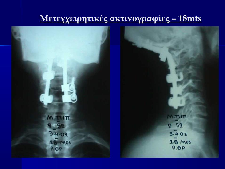
Μετεγχειρητικές ακτινογραφίες – 18mts

FRACTURES- FRACTURES- DISLOCATIONSDISLOCATIONSOF THEOF THELOWER CERVICAL LOWER CERVICAL SPINESPINE

Compression fractures are characterized by Compression fractures are characterized by failure of the anterior column under failure of the anterior column under compression with intact middle and posterior compression with intact middle and posterior columns: these are stable injuriescolumns: these are stable injuries
When the anterior and middle columns fall When the anterior and middle columns fall under axial loading forces, a burst fracture is under axial loading forces, a burst fracture is producedproduced

Distraction of the Distraction of the middle and posterior middle and posterior columns produces a columns produces a seat-belt type of seat-belt type of flexion distraction flexion distraction injury. These two injury. These two injury patterns can injury patterns can express varying express varying degrees of degrees of instabilityinstability
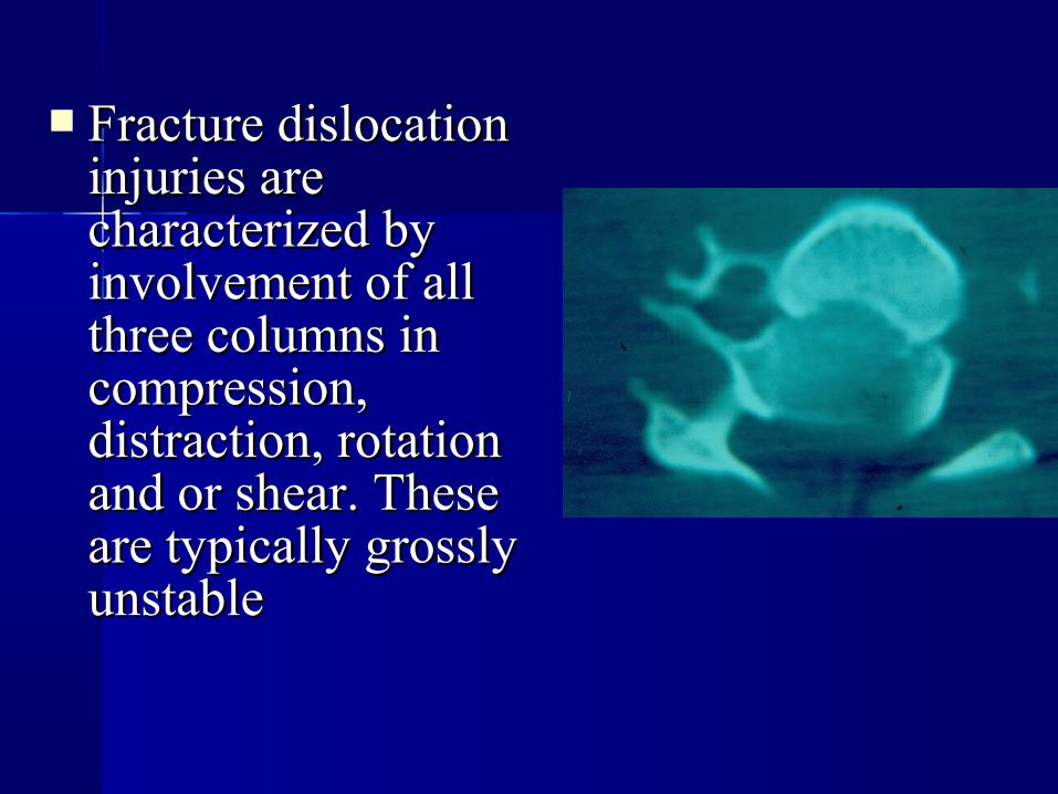
Fracture dislocation Fracture dislocation injuries are injuries are characterized by characterized by involvement of all involvement of all three columns in three columns in compression, compression, distraction, rotation distraction, rotation and or shear. These and or shear. These are typically grossly are typically grossly unstableunstable

Classification of spinal injuries of Classification of spinal injuries of the lower cervical spine (Cthe lower cervical spine (C33 – C – C77))

Compression Compression fracturesfractures

Simple wedge fractureSimple wedge fracture Resulting from hyperflexion Resulting from hyperflexion
of the cervical spine, a simple of the cervical spine, a simple wedge fracture generally wedge fracture generally occurs in the mid-cervical or occurs in the mid-cervical or lower cervical segment. There lower cervical segment. There is anterior compression of the is anterior compression of the vertebral body and although vertebral body and although the posterior ligament the posterior ligament complex is stretched it complex is stretched it remains intact making this a remains intact making this a stable fracturestable fracture

Burst fracturesBurst fractures DefinitionDefinition
– Comminuted vertebral body fracture with Comminuted vertebral body fracture with retropulsion into spinal canalretropulsion into spinal canal
– End plate fracturesEnd plate fractures– Middle column failureMiddle column failure– Posterior arch failurePosterior arch failure

Biomechanics Biomechanics etiologyetiology–Axial loadAxial load
–MVA, falls, MVA, falls, divingdiving

EvaluationEvaluation– ImmobilizationImmobilization
Orthoses – some better than othersOrthoses – some better than othersTongs tractionTongs traction
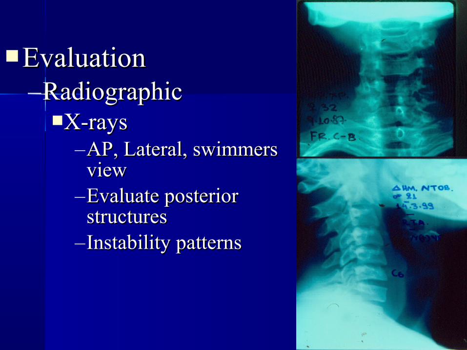
EvaluationEvaluation–RadiographicRadiographic
X-raysX-rays–AP, Lateral, swimmers AP, Lateral, swimmers
viewview–Evaluate posterior Evaluate posterior
structuresstructures–Instability patternsInstability patterns

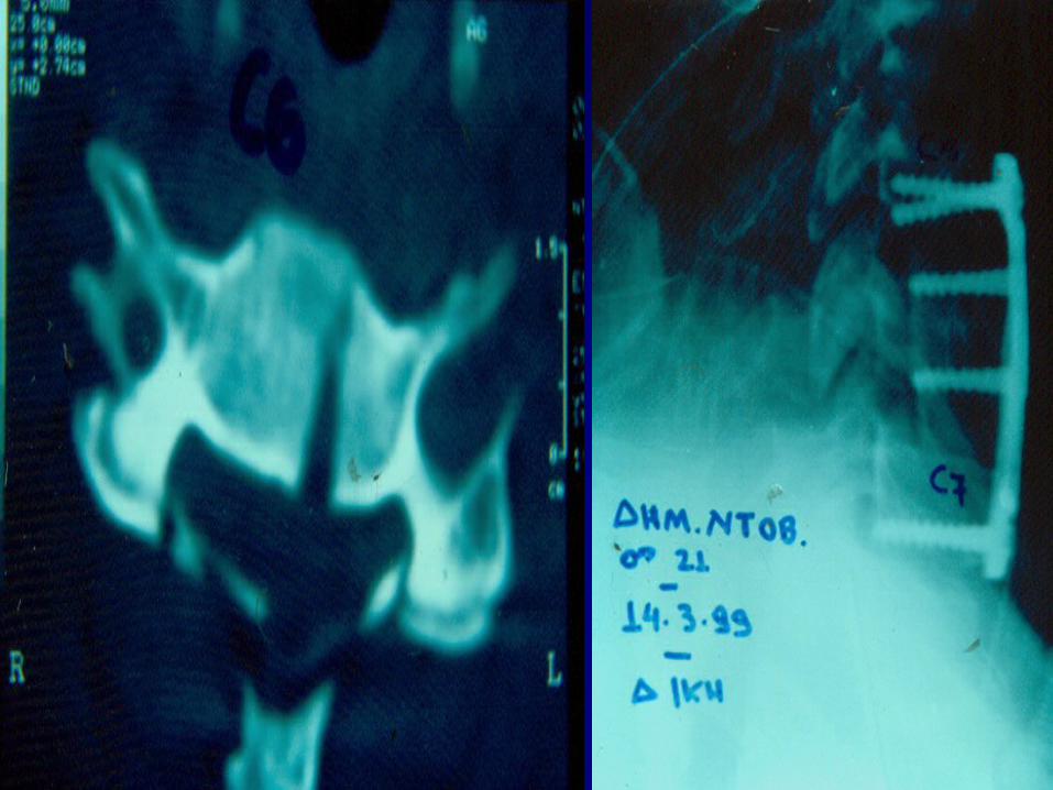
CT - scanCT - scan–Posterior arch Posterior arch
well definedwell defined–Retropulsed Retropulsed
fragnents fragnents cord - nerves cord - nerves compromisedcompromised
–Adjacent level Adjacent level injury – 10%injury – 10%

MRIMRI– Useful to evaluate Useful to evaluate
discrepancy discrepancy between clinical between clinical evaluation and x-evaluation and x-ray / CT findingsray / CT findings
– Disc herniationDisc herniation– Ligamentous Ligamentous
injuryinjury

Treatment Treatment

Determine neurologic Determine neurologic statusstatus
Evaluate stabilityEvaluate stability

Non operativeNon operative
– 2 column burst 2 column burst (complete, intact)(complete, intact)
– No posterior No posterior ligament injuryligament injury
– No indication for No indication for decompression (no decompression (no additional root additional root recovery expected)recovery expected)
– Cervical orthosisCervical orthosis
– Halo-vestHalo-vest


Operative managementOperative management

OperativeOperative
– 3 column burst3 column burst
– Incomplete SCIIncomplete SCI
– Complete SCI, canal Complete SCI, canal compromise and compromise and inadequate root inadequate root recoveryrecovery
– Contraindication to Contraindication to halohalo
TimingTiming– Allow for medical stabilizationAllow for medical stabilization– SCI dependent upon pressure and timeSCI dependent upon pressure and time– Early ischemic changes in cordEarly ischemic changes in cord– Earlier mobilization with surgery Earlier mobilization with surgery – Decreased pulmonary complicationsDecreased pulmonary complications

ApproachApproach–Anterior decompression / stabilizationAnterior decompression / stabilization–Posterior decompression / stabilizationPosterior decompression / stabilization–Combined Combined

Anterior decompressionAnterior decompression– Traction Traction – Intraoperative distractionIntraoperative distraction– Midline troughMidline trough– Bone/disc fragment removalBone/disc fragment removal
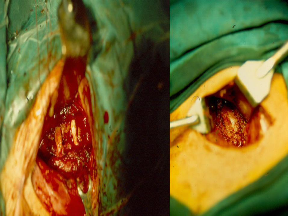

GraftingGrafting– Notched (no plate)Notched (no plate)– Allograft – fibulaAllograft – fibula– Autograft – iliac Autograft – iliac
crestcrest– High rare of High rare of
dislodgement dislodgement without fixationwithout fixation

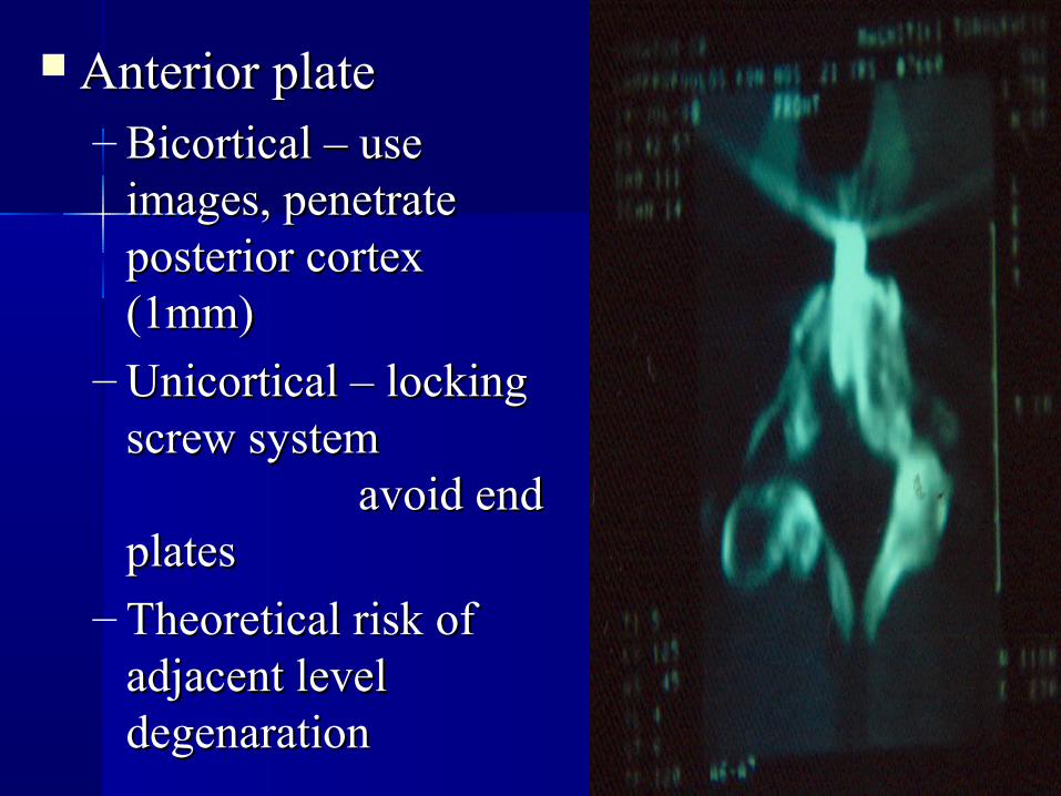
Anterior plateAnterior plate
– Bicortical – use Bicortical – use images, penetrate images, penetrate posterior cortex posterior cortex (1mm)(1mm)
– Unicortical – locking Unicortical – locking screw systemscrew system
avoid end avoid end platesplates
– Theoretical risk of Theoretical risk of adjacent level adjacent level degenarationdegenaration


Tear drop fracturesTear drop fractures

DefinitionDefinition– Acute flexion injury of Acute flexion injury of
the cervical spine the cervical spine characterized by characterized by compression of the compression of the vertebral body with vertebral body with anterior displacement anterior displacement of the anteroinferior of the anteroinferior cornercorner

Biomechanics etiologyBiomechanics etiology–Flexion and vertebral comressionFlexion and vertebral comression
MVA head striking solid objectMVA head striking solid objectDivingDivingFall – landing head firstFall – landing head first


Treatment Treatment

Non operativeNon operative– Immobilization (collar or halo)Immobilization (collar or halo)
– Neuro intactNeuro intact
– Anterior injury onlyAnterior injury only
– Compression – flexion I, II, IIICompression – flexion I, II, III
– Less than 11Less than 11oo angulation angulation


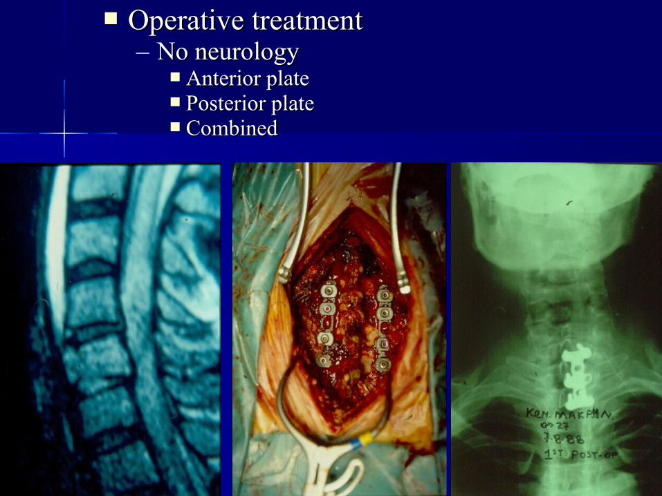
Operative treatmentOperative treatment– No neurologyNo neurology
Anterior plateAnterior plate Posterior platePosterior plate CombinedCombined

– Neurologic deficitNeurologic deficitAnterior Anterior
decompression, fusion, decompression, fusion, plateplate
Consider posterior Consider posterior stabilization for stabilization for significant posterior significant posterior injury for longer injury for longer segment involvementsegment involvement

Facet fractures and Facet fractures and DislocationsDislocations

DefinitionDefinition
–Disruption of Disruption of spinal integrity spinal integrity as result of as result of flexion flexion distraction forces distraction forces result in either result in either soft tissue injury soft tissue injury or fractureor fracture

Biomechanics etiologyBiomechanics etiology–Flexion distraction with or without Flexion distraction with or without
lateral flexion and rotation. lateral flexion and rotation. –Majority are MVA with C5-6, C6-7 Majority are MVA with C5-6, C6-7
most common level. most common level. –50% of injured have associated 50% of injured have associated
facet fracturesfacet fractures

Bilateral facet Bilateral facet
dislocationdislocation– Significant disc Significant disc
disruption in addition disruption in addition to capsular ligamnets to capsular ligamnets and posterior and posterior ligamentsligaments
– Translation (>50%)Translation (>50%)

Unilateral ligamentous Unilateral ligamentous
injuryinjury– Rotational componentRotational component
– Facets, spinous process Facets, spinous process rotated on one siderotated on one side
– Capsular ligament Capsular ligament disruption on dislocated disruption on dislocated side, posterior side, posterior longitudinal ligament longitudinal ligament disruption as well as disruption as well as interspinous ligament interspinous ligament disruptiondisruption

Fractures – distinct Fractures – distinct injuriesinjuries
Bilateral facet fracturesBilateral facet fractures– Superior, inferior, Superior, inferior,
combinationcombination– Translation, slight Translation, slight
flexionflexion– Best visualized with CTBest visualized with CT– Ligaments may be intactLigaments may be intact

Unilateral facet Unilateral facet
fracturesfractures– SuperiorSuperior
Most commonMost common Flexion, rotationFlexion, rotation
– InferiorInferior
– Fracture separation Fracture separation of articular massof articular mass
Pedicle and Pedicle and lamina fracture lamina fracture (unilateral)(unilateral)

EvaluationEvaluation– NeurologicNeurologic
Dependent upon Dependent upon canal size, amount canal size, amount of translation, disc of translation, disc disruptiondisruption
Bilateral – more Bilateral – more cord injuriescord injuries
Unilateral – more Unilateral – more root injuriesroot injuries

Plain films for Plain films for unilateral facet unilateral facet dislocationdislocation– Lateral film: “bow-tie Lateral film: “bow-tie
sign” with body and sign” with body and facet rotation creating facet rotation creating overlapoverlap

Plain films for unilateral Plain films for unilateral facet dislocationfacet dislocation– A/P filmA/P film
Spinous process displaced Spinous process displaced towards side of injurytowards side of injury
Transverse facet indicates Transverse facet indicates pedicle and lamina fracture pedicle and lamina fracture allowing facet rotationallowing facet rotation

Plain films for unilateral facet dislocationPlain films for unilateral facet dislocation– A/P filmA/P film
Oblique films may better demonstrate dislocation and Oblique films may better demonstrate dislocation and reveal associated facet fracturesreveal associated facet fractures

Plain films for bilateral dislocationsPlain films for bilateral dislocations–Anterior translation on lateral filmAnterior translation on lateral film–Appearance of jumped facets on Appearance of jumped facets on
lateral filmlateral film

CT scans good for CT scans good for visualizing visualizing fractures but does fractures but does not predict not predict instabilityinstability

MRIMRI
– Good for visualizing Good for visualizing fracture and multilevel fracture and multilevel pathologypathology
– Predicts instability Predicts instability benefiting from fusion benefiting from fusion procedure: disruption of procedure: disruption of any 3 of the anterior any 3 of the anterior longitudinal, posterior longitudinal, posterior longitudinal, longitudinal, interspinous and facet interspinous and facet capsule ligamentscapsule ligaments

Treatment Treatment

LigamentousLigamentous–Subluxation – may heal with Subluxation – may heal with
orthosis if spontaneous arthrodesis orthosis if spontaneous arthrodesis occursoccurs
–Perched – significant posterior Perched – significant posterior ligament injury, usually requires ligament injury, usually requires posterior reduction fusionposterior reduction fusion

Bilateral facet injuryBilateral facet injury– Awake closed reductionAwake closed reduction– MRI considerations (timing, neuro)MRI considerations (timing, neuro)– Gardner – Wells tongs, 5lb. Gardner – Wells tongs, 5lb.
IncrementsIncrements– Avoid overdistraction Avoid overdistraction
– Posterior fusion, instrumentationPosterior fusion, instrumentation– Anterior discectomy, fusion, Anterior discectomy, fusion,
instrumentationinstrumentation

Unilateral facet injuryUnilateral facet injury–Radicular symptoms rare cord Radicular symptoms rare cord
lesionslesions–Dislocation – Harder to reduceDislocation – Harder to reduce–Fracture – Evaluate with CTFracture – Evaluate with CT

DislocationDislocation– Closed reduction maneuver Closed reduction maneuver
Traction, flexion, rotate neck to unlock Traction, flexion, rotate neck to unlock facet, increase flexion followed by facet, increase flexion followed by dislocation of neck dislocation of neck
– Avoid excessive weight – can lead to Avoid excessive weight – can lead to increased ligamentous injuryincreased ligamentous injury
– Posterior open reduction Posterior open reduction instrumentationinstrumentation

FractureFracture–Relatively easy reduction, but Relatively easy reduction, but
difficult to maintain difficult to maintain –Counteract rotational forcesCounteract rotational forces–Consider operative stabilization if Consider operative stabilization if
radiculopathy persistsradiculopathy persists


Whiplash injuriesWhiplash injuries

Extension followed by flexionExtension followed by flexion– May have severe injuriesMay have severe injuries
Cervical ligamentsCervical ligamentsAnterior or posterior musculatureAnterior or posterior musculatureTemporo – mandibular jointTemporo – mandibular jointEsophagus/tracheaEsophagus/trachea
– Difficult to diagnoseDifficult to diagnose


Recovery periodRecovery period– 3-4 days3-4 days– 3-4 weeks3-4 weeks– 6 months6 months– 2 years2 years
10% have long term pain10% have long term pain RxRx
– Soft collarSoft collar– Isometric strengtheningIsometric strengthening– Repeat flex/ext lateral x-rays if no Repeat flex/ext lateral x-rays if no
improvement in 3-4 weeksimprovement in 3-4 weeks

Cord contusionsCord contusions

SCISCI All radiologic studies normalAll radiologic studies normal Etiology elastic deformation of spinal Etiology elastic deformation of spinal
column within physiologic range of column within physiologic range of bone/ligaments, but beyond physiologic bone/ligaments, but beyond physiologic range of cord range of cord
Rx depends on levelRx depends on level– HaloHalo– CTOCTO– CO CO


Thoraco - Thoraco - Lumbar Lumbar InjuriesInjuries
ClassificationClassification

THE THREE COLUMN SPINE THE THREE COLUMN SPINE and its significance in the classification of acute and its significance in the classification of acute thoracolumbar spinal injuries.thoracolumbar spinal injuries. Denis F. Denis F. ((Spine 1983Spine 1983))

DESTRUCTIONDESTRUCTION22
out of theout of the33
COLLUMNSCOLLUMNS
MAJOR INSTABILITYMAJOR INSTABILITY

A comprehensive classification of A comprehensive classification of thoracic and lumbar injuries.thoracic and lumbar injuries.
Magerl F, Aebi M, Gertzbein SD, Magerl F, Aebi M, Gertzbein SD, Harms J, Nazarian S. Harms J, Nazarian S.
Eur Spine J 1994Eur Spine J 1994


Type AType A


Type BType B


Type CType C


DESTRUCTIONDESTRUCTIONof theof the
33COLLUMNSCOLLUMNS
MAXIMUM INSTABILITYMAXIMUM INSTABILITY
ROTATIONAL ROTATIONAL INJURIESINJURIES

TREATMENTTREATMENT

Check listCheck listofof
Thoraco-LumbarThoraco-Lumbarinstabilityinstability

White A- Panjabi M White A- Panjabi M Clinical Biomechanics of the Spine Clinical Biomechanics of the Spine
1978.1978.

RadiologicalRadiological evaluation evaluation

Radiological parametersRadiological parameters
Kyphosis of the Kyphosis of the vertebral bodyvertebral body
Segmental Segmental kyphosiskyphosis
Disc heightDisc height Vertebral body Vertebral body
height:Beck indexheight:Beck index


Purposes Of The Operative Purposes Of The Operative TreatmentTreatment
A.A. The stabilization of the spineThe stabilization of the spine
B.B. The decompression of the spinal cord-The decompression of the spinal cord-nervesnerves
C.C. The correction of the spinal deformityThe correction of the spinal deformity
PROCEDURESPROCEDURES

Posterior ProcedurePosterior Procedure


In case of severe neurological In case of severe neurological deficit(Frankel-A.S.I.A: A-B-C)deficit(Frankel-A.S.I.A: A-B-C)
LAMINECTOMYLAMINECTOMY

ShortStabilization

Long stabilizationLong stabilization

GraftsGrafts
One level above and below One level above and below
the fractured vertebrathe fractured vertebra
•Local autograftsLocal autografts•Frozen femoral headsFrozen femoral heads•AllograftsAllografts

Anterior ProcedureAnterior Procedure


Anterior – Posterior decompression and stabilizationAnterior – Posterior decompression and stabilizationone – two sessionsone – two sessions

IMPLANTSIMPLANTS

Implants for Implants for Anterior Anterior CorrectionCorrectionandandStabilizationStabilization


Implants Implants for for
Posterior Posterior CorrectionCorrection
andandStabilizationStabilization

ImplantsImplantsforfor
Anterior - Anterior - PosteriorPosterior
Correction and Correction and StabilizationStabilization

SACRAL SACRAL
FRACTURES FRACTURES

PELVIC ANATOMY ANTERIOR VIEW PELVIC ANATOMY ANTERIOR VIEW

PELVIC ANATOMY POSTERIOR VIEW

PELVIC ANATOMY INLET VIEW

SACRUM ANATOMY

SACRUM ANATOMY

POSTERIOR WALL OF PELVIS

LATERAL WALL OF PELVIS

SACRAL PLEXUS

SACRAL PLEXUS

SACRAL PLEXUS

SACRUM FRACTURES – NERVE ROOTS


SACRUM FRACTURES – DENIS CLASSIFICATIONSACRUM FRACTURES – DENIS CLASSIFICATION
ZONE IAcross sacralNeurological injuries
•due to superior migration of fragments•6% of the whole•lumbrosacral plexus L5,S1 (24%)•Femoral nerve

ZONE II
• Through the neuroforamina
• Neurological injuries → L5, S1 (50%)
• Unilateral sacral anesthesia• Incontinence• Flaccid bowel and bladder• impotence
• Evaluation • Achilles reflex• Bulbocaverosus reflex• Rectal tone
SACRUM FRACTURES – DENIS CLASSIFICATION

SACRUM FRACTURES – DENIS CLASSIFICATION
ZONE III
• through the body of the sacrum
• Neurological injuries
• 56% of the whole• Cauda equina• Neurogenic bladder• Saddle anesthesia• Loss of sphincter tone• Bowel, bladder dysfunction 70%

MISCELLANEOUS FRACTURES
• Transverse fractures
• From landing on the buttocks
• U shaped fractures

• One hand is placed on the iliac crest
• The other hand applies traction to the leg
→ Displacement in vertical plane
PHYSICAL EXAMINATION
RADIOGRAPHIC INVESTIGATION
• AP radiographs, inlet and outlet views
• Difficult – complex shape (50% are missed)
• Findings – low lumbar transverse process fractures
- asymmetrical sacral foramen
- irregular trabeculation of the lateral
masses
• Sacral arcuate lines → asymmetry: uncomplicated
sacral frx
→ disorganized: comminuted
sacral frx


RADIOGRAFIC INVESTIGATION

• The most accurateThe most accurate
• Especially for transverse fracturesEspecially for transverse fractures
• Useful for detecting large defects as tarlov cystsUseful for detecting large defects as tarlov cysts
• Diagnosis of coexisting malignant lesionsDiagnosis of coexisting malignant lesions
CT SCAN


CD SCAN

• The most sensitive
in detection of fractures
- soft tissue edema
- marrow changes
MRI

TREATMENT
ZONE I
• Without neurologic deficits and stable
• Symptom relief
• Bed rest (7-10 days)
• Log-rolled

TREATMENT
ZONE II and III
• Without neurologic deficits
• Bed rest for 4-8 weeks
• Weight bearing at 4-8 weeks on the fractured side

TREATMENT
ZONE III
• Without neurologic deficits
• Observation: neuropraxia that will resolve
• Symptoms beyond 6-8 weeks: foraminal decompression

TREATMENT
ZONE III
• With neurologic injury
• Aggressive radiologic examination
• Early posterior
decompression
forReturn of – bowel, bladder
control
Reserval of foot drop

COMPLICATIONS OF CONSERVATIVE
TREATMENT
• chronic pain
• sacroiliac joint arthritis
• changes in the alignment on the sacrum
• bowel, bladder disability

DETERMINATION OF FRACTURE STABILITY
• Stable fractures
• Impacted vertical fracture
• Nondisplaced fracture of posterior sacroiliac complex
• Fracture of the upper sacrum

DETERMINATION OF FRACTURE STABILITY
• Unstable
• Fracture diastasis of more than 0,5 – 1cm along with an anterior unstable injury

SURGICAL INDICATION
• posterior or vertical displacement or both (>1cm)
• Rotationally unstable pelvic ring injuries
• Sacral fractures with unstable pelvic ring that requires mobilization
• Neurological injury

PROCEDURE PRONE POSITION

PERCUTANEOUS ILIOSACRAL SCREW FIXATION
• For unilateral sacral fractures zone I or zone II
• Under fluoroscopic control the reduction is obtained and
held by iliac screws (cannulated)

OPEN REDUCTION AND INTERNAL FIXATION

MISCELLANEOUS CASES

CASE 4


CONCLUSION
• Neurological deficities
• Stable Fractures : conservative treatment
• Unstable Fractures : operative treatment
• Neurologic injury :posterior decompression